Radial Nerve Palsy Tendon Transfers Episode II What is a tendon transfer? The tendon of a...
35
-
Upload
godfrey-lloyd -
Category
Documents
-
view
224 -
download
0
Transcript of Radial Nerve Palsy Tendon Transfers Episode II What is a tendon transfer? The tendon of a...


Radial Nerve Palsy Tendon TransfersEpisode II

What is a tendon transfer?
• The tendon of a functioning muscle is detached from its insertion and reattached to another tendon or bone to replace the function of a paralysed muscle or injured tendon. The transferred tendon remains attached to its parent muscle with an intact neurovascular pedicle.

What is a tendon transfer?
• “Using the power of a functioning muscle unit to activate a non functioning nerve/muscle/tendon unit”.
• Tendon transfers work to correct:– instability– imbalance – lack of co-ordination – restore function by redistributing remaining muscular
forces

Indications• Paralysed muscle
– Nerve injury – peripheral or brachial plexus– High cervical quadriplegia (needs some input to brachial plexus/hand)– Neurological disease– Nerve repair with early transfer as internal splint
• Injured (ruptured or avulsed) tendon or muscle– Considerations
• Graft vs. transfer (adhesions more likely in graft – 2 anastomoses)• Quality of available donors• Length of time since injury• Nature of tendon bed
• Balancing deformed hand e.g. cerebral palsy or rheumatoid arthritis• Some congenital abnormalities
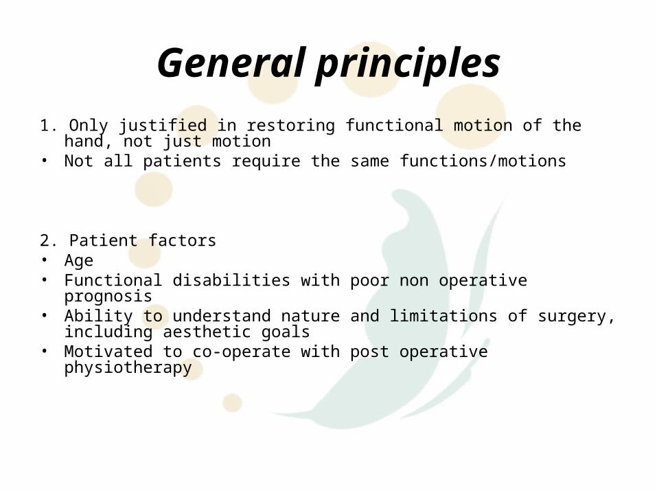
General principles1. Only justified in restoring functional motion of the hand, not just motion• Not all patients require the same functions/motions
2. Patient factors• Age• Functional disabilities with poor non operative prognosis • Ability to understand nature and limitations of surgery, including aesthetic goals• Motivated to co-operate with post operative physiotherapy

General principles3. Recipient site• “Tissue Equilibrium” concept as per Steindler/Boyes• Tissue bed into which transfer is placed should be soft and supple• Good soft tissue coverage• Stable underlying skeleton• Full passive range of motion of joints to be powered• Area to be powered must be sensate

General principles4. Donor muscle factors (APOSLE)
Amplitude of the donor muscle– Should be matched to the unit being replaced
• Finger flexors 60 - 70mm, • finger extensors and EPL 40 - 50mm, • wrist flexors / extensors 30 - 40mm, • brachioradialis 20 - 30mm
– Amplitude of motion of any tendon can be increased by :-• Increasing the number of joints its crosses eg the amplitude of a tendon crossing
the wrist joint is increased by 20 – 30mm by full ROM of wrist• Tenodesis effect during active movement• Freeing fascial attachments to donor tendons• Inserting the tendon closer to the joint being moved, but this requires a motor unit of
increased power (due to leverage); and vice versa

General principlesPower of the donor muscle– Any transferred muscle loses at least one grade of strength, so only Grade 5
muscles are satisfactory (Grade 4, or 85% normal strength, can be sufficient for some transfers). Donor muscle strength should be maximised pre-operatively.
– Strongest are brachioradialis and FCU. Donor power correlated roughly with cross sectional area of muscle and fibre length
– Overly powerful muscles will unbalance and, over time, deform a joint. So muscle power should be matched if possible.
– Effective power of a transfer can be increased by placing the tendon insertion farther from the joint axis and as close to 90° as possible

General principlesOne tendon, One function– Effectiveness reduced in transfer designed to produce multiple functions
Synergistic muscle groups are generally easier to retrain– Fist group – wrist extensors, finger flexors, digital adductors, thumb flexors,
forearm pronators, intrinsics– Open hand group – wrist flexors, finger extensors, digital abductors, forearm
supinators– Use of synergistic muscles tends to help retain joint balance

General principlesLine of transfer– Should approximate pull of original tendon if possible– Acute angles should be avoided
Expendability– Transfer must not cause loss of an essential function
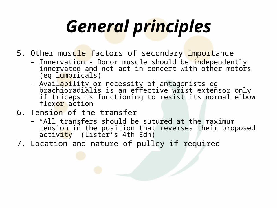
General principles5. Other muscle factors of secondary importance
– Innervation - Donor muscle should be independently innervated and not act in concert with other motors (eg lumbricals)
– Availability or necessity of antagonists eg brachioradialis is an effective wrist extensor only if triceps is functioning to resist its normal elbow flexor action
6. Tension of the transfer– “All transfers should be sutured at the maximum tension in the position that
reverses their proposed activity” (Lister’s 4th Edn) 7. Location and nature of pulley if required
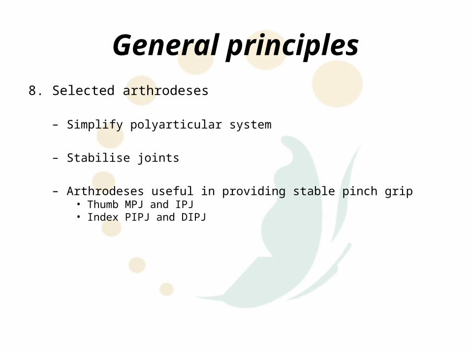
General principles8. Selected arthrodeses
– Simplify polyarticular system
– Stabilise joints
– Arthrodeses useful in providing stable pinch grip• Thumb MPJ and IPJ• Index PIPJ and DIPJ

General principles9. Timing
– “The timing of tendon transfers depends upon the aetiology and prognosis of motor imbalance, the neurophysiologic problems for the patient, and the constitution of the involved extremity” (Omer GE: Timing of tendon transfers to the hand. Hand Clin 4(2):317, 1988)
– Usually last stage in reconstruction, after skeletal stability, soft tissue coverage, sensation and joint mobility “tissue equilibrium”
– Brown suggested early transfer if expected poor results• Nerve gap>4cm• Large wound or extensive scarring• Skin loss over nerve
10. Comparison to alternatives– Nerve repair or transfer– Tendon repair or graft – Tenodesis (joint stabilisation by anchoring tendons that move the joint)– Arthrodesis– Amputation– Muscle lengthening, release or denervation (in spasticity)

General principles11. Contraindications
– Age – due to joint stiffness, decreased need for power movements and difficult rehabilitation
– Motivation – patients must be concerned about disability and highly motivated to perform hand rehabilitation
– Task analysis – transfers must be designed to accomplish tasks rather than just specific motions. Eg opening doors requires grasp and twist
– Nature of disability – systemic and local disease factors must be controlled before reconstruction attempted
12. Disadvantages– No increase in strength– Normal function of transferred muscle is lost– Transferred tendon may perform a different force, amplitude of movement
and functional pattern– Transferred tendon must learn a new movement/function

Selecting donor tendons• Based on Smith & Hastings (Principles of tendon transfers to the hand. Instr
Course Lect 29:129, 1980)1. List functioning muscles2. List which of those muscles are expendable3. List hand functions requiring restoration4. Match #2 and #35. Staging

Maximising Success / Surgical Technique
1. Incisions should not cross the path of the transferred tendon
2. Avoid interference with normal structures
3. Tendon should insert into the joint of motion at 90 to maximise power and excursion. Insertion can be moved away from the joint to improve power, but this is at the expense of decreased excursion
4. The transferred tendon should insert into another tendon or bone. Strong insertions allow earlier mobilisation.

Maximising Success / Surgical Technique
5. A single insertion is best. Dual insertions tend to provide motion to the tighter insertion. Can be an advantage in complex movements, where one insertion is tighter during one phase of motion, and the other takes over during another phase
.
6. Tension should be set to produce the necessary joint movement with maximal muscle contraction. Some initial over correction should be planned, as some tendon stretch is usual.

Maximising Success / Surgical Technique
7. Joint should be initially immobilised in a position that relieves tension at the insertion of the transfer
8. Reverse order – harvest grafts, prepare recipient site and tunnel before raising muscle
General Post Operative Management
• Rehabilitation is equally important in tendon transfer success as surgical execution
• Rehabilitation / physiotherapy is essential in– Regaining joint mobility lost during splinting– Training tendon to glide in new course– Teaching patients to activate a new muscle to achieve a certain
function, which requires development of new neural pathways• The more that a patient notices a disability, the greater the
motivation, so the easier the retraining• Children are usually managed with static protocols or longer
protective phase
Basic Principles of Post Operative Rehabilitation
• Described by Toth 19861. Protective phase• Begins at surgery and lasts 3 – 5 weeks• Objectives:-
– Protective splinting– Oedema control– Mobilise uninvolved joints
2. Mobilisation phase• Begins when tendon healing is adequate for activation (usually 3 – 5 weeks post op)• Objectives
– Mobilise tendon transfer– Immobilise soft tissue– Continue immobilisation of uninvolved joints to prevent joint stiffness from disuse– Reinforce preoperative teaching and patient education– Continue oedema control and protective splinting– Begin home rehabilitation program
• Usually day time dynamic splinting with nightly static splinting

Basic Principles of Post Operative Rehabilitation
3. Intermediate phase• Begins 5 – 8 weeks post operatively• Gradually increases hand activity and passive range of motion exercises• Limited functional movements permitted4. Resistive phase• Beginning at 8 – 12 weeks• Tendon junctions are strong enough to withstand increasing resistance• Therapeutic objective is to increase endurance and strength of transferred
muscles• Work related simulated tasks are begun to patient tolerance

Radial Nerve Palsy• Need to differentiate between complete radial nerve palsy (includes
triceps) and posterior interosseous palsy– Brachioradialis and ECRL are innervated prior to termination into posterior
interosseous and sensory branches of radial nerve
• Severe impairment due to loss of extension power to the wrist, fingers, thumb and loss of radial abduction of the thumb
• Wrist extension is critical for stability, which is essential for grip and assisting the function of many tendons crossing the wrist
Tendon Transfers
• Well defined and highly effective, aiming to replace– Wrist extension– Finger extension– Thumb extension and abduction
• Standard – accredited to Riordan 1964

Radial Nerve Palsy
• Non-Operative Treatment– Splintage
• Burkhalter observed grip strength increased 3-5 by simply stabilising the wrist with splintage
• Tailor to needs of patient• Brand recommended that if wrist splint during day then need
night finger extension splint because lose length of flexor muscle fibres making it more difficult to achieve normal balance after nerve recovery or after tendon transfers
– Maintenance of full passive ROM in all joints of the wrist/hands and prevent contractures

Radial Nerve Palsy
• Early transfers (“Internal Splintage”)– Burkhalter believes greatest functional loss is grip strength
therefore advocated early PT to ECRB• Therefore eliminate need for external splint plus also restore grip
strength
– 3 indications:• Works as substitute during early regeneration• Works as helper by adding power to reinnervated muscle• Acts as substitute in cases which results of nerve repair are poor
(eg chronic/crush injuries or elderly)

Riordan TransferDonor Insertion Function
PT ECRB Wrist dorsiflexion
FCU EDC IF - LF Finger extension
PL EPL (rerouted) Thumb extension

PT to ECRB
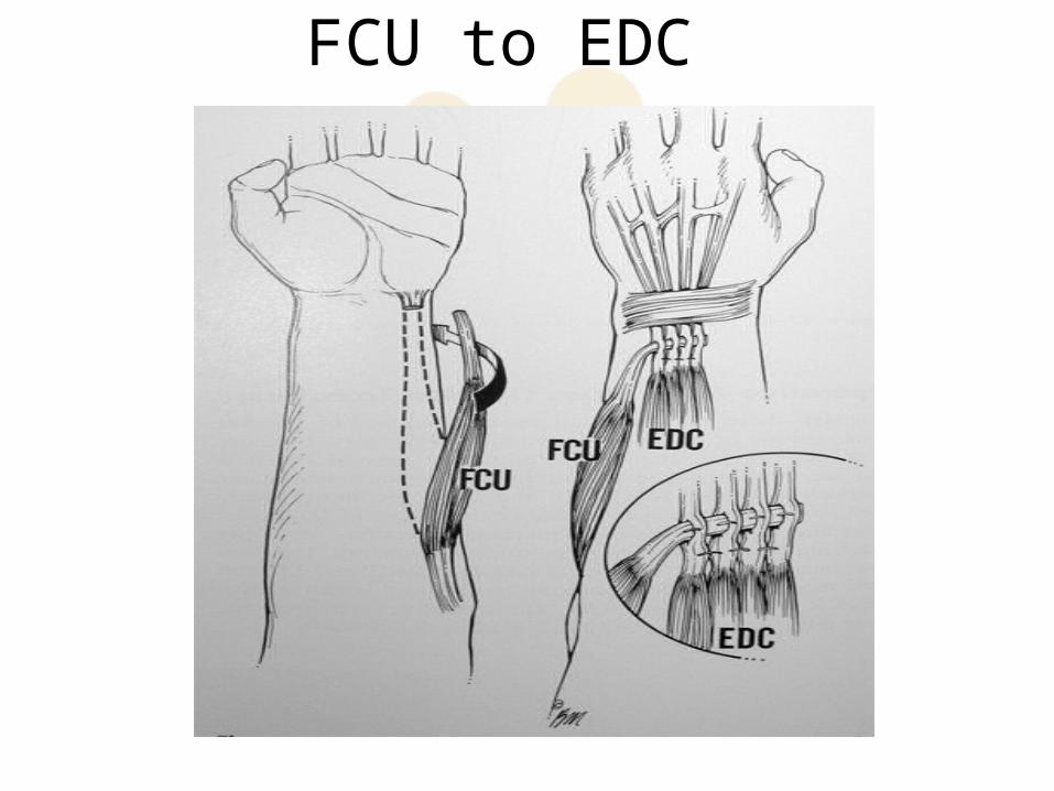
FCU to EDC

PL to EPL

Donor Insertion Function Reference
PTFCRFCU
ECRL & ECRBEPL, EPB, APL & EDC IFEDC MF – LF
Wrist extensionThumb & index extension, thumb abductionFinger extension
Jones 1921
PTFDS MFFCU
ECRBEPL (re routed)EDC
Wrist extensionThumb extension & abductionFinger extension
Goldner 1974
PTPLFCR
ECRBEPLEDC
Wrist extensionThumb extensionFinger extension
Brand 1975
PTFCRFDS RFFDS MF
ECRL & ECRBAPL & EPBEPL & EIPEDC via interosseous m
Wrist extensionThumb abductionThumb & index extensionFinger extension
Boyes 1970
PTPLFDS LFFDS RF
ECRBAPLEPLEDC
Wrist extensionThumb abductionThumb extensionFinger extension
Beasley 1970
PTFCU
ECRBEDC & EPL
Wrist extensionDigit extension
Smith & Hastings

Brand transfers for radial nerve palsy
Brand transfers for radial nerve palsy
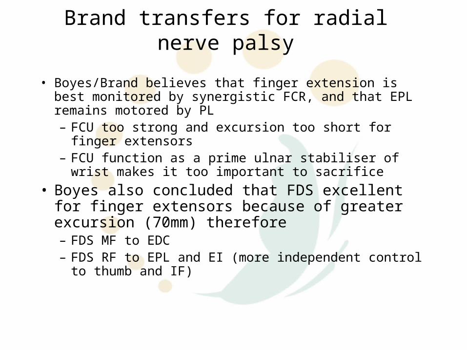
• Boyes/Brand believes that finger extension is best monitored by synergistic FCR, and that EPL remains motored by PL– FCU too strong and excursion too short for finger extensors– FCU function as a prime ulnar stabiliser of wrist makes it too
important to sacrifice• Boyes also concluded that FDS excellent for finger
extensors because of greater excursion (70mm) therefore – FDS MF to EDC– FDS RF to EPL and EI (more independent control to thumb
and IF)

Direct Nerve Transfers
• Transfer of intact nerves to denervated muscles.
• MacKinnon and associates– Median nerve supplies redundant branches to FDS
and therefore available for transfer– Or branches to PL and FCR (if these tendons not
used for transfer)

Post-Operatively• Long arm splint immobilisation for 4 weeks
– 15-30 pronation– wrist 40 extension– MPJ 10-15 flexion– Thumb in maximal extension and abduction– PIPJ fingers left free
• ROS & change splint at 10-14 days• AROM hand therapy begins at 4 weeks• Removable short arm splint to extend fingers, thumb and
wrist for further 2 weeks, only removed for exercises


![EfficacyofManipulativeAcupunctureTherapyMonitoredbyLSCI ...Bell’s palsy is an acute peripheral facial nerve palsy of un-knowncauseandaccountsfor50%ofallcasesoffacialnerve palsy [1].](https://static.fdocuments.in/doc/165x107/60a4deb9e0003e748e568e41/efficacyofmanipulativeacupuncturetherapymonitoredbylsci-bellas-palsy-is-an.jpg)