Radial nerve palsy
46
RADIAL NERVE PALSY Dr Manoj Das Ortho Resident Institute Of medicine, TUTH, Nepal
-
Upload
manoj-das -
Category
Health & Medicine
-
view
787 -
download
0
Transcript of Radial nerve palsy
RADIAL NERVE PALSY
Dr Manoj DasOrtho ResidentInstitute Of medicine, TUTH, Nepal
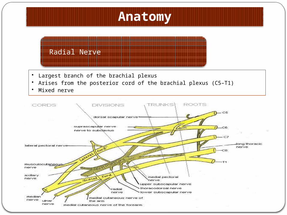
Radial Nerve
• Largest branch of the brachial plexus• Arises from the posterior cord of the brachial plexus (C5–T1)• Mixed nerve
Anatomy
Course of Radial Nerve (RN) in the arm
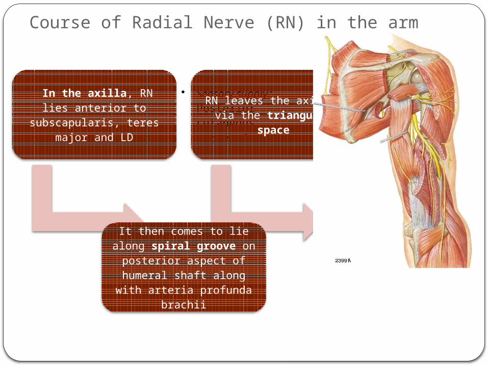
In the axilla, RN lies anterior to subscapularis,
teres major and LD
• Sensory supply: Posterior cutaneous nerve of arm
RN leaves the axilla via the triangular space
• Motor supply: long head of Triceps
It then comes to lie along spiral groove on posterior aspect of
humeral shaft along with arteria profunda brachii
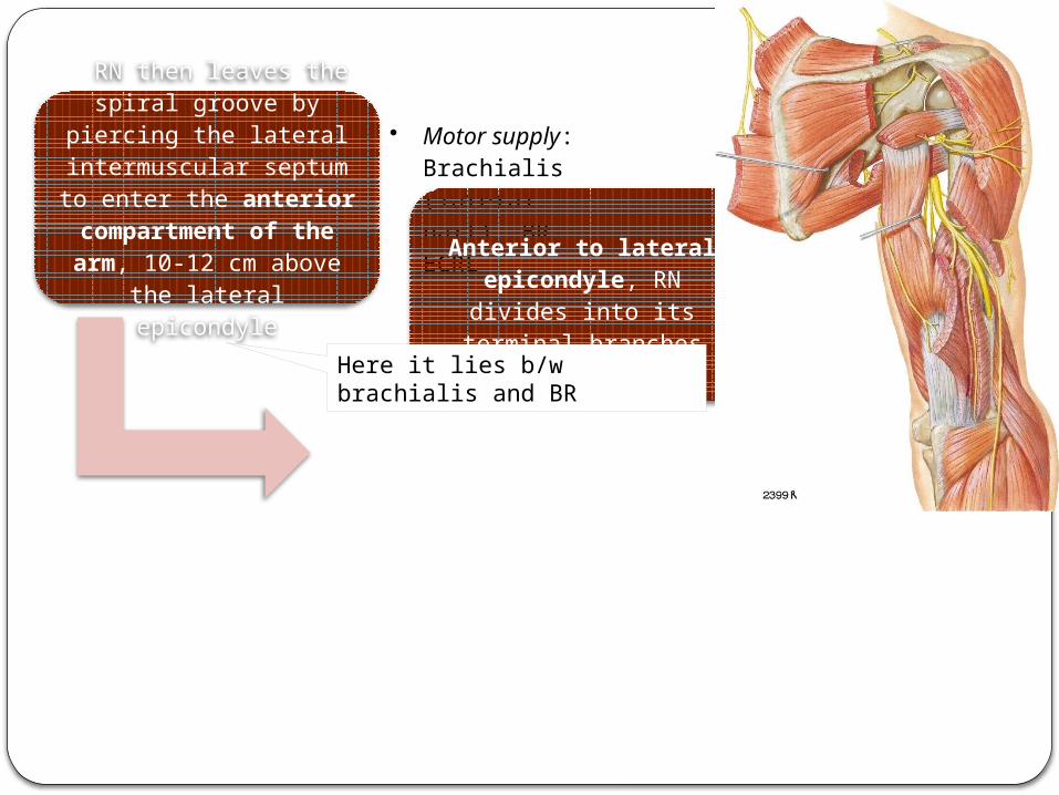
RN then leaves the spiral groove by piercing the lateral intermuscular
septum to enter the anterior compartment of the arm, 10-12 cm
above the lateral epicondyle
• Motor supply: Brachialis (lateral part), BR, ECRL
Anterior to lateral epicondyle, RN divides
into its terminal branchesHere it lies b/w brachialis and BR
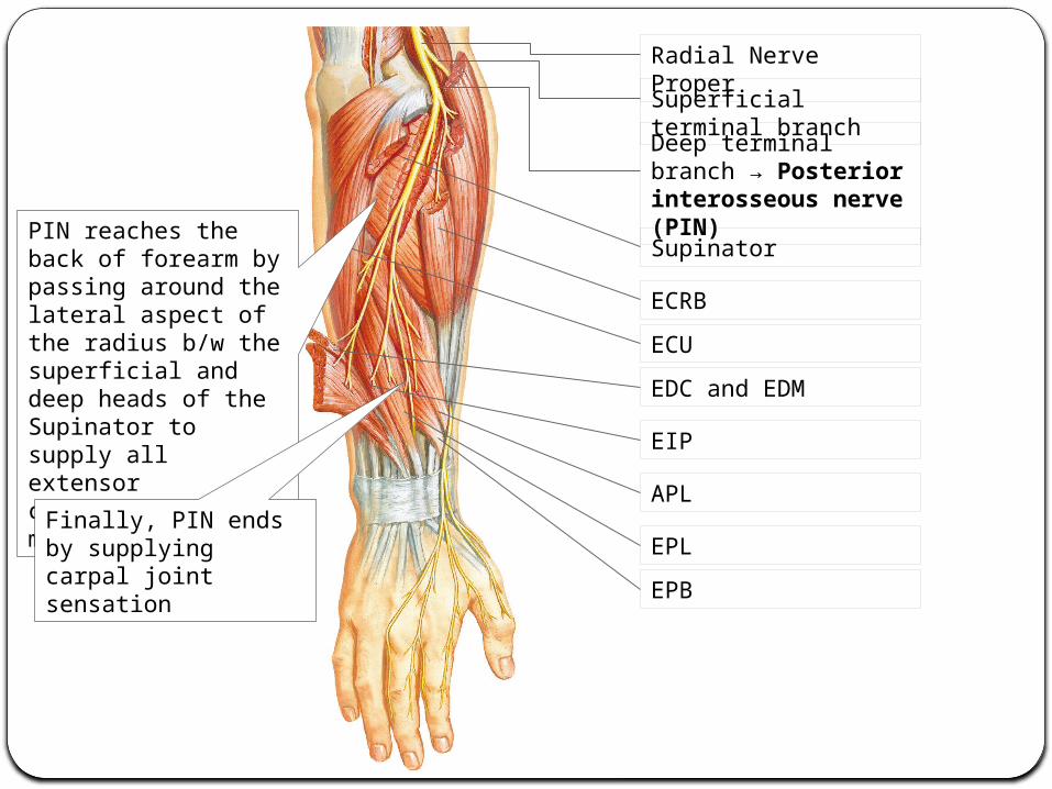
Deep terminal branch → Posterior interosseous nerve (PIN)Supinator
EIP
EDC and EDMECUECRB
Superficial terminal branch
Radial Nerve Proper
EPLEPB
APL
PIN reaches the back of forearm by passing around the lateral aspect of the radius b/w the superficial and deep heads of the Supinator to supply all extensor compartment muscles
Finally, PIN ends by supplying carpal joint sensation
BRECRL
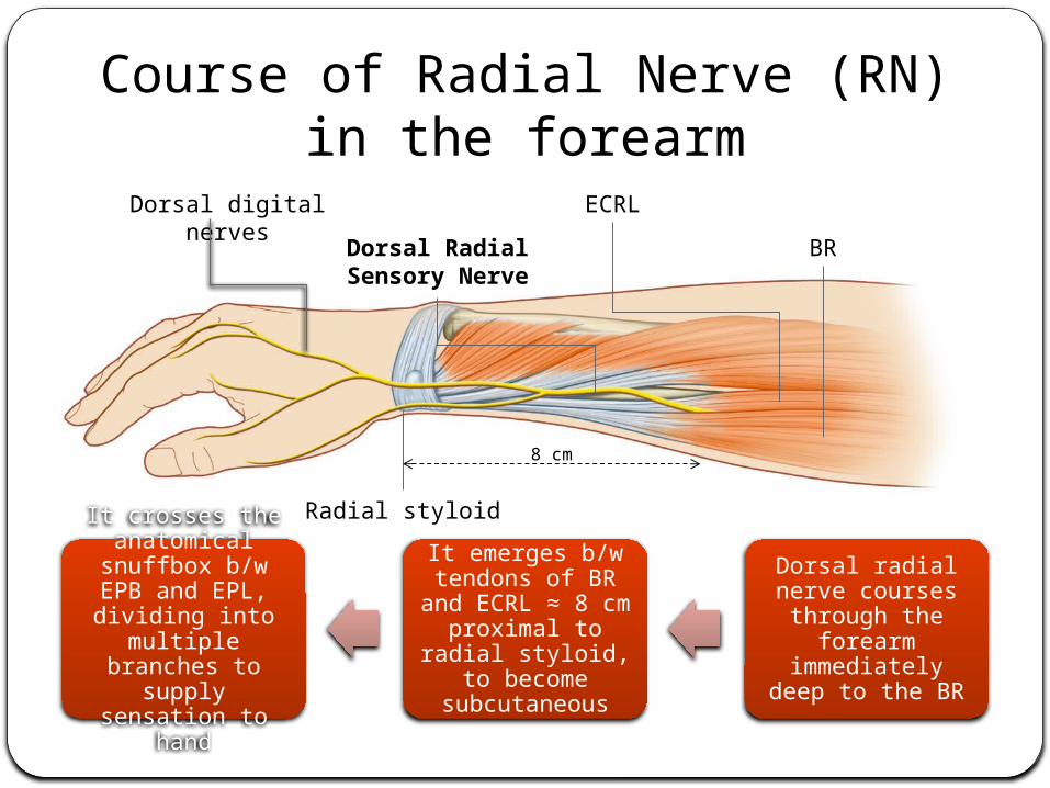
Dorsal Radial Sensory Nerve
Dorsal digital nerves
Radial styloid
8 cm
Dorsal radial nerve courses through the
forearm immediately deep
to the BR
It emerges b/w tendons of BR and
ECRL ≈ 8 cm proximal to radial styloid, to become
subcutaneous
It crosses the anatomical
snuffbox b/w EPB and EPL, dividing
into multiple branches to
supply sensation to hand
Course of Radial Nerve (RN) in the forearm
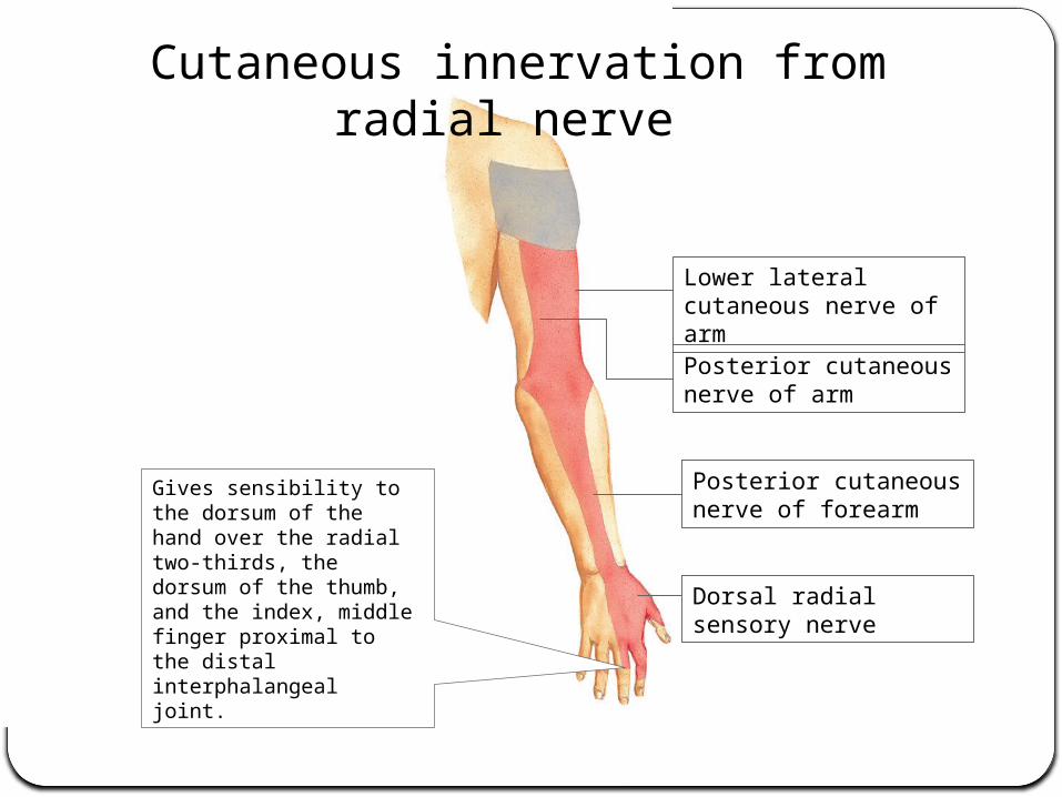
Lower lateral cutaneous nerve of armPosterior cutaneous nerve of arm
Posterior cutaneous nerve of forearm
Dorsal radial sensory nerve
Gives sensibility to the dorsum of the hand over the radial two-thirds, the dorsum of the thumb, and the index, middle finger proximal to the distal interphalangeal joint.
Cutaneous innervation from radial nerve
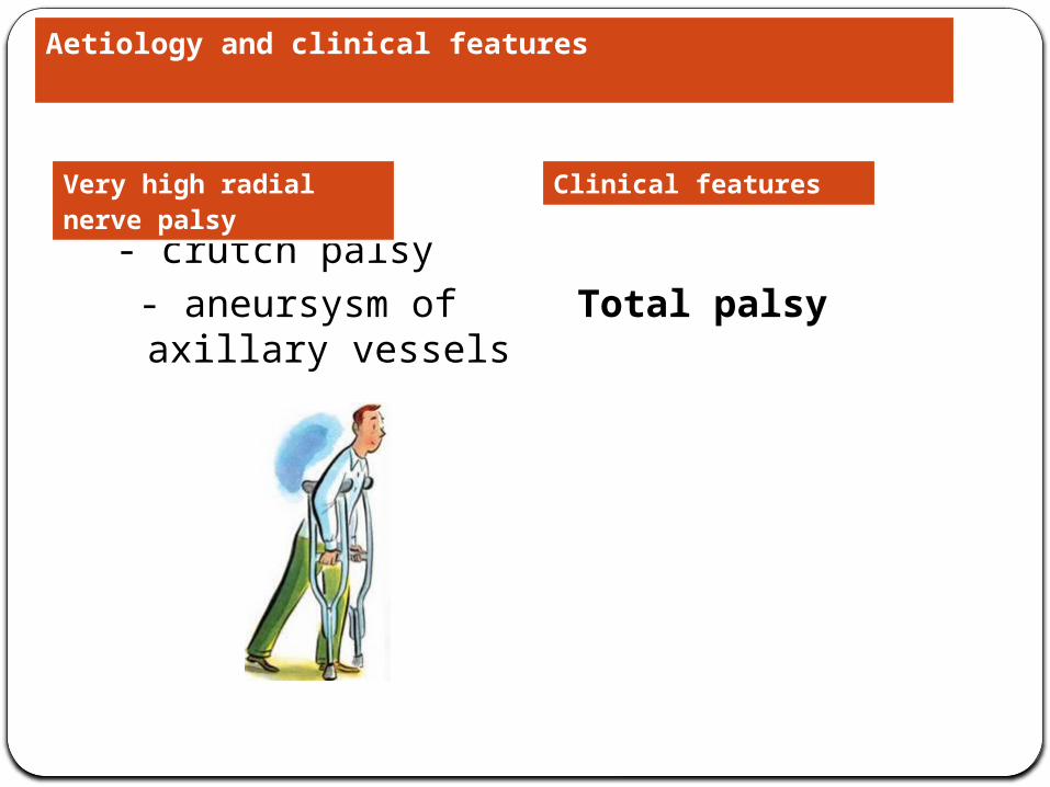
- crutch palsy - aneursysm of
axillary vesselsTotal palsy
Aetiology and clinical features
Very high radial nerve palsy
Clinical features
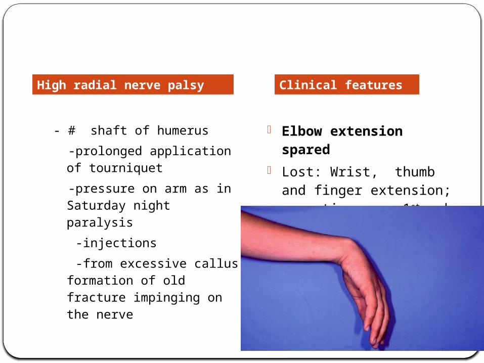
- # shaft of humerus -prolonged application of
tourniquet -pressure on arm as in
Saturday night paralysis -injections -from excessive callus
formation of old fracture impinging on the nerve
- Elbow extension spared
- Lost: Wrist, thumb and finger extension; sensation over 1st web space
High radial nerve palsy Clinical features
-Dislocation of elbow
-#neck of radius
-Enlarged bursae
-Rheumatoid synovitis of elbow
-During operation for excision of radius head
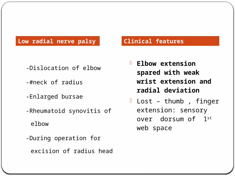
- Elbow extension spared with weak wrist extension and radial deviation
- Lost – thumb , finger extension: sensory over dorsum of 1st web space
Low radial nerve palsy Clinical features
Diagnosis
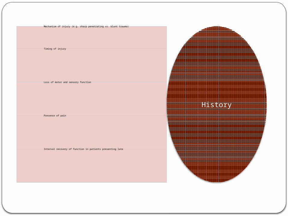
Mechanism of injury (e.g. sharp penetrating vs. blunt trauma)
Timing of injury
Loss of motor and sensory function
Presence of pain
Interval recovery of function in patients presenting late
History
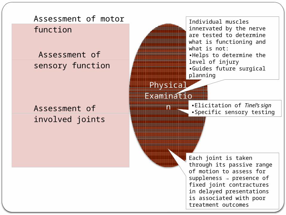
Assessment of motor function
Assessment of sensory function
Assessment of involved joints
Physical Examinatio
n
Individual muscles innervated by the nerve are tested to determine what is functioning and what is not:▪Helps to determine the level of injury▪Guides future surgical planning
▪Elicitation of Tinel’s sign▪Specific sensory testing
Each joint is taken through its passive range of motion to assess for suppleness → presence of fixed joint contractures in delayed presentations is associated with poor treatment outcomes
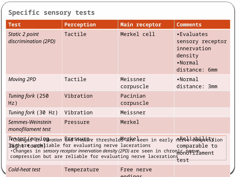
Specific sensory testsTest Perception Main receptor CommentsStatic 2 point discrimination (2PD)
Tactile Merkel cell ▪Evaluates sensory receptor innervation density▪Normal distance: 6mm
Moving 2PD Tactile Meissner corpuscle ▪Normal distance: 3mm
Tuning fork (250 Hz)
Vibration Pacinian corpuscle
Tuning fork (30 Hz) Vibration MeissnerSemmes-Weinstein monofilament test
Pressure Merkel
Ten test (moving light touch)
Pressure Merkel ▪Reliability comparable to monofilament test
Cold-heat test Temperature Free nerve endings•Changes in Vibration and Pressure thresholds are seen in early nerve compression but are unreliable for evaluating nerve lacerations•Changes in sensory receptor innervation density (2PD) are seen in chronic nerve compression but are reliable for evaluating nerve lacerations
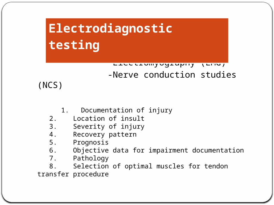
Commonly used EDT -Electromyography (EMG) -Nerve conduction studies (NCS)
1. Documentation of injury 2. Location of insult 3. Severity of injury 4. Recovery pattern 5. Prognosis 6. Objective data for impairment documentation 7. Pathology 8. Selection of optimal muscles for tendon transfer procedure
Electrodiagnostic testing
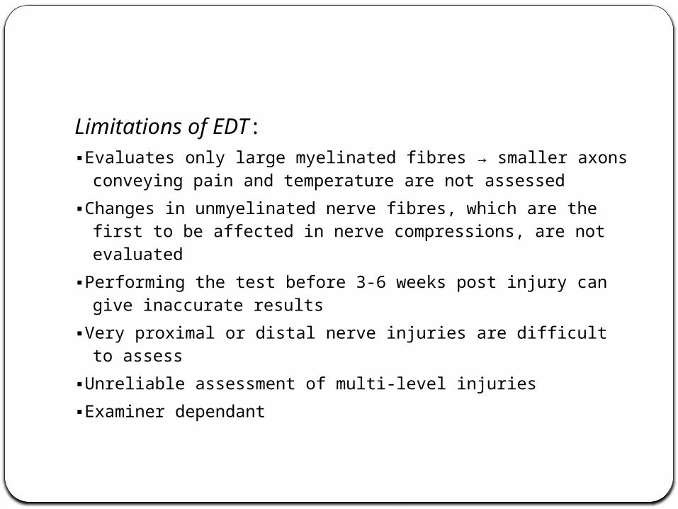
Limitations of EDT:▪Evaluates only large myelinated fibres → smaller axons
conveying pain and temperature are not assessed▪Changes in unmyelinated nerve fibres, which are the first
to be affected in nerve compressions, are not evaluated▪Performing the test before 3-6 weeks post injury can give
inaccurate results▪Very proximal or distal nerve injuries are difficult to assess▪Unreliable assessment of multi-level injuries▪Examiner dependant
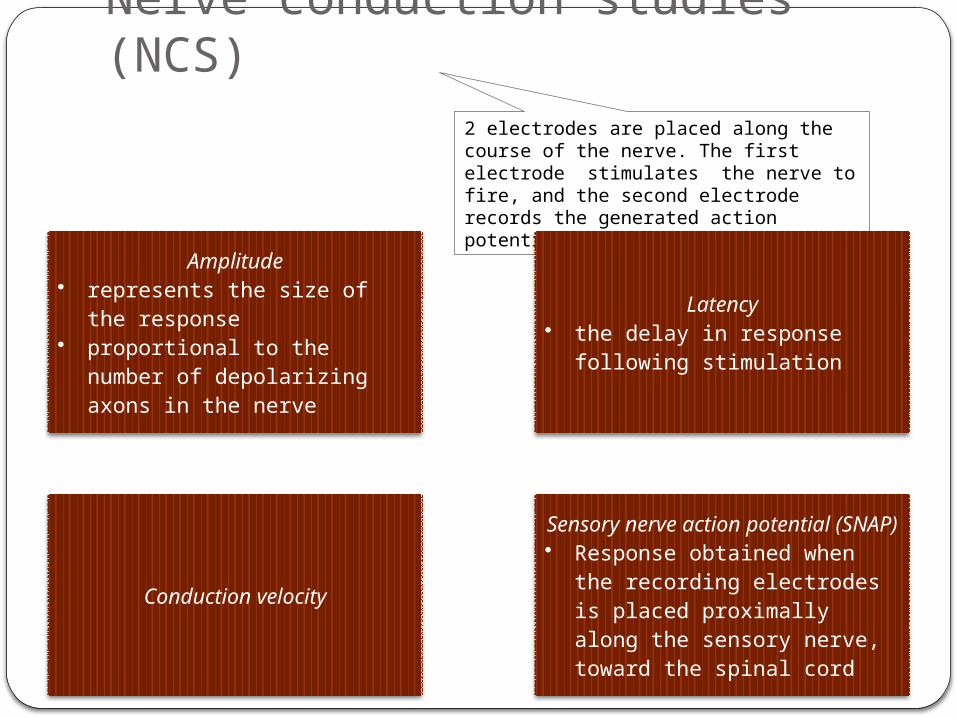
Nerve conduction studies (NCS)2 electrodes are placed along the course of the nerve. The first electrode stimulates the nerve to fire, and the second electrode records the generated action potential
Amplitude• represents the size of the
response• proportional to the number of
depolarizing axons in the nerve
Latency• the delay in response
following stimulation
Conduction velocity
Sensory nerve action potential (SNAP)
• Response obtained when the recording electrodes is placed proximally along the sensory nerve, toward the spinal cord
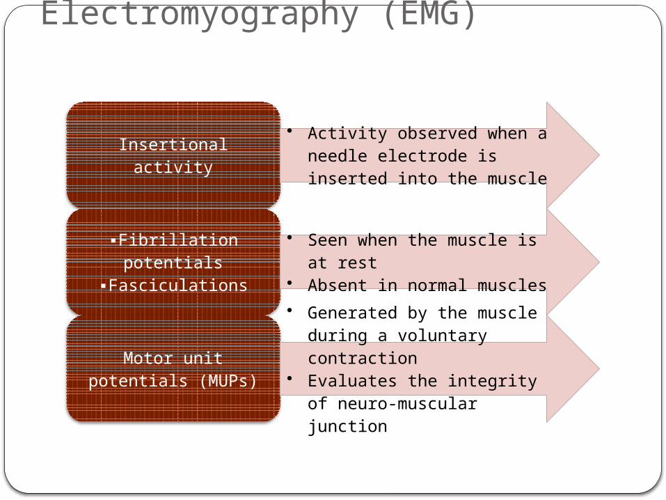
Electromyography (EMG)
Insertional activity• Activity observed when a
needle electrode is inserted into the muscle
▪Fibrillation potentials ▪Fasciculations
• Seen when the muscle is at rest
• Absent in normal muscles
Motor unit potentials (MUPs)
• Generated by the muscle during a voluntary contraction
• Evaluates the integrity of neuro-muscular junction
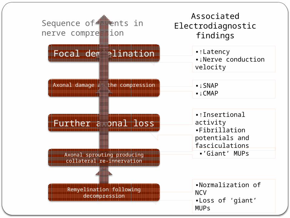
Sequence of events in nerve compression
Focal demyelination
Axonal damage at the compression site
Further axonal loss
Axonal sprouting producing collateral re-innervation
Remyelination following decompression
▪↑Latency▪↓Nerve conduction velocity
Associated Electrodiagnostic findings
▪↓SNAP▪↓CMAP
▪↑Insertional activity▪Fibrillation potentials and fasciculations
▪’Giant’ MUPs
▪Normalization of NCV▪Loss of ‘giant’ MUPs
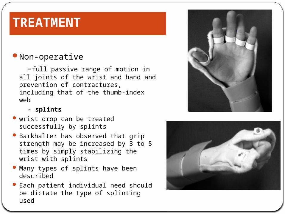
Non-operative -full passive range of motion in all
joints of the wrist and hand and prevention of contractures, including that of the thumb-index web
- splints wrist drop can be treated successfully
by splints Barkhalter has observed that grip
strength may be increased by 3 to 5 times by simply stabilizing the wrist with splints
Many types of splints have been described
Each patient individual need should be dictate the type of splinting used
TREATMENT
INTERNAL SPLINTBurkhalter proposed early transfer of PT-ECRB to restore wrist
extension as an adjunct to nerve repair.
It restores the power grip quickly and effectively since wrist extension is restored
Advantages are: It works as a substitute during nerve regrowth and largely eliminates
an external splint
Subsequently the transfer aids the newly innervated and weak wrist extensor
It continues to act as a substitute in case nerve regeneration is poor or absent
Green’s operative hand surgery
In a sharp injury exploration is indicated for diagnostic, therapeutic and prognostic purposes
In avulsion , blasting injures –to identification of the nerve injury and making the ends of the nerve with sutures for later repair.
When a nerve deficit follows blunt or closed trauma, and no clinical or electrical evidence of regeneration has occurred after an appropriate time, exploration of the nerve is indicated.
INDICATIONS FOR SURGERY
-primary repair gives the best result with respect to motor,sensory recovery, is indicated in clean sharp nerve injuries and carried out in first 6-8 hours.
-delayed,primary repair carried out between 7-18days -primary repair fascicular alignment because of minimal excision of the nerve ends.
-Secondary repair-preferable only in crushed,avulsed injuries where patients life is seriously endangered.it is done at delay of 3-6 wks.
Time of surgery
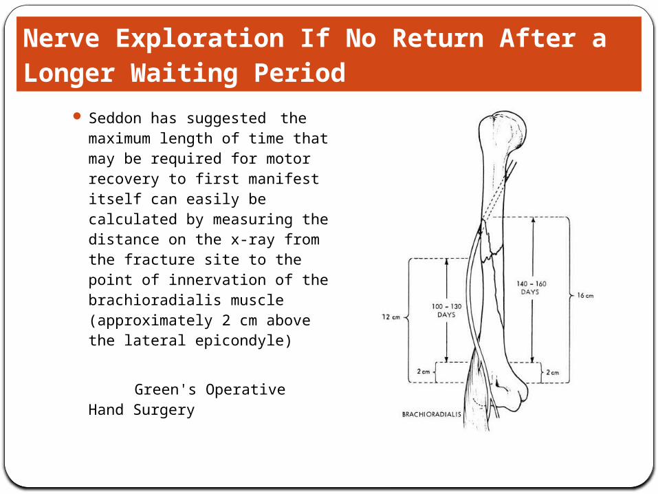
Seddon has suggested the maximum length of time that may be required for motor recovery to first manifest itself can easily be calculated by measuring the distance on the x-ray from the fracture site to the point of innervation of the brachioradialis muscle (approximately 2 cm above the lateral epicondyle)
Green's Operative Hand Surgery
Nerve Exploration If No Return After a Longer Waiting Period
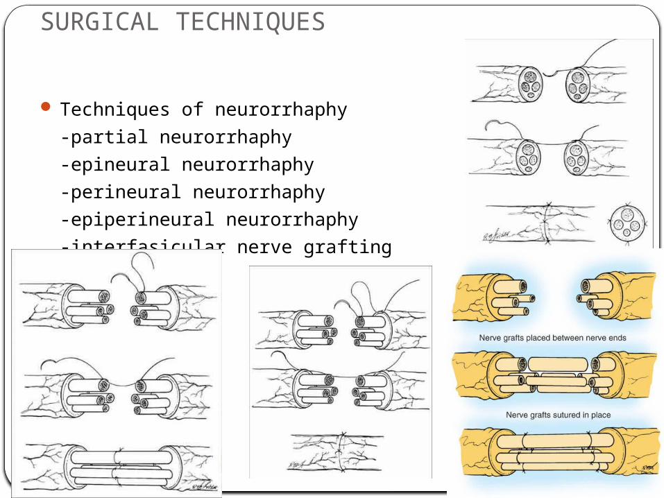
SURGICAL TECHNIQUES
Techniques of neurorrhaphy -partial neurorrhaphy -epineural neurorrhaphy -perineural neurorrhaphy -epiperineural neurorrhaphy -interfasicular nerve grafting
Tendon transfersArthodesisTendon transfers work to correct:
instabilityimbalance lack of co-ordination restore function by redistributing remaining
muscular forces
RECONSTRUCTIVE PROCEDURES
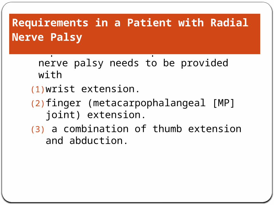
A patient with irreparable radial nerve palsy needs to be provided with
(1) wrist extension.(2) finger (metacarpophalangeal [MP] joint)
extension.(3) a combination of thumb extension and
abduction.
Requirements in a Patient with Radial Nerve Palsy
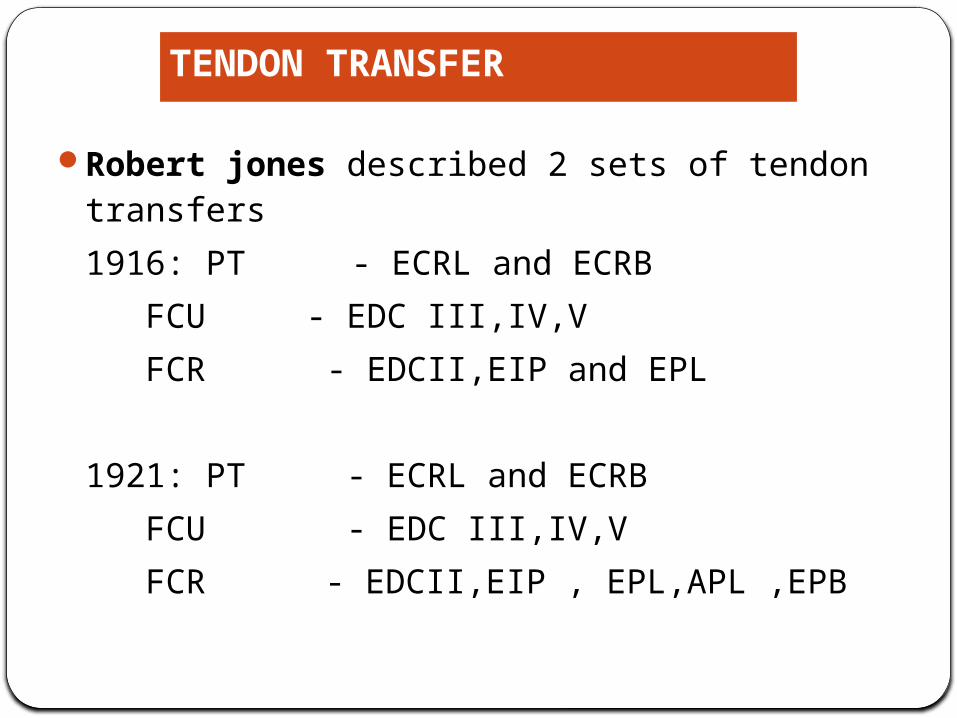
Robert jones described 2 sets of tendon transfers1916: PT - ECRL and ECRB
FCU - EDC III,IV,V FCR - EDCII,EIP and EPL
1921: PT - ECRL and ECRB FCU - EDC III,IV,V FCR - EDCII,EIP , EPL,APL ,EPB
TENDON TRANSFER
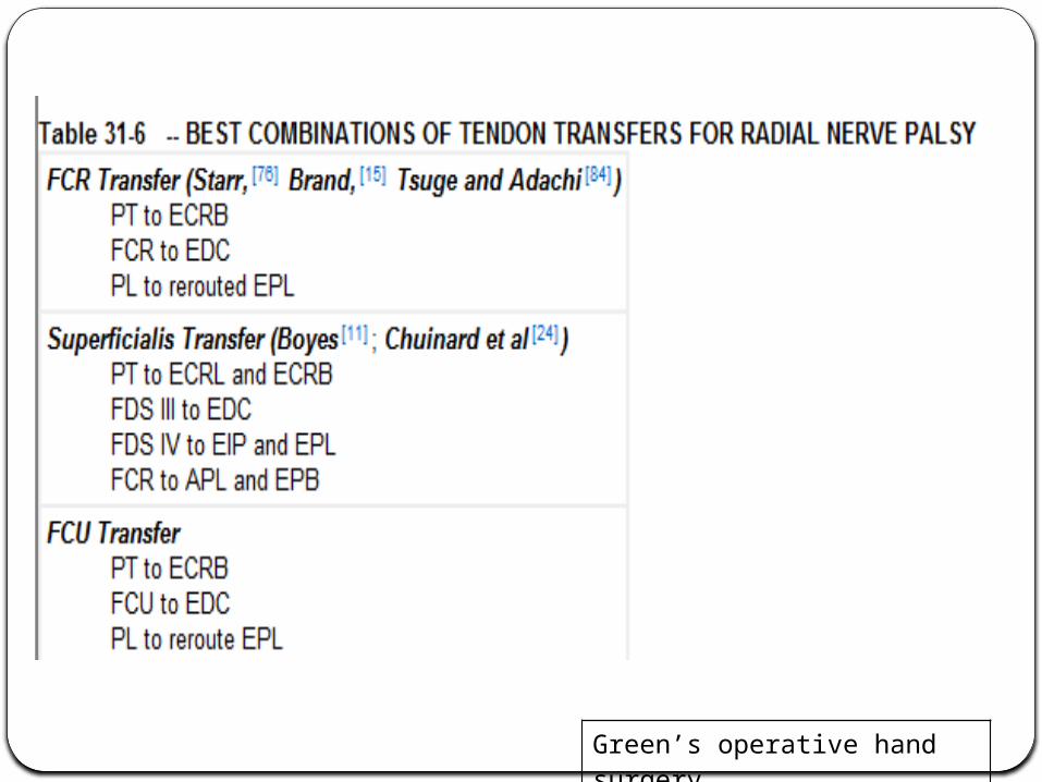
Green’s operative hand surgery
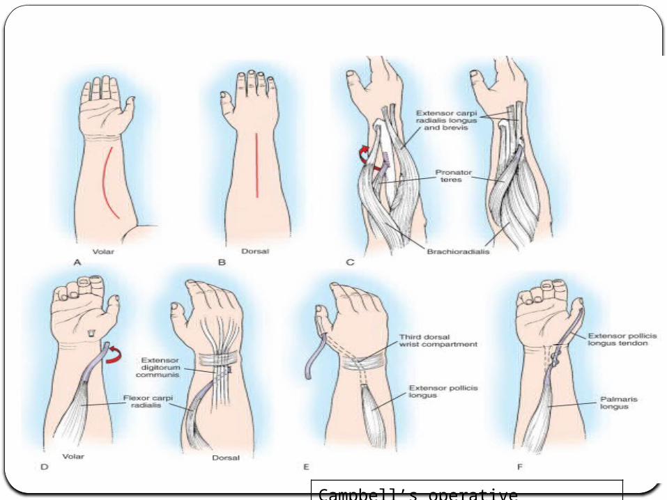
Campbell’s operative orthopaedics
a long arm splint is applied that immobilizes the forearm in 15 to 30degrees of
pronation. the wrist in approximately 45 degrees of extension. the MP joints in slight (10 to 15 degrees) flexion. the thumb in maximum extension and abduction. The proximal interphalangeal joints of the fingers are
left free.
The cast is removed 4 weeks postoperatively; removable short arm splints to hold the wrist, fingers, and thumb in extension are made, which the patient wears for an additional 2 weeks, removing them only for exercise.
Postoperative Management
Radial Nerve Compression Syndromes

Wartenberg’s syndrome
• Aka: Cheiralgia paresthetica• D/t compression of Superficial radial nerve
as it emerges b/w ECRL and BR, 8 cm proximal to radial styloid

isolated pain or paresthesias over the dorsoradial aspect of the hand
preceding history of trauma to the area (i.e., handcuffs, forearm fracture)
Differentiating Wartenberg’s syndrome from de Quervain’s tenosynovitis
A Tinel’s sign over the superficial sensory radial nerve is the most common exam finding
Clinical features
presence of motor weakness suggests a more proximal site of compression
Also seen in patients who use forearms in pronated position for extended periods → in pronation, the tendons of BR and ECRL approximate and may compress the nerve
▪In WS, pain is exacerbated by pronation, while in DQT pain is elicited with changes in thumb and wrist position▪DQT - normal sensation in the dorso-radial hand▪DQT - pain on percussion over the 1st extensor compartment
Electrodiagnostic testing is of limited value in Wartenberg’s syndrome

Posterior interosseous nerve (PIN) syndrome
• D/t compression of PIN in the radial tunnel• Most common causes include:
▪Tumors such as lipomas, ganglia ▪Rheumatoid synovitis ▪Septic arthritis ▪Vasculitis

The radial tunnel is a 5 cm space bounded by:▪Dorsally: capsule of the radiocapitellar joint ▪Volarly: the BR▪Laterally: the ECRL and ECRB muscles ▪Medially: the biceps tendon and brachialis muscles
Within radial tunnel, there are 5 potential sites of compression: ▪fibrous bands to the radiocapitellar joint between the brachialis and BR ▪the recurrent radial vessels (leash of Henry)▪the proximal edge of the ECRB ▪the proximal edge of the Supinator (arcade of Fröhse)▪the distal edge of the Supinator
BR
Supinator
arcade of Fröhse
ECRL
PIN

Diagnosis
loss of finger and thumb extension
Weak wrist extension with radial deviation (since ECRL innervation is intact)
Intact passive tenodesis effect (rules out extensor tendon rupture)
EMG testing is helpful to confirm the diagnosis and monitor motor recovery

Radial Tunnel syndrome
• Similar to PIN syndrome, it is also d/t compression of PIN in the radial tunnel
• Not considered a true compression neuropathy by some

Radial Tunnel Syndrome is a clinical diagnosis
Radial Tunnel
Syndrome
Tenderness over radial tunnel (lateral proximal
forearm, 3-4 cm distal to lateral epicondyle over the mobile wad)
Pain at ECRB origin with
resistance of middle finger
extension
Pain with resisted forearm
supination↑ Pain on
combined elbow extension, forearm
pronation, and wrist flexion

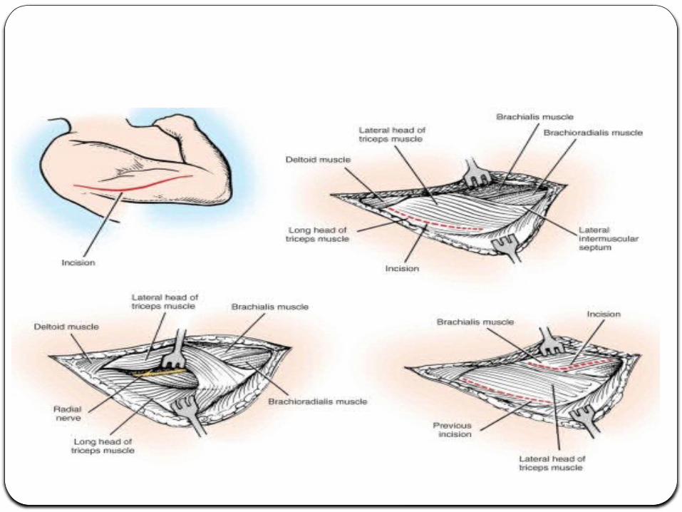
Transmuscular Brachioradialis-Splitting Approach

Posterior (Henry or Thompson Approach)

Anterior (Modified Henry) Approach

THANK YOU