Rad Agrawal MD FACP FACG AGAF FASGE A … · A Review of the Pathogenesis, Management and...
11
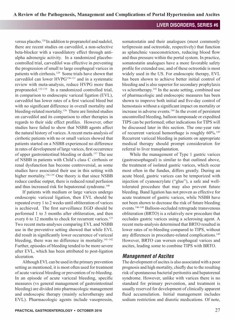
20 PRACTICAL GASTROENTEROLOGY • OCTOBER 2016 LIVER DISORDERS, SERIES #6 Rad Agrawal, MD, FACP, FACG, AGAF, FASGE Courtney Reynolds, MD/PhD 1 Emily Law, MD 1 Duminda Suraweera, MD 1 Gaurav Singhvi, MD 2 1 Department of Medicine, Olive View-UCLA Medical Center, Sylmar, CA 2 David Geffen School of Medicine at UCLA, Los Angeles, CA A Review of the Pathogenesis, Management and Complications of Portal Hypertension and Ascites Gaurav Singhvi Duminda Suraweera Emily Law Courtney Reynolds INTRODUCTION W orldwide, the etiology of portal hypertension is divided between Western and non-Western countries, where 90% of cases in the former are caused by cirrhosis. In the latter, non-cirrhotic conditions such as schistosomiasis or portal vein thrombosis predominate. 1 In some cases, the exact cause of portal hypertension is unclear. Globally, idiopathic non-cirrhotic portal hypertension (INCPH) is a rare disorder associated with infections such as human immunodeficiency virus (HIV) and an array of autoimmune and immunodeficiency disorders ranging from combined variable immunodeficiency to Crohn’s disease. 2 Here, we will review portal hypertension focusing upon the etiology of cirrhosis. In the US, the prevalence of cirrhosis has been calculated between 0.15% and 0.27% of the population (roughly 400,000 to 660,000 people). 3-4 Among those with cirrhosis, it is estimated that 80-90% have portal hypertension, even if they are otherwise asymptomatic. 1 In 2013, the Center for Disease Control (CDC) reported that chronic liver disease and cirrhosis caused approximately 36,000 deaths in the United States. 5 In other words, there is a 5-9% annual mortality associated with cirrhosis; this high mortality is largely attributed to complications of portal hypertension. Alterations in the Circulatory System The pathophysiology of portal hypertension involves alteration of both the splanchnic and the systemic circulatory systems (Figure 1). While portal hypertension had previously been conceptualized as the result simply of increased resistance within the portal system, there is mounting evidence that elevated pressure is also the consequence of increased blood volume or hyperemia, particularly in later stages. It is thought that early hypoxia due to resistance to blood flow triggers the development of collateral blood supply and a local hyperdynamic state characterized by vasodilation. This vasodilation is driven primarily by increased splanchnic production of nitrous oxide (which also contributes to the collateral angiogenesis), leading to decreased responsiveness to vasoconstrictors and overall increased blood volume within the portal system. These changes lead to decreased blood volume and pressure sensed
Transcript of Rad Agrawal MD FACP FACG AGAF FASGE A … · A Review of the Pathogenesis, Management and...

LIVER DISORDERS, SERIES #6
20 PRACTICAL GASTROENTEROLOGY • OCTOBER 2016
LIVER DISORDERS, SERIES #6
Rad Agrawal, MD, FACP, FACG, AGAF, FASGE
Courtney Reynolds, MD/PhD1 Emily Law, MD1 Duminda Suraweera, MD1 Gaurav Singhvi, MD2 1Department of Medicine, Olive View-UCLA Medical Center, Sylmar, CA 2David Geffen School of Medicine at UCLA, Los Angeles, CA
A Review of the Pathogenesis, Management and Complications of Portal Hypertension and Ascites
Gaurav SinghviDuminda Suraweera Emily LawCourtney Reynolds
INTRODUCTION
Worldwide, the etiology of portal hypertension is divided between Western and non-Western countries, where 90% of cases in the former
are caused by cirrhosis. In the latter, non-cirrhotic conditions such as schistosomiasis or portal vein thrombosis predominate.1 In some cases, the exact cause of portal hypertension is unclear. Globally, idiopathic non-cirrhotic portal hypertension (INCPH) is a rare disorder associated with infections such as human immunodeficiency virus (HIV) and an array of autoimmune and immunodeficiency disorders ranging from combined variable immunodeficiency to Crohn’s disease.2 Here, we will review portal hypertension focusing upon the etiology of cirrhosis. In the US, the prevalence of cirrhosis has been calculated between 0.15% and 0.27% of the population (roughly 400,000 to 660,000 people).3-4 Among those with cirrhosis, it is estimated that 80-90% have portal hypertension, even if they are otherwise asymptomatic.1 In 2013, the
Center for Disease Control (CDC) reported that chronic liver disease and cirrhosis caused approximately 36,000 deaths in the United States.5 In other words, there is a 5-9% annual mortality associated with cirrhosis; this high mortality is largely attributed to complications of portal hypertension.
Alterations in the Circulatory SystemThe pathophysiology of portal hypertension involves alteration of both the splanchnic and the systemic circulatory systems (Figure 1). While portal hypertension had previously been conceptualized as the result simply of increased resistance within the portal system, there is mounting evidence that elevated pressure is also the consequence of increased blood volume or hyperemia, particularly in later stages. It is thought that early hypoxia due to resistance to blood flow triggers the development of collateral blood supply and a local hyperdynamic state characterized by vasodilation. This vasodilation is driven primarily by increased splanchnic production of nitrous oxide (which also contributes to the collateral angiogenesis), leading to decreased responsiveness to vasoconstrictors and overall increased blood volume within the portal system. These changes lead to decreased blood volume and pressure sensed

LIVER DISORDERS, SERIES #6
A Review of the Pathogenesis, Management and Complications of Portal Hypertension and Ascites
PRACTICAL GASTROENTEROLOGY • OCTOBER 2016 21
LIVER DISORDERS, SERIES #6
at carotid and renal baroreceptors, leading to similar neurohumoral activation as seen in heart failure (i.e., upregulation of the renin-angiotensin system and anti-diuretic hormone). Thus, portal hypertension spurs a hyperdynamic response in the systemic circulation characterized by increased cardiac output, expansion of plasma volume and reduced systemic vascular resistance.6-15
Alterations in Liver Structure and FunctionIn the setting of cirrhosis, there are characteristic structural and vascular changes within the liver that contribute to portal hypertension. It is well known that hepatic stellate cells (HSC), which function as quiescent lipid and vitamin storage cells in normal liver, become activated as a result of ongoing hepatic injury. This activation results in altered gene activity thought to produce the characteristic fibrotic changes of cirrhosis. However, preceding this development there are substantial changes to the sinusoid endothelial cells as well. The sinusoids ordinarily allow passage of macromolecules to the liver parenchyma through large fenestrations. Capillarization, or loss of endothelial cell fenestration, is an early response to liver injury that appears to occur prior to HSC activation and leads to increased vascular resistance. Interestingly, animal models suggest that reversal of the capillarization process can restore HSC quiescence and reverse fibrosis.6,16 Thus, cirrhosis triggers alterations in liver architecture that contribute to portal hypertension by increased mechanical and vascular resistance.
Pathophysiology of Ascites In the Setting of Portal HypertensionWhile the etiologies of ascites are diverse—including malignancy, infection, hypoalbuminemia and lymphatic obstruction—the overwhelming majority of cases are due to portal hypertension from cirrhosis.17 In the US in particular, an estimated 80% of patients with ascites are due to cirrhosis. Ascites is the most common complication of portal hypertension. The development of ascites is a poor prognostic indicator; median survival for patients with refractory ascites is six months.18 The formation of ascites is similar to edema developing in other parts of the body: ascites emerges when there is a gradient in the hydraulic and oncotic pressures within blood vessels versus the interstitial space. With portal hypertension, ascites is partly the result of the arterial vasodilation that occurs as mentioned above;
this vasodilation and the resulting increased blood volume render increased hydraulic pressure within the vascular bed causing ascites. On the other hand, decreased oncotic pressure, which also contributes to ascites, is primarily due to decreased synthetic function of the cirrhotic liver rather than from portal hypertension directly.
The development of ascites exacerbates the neurohumoral responses activated by portal hypertension. Venous return and renal perfusion are further compromised by ascites and lead to water and sodium retention. It is believed that the presence of ascites corresponds to a decrease in liver function of 60% or less, according to perfused hepatic mass imaging.19 Renal hypoperfusion may initially be countered by increased production of nitric oxide and prostaglandins, however long-standing decompensated cirrhosis usually leads to chronic kidney disease and in some cases the often fatal hepatorenal syndrome. Clinically, patients with ascites develop volume overload and dilutional hyponatremia despite increased total body sodium. Hyponatremia is associated with
Figure 1. Pathophysiology of Portal Hypertension (http://www.cmaj.ca)

22 PRACTICAL GASTROENTEROLOGY • OCTOBER 2016
LIVER DISORDERS, SERIES #6
A Review of the Pathogenesis, Management and Complications of Portal Hypertension and Ascites
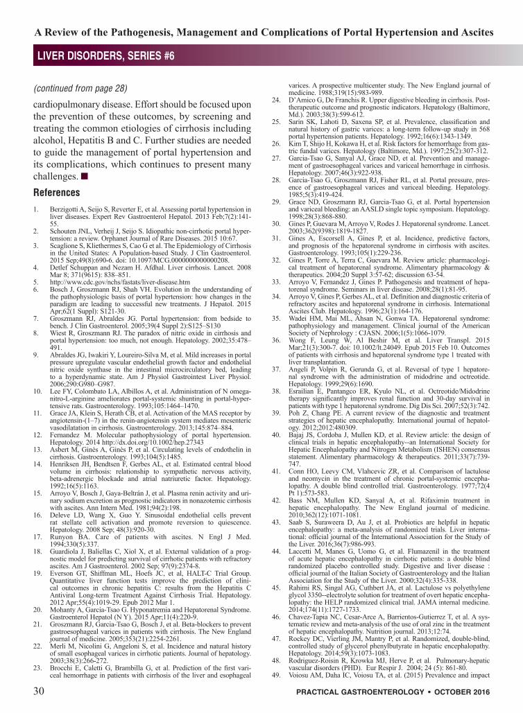
subdivided into two types by distribution: Type 1 are located in the fundus while Type 2 are located in the body, antrum or around the pylorus (Figure 2).
Esophagogastroduodenoscopy (EGD) is the gold standard for the diagnosis of gastroesophageal varices. Varices can be classified as small, medium or large. Small varices are minimally elevated veins above the mucosal surface, medium varices are tortuous veins occupying less than one-third of the esophageal lumen while large varices occupy greater than one-third of the lumen. It is recommended by the American College of Gastroenterology that patients undergo screening for varices at the time of diagnosis of cirrhosis.27-29
Hepatorenal SyndromeHepatorenal syndrome (HRS) is a manifestation of acute renal dysfunction that is seen in severe cirrhosis.30 Risk of developing hepatorenal syndrome from cirrhosis is estimated at 20% after one year and 40% after five years with an incidence of 10% among hospitalized patients with cirrhosis and ascites.31 While the exact mechanism is unknown, it is likely due to a decrease in peripheral arterial circulation from arterial vasodilation in the splanchnic circulation.32 A reduction in cardiac output may also play a concurrent role.33 Patients often present with profound volume overload and electrolyte abnormalities. The diagnosis of hepatorenal syndrome is one of exclusion. Criteria include a plasma creatinine concentration of greater than 1.5 mg/dL, presence of liver disease and portal hypertension, absence of apparent other causes of kidney injury and lack of improvement in renal function after volume expansion with intravenous albumin.34 There are two described types of hepatorenal syndrome. Type 1 is a rapidly developing renal failure defined as a doubling of the serum creatinine to above 2.5 mg/dL or a decrease in glomerular filtration by more than 50% in less than two weeks.35 In contrast, Type 2 hepatorenal syndrome is a gradually developing renal failure with creatinine above 1.5 mg/dL (Table 1).34
Ideally, hepatorenal syndrome is treated with
a poor prognosis and has been shown to predict the development of hepatic encephalopathy, the hepatorenal syndrome and mortality both from cirrhosis and in the short term following liver transplantation.20
Complications of Portal HypertensionPortal hypertension can result in several severe complications leading to significant morbidity and mortality. Generally these complications manifest when hepatic venous pressure gradient exceeds 10 to 12 mm Hg.21 Ascites is the most common complication of portal hypertension as discussed above.
Gastroesophageal VaricesIt is estimated that 5-15% of cirrhotic patients develop gastroesophageal (GE) varices per year, with the development of GE varices correlating with the degree of severity of cirrhosis. About 40% of Child-Pugh A patients have varices as compared to 85% of Child-Pugh C patients.22 Approximately 50% of patients with cirrhosis have gastroesophageal varices at any given time, while the majority of patients with cirrhosis develop GE varices at some point during their lifetime.
Esophageal variceal bleeding occurs at a yearly rate of 5-15%.23 Risk factors for esophageal variceal hemorrhage include size of varices, severity of cirrhosis, variceal pressure and endoscopic presence of variceal red spots. An acute episode of variceal hemorrhage carries a six week mortality rate in excess of 20%.24
Gastric varices are present in 5-44% of patients with portal hypertension.25 Risk factors for gastric variceal hemorrhage include the size of fundal varices, Child-Pugh class and endoscopic presence of variceal red spots.26 Gastric varices can be subdivided into two groups:27 those associated with esophageal varices (gastroesophageal varices) and those not associated with esophageal varices (isolated gastric varices). Gastroesophgeal varices can be further subdivided into two groups depending on their distribution. Type 1 extend along the lesser curvature, and Type 2 extend along the fundus. Isolated gastric varices can also be
Table 1. Diagnostic Criteria for Hepatorenal Syndrome (HRS)Type 1 HRS Type 2 HRS BothCr* increase to >2.5mg/dL Cr increase to >1.5mg/dL Advanced liver failureARF** within 2 weeks ARF over >2 weeks Portal hypertension– – Absence of other causes of ARF
*Cr = serum creatinine**ARF = acute renal failure

LIVER DISORDERS, SERIES #6
A Review of the Pathogenesis, Management and Complications of Portal Hypertension and Ascites
PRACTICAL GASTROENTEROLOGY • OCTOBER 2016 23
defined by liver disease, increased alveolar-arterial oxygen gradient and intrapulmonary vascular dilatations.48 It is more common than portopulmonary hypertension, but both can occur in the same patient. Prevalence ranges from 4 to 34% of patients with liver disease.49-50 While the development of HPS does not require the presence of cirrhosis, it is more common in this setting.51-53 Still, HPS does not correlate with the severity of liver disease.54 The proposed pathophysiology of HPS involves pulmonary production of excess vasoactive mediators, nitric oxide (NO) and carbon monoxide (CO). Arterial hypoxemia is then caused by intrapulmonary vascular dilatation. Other mechanisms or pathways are under investigation, however some studies suggest that there may be increased pulmonary angiogenesis, resulting from greater macrophage production of vascular endothelial growth factor (VEGF)-A.55-56 Screening for HPS with an arterial blood gas is recommended in liver transplant candidates and patients with liver disease who have shortness of breath. The ABG then directs whether the patient needs a contrast-enhanced echocardiography (CEE) which is diagnostic.48
Clinical features of HPS include dyspnea, cyanosis and progressive hypoxemia.57-59 A hallmark finding is platypnea or increased dyspnea with upright positioning that is relieved by lying down; quantitatively platypnea corresponds with orthodeoxia or a decrease in arterial oxygenation by more than 4mmHg moving from recumbency to sitting. A variety of medical therapies exist for HPS but there is a dearth of evidence on their efficacy in improving oxygenation or dyspnea; these agents include somatostatin analogues, beta-blockers, cyclooxygenase inhibitors, glucocorticoids, immunosuppression, pulmonary vasoconstrictors, NO inhibitors, inhaled NO, antimicrobials and garlic.60-77 Supplemental oxygen is often used for symptom relief. Case reports suggest a benefit from TIPS, however this is not routinely recommended due to otherwise variable outcomes and theoretical risk of worsening HPS.78-81 Definitive treatment of HPS is liver transplantation, which results in complete resolution of HPS in greater than 80% of patients.82-88
Porto-Pulmonary HypertensionPulmonary hypertension is a complication of portal hypertension, with or without cirrhosis, and is considered to be a type of pulmonary arterial hypertension.89 Portopulmonary hypertension (POPH)
recovery of liver function either through treatment of the underlying cause (abstinence from alcohol, antiviral therapy, etc.) or through liver transplantation. One study of liver transplantation for Type 1 hepatorenal syndrome found 75% of patients had complete recovery of kidney function after transplant; non-response was associated with prolonged courses of dialysis proceeding transplant, suggesting that prompt referral is key.36 Medical therapy targeted at HRS itself aims to increase perfusion to the kidneys by increasing arterial pressure. In the United States, a combination of octreotide, midodrine and albumin is most frequently used, and the usual course of treatment is two weeks. Alternatives include norephinephrine and vasopressin. Although small studies suggest the effectiveness of vasoconstrictors in this setting, the mortality of HRS remains high.37-38 Patients who fail medical therapy but are either expected to recover liver function or await liver transplantation can transition to dialysis.
Hepatic EncephalopathyHepatic encephalopathy is a neurologic dysfunction seen in patients with liver disease and portal hypertension. The pathogenesis of hepatic encephalopathy is likely multifactorial. Ammonia produced by gut bacteria is typically processed in the liver. However, in the setting of portal hypertension, portosystemic shunts result in ammonia bypassing the liver and accumulating in the systemic circulation and crossing the blood-brain barrier.39 Patients can present with a wide spectrum of neurocognitive manifestations. Hepatic encephalopathy can be divided into minimal hepatic encephalopathy—patients with abnormal psychometric tests but no obvious clinical changes—and overt hepatic encephalopathy, in which patients have obvious clinical manifestations. These manifestations include personality changes, irritability and disinhibition. The West Haven Criteria is used to grade hepatic encephalopathy.40 Grade 1 is considered minimal hepatic encephalopathy, grades 2-3 are intermediate, and grade 4 is a comatose patient. Management of encephalopathy is primarily with non-absorbable disaccharides, such as lactulose and non-absorbable antibiotics, such as rifaximin.41-42 Probiotics, polyethylene glycol, flumazenil ammonia scavengers and zinc have also been shown to be of benefit in the management of hepatic encephalopathy.43-47
Hepatopulmonary Syndrome Hepatopulmonary syndrome (HPS) is a syndrome

A Review of the Pathogenesis, Management and Complications of Portal Hypertension and Ascites
LIVER DISORDERS, SERIES #6
24 PRACTICAL GASTROENTEROLOGY • OCTOBER 2016
defects.102 This occurs more frequently on the right side than the left, possibly due to embryogenic defects.103-104 Subsequently, the hydrothorax can cause an acute tension hydrothorax or infection, namely spontaneous bacterial empyema. The diagnosis is often clinical, and symptoms include shortness of breath, nonproductive cough, chest discomfort and hypoxia. Thoracentesis is performed mainly to exclude other causes, whereas treatment options for HH include medical management with dietary sodium restriction and combined loop diuretic and aldosterone receptor antagonist therapy.105 When HH is refractory to medications, therapeutic thoracentesis can be pursued but it has a high rate of recurrence. Similarly, pleurodesis has a limited role in the management of non-malignant pleural effusions and has been associated with recurrence and significant morbidity such as infection.106 Other options available are transjugular intrahepatic portosystemic shunt (TIPS) and liver transplantation, although due to high associated morbidity TIPS is reserved for patients with relatively preserved liver function (Child-Pugh score <13 or MELD <15).107-108
Diagnosis and Management of Portal Hypertension and AscitesDiagnosis of Portal Hypertension and AscitesThe gold standard for diagnosis of portal hypertension is hepatic venous pressure gradient testing (HVPG), which indirectly measures portal pressure as the difference between the wedged and free hepatic venous pressures. Normal values for HVPG are 1-5mmHg. Any pressure above this range is considered portal hypertension, however HVPG of >10mmHg has been termed “clinically significant” as this level is predictive of the development of ascites and varices. Variceal bleeding becomes more likely with HVPG of 12 or more.109 While less invasive diagnostic techniques are being investigated, such as contrast enhanced ultrasound, in practice, most patients with cirrhosis (or other conditions known to cause portal hypertension) who develop complications such as ascites or varices are presumed to have portal hypertension without further testing.110 The diagnosis of ascites is usually prompted by patient presentation of increased abdominal girth, weight gain and dyspnea. Free fluid within the abdomen can be visualized and graded by imaging, most often ultrasound, while paracentesis allows sample collection to analyze the fluid. The range of tests performed on
is more commonly found in females and in patients with autoimmune liver diseases, namely primary biliary cholangitis and autoimmune hepatitis.90 It is not, however, found to be related to the severity of liver dysfunction, whether by Child Turcotte Pugh (CTP) classification or model for endstage liver disease (MELD) score.91 The pathophysiology of POPH is not clearly defined, however current research has shown remodeling of the pulmonary arterial wall which causes an obstructive thickening and fibrosis of the arteries.92-93 The remodeling is a consequence of the hyperdynamic state caused by splanchnic vasodilation, and the dysfunctional imbalance of mediators such as endothelin-1, prostacyclin and nitric oxide.
Right heart catheterization is required to establish the diagnosis of POPH. According to the criteria established by the 2004 European/US Consensus Study Group, the diagnosis requires 1) portal hypertension with or without hepatic cirrhosis and 2) pulmonary arterial hypertension by right heart catheterization (RHC) with mPAP > 25 mmHg, PVR > 240 dynes.s.cm^-5 and PAWP < 15 mmHg. The severity of portopulmonary hypertension depends on the mPAP: mild is mPAP 25-34 mmHg, moderate is mPAP 35-44 mmHg, and severe is 45 mmHg and greater. In terms of screening, the American Association for the Study of Liver Disease (AASLD) recommends patients being evaluated for liver transplant undergo echocardiogram followed by right heart cardiac catheterization if the RVSP is greater than or equal to 45 mmHg. There is currently no screening recommendation regarding patients with portal hypertension not undergoing liver transplant.48
Medical therapies for POPH include agents used for pulmonary arterial hypertension: endothelin receptor antagonists, prostanoids, phosphodiesterase-5 inhibitors and soluble guanylate cyclase stimulators. Liver transplantation is the only potentially curative option; after transplant, about half of patients can be weaned from POPH medications.94-95
Hepatic Hydrothorax Hepatic hydrothorax (HH) is an uncommon complication in patients with liver disease, found in only 5-10% of patients.96-98 It is defined as a transudative pleural effusion greater than 500 mL in a patient with portal hypertension without any other etiology of the effusion.99-101 The pathologic process is presumed to result from translocation of peritoneal ascetic fluid into the pleural cavity through small diaphragmatic (continued on page 26)

26 PRACTICAL GASTROENTEROLOGY • OCTOBER 2016
LIVER DISORDERS, SERIES #6
A Review of the Pathogenesis, Management and Complications of Portal Hypertension and Ascites
ascitic fluid depends on clinical suspicion, however one essential test for diagnostic paracentesis is to calculate the serum ascites albumin gradient (SAAG) comparing the serum and ascites albumin levels. A high gradient indicates ascites with a low protein content, consistent with cirrhosis or heart failure. Low gradients occur in the setting of malignancy or infection.111 Beyond this, it is standard to obtain cytology, cell count, culture and Gram stain on an initial, diagnostic paracentesis and to evaluate for the presence of spontaneous bacterial peritonitis (SBP), which is heralded by the presence of >250 polymorphonuclear cells/mm3.
Primary Management of Portal HypertensionWhile management of portal hypertension most often focuses upon its complications, there is evidence to support treating the underlying cause as well. In the case of portal hypertension caused by cirrhosis, the regression of cirrhosis after stopping the offending agent or treating the underlying cause has been demonstrated for several disparate etiologies (autoimmune hepatitis, biliary obstruction, iron overload, NASH and hepatitis B and C).112-118 Treatment response to antiviral therapy in patients with Hepatitis C has been correlated with improvement in hepatic fibrosis.119-121 Similar findings have been demonstrated in chronic hepatitis B, where regression of cirrhosis is feasible with long-term suppression with tenofovir.122 The evidence is more limited for improvement of fibrosis following treatment of alcoholic cirrhosis, however abstinence from alcohol has been shown to lead to improved liver function and decreased inflammation and is associated with significantly improved survival compared to cirrhotic patients who continue to drink.123 Nonetheless, although there is evidence to suggest regression in fibrosis, the degree of regression is highly variable, and an actual reversal of cirrhosis has not been demonstrated in humans.124
Management of Gastroesophageal VaricesManagement of varices consists of primary prevention, acute treatment of variceal bleeding and secondary prevention. In patients newly diagnosed with cirrhosis, it is currently a Class IIa recommendation from the American College of Gastroenterology (ACG) to perform a baseline upper endoscopy to assess for gastroesophageal varices.125 On the initial EGD screening, varices should be graded as small, medium
or large as mentioned above, and evaluated for the presence or absence of red signs (wale marks or red spot). In patients with compensated cirrhosis and no varices on the initial EGD, an EGD screening should be repeated in three years. In patients with decompensated cirrhosis and no varices, EGD should be repeated annually.126
In patients with small varices that have not bled and who are not on a non-selective beta-blocker (NSBB), an EGD should be repeated in 2 years. However, in patients with small varices who are on a NSBB, no follow-up EGD is needed. In patients with medium/large varices who are on a NSBB, the dose should be adjusted to the maximum tolerated and a follow-up or surveillance EGD is not needed.125 NSBB is an accepted therapy for primary prophylaxis of variceal hemorrhage. Through blockade of beta-1 receptors, these agents reduce cardiac output and thereby portal pressure. Through blockade of beta-2 receptors, they reduce portal blood inflow from splanchnic vasoconstriction. Propranolol and nadolol are NSBBs that have demonstrated efficacy in much of the literature. They can decrease the incidence of a first variceal hemorrhage from 25 to 15% in a median follow-up of 24 months.127 There is also a lower mortality in patients on NSBBs (propranolol or nadolol)
Figure 2. Types of Gastric Varices (source: www.nursingcenter.com)
(continued from page 24)
LIVER DISORDERS, SERIES #6
A Review of the Pathogenesis, Management and Complications of Portal Hypertension and Ascites
PRACTICAL GASTROENTEROLOGY • OCTOBER 2016 27
versus placebo.128 In addition to propranolol and nadolol, there are recent studies on carvedilol, a non-selective beta-blocker with a vasodilatory effect through anti-alpha adrenergic activity. In a randomized placebo-controlled trial, carvedilol was effective in preventing the progression of small to large esophageal varices in patients with cirrhosis.129 Some trials have shown that carvedilol can lower HVPG130-131 and in a systematic review with meta-analysis, reduce HVPG more than propranolol.132-135 In a randomized controlled trial, in comparison to endoscopic variceal ligation (EVL), carvedilol has lower rates of a first variceal bleed but with no significant difference in overall mortality and bleeding-related mortality.136 There are limited studies on carvedilol and its comparison to other therapies in regards to their side effect profiles. However, other studies have failed to show that NSBB agents affect the natural history of varices. A recent meta-analysis of cirrhotic patients with no or small varices showed that patients started on a NSBB experienced no difference in rates of development of large varices, first occurrence of upper gastrointestinal bleeding or death.137 The use of NSBB in patients with Child’s class C cirrhosis or renal dysfunction has become controversial, as some studies have associated their use in this setting with higher mortality.138-139 One theory is that since NSBB reduce cardiac output, there is reduced renal perfusion and thus increased risk for hepatorenal syndrome.140
If patients with medium or large varices undergo endoscopic variceal ligation, then EVL should be repeated every 1 to 2 weeks until obliteration of varices is acchieved. The first surveillance EGD should be performed 1 to 3 months after obliteration, and then every 6 to 12 months to check for recurrent varices.125 Two recent meta-analyses comparing EVL and NSBB use in the preventive setting showed that while EVL did result in significantly lower occurrence of variceal bleeding, there was no difference in mortality.141-142 Further, episodes of bleeding tended to be more severe after EVL, which has been attributed to post-ligation ulceration.
Although EVL can be used in the primary prevention setting as mentioned, it is most often used for treatment of acute variceal bleeding or prevention of re-bleeding. In an episode of acute variceal bleeding, specific measures (vs general management of gastrointestinal bleeding) are divided into pharmacologic management and endoscopic therapy (mainly sclerotherapy and EVL). Pharmacologic agents include vasopressin,
somatostatin and their analogues (most commonly terlipressin and octreotide, respectively) that function as splanchnic vasoconstrictors, reducing blood flow and thus pressure within the portal system. In practice, somatostatin analogues have a more favorable safety profile for extended use, and of these octreotide is most widely used in the US. For endoscopic therapy, EVL has been shown to achieve better initial control of bleeding and is also superior for secondary prophylaxis vs sclerotherapy.143 In the acute setting, combined use of pharmacologic and endoscopic measures has been shown to improve both initial and five-day control of hemostasis without a significant impact on mortality or increase in adverse events.144 In the event of persistent uncontrolled bleeding, balloon tamponade or expedited TIPS can be performed; other indications for TIPS will be discussed later in this section. The one-year rate of recurrent variceal hemorrhage is roughly 60%.145 Recurrent variceal bleeding in patients on appropriate medical therapy should prompt consideration for referral to liver transplantation.
While the management of Type 1 gastric varices (gastroesophageal) is similar to that outlined above, the treatment of isolated gastric varices, which occur most often in the fundus, differs greatly. During an acute bleed, gastric varices can be temporized with injection of cyanoacrylate (“glue”), a safe and well-tolerated procedure that may also prevent future bleeding. Band ligation has not proven as effective for acute treatment of gastric varices, while NSBB have not been shown to decrease the risk of future bleeding events.146-148 Balloon-occluded retrograde transvenous obliteration (BRTO) is a relatively new procedure that occludes gastric varices using a sclerosing agent. A recent meta-analysis determined that BRTO resulted in lower rates of re-bleeding compared to TIPS, without any differences in procedure-related complications.149 However, BRTO can worsen esophageal varices and ascites, leading some to combine TIPS with BRTO.
Management of AscitesThe development of ascites is also associated with a poor prognosis and high mortality, chiefly due to the resulting risk of spontaneous bacterial peritonitis and hepatorenal syndrome. However, unlike with varices there is no standard for primary prevention, and treatment is usually reserved for development of clinically apparent fluid accumulation. Initial management includes sodium restriction and diuretic medications. Of note,

28 PRACTICAL GASTROENTEROLOGY • OCTOBER 2016
LIVER DISORDERS, SERIES #6
A Review of the Pathogenesis, Management and Complications of Portal Hypertension and Ascites
sodium restriction (to less than two grams daily) is most effective in patients with relatively intact renal function, as sodium excretion becomes more impaired with disease progression. Concomitant fluid restriction is usually only implemented if severe hyponatremia has developed (i.e., serum sodium less than 120 mEq/L).150 In one randomized controlled trial, cirrhotic patients with ascites on diuretics were randomized to a low sodium diet versus unrestricted sodium intake. There was no significant difference between the two groups among the endpoints measured (mortality, time for complete resolution of ascites, hospital stays and cost).151 However, in patients with no previous history of gastrointestinal bleeding, there was a higher survival rate in those on a low sodium diet. In practice, the effectiveness of sodium restriction is limited by patient compliance.
Diuretic therapy is a complement to, rather than a replacement for, sodium restriction and is usually instituted concurrently. The diuretic of choice is spironolactone, as it works to combat the renin-angiotensin system activation triggered by portal hypertension and ascites.152 Patients who do not respond to an adequate dose of spironolactone (200 to 400mg daily), may also receive oral furosemide; the ratio of spironolactone to furosemide dosing is generally 100mg: 40mg respectively. Rapid fluid or weight loss from diuretics should be avoided, and patients in the dose titration phase need to be monitored closely for complications of diuretic treatment including hyponatremia, hyperkalemia, encephalopathy and renal failure. The additional benefit of albumin to diuretic therapy has been controversial. In one randomized controlled trial, cirrhotic patients in an outpatient setting received either diuretics alone versus diuretics with albumin. They found a higher clinical response rate in those who received diuretics with albumin compared to diuretics alone, resulting in shorter hospital stays, lower probability of re-developing ascites and lower probability of readmission to the hospital.153 Practically, the routine use of albumin is limited by expense and patient adherence. Finally, therapeutic paracentesis is often used in the setting of severe or tense ascites. Refractory ascites is defined as ascites that fails the above measures or recurs rapidly after therapeutic paracentesis; it occurs in approximately 5-10% of patients with ascites. Treatment options include large volume paracentesis (up to 5L), liver transplantation or TIPS.
Transjugular Intrahepatic Portosystemic Shunting for Refractory Bleeding or AscitesTIPS, which creates a shunt from the portal vein to the hepatic vein, has emerged as a second line treatment for severe complications of portal hypertension including recurrent variceal bleeding and refractory ascites. Before TIPS is performed, the patient must be evaluated to determine if they are an appropriate candidate. Risk factors for poor outcome and complications from the procedure include prior encephalopathy, hyperbilirubinemia and cardiopulmonary disease. These risks must be considered, along with the possibility of referral for definitive treatment with liver transplant. Absolute contraindications to TIPS include congestive heart failure, multiple hepatic cysts, uncontrolled sepsis, biliary obstruction and severe pulmonary hypertension. For variceal bleeding, TIPS has been shown to be superior to NSBB plus sclerotherapy in preventing recurrence in one meta-analysis; despite this, no difference in mortality has been proven.154 One retrospective study comparing TIPS with EVL did find a survival benefit, however this has yet to be demonstrated in controlled prospective trials.155 For refractory ascites, there is conflicting evidence from randomized controlled trials (and meta-analyses of these trials) about whether TIPS confers a survival benefit compared to large volume paracentesis. Available trials are limited by small sample size and heterogeneous patient selection, however there may be an advantage for using TIPS in patients with ascites and relatively preserved renal function.156-158 In one retrospective study, patients who had MELD scores greater than 15 were evaluated in two groups, those who received TIPS and whose who did not. In the first two months post-TIPS, patients had increased mortality compared to their counterparts, however this was not statistically significant. After two months, TIPS was associated with lower mortality and need for liver transplantation versus cirrhotic patients who did not undergo TIPS.159 Further, prospective, controlled studies are needed to confirm this result.
CONCLUSIONPortal hypertension is an important cause of mortality globally and a frequent consequence of end stage liver disease in the United States. If untreated, portal hypertension results in the associated conditions of ascites, variceal bleeding, hepatorenal syndrome and
(continued on page 30)
30 PRACTICAL GASTROENTEROLOGY • OCTOBER 2016
LIVER DISORDERS, SERIES #6
A Review of the Pathogenesis, Management and Complications of Portal Hypertension and Ascites
cardiopulmonary disease. Effort should be focused upon the prevention of these outcomes, by screening and treating the common etiologies of cirrhosis including alcohol, Hepatitis B and C. Further studies are needed to guide the management of portal hypertension and its complications, which continues to present many challenges. References1. Berzigotti A, Seijo S, Reverter E, et al. Assessing portal hypertension in
liver diseases. Expert Rev Gastroenterol Hepatol. 2013 Feb;7(2):141-55.
2. Schouten JNL, Verheij J, Seijo S. Idiopathic non-cirrhotic portal hyper-tension: a review. Orphanet Journal of Rare Diseases. 2015 10:67.
3. Scaglione S, Kliethermes S, Cao G et al. The Epidemiology of Cirrhosis in the United States: A Population-based Study. J Clin Gastroenterol. 2015 Sep;49(8):690-6. doi: 10.1097/MCG.0000000000000208.
4. Detlef Schuppan and Nezam H. Afdhal. Liver cirrhosis. Lancet. 2008 Mar 8; 371(9615): 838–851.
5. http://www.cdc.gov/nchs/fastats/liver-disease.htm6. Bosch J, Groszmann RJ, Shah VH. Evolution in the understanding of
the pathophysiologic basis of portal hypertension: how changes in the paradigm are leading to successful new treatments. J Hepatol. 2015 Apr;62(1 Suppl): S121-30.
7. Groszmann RJ, Abraldes JG. Portal hypertension: from bedside to bench. J Clin Gastroenterol. 2005;39(4 Suppl 2):S125–S130
8. Wiest R, Groszmann RJ. The paradox of nitric oxide in cirrhosis and portal hypertension: too much, not enough. Hepatology. 2002;35:478–491.
9. Abraldes JG, Iwakiri Y, Loureiro-Silva M, et al. Mild increases in portal pressure upregulate vascular endothelial growth factor and endothelial nitric oxide synthase in the intestinal microcirculatory bed, leading to a hyperdynamic state. Am J Physiol Gastrointest Liver Physiol. 2006;290:G980–G987.
10. Lee FY, Colombato LA, Albillos A, et al. Administration of N omega-nitro-L-arginine ameliorates portal-systemic shunting in portal-hyper-tensive rats. Gastroenterology. 1993;105:1464–1470.
11. Grace JA, Klein S, Herath CB, et al. Activation of the MAS receptor by angiotensin-(1–7) in the renin-angiotensin system mediates mesenteric vasodilatation in cirrhosis. Gastroenterology. 2013;145:874–884.
12. Fernandez M. Molecular pathophysiology of portal hypertension. Hepatology. 2014 http://dx.doi.org/10.1002/hep.27343
13. Asbert M, Ginès A, Ginès P, et al. Circulating levels of endothelin in cirrhosis. Gastroenterology. 1993;104(5):1485.
14. Henriksen JH, Bendtsen F, Gerbes AL, et al. Estimated central blood volume in cirrhosis: relationship to sympathetic nervous activity, beta-adrenergic blockade and atrial natriuretic factor. Hepatology. 1992;16(5):1163.
15. Arroyo V, Bosch J, Gaya-Beltrán J, et al. Plasma renin activity and uri-nary sodium excretion as prognostic indicators in nonazotemic cirrhosis with ascites. Ann Intern Med. 1981;94(2):198.
16. Deleve LD, Wang X, Guo Y. Sinusoidal endothelial cells prevent rat stellate cell activation and promote reversion to quiescence. Hepatology. 2008 Sep; 48(3):920-30.
17. Runyon BA. Care of patients with ascites. N Engl J Med. 1994;330(5):337.
18. Guardiola J, Baliellas C, Xiol X, et al. External validation of a prog-nostic model for predicting survival of cirrhotic patients with refractory ascites. Am J Gastroenterol. 2002 Sep; 97(9):2374-8.
19. Everson GT, Shiffman ML, Hoefs JC, et al, HALT-C Trial Group. Quantitative liver function tests improve the prediction of clini-cal outcomes in chronic hepatitis C: results from the Hepatitis C Antiviral Long-term Treatment Against Cirrhosis Trial. Hepatology. 2012 Apr;55(4):1019-29. Epub 2012 Mar 1.
20. Mohanty A, Garcia-Tsao G. Hyponatremia and Hepatorenal Syndrome. Gastroenterol Hepatol (N Y). 2015 Apr;11(4):220-9.
21. Groszmann RJ, Garcia-Tsao G, Bosch J, et al. Beta-blockers to prevent gastroesophageal varices in patients with cirrhosis. The New England journal of medicine. 2005;353(21):2254-2261.
22. Merli M, Nicolini G, Angeloni S, et al. Incidence and natural history of small esophageal varices in cirrhotic patients. Journal of hepatology. 2003;38(3):266-272.
23. Brocchi E, Caletti G, Brambilla G, et al. Prediction of the first vari-ceal hemorrhage in patients with cirrhosis of the liver and esophageal
varices. A prospective multicenter study. The New England journal of medicine. 1988;319(15):983-989.
24. D’Amico G, De Franchis R. Upper digestive bleeding in cirrhosis. Post-therapeutic outcome and prognostic indicators. Hepatology (Baltimore, Md.). 2003;38(3):599-612.
25. Sarin SK, Lahoti D, Saxena SP, et al. Prevalence, classification and natural history of gastric varices: a long-term follow-up study in 568 portal hypertension patients. Hepatology. 1992;16(6):1343-1349.
26. Kim T, Shijo H, Kokawa H, et al. Risk factors for hemorrhage from gas-tric fundal varices. Hepatology (Baltimore, Md.). 1997;25(2):307-312.
27. Garcia-Tsao G, Sanyal AJ, Grace ND, et al. Prevention and manage-ment of gastroesophageal varices and variceal hemorrhage in cirrhosis. Hepatology. 2007;46(3):922-938.
28. Garcia-Tsao G, Groszmann RJ, Fisher RL, et al. Portal pressure, pres-ence of gastroesophageal varices and variceal bleeding. Hepatology. 1985;5(3):419-424.
29. Grace ND, Groszmann RJ, Garcia-Tsao G, et al. Portal hypertension and variceal bleeding: an AASLD single topic symposium. Hepatology. 1998;28(3):868-880.
30. Gines P, Guevara M, Arroyo V, Rodes J. Hepatorenal syndrome. Lancet. 2003;362(9398):1819-1827.
31. Gines A, Escorsell A, Gines P, et al. Incidence, predictive factors, and prognosis of the hepatorenal syndrome in cirrhosis with ascites. Gastroenterology. 1993;105(1):229-236.
32. Gines P, Torre A, Terra C, Guevara M. Review article: pharmacologi-cal treatment of hepatorenal syndrome. Alimentary pharmacology & therapeutics. 2004;20 Suppl 3:57-62; discussion 63-54.
33. Arroyo V, Fernandez J, Gines P. Pathogenesis and treatment of hepa-torenal syndrome. Seminars in liver disease. 2008;28(1):81-95.
34. Arroyo V, Gines P, Gerbes AL, et al. Definition and diagnostic criteria of refractory ascites and hepatorenal syndrome in cirrhosis. International Ascites Club. Hepatology. 1996;23(1):164-176.
35. Wadei HM, Mai ML, Ahsan N, Gonwa TA. Hepatorenal syndrome: pathophysiology and management. Clinical journal of the American Society of Nephrology : CJASN. 2006;1(5):1066-1079.
36. Wong F, Leung W, Al Beshir M, et al. Liver Transpl. 2015 Mar;21(3):300-7. doi: 10.1002/lt.24049. Epub 2015 Feb 10. Outcomes of patients with cirrhosis and hepatorenal syndrome type 1 treated with liver transplantation.
37. Angeli P, Volpin R, Gerunda G, et al. Reversal of type 1 hepatore-nal syndrome with the administration of midodrine and octreotide. Hepatology. 1999;29(6):1690.
38. Esrailian E, Pantangco ER, Kyulo NL, et al. Octreotide/Midodrine therapy significantly improves renal function and 30-day survival in patients with type 1 hepatorenal syndrome. Dig Dis Sci. 2007;52(3):742.
39. Poh Z, Chang PE. A current review of the diagnostic and treatment strategies of hepatic encephalopathy. International journal of hepatol-ogy. 2012;2012:480309.
40. Bajaj JS, Cordoba J, Mullen KD, et al. Review article: the design of clinical trials in hepatic encephalopathy--an International Society for Hepatic Encephalopathy and Nitrogen Metabolism (ISHEN) consensus statement. Alimentary pharmacology & therapeutics. 2011;33(7):739-747.
41. Conn HO, Leevy CM, Vlahcevic ZR, et al. Comparison of lactulose and neomycin in the treatment of chronic portal-systemic encepha-lopathy. A double blind controlled trial. Gastroenterology. 1977;72(4 Pt 1):573-583.
42. Bass NM, Mullen KD, Sanyal A, et al. Rifaximin treatment in hepatic encephalopathy. The New England journal of medicine. 2010;362(12):1071-1081.
43. Saab S, Suraweera D, Au J, et al. Probiotics are helpful in hepatic encephalopathy: a meta-analysis of randomized trials. Liver interna-tional: official journal of the International Association for the Study of the Liver. 2016;36(7):986-993.
44. Laccetti M, Manes G, Uomo G, et al. Flumazenil in the treatment of acute hepatic encephalopathy in cirrhotic patients: a double blind randomized placebo controlled study. Digestive and liver disease : official journal of the Italian Society of Gastroenterology and the Italian Association for the Study of the Liver. 2000;32(4):335-338.
45. Rahimi RS, Singal AG, Cuthbert JA, et al. Lactulose vs polyethylene glycol 3350--electrolyte solution for treatment of overt hepatic encepha-lopathy: the HELP randomized clinical trial. JAMA internal medicine. 2014;174(11):1727-1733.
46. Chavez-Tapia NC, Cesar-Arce A, Barrientos-Gutierrez T, et al. A sys-tematic review and meta-analysis of the use of oral zinc in the treatment of hepatic encephalopathy. Nutrition journal. 2013;12:74.
47. Rockey DC, Vierling JM, Mantry P, et al. Randomized, double-blind, controlled study of glycerol phenylbutyrate in hepatic encephalopathy. Hepatology. 2014;59(3):1073-1083.
48. Rodriguez-Roisin R, Krowka MJ, Herve P, et al. Pulmonary-hepatic vascular disorders (PHD). Eur Respir J. 2004; 24 (5): 861-80.
49. Voiosu AM, Daha IC, Voiosu TA, et al. (2015) Prevalence and impact
(continued from page 28)

LIVER DISORDERS, SERIES #6
A Review of the Pathogenesis, Management and Complications of Portal Hypertension and Ascites
PRACTICAL GASTROENTEROLOGY • OCTOBER 2016 31
on survival of hepatopulmonary syndrome and cirrhotic cardiomyopa-thy in a cohort of cirrhotic patients. Liver Int.
50. El Makarem MA, Elakad A, Ali A, et al. (2011) Hepatopulmonary syn-drome: prevalence and predictors in Egyptian cirrhotic patients. Trop Gastroenterol 32:25-30
51. De BK, Sen S, Sanyal, R. Hepatopulmonary syndrome in non-cirrhotic portal hypertension. Ann Intern Med. 2000; 132: 924.
52. Regev A, Yeshurun M, Rodriguez M, et al. Transient hepatopulmonary syndrome in a patient with acute hepatitis A. J Viral Hepat. 2011; 8:83-86.
53. Gupta D, Vijaya DR, Gupta R, et al. Prevalence of hepatopulmonary syndrome in cirrhosis and extrahepatic portal venous obstruction. Am J Gastroenterol. 2001; 96: 3395-3399.
54. Krowka MJ, Wiseman GA, Burnett OL, et al. Hepatopulmonary syn-drome: a prospective study of relationships between severity of liver disease, PaO2 response to 100% oxygen, and brain uptake after 99mTc MAA lung scanning. Chest 2000; 118: 615-624.
55. Thenappan T, Goel A, Marsboom G, Fang YH, et al. A central role for CD68(+) macrophages in hepatopulmonary syndrome. Reversal by macrophage depletion. Am J Respir Crit Care Med. 2011; 183: 1080-1091.
56. Zhang J, Luo B, Tang L, Wang Y, et al. Pulmonary angiogenesis in a rat model of hepatpulmonary syndrome. Gastroenterology 2009; 136: 1070-1080.
57. Anand AC, Mukherjee D, Rao KS, et al. Hepatopulmonary syndrome: prevalence and clinical profile. Indian J Gastroenterol. 2001; 20:24-27.
58. Mohammad AA, Fatemi SR, Mirzaee V, et al. Clinical features of hepatopulmonary syndrome in cirrhotic patients. World J Gastroenterol. 2006; 12:1954-1956.
59. Swanson KL, Wiesner RH, Krowka MJ. Natural history of hepatopul-monary syndrome: impact of liver transplantation. Hepatology. 2005; 41: 1122-1129.
60. Krowka MJ, Dickson ER, Cortese DA. Hepatopulmonary syndrome. Clinical observations and lack of therapeutic response to somatostatin analogue. Chest 1993;104:515–521.
61. Agustí AGN, Roca J, Bosch J, et al. Effects of propranolol on arterial oxygenation and oxygen transport to tissues in patients with cirrhosis. Am Rev Respir Dis 1990;142:306–310.
62. Lambrecht GL, Malbrain ML, Coremans P, et al. Orthodeoxia and platypnea in liver cirrhosis: effects of propranolol. Acta Clin Belg 1994;49:26–30.
63. Shijo H, Sasaki H, Yuh K, et al. Effects of indomethacin on hepatogenic pulmonary angiodysplasia. Chest 1991;99:1027–1029.
64. Song JY, Choi JY, Ko JT, et al. Long-term aspirin therapy for hepato-pulmonary syndrome. Pediatrics 1996;97:917–920.
65. Cadranel J, Milleron B, Cadranel JF, et al. Severe hypoxemia associated intrapulmonary shunt in a patient with chronic liver disease: improve-ment after medical treatment. Am Rev Respir Dis 1992;146:526–527.
66. Krowka MJ, Cortese DA. Severe hypoxemia associated with liver disease: Mayo Clinic experience and the experimental use of almitrine bismesylate. Mayo Clin Proc 1987;62:164–173.
67. Nakos G, Evrenoglou D, Vassilakis N, et al. Haemodynamics and gas exchange in liver cirrhosis: the effect of orally administered almitrine bismesylate. Respir Med 1993;87:93–98.
68. Rolla G, Bucca C, Brussino L. Methylene blue in the hepatopulmonary syndrome. N Engl J Med 1994;331:1098.
69. Schenk P, Madl C, Rezale-Majd S, et al. Methylene blue improves the hepatopulmonary syndrome. Ann Intern Med 2000;133:701–706.
70. Brussino L, Bucca C, Morello M, et al. Effect on dyspnoea and hypox-aemia of inhaled N G-nitro-l-arginine methyl esther in hepatopulmo-nary syndrome. Lancet 2003;362:43–44.
71. Rolla G, Brussino L, Dutto L, et al. Smoking and hypoxemia caused by hepatopulmonary syndrome before and after liver transplantation. Hepatology 2001;34:430–431.
72. Boker KHW, Hoper M, Lehman M, et al. Effect of nitric oxide synthase inhibitor l-NAME on vascular resistance and arterial oxygenation in patients with hepatopulmonary syndrome. Hepatology 1994;20:Suppl. 1, 333A (abstract).
73. Durand P, Baujard C, Grosse AL, et al. Reversal of hypoxemia by inhaled nitric oxide in children with severe hepatopulmonary syn-drome, type 1, during and after liver transplantation. Transplantation 1998;65:437–439.
74. Maniscalco M, Sofia M, Higenbottam T. Effects of an NO-synthase inhibitor l-NMMA in the hepatopulmonary syndrome. Respiration 2001;68:226.
75. Rabiller A, Nunes H, Lebrec D, et al. Prevention of Gram-negative translocation reduces the severity of hepatopulmonary syndrome. Am J Respir Crit Care Med 2002;166:514–517.
76. Anel RM, Sheagren JN. Novel presentation and approach to manage-ment of hepatopulmonary syndrome with use of antimicrobial agents. Clin Infect Dis 2001;32:131–136.
77. Abrams GA, Fallon MB. Treatment of hepatopulmonary syndrome
with Allium sativum L. (garlic): a pilot trial. J Clin Gastroenterol 1998;27:232–235.
78. Chevallier P, Novelli L, Motamedi JP, et al. Hepatopulmonary syn-drome successfully treated with transjugular intrahepatic portosystemic shunt: a three-year follow-up.. J Vasc Interv Radiol. 2004;15(6):647.
79. Paramesh AS, Husain SZ, Shneider B, et al. Improvement of hepa-topulmonary syndrome after transjugular intrahepatic portasystemic shunting: case report and review of literature.. Pediatr Transplant. 2003;7(2):157.
80. Lasch HM, Fried MW, Zacks SL, et al. Use of transjugular intrahepatic portosystemic shunt as a bridge to liver transplantation in a patient with severe hepatopulmonary syndrome. Liver Transpl. 2001;7(2):147.
81. Martinez-Palli G, Drake BB, Garcia-Pagan JC, et al. Effect of tran-sjugular intrahepatic portosystemic shunt on pulmonary gas exchange in patients with portal hypertension and hepatopulmonary syndrome. World J Gastroenterol. 2005;11(43):6858.
82. Krowka MJ, Cortese DA. Hepatopulmonary syndrome: an evolving perspective in the era of liver transplantation. Hepatology 1990;11:138–141.
83. Rodriguez-Roisin R, Krowka MJ. Is severe hypoxaemia due to hepatic disease an indication for liver transplantation? A new therapeutic approach. Eur Respir J 1994;7:839–842.
84. Fewtrell MS, Noble-Jamieson G, Revell S, et al. Intrapulmonary shunt-ing in the biliary atresia/polysplenia syndrome: reversal after liver transplantation. Arch Dis Child 1994;70:501–504.
85. Battaglia SE, Pretto JJ, Irving LB, et al. Resolution of gas exchange abnormalities and intrapulmonary shunting following liver transplanta-tion. Hepatology 1997;25:1228–1232.
86. Krowka MJ. Hepatopulmonary syndrome: recent literature (1997 to 1999) and implications for liver transplantation. Liver Transpl 2000;6: Suppl. 1, S31–S35.
87. De Goyet JV, Gibbs P, Clauyput P, et al. Original extrahepatic approach for hepatic portal revascularization and relief of portal hyperten-sion related to late portal vein thrombosis after liver transplantation. Transplantation 1996;62:71–75.
88. Collisson EA, Nourmand H, Fraiman MH, et al. Retrospective analysis of the results of liver transplantation for adults with severe hepatopul-monary syndrome. Liver Transpl 2002;8:925–931.
89. https://www.nhlbi.nih.gov/health/health-topics/topics/pah/types90. Kawut SM, Krowka MJ, Trotter JF, et al. Clinical risk factors for porto-
pulmonary hypertension. Hepatology. 2008; 48 (1) 196-203.91. Krowka MJ. Portopulmonary hypertension. Semin Respir Crit Care
Med 2012; 33:17-25. 92. Krowka MJ, Edwards WD. A spectrum of pulmonary vascular pathol-
ogy in portopulmonary hypertension. Liver Transpl 2000; 6: 241-2. 93. Edwards BS, Weir EK, Edwards WD, et al. Coexistent pulmonary
and portal hypertension: morphologic and clinical features. J Am Coll Cardiol 1987; 10: 1233-8.
94. Bandara M, Gordon FD, Sarwar A, et al. Successful outcomes follow-ing living donor liver transplantation for portopulmonary hypertension. Liver Transpl. 2010; 16 (8) 983-9.
95. Khaderi S, Khan R, Safdar Z, et al. Long-term follow-up of portopul-monary hypertension patients after liver transplantation. Liver Transpl. 2014; 20 (6): 724-7.
96. Krok KL, Cardenas A. Hepatic hydrothorax. Semin Respir Crit Care Med 2012; 33 (1): 3-10.
97. Cardenas A, Kelleher T, Chopra S. Review article: hepatic hydrothorax. Aliment Pharmacol Ther 2004; 20 (3): 271-9.
98. Baikati K, Le DL, Jabbour II, et al. Hepatic hydrothorax. Am J Ther 2014; 21 (1): 43-51.
99. Xiol X, Castellote J, Cortes-Beut R, et al. Usefulness and complications of thoracentesis in cirrhotic patients. Am J Med 2001; 111 (1) 67-9.
100. Chen CH, Shih CM, Chou JW, et al. Outcome predictors of cirrhotic patients with spontaneous bacterial empyema. Liver Int 2011; 31 (3): 417-24.
101. Chen TA, Lo GH, Lai KH. Risk factors for spontaneous bacterial empyema in cirrhotic patients with hydrothorax. J Chin Med Assoc 2003; 66 (10): 579-86.
102. Lieberman FL, Hidemura R, Peters RL, et al. Pathogenesis and treat-ment of hydrothorax complicating cirrhosis with ascites. Ann Intern Med 1966; 64 (2): 341-51.
103. Lazaridis KN, Frank JW, Krowka MJ, et al. Hepatic hydrothorax: pathogenesis, diagnosis, and management. Am J Med 1999; 107 (3): 262-7.
104. Strauss RM, Boyder TD. Hepatic hydrothorax. Semin Liver Dis 1997; 17 (3): 227-32.
105. Runyon BA, Committee AP. Management of adult patients with ascites due to cirrhosis: an update. Hepatology 2009; 49 (6): 2087-107.
106. Milanez de Campos JR, Filho LO, de Campos Werebe E, et al. Thoracoscopy and talc poudrage in the management of hepatic hydro-thorax. Chest. 2000;118(1):13.
107. Dhanasekaran R, West JK, Gonzales PC, et al. Transjugular intrahepatic

32 PRACTICAL GASTROENTEROLOGY • OCTOBER 2016
LIVER DISORDERS, SERIES #6
A Review of the Pathogenesis, Management and Complications of Portal Hypertension and Ascites
portosystemic shunt for symptomatic refractory hepatic hydrothorax in patients with cirrhosis. Am J Gastroenterol. 2010;105(3):635.
108. Wilputte JY, Goffette P, Zech F, et al. The outcome after transjugular intrahepatic portosystemic shunt (TIPS) for hepatic hydrothorax is closely related to liver dysfunction: a long-term study in 28 patients. Acta Gastroenterol Belg. 2007;70(1):6.
109. Kumar A, Sharma P, Sarin SK. Indian J Gastroenterol. 2008 Mar-Apr;27(2):74-80. Hepatic venous pressure gradient measurement: time to learn!
110. Jeong WK, Kim TY, Sohn JH et al. Severe Portal Hypertension in Cirrhosis: Evaluation of Perfusion Parameters with Contrast-Enhanced Ultrasonography. PloS one 10(3):e0121601
111. Mauer K, Manzione NC. Usefulness of serum-ascites albumin differ-ence in separating transudative from exudative ascites. Another look. Dig Dis Sci. 1988;33(10):1208.
112. Dufour JF, DeLellis R, Kaplan MM. Reversibility of hepatic fibrosis in autoimmune hepatitis. Ann Intern Med. 1997; 127: 981-5.
113. Hammel P, Couvelard A, O’Toole D, et al. Regression of liver fibrosis after biliary drainage in patients with chronic pancreatitis and stenosis of the common bile duct. N Engl J Med. 2001; 344: 418-23.
114. Muretto P, Angelucci E, Lucarelli G. Reversibility of cirrhosis in patients cured of thalassemia by bone marrow transplantation. Ann Intern Med. 2002; 136: 667-72.
115. Dixon JB, Bhathal PS, Hughes NR, et al. Nonalcoholic fatty liver disease: Improvement in liver histological analysis with weight loss. Hepatology. 2004; 39: 1647-54.
116. Kral JG, Thing SN, Biron S, et al. Effects of surgical treatment of the metabolic syndrome on liver fibrosis and cirrhosis. Surgery. 2004; 135: 48-58.
117. Kweon YO, Goodman ZD, Dienstag JL, et al. Decreasing fibrogenesis: An immunohistochemical study of paired liver biopsies following lami-vuding therapy for chronic hepatitis B. J Hepatol. 2001; 35: 749-55.
118. Poynard T, Bedossa P, Opolon P. Natural history of liver fibrosis progression in patients with chronic hepatitis C: The OBSVIRC, METAVIR, CLINIVIR, and DOSVIRC groups. Lancet. 1997; 349: 825-32.
119. Pol S, Carnot F, Nalpas B, et al. Reversibility of hepatitis C virus-related cirrhosis. Human Pathology, 2004 Jan. 35 (1): 107-112.
120. Shiratori Y, Imazeki F, Moriyama M, et al. Histologic improvement of fibrosis in patients with hepatitis C who have sustained response to interferon therapy. Ann Intern Med. 2000; 132(7):517-24
121. Yada N, Sakurai T, Minami T, et al. Ultrasound Elastography Correlates Treatment Response by Antiviral Therapy in Patients with Chronic Hepatitis C. Oncology 2014. 87 Suppl 1:118-23
122. Marcellin P, Gane E, Buti M, et al. Regression of cirrhosis, during treatment with tenofovir disoproxil fumarate for chronic hepatitis B: a 5-year open-label follow-up study. Lancet 2013. 381(9865):468-75.
123. W.J. Powell Jr, G. Klatskin. Duration of survival in patients with Laennec’s cirrhosis. Influence of alcohol withdrawal, and possible effects of recent changes in general management of the disease. Am J Med, 44 (1968), pp. 406–420
124. Ismail, MH and Pinzani, Massimo. Reversal of liver fibrosis. Saudi J Gastroenterol 2009. 15(1):72-9.
125. Garcia-Tsao G, Sanyal AJ, Grace ND, et al, the Practice Guidelines Committee of the American Association for the Study of Liver Diseases and the Practice Parameters Committee of the American College of Gastroenterology. Am J Gastroenterology 2007. 102:2086-2102
126. Garcia-Tsao G, Sanyal AJ, Grace ND et al. AASLD Practice Guidelines: Prevention and Management of Gastroesophageal Varices and Variceal Hemorrhage in Cirrhosis. Hepatology Volume 46, No. 3, 2007.
127. D’Amico G, Pagliaro L, Bosch J. Pharmacological treatment of por-tal hypertension: an evidence-based approach. Semin Liv Dis 1999; 19:475-505.
128. Chen W, Nikolova D, Frederiksen SL et al. Beta-blockers reduce mortality in cirrhotic patients with oesophageal varices who have never bled. J Hepatology 2004; 40 (Suppl 1): 67.
129. Bhardwaj A, et al. Carvedilol delays the progression of small oesopha-geal varices in patients with cirrhosis: a randomized placebo-controlled trial. Gut. 2016 Jun 13.
130. Forrest EH, Bouchier IA, Hayes PC. Acute haemodynamic changes after oral carvedilol, a vasodilating beta-blocker, in patients with cir-rhosis. J Hepatol. 1996 Dec; 25(6):909-15.
131. Bruha R, Vitek L, Petrtyl J, et al. Effect of carvedilol on portal hyperten-sion depends on the degree of endothelial activation and inflammatory changes. Scand J Gastroenterol. 2006. 41(12):1454-63.
132. Bañares R, Moitinho E, Piqueras B, et al. Carvedilol, a new nonselec-tive beta-blocker with intrinsic anti- Alpha1-adrenergic activity, has a greater portal hypotensive effect than propranolol in patients with cir-rhosis. Hepatology. 1999 Jul; 30(1):79-83.
133. Lin HC, Yang YY, Hou MC, et al. Acute administration of carvedilol is more effective than propranolol plus isosorbide-5-mononitrate in the reduction of portal pressure in patients with viral cirrhosis Am J
Gastroenterol. 2004 Oct; 99(10):1953-8.134. Bañares R, Moitinho E, Matilla A, et al. Randomized comparison of
long-term carvedilol and propranolol administration in the treatment of portal hypertension in cirrhosis. Hepatology. 2002 Dec; 36(6):1367-73.
135. Sinagra E, Perricone G, D’Amico M, et al. Systematic review with meta-analysis: the haemodynamic effects of carvedilol compared with propranolol for portal hypertension in cirrhosis. Aliment Pharamacol Ther. 2014 Mar; 39 (6): 557-68.
136. Jutabha R, Jensen DM, Martin P, et al. A randomized prospective study of prophylactic rubber band ligation compared to propranolol for prevention of first varcieal hemorrhage in cirrhotics with esophageal varices. Gastrointest Endosc 2001; 53:568.
137. Qi XS, Bao YX, Bai M, et al. World J Gastroenterol. 2015 Mar 14;21(10):3100-8. doi: 10.3748/wjg.v21.i10.3100. Nonselective beta-blockers in cirrhotic patients with no or small varices: A meta-analysis.
138. SerstéT, Melot C, Francoz C, et al. Deleterious effects of beta-blockers on survival in patients with cirrhosis and refractory ascites. Hepatology. 2010;52(3):1017.
139. Wong F, Salerno F. Beta-blockers in cirrhosis: friend and foe? Hepatology. 2010;52(3):811.
140. Bendtsen F, Henriksen JH, Sørensen TI. Long-term effects of oral propranolol on splanchnic and systemic haemodynamics in patients with cirrhosis and oesophageal varices. Scand J Gastroenterol. 1991;26(9):933.
141. Khuroo MS, Khuroo NS, Farahat KL, et al. Meta-analysis: endoscopic variceal ligation for primary prophylaxis of oesophageal variceal bleed-ing. Aliment Pharmacol Ther 2005;21:347–361.
142. Garcia-Pagan JC, Bosch J. Endoscopic band ligation in the treat-ment of portal hypertension. Nat Clin Pract Gastroenterol Hepatol 2005;2:526–535.
143. Romano G, Agrusa A, Amato G, et al. G Chir. 2014 Mar-Apr;35(3-4):61-4. Endoscopic sclerotherapy for hemostasis of acute esophageal variceal bleeding.
144. Bañares R, Albillos A, Rincon D, et al. Endoscopic treatment versus endoscopic plus pharmacologic treatment for acute variceal bleeding: A meta-analysis. Hepatology 2002;305:609–615.
145. Bosch J, Garcia-Pagan JC. Prevention of variceal rebleeding. Lancet 2003; 361: 952-4.
146. Sarin SK, Jain AK, Jain M, et al. A randomized controlled trial of cyanoacrylate versus alcohol injection in patients with isolated fundic varices. Am J Gastroenterol 2002;97(4):1010.
147. Romero-Castro R, Ellrichmann M, Ortiz-Moyano C, et al. EUS-guided coil versus cyanoacrylate therapy for the treatment of gastric varices: a multicenter study (with videos). Gastrointest Endosc. 2013 Nov;78(5):711-21. Epub 2013 Jul 25.
148. Mishra SR, Chander Sharma B, et al. Endoscopic cyanoacrylate injec-tion versus beta-blocker for secondary prophylaxis of gastric variceal bleed: a randomised controlled trial. Gut. 2010;59(6):729.
149. Wang YB, Zhang JY, Gong JP, et al. Balloon-occluded retrograde transvenous obliteration versus transjugular intrahepatic portosystemic shunt for treatment of gastric varices due to portal hypertension: A meta-analysis. J Gastroenterol Hepatol. 2016 Apr;31(4):727-33. doi: 10.1111/jgh.13248.
150. Runyon BA, AASLD. Introduction to the revised American Association for the Study of Liver Diseases Practice Guideline management of adult patients with ascites due to cirrhosis 2012. Hepatology. 2013;57(4):1651.
151. Gauthier A, Levy VG, Quinton A, et al. Salt or no salt in the treatment of cirrhotic ascites: A randomized study. Gut, 1986: 27: 705-709.
152. Biecker E. Diagnosis and therapy of ascites in liver cirrhosis. World J Gastroenterol. 2011 Mar 14; 17(10): 1237–1248.
153. Gentilini P, Casini-Raggi V, et al. Albumin improves the response to diuretics in patients with cirrhosis and ascites: results of a randomized, controlled trial. J Hepatol 1999 Apri; 30 (4) 639-45.
154. Khan S, Tudur Smith C, Williamson P, et al. Portosystemic shunts ver-sus endoscopic therapy for variceal rebleeding in patients with cirrhosis. 2006 Oct 18; (4):CD000553.
155. Hui X, Zhang M, Pang JX, et al. Transjugular intrahepatic portosys-temic shunt vs endoscopic therapy in preventing variceal rebleeding. World J Gastroenterol. 2012 Dec 28; 18(48): 7341–7347.
156. D’Amico G, Luca A, Morabito A, et al. Uncovered transjugular intra-hepatic portosystemic shunt for refractory ascites: a meta-analysis. Gastroenterology. 2005 Oct; 129(4):1282-93.
157. Saab S, Nieto JM, Lewis SK, et al. TIPS versus paracentesis for cir-rhotic patients with refractory ascites. 2006 Oct 18; (4):CD004889.
158. Narahara Y, Kanazawa H, Fukuda T, et al. Transjugular intrahepatic portosystemic shunt versus paracentesis plus albumin in patients with refractory ascites who have good hepatic and renal function: a prospec-tive randomized trial. J Gastroenterol. 2011 Jan; 46(1):78-85.
159. Ascha M, Hanouneh M, Ascha MS et al. Transjugular Intrahepatic Porto-Systemic Shunt in Patients with Liver Cirrhosis and Model for End-Stage Liver Disease ≥ 15. Hepatology. 2016 July: 64 (1): 224-231.