R. Laeijendecker - Erasmus University Rotterdam · 2016-03-10 · “Si tibi videtur quod multa...
240
ORAL LICHEN PLANUS R. Laeijendecker
Transcript of R. Laeijendecker - Erasmus University Rotterdam · 2016-03-10 · “Si tibi videtur quod multa...

ORAL LICHEN PLANUS
R. Laeijendecker
Dit proefschrift is tot stand gekomen zonder enige vorm van externe financie-ring of sponsoring.
Academic thesis to obtain Ph.D. degree in Medical Sciences at the Erasmus University Rotterdam.
Photograph on the cover: ‘La Bocca della Verità’. ‘The Mouth of Truth’. In the portico of the church of Santa Maria in Cosmedin, Rome, ITALY.
ISBN 90-5335-068-3
© 2005, R. Laeijendecker
Lay-out: Grafische Vormgeving Kanters, SliedrechtPrinted by Drukkerij Ridderprint B.V., Ridderkerk
No part of this thesis may be reproduced or transmitted in any forms by means, elec-tronic or mechanical, including photocopying, recording or any information storage and retrieval system, without permission in writing from the publisher (R. Laeijen-decker, dermatologist, Albert Schweitzer Hospital, Albert Schweitzerplaats 25, 3318 AT Dordrecht, The Netherlands).

ORAL LICHEN PLANUS
Orale lichen planus
Proefschrift
ter verkrijging van de graad van doctor aan de
Erasmus Universiteit Rotterdam
op gezag van de
rector magnificus
Prof.dr. S.W.J. Lamberts
en volgens besluit van het College voor Promoties.
De openbare verdediging zal plaatsvinden op
donderdag 17 november 2005 om 13.30 uur
door
Ronald Laeijendecker
geboren te Dordrecht

Promotiecommissie
Promotor: Prof.dr. H.A.M. Neumann
Overige leden: Prof.dr. J.W. Oosterhuis Prof.dr. E.P. Prens Prof.dr. I. van der Waal
Copromotor: Dr. B. Tank

“Si tibi videtur quod multa scis, et satis bene intellegis,scito tamen quia sunt multo plura quae nescis”.
“Als het u schijnt, dat gij veel weet, en vrij goed begrijpt,weet dan, dat er nog veel meer is, dat gij niet weet”.
Thomas A Kempis (1380-1471), De imitatione Christi 1. 2. 3.
“Artes serviunt vitae, sapientia imperat”.“Wetenschappen dienen het leven, wijsheid beheerst het”.
Lucius Annaeus Seneca (3 B.C.-65), Epistulae 85. 32.
Voor de zorg van de patiënten met OLP
Voor Marjon, Annelien, Michiel en Esther
Voor mijn ouders
In herinnering: mijn broer Erik


CONTENTS
List of abbreviations 9
Chapter 1 General introduction and aim of the thesis 13
Chapter 2 Oral lichen planus: A review of the literature 25
Chapter 3 A: Oral lichen planus and allergy to dental amalgam restorations 109 B: Lichenoid contact stomatitis (Editorial) 123
Chapter 4 A: Oral manifestations of gold allergy 129 B: An update on gold allergy 141
Chapter 5 Premalignant nature of oral lichen planus 147
Chapter 6 Oral lichen planus and hepatitis C virus infection 159
Chapter 7 Oral lichen planus in childhood 167
Chapter 8 A comparison of treatment of oral lichen planus with topical tacrolimus and triamcinolone acetonide ointment 179
Chapter 9 Clinical guidelines on the management of oral lichen planus 187
Chapter 10 Summary, general discussion and recommendations for further research 195
Chapter 11 Samenvatting, algemene discussie en aanbevelingen voor verder onderzoek 207
Figures and legends to the figures 219
Dankwoord 229
Curriculum vitae 233
Bibliography 237

9
LIST OF ABBREVIATIONS
ALAT Alanine aminotransferaseAlc in alcohol 70% (ethanol)ALP Alkaline phosphataseANA Anti-nuclear antigenANOVA (Statistical) Analysis of VarianceAq Aqueous solution ASAT Aspartate aminotransferaseAu Gold B-cells “Bursa of Fabricius” (Bone marrow-dependant) cellsBCG Bacille Calmette-Guérin Bis-GMA 2,2-bis[4-(2-hydroxy-3-methacryloxypyloxy)phenyl]- propaneBMS Burning mouth syndromeBMZ Basal membrane zoneC ComplementCD Cluster determinant or cluster of differentiation antigencm centimeter(s)Cl ChlorideCLP Cutaneous lichen planusCO2 Carbon dioxideCOX CyclooxygenaseDNA Deoxyribonucleic acidELISA Enzyme-linked immunosorbent assayF FemaleFSH Follicle-stimulating hormoneG0 no growth in the cell cycleG1 post-mitotic growth phaseGGT Gamma-glutamyl transpeptidaseGVHD Graft-versus-host diseaseHAV Hepatitis A virusHBV Hepatitis B virusHCV Hepatitis C virusHDL High density lipoproteinHE Hematoxylin and EosinHEMA Hydroxyethyl methacrylate HIV Human immunodeficiency virusHLA Human leukocyte antigen (or Human leukocyte system A)

10
HSP Heat shock proteinGM-CSF Granulocyte-macrophage colony-stimulating factorI InternationalICAM Intercellular adhesion moleculeIF ImmunofluorescenceIFN InterferonIg ImmunoglobulinIL InterleukinKDa kilodaltonKg kilogramLDE(s) Lichenoid drug eruption(s)LDH Lactate dehydrogenaseLDL Low density lipoproteinLE Lupus erythematosusLFA Lymphocyte function-associated antigenLH Luteinizing hormoneLP Lichen planus (OLP + CLP)LPSA Lichen planus-specific antigenM (or M in TNM-system) Male (or distant metastases)M. MorbusMHC Major Histocompability Complexmg milligram(s)ml milliliter(s)MTX MethotrexateN Lymph nodesND:YAG Neodynium: yttrium-aluminium-garnetnm nanometerOLP Oral lichen planusOLPa Oral lichen planus in adulthood (age older than 17 years)OLPc Oral lichen planus in childhood (age younger than 18 years)OSCC Oral squamous cell carcinomap the short arm of a chromosome P ProbabilityPAS Periodic acid-SchiffPet in PetrolatumPLEVA Pityriasis lichenoides et varioliformis acutaPPDA Paraphenylenediamine(P)UVA (Psoralens and) ultraviolet-ARNA Ribonucleic acidT Primary tumorTB Total bilirubin
11
T3 TriiodothyronineT4 ThyroxinT-cells Thymus-dependant cellsTCR T-cell receptorTEGDMA Triethyleneglycol dimethacrylateTEN Toxic epidermal necrolysisTNF Tumor necrosis factorTSH Thyroid-stimulating hormoneU UnitsUVB Ultraviolet-BVCAM Vascular cell adhesion moleculeVLDL Very low density lipoproteinVVGS Vulvovaginal-gingival syndromeWHO World Health Organization


CHAPTER 1
GENERAL INTRODUCTION
AND AIM OF THE THESIS


15
General introduction
Oral diseases are usually local, but may also be the signs of systemic diseases, including dermatological disorders.1,2 Generally, the disorders of the oral cav-ity are studied by the dentist, the general practitioner, several dental and medi-cal specialisms such as the Oral and Maxillofacial Surgery, the Periodontology, the Otorhinolaryngology, the Internal Medicine and the Dermatology.Dermatology may be defined literally as the study of the skin and its dis-eases.3 However, today dermatology is a separate medical specialism, which is not only confined to the skin, but also includes the study of the disorders of the adjacent mucous membranes (for example, the oral cavity), many internal diseases, environmental (chemicals, plants and radiation) and psychological factors which may influence the skin, phlebology, oncology, dermatological surgery, venereology, allergology, microbiology, immunology, histopathol-ogy, genetics and pharmacotherapy.3 In the second half of the 20th century, there was a considerable increase in the dermatological knowledge especially on sophisticated research techniques in dermatology. Research techniques involving biochemistry, electron microscopy, immunology, immuno-cyto-chemistry and molecular biology have provided a better understanding of the pathogenesis and the treatment of many skin diseases.3
A short treatise on the history of medicine (historia medicinae) and dermatology
“L’histoire de la science, c’est la science même”.Auguste Comte (1798-1857).
The medical science is as old as the mankind itself. The same applies to the study of skin diseases.4 However, the expression “dermatology” is from more recent times.5 The important ancient nations such as Egypt, Greece, the Roman Empire, India and China have largely influenced the medical science. A clay tablet with a text in cuneiform writing on the preparation of medication from the Babylonian period (more than 4000 years ago) is perhaps one of the oldest remnants of medi-cine (nowadays in the Museum of the University of Pennsylvania, U.S.A.). More than 2000 years ago specialists on skin diseases from Egypt were invited to Rome for their expertise and knowledge. In the Bible, there are many reports on skin diseases.4 “Leprosy” in the Old Testament probably also includes disorders such as scabies, psoriasis, pellagra, tuberculosis, syphilis and vitiligo.6 Hippocrates (460-377 B.C.) from Greece has been considered to be “the father of the medical science” (Figure A). The intrinsic power of healing of an individual overcomes most diseases. The doctor is only the servant, not the master of nature (“minister non magister naturae”). Hippocrates had high ethical regard for the medical pro-
16
fession (“Officium nobile”). This is seen by the famous “Oath of Hippocrates”, which is still taken after qualifying medical examination. The hospital “Askle-pion” of Hippocrates is on the Greek island of Kos with a view of Turkey. The remnants may still be visited today.4
Several famous names in the history of medicine are Celsus, da Vinci, Para-celsus, Vesalius, Sydenham, Harvey, Virchow, Pasteur, Van Foreest, Semmel-weis, Lister, Boerhaave, Dunant, Fleming, Einthoven, Röntgen, Curie, Osler, Freud, Schweitzer and Kolff. Each of them has influenced the medical science significantly, but the exact contribution made by each is beyond the scope of this thesis.4 One exception is Albert Schweitzer (1875-1965) from the Elzas, who was a physician, theologian, philosopher, historian and a musician. He gave up a brilliant medical career to heal the poor and needy in Lambarene (Gabon in Africa). His basic principle was “the respect for all living things”. In his opinion the vocation for a doctor should be “being beneficial to the health of all patients”, which is still true today.7
G. Mercuriale (1530-1606) of Venice in Italy is credited with writing the first treatise on dermatology in 1572, also considered to be the first systematic text-book on diseases of the skin.5,8 It was written in Latin and was essentially a com-pilation of ancient writers.9 In 1714, D. Turner (1667-1741) of London in Eng-land authored “De morbis cutaneis. A treatise of diseases incident to the skin”, the first textbook on dermatology in the English language or in any vernacular other than Latin.10 In 1777, A. Lorry (1726-1783) of Paris in France described for the first time the skin as a living organ and noted its interaction with other organs. This contrasted with the previously held view that the skin was merely an enclosure for the body.9 J.J.R. (von) Plenck (1738-1807) of Vienna in Austria proposed a new classification of skin diseases in “Doctrina de morbis cutaneis” mostly based on the morphology of the characteristic skin lesions.11 More than 20 years later R. Willan (1757-1812) from England improved this work and Th. Bateman (1778-1821) completed it. Dermatology was begun as a specialty in France in 1801 by J.L. Alibert (1768-1837).12 V. Chiarugi (1759-1820) was the first professor of dermatology (1802) in Europe at the University of Pisa, Italy.9
F. Hebra was the founder of the Vienna School, the first and the most impor-tant school of dermatology within the German-speaking area in Europe. By categorizing skin diseases on the basis of the pathological anatomy of the skin, he can be regarded as the founder of modern dermatology. In 1849, Hebra was one of the first professors of exclusively skin diseases and the only professor in Vienna (Vienna Allgemeines Krankenhaus) to be appointed for life.9 He published the famous “Atlas der Hautkrankheiten” [German] (“Atlas of skin diseases”), a huge undertaking and a masterpiece of Viennese medical illustra-tion.13 M. Kaposi was Hebra’s son-in-law and also his pupil and successor. In 1836, H.D. Bulkley (1804-1872) established the first institution in New York in the United States of America for the treatment of cutaneous diseases. L.A.

17
Duhring (1845-1913) was a famous professor of dermatology at the Univer-sity of Pennsylvania for 40 years.9 Towards the end of the 19th century, skin diseases, especially syphilis and tuberculosis formed a substantial part of the general physician’s practice.5 Important names in dermatology in the later 19th and early 20th century are Besnier, Sabourad and Darier.9 P.G. Unna (1850-1929) gave the histopathology of the skin diseases a central position, which also resulted in adequate treatment options in several dermatoses.14 In 1895, J. Jadassohn described contact allergy to mercury and he can be considered as the “father” of the concept of contact dermatitis.15
In The Netherlands, the skin and the venereal diseases were treated by surgeons and quacks until the 19th century.5 In about 1800, there was a rearrangement in the science of the skin which resulted in dermatology as a separate specialty in medicine.3 In 1790, R. Arends, a surgeon in Dordrecht, The Netherlands, trans-lated the “Doctrina de morbis cutaneis” (“Treatise on diseases of the skin”) by Plenck in Dutch “Leerstuk wegens de Huidziekten”.16 Dermatology was historically a part of surgery and many skin diseases were previously described in surgical textbooks.3 However, because of the overlap between internal dis-eases and dermatoses, dermatology was attached to the internal medicine.3 In about 1900, dermatology consisted partly of urology probably because of the venereal diseases especially syphilis.5 In 1896, the Dutch Society of Derma-tology was founded.5 The first Dutch textbook of dermatology was written in 1897 by S. Mendes da Costa and A.N. van Praag.17 Treatment of several skin diseases such as lichen planus, psoriasis, chronic dermatitis, lupus vulgaris at that time consisted of arsenic in pills or in liquor Fowleri because it would positively influence the metabolism.5 The first professor in dermatology was J.L.C. (Chanfleury) van IJsselsteijn (1819-1905) in Amsterdam in 1867. Other important names in the history of dermatology in The Netherlands were D. van Haren Noman, S. Mendes da Costa and S.B. Selhorst.5
Several aspects of the oral cavity
The oral cavity
The first part of the digestive process commences in the oral cavity with the ingestion, fragmentation and moistening of food. Moreover, the oral cavity is involved in speech, facial expression, sensory perception and breathing. The major structures of the oral cavity are the lips, the teeth, the tongue and the oral mucosa with the associated salivary glands. The entire oral cavity is lined by a protective mucous membrane, the oral mucosa, which has numerous sensory receptors. Saliva plays a considerable role in the digestion through the action of enzymes, such as amylase and maltase.18

18
The structure of the oral mucosa
The mucosa consists of an epithelium and a lamina propria. The epithelium is of the stratified squamous type, which tends to keratinize in the areas of great friction. The oral epithelium is supported by dense connective tissue, the lamina propria. The epithelium consists of a functional compartment which is the site of cell division with the basal and the parabasal cells (progenitor cells), a maturation compartment with spinous or granular cells where the cells become more differentiated and superficial cornified compartment with areas of either orthokeratotic or parakeratotic keratinization and non-keratinized regions in which granular cells are absent and the surface cells are flattened, such as the buccal and the floor of the mouth mucosae. The lamina propria is connected with the underlying muscle by loose submucosal connective tissue in highly mobile areas in the oral cavity. In contrast, in areas where the oral mucosa over-lies bone, such as the hard palate and tooth-bearing ridges, the lamina propria is tightly bound to the periosteum by a relatively thin, dense fibrous submucosa. Throughout the oral mucosa, numerous small accessory salivary glands of both serous and mucous types are distributed in the submucosa.1,18-21
The mucosa is divided into masticatory, lining and specialized types. The masticatory mucosa (hard palate, gingiva) is adapted to the forces of pressure and friction and is keratinized with numerous tall rete ridges and connective tissue papillae and little submucosa. The lining mucosa (buccal, labial and alveolar mucosa, floor of the mouth mucosa, ventral surface of the tongue, soft palate and the lips) is non-keratinized with broad rete ridges, connective tissue papillae and abundant elastic fibres in the lamina propria. Specialized mucosa on the dorsum of the tongue, adapted for taste and mastication is keratinized with numerous rete ridges and connective tissue papillae, abun-dant elastic and collagen fibres in the lamina propria and no submucosa.The various papillae on the dorsum of the tongue are the filiform, the fungi-form, the circumvallate and the foliate papillae. The dentogingival junction represents a unique anatomical feature concerned with the attachment of the gingival mucosa (gum) to the tooth. Non-keratinized gingival epithelium forms a cuff surrounding the tooth, and its lowest point on the tooth adheres to the enamel or the cement. This “junctional”epithelium is unique because it is in bonded to both its tooth and lamina propria aspects by basement membranes. The lingual tonsils are round or oval prominences with intervening crypts lined by non-keratinized epithelium. The external surface of the lip is covered by hairy skin which passes through a transition zone to merge with the oral mucosa of the inner surface. The transition zone constitutes the free vermilion border of the lip and derives its color from the rich vascular dermis, which here only has a thin, slightly keratinized epidermal covering. The vermilion zone contains no hair or sweat glands. Moreover, the free border has a rich sensory innerva-tion.1,18-21

19
Immunity of the oral cavity
Movement of the soft tissues during speech and swallowing, and salivation ensures that much of the foreign material is swallowed. This prevents an accu-mulation of oral debris and subsequent infection, irritants and possible sensi-tizers. Saliva also aggregates bacteria and deters their attachment to surfaces. Salivary lysozyme, thiocyanate, peroxides and various mucins and other com-ponents are inhibitory to various microbial agents. Salivary tissue derives its B-cells from the gastrointestinal-associated lymphoid tissue (GALT) system. Salivary acinar cells produce a secretory component (transport piece) needed for transport of IgA into the saliva and its stability in the presence of salivary or gastric proteolytic enzymes. The exact contribution made by salivary IgA antibodies in the oral defence is difficult to assess. However, patients with IgA deficiency suffer from various oral infections. Neutrophils and other leucocytes are also essential in oral health. Neutropenia and/or leucopenia predisposes to severe gingivitis, oral ulceration and infections. The lingual tonsils are a part of “Waldeyer’s oropharyngeal ring” of lymphoid tissue.1,20
The dentition
The subject of the dentition is very well known by the dentist and the oral and maxillofacial surgeon. Unfortunately, it may often be less known by other medical specialists. Each tooth may roughly be divided into two segments, the crown and the root. The crown is that portion, which projects into the oral cavity and is protected by a layer of highly mineralized enamel. The bulk of the tooth consists of dentine, a mineralized tissue with a similar chemical composition to the bone. The dentine has a central pulp cavity containing the dental pulp which consists of specialized connective tissue containing many sensory nerve fibres. The root is embedded in a bony ridge in the jaw called the alveolar ridge. The tooth socket is known as the alveolus. The root of the tooth is invested by a thin layer of cementum which is connected to the bone of the socket by a thin, fibrous layer called the periodontal ligament or periodontal membrane. The potential space between the gingival cuff and the enamel is called the gingival crevice. All of the tissues which surround the tooth are col-lectively known as the peridontium.18,19 There are 10 deciduous (primary or milk) teeth (4 incisors, 2 canines and 4 molars) in each jaw. All elements are fully emerged by the age of about 3 years. The secondary or permanent teeth begin to emerge about the age of 6-7 years. However, some milk teeth may still be present at the age of 12-13 years. The full permanent dentition has generally emerged by the age of 12-14 years and finally consists of 16 teeth in each jaw (4 incisors, 2 canines, 4 premolars and 6 molars). However, the last molars (third or wisdom teeth), if present, often emerge later or may be impacted and never appear in the oral cavity.1

20
The nomenclature of the permanent teeth given to the right upper quadrant is number 1 (in primary teeth number 5), the left upper quadrant is number 2 (in primary teeth number 6), the left lower quadrant is number 3 (in primary teeth number 7) and the right lower quadrant is number 4 (in primary teeth number 8). In addition to these numbers, a second number follows depending on the specific place of the tooth starting from the median line. Thus, the permanent canine in the right upper quadrant is designated as 13 (pronounced as one-three) and the primary last molar in left lower quadrant is designated as 75 (pronounced as seven-five).
The outline for the primary teeth is as follows:
Right 55 54 53 52 51 61 62 63 64 65 Left
85 84 83 82 81 71 72 73 74 75
The outline for the secondary teeth is as follows:
Right 18 17 16 15 14 13 12 11 21 22 23 24 25 26 27 28 Left
48 47 46 45 44 43 42 41 31 32 33 34 35 36 37 38
The terminology of the anatomical positions of the teeth differ from the medi-cal terminology elsewhere in the human body. The sides of the teeth located respectively towards the cheeks are the “buccal” sides and towards the lips are the “labial” sides. The sides of the teeth in the maxilla located towards the pal-ate are the “palatinal” sides and the sides of the teeth in the mandibula located towards the tongue are the “lingual” sides. The sides of mastication of the (pre)molars are the “occlusal” sides and of the incisors the “incisal” edges. The sides between the teeth are the “approximal” sides. These are designated as “mesial” if the side is located towards the median line and as “distal” if the side is located towards the lateral part of the oral cavity.1,22
History of oral lichen planus
Erasmus Wilson (1809-1884) was a leading figure in the British dermatology and his most important text “A practical and theoretical treatise on the diagno-sis, pathology and treatment of the skin arranged according to a natural system of classification, and preceded by an outline of the anatomy and the physiol-ogy of the skin” in 1842 was regarded as the standard work in England and a landmark of the English School of Dermatology.9,23
Nonetheless, it did not include any illustration of “lichen planus” (LP) (or Wil-son’s disease), which he was the first to name and describe in 1869.24 In a study of 50 cases of LP, he reported that 3 patients also had involvement of the oral

21
mucosa. The clinical descriptions of the three cases of oral lichen planus (OLP) were brief and he attributed the disease to summer heat.24 Wilson considered it to be the same disease “leichen ruber” described by F. (von) Hebra in 1860. However, Hebra introduced the disease “lichen ruber acuminatus” which later appeared to be “pityriasis rubra pilaris”.13 Because Wilson thought that there was an association between “his” LP and Hebra’s “lichen ruber acuminatus” the synonyms “lichen planus”, “lichen ruber” and “lichen ruber planus” were introduced and still exist today.25 In 1953, R. Degos reported that there was a confusion in the nomenclature of LP because of the historical terminology.26 In 1892, the first variant of LP “lichen ruber pemphigoides” was reported by M. Kaposi.27 In 1895, L.F. Wickham noted the punctations and striae atop the lesions of LP that currently bear his name.28 In 1909, J. Darier defined the his-topathological characteristics of the skin lesions of LP and W. Dubreuilh did the same for the oral lesions of LP in 1906.29,30 In 1910, H. Hallopeau reported a case of OLP with malignant degeneration.31 In 1953, H. Gougerot and A. Civatte described the presence of colloid bodies (Civatte’s bodies) in microscopical examination of LP.32 Civatte’s bodies are degenerated epidermal basal cells.33,34
Nomenclature in oral lichen planus
The term “lichen” is probably derived from the Greek word “leichen (λειχην)”, which means “treemoss”. The word “planus” comes from “pla-num” [Latin] which means “smooth, level and even”. The word “oral” origi-nates from “os, oris” [Latin] which means “mouth”. The word “ruber” comes from “ruber,-bra,-brum” [Latin] which means “red”. The term “cutaneous” comes from “cutis,-is” [Latin] which means “skin”. The word “mucosa” comes from “mucus” [Latin] which means “slime or mucus”.Dermatology comes from the Greek words “derma (δερµα)” which means “skin” and “logos (λογοζ)” which is translated as “study”.35,36
General introduction on oral lichen planus
The clinical spectrum of lichen planus (LP) is broad and may involve the skin surface, the mucous membranes, the hair and the nails.33,34 Oral lichen planus (OLP) may affect up to 0.5-2% of the population and is more common than cutaneous lichen planus (CLP).22,37 Oral lichen planus appears to affect women preferentially.34,38 The oral mucosa may be involved alone or in combination with lesions on the skin or other mucosa.39,41 The diagnosis of OLP is based on a combination of characteristic clinical findings, history and histopathologi-cal examination.33,34 Oral lichen planus is generally a very persistent disorder

22
despite several kinds of treatment.34,40 The exact cause of OLP is unknown, but an immune-mediated (T-cell dependant) pathogenesis is proposed. Current concepts on the pathogenesis of OLP include immunological, environmental and genetic factors.33,34,37 Cutaneous lichen planus is a benign, self-limiting inflammatory dermatosis, which in its classical presentation is characterized by pruritic violaceous pap-ules most commonly on the extremities of middle-aged adults. However, there are numerous clinical variants of CLP with other clinical features than the classical form.33,41
The aim of the thesis
The aim of the studies described in this thesis was to review the literature on oral lichen planus (Chapter 2), to give an update on gold allergy (Chapter 4), to give clinical guidelines on the management of oral lichen planus (Chapter 9) and to attempt to clarify several persistent controversies in oral lichen planus. Answers to the following questions were sought whereby various approaches were persued. 1 Is it possible to identify specific subgroups of patients with oral lichen pla-
nus depending on the differences in the relationship between the oral lesions and dental amalgam fillings? Are there contact allergies in patients with oral lichen planus and amalgam fillings? What are the results of partial or complete replacements of amalgam following a positive patch test reaction to ammoniated mercury, metallic mercury or amalgam? (Chapter 3)
2 What is the frequency of sensitization to gold in patients with symptoms of persistent oral mucosal or cutaneous lesions that were possibly related to allergy to constituents of dental gold alloys or gold jewelry? (Chapter 4)
3 Is oral lichen planus a premalignant disorder? Is there an intrinsic malignant potential of oral lichen planus or are there contributing external risk factors, which may also be responsible for the transformation of oral lichen planus into an oral squamous cell carcinoma? (Chapter 5)
4 Is there a relationship between oral lichen planus and hepatitis C virus infec-tion in The Netherlands? Is screening for anti-hepatitis C virus antibodies and liver enzymes necessary in our patients in The Netherlands? Are there any reports in the literature on the association of oral lichen planus and hepatitis C virus infection? (Chapter 6)
5 What is the incidence of oral lichen planus in childhood encountered in der-matological practice? What are the clinical characteristics of these patients? Are there any differences between oral lichen planus in childhood and oral lichen planus in adulthood? Are there any reports on oral lichen planus in childhood in the literature? (Chapter 7)

23
6 Is treatment with topical tacrolimus ointment more effective than treatment with topical triamcinolone acetonide ointment in patients with symptomatic oral lichen planus? Are there differences in the remission periods between the two treatments? (Chapter 8)
References
1. Scully C. The oral cavity. In: Rook/Wilkinson/Ebling. Textbook of Dermatology. Champion RH, Burton JL, Burns DA, Breathnach SM. 6th edn. Vol 4. Oxford: Blackwell Science Ltd 1998; 3074-147.
2. van der Waal RIF. Thesis. Oral manifestations of systemic diseases and malignan-cies located elsewhere in the body. University of Amsterdam. Ponsen & Looijen BV, Wageningen 2003.
3. Burton JL, Champion RH. Introduction and historical bibliography. In: Rook/Wilkinson/Ebling. Textbook of Dermatology. Champion RH, Burton JL, Burns DA, Breathnach SM. 6th edn. Vol 1. Oxford: Blackwell Science Ltd 1998; 1-15.
4. Lindeboom GA. Inleiding tot de geschiedenis der geneeskunde. [Dutch]. Introduc-tion on the history of medicine. 6th edn. Rodopi BV, Amsterdam 1985.
5. Mesander B. Van Praedermatoloog tot Dermatoloog. De historie van het ontstaan van het specialisme dermatologie in Nederland. [Dutch]. From praedermatologist to dermatologist. The history of the specialty of dermatology in The Netherlands. Belvédère/Medidact. Overveen/Alphen aan de Rijn 2001.
6. Crijns M, van Leeuwen R. Huidziekten in de beeldende kunst. [Dutch]. Skin dis-eases in the visual arts. Edn 1. Glaxo BV, Nieuwegein 1992.
7. Daeter B. Albert Schweitzer. Een pionier in het oerwoud. [Dutch]. Albert Schweitzer. A pioneer in the jungle. Tirion Uitgevers BV, Baarn 2002.
8. Mercuriale G. De morbis cutaneis et omnibus corporis humani excrementis trac-tatus locupletissimi… Ex ore Hieronymi Mercurialis…excepti, atque in libros quinque digesti, opera Pauli Aicardii, Venetiis [Latin]; Apud Paulum & Antonium Meietos; 1572.
9. Potter BS. Bibliographic landmarks in the history of dermatology. J Am Acad Dermatol 2003; 48(6): 919-32.
10. Turner D. De morbis cutaneis. A treatise of diseases incident to the skin. London: Bonwicke R, Freeman W, Goodwin T, Walthoe J, Wotton M, Manship S, Nichol-son J, Parker R, Tooke B, and Smith R; 1714.
11. Plenck JJ. Doctrina de morbis cutaneis: qua hi morbi in suas classes, genera & species rediguntur. [Latin]. Viennae: Apud Rudolphum Graeffer; 1776.
12. Alibert JL. Description des maladies de la peau observées a l’Hôspital Saint-Louis, et exposition des meilleures méthodes suivies pour leur traitement. [French]. Paris: Barrois l’ainé et fils; 1806.
13. Hebra F, Elfinger A, [Heitzmann C]. Atlas der Hautkrankheiten. [German]. Atlas of skin diseases. Wien: Kaiserliche Akademie der Wissenschaften; 1856-76.
14. Unna PG. Die Histopathologie der Hautkrankheiten. [German]. Histopathology of skin diseases. In: Orth J, editor. Lehrbuch der speciellen pathologischen Anatomie. [German]. Textbook of Histopathology. Vol 8. Berlin: August Hirschwald; 1894.
15. Jadassohn J. Zur Kenntnis der Medikamentösen Dermatosen. [German]. Review of drug-induced skin disorders. Verh Dtsch Derm Gesellschaft v Kongress 1895; 103.
16. Arends R, Leerstuk wegens de huidziekten, waar in deze Gebreken onder hunne Orden, Geslagten en Soorten gebragt worden van Josef Jacob Plenck. [Dutch]. Textbook of skin diseases. Nicolaas de Rot, Johannes Crevel; 1790.
24
17. Mendes da Costa S, van Praag AN. Leerboek Dermatologie. [Dutch]. Textbook of Dermatology. De Erven F. Bohn. Haarlem; 1897, 1899, 1901.
18. Wheater PR, Burkitt HG. Daniels VG. Functional histology. A text and colour atlas. Churchill Livingstone. Edinburgh London New York 1979; 171-81.
19. Junqueira LC, Carneiro J. Functionele histologie. [Dutch]. Basic Histology. Edn 3. Bunge, Utrecht 1984: 360-9.
20. Prime SS. Development, structure and function of oral mucosa. In: Scully C, ed. The mouth in health and disease. Heinemann Medical, Oxford 1989: 124-44.
21. Meyer J, Squier CA, Gerson SJ. The structure and function of oral mucosa. Per-gamon Press, Oxford 1984.
22. van der Waal I, van der Kwast WAM, van der Wal JE. Pathologie van de mond-holte. [Dutch]. Pathology of the oral cavity. Edn 3. Bohn Stafleu Van Loghum, Houten/Diegem 1996.
23. Wilson E. A practical and theoretical treatise on the diagnosis, pathology and treatment of diseases of the skin: arranged according to a natural system of clas-sification, and preceded by an outline of the anatomy and physiology of the skin. John Churchill, London 1842.
24. Wilson E. On lichen planus. J Cutan Med Dis Skin 1869; 3: 117-32.25. Eerdekens M, Matthieu L, Jacob W et al. Lichen ruber planus. [Dutch]. Lichen
planus. Cilag. Derma Forum, Kaatsheuvel/Herentals 1989, 13 H. 26. Degos R. Dermatologie. Edn 1. Paris, Editions médicales Flammarion. [French].
1953; 1: 270.27. Kaposi M. Lichen ruber pemphigoides. Arch Dermatol Syph (Berl) 1892; 24: 343-6.28. Wickham LF. Sur un signe pathognomique du lichen de Wilson (lichen plan).
Stries et ponctuations grisâtres. [French]. Ann Derm Syph 1895; 6: 517-26.29. Darier J. Précis de dermatologie. [French]. Masson, Paris 1909: 118-20.30. Dubreuilh W. Histologie du lichen plan des muqueuses. [French]. Ann Derm Syph
1906; 7: 123-9.31. Hallopeau H. Sur un cas de lichen de Wilson gingival avec neoplasie voisine dans
la region maxillaire. [French]. Bull Soc Fr Derm Syph 1910; 17: 33.32. Gougerot H, Civatte A. Critères cliniques et histologiques des lichen plans cutanés
et muqueux: délimitations. [French]. Ann Derm Syph 1953; 80: 5-29.33. Boyd AS, Neldner KH. Lichen planus. J Am Acad Dermatol 1991; 25: 593-619.34. Scully C, Beyli M, Ferreiro MC et al. Update on oral lichen planus: etiopathogen-
esis and management. Crit Rev Oral Biol Med 1998; 9: 86-122.35. Muller F, Thiel JH. Grieks-Nederlands woordenboek. [Dutch]. Greek-Dutch dic-
tionary. Wolters Noordhoff, Groningen, The Netherlands, 11th edn, 1969. 36. Muller F, Renkema EH. Latijn-Nederlands woordenboek. [Dutch]. Latin-Dutch
dictionary. Wolters Noordhoff, Groningen, The Netherlands, 11th edn, 1967. 37. Vincent SD, Fotos PG, Baker KA et al. Oral lichen planus: the clinical, historical and
therapeutic features of 100 cases. Oral Surg Oral Med Oral Pathol 1990; 70: 165-71.38. Lozada-Nur F, Miranda C. Oral lichen planus: epidemiology, clinical characteris-
tics, and associated diseases. Semin Cutan Med Surg 1997; 16(4): 273-7.39. Eisen D. The evaluation of cutaneous, genital, scalp, nail, esophageal, and ocular
involvement in patients with oral lichen planus. Oral Surg Oral Med Oral Pathol Oral Radiol Endod 1999; 88(4): 431-6.
40. Setterfield JF, Black MM, Challacombe SJ. The management of oral lichen planus. Clin Exp Dermatol 2000; 25: 176-82.
41. Black MM. Lichen planus and lichenoid disorders. In: Rook/Wilkinson/Ebling. Textbook of Dermatology. Champion RH, Burton JL, Burns DA, Breatnach SM. 6th edn. Vol 4. Oxford: Blackwell Science Ltd. 1998; 1899-926.
CHAPTER 2
ORAL LICHEN PLANUS:
A REVIEW OF THE LITERATURE


27
Introduction
The various terms for oral lichen planus (OLP) in the literature are oral lichenoid lesions, oral lichenoid reactions, (lichenoid) contact lesions, lichenoid con-tact stomatitis, lichen planus - like lesions, and lichen planus mucosae oris. These terms are used interchangeably, which is confusing. Generally, the term “oral lichen planus” is used here because there is no reliable differ-ence in clinical practice based on symptomatology, clinical examination and histopathology.1-5
Epidemiology
Oral lichen planus is a rather common benign inflammatory disease. The preva-lence of OLP is about 0.5 to 2% in the general population, but there is a varia-tion of 0.1 to 4% in the literature, probably depending on the selected popula-tion.6-9 Generally, it is a disease of the middle-aged and the elderly and the female-to-male ratio is about 2: 1. The peak age in OLP is between 50 and 65 years.6-10 The disease is rare in childhood.10,11 There is an increased incidence in India and this is probably correlated with betel nut chewing.12 In a demo-graphic study by Axell and Rundquist in 20,333 Swedish individuals aged 15 years and older, OLP was found in 1.9% of these individuals (1.6% in men and 2.2% in women). The highest prevalences were noted in the age groups 65-74 years and 55-64 years.13 The prevalence of OLP may show a significant varia-tion between different studies, because a significant proportion of the patients is asymptomatic and does not seek medical help. It has been reported that one-third of the patients with OLP have no symptoms, whereas two-thirds of the patients have one or more symptoms.6,10
The clinical characteristics of the variants of oral lichen planus
In 1978, the WHO formulated a clinical definition of OLP: “Lichen planus commonly affects the oral mucosa, and lesions may occur in the mouth in the absence of skin lesions. Mucosal lesions are usually multiple and often have a symmetrical distribution. They commonly take the form of minute white papules which gradually enlarge and coalesce to form either a reticular, annu-lar, or plaque pattern. A characteristic feature is the presence of slender white lines (Wickham’s striae) radiating from the papules. In the reticular form, there is a lacelike network of slightly raised gray-white lines, often interspersed with papules or rings. The plaque form may be difficult to distinguish from leuko-plakia, but in lichen planus there is usually no change in the flexibility of the

28
affected mucosa. In some patients the lesions are atrophic, with or without erosions. Oral lesions of lichen planus may also include bullae, but these are rare”.14
Oral lichen planus can be categorized into 6 different clinical subtypes namely reticular, papular, plaque, erosive, ulcerative (or bullous or vesicular), and atrophic (or erythematous) (Table I ).6,15,16 A division of OLP into 3 subtypes is also possible: reticular, including white lines, papules and plaques (hyper-keratotic lesions); erosive, including ulcerations and bullae; and atrophic or erythematous (Table I ).10,17 The extremely rare pigmented variant of OLP is not categorized as a separate form in these clinical variants of OLP. The pigmented variant is characterized by pigmented papules arranged in a reticular pattern interspersed with whitish lesions. This form is due to melanin overproduction during the acute phase of the disease.21
The reticular form with Wickham’s striae, the papular form and the plaque form are usually asymptomatic and show hyperkeratotic (white) lesions (Figure 1). The atrophic or erythematous (red) variant and the erosive or ulcerative (or bullous) (yellow) variant generally have persistent symptoms (Figures 2 and 3). The symptoms are pain (the single most frequent complaint) or stinging in the oral cavity aggravated during eating and drinking, and difficulties with the speech and overall functioning. These symptoms can be extremely distressing and even disabling, and may affect the quality of life. Sometimes, there is a burn-ing sensation with a dry mouth or bleeding while brushing the teeth.6,10,17-19 A metallic taste in the mouth has also been reported.20 Only a few patients with the reticular variant complain of a little oral discomfort or soreness, but reticular lesions on the tongue may produce dysgeusia (taste disturbance) (Figure 4).10,17 The different variants of OLP may occur together in one patient or may transform into each other.15 The erosive and the atrophic variants often also have minor signs of the hyperkeratotic (reticular) variant in the surrounding mucosa (Figures 2 and 3). This may be an additional clue in the clinical diagnosis of OLP.10,17 The lesions are found (in diminishing frequency) on the buccal mucosa (typically in the posterior part), the lateral margins of the tongue, the gingiva, the labial mucosa and the vermilion of the lower lip (Figures 5 and 6). The upper lip, the palate and the floor of mouth are not commonly involved. Symmetrical or bilateral involvement of the oral mucosa is very common (more than 90% of the cases) and this may also be helpful in the clinical diagnosis of OLP.6,10,17,21,22 Generally, the reticular variant is by far the most common form of OLP and it may be regarded as the classical variant of OLP (Figure 1).18,23 In the study by Axell and Rundquist, 77.3% of the patients had the reticular form of OLP.13 However, in the hyperkeratotic variant (in sharp contrast with the atrophic and erosive variant), the patients often do not consult a physician
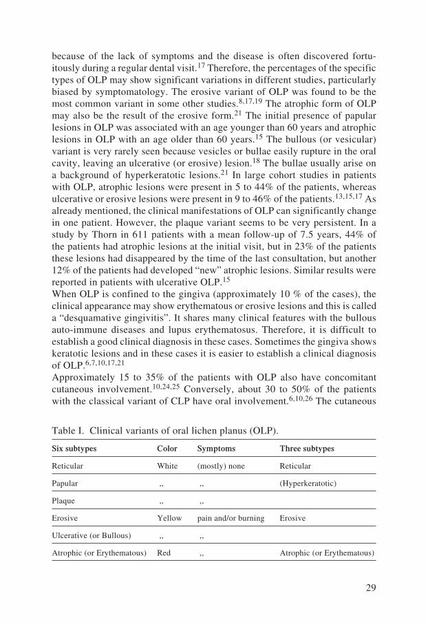
29
because of the lack of symptoms and the disease is often discovered fortu-itously during a regular dental visit.17 Therefore, the percentages of the specific types of OLP may show significant variations in different studies, particularly biased by symptomatology. The erosive variant of OLP was found to be the most common variant in some other studies.8,17,19 The atrophic form of OLP may also be the result of the erosive form.21 The initial presence of papular lesions in OLP was associated with an age younger than 60 years and atrophic lesions in OLP with an age older than 60 years.15 The bullous (or vesicular) variant is very rarely seen because vesicles or bullae easily rupture in the oral cavity, leaving an ulcerative (or erosive) lesion.18 The bullae usually arise on a background of hyperkeratotic lesions.21 In large cohort studies in patients with OLP, atrophic lesions were present in 5 to 44% of the patients, whereas ulcerative or erosive lesions were present in 9 to 46% of the patients.13,15,17 As already mentioned, the clinical manifestations of OLP can significantly change in one patient. However, the plaque variant seems to be very persistent. In a study by Thorn in 611 patients with a mean follow-up of 7.5 years, 44% of the patients had atrophic lesions at the initial visit, but in 23% of the patients these lesions had disappeared by the time of the last consultation, but another 12% of the patients had developed “new” atrophic lesions. Similar results were reported in patients with ulcerative OLP.15 When OLP is confined to the gingiva (approximately 10 % of the cases), the clinical appearance may show erythematous or erosive lesions and this is called a “desquamative gingivitis”. It shares many clinical features with the bullous auto-immune diseases and lupus erythematosus. Therefore, it is difficult to establish a good clinical diagnosis in these cases. Sometimes the gingiva shows keratotic lesions and in these cases it is easier to establish a clinical diagnosis of OLP.6,7,10,17,21 Approximately 15 to 35% of the patients with OLP also have concomitant cutaneous involvement.10,24,25 Conversely, about 30 to 50% of the patients with the classical variant of CLP have oral involvement.6,10,26 The cutaneous
Table I. Clinical variants of oral lichen planus (OLP).
Six subtypes Color Symptoms Three subtypes
Reticular White (mostly) none Reticular
Papular ,, ,, (Hyperkeratotic)
Plaque ,, ,,
Erosive Yellow pain and/or burning Erosive
Ulcerative (or Bullous) ,, ,,
Atrophic (or Erythematous) Red ,, Atrophic (or Erythematous)

30
and oral involvement does not necessarily occur at the same time.24 In most instances, cutaneous lesions typically develop within several months after the appearance of the oral lesions. The severity of OLP usually does not correlate with the extent of CLP.10,24 However, in a number of Scandinavian studies, it was reported that oral involvement was eight times more common than cutane-ous involvement.27,28 These percentages show a large variation in the literature also depending on the medical specialty. It appears to be easier for a dermatolo-gist to look into the mouth of a patient than for a dentist to look at the skin of a patient with LP.6,17,24
Etiology
The exact cause of OLP remains unknown, but an immune-mediated (T-cell dependant) pathogenesis has been proposed.29,30 Abnormal metabo-lism, impaired production of tonofilaments, and defects in the assembly of desmosomes have also been implicated as etiological factors.31 Numerous studies support the hypothesis that OLP is a complex immunological disease mediated by cytotoxic T-cells directed against basal keratino-cytes.6,32,33 In the etiology, there is an interplay of host factors, environ-mental factors and lifestyle.6 Nevertheless, this is the rule in many diseases. Cell-mediated immunity appears to play a major role in OLP.30 Endogenous and exogenous factors are probably necessary for developing of OLP in a person with a genetic predisposition.34,35 Oral lichen planus is considered to be idiopathic in most cases.1,10,23,25
Genetic background
The Human Leucocyte system A (HLA) is a group of cellular proteins on the surface of the cell-membrane of all nucleated cells and thrombocytes. These histocompability antigens are genetically determined by genes on 4 associated loci: HLA-A, HLA-B, HLA-C and HLA-D. These 4 loci are located on chro-mosome 6.39 Generally, the associations between HLA-antigens and OLP are inconsistent in the literature.6,36-38 Lowe et al evaluated 57 patients and 300 control subjects. A significantly higher prevalence of HLA-A3 was found (54% versus 29.7%) and more than twice as many affected patients (19.3% versus 9%) carried HLA-A5. Additional HLA-loci were not evaluated.37 Simon et al reported elevated prevalence of HLA-B16, HLA-B8 and HLA-Bw35. HLA-Bw35 was more common in patients with only cutaneous lesions, whereas HLA-B8 was more common in patients with oral lesions.40 Powell et al studied 72 patients with LP. Eighty percent of the patients with generalized disease, 56% of the patients with a lichenoid drug eruption, 54% of those with local-ized lesions, 31% of the patients with OLP, and 25% of the control group were positive for HLA-DR1 (HLA-class II-antigen). HLA-DQw1 was reported in

31
62% of the control sera and in 83% of the affected patients.41 Porter et al investigated the prevalence of HLA-A, HLA-B, HLA-C, HLA-DR, and HLA-DQ1 in a group of 40 British patients with OLP compared with healthy control individuals. In the group of patients with OLP, an increase in the frequency of HLA-Bw57 and a decrease in the frequency of HLA-DQ1 were found. The authors suggested that OLP may represent a heterogenous group of diseases and that HLA-Bw57 may predispose a person to OLP, whereas HLA-DQ1 may be associated with resistance to it.36 Lin et al found an increase in HLA-DR9 in Chinese patients with OLP.38
The familial LP also indicated that there is a substantial contribution of a specific genetic predisposition in the development of LP. A possible explana-tion for familial LP would be similar HLA-alleles.22 However, in familial LP conflicting results on HLA-alleles were reported.42-44
Emotional stress and lifestyle
Stress was implicated as an important etiological factor in the pathogenesis of OLP.45,46 Stress was identified as the most frequent cause of exacerbations of OLP.6,47 However, the chronic discomfort that can afflict patients with symp-tomatic OLP may be itself a stress factor and may perhaps partially explain the cases in which this association was documented. Furthermore, patients with OLP may be concerned about the possibility of malignancy, the (not justified) contagious nature of the lesions and the lack of available educational mate-rial.6,17 Education of patients with OLP on their disease can alleviate much of the anxiety.17 Altman and Perry reported in a study in 197 patients with LP that only 10% of the affected patients could recall a stressful situation at the onset of their disease, whereas 60% of the patients believed that chronic stress aggra-vated its course.46 Most studies on LP accentuate the association between oral lesions and stress.22 Patients with OLP frequently report worsening of the dis-ease during periods of stress and others noticed onset or exacerbation of OLP with fatigue.45 Nervous, highly stressed individuals suffer from OLP rather frequently.48 Lowental and Pisanti reported an association between emotional stress and erosive OLP, but not with the reticular variant of OLP.49 However, Allen et al reported that there was no significant association between stress and OLP.50 In a study by Rojo-Moreno et al there were 100 patients with OLP and 50 control subjects. In a psychometric evaluation, the patients with OLP (par-ticularly with symptomatic OLP) were found to have a higher vulnerability for psychic disorders, higher anxiety and increased depression scores than those in the control group. However, it could not be concluded that both the observed psychologic alterations acted as a direct etiological factor of OLP and these alterations were a consequence of OLP.51
Ostman et al reported different lifestyle patterns in patients with OLP compared with an age- and sex-matched control group in Sweden. In the patients with

32
OLP, the frequency of physical activity was significantly higher than in the patients in the control group. In the group with OLP, more individuals were divorced or whose spouse had died. Furthermore, the daily intake of carbo-hydrates, fibers and iron was statistically significant higher in patients with OLP.52 Unfortunately, the exact relationship with the pathogenesis of OLP in those patients could not be established.
Tobacco
Generally, there is no increased prevalence of cigarette smoking in patients with OLP.53 Murti et al examined 722 Indian patients with OLP and found a strong association between OLP and the use of tobacco.54 Betel nut chewing is also more prevalent in Indian patients with OLP.12 In a prospective study by Silverman et al in San Francisco in the U.S.A., there was no correlation between the onset of OLP and smoking.8 However, the plaque form of OLP is more often encountered in patients who used tobacco.15
Exposure to tobacco should be discouraged strongly in patients with OLP, because it is a well-known risk factor for oral cancer, and there is also a pos-sible synergistic premalignant effect in patients with OLP.55,56,389
Trauma
The Koebner’s phenomenon or isomorphic response is a common feature in LP, and develops in areas subjected to some type of trauma in the absence of previous clinically visible lesions.26,57 In a study by Eisen in 723 patients in 2002, mechanical traumata, dental interventions, heat and irritants from tobacco products, friction from sharp cusps, rough dental restorations, poorly fitting dental prostheses and oral habits as lip chewing and cheek biting (morsicatio buccarum) were frequently found to be exacerbating factors that resulted in (symptomatic) OLP. When these factors were minimized or eliminated, the oral lesions either reverted to the less severe forms of the disease or sometimes resolved completely. Dental plaque and calculus are particularly associated with a significantly higher incidence of gingival OLP lesions.17
Micro-organisms and infections
Dental plaque consists of micro-organisms (more than 70%), mucines and degenerated epithelial cells, and may frequently produce a (chronic) gingi-vitis.48,58 This chronic infection of the gingiva (possibly also in combination with the Koebner’s phenomenon) may aggravate OLP.17,59 In long-stand-ing dental plaques, calcifications may occur and produce dental calculus.48 Ramon-Fluixa reported in a study in 90 patients with OLP that increased

33
dental plaque and calculus deposits were associated with a significantly higher incidence of erythematous and erosive gingival lesions.59 Oral hygiene in patients with OLP should be optimized by both the patient and the patient’s dentist or periodontist to reduce the severity of gingival inflam-mation and to avoid periodontal surgical interventions that may exacerbate OLP.17,57
Oral lichen planus was suggested to be related to bacteria such as a Gram-nega-tive anaerobic bacillus (Jacob and Helmbold, 1933) and spirochetes (Lehnhoff, 1948), but this has not yet been confirmed.60,61 Lichenoid reactions have been seen in syphilis (Lochner and Pomeranz, 1974), chronic bladder infection (Shel-ley and Shelley, 1989), and intestinal amebiasis (Wahba-Yahav, 1989).62-64 In the latter two cases the lesions disappeared after systemic treatment with metronidazole.63,64
The involvement of viral agents in OLP has been suggested.65 Oral lichen planus was reported in HIV infection.66 Human papillomaviruses (HPV) were found in the lesions of OLP, but any causal role remains speculative.67,68 Walsh et al suggested that low-grade or persistent infection of epithelial cells with herpes viruses may be a possible etiologic factor in OLP.29,69 The possible relationship between OLP and hepatitis C virus infection has been frequently reported.70-75 The possible association between OLP and hepatitis B virus infection has been reported less often.76,77 Interestingly, hepatitis B vaccina-tion has also been implicated in the development of OLP.78,79
Generally, there is an increased prevalence of candidal carriage and infection in patients with OLP and candida albicans has been considered as a provoca-tive factor in OLP.80-82 Oral candidosis may also be provoked by endocrinop-athies, immune-dysfunction and specific drugs.80 Candida was demonstrated in cultures in 37 to 50% of the patients with OLP.80,82 Candidal infection has been demonstrated in 0 to 17% of the biopsies of OLP without predilec-tion for a specific variant of OLP.83,84 However, candida albicans can be detected in the oral cavity without any symptoms in about 40% of the normal population.80,82 The natural barrier of the oral mucosa in OLP is decreased and the treatment of OLP with corticosteroids and other immunomodulators frequently predisposes to candidosis.80 Symptoms of OLP may be worsened by candidal overgrowth or by infection.80-84 Furthermore, candida albicans can enzymatically catalyze the formation of N-nitrosobenzylmethylamine, a potential carcinogen.84 However, the presence of this yeast and the develop-ment of OLP and a higher rate of candida albicans colonization in OLP was not correlated in several other studies.8,83 It seems reasonable to start with antifungal treatment to avoid the role of candidosis in (symptomatic) OLP.85 It has been reported that antifungal treatment of erosive lesions of OLP from which candida had been isolated by culture often transformed the lesions to reticular form.6,80 Unfortunately, we are unable to corroborate this statement from our experience.

34
Contact allergy
Contact allergens are mostly low molecular weight (less than 500 daltons) sub-stances, which are able to penetrate the oral mucosa. These substances must be presented by antigen-presenting cells, principally Langerhans cells, to T-lym-phocytes to induce contact allergy. Antigenicity is also accomplished by the conjugation of these allergens with autologous proteins present in the mucosa. An antigen with a molecular weight of at least 5,000 daltons is required to induce and elicit a contact allergy. The effector cells, which mediate delayed-type allergic reactions are the progeny of these T-lymphocytes.86 The patho-genesis of an allergic contact stomatitis shows significant similarities with the pathogenesis of OLP.30,87,89 Generally, allergic contact sensitivity affects both the skin and the oral mucosa. In general, allergic contact stomatitis occurs less frequently than allergic contact dermatitis probably because of the following reasons. The epidermis of the skin consists of proteins that combine more readily with low molecular weight substances to form allergens. The period of contact of sensitizers with the oral mucosa is brief (with the exception of den-tal appliances) because the saliva dilutes and removes the potential allergens or neutralizes chemicals. The extensive vascularization of the mucosa aids in rapid dispersion and absorption of the allergens.87 The patch test is a scientific in vivo method of investigating contact allergies. A possible role of contact allergy in OLP should be investigated by patch tests, which are generally per-formed on the upper part of the back conform patch tests in allergic contact dermatitis.86-88 Generally, patch testing is a routine procedure in dermatol-ogy.91 However, properly applied allergens and correctly interpreted patch test reactions with clinical consequences may be more complicated (Figure 16).86 Several in vitro tests such as the lymphocyte transformation test and the mac-rophage inhibition test, often parallel clinical contact sensitivity.92,93 However, these tests may show a significant variation in results and have been optimized for only a few contact allergens.94
Allergy to dental metal restorations and electrogalvanism
Generally, there is no evidence of any association between allergy and dental restorative materials in the majority of the patients with OLP.17,95,96 How-ever, contact with or proximity to dental restorations may cause OLP.97-100 These reactions are typically induced by a contact allergy, but friction by dental fillings (via the Koebner’s phenomenon), toxic reactions to compounds released or generated, and dental plaque should be excluded.17,100-103
The hypothesis is that dental metal restorations in the oral cavity are prone to corrosion and by releasing ions may be responsible for sensitization and aller-gic reactions (type IV, T-cell dependant). This process may lead to chronic antigenic stimulation with mucosal changes and ultimately to OLP.100,104 An
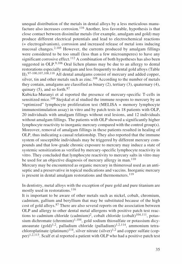
35
unequal distribution of the metals in dental alloys by a less meticulous manu-facture also increases corrosion.116 Another, less favorable, hypothesis is that close contact between dissimilar metals (for example, amalgam and gold) may produce different electrical potentials and lead to electrochemical reactions (= electrogalvanism), corrosion and increased release of metal ions inducing mucosal changes.2,105 However, the currents produced by amalgam fillings were considered to be too small (less than a few microamperes) to have any significant corrosive effect.112 A combination of both hypotheses has also been suggested in OLP.9,106 Oral lichen planus may be due to an allergy to dental restorations especially amalgam and less frequently to dental gold alloys (Table II).97-100,107,108,118 All dental amalgams consist of mercury and added copper, silver, tin and other metals such as zinc.108 According to the number of metals they contain, amalgams are classified as binary (2), tertiary (3), quaternary (4), quinary (5), and so forth.87
Kubicka-Muranyi et al reported the presence of mercury-specific T-cells in sensitized mice.109 Stejskal et al studied the immune respons to mercury by an “optimized” lymphocyte proliferation test (MELISA = memory lymphocyte immunostimulation assay) in vitro and by patch tests in 18 patients with OLP, 20 individuals with amalgam fillings without oral lesions, and 12 individuals without amalgam fillings. The patients with OLP showed a significantly higher lymphocyte reactivity to inorganic mercury compared with the control group(s). Moreover, removal of amalgam fillings in these patients resulted in healing of OLP, thus indicating a causal relationship. They also reported that the immune system of susceptible individuals may be triggered by different mercury com-pounds and that low-grade chronic exposure to mercury may induce a state of systemic sensitization as verified by mercury-specific lymphocyte reactivity in vitro. They concluded that lymphocyte reactivity to mercury salts in vitro may be used for an objective diagnosis of mercury allergy in man.110 Mercury may be encountered as organic mercury in thimerosal used as an anti-septic and a preservative in topical medications and vaccins. Inorganic mercury is present in dental amalgam restorations and thermometers.119
In dentistry, metal alloys with the exception of pure gold and pure titanium are mostly used in restorations.116
It is important to be aware of other metals such as nickel, cobalt, chromium, cadmium, gallium and beryllium that may be substituted because of the high cost of gold alloys.87 There are also several reports on the association between OLP and allergy to other dental metal allergens with positive patch test reac-tions to cadmium chloride (cadmium)2, cobalt chloride (cobalt)104,111, potas-sium dichromate (chromium)1,104, gold sodium thiosulfate or potassium dicy-anoaurate (gold)1,2, palladium chloride (palladium)1,2,114, ammonium tetra-chloroplatinate (platinum)114, silver nitrate (silver)1,2 and copper sulfate (cop-per)1,2,113. Scalf et al reported a patient with OLP who had a positive patch test

36
to iridium (III) chloride hydrate (1% aq) (iridium) and indium (III) sulfate (10 % aq) (indium) (Table II ).1 Yiannis et al reported a positive patch test to beryl-lium sulfate tetrahydrate (1% aq) (beryllium) in 2 patients with OLP (Table II).2 However, in both the studies the clinical relevance of the findings was debatable because other allergens could also be relevant in these patients.1,2 Mizoguchi et al described a case of linear, facial LP with a “peculiar sensation” in the mouth that was caused by allergy to palladium. Patch tests were positive to both palladium and platinum. Removal of the palladium-containing dental work resulted in total resolution of the symptoms.115
Dental alloys may contain small proportions of nickel, cadmium, beryllium, indium, gallium, platinum, and palladium.116,117,123 These constituents may produce local pathogenic effects including stomatitis, gingivitis, parodonti-tis, and even alveolar bone loss.120,122 Local toxic reactions by dental metals occur more frequently in women than in men. Generally, such reactions occur shortly after the placement of the dental metal restorations. A metallic taste is commonly an important symptom of the local toxic reaction.116,120 It was also reported that local reactions may be possibly involved in the pathogenesis of the Burning Mouth Syndrome (BMS).96,124 The exact composition of dental metal restorations in the oral cavity of a patient may be unknown.Screening for dental metal restorations in the oral cavity may be performed by an Orthopantogram (X-ray). Chemical analysis for establishing the exact composition may be performed after complete removal of the dental metal restoration. The exact composition of the dental metal restorations may also be investigated by analyzing a small sample of the alloy with scanning electronmicroscopy and a subsequent rönt-gen diffraction spectrum.116,124 It was reported that dental metal restorations, particularly amalgam, were associated with several disorders such as chronic fatigue syndrome, aspecific symptoms in the oral cavity, abnormal psychological signs, and a feeling of decreased general well-being.89 Moreover, neurological disorders such as mul-tiple sclerosis and Alzheimer’s disease were reported to be caused by chronic mercury poisoning.121 The leakage of very small amounts of mercury from
Table II. Allergy to dental metal restorations and OLP.
Metals:
Mercury (Amalgam) Gold Platinum Indium
Cobalt Chromium Silver Iridium
Cadmium Palladium Copper Beryllium

37
dental amalgam was detected but intensive investigation showed that amalgam did not cause a significant health risk for the general population. However, volatile free mercury in dental clinics should be monitored to avoid occupa-tional health risks.125
Allergy to other allergens
In 2000, Yiannis et al reported in a study in 46 patients with a clinical and histopathological diagnosis of OLP that 6 patients had positive patch tests to flavorings such as vanillin, cinnamic aldehyde, fragrance mix and balsam of Peru. These patients noted a substantial aggravation of their symptoms when exposed to these allergens. Avoidance of these flavoring agents (in specific foods or oral hygiene products (fragrance mix)) resulted in clini-cal and symptomatic improvement in all 6 patients underlining the clinical relevance. One patient had a positive patch test to acrylic resin monomer (used as an adhesive material) and a moderate improvement was noted after removal of an acrylate dental retainer. Besides these cases, there were several positive reactions to dental metals in other patients. In this study, nearly 40% of the patients with OLP had an exacerbating contact hypersensi-tivity.2 An important aspect in this study was possibly that macular erythema as a result of the patch test was already regarded as positive in contrast with regular patch tests.86 A few other studies also reported that a small minority of the patients with OLP reacted to certain foods and to food additives such as cinnamic aldehyde.126-128 Contact allergy in association with OLP to one or more of the acrylic denture materials or composite fillings has also been described.2,129
Hensten-Pettersen reported the introduction of more than 100 similar new brands of resin-based cold-curing materials.130 Resin-based unfilled and com-posite materials, pit and fissure sealants, orthodontic adhesives, glazes, veneers and repair kits for porcelain-fused-to-metal restorations, root canal sealers, and temporary crowns are used in a large segment of the population and may also cause allergic reactions. After cold-curing, it is likely that small amounts of the monomers will be left unpolymerized. Depending on the composition, the polymerization is induced by chemical catalysts, visible, or ultraviolet light sources. The materials are based on different types of monomers such as methacrylate monomers, urethane-based-dimethacrylates, epoxybisphenol resins, and ethylene-amino derivates.130 Moreover, many chemicals such as benzoyl peroxide, hydroquinone, phthalates, tertiary aromatic and aliphatic amines, ultraviolet stabilizers and antioxidants involved in the polymerization process may still be present. Saliva, water and alcohol may leach out any unpo-lymerized residual material in “cured” orthodontic bonding resins, which may cause allergic reactions. Frequently occurring methacrylates in bonding resins
38
are 2,2-bis[4-(2-hydroxy-3-methacryloxypyloxy)phenyl]-propane (bis-GMA) and 2-hydroxyethyl methacrylate (2-HEMA). “Bis” stands for the epoxy resin bisphenol A, which reacts with glycidyl methacrylate. The plastic is generally combined with quartz, lithium aluminium silicate, glass, or silicon dioxide to modify the physical properties of the resin. Bis-GMA and triethyleneglycol dimethacrylate (TEGDMA) frequently occur in composite resins. Glass iono-mers may include 2-HEMA or trimethylolpropane trimethacrylate. Most den-tures are made of acrylic resins that are heat-cured. Heat-cured acrylic dentures rarely cause allergic reactions. Self-curing acrylics that harden without heat are available for repairs and relining.87,131,132 In our opinion, it cannot be ruled out with certainty that these allergens may also play a role in the pathogenesis of OLP.
Specific foods
Specific foods, such as citrus juices, tomatoes, spicy ingredients, strong alco-holic liquors, and crisp foods such as corn chips and toasts may aggravate the symptoms of OLP.17,134,294 Generally, such reactions are not based on allergy, but on local irritation or toxicity.17,133 An adequate nutrition is also essential for a healthy oral mucosa. Nutrients related to energy support, enzyme activity, iron, zinc and vitamin A are documented to affect the state of epithelial cells and the mucosa. Trauma and infections increase the nutritional demands, but painful oral lesions may reduce adequate dietary intake.52
Histopathology
Generally, histopathological examination is necessary to establish the diagno-sis of OLP and to exclude other diagnoses especially (pre)malignant disorders, bullous auto-immune diseases and lupus erythematosus.6,9,17,135 The clinical presentation of the (chronic) lesions in OLP may essentially change in the time in one patient and may resemble other diseases.15 An exception against the rule for taking a biopsy could be made for the reticular variant of OLP with-out any symptoms with localized and symmetrical lesions for example on the buccal mucosa without contributing external risk factors such as smoking and alcohol abuse. Furthermore, the clinical aspects of OLP may be sufficient to establish a correct diagnosis if there are also concomitant characteristic CLP lesions.6,9,10
The punch biopsy of the skin is familiar to all dermatologists. It is also a safe and useful technique for the diagnosis of diseases of the oral cavity. However, knowledge of the anatomical structures of the oral cavity is essential and neu-rovascular structures in the specific regions must be avoided by taking rather superficial biopsies. After local anesthesia with lidocaine (1% or 2%) and
39
epinephrine (1:100,000) one or more punch biopsies of at least 3 millimeters diameter can be taken. Suturing is not necessary and healing occurs by second-ary intention and almost complete re-epithelialization occurs within 2 weeks. Bleeding is one of the most dreaded complications of oral surgery, but can be adequately controlled by applying pressure during several minutes in the majority of the cases.136,137 Punch biopsies should be taken from the hyper-keratotic or erythematous lesions and from the edge of the lesions in case of erosions or ulcerations to avoid non-specific histopathological features.9 Rou-tinely, the biopsies are fixed in buffered 4% formalin and stained with hema-toxylin and eosin (HE) and also with periodic acid-Schiff (PAS) reagent. If there are obvious erosions, ulcerations or bullae, without concomitant signs of the hyperkeratotic (reticular) variant, the biopsies must be transported in physi-ological saline for direct immunofluorescence examination to exclude bullous auto-immune diseases or lupus erythematosus.137 Direct immunofluorescence should also be performed in lesions confined to the gingiva for a correct diag-nosis, particularly when there is the clinical presentation of a desquamative gingivitis.7,138 Generally, it is much easier to biopsy non-gingival lesions.6
Light microscopy
The histopathological features of OLP and of CLP are essentially the same, but in OLP these features may be less distinct than in CLP (Figures 7, 8, 9,10 and 11).9,23 A histopathological definition of OLP was formulated by the WHO in 1978 as follows:“The histopathological features of OLP are characteristic. There is usually a keratinized layer, and this may be either ortho- or parakeratinized. If keratiniza-tion is normally found at the affected site, then the keratinized layer is thick-ened. If the site is normally non-keratinized (for example, the buccal mucosa), the keratinized layer in the lichen planus lesion may be very thin; if there is normally a stratum granulosum this will be thickened. If there is normally no stratum granulosum, then the granular cells may be present in small numbers. The ‘saw-tooth’ appearance of the rete processes that is a common feature of skin lesions is less frequently seen in the oral mucosa. The thickness of the epithelium varies and atrophy is often seen. Civatte’s (colloid) bodies may be present in the region of the basal-cell layer, lying either in the epithelium or within the superficial part of the connective tissue. These are rounded or lobulated acidophilic structures which sometimes contain a pyknotic nucleus or nuclear fragments. The changes in the basal cell layer often include “lique-faction degeneration”, and there may be a narrow band of eosinophilic material in the position of the basement membrane. There is a well-defined zone of cel-lular infiltration that is confined to the superficial part of the connective tissue (lamina propria), and the infiltrate consists mainly of lymphocytes except in the vicinity of an erosion”.14
40
Histologically, the principal feature is the dense band-like inflammatory infil-trate that mainly consists of T-lymphocytes and a low number of histiocytes in the superficial stroma and the basement membrane zone (BMZ) associated with liquefaction degeneration of basal epithelial cells.6,9,26 Another feature is the presence of Civatte’s bodies, which are dyskeratotic basal keratino-cytes that have undergone premature keratinization and have been extruded into the papillary mesenchyma.140,141 Civatte’s bodies were noted in 27% of patients with OLP and are considered to be among the earliest histopathological changes.26,142 As the BMZ becomes vacuolated, fluid accumulates and leads to the formation of clefts known as Max Joseph spaces (or Caspary-Joseph spaces).143,144 Plasma cells and melanophages may also be present.145 Para-keratosis may be more common in OLP than in CLP (Figures 7, 8, 9, 10 and 11).16,65 The Wickham’s striae appear to be correlated with an increased granu-lar cell layer.147 Unfortunately, there are no defined histopathological features for the different clinical subtypes of OLP.22,146 Although the above characteristics of OLP seem to result in a well-described entity, Van der Meij et al reported in a study in 1999 that there was a significant inter-observer and intra-observer variation in the histological assessment of OLP between 5 oral pathologists.148 Some pathologists reported conclusions of “evident OLP” or “compatible with OLP” in a considerable number of cases, while others classified the same cases as “no histological support for OLP”. They concluded that “the histopathological assessment of OLP, based on the available WHO-definition, is a rather subjective and insufficiently reproduc-ible process. Stricter diagnostic criteria are required in order to obtain a more reproducible diagnosis of OLP”.139,148,389 Unfortunately, the pathologists were not provided with any clinical information and patient data, and similar studies in nearly all other diseases are lacking.
Electron microscopy
The ultrastructural changes of OLP are closely related with the findings reported in conventional light microscopy and include disruption and thickening (or duplication) of the basement membrane with degenerative changes in the cells of the basal cell layer and the stratum spinosum, the presence of inflammatory cells in intercellular spaces of the epithelium, pools of fluid filling epithelial intercellular spaces (Max Joseph spaces or Caspary-Joseph spaces) with main-tenance of desmosomes, distortion of cytoplasmatic membranes of the epithe-lium adjacent to desmosomes because intercellular pools forming cytoplasmatic “villi”, irregularity of the nuclear membrane of epithelial cells, and increased thickening and granularity of epithelial tonofibrils.149,150 The cellular infil-trates in OLP and CLP essentially have similar ultrastructural characteristics.151 The infiltrate consists of predominantly T-lymphocytes and a low number of macrophages.151 In the cytoplasm of the lymphocytes, there is a decreased

41
number of mitochondria and the Golgi apparatus is poorly developed. Trans-forming lymphocytes (lymphoblasts) were not encountered. The cytoplasm of the macrophages contained primary and secondary lysosomes, some of which contained phagocytosed material.151 Civatte’s bodies are believed to be formed by a meshwork of 70 ängström (= 7 nanometers) strands probably derived from intracellular tonofilaments. As the disease process continues, there is a steady loss of tonofilaments, desmosomes, and hemidesmosomes. The degeneration of hemidesmosomes in the basal layer may explain the blistering.26,140,153 In a study by Tyldesley and Appleton, it was explicitly mentioned that no viral inclusions were observed.152
Immunopathology
Direct immunofluorescence shows fibrin and (shaggy) fibrinogen in a fibril-lar or linear pattern at the basal membrane zone (BMZ) in a high number of OLP lesions, which is an important feature.141,154,156 IgM and complement components, principally C3, C4, and C5, have also been observed in the BMZ. IgA could not be demonstrated in the BMZ. Colloid bodies most often stain positively for IgM, C3, and C4.141,155 IgA, IgG, C1, and C5 may sometimes also be detected.155,156 IgM is particularly found in early stages of colloid body formation. Both fibrin and albumin were also noted.22,155 However, it is important to stress that these findings when combined with histopathology are only highly indicative, and not diagnostic for OLP.6,9,104 Indeed, oral lichenoid drug reactions also have the same features.14,95,157,160,464,467
When there are oral lesions with erosions, ulcerations, or bullae, without concom-itant signs of the hyperkeratotic (reticular) variant in the surrounding mucosa, direct immunofluorescence should be performed for distinguishing between OLP and bullous auto-immune diseases and lupus erythematosus.6,158-160 Direct immunofluorescence should also be performed when the oral lesions are confined to the gingiva because the histopathological features of OLP on the gingiva are often non-diagnostic, particularly in case of the clinical picture of a desquamative gingivitis.17,138
Immunohistochemical examination shows a band-like infiltrate mainly consist-ing of T-lymphocytes in the BMZ characteristic of OLP.161,162 The duration of the lesions seems to influence the composition of this inflammatory infiltrate. The infiltrate in early lesions principally consists of T-helper/inducer cells (CD4 +, Leu-3a) and a low number of macrophages. The infiltrate in advanced lesions primarily consists of T-suppressor/cytotoxic cells (CD8+, Leu-2a) that also express HLA-DR antigens.146,159,163 A few or no B-cells were identi-fied.22,163 Boisnic et al reported in a histochemical study on OLP that the band-like infiltrate consisted of 94% T-lymphocytes and 6% B-lymphocytes.164
There is an increased number of Langerhans cells in the epithelium of OLP and these cells are important in antigen presentation.165 Furthermore,
42
numerous basal keratinocytes are positive for HLA-DR class II antigens, whereas in normal epithelium, keratinocytes were found not to express those antigens. HLA-DP and HLA-DQ antigens were not observed to be expressed.164,166,167 In 1983, Olsen et al identified lichen planus-specific antigen (LPSA) by indi-rect immunofluorescence in lesional tissue from patients with LP.168 Subse-quent investigation showed that this antigen was present in 20 patients (80%) with CLP.169 However, not all lesions of CLP in the same patient have LPSA. The circulating antibodies to LPSA are more likely to be markers of the disease than to be causative.170 In OLP, LPSA seems to be less prevalent than in CLP, but patients with OLP may also have antibodies directed against this antigen.170
The LPSA was found in the granular or spinous layer and seems to be specific for LP because it was not found in patients with other dermatoses. Moreover, LPSA was reported in childhood bullous LP.171
The immunohistopathological changes that are typical in OLP may also be observed in the oral lichenoid tissue reactions.159
Pathogenesis
To date, the exact cause of OLP remains unknown.6,22,26 A number of etiological factors and associated mechanisms have been put forward, but none of them pro-vide a satisfactory unifying explanation on the underlying antigenic trigger which ultimately leads to the histopathological changes that are observed in this dis-ease.30 Abnormal metabolism, impaired production of tonofilaments, and defects in the assembly of desmosomes have also been implicated as pathogenetic fac-tors.31 Basal cells behave more like keratinocytes because of decreased meta-bolic and regenerative capacity. This leads to an increased production of granular cells.172 Although immunological involvement in the pathogenesis was demon-strated in previous studies, the primary event in the disease was not identified and it is still a matter of speculation and remains to be elucidated.30,70,161,162
The levels of epidermal glucose-6-phosphate dehydrogenase were observed to be inconsistent (normal, decreased or increased).173,174 A reduced quantity of this enzyme may predispose patients to develop LP after ingestion of anti-malarial drugs.175 Reduced levels of respiratory enzymes such as cytochrome oxidase, succinic-dehydrogenase, reduced form of nicotinamide-adenine-dinucleotide (-phosphatase), and ß-hydroxyacyl-coenzyme-A-dehydrogenase were also reported.172,176,177 Holmstrup and Dabelsteen reported that two plant agglutinins, concanavalin-A and Ricinus communis agglutinin fraction-I, did not bind to the cell membranes of basal cells in OLP. They postulated that an antigenic change had taken place in the cells involved and that this change might play a role in the pathogenesis of OLP.178
The genes of the Major Histocompability Complex (MHC) encode for cell determinants that regulate immune responsiveness. Class I MHC molecules
43
are present on all nucleated cells of the body in varying amounts from scarcely detectable to very strong expression. The amount of these cell-surface mol-ecules may markedly increase during immune reactions. They are the core of the individuality antigens of each person, and form the recognition signal for CD8+ cytotoxic T-cells. In humans, the HLA-system fulfils the role of histo-compability.179
Humoral immunity
There are contradictory reports on the levels of immunoglobins IgM, IgA, IgG, IgD and IgE in the sera of patients with LP.180 The levels of complement were reported to be normal.181 Circulating immune complexes and complement components, such as C3, were reported in patients with OLP.182 Mahood et al evaluated 45 patients with LP and found that when the disease was active or recently resolved (< 2 years), serum IgM levels were slightly low. Patients with a history of LP (> 2 years) had normal levels of serum immunoglobulins.183 In one study, serum cryoglobulins were found in 31% of the patients with LP.184 The exact role played by humoral (auto) immunity in OLP still remains obscure and its contribution appears to be minor as compared with that of the cellular (auto)immunity.30,162
Cellular immunity
The pathogenic mechanism responsible for OLP has been mainly attributed to dermal T-cells. Evidence for the central role of T-cells in the evolution of OLP has been reported in various studies.30,70,161,162 T-cell receptor gene and rear-rangement studies indicated that clonal T-cells are present in LP tissue.90 These T-cells significantly showed more cytotoxicicity against autologous lesional keratinocytes than T-cells from non-lesional tissue.189 There is a selective recruitment of T-cells in OLP.30,161,162 The histopathological abnormalities observed in LP disappeared when lesional skin was transplanted onto athymic mice (lacking functional T-cells).185 Similar changes were reported to occur in explants of lesional skin maintained in organ cultures.186 These findings indicated that the migration of T-cells and other components of the cellular immune response is a crucial factor in the pathogenesis of LP.151,164,185 A diagrammatic representation of the events leading to the evolution of OLP is shown in Figure 12 and was modified from Boyd and Neldner.26,30,98 It is also a modification of the proposed immunological lichenoid tissue reaction from Morhenn and Shiohara et al.187,188,198.
It has been postulated that the primary event in OLP is a delayed-type hyper-sensitivity (Type IV) in which an unidentified “neoantigen” of basal kerati-nocytes is processed in the Langerhans cells followed by the recruitment of T-cells.9,26,159 This process is mediated via adhesive interactions between

44
specific cell surface molecules and directed by chemotactic cytokines pro-duced by epithelial cells.190,191 Cytokines are soluble messenger proteins elaborated by cells, usually involved in inflammatory processes.26,192 Inter-action of intercellular adhesion molecule-1 (ICAM-1, CD54) on Langerhans cells (CD 6+) and lymphocyte function-associated antigen-1 (LFA-1, CD11a) on T-cells results in the generation, among others, of either CD8+ (MHC class I restricted) or CD4+ (MHC class II restricted) cytotoxic T-cells.159,193-195 Other specific adhesive interactions include CD3/T-cell receptor: antigen, CD8: MHC class I and CD4: MHC class II.190,196 The cell-mediated response initiated by Langerhans cells thus includes cytotoxic T-cells that are directed against the “altered” keratinocytes and which may also be responsible for damaging cells of other organ systems such as hepatocytes.197 The mucosa in OLP contains an increased number of Langerhans cells compared with normal oral mucosa.30 There is an interaction of an antigen (drugs, contact allergens or viruses) with either keratinocytes or Langerhans cells, resulting in the production of interleukin-1 (IL-1) (Figure 12).26,98 This IL-1, in turn, stimulates the production of interleukin-2 (IL-2) by T-cells.30,187 In addition, HLA-DR+ keratinocytes produce pro-inflammatory cytokines.198 The pro-duction of interferon gamma (IFN-γ) by activated T-cells is increased.26,188 This, in turn, induces the expression of HLA-DR on keratinocytes which progress into lesional cells.187 The expression of LFA-1 on monocytes was observed to be induced by IFN-γ produced by T-cells, thereby facilitating the attachment of these cells to keratinocytes.198 Helper T-cells (CD4+) and cytotoxic T-cells (CD8+) destroy keratinocytes via interaction with HLA-DR (MHC class II) and class I antigens, respectively.163,164 Lymphokines may also directly damage basal cells or down-regulate (CD8+) cytotoxic/sup-pressor cells.199 It was also suggested that activated T-cells may recognize keratinocytes as “target cells” and interact directly with them.161 In early lesions of OLP, helper T-cells and macrophages were predominant, but in older lesions cytotoxic/suppressor T-cells were mainly observed within the infiltrate.156,159,163,164 An imbalance existed between T-helper and T-sup-pressor activity.200,201 T-lymphocytes in OLP generate increased levels of interleukin-6 (IL-6) and granulocyte-macrophage colony-stimulating factor (GM-CSF). The local cytokine production may be important in the perpetu-ation of OLP.202 Calprotectin and cutaneous lymphocyte antigen (involved in homing T-cells) are up-regulated in OLP.30,472,473 Heat shock proteins (HSPs), also called “stress proteins”, are expressed by most living cells. They play a role in case of stress, including heat, oxidative injury, infection, apoptosis, protection againsts ultraviolet damage, wound healing and other protein remodelling processes. These proteins also play an essential role in cellular survival and are encountered in many immune and inflammatory responses in the skin and the mucosal membranes. Heat shock proteins are classified according to molecular weight, homology, and func-
45
tion. An altered HSPs 60 and 70 expression in the oral mucosal epithelial cells was reported in OLP and was claimed to have a role in the pathogen-esis of OLP.203-206 The T-cells in OLP consist of a distinctive population of gamma-delta T-cells. This subtype of T-cells may interact with certain parts of HSPs. Heat shock proteins and vascular adhesion molecule-1 (VCAM-1) may also be expressed under the influence of IFN-γ and facilitate the lym-phocyte-to-keratinocyte adherence which determines the apoptotic death of keratinocytes.32,203-206 Laminin and types IV and VII collagen extracellular matrix are significantly increased at the epithelio-mesenchymal junction in OLP and may bind to beta-1 integrins on the surfaces of infiltrating lymphocytes.159 In addition, fibrinogen is deposited at the BMZ and adjacent areas of lymphocyte accu-mulation.6,159 Oral lichen planus differs from CLP in the expression of alpha-beta integrin, which is up-regulated in OLP but not in CLP.473 There are con-tradictory reports on the peripheral CD8 count and the CD4/CD8-ratio.207,208 The possible contribution of auto-reactivity in the pathogenesis of OLP was also indicated on the basis of studies demonstrating changes in T-lympho-cytes in the peripheral blood, including a depressed number of CD4+ and CD45RA+ cells in patients with OLP compared with age- and sex-matched healthy individuals.6,217 Simon et al reported that the activity of natural killer cells in patients with LP was decreased and it seemed to be related to the severity of LP.209 A normal amount of β2-microglobulin was reported to be produced by activated lymphocytes in patients with OLP.210 As the lesions evolve, cytokines derived from lymphocytes (IFN-γ) and keratinocytes (IL-1, IL-6) influence the behavior of the lesional cells and the overlying epi-dermis.30,209 The majority of T-lymphocytes within the epithelium express the alpha-beta T-cell receptor (TCR) and are “memory” T-cells.69,70 The TNFα-receptor is known to play an important role in apoptosis which is a very important phenomenon in the histopathology of OLP.474
Associated diseases
Oral lichen planus has been reported to be associated with a variety of disor-ders and particularly with disorders involving altered or disturbed immunity. However, it may be difficult to determine whether there is a causal or a purely fortuitous association.22,26 A small number of patients with OLP may coinci-dentally suffer from systemic disease not necessarily linked causally with OLP, because OLP is a relatively common disease. Furthermore, OLP is predomi-nantly seen in individuals older than 50 years. This also increases the chances of coincidental systemic disease.6 There is also the difficulty in the differential diagnosis between OLP and oral lichenoid drug eruption (LDE).10,157 It may be impossible to distinguish both disorders based on clinical and histopathologi-

46
cal examination.157 Since many different drugs may be involved in oral LDE (Table VIII), some reports on apparent association of OLP with systemic dis-ease were likely because of side effects of drugs rather than associations with a specific systemic disease.6 There are contradictory reports on associations between OLP and/or CLP and disorders such as alopecia areata211,243, chronic active hepatitis212,218,219, der-matomyositis211, dermatitis herpetiformis213, diabetes mellitus19,214-216,222, Hashimoto’s thyroiditis220,221, HIV infection66, hypertension223-225, hyper-thyroidism250, hypogammaglobulinemia211,226, keratoconjunctivitis sicca and xerostomia (Sjögren’s syndrome)227,274,356, lupus erythematosus228,229, malignancies6,230,232, morphea233, myasthenia gravis234,235, pemphigus folia-ceus236, pemphigus vulgaris236,237, pernicious anemia238,239, primary biliary cirrhosis240,241, rheumatoid arthritis269, systemic sclerosis242, thymoma211,234, ulcerative colitis243,244 and vitiligo211,233. Marren et al reported an associa-tion between vulval lichen sclerosus and OLP in older women.245 Occasion-ally, associations were also reported between LP and other conditions such as coeliac disease246, Crohn’s disease247, vitamin deficiencies (B1, B6, C, iron or folate acid)52,248, erythema dyschromicum249, mesangioproliferative glomerulonephritis250, psoriasis vulgaris251, Turner’s syndrome with endocri-nopathies252, and urolithiasis253. It was reported that OLP was most strongly related to chronic liver diseases, especially chronic active hepatitis212,218,219 and primary biliary cirrhosis240,241. However, the treatment of primary biliary cirrhosis with penicillamine may pro-duce an oral LDE.254,255 Bagan et al reported that the erosive form of OLP was more commonly associated with chronic liver disease than other forms of OLP. They suggested that the clinical presentation of OLP seems to be influenced by the concomitant disease.256 The reports on increased associations between OLP and chronic liver disease were particularly from Italy257, Spain258 and Japan259. In British219, North American260 and North European240 studies, the association between chronic liver disease and OLP was not observed.
The bullous (or vesiculo-bullous) variant of CLP and LP pemphigoides were most commonly reported in association with underlying malignancies.6 Concur-rent LP and neoplastic disorders were reported in breast cancer261, Castleman’s tumor232, craniopharyngioma231, lymphosarcoma231, malignant fibrohistio-cytoma262, metastatic adenocarcinoma230, neuroblastoma263, non-Hodgkin’s lymphoma230,264, pituitary adenoma231, retroperitoneal sarcoma230, stomach cancer220 and thymoma211,234. However, some other authors reported no higher risk either of skin malignancy or of internal organs in CLP.26,46,265,266 Shuttleworth et al studied 54 patients with LP and an equal number of matched control individuals and found no association between LP and auto-immune diseases.267Altman and Perry did not find a consistent link with other diseases in a study of 307 patients with LP.46 The association between ulcerative colitis

47
and LP was reported as “epidemiologically evident” in a multicenter, case-control survey.243
Some authors found an impaired glucose metabolism in a higher number of patients with OLP215,268, whereas other authors only found low prevalences of OLP in large groups of patients with diabetes mellitus8,216. Bagan et al reported that there may be a higher prevalence of lingual involvement and ero-sive lesions in patients with diabetes mellitus and OLP.270 Powell et al reported that LP could antedate diabetes, like necrobiosis lipoidica.271
In 1966, Grinspan et al described seven cases of OLP associated with diabetes and hypertension, a triad which became known as Grinspan’s syndrome.223 This is still seen but is probably a coincidental combination of three common disorders or may be an oral LDE provoked by the drugs used to control diabetes and/or hypertension.225 Oral lichen planus and hypertension probably also have no significant correlation.272
In 2000, Blanco Carrion et al investigated possible associations between OLP and biochemical alterations in 52 patients with OLP and 54 control individu-als.273 Basic biochemical parameters such as globulin, albumin, glucose, urea, creatinine, uric acid, total cholesterol, triglycerides, lipoprotein fractions (LDL, HDL, VLDL), liver enzymes (ASAT, ALAT, GGT, ALP), LDH, calcium, phos-phorus, thyroid hormones (TSH, T3, T4) and sex hormones (FSH, LH, estra-diol, progesterone) were determined. The results showed that the mean values of the different parameters were within the normal range, with the exception of glucose, which exhibited borderline values, and LDL, which exceeded the reference concentrations. Patients with OLP showed higher mean cholesterol levels than the controls. Patients with atrophic or erosive lesions presented an increase in VLDL versus patients with reticular lesions. The conclusions were that the endocrine changes associated with the menopause did not seem to influence the development of OLP (or a specific variant) and that minor altera-tions were observed in lipid metabolism in patients with OLP.273
Differential diagnosis
The differential diagnosis of OLP depends largely on the clinical variant of OLP, the involved anatomical location in the oral cavity, the symmetry, the persistence and the severity of the lesions, the age and the sex of the patient, the possible concomitant diseases, the potential risk factors such as exposure to tobacco, prior treatment, and the possible involvement of the skin. Generally, the diagnosis of the reticular type of OLP can be established on the basis of clinical features such as the characteristic Wickham’s striae (Figure 1).13,18,22 The papular variant of OLP has also a typical, clinical appearance, although it is not very common.15,18 The plaque-type OLP should be par-

48
ticularly distinguished from leucoplakia, several forms of keratosis (frictional, smoker’s, idiopathic or benign oral), and candidosis (especially the chronic hyperplastic form) (Table III).18,275-277
The differential diagnosis of the hyperkeratotic form of OLP may also include chronic discoid lupus erythematosus, geographic stomatitis, morsicatio buc-carum, papillomas, psoriasis, hairy leukoplakia (mainly associated with HIV infection), syphilis, oral lesions in chronic renal failure, burns, grafts, lichen sclerosus and carcinoma (Table III).6,65,275,278 White lesions are also seen in rare disorders including white sponge nevus, chronic mucocutaneous candi-diasis, dyskeratosis congenita, dyskeratosis follicularis, pachyonychia con-genita, Clouston’s syndrome, oral hyperkeratosis syndrome, hereditary benign intraepithelial dyskeratosis.275,279,280 Histopathological examination should be undertaken in these cases. Direct immunofluorescence may also be valuable in differentiating the plaque variant of OLP from leucoplakia.9
In 2002, Van der Meij et al published a study on 4 experienced clinicians who conducted a clinical examination of 60 patients with white oral lesions based on clinical pictures with an interval of three months.135 The diagnosis varied from diagnosis of OLP (based on the WHO definition), leukoplakia or other definable lesion. The results showed that there was a moderate to substantial inter-observer agreement, whereas the intra-observer agreement varied from substantial to good. Their conclusions were that although the clinical WHO definition of OLP seemed to be more reproducible than the histopathological one, there was still a significant level of subjectivity in using this definition. They suggested that there should be a set of clinical and histopathological diag-nostic criteria with good inter-observer and intra-observer agreements.135,389 This is very important for reproducible and reliable studies on OLP.
Clinically, the atrophic, the erosive and the ulcerative variants are the most difficult forms of OLP to distinguish from other disorders (Figures 2 and 3) (Table IV).22,281 Consecutive histopathological examination and direct immu-nofluorescence should be performed in these cases.17,275 These variants often also have minor signs of the hyperkeratotic (reticular) variant in the surrounding mucosa, which may be an additional clue in the diagnosis of OLP.17 Further-more, in patients who underwent multiple biopsies of different forms of OLP, reticular lesions were histologically diagnosed as OLP much more commonly than erythematous or erosive lesions, which were often interpreted as non-spe-cific mucositis.17,146 Approximately 15 to 35% of the patients with OLP also have concomitant CLP and generally the diagnosis of the skin can be established more easily.6 Many diseases may resemble the atrophic, the erosive and the ulcerative variants of OLP including particularly lupus erythematosus (systemic, subacute cutaneous, and chronic discoid), (allergic) contact stomatitis, cicatri-cial pemphigoid (benign mucous membrane pemphigoid), erythema exudativum multiforme (major), erythematous (atrophic) candidiasis, erythroplakia, pem-

49
Table III. Main causes of white oral lesions.
Oral lichen planus (OLP) Oral lichenoid drug eruption (LDE) Burns
Leucoplakia Lupus erythematosus (LE) Papillomas (some)
Idiopathic keratosis Geographic stomatitis Grafts
Frictional keratosis Morsicatio buccarum Lichen sclerosus
Smoker’s keratosis Carcinoma Psoriasis
Benign oral keratosis Candidosis Hairy leucoplakia
Syphilitic keratosis Chronic renal failure Several inherited lesions (e.g.
white sponge nevus)
Table IV. Main causes of erythematous or ulcerative oral lesions.
Oral lichen planus (OLP) Oral lichenoid drug eruption Malignant neoplasms
(LDE)
Lupus erythematosus (LE) (Allergic) contact stomatitis Cicatricial pemphigoid
Erythema multiforme (major) Erythematous candidosis Erythroplakia
Pemphigus vulgaris Recurrent aphthous stomatitis Mucoceles
Several viral infections Mucha-Habermann’s disease Immunodeficiencies
(e.g. herpes simplex) (PLEVA)
Pyostomatitis vegetans M. Behçet Sweet’s syndrome
Dermatitis herpetiformis Linear IgA disease Epidermolysis bullosa
Toxic epidermal necrolysis Acute necrotizing gingivitis Kawasaki’s disease
(TEN)
Several fungal infections Leishmaniasis Noma
M. Crohn Ulcerative colitis Coeliac disease
Reiter’s disease Tuberculosis Syphilis
(Cytotoxic) drugs (e.g. MTX) Radiation-induced mucositis Ulcerative stomatitis
Hypereosinophilic syndrome Angina bullosa hemorrhagica Eosinophilic ulcer
Several blood disorders (e.g. Nutrition deficiency states Necrotizing sialometapla-
anemia, leukemia, sia
neutropenia)

50
phigus vulgaris, recurrent aphthous stomatitis, superficial mucoceles, malignant neoplasms and viral infections such as Herpes simplex, Chickenpox, Herpes zoster, Epstein-Barr (Infectious mononucleosis), Coxsackie (Herpangina, and Hand-foot-mouth disease), Cytomegalovirus and HIV.6,9,19,275,279 Erosions or ulcerations in the mouth may be encountered in many other diseases such as dermatitis herpetiformis, epidermolysis bullosa, linear IgA disease, pemphi-gus vegetans, toxic epidermal necrolysis (TEN), immunodeficiencies, several blood disorders (anemia, leukemia, leukopenia, neutropenia, other white cell dyscrasias), nutitrion deficiency states (low iron, folate or vitamin B12), Kawa-saki’s disease, acute necrotizing (ulcerative) gingivitis, noma, fungal infections (histoplasmosis, aspergillosis, cryptococcosis, mucormycosis, blastomycosis), Leishmaniasis, glucagonoma, M. Behçet, Sweet’s syndrome, Reiter’s disease, M. Crohn, ulcerative colitis, coeliac disease, tuberculosis, secondary syphilis, drugs (cytotoxic (especially methotrexate) and other agents, acrodynia (mercury poisoning)), radiation-induced mucositis, angina bullosa hemorrhagica, hypere-osinophilic syndrome, eosinophilic ulcer, necrotizing sialometaplasia, mono-clonal plasmacytic ulcerative stomatitis, pyostomatitis vegetans, and Mucha-Habermann’s disease (pityriasis lichenoides et varioliformis acuta (PLEVA)) (Table IV).275,279 Nevertheless, many of those disorders have other specific clinical features which may be easily distinguished from OLP.When OLP is confined to the gingiva, the clinical appearance in the form of “desquamative gingivitis” shares many clinical features with bullous auto-immune diseases and lupus erythematosus and it may be very difficult to dis-tinguish from these disorders.7,17,65 The histopathological and immunological examination of oral LDE generally show features identical or very similar to those of “idiopathic” OLP. Such investigations cannot be used to reliably distinguish between oral LDE and “idiopathic” OLP.10,157,159
The Burning Mouth Syndrome (BMS) may also be encountered in the differ-ential diagnosis of symptomatic OLP, particularly when there are nearly no objective lesions of OLP. The BMS is characterized by chronic intermittent or constant burning or tingling in the oral cavity without macroscopically visible lesions of the oral mucosa.96
Treatment
“Primum non nocere”. “First, do no harm”. Hippocrates (?) (460-377 B.C.)
Many of the currently used treatments for OLP are empirical and are based on anecdotal reports or open clinical trials without adequate control groups. Randomized placebo-controlled clinical trials have rarely been reported and

51
usually involve small populations.282 Much of the data cannot directly be com-pared and meta-analysis is not possible because of the heterogeneity of the publications concerning the exact definition of OLP, the design of the study, associated disorders, the therapeutic response and the follow-up.133,295 The most common cause of treatment failure in OLP is lack of an appropiate diag-nosis, particularly in the erosive and the atrophic forms.134 In the literature, it has been reported that only symptomatic OLP requires treat-ment.10,85,282 The chronic oral symptoms and the possible small risk for malig-nant transformation necessitate adequate treatment.10,133 Generally, we agree with this statement. However, malignant transformation was also reported in the hyperkeratotic (asymptomatic) variant of OLP.6,18,283,284 Moreover, the clini-cal variant of OLP in one patient may show a variation in the (near) future.15 Therefore, in our opinion, it may also be necessary to treat those forms, espe-cially when there are extensive or persistent lesions or when contributing risk factors such as use of tobacco and alcohol abuse for malignant transformation are present. An exception for the treatment could be made for circumscribed forms such as the asymptomatic reticular variant of OLP with bilateral lesions on the buccal mucosa.10,18 However, treatment in (symptomatic) OLP is com-monly not effective.9,10 The large number of agents used in the management of OLP reflects the inadequacy of any agent to control the symptoms in all patients (Table V).6,85 The clinical guidelines on the management of OLP are summarized in chapter 9.Silverman et al reported that symptomatic OLP was fairly common, with 61% of the patients with OLP having lesion-associated pain and an additional 21% of the patients having non-painful irritation. Most patients with symptomatic OLP were women with erosive or ulcerative lesions. Complete remissions were either absent or infrequent, particularly in patients with erosive or ulcerative lesions.285
General measures
General therapeutic measures may include an increase in oral hygiene some-times by referral to a dental hygienist.102 Accumulation of plaque may aggra-vate mucosal inflammation and causes additional pain.6 In these cases, an antibacterial mouthwash with chlorhexidine 0.2 % (or 0.12%) may be help-ful.101,282 The patients are strongly urged to disengage from use of tobacco and alcohol abuse.6,55,56,286 Information on possible aggravating factors such as stress, lifestyle and specific foods (crisp foods, tomatoes, citrus juices and spicy ingredients) should be provided.17,47,52 Traumatizing factors such as morsicatio buccarum, lip chewing, sharp or roughened teeth, ill-fitting dentures or oral appliances or intensive tongue thrusting should be eliminated as much as possible.17

52
Possible extra-oral manifestations of LP should be distinguished, because it may influence the modality of the treatment.24,266 Possibly associated disorders such as diabetes mellitus, Hashimoto’s thyroiditis, ulcerative colitis and LE must also be identified.6 Contact allergy as a cause of OLP should be consid-ered when the oral lesions are in close contact with specific dental restorative materials such as amalgam, gold, palladium, acrylic denture materials and com-posite fillings, because partial or complete replacement of the dental restorative material will lead to a significant improvement in many patients (Figures 13, 14 and 16).3,10,108,114 Contact allergy to flavorings such as vanillin, cinnamic aldehyde, fragrance mix or balsam of Peru may also be encountered.2 Many drugs (Table VIII) may be involved in oral LDE and these reactions gener-ally show features identical or very similar to those of “idiopathic” OLP. The distinction between “idiopathic” OLP and oral LDE may be very difficult in clinical practice. The oral LDE will resolve within several weeks or months when the specific drug is withdrawn.10,157,159
Specific treatment
Generally, topical agents do not easily adhere to the moist mucous mem-branes.85 Sometimes, it may also be difficult to apply the topical agent to all of the affected sites in the oral cavity, particularly in the elderly, who form a con-siderable proportion of the patients with OLP. Furthermore, a high frequency of application is commonly necessary and may lead to a lower compliance. Opti-mal effects are generally achieved with 4 (to possibly 10) applications daily.85 Generally, patients are instructed not to eat or rinse their mouths for at least 1 hour after application. Topical agents may be applied as ointments, pastes, lozenges, mouthwashes or inhalers.133 The mode of application may vary with the extent and the severity of the disease.134 The decision on which topical vehicle to use, is largely based on the practitioner’s personal preference.85 Ointments tend to be the most helpful vehiculum in oral lesions, because gels may often irritate and creams are often bitter.9,22 Systemic immunosuppressive agents should generally be reserved for acute exacerbations of OLP which are characterized by multiple or persistent erosions or ulcers, or widespread symp-tomatic disease.134 Combinations of treatment are commonly used in practice (Table V).10,127
Antifungal agents
Oral candidosis superimposed on OLP is rather common, especially when topi-cal or systemic immunosuppressive agents are used.18,287 Vincent et al reported superinfection of candida albicans in 25 of 100 patients with OLP.7 Generally, it seems reasonable to start with an antifungal treatment to avoid a contributing

53
factor of candidosis in OLP.6,282 Nystatin oral suspension (100,000 U/ml, 3-4 times daily 5 ml during 1-2 weeks), miconazole gel (20 mg/g, 3-4 times daily during 1-2 weeks), amphotericin lozenges (10 mg, 3-4 times daily during 1-2 weeks) are effective. In chronic resistant or frequently relapsing candido-sis, systemic itraconazole (capsule 100 mg, once daily during 1-2 weeks) and systemic fluconazole (capsule 50 mg, once daily one or two capsules during 1-2 weeks) are effective.275,288 Some authors advise regular use of topical antifungal suspensions (miconazole gel once daily) to prevent superimposed candidosis on OLP.298
Analgesics
Lidocaine gel can be used safely against persistent lesion-associated pain in patients with OLP alongside other therapeutic agents.282 Lidocaine gel can be used in the mouth, but is limited to restricted areas of pain with a maxi-mum of 6 doses per day. The patient should not eat or drink shortly after the use of the gel in the whole oral cavity because of an increased risk of choking.288
Corticosteroids
The mainstay of treatment of OLP remains corticosteroids, which can be used topically, intralesionally, or systemically.7,85 Corticosteroids both inhibit the membrane enzyme phospholipase A2 and stimulate the cell-membrane protein lipocortin. These factors indirectly influence prostaglandins, leukotrienes, and platelet-activating factor.289 Other proposed mechanisms include a cytostatic action, a “stabilizing” action on lysosomal membranes and cytokine suppres-sion. Generally, corticosteroids are anti-inflammatory, immunosuppressive, antiproliferative and vasoconstrictive.289
Topical corticosteroids
In The Netherlands, hypromellose 20% ointment (hydroxypropylmethylcellu-lose) is an appropriate base used for mucous membranes.288 In other countries, the base called Orabase is commonly used. It consists of carboxymethylcel-lulose, pectin and gelatin.134,290
Triamcinolone acetonide 0.1% in hypromellose 20% ointment administered at least 4 times per day may be useful in patients with mildly symptomatic OLP.134,282,291 Lidocaine 1-2 % may be supplementary to this ointment.288
Fluocinolone acetonide was reported to be more effective than triamcinolone acetonide.291,292 Unfortunately, fluocinolone acetonide is no longer available in The Netherlands.288 Greenspan reported that corticosteroids had no signifi-cant effect on the hyperkeratotic lesions of OLP.293

54
Betametasone valerate as an aerosol was compared with placebo in a double-blind study in 23 patients with symptomatic OLP. During 2 months of treat-ment, 8 out of 11 patients responded well compared with 2 in the placebo group.296 The safety profile of betametasone used regularly for several weeks in patients with erosive OLP was studied, but significant adrenal suppression could not be established.297 Nevertheless, if swallowed, the systemic absorp-tion of 750 microgram betametasone is equivalent to 5 mg prednisolone.297
Clobetasol proprionate ointment was reported to be highly effective in OLP and may also be more effective than fluocinolone.299,300 No significant adre-nal suppression or other adverse effects were found during a 6-month fol-low-up.301
In 2002, Hegarty et al reported a randomized cross-over study in 44 patients (9 males and 35 females; aged 26-86 years, mean age 54 years) with symptomatic OLP.302 Fluticason propionate was administered for 6 weeks as a spray (50 micrograms aqueous solution per dosis, 2 puffs applied onto the lesion(s), 4 times daily) and betametasone sodium phosphate was also administered for 6 weeks as a mouthwash (0.5 mg tablet dissolved in 10 ml water). The resulting solution was held in the mouth against the lesion(s) for 3 minutes, 4 times daily). Subjective improvement of the symptoms occurred in 35 patients after fluticason propionate treatment and in 32 patients after betametasone sodium phosphate treatment. The conclusions were that both fluticason propio-nate spray and betametasone sodium phosphate solution were effective in the short-term management of symptomatic OLP. Fluticason propionate spray was slightly more acceptable to the patients because of the convenience of the spray form, but there were significantly more minor side effects such as bad taste and smell, nausea and a dry mouth associated with the spray.302 “Generalized” OLP may possibly be managed better by the oral mouthwash of betametasone sodium phosphate tablets 0.5 mg dissolved in 10 ml water, whereas localized erosions may possibly be controlled better by fluticason propionate spray or a topical corticosteroid ointment.282,302 An alternative spray is budesonide spray, 100 microgram per dosis, at least two puffs twice daily on the individual lesions.282 Rodström et al advised an initial treatment with a highly potent steroid (e.g. clobetasol proprionate) and for the maintenance treatment a mod-erately potent steroid (e.g. triamcinolone acetonide).291
It is important to state that all of these corticosteroid preparations (classes II, III and IV) may be associated with adrenal suppression or secondary oral candidosis if used for prolonged periods.303 Atrophy of the oral mucosa is rarely observed.10 Moreover, pain and ulceration cannot be alleviated in all patients.85 Other modalities of treatment may be necessary.85,133,305 Beckman reported that betametasone valerate aerosol could be harmful and even fatal when applied to mucous membranes.304 A poorly evaluated aspect of the use of topical corticosteroids in OLP could be the reduced efficacy on repeated use (= tachyphylaxis).306

55
Intralesional corticosteroids
Intralesional corticosteroids may be of benefit in the management of symptom-atic, localized, erosive or ulcerative lesions which have been unresponsive to other topical agents.305 Triamcinolone acetonide 5-10 mg/ml injected at weekly or twice weekly intervals over 3 to 4 weeks, or every 4 weeks, is generally pre-ferred (with a maximum of 0.1 ml per 1 square cm of tissue per injection).85,305 Moreover, hydrocortisone, dexamethesone, or methyl-prednisolone have also been used.6 A 50% mixture in local anesthetic has been recommended to lessen the pain of the injections.305 Side effects such as tissue atrophy, oral candidosis and intravascular injection are certainly possible together with systemic side effects associated with corticosteroids.18,307 Furthermore, it is very difficult to inject sufficient quantities into gingival lesions for an adequate effect.307
Systemic corticosteroids
Various regimens have been used, but doses of prednisone 30-80 mg are gener-ally administered once daily in the morning for two or three weeks, depending on the severity of symptomatic OLP.10,134 Prolonged use of systemic cortico-steroids may be limited by toxicity and should only be used in exceptional cir-cumstances.85,310 However, adverse effects may also occur during short treat-ment courses.310 Many side effects from long-term treatment can be avoided if combination therapy or alternate-day therapy is established as soon as pos-sible.308,309 However, relapses often occur when the dose of corticosteroids is lowered or discontinued and the frequent need to reinstitute therapy indicates the need for a corticosteroid-sparing agent.10 Moreover, the outcome after systemic treatment may be worse than that after topical treatment.285 Many side effects of treatment with systemic corticosteroids have been reported and include iatrogenic Cushing’s syndrome, relative adrenal insufficiency, weight gain, altered fat deposition, muscle wasting, osteoporosis, psychosis, mood alteration, insomnia, cataract, glaucoma, hypertension, edema, peptic ulcers, occult infections, (dysregulation of) diabetes mellitus, steroid acne, thinning of the skin, striae and bruising.133,308,309
Topical tacrolimus
Treatment of OLP with topical tacrolimus ointment is described in Chapter 8.
Topical cyclosporin
The efficacy of topical cyclosporin in symptomatic OLP remains controver-sial.85,134,282 In 1990, Eisen et al reported a double-blind, placebo-controlled trial in 16 patients with symptomatic OLP.311 The patients received either

56
topical cyclosporin (containing 100 mg per milliliter) or its vehicle and they swished and expectorated 5 ml of medication after 5 minutes three times daily during 8 weeks. The 8 patients treated with cyclosporin showed a marked improvement, whereas the 8 patients with the vehicle showed no or minimal improvement. After a switch to cyclosporin for 8 weeks, the vehicle-treated patients had improvement similar to that seen in patients who initially received cyclosporin. There were no systemic side effects. In most cases, blood cyclo-sporin levels were low or undetectable. However, there were relapses of OLP in 4 patients (25%) within 1 month after the end of treatment. Eight patients (50%) showed a complete remission in a follow-up period of 3 to 8 months. Unfortunately, there was no follow-up possible in 4 patients.311 Because of the prohibitively high costs of this form of treatment, we advised a modification of this therapy by applying cyclosporin suspension 4 times daily with a cotton swab onto the symptomatic lesions with variable results in many patients (unpublished observations). The volume of medication used in this application is significantly lower. Several authors have reported that topical cyclosporin may be of significant benefit in the management of OLP.312-314,322 Nevertheless, other groups reported less encouraging results.315-318 Cyclospo-rin was not detected in the sera of treated patients in most of the studies.133,311 Surber et al suggested that the efficacy of topically applied cyclosporin in OLP could be related to a systemic effect of the drug.319 Generally, the systemic absorption is low and the efficacy of treatment does not correlate with cyclo-sporin blood levels.311 In patients who respond to topical cyclosporin treat-ment, the effect is typically observed within 4 to 8 weeks of the treatment.10
Systemic cyclosporin
Cyclosporin is a cyclic polypeptide made up of 11 amino acids. It was iso-lated and purified in 1972 from a soil fungus found in Norway. Cyclosporin is an immunosuppressive agent which inhibits the production and the release of interleukin-1 (IL-1) from monocytes and interleukin-2 (IL-2) from helper/inducer T-cells. The proliferation and function of helper/inducer T-cells are selectively inhibited and cyclosporin may reduce IFN-α leading to reduced expression of intracellular adhesion molecule-1 (ICAM-1) on keratinocytes. This specific and reversible inhibitory effect on immunocompetent lymphocytes occurs in the G0 or G1 phase of the cell cycle. Cyclosporin is also reported to inhibit transcription of IL-3, IL-4, GM-CSF, TNF-alpha and IFN-γ.289 Apart from its use in transplantation patients, cyclosporin has been used in a wide range of T-cell mediated diseases such as psoriasis, atopic dermatitis and LP.266,289 Cyclosporin may significantly interfere in the pathogenesis of OLP.26,88,266,289 Generally, treatment at low doses (3-5 mg/kg/day) are used in dermatology for several months.289 Cyclosporin in low doses (1-2 mg/kg/day) was reported to
57
be beneficial in OLP. Side effects of cyclosporin are common, even with care-ful monitoring of the patient. Cyclosporin may have a considerable, and largely reversible, toxicity on the kidney (renal dysfunction), especially when it is used for long periods. Other side effects include nausea and vomiting, hypertension, hypertrichosis, tremor, hyperkalemia, gingival hypertrophy. Long-term immu-nosuppression leads to a higher risk of infections and malignancies, particularly lymphomas. Moreover, many drugs show pharmacological interactions with cyclosporin.288,320 Systemic cyclosporin at doses ranging from 3-6 mg/kg/day have been shown to control OLP and in some patients a more prolonged remission was achieved.294,321 Generally, systemic treatment with cyclosporin is limited to patients with severe and persistent symptomatic OLP, when most other treat-ments have failed.134 The adverse effects of cyclosporin and the chronicity of the oral lesions have dissuaded many clinicians from using it for systemic treatment of OLP.26
Topical retinoids
Sloberg et al reported in a placebo-controlled study in 23 patients that topi-cal tretinoin 0.1% in Orabase, applied 4 times per day for 2 months led to an improvement or healing in 71% of the patients with atrophic or erosive lesions or with reticular or plaque lesions. The side effect of transient burning directly after application was controlled by reducing the number of applications.323 Buajeeb et al reported no side effects with topical tretinoin 0.05% in an oral base, but only partial improvement occurred in 50% of the patients with ero-sive OLP.324 Generally, topical corticosteroids (0.1% fluocinolone acetonide) have shown to be more effective than topical retinoids (0.05% retinoic acid) in the treatment of the atrophic or the erosive variants of OLP.305,323,324 Topical tretinoin 0.1% seems to be more effective than tretinoin 0.05%, but tretinoin 0.1% may have more side effects such as irritation and burning.85,295,325 We agree with some of the authors who reported that tretinoins were more effec-tive in the hyperkeratotic variants of OLP than in the erosive or atrophic vari-ants.9,325 Moreover, retinoids may be very irritating, particularly in patients with erosive lesions.9 Tradati et al used a new topical retinoid fenretinide (4-HPR) effectively in a few patients with OLP. Side effects were reported to be minimal.326
Systemic retinoids
Retinoids have profound effects on differentiation, cell growth, and immune response.289 They have anti-inflammatory properties, because of macrophage activation and antibody-dependent cell-mediated cytotoxicity.327 Moreover, retinoids may also reduce the CD4+ lymphocyte infiltrate and increase the
58
macrophages in OLP, thus accelerating the healing process.328 Several drugs such as isotretinoin, etretinate, acitretin and temarotene belong to the group of systemic retinoids. Isotretinoin is very effective in the treatment of severe acne.289,294 A wide group of keratinization disorders were treated with etreti-nate, which has now been superceded by acitretin. The major disadvantage of etretinate is its binding to body fat for up to 2 years after the treatment was completed. The elimination half-life of etretinate is over 100 days, whereas acitretin is 2 days.289 A number of side effects to the retinoids are common, appear to be dose related and are mostly reversible. The side effects include cheilitis, conjunctivitis, dryness of the mucous membranes and the skin, epi-staxis, desquamation of the hands and the feet, pruritus, myalgia, arthralgia, lethargy, depression, headache, increased photosensitivity, hyperostosis, pre-mature closure of the epiphyses, hair loss and alopecia. Intracranial hyperten-sion may occur and this is also the reason for not combining isotretinoin with tetracyclines. Abnormal liver enzyme levels, increase in very low density lipo-protein (VLDL) cholesterol, and reduction in high density lipoprotein (HDL) cholesterol were reported after retinoid therapy. Retinoids are known to be tera-togenic. Therefore, it is important that women of childbearing potential use an effective form or forms of contraception shortly before, during and a variable period (depending on which agent is used) after treatment.288,289,309 Informed consent must be provided by the female patient before the start of treatment.Hersle et al used etretinate (mean dose: about 1 mg/kg/day) for 2 months in a double-blind study in 28 patients with severe OLP.330 A complete or partial response was seen in 93% of the patients, but all patients developed side effects (cheilitis, conjunctivitis, dryness of skin and mouth, hair loss, pruritus, and headache), which resulted in a drop-out rate of 26%. Lower doses of etretinate produced fewer side effects, but also showed lower improvement in patients with OLP.330 Ferguson et al reported that etretinate in doses of 25-75 mg per day for 2 months was of minimal benefit in the management of erosive OLP.327 Camisa and Allen drew a similar conclusion for isotretinoin at doses of 10-60 mg per day for 2 months.331 Laurberg reported in a study in patients with CLP, some of whom also had oral lesions, who were treated with acitretin 30 mg per day for 2 months that there was an improvement, but the oral lesions were not specified, which negatively influenced the importance of their study.332 Bollag and Ott reported an open pilot study in 13 LP patients who were treated with temarotene at doses of 800 to 4800 mg per day.329 Temarotene is a newer retinoid (aretinoid) which lacks the conventional cutaneous adverse effects of the other systemic retinoids. It was beneficial in the cutaneous lesions, but less effective in the oral lesions. Moreover, the optimal dose still remains uncer-tain and 4 patients experienced raised transaminase levels and one patient had nausea and vomiting.329
In conclusion, complete remission in patients with OLP is difficult to achieve with systemic retinoids alone and the possible adverse effects currently dissuade

59
their use.133 Retinoids should probably be used as adjuvant therapy or when alter-native therapy failed in the treatment.10 Retinoids combined with topical cortico-steroids may improve the efficacy of the treatment.134 Larger placebo-controlled studies with temarotene should be performed in the near future to investigate its beneficial effects and possible side effects.294
Azathioprine
Azathioprine is converted in the body to mercaptopurine, an inhibitor of purine synthesis and it is a systemic immunosuppressive agent with potent anti-inflammatory properties. Azathioprine is particularly used in dermato-logical practice as a steroid-sparing drug to minimize adverse effects when systemic corticosteroids are needed for a long-term treatment. However, aza-thioprine may generally produce bone marrow suppression, which may be severe.289,309 Lozada reported that azathioprine may be a successful steroid-sparing adjunct to systemic prednisone therapy in patients with OLP.333 Lear and English chose azathioprine as monotherapy at a dose of 75-150 mg/day as an alternative for systemic corticosteroids in the treatment of OLP.334 The response to treatment is slow, and the maximum benefits were achieved after 3 to 6 months.10
Photochemotherapy: psoralen and oral ultraviolet A (oral puva)
Certain plastics used in dental restorations are cured in the mouth by UV light. The units designed for this purpose may be modified to provide UVA in the oral cavity.335
Lundquist et al reported that UVA was applied to one buccal mucosa 2 hours after ingestion of 8-methoxypsoralen (dose: 0.6 mg/kg).336 The results were compared with the non-exposed buccal mucosa (control side) after 12 treat-ments (total dose, 16.5 Joules/cm2). Thirteen of the 18 treated patients’ buccal mucosae showed some degree of improvement compared with 6 from 18 con-trol sides, with a sustained benefit observed during the 12 months follow-up. Sixteen patients complained of side effects including nausea, dizziness, eye-symptoms, paraesthesia and headache, and 2 of whom droped-out because of severe nausea.336 The basic design of this study was debated because in the confined space of the oral cavity, it is unlikely that the successful treatment of one side would not influence the other side.337 Kuusiletho et al reported in a preliminary trial the use of topical PUVA by topical application of 0.01% triox-salen to avoid the common side effects of “systemic” PUVA.338 One potential drawback of PUVA therapy is the risk of development of squamous cell carci-noma in a condition with a possible premalignant potential.339,340 Until further evaluation of safety is undertaken, photochemotherapy has to be considered as experimental.6,85
60
Surgical techniques
Surgical excision, cryotherapy, carbon dioxide (CO2) laser, ND:YAG laser and low-dose excimer 308 nm laser have been used for the treatment of symp-tomatic OLP with variable results.85,282,475 There are insufficient double-blind placebo-controlled studies to allow any definite conclusions.133 The well- recognized Koebner’s phenomenon may possibly be a contraindication for frequent use of these techniques.85,282
Emslie and Hardman reported 4 patients with symptomatic OLP. They were treated with primary excision and closure. The results were satisfactory in all the 4 patients.341 Hovick and Kalkwarf reported treatment of localized erosive OLP with free intra-oral soft tissue grafts.342 Two patients initially demonstrated unresolvable discomfort of localized gingival lesions. Therapy consisted of grafting normal appearing palatal tissue onto the gingiva. Evalu-ation at 10 months post-operatively revealed no recurrence of symptomatic OLP in both patients.342 Generally, surgery should be reserved for removal of (lichenoid) dysplastic circumscribed areas in patients at high risk of malig-nancy.282 Cryosurgery has also been used in a number of patients with OLP.343-345 Cryo-surgery destroys local tissue and may cause erosions to enlarge, necessitating close and regular review. Healing generally occurs within several weeks, but relapses are common.85 Carbon dioxide lasers were used to treat erosions with varying rates of recur-rence.346,347,363 The procedure causes little discomfort and the wounds heal without contraction or fibrosis. Frame et al treated 3 patients with erosive OLP with a carbon dioxide laser. They noted re-epithelialization, minimal wound contraction, and a normalization of the oral function within 4 to 6 weeks.346 In our opinion, this laser treatment may be performed in patients with severe, persistent symptomatic OLP with circumscribed areas. The initial results were good but recurrences commonly occurred after about 6 to 8 weeks (unpublished observations).
Other agents
A variety of other agents have less thoroughly been evaluated for the treatment of OLP.133 Generally, these agents have variable results in the treatment of small groups of patients with OLP (Table V).In 1989, Chen used UVA alone in once weekly doses in 35 patients with OLP. In 30 patients (86%), the oral lesions improved or were cured after 8 weeks.348
In 1995, Nagao et al reported a single case of OLP with chronic hepatitis C virus infection which was successfully treated by glycyrrhizin which is an active component of liquorice roots.133,349 The efficacy of griseofulvin in OLP is debatable.85 The earliest studies of
61
griseofulvin in OLP were very encouraging350-352, but subsequent studies have failed to show any benefit353-355. Dapsone may be considered in resistant cases, particularly when bullous or severely eroded lesions are present, but it may have considerable adverse effects which may be outweigh any likely moderate benefit.85,133,294,357 However, Matthews et al reported good results with Dapsone particularly in patients with desquamative gingivitis.358
In 1990, Bogaert and Sanchez in a study of 4 patients with OLP reported a complete healing in 2 patients on phenytoin therapy.359 In 1984, Lundström et al reported that amphotericin B treatment resulted in a 94% clinical improvement in patients with OLP and positive Candida albicans cultures.80 However, Carbone et al reported in 1999 that treatment against candida albicans only played a marginal role (if any) in the management of OLP.298
Aureomycin and tetracycline mouthwashes have also been used with some success in patients with OLP.20,361,362
Psychotherapy and psychiatric drugs have been successfully used in patients with OLP.45,47,360 In 1989, Pelisse described a patient with the vulvovaginal-gingival syndrome who was successfully treated with sulpiride.364 In 1977, Jolly and Nobile investigated the vitamin status of patients with OLP and reported that vitamins could be beneficial in these patients.348
Levamisole has been used in patients with OLP.365 Clinical efficacy could not been established in these patients despite significant immune alterations. The side effects of levamisole preclude its routine use.366 Combination of levami-sole (150 mg a day for three consecutive days in one week) and low-dose pred-nisolone may be helpful in the control of severe erosive OLP.367 However, this improvement may have resulted from the corticosteroids.85
Antibiotics, such as penicillins, chloramphenicol, tetracyclines, and doxycy-cline were used in patients with OLP with variable results.368-370 Doxycy-cline (200 mg a day) may improve desquamative gingivitis, but not invari-ably.370,371
In 1993, Eisen reported in a open trial, a beneficial effect of hydroxychloro-quine sulfate (200 to 400 mg a day) in 9 of the 10 patients with OLP, mainly in combination with topical corticosteroids, because the effect of both agents appear to be additive.372 Hydroxychloroquine can be administered long-term to patients with symptomatic OLP with few adverse effects. Treatment is not always effective and improvement often takes 4 to 6 months.10,372 However, antimalarial agents were also implicated as a cause of an oral lichenoid drug reaction.157 In 1985, Sato et al reported a complete resolution in 10 patients with OLP after the use of topical human interferon-β.373 Systemic IFN-α (3 to 10 million IU, three times weekly) may be used in the treatment of OLP both in patients with

62
Table V. Agents used in the treatment of oral lichen planus (OLP)
Group of drugs Systemic drugs Topical drugs
Analgesics Paracetamol Lidocaine
Antibiotics Doxycycline, Penicillin Tetracycline, Aureomycin Chloramphenicol, Tetracycline
Anticonvulsants Phenytoin [Phenytoin]
Antifungals Itraconazole, Fluconazole Nystatin, Miconazole, Griseofulvin, Amphotericin B Amphotericin B
Antileprotics Dapsone
Antimalarials Hydroxychloroquine sulfate
Antivirals Interferon-alpha Interferon-beta
Corticosteroids Betametasone, Prednisolone Betamethasone valerate, Prednisone Clobetasole propionate, Fluocinonide, Hydrocortisone Triamcinolone acetonide (classes I-IV). INTRALESIONAL: Dexamethasone, Hydrocortisone, Methyl-prednisolone, Triamcinolone acetonide
Cytotoxic agent Azathioprine
Hormones Adrenocorticotrophic hormone (ACTH)
Immunostimulants Levamisole
Immunosuppressants Cyclosporin, Mycophenolate Cyclosporin, Tacrolimus mofetil
Miscellaneous Diethyldithiocarbamate, Eiconol, Enoxaparin, Glycyrrhizin, Oxpentifylline, Thalidomide, Vitamins, [Magnetism, Reflexotherapy]
Psychological interventions Psychiatric drugs (e.g. Sulpiride), Psychotherapy
Retinoids Acitretin, Etretinate, Tretinoin, Isotretinoin, Isotretinoin, Temarotene Fenretinide
Surgery Conventional, Cryosurgery, Grafting, Laser (CO2, ND:YAG, Low-dose excimer)
UV-radiation PUVA Local PUVA, UVA

63
HCV infection and in patients without HCV infection.374,375 However, IFN-α may also trigger or worsen OLP.376,377 Antibodies against IFN-γ were reported to abrogate experimentally induced lichenoid reactions.378
In 1985, Naafs and Faber reported a successful treatment of a patient with CLP with thalidomide.379 In 1996, Dereure et al reported successful treat-ment with thalidomide (150 mg a day) in 2 patients with severe erosive OLP. The only side effect was a mild lymphopenia, which resolved after the treat-ment was discontinued.380 Thalidomide has severe teratogenic effects and patients should also be monitored for the development of peripheral nerve damage.381 In 1998, Hodak et al reported a study in 6 CLP patients and 4 patients with CLP and active OLP that low-molecular weight, low-dose heparin, enoxaparin (3 mg subcutaneously once a week) was beneficial in 1 patient with oral lesions and in 9 patients with cutaneous lesions. This low dose of enoxaparin did not influence the coagulation activity in the patients.382
In 1991, Sun et al reported a beneficial effect of diethyldithiocarbamate in a few patients with OLP by influencing IL-2 production.383
In 1995, Barer and Polovets reported the treatment with eiconol in patients with OLP.384 In 2003, Eisen reported that mycophenolate mofetil, a new immunosuppressive drug, at doses of 2 to 3 g/day is both well tolerated and effective in patients with OLP who were unresponsive to other treatments. The response to treatment was achieved over a course of several months. Moreover, this treatment is very expensive and gastrointestinal adverse reactions may require discontinuation of the drug.10
In 2005, Wongwatana et al reported that it was unlikely that oxpentifylline at a dose of 400 mg 3 times daily, had any benefit in the treatment of OLP.476
Alternative therapies such as magnetism and reflexotherapy were also used in an attempt to control OLP.385,386
Prognosis
Generally, OLP is a very persistent disorder and spontaneous remission is not very common in contrast with CLP.17,18,294 Despite several kinds of treatment, OLP may persist for many years (for up to 25 years).65,133 The long-term effect of treatment upon the course of OLP is uncertain.85 Generally, OLP follows a chronic course, with periods of quiescence and periods of flares.85,133,134 Thorn et al reported a complete remission rate of 17% in the 611 patients with OLP with a mean follow-up period of 7.5 years.15 Silverman et al reported an esti-mated overall spontaneous remission in 2.8% of the 570 patients and in 6.5% of the 214 patients during a mean follow-up of 7.5 years.8,285 In another study, Sil-verman reported no spontaneous remissions in 95 OLP patients with a female-

64
to-male ratio of 2.3: 1 and with more than 50% of the patients with the erosive form in a follow-up period of 1 to 20 years (mean 6.1 years). Furthermore, OLP was quite stable with only 11.6% of the patients developing new lesions in the follow-up period.25 The various clinical variants of OLP follow some-what different courses.6,15 Andreasen observed that 41% of the reticular oral lesions, 12% of the atrophic lesions, 7% of the plaque-type lesions, and none of the erosive lesions underwent spontaneous resolution.16 Fulling reported a clearance rate of 15% with therapy in patients with OLP in an average of 3.6 years.387 Generally, remission rates are higher for the reticular variant than for the erosive or the ulcerative variant.18 Papular lesions are mainly seen in the initial phase and have a transitory course.15 Complete spontaneous remission is most commonly seen in patients with an initial presence of the papular vari-ant. Ulcerative and erosive lesions are rather persistent. Atrophic lesions show remissions and exacerbations.6,22 The plaque type is a very chronic variant.15,16 Thorn et al described that after a few years many patients had persistent OLP lesions that no longer included the characteristic, initial features of the reticular and papular form OLP.15
Complications
Oral candidosis is a common complication of OLP particularly when topi-cal or systemic immunosuppressive therapeutic agents are used.7,18,287 The possible malignant transformation of OLP is a very important issue and still remains controversial in the literature.17,18,388,389 The malignant transforma-tion of OLP into an oral squamous cell carcinoma ranges from 0-10% in the literature.390 In 2003, Van der Meij et al suggested that immunomodulating agents, such as topical and systemic corticosteroids, which are commonly used in the treatment of OLP, may suppress local cell-mediated immunity and may increase transformation of OLP into an oral malignancy. More-over, the clinical signs of an oral malignancy may be less obvious during sustained treatment with corticosteroids, which may also increase the chance of progression into an advanced state of malignancy before establishing the diagnosis and the treatment.389
Chronic, symptomatic OLP may negatively influence the quality of life of the patients and may lead to feelings of chronic discomfort. The uncertainty on the possible inadequate treatment of OLP, the possible involvement of extra-oral sites and the possible malignant transformation may enhance feel-ings of anxiety and stress.6,10,17 Moreover, prolonged complaints of oral pain may cause deranged alimentary canal. Specific nutritional deficiencies and even significant loss of weight may occur during prolonged symptomatic disease.52 Persistent treatment of chronic OLP may also cause several side effects related to the administration of the specific drug.133,134 Patients with
65
desquamative gingivitis frequently complain of pain and bleeding while brushing the teeth.10 This results in reduced oral hygiene with an increased accumulation of plaque and calculus and this, in turn, may result in wors-ening of the gingival lesions.48,57 Periodontal surgery may be necessary in such cases to prevent advanced periodontal disease and rarely tooth loss.57 Chronic complaints of pain and burning in the oral cavity may cause difficul-ties with speech and overall functioning and may also interfere with work and relationships.10,17
Follow-up
Generally, the schedule of follow-up of patients with OLP should include at least one or two visits per year as long as OLP persists.6,389,391 More frequent visits may be necessary depending on symptomatology, signifi-cant changes of symptoms, the extent and severity of OLP, possible extra-oral involvement of LP, the necessity of monitoring of a specific treatment, and the possible accompanying risk factors of oral malignancy such use of tobacco, alcohol abuse, (chronic) candidosis, and poor nutrition.6,55,133,388 A careful physical examination at each visit is necessary and histopathological examination should be repeated when a malignancy is suspected, because an early detection of an oral squamous cell carcinoma favors the progno-sis significantly.390-392 In such cases, red mucosal areas (rather than white), which may show more frequently epithelial dysplasia, should preferentially be biopsied.275 When risk factors of oral malignancy such as use of tobacco and/or alcohol abuse are present, the patients are strongly urged to disengage from the (bad) habit.6,393 Verbal and written general information on OLP including diagnosis, symp-toms, possible extra-oral LP involvement, treatment options, possible side effects, contributing risk factors, the possible risk of malignant transforma-tion, the schedule of follow-up and the probable aggravating factors of OLP such as stress, specific foods (acidic fruits, spicy ingredients and crisp foods), mechanical trauma, irritation or allergy to dental restorations and poor oral hygiene should be provided to the patients.17,85,294,391
Written information on OLP of the patient including diagnosis, symptoms, probable aggravating factors, results of further investigations, possible extra-oral LP involvement, recommendations, treatment, contributing risk factors, possible risk of malignant transformation and schedule of follow-up should also be provided to the general practitioner, the dentist and to other possibly involved dental and medical specialists.A multi-disciplinary approach may be necessary for adequate diagnosis, treat-ment and follow-up when extra-oral sites are concomitantly involved in patients with OLP.24,26,294
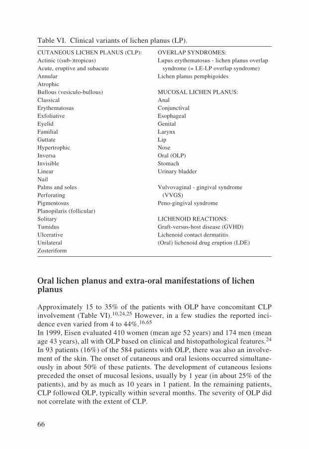
66
Oral lichen planus and extra-oral manifestations of lichen planus
Approximately 15 to 35% of the patients with OLP have concomitant CLP involvement (Table VI).10,24,25 However, in a few studies the reported inci-dence even varied from 4 to 44%.16,65 In 1999, Eisen evaluated 410 women (mean age 52 years) and 174 men (mean age 43 years), all with OLP based on clinical and histopathological features.24 In 93 patients (16%) of the 584 patients with OLP, there was also an involve-ment of the skin. The onset of cutaneous and oral lesions occurred simultane-ously in about 50% of these patients. The development of cutaneous lesions preceded the onset of mucosal lesions, usually by 1 year (in about 25% of the patients), and by as much as 10 years in 1 patient. In the remaining patients, CLP followed OLP, typically within several months. The severity of OLP did not correlate with the extent of CLP.
CUTANEOUS LICHEN PLANUS (CLP):Actinic ((sub-)tropicus) Acute, eruptive and subacute AnnularAtrophicBullous (vesiculo-bullous)Classical Erythematosus ExfoliativeEyelidFamilial GuttateHypertrophic InversaInvisible Linear Nail Palms and soles Perforating Pigmentosus Planopilaris (follicular) Solitary Tumidus Ulcerative Unilateral Zosteriform
OVERLAP SYNDROMES: Lupus erythematosus - lichen planus overlap syndrome (= LE-LP overlap syndrome)Lichen planus pemphigoides
MUCOSAL LICHEN PLANUS: AnalConjunctival Esophageal Genital Larynx Lip Nose Oral (OLP) Stomach Urinary bladder
Vulvovaginal - gingival syndrome (VVGS) Peno-gingival syndrome
LICHENOID REACTIONS: Graft-versus-host disease (GVHD) Lichenoid contact dermatitis (Oral) lichenoid drug eruption (LDE)
Table VI. Clinical variants of lichen planus (LP).
67
Six female patients were diagnosed with lichen planopilaris. The development of the lesions of the scalp preceded the onset of OLP in 83% of the patients by 1 to 3 year(s). Lichen planopilaris and the oral lesions simultaneously developed in one patient. Nail changes typical of LP were noted in 11 patients and preceded the onset of OLP in all patients by approximately 2 to 5 years. The most common clinical manifestations included thinning, ridging, and distal splitting of the nail plate. Six patients had involvement of all fingernails, 3 patients showed LP lim-ited to several fingernails, and 2 patients had involvement of both the fingernails and the toenails. Vulvar and vaginal LP was identified in 77 (19%) of the 399 women who were examined. The erosive form of the disease was the predomi-nant type, and the asymptomatic reticular lesions were identified in 25% of the patients. The simultaneous appearance of the genital and oral lesions was noted in 50% of the patients. Genital LP preceded OLP in 20% of the cases, and OLP preceded genital LP in 30% of the cases, usually within 1 year. Eight men of the 174 examined patients had genital LP with simultaneous involvement of the oral mucosa. Four of the patients showed LP of the esophagus upon endoscopy with symptoms of dysphagia and one patient showed ocular LP. The conclusions were that a thorough evaluation, if necessary by a multi-disciplinary group of healthcare providers, in patients with OLP should be performed, because a relatively high percentage of patients with OLP may display extra-oral manifestations of LP.24
Mucosal lichen planus
The vulvovaginal-gingival syndrome
In 1982, Pelisse et al described a new entity of erosive LP: the vulvovaginal-gingival syndrome (VVGS). It consists of the triad of erosive or desquamative vulvitis, vaginitis, and gingivitis.394 The term “ plurimucosal LP” was already reported by Gougerot and Burnier in 1937 and described the occurrence of LP involving the buccal mucosa, the cervix and the stomach without skin involve-ment.395 Bermejo et al suggested that the term “plurimucosal LP” was more appropriate than “the vulvovaginal-gingival syndrome”.396 We suggest the term “oro-genital LP”, which describes this disease more accurately and can easily be distinguished as a clinical entity in the spectrum of LP. However, the term “vulvovaginal-gingival syndrome” will be used here because it is mostly used in the literature.24,364,394 In 1989, M. Pelisse reported 19 women aged 27 to 70 years (median age 44 years) with VVGS.364 Eighteen women had an erosive vulvitis with complaints of burning and/or pain and 12 women had seri-ous complaints of dyspareunia. All 18 patients showed several erythematous plaques with epithelial desquamation or frank erosions. In most instances, there was an easily overlooked narrow rim of white reticulation at the periphery of the red plaques. In 6 patients, vulvar adhesions or atrophic lesions were found.
68
Eleven patients showed a desquamative vaginitis with dyspareunia and post-coital bleeding. Four patients developed a greenish, somewhat bloody vaginal discharge. Vaginal examination was difficult to accomplish because of pain and bleeding. When inspection was successful, it showed either a diffuse, erosive vaginitis or a slightly less erosive, red, desquamative vaginitis with indica-tions of a fine white reticular network. Cervical involvement was noticed in 2 women. Even in the quiescent stages, the vaginal mucosa was atrophic and fragile. Twelve patients also showed a desquamative or erosive gingivitis with discomfort and easy bleeding. The gingival surfaces were diffusely red and were usually eroded. In 5 patients, there were also typical white, reticular lesions on the buccal mucosa. The labial aspect of the maxillar gingival surface was the single area most commonly involved. Biopsy specimens taken from the white, reticulated border generally showed the typical histology of LP, whereas biopsy specimens taken from the eroded surface more often showed only a non-specific inflammatory reaction. Direct immunofluorescent examination in these patients did not show significant deposition of immuno-reactants. It is impor-tant that all patients had lesions on the vulvar, vaginal, and gingival mucosal surfaces, but often, at a single point in time, only one or two of the three sites were involved. The time interval between involvement of the different sites may show a variation of several years. Seven patients had concomitant involve-ment at the three sites. Vulvo-vaginitis preceded oral involvement (by up to 7 years) in 7 patients. In 5 patients, oral lesions preceded symptomatic genital disease by 2 to 9 years. Three patients also developed typical CLP. Treatment in all patients consisted of topically applied corticosteroids. However, only 5 patients showed satisfactory improvement. Ten patients were treated with sys-temic prednisone at a dose of at least 0.5 mg/kg/day. The initial response was excellent and required at least three weeks of therapy. Unfortunately, when the dose was tapered, relapse generally occurred at a dose of 10 mg/day. Systemic retinoids, griseofulvin and dapsone were administered in a few patients but generally had disappointing results.364
In 1994, Eisen reported 22 patients with VVGS and reported that therapy with potent topical corticosteroids was generally beneficial.370
In 1999, the same author reported that from the 399 women with OLP, 77 (19%) women also had vulvar and vaginal LP.24 The erosive form of the dis-ease was the predominant type in the genital region, although asymptomatic reticular lesions were identified in 25% of the patients. The patients with severe oral lesions commonly displayed mild genital disease, whereas the patients with asymptomatic gingival disease frequently suffered from severe erosive vulvovaginal disease. It is important to state that in many patients whose geni-tal symptoms had been evaluated by other physicians, the condition was com-monly misdiagnosed or undiagnosed. Generally, in patients with OLP, 6% will manifest simultaneous involvement at three or more sites, thus highlighting the importance of thorough examination and the need for a multi-disciplinary

69
approach in these patients. Lichen planus is the most common cause of desqua-mative vulvitis associated with desquamative vaginitis.24
In 2003, Ramer et al reviewed 127 cases of the VVGS found in the literature and added 5 new ones.397 The clinical characteristics of those patients are shown in Table VII. The onset of the erosive or ulcerative oral lesions may precede or fol-low the onset of vulvovaginal lesions by months or even years. It is possible that the initial diagnosis of OLP or genital LP has to be revised into VVGS after a while, because other mucosal membranes are consecutively affected. Generally, histopathological and immunopathological examinations in patients with VVGS show the typical features of mucosal LP. Complications of progressive disease in genital LP or VVGS are vulvar scarring, vaginal adhesions and synechiae that prohibit sexual intercourse. Gynecological surgery (adhesiolysis) may be neces-sary in these cases. Nevertheless, adhesions frequently recur after treatment.397 Insertion of intravaginal tampons may prevent new adhesions.398,401
Peno-gingival syndrome
In 1993, Cribier et al reported the male equivalent of the VVGS.399 In 1999, Eisen reported in a study of 584 patients with OLP that 8 patients also had genital LP.24 The mean age of onset of these 8 patients was 40 years. There was a simultaneous appearance of the mucosal lesions in all patients. Two patients had symptomatic, erosive genital lesions, 5 patients had asymptomatic hyper-
Table VII. A review of the clinical characteristics of the patients with the vul-vovaginal-gingival syndrome (VVGS) (Ramer et al (2003)).397
Investigated number of patients Patients
Mean age 113 52 years
Age range 113 29-79 years
Dyspareunia 132 88 (67%)
Burning/ pruritus/ pain 132 105 (80%)
Vaginal discharge 132 62 (47%)
Desquamation or erosions 132 95 (72%)
Vulvar itching 113 64 (57%)
Premenopausal 27 14 (52%)
Postmenopausal 27 13 (48%)
CLP 55 7 (13%)
Initial presentation of:
Oral lesions 132 33 (25%)
Genital lesions 132 34 (26%)
Both oral/genital lesions 132 65 (49%)

70
keratotic lesions (2 patients annular, 2 patients papular and 1 patient plaque form), and 1 patient had an asymptomatic, erythematous form. The glans penis was the most common site of involvement. Two patients also had lesions on the penile shaft and one patient also had lesions on the scrotum. Unlike female patients with VVGS, only 4 of the 8 males with oral and genital disease had gingival lesions.24 In our opinion, the term “oro-genital LP” also seems to be more appropriate in such cases.
Genital lichen planus
Females
The clinical signs and symptoms of genital LP in females were described in the VVGS.364,394,397 Generally, genital LP may occur solitary or within the VVGS.24,400 The exact percentage of involvement of the female genitalia in CLP is unknown.26,144 It seems to be likely that (especially asymptomatic) genital lesions in women with LP are unnoticed or misdiagnosed.24 Genital LP may be even more severe and recalcitrant to treatment compared with OLP. Treatment modalities with changing results consist of topical agents such as corticosteroids (class III or IV), lidocaine gels, antifungal agents, retinoids, estrogens, and cyclosporin, as well as systemic agents such as corticosteroids, retinoids, antifungal agents, hydroxychloroquine, dapsone and cyclospo-rin.397,400 In 2004, Byrd et al reported a significant improvement of clinical signs and symptoms with topical tacrolimus 0.1% ointment twice daily within 3 months (mean 4.2 weeks) in 15 (94%) of 16 patients with symptomatic vulvar LP, recalcitrant to many other treatments. Six patients (38%) reported mild adverse effects, including irritation, burning and tingling. These adverse effects resolved with continued medication. Genital LP returned in 10 (83%) of the 12 patients within 6 months after discontinuation of therapy over a period of several weeks, but in 6 patients the lesions were less severe than the lesions before treatment. They concluded that topical tacrolimus therapy effectively controlled symptoms and improved lesions in all but 1 patient. The effect may be temporary requiring continued use of tacrolimus, which appeared to be safe and effective in controlling the disease.400 There are some reports on the malignant transformation of genital LP into a squamous cell carcinoma, which particularly underlines the clinical importance of a thorough investigation with adequate histopathological examination and a multi-disciplinary approach.402-404 The differential diagnosis includes atrophic vulvovaginitis, desquamative vulvovaginitis, lichen sclerosus (et atrophicus), and localized bullous auto-immune diseases such as localized bullous pem-phigoid and cicatricial pemphigoid.405,406 In 1994, Marren et al reported the association of vulvar lichen sclerosus and OLP in older women.245
71
Males
It has been reported that the genitalia in males (particularly the glans penis) were involved in 25% in the classical form of CLP.26 Involvement of the genitalia in men in the classical form of CLP has to be considered as a site of predilection.22,30,266 Isolated genital LP does occur in men, especially the annular variant.266 Malignant transformation of LP of the penis into a squa-mous cell carcinoma was rarely reported.407,408
Other sites of mucosal lichen planus
Lichen planus has also been described in the anal region, the conjunctivae, the larynx, the lip, the nose, the esophagus, the stomach and in the urinary bladder (Table VI).26,65
Anal region
Lichen planus may affect the anal and the perianal region and may produce intense localized pruritus.409
Conjunctivae
Conjunctival LP was recognized as a rare cause of cicatrizing conjunctivitis and in most cases these patients also had concomitant OLP.24 The institution of prompt treatment controlled inflammation and prevented cicatrization.410,411
Lips
Formally, the vermilion border of the lip does not belong to the oral mucous membrane, but in the literature the disorders of the lips are often included in the section on oral diseases. Moreover, the vermilion border of the lips generally shows LP lesions which resemble the clinical features of the different subtypes of OLP (Figure 6).412 Generally, the lower lip is more affected than the upper lip. However, isolated LP of the lips is rarely seen. The reticular variant is most commonly seen, but the other variants also occur. The differential diagnosis includes chronic discoid lupus erythematosus, leukoplakia, actinic cheilitis, bullous auto-immune diseases, infective cheilitis, plasma-cell cheilitis and squamous cell carcinoma. Histopathological and/or immunopathological exam-ination is often necessary to establish the diagnosis LP on the lips.413,414 Squamous cell carcinoma is the most common malignancy affecting the vermilion border and occurs on the lower lip in 89%, on the upper lip in 3%, and at the commissures in 8% of the patients (Figure 18). Risk factors for this malignancy

72
are exposure to sunlight (actinic damage), tobacco smoking, low social class, poor nutrition, poor dentition, and immune suppression. Malignant transforma-tion of LP of the vermilion border of the (lower) lip both into a squamous cell carcinoma, and rarely into a verrucous carcinoma was reported.412,415,416 It is difficult to establish whether there is a synergistic premalignant effect in case of exposure to external carcinogens and the persistence of LP on the lips, which may act as an intrinsic risk factor.
Esophagus
In 1990, Dickens et al reported in a study of 19 patients with LP that 5 patients also had esophageal LP diagnosed via endoscopy. Four of the 5 patients were asymptomatic yet they displayed subtle papular lesions typical of LP. One patient with dysphagia showed erosive LP. Four of the 5 patients also had OLP.417
In 1997, Eisen reported in a study of 584 patients with OLP that 4 patients had dysphagia and esophageal LP was discovered via endoscopy.24 The incidence of esophageal disease was probably higher, given the fact that only symptomatic patients were screened and only erosive esophageal LP was detected. Endos-copy is probably not performed in patients with asymptomatic hyperkeratotic (reticular) esophageal LP and therefore it is not detected. A thorough history of symptoms like dysphagia should be obtained in patients with OLP.24 Although malignant transformation has not yet been reported, untreated erosive esopha-geal LP may result in chronic pain, dysphagia, strictures and stenosis.418
Further discussion of LP of the larynx, the nose, the stomach and the urinary bladder is beyond the scope of this thesis.
Clinical aspects of cutaneous lichen planus
Cutaneous lichen planus (CLP) is a benign, self-limiting inflammatory dermatosis characterized by a combination of clinical and histopathological features that most commonly affects middle-aged adults.266 It has a worldwide distribution with no overt racial predisposition and the estimated prevalence is about 0.5% in the Euro-pean and in the North American populations. However, there is a considerable variation in its incidence in several countries.419 In Nigeria, CLP is one of the most common dermatological diseases.420 Outbreaks of CLP occur throughout the year, although in one study it was suggested that an unexplained seasonal influence existed, showing an increased incidence between January and July.421
The classical form of cutaneous lichen planus
Cutaneous lichen planus is generally an idiopathic dermatosis with an insidious onset and its classical form commonly lasts 8 to 12 months (Table VI). The
73
lesions clear without any treatment within 18 months in 85% of the patients. However, CLP may even persist indefinitely.26,266 Current concepts on patho-genesis include immunological, genetic and environmental factors.11,30 The classical form of CLP comprises pruritic, faintly erythematous to violaceous, shiny, flat-topped papules, which may be polygonal in morphology and are usually symmetrically distributed (Figure 15). Fine white lines known as Wickham’s striae may be present on the surface of the lesions. Application of mineral oil on the surface of the lesions to fill air spaces in the keratin makes the striae more easily visible. The lesions may occur anywhere on the body but there appears to be a predilection for the flexor aspects of the wrists and the forearms, the ankles, the lumbar area, the (male) genitals and the shins. The face and the scalp are usually spared in classical CLP. The lesions vary in size from less than a millimeter to over a centimeter in diameter and may be scattered or occur in clusters. Scaling may be present in plaques and in lesions of prolonged duration. Linear lesions often appear along scratch marks or in scars (Koebner’s phenomenon).22,26,266,424 Cutaneous lichen planus may be intensely pruritic. However, physical evidence of scratching, such as excoria-tions, secondary infections and blood crusts, is infrequently seen, in contrast with many other itchy dermatological diseases such as atopic dermatitis, aller-gic contact dermatitis and prurigo nodularis.266 Twenty percent of the patients with CLP may be asymptomatic.419 The nails (particularly of the fingers) are also involved in about 10% of the cases of CLP, but this is usually a minor feature of the disease.26
The female-to-male ratio in CLP is probably 3:2. The ages of highest preva-lence of CLP differ significantly in men and in women and is about 30 years in men and about 50 years in women. There is oral involvement in about 50% of the patients with classical CLP, with a significant variation in several studies, probably depending on symptomatology and medical specialty.22,266 Lichen planus in childhood is rare and only 2-3% of all patients with LP are pediatric patients.11,422 The differential diagnosis may include psoriasis guttata, secondary syphilis, lichen nitidus, verrucae planae, (lichenoid) drug eruption, pityriasis lichenoides chronica, eczematous eruptions with lichenification, prurigo, (papular) lichen amyloidosus and Darier’s disease.144,419
Other remarks on the terms lichen, lichenoid and lichenification
Many diseases have the word “lichen” in common, but do not belong to the spectrum of LP.26,144 However, some of these disorders may be included in the differential diagnosis of a variant of LP (Table VI). In this thesis only an enu-meration is given: lichen amyloidosus, lichen aureus (lichen purpuricus), lichen

74
myxoedematosus (lichen fibromucinodosis), lichen nitidus, lichen nuchae (a variant of lichen simplex), lichen sclerosus et atrophicus, lichen scrofulosum, lichen simplex (chronicus), lichen spinulosus, lichen striatus, lichen verrucosus et reticularis (Nékam’s disease, keratosis lichenoides chronica, lichen ruber moniliformis).The term “lichenoid” may be used by clinicians to describe a flat-topped, shiny, papular eruption resembling LP, or by histopathologists to describe a type of tissue reaction principally consisting of a basal cell liquefaction degeneration and a band-like inflammatory infiltrate in the papillary dermis or in the lamina propria (“interface dermatitis”), consisting predominantly of mononuclear cells. Colloid bodies may be present. This pattern may be seen in a wide vari-ety of conditions: LP, lupus erythematosus, lichen sclerosus, poikilodermas, several cutaneous drug reactions.266,419 The word “lichenoid” is also used in: pityriasis lichenoides chronica, pityriasis lichenoides et varioliformis acuta (PLEVA, M. Mucha-Habermann), pigmented purpuric lichenoid dermatosis of Gougerot and Blum and exudative discoid and lichenoid dermatitis (Sulzberger-Garbe syndrome).Lichenification is a pattern of cutaneous response to repeated rubbing or scratching. It is characterized histologically by acanthosis and hyperkeratosis, and clinically by a thickened appearance of the skin, with accentuation of the surface markings so that the affected skin surface resembles “tree bark”. It is common in patients with atopic dermatitis, but may also occur in other “irri-tant” dermatoses.423
Other clinical variants of cutaneous lichen planus (Table VI)
Actinic LP (LP (sub-)tropicus) mostly affects young individuals from the Middle East, East Africa or India. It begins during spring or summer and mainly involves sun-exposed skin particularly of the face and of the dorsum of the hands and the arms. The lesions are characterized by well-defined num-mular patches, which have a deeply hyperpigmented center surrounded by a strikingly hypopigmented zone. Sunlight appears to be the precipitating factor and lesions have been provoked experimentally with UVB. Actinic LP may mimic melasma and lupus erythematosus.26 Acute, eruptive and subacute LP shows many, small and dissiminated lesions with a rapid evolution. This form may mimic a lichenoid drug eruption and lichen nitidus.266,424
Annular LP shows a few, rather large annular lesions. In males, these lesions are typically found on the penis and are more common in negroid patients. This form may resemble psoriasis, granuloma annulare and sarcoidosis.266,419 Atrophic LP has only a few lesions and may be the result of faded annular or hypertrophic lesions. It may mimic lichen sclerosus et atrophicus.266

75
Bullous LP (or vesiculo-bullous LP) is uncommon and refers to the develop-ment of vesicles and bullae on pre-existing lesions of CLP. Histopathological examination shows findings compatible with those of typical CLP and a sub-epidermal bulla with a negative direct immunofluorescence. It may resemble several bullous auto-immune diseases.26,424
Lichen planus erythematosus is rare. Deep red, asymptomatic papules blanch-ing with pressure appear on the trunk and extremities. Histopathological findings are consistent with CLP.26
Exfoliative LP resembles a general exfoliative dermatitis, which may be asso-ciated with erythroderma or toxic epidermal necrolysis.424 Eyelid LP shows isolated lesions on the skin of the eyelids which may be typi-cal for CLP or may mimic a brown retiform-like eruption resembling erythema ab igne.424 Familial LP is uncommon and occurs in individuals of the same family. It has a longer duration and more common relapses than the classical form of CLP. Familial LP commonly shows dissiminated skin lesions with frequent involve-ment of the oral mucosa. Familial LP occurs at a younger age particularly in the third decade, but may occur rather frequently under the age of 20 years.11,266
There may be an increased incidence of transformation into squamous cell carcinoma in children with familial LP.422 Guttate LP shows widely scattered, 1-2 mm lesions without the tendency of forming plaques. Guttate LP has a relatively good prognosis.266 Hypertrophic LP is characterized by thickened, hyperkeratotic, intensely pru-ritic lesions on the ventral parts of the lower limbs and the ankles and the lesions may persist for many years. It may resemble lichen simplex chronicus, lichen amyloidosus and lichen myxoedematosus.26,419 The occurrence of squa-mous cell carcinomas in hypertrophic LP has been reported.425
Lichen planus inversa was described, with violaceous papules in the axillae, groin and inframammary areas.26 The disease “lichen invisible” introduced by H. Gougerot in 1926 would only show subjective symptoms of LP without any clinical sign.426 In the oral cav-ity, it is probably similar to the Burning Mouth Syndrome (BMS).96 In such cases, the term “lichen invisible” should be avoided in our opinion. In 1966, Cram et al described another type of “LP invisible”. Premonitory pruritus is found, but the lesions are not apparent to the naked eye, and may be visualized by illumination with a Wood’s lamp. Histopathological examination shows features compatible with those of CLP.20
Linear lesions seen as a Koebner’s phenomenon are frequently found in many variants of LP.26 However, linear LP is rare, although rather more common in childhood. This variant shows isolated, long, narrow linear lesions which may extend the whole length of the limb and may occur in Blaschko’s lines (Figure 17). Linear LP has more often been described on one side of the body and may resemble lichen striatus, linear epidermal nevi, inflammatory linear verrucous

76
epidermal nevus (ILVEN) and linear forms of psoriasis.422 In 2004, Agaki et al reported three patients with a linear LP pigmentosus.427
In nail LP, the fingernails are more frequently affected than the toenails and initially only two or three fingernails are involved before spreading to the remaining digits. The most common change is a slight thinning of the nail plate, which may be observed to emerge from beneath the cuticle and extend forward with the growing nail. The nails may show longitudinal ridging, linear depressions and grooving, splitting (onychoschizia), shedding (onychomade-sis), longitudinal striation (onychorrhexis), absence (anonychia), subungual hyperkeratosis and a pterygium. Pterygium is formed by an adhesion between the dorsal nail fold and the nail bed with a partial destruction of the matrix resulting in the destruction of the nail. Lichen planus of the nail bed may pro-duce longitudinal melanonychia, hyperpigmentation, subungual hyperkeratosis or onycholysis and may mimic psoriasis unguium. However, nail abnormalities in LP are neither specific nor pathognomic.24,26,266
Lichen planus on the palms and the soles lacks the characteristic shape and color of most lesions of CLP elsewhere. The firm, yellow lesions are papular or nodular and may mimic hyperkeratotic eczema, atypical psoriasis, warts, callosity and keratosis palmoplantaris areata.266,419
Perforating LP has been reported and show papules with crustae. Histopatho-logical examination shows the classical features of CLP in addition to areas of perforation in the epidermis with numerous hyaline bodies present at the base of the perforation.26
Lichen planus pigmentosus is a pigmentary disorder, which may be associated with the typical papular CLP lesions seen in India and in the Middle East. The macular hyperpigmentation mainly involves the face and the upper limbs. It may mimic erythema dyschromicum perstans.422
Lichen planopilaris or follicular LP presents with follicular involvement of hair-bearing areas. It is more common in women and there are follicu-lar spinous lesions and often classical LP lesions elsewhere on the body. It may occur on the scalp and is likely to produce a cicatricial alopecia because of the deep inflammatory infiltrate around the hair follicles. It should be distinguished from chronic discoid lupus erythematosus, fol-liculitis decalvans, tufted hair folliculitis, perifolliculitis capitis abscedens et suffodiens and the final stage of pseudopelade of Brocq.24,266 There are obviously overlapping clinical and histopathological aspects between lichen planopilaris and the so called Graham-Little-Piccardi-Lassueur syn- drome.144,428 In solitary LP, there is only one lichenoid lesion which looks like a (pre)malignant epidermal tumor and will frequently be treated by excision. However, histo-pathological examination shows typical features of CLP.429
Lichen planus tumidus is a variant of follicular LP with a deep inflammatory infiltrate with clusters of milium cysts and comedones.430

77
Ulcerative LP is very rare. Large and painful ulcerations mostly occur on the sole of one or both feet. The lesions heal with great difficulty.26
In unilateral LP only a part of one side of the body is involved.431 In zosteriform LP, the lesions follow a segmental distribution like those in herpes zoster.266
Histopathology of cutaneous lichen planus
Histopathological features of the classical form of CLP are characteristic and show an irregularly thickened granular layer with orthokeratotic hyperkera-tosis. Parakeratosis is rarely found. Irregular acanthosis is usually present, with rete ridges that form a “sawtooth” pattern. The focal increase in thick-ness of the granular layer and infiltrate corresponds to the presence of the Wickham’s striae.26 A dense, band-like inflammatory infiltrate in the papil-lary dermis is an important feature and may obliterate the dermo-epidermal interface. The infiltrate consists almost entirely of lymphocytes intermingled with a few histiocytes and melanophages.266,422 There may be a vacuolar (“hydropic”) degeneration of the basal layer. Eosinophilic-staining Civatte’s bodies (hyaline bodies, cytoid bodies or colloid bodies) represent degener-ated basal epidermal cells, which have probably undergone premature kerati-nization and have extruded into the papillary dermis.419 Civatte’s bodies are found in 37-100% of the biopsy specimens of CLP and are considered to be among the earliest histopathological changes, but are not specific for LP.22,30 If the basal membrane zone becomes vacuolated, fluid accumulates and leads to the formation of clefts, known as Max Joseph spaces (or Caspary-Joseph spaces), which may be seen in up to about 17% of the specimens.144,432 Mela-nocytes are considerably decreased in number or even absent. The liberated melanin granules are ingested by dermal macrophages (“melanophages”) in the papillary dermis (Figures 7, 8, 9, 10 and 11).26,266 The histological fea-tures of the several clinical variants of LP may be less obvious or may show some other characteristics. For example in lichen planopilaris, a perifollicular inflammatory cell infiltrate is seen in early stages, with subsequent destruc-tion of the follicles.26 In LP pigmentosus, there is an increase of deposition of melanin in the basal and malpighian layers.422 Hypertrophic LP usually shows extensive acanthosis.26,266
Treatment
Treatment of CLP is mostly necessary, because it relieves pruritus and induces a remission.266 Treatment depends on the symptoms of LP, the involved variant (Table VI), the extent and the anatomical location of the lesions,
78
the age of the patient, concomitant disorders and co-medication.22,26,294,419 It is important to state that there are no large randomized trials with defi-nite results in the literature examining the efficacy of the various drugs or physical treatments in LP. Many published trials are observational and are not always prospective and the recommendations of the experts are based on their personal experience and not on “evidence-based medicine”.294,295 Generally, potent topical (class III or IV) or systemic corticosteroids (pred-nisolone 20-40 mg once a day) are used in the treatment of CLP during 6 to 8 weeks. Intralesional corticosteroids or very potent topical corticoste-roids under occlusion are particularly used in hypertrophic LP lesions. Fur-thermore, systemic retinoids (acitretin 0.5-1.0 mg/kg/day), antihistamines, cyclosporin (3-5 mg/kg/day) and ultraviolet radiation ((bath)PUVA, UVA and UVB) are taken into account. Griseofulvin, antibiotics (penicillins, tri-methoprim and metronidazole), dapsone, azathioprine, methotrexate, antima-larial drugs, isoniazid, cyclophosphamide, mycophenolate mofetil, enoxapa-rin, interferon-α2b and thalidomide are far less common variants of treat-ment.26,266,294,295,379,382,422 Even skin grafting may be required in persistent ulcerative lesions on the palms or the soles.20,294
Prognosis
When the lesions in CLP have disappeared (with or without treatment), post-inflammatory hyperpigmentation occurs rather frequently and intensely and may persist for several months. Generally, there is a recurrence in about 12 to 20 % of the patients with the classical CLP after initial clearing.22,26 The period of healing of the classical variant of CLP with intensive treatment is mostly 1 to 3 months.266 In general, CLP has no increased risk of malignancies of the skin or internal organs. Lichen planus pemphigoides and possibly also vesiculo-bullous LP have been described in association with several forms of internal malignancies.265 Malignant transformation of the involved skin was rarely reported in hypertrophic LP and familial LP.266,422
Overlap syndromes
Lichen planus-lupus erythematosus overlap syndrome
The lichen planus-lupus erythematosus overlap syndrome (LP-LE overlap syndrome) was described by Copeman et al in 1970. In that study, 4 patients showed overlapping clinical and histopathological features of LP and LE.433 Later, further combinations of CLP and OLP with either chronic discoid LE, subacute cutaneous LE or systemic LE were described.229 However, some
79
authors prefer to diagnose LP and LE separately. The lesions are often seen on the hands and the feet. The presence of systemic immunological markers such as ANA titers is also controversial.26,266,424 Distinctions between LP and LE include colloid bodies, basement membrane changes and direct immunofluorescence. However, colloid bodies may also be seen in LE, but in LP they tend to be more numerous and are situated deeper, even into the upper area of the reticular dermis. In LP, basement membrane clefts may be due to lysis of keratinocytes, whereas in LE vacuolization forms on either side of this membrane. Direct immunofluorescence shows immunoglobulin and complement deposition in a granular, linear band along the dermo-epi-dermal junction in LE, whereas in LP these immunoreactants tend to be limited to colloid bodies. The demonstration of the LP-specific antigen in the granular layer by indirect immunofluorescence in LP can also be helpful in distinguishing from LE. Some studies on the basement membrane zones showed that there are possibly several similar steps in the pathogenesis of LP and LE.22,26,434
Lichen planus pemphigoides
Lichen planus pemphigoides shows a combination of clinical features of LP and bullous pemphigoid with subepidermal bullae.26,266,435 The bullae may also present on a previously healthy skin in contrast with bullous LP. In con-trast with bullous LP, LP pemphigoides also shows linear C3 and IgG deposits at the BMZ, like bullous pemphigoid in direct immunofluorescence. Indirect immunofluorescence may also be positive in LP pemphigoides.422 Recent immunoblotting data revealed a 180-KDa antigen and a 200-KDa antigen simi-lar to that found in bullous pemphigoid. These data indicated the coexistence of two separate diseases (LP and bullous pemphigoid) in the same patient.22,419 However, some authors take the view that the antigen in LP pemphigoides is different from the antigen in bullous pemphigoid. The mean age of patients with LP pemphigoides is lower and the course tends to be less severe than in classical bullous pemphigoid.26,144 Lichen planus pemphigoides was reported in association with several forms of internal malignancies including stomach cancer, lymphosarcoma, reticulum cell carcinoma, craniopharyngioma, and neuroblastoma.26
Lichenoid reactions
Lichenoid reactions may resemble LP in many respects and may be induced by contact, inhalation or ingestion of a large number of different drugs and chemicals. Lichenoid reactions may clinically resemble the classical form of CLP.26

80
Graft-versus-host disease
Graft-versus-host disease (GVHD) is important because skin manifestations are prominent and LP can be closely simulated both clinically and histologi-cally. Furthermore, cutaneous GVHD provides an excellent biological model for the study of interactions between lymphocytes and the skin and can also provide a clue in the pathogenesis of LP and lichenoid eruptions.266,422,438 Bone-marrow transplantation is a well-accepted treatment for a variety of severe hematological disorders, including acute leukemias, aplastic anemia, certain immunodeficiency disorders and some inborn errors of metabolism. A major obstacle to successful transplantation is the threat of GVHD.26,419 Graft-versus-host disease occurs when lymphoid cells from an immunocom-petent donor are introduced into a histo-incompatible recipient who is inca-pable of rejecting them. Although GVHD usually occurs in patients undergo-ing allogeneic bone-marrow transplantation, it may also occur in immuno-deficient children after maternal-fetal cell transfer and surprisingly also in otherwise healthy patients receiving blood transfusions after heart surgery. Rarely, GVHD can develop “spontaneously” during a disseminated carci-noma.22,30,439
The main targets of GVHD in man are the skin, the gastrointestinal tract and the liver. The severity of the reaction, clinically and histologically, varies from mild to severe (grades 1 to 4). There are two main forms of GVHD namely the acute form, which develops between 1 week and 3 months after transplantation and the chronic form, which develops after 3 months. The chronic form is not preceded by the acute phase in about 15% of the cases.266,436 The oral manifestations of acute GVHD consist of painful mucosal ulceration, cheilitis, and the presence of lichenoid striae or plaques. Small, white lesions affect the buccal and lingual mucosa early on, but disappear after 2 weeks. Erythema and ulceration are most pronounced at 7 to 11 days and may be asso-ciated with obvious infection.275,437 Candidosis or herpes simplex stomatitis (occasionally herpes zoster) is common and there may also be oral purpura, especially in adults.438,439 The oral lesions in chronic GVHD occur together with skin lesions, and include generalized mucosal erythema, lichenoid lesions, mainly in the buccal mucosa, and xerostomia. Xerostomia is most significant in the first 2 weeks after transplantation and is a consequence of drug treat-ment, irradiation and/or GVHD.275,440 Lip biopsy is useful in the diagnosis of chronic GVHD and should include both the mucosa and the underlying minor salivary glands. Histology shows changes similar to those seen in Sjögren’s syndrome.441
The severity of the cutaneous eruption often parallels that of GVHD. In acute GVHD, the earliest cutaneous features include a malar flush, erythema of the palms or soles and a generalized morbilliform rash. Occasionally, erythematous to violaceous follicular papules occur and rarely a toxic epidermal necrolysis

81
or a severe exfoliative erythroderma occur.266 Characteristic histopathological features are present in the patients with acute GVHD. The epidermis shows focal vacuolar alteration of the basal layer with a few lymphoid cells at the dermo-epidermal interface. The numbers of the mononuclear inflammatory cells correlate positively with the severity of the disease. IgM deposits may occur at the dermo-epidermal junction.442
Chronic GVHD develops in 10% of all patients undergoing allogeneic bone-marrow transplantation and even in 30% of all long-term survivors (over 150 days). The cutaneous changes may be localized or generalized. The localized forms may be linear or whorled along Blaschko’s lines. The initial eruption is usually lichenoid and may involve the oral cavity, the skin, the nails and the hair. Clinically and histologically, it closely resembles “idiopathic” LP. However, hyper- and hypopigmentation are often more prominent features. Chronic GVHD may also resemble Sjögren’s syndrome, LE and dermato-myositis. A very serious disorder is a severe poikiloderma with widespread cutaneous sclerosis, contractions, malabsorption, wasting, alopecia and areas of ulceration. 26,30,438 There is an increased frequency of HLA-A1, HLA-B1 and HLA-B2 in chronic GVHD with scleroderma-like complications which indicates that a specific genetic predisposition is important.266 A malig-nant transformation into a squamous cell carcinoma has been reported in an indolent ulcer in chronic GVHD.443 In the early phase of chronic GVHD, histopathological examination may resemble LP. However, the infiltrate at the dermo-epidermal interface is often less dense than that in LP and may contain eosinophils. It was observed that the number of CD3+ infiltrating T-cells was lower than that in OLP. The final stages of chronic GVHD show an atrophy of the epidermis with gross fibrosis of the dermis, skin append-ages or even subcutis.30,439,442 The mortality in chronic GVHD is about 10%.266 Epithelial damage in GVHD appears to be mediated particularly by cytotoxic T-cells in which Langerhans cells are selectively lost. Various lymphokines such as interferons and IL-2 may be released by T-cells which were sensitized via interaction with host cells and this process will activate both donor and host mononuclear cells.444 The future success of bone-mar-row transplantation largely depends on the extent to which GVHD can be prevented. The mortality of GVHD grades 2 to 4 exceeds 75%. The threat of GVHD increases when the donor and recipient genotypes are not identical. Prospective CD31 typing may reduce the risk of acute GVHD. Transplanta-tion of bone-marrow that has been depleted of T-cells by the use of a cocktail of anti-T-cell monoclonal antibodies has made a major contribution in the prevention of GVHD. The most effective combinations of systemic steroids and immunosuppressive agents have yet to be established. Cyclosporin has also failed to reduce the incidence and the severity of GVHD. Phototherapy (UVB) and photochemotherapy (PUVA) are also used in the treatment of GVHD.26,30,266,438

82
Lichenoid contact dermatitis
A lichenoid contact dermatitis has been described after exposure to nickel445, mercury446, gold114, aminoglycoside antibiotics447, methacrylic acid esters448, epoxy resin449 and color film developing chemicals based on paraphenylene-diamine450. The “closed” patch test is the most convenient method for testing lichenoid contact dermatitis in conformation with allergic contact dermatitis. However, the typical lichenoid features often occur after the regular (last) read-ing of the patch tests after 72 hours.114,451 The active ingredients in the color film developer responsible for the reaction are certain substituted paraphenylenediamine (PPDA) compounds. The following types of reaction are observed: an acute or subacute eczematous or papular der-matitis and an (subacute) lichenoid dermatitis both clinically and histologically identical to CLP. These reactions may occur individually or may evolve into each other. However, it is not quite clear why exposure to color film developers induces an eczematous reaction in some patients and a lichenoid dermatitis in others. There are gradations both in clinical and histological aspects between the spectrum of typical eczematous dermatitis and typical CLP. The fact that the condition begins on areas exposed to the chemical strongly indicates that direct contact with the developer is the cause of the eruption, but ingestion or inhalation cannot be ruled out entirely. Moreover, oral lichenoid lesions affect-ing the buccal mucosa or the tongue occasionally appear in patients with a lichenoid contact dermatitis to color film developers. Sensitization in exposed workers is fairly high and may take 1 month to 3 years to develop. The skin reaction subsides after several weeks, leaving marked hyperpigmentation. Patch test reactions are usually positive to PPDA and are eczematous in nature but may become lichenoid.26,266,451,452 A lichenoid contact dermatitis associated with PPDA itself (in hair dyes) has been reported, but is not common.453 More-over, a lichenoid pigmented contact dermatitis from isopropyl-PPDA (IPPD) has been reported.454 Other allergens are diethylparaphenylenediamine (CD2) and amino-4-N-ethyl-N-methane-sulfon-aminoethyl-m-toluidine (CD3).451,455
A lichenoid dermatitis has been described in the parts of tattoos in case of a coexisting mercury hypersensitivity to the injected red dye, indicating a lichen-oid reaction induced by delayed-type cellular hypersensitivity. The reaction may eventually subside spontaneously, but the risk of a generalized eruption is high. It is possible that there is an accompanying OLP in these patients in case of contact with dental amalgam fillings.446,456 A photo-contact lichenoid eruption has been occurred after application of musk ambrette.457
Cutaneous lichenoid drug eruptions
A cutaneous lichenoid drug eruption (cutaneous LDE) generally shows a less typical distribution and more frequently a photodistribution of the clini-

83
cal lesions with more polymorphism than the classical form of CLP.266,419 Eczematization, hypertrophy, unusual pigmentation, scaling (sometimes more psoriasiform), and intense post-inflammatory hyperpigmentation are com-mon.458 This hyperpigmentation regresses very slowly and may even be per-manent.459 Alopecia may be profound and unrelenting. Rarely, atrophy of the skin may occur with anhidrosis of the affected areas.458,459 Concomitant oral involvement is less common in cutaneous LDE than in CLP.460 Many Allied troops developed a cutaneous LDE (called “New Guinea LP”) after receiving quinacrine as malaria prophylaxis while serving in the South Pacific during World War II. Follicular involvement frequently ensued, leading to alopecia. The lesions commonly start some months after administration of the drug.461 Histopathological features more indicative of a cutaneous LDE than of CLP include the following: focal parakeratosis, focal interruption of the granular layer, colloid bodies situated higher in the granular and cornified layers, pres-ence of a few eosinophils, exocytosis of lymphoid cells into the epidermis and a deeper perivascular infiltrate.462 Moreover, plasma eosinophilia occurs more frequently in cutaneous LDE than in CLP.463 Unfortunately, cutaneous LDE may sometimes be difficult to distinguish from CLP both on clinical and his-topathological grounds.26,266 Watanabe et al evaluated cutaneous LDE using direct immunofluorescence of the lesions and noted no significant differences from CLP.464 In 1974, Van Joost reported the presence of circulating antibod-ies reactive with basal cells of the skin in drug eruptions.471 This was shown to be a useful corroborative test of LDE, but its full implications are not yet understood.The list of drugs that may produce a cutaneous LDE is extensive and continues to increase steadily (Table VIII). Several groups of drugs may be involved: analgesics, antibiotics, antimalarials, antirheumatics, cardiovascular drugs, oral antidiabetics, psychotropical and neurological agents.465,466 The resolution of the lesions after withdrawal of the specific drug can take several months.266 Therefore, it is often difficult to determine if a specific drug is reponsible for the lichenoid cutaneous reaction, particularly when a patient uses several dif-ferent drugs at the same time. Photosensitive cutaneous LDEs particularly occur in patients treated with tetra-cyclines, thiazide diuretics, anti-malarials, and diltiazem. Photosensitive cuta-neous LDEs occur more commonly in patients with a dark skin type (Type VI, Fitzpatrick) or in patients with advanced HIV infection.467
Oral lichenoid drug eruptions
Generally, the reports on oral lichenoid drug eruptions (oral LDEs) are con-siderably fewer than those on cutaneous LDEs and fewer drugs have been reported as causing oral LDEs rather than cutaneous LDEs. However, data
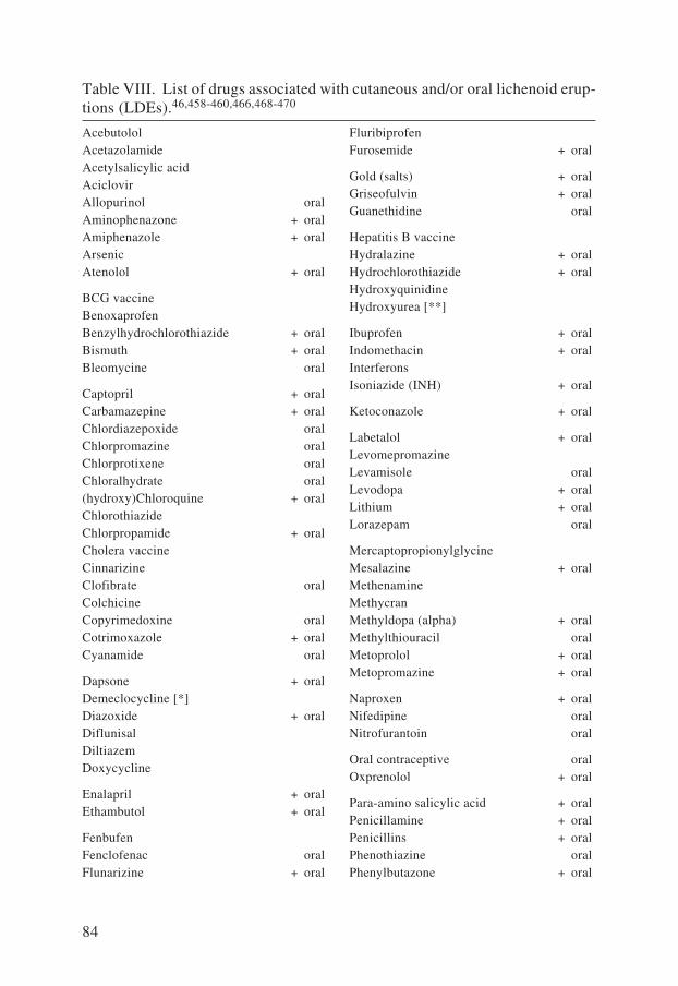
84
Acebutolol AcetazolamideAcetylsalicylic acid AciclovirAllopurinol oralAminophenazone + oralAmiphenazole + oralArsenic Atenolol + oral
BCG vaccineBenoxaprofen Benzylhydrochlorothiazide + oralBismuth + oralBleomycine oral
Captopril + oralCarbamazepine + oralChlordiazepoxide oralChlorpromazine oralChlorprotixene oralChloralhydrate oral(hydroxy)Chloroquine + oralChlorothiazide Chlorpropamide + oralCholera vaccine Cinnarizine Clofibrate oralColchicineCopyrimedoxine oralCotrimoxazole + oralCyanamide oral
Dapsone + oralDemeclocycline [*] Diazoxide + oralDiflunisal Diltiazem Doxycycline
Enalapril + oralEthambutol + oral
FenbufenFenclofenac oralFlunarizine + oral
FluribiprofenFurosemide + oral
Gold (salts) + oralGriseofulvin + oralGuanethidine oral
Hepatitis B vaccine Hydralazine + oralHydrochlorothiazide + oralHydroxyquinidine Hydroxyurea [**]
Ibuprofen + oralIndomethacin + oralInterferons Isoniazide (INH) + oral
Ketoconazole + oral
Labetalol + oralLevomepromazine Levamisole oralLevodopa + oralLithium + oralLorazepam oral
Mercaptopropionylglycine Mesalazine + oralMethenamine Methycran Methyldopa (alpha) + oralMethylthiouracil oralMetoprolol + oralMetopromazine + oral
Naproxen + oralNifedipine oralNitrofurantoin oral
Oral contraceptive oralOxprenolol + oral
Para-amino salicylic acid + oralPenicillamine + oralPenicillins + oralPhenothiazine oralPhenylbutazone + oral
Table VIII. List of drugs associated with cutaneous and/or oral lichenoid erup-tions (LDEs).46,458-460,466,468-470

85
Phenytoin + oralPindolol + oralPiroxicam Practolol + oralPrazosine + oralPrimidon oralProbenecid Procainamide oralPropranolol + oralPropylthiouracil oralPyrimethamine + oralPyrithioxine Pyritinol + oral
Quinacrine (Mepacrine) + oralQuinidine + oralQuinine + oral
Radiocontrast media (iodine) + oralReserpine oral
Simvastatin Smallpox vaccine
Spironolactone + oralStreptomycin + oralSulindac Sulphamethoxazole oralSulphasalazine + oralSulphonylureas
Temazepam + oralTestosterone Tetracyclines + oralThiacetazone Tick-borne meningoencephalitis vaccine Tiopronine Tolamulol Tolazamide + oralTolbutamide + oralTrihexyphenidyl Triprolidine
Verapamil
Drug: capable of inducing cutaneous lichenoid eruption.
Drug + oral: capable of inducing oral and/or cutaneous lichenoid eruption.
Drug oral: capable of inducing oral lichenoid eruption.
[*]: requires exposure to light.
[**]: induces ulcerative LP on the hands and the feet.
searches on LDEs showed that many drugs may also be involved in oral LDEs (Table VIII).157,465,467-469 Nevertheless, many of the reports on oral LDEs are anecdotal, or do not state histological verification, or do not specify the details of the oral cases in reports on both cutaneous LDEs and oral LDEs.10,157 Moreover, sometimes it may be impossible to attribute the causative drugs or histological findings to individual cases.157 Occasionally, oral LDE can be severe and may also involve the esophagus.460 Some histopathological fea-tures may occur more often in oral LDE than in OLP: a more diffuse and more deeply extending subepithelial infiltrate containing eosinophils, a perivascular infiltrate, parakeratosis, and the presence of colloid bodies higher in the epi-thelium.462,467 However, histopathological and immunological examinations of oral LDE generally show features identical or very similar to those of OLP and such investigations cannot be used reliably to distinguish between oral LDE and OLP.462,464,467 Lamey et al suggested that the clinical feature of unilateral-ity occurred more commonly in oral LDE.470 Labial LDE has been reported to occur more frequently in patients with HIV infection.467

86
There is as yet no specific test for the diagnosis of oral LDE.157 Although, reso-lution and recurrence of oral LDE on withdrawal and re-exposure to the drug is probably diagnostic.10,65 However, oral LDE may persist for a long period after the withdrawal of the drug.9 This makes the distinction between oral LDE and OLP very difficult in clinical practice. The exact cause of oral LDE remains unclear. The adulteration of cellular molecules by the offending agents may be operative.26 The etiopathogenic pathways of oral LDE show many striking similarities with those in OLP (Fig-ure 12).30,157,266
References
1. Scalf LA, Fowler JF, Morgan KW et al. Dental metal allergy in patients with oral, cutaneous, and genital lichenoid reactions. Am J Contact Dermatitis 2001; 12: 146-50.
2. Yiannias JA, el-Azhary RA, Hand JH et al. Relevant contact sensitivities in patients with the diagnosis of oral lichen planus. J Am Acad Dermatol 2000; 42: 177-82.
3. Camisa C, Taylor JS, Bernat JR et al. Contact hypersensitivity to mercury in amal-gam restorations may mimic oral lichen planus. Cutis 1999; 63: 189-92.
4. Pecegueiro M, Sachse MF, Amaro J et al. Oral lichen planus versus oral lichenoid eruption as a manifestation of contact allergy. Contact Dermatitis 1999; 40: 333-4.
5. Dunsche A, Kastel I, Terheyden H et al. Oral lichenoid reactions associated with amalgam. Br J Dermatol 2003; 148: 70-6.
6. Scully C, Beyli M, Ferreiro MC et al. Update on oral lichen planus: etiopathogen-esis and management. Crit Rev Oral Biol Med 1998; 9: 86-122.
7. Vincent SD, Fotos PG, Baker KA et al. Oral lichen planus: the clinical, historical and therapeutic features of 100 cases. Oral Surg Oral Med Oral Pathol 1990; 70: 165-71.
8. Silverman S, Gorsky M, Lozada-Nur F. A prospective follow-up study of 570 patients with oral lichen planus. Oral Surg Oral Med Oral Pathol 1985; 60: 30-4.
9. Conklin RJ, Blasberg B. Oral lichen planus. Dermatol Clin 1987; 5: 663-73.10. Eisen D. The clinical manifestations and treatment of oral lichen planus. Dermatol
Clin 2003; 21: 79-89.11. Alam F, Hamburger J. Oral mucosal lichen planus in children. Int J Paed Dent
2001; 11: 209-14.12. Pindborg JJ, Mehta FS, Daftary DK et al. Prevalence of oral lichen planus among
7639 Indian villagers in Kerala, South India. Acta Derm Venereol (Stockholm) 1972; 52: 216-20.
13. Axell T. Rundquist L. Oral lichen planus - a demographic study. Comm Dent Oral Epidemiol 1987; 15: 52-6.
14. WHO Collaborating centre for oral precancerous lesions. Definition of leukoplakia and related lesions: an aid to studies on oral precancer. Oral Surg Oral Med Oral Pathol 1978; 46: 518-39.
15. Thorn JJ, Holmstrup P, Rindum J et al. Course of the various clinical forms of oral lichen planus. J Oral Pathol 1988; 17: 213-8.
16. Andreasen JO. Oral lichen planus. A clinical evaluation of 115 cases. Oral Surg Oral Med Oral Pathol Oral Radiol Endod 1968; 61: 373-81.
87
17. Eisen D. The clinical features, malignant potential, and systemic associations of oral lichen planus: a study of 723 patients. J Am Acad Dermatol 2002; 46: 207-14.
18. Mollaoglu N. Oral lichen planus: A review. Br J Oral Maxillofac Surg 2000; 38: 370-7.
19. Lozada-Nur F, Miranda C. Oral lichen planus: epidemiology, clinical characteris-tics and associated diseases. Semin Cutan Med Surg 1997; 16 (4): 273-7.
20. Cram DL, Muller SA. Unusual variations of lichen planus. Mayo Clin Proc 1966; 41: 677-88.
21. Laskaris G. Skin Diseases. In: Color Atlas of Oral Diseases. Edited by Thieme. 3rd edn. 2003; 24: 264-8.
22. Voute ABE. Thesis. Oral lichen planus: a clinical study. University of Amsterdam. Elinkwijk BV, Utrecht 1994.
23. Scully C. The oral cavity. In: Rook/Wilkinson/Ebling. Textbook of Dermatology. Champion RH, Burton JL, Burns DA, Breathnach SM. 6th edn. Vol 4. Oxford: Blackwell Science Ltd 1998: 3081-3.
24. Eisen D. The evaluation of cutaneous, genital, scalp, nail, esophageal, and ocular involvement in patients with oral lichen planus. Oral Surg Oral Med Oral Pathol Oral Radiol Endod 1999; 88: 431-6.
25. Silverman S, Bahl S. Oral lichen planus update: clinical characteristics, treatment responses, and malignant transformation. Am J Dent 1997; 10: 259-63.
26. Boyd AS, Neldner KH. Lichen planus. J Am Acad Dermatol 1991; 25: 593-619.27. Axell T. A preliminary report of prevalence of oral mucosal lesions in a Swedish
population. Comm Dent Oral Epidemiol 1975; 3: 143-5.28. Hellgrin L. Prevalence of lichen ruber planus in different geographic areas of
Sweden. Derm Venereol 1970; 50: 374-80.29. Walsh LJ, Savage NW, Ishii T et al. Immunopathogenesis of oral lichen planus.
J Oral Pathol Med 1990; 19: 389-96.30. Tank B, van Joost Th. Lichen planus and Graft-vs-Host disease. In: Skin Immune
System (SIS). Cutaneous immunology and clinical immunodermatology. Edited by Bos JD. 2nd edn. CRC Press, Boca Raton, Florida, U.S.A. 1997: 531-41.
31. Clausen J, Kjaergaard J, Bierring F. Ultrastructure of the dermo-epidermal junction in lichen planus. Acta Derm Venereol (Stockholm) 1981; 61: 101-6.
32. Walton LJ, Thornhill MH, Farthing PM. VCAM-1 and ICAM-1 are expressed by Langerhans cells, macrophages and endothelial cells in oral lichen planus. J Oral Pathol Med 1994; 23: 262-8.
33. Nickoloff BJ, Lewinsohn DM, Butcher EC. Enhanced binding of peripheral blood mononuclear leukocytes to gamma-interferon-treated cultured keratinocytes. Am J Dermatopathol 1987; 9: 413-8.
34. Eversole LR. Immunopathogenesis of oral lichen planus and recurrent aphthous stomatitis. Semin Cutan Med Surg 1997; 16: 284-94.
35. Porter SR, Kirby A, Olsen I et al. Immunologic aspects of dermal and oral lichen planus: a review. Oral Surg Oral Med Oral Pathol Oral Radiol Endod 1997; 83: 358-66.
36. Porter K, Klouda P, Scully C et al. Class I and II antigens in British patients with oral lichen planus. Oral Surg Oral Med Oral Pathol 1993; 75: 176-80.
37. Lowe NJ, Cudworth AG, Woodrow JC. HLA antigens in lichen planus. Br J Der-matol 1976; 96: 169-71.
38. Lin SC, Sun A. HLA-DR and DQ antigens in Chinese patients with oral lichen planus. J Oral Pathol Med 1990; 19: 298-300.

88
39. Harper JI. Genetics and genodermatoses. In Rook/Wilkinson/Ebling. Textbook of Dermatology. Champion RH, Burton JL, Burns DA, Breathnach SM. 6th edn. Vol 1. Oxford: Blackwell Science Ltd 1998; 371-2.
40. Simon M, Djawari D, Schonberger A. HLA-antigens associated with lichen planus. Clin Exp Dermatol 1984; 9: 435.
41. Powell FC, Rogers RS, Dickson ER et al. An association between HLA-DR1 and lichen planus. Br J Dermatol 1986; 114: 473-8.
42. Copeman PWM, Tan RSH, Timlin D et al. Familial lichen planus. Br J Dermatol 1978; 98: 573-7.
43. Mahood JM. Familial lichen planus. Arch Dermatol 1983; 119: 292-4.44. Grunnet N, Schmidt H. Occurence of lichen planus in a family. Genetic susceptibil-
ity or coincidence? Clin Exp Dermatol 1983; 8: 397-400.45. Hampf GC, Malmstrom JM, Aalberg VA et al. Psychiatric disturbance in patients
with oral lichen planus. Oral Surg Oral Med Oral Pathol 1987; 63: 429-32.46. Altman J, Perry HO. The variations and course of lichen planus. Arch Dermatol
1961; 84: 179-91.47. McCartan BE. Psychological factors associated with oral lichen planus. J Oral
Pathol Med 1995; 24: 273-5.48. Shafer WG, Hine MK, Levy BM. A textbook of oral pathology. 4th edn. WB
Saunders. Philadelphia 1983.49. Lowenthal U, Pisanti S. Oral lichen planus according to the modern model. J Oral
Med 1984; 39: 224-6.50. Allen CM, Beck FM, Rossie KM et al. Relation of stress and anxiety to oral lichen
planus. Oral Surg Oral Med Oral Pathol 1986; 61: 44-6.51. Rojo-Moreno JL, Bagan JV, Rojo-Moreno J et al. Psychologic factors and oral
lichen planus. A psychometric evaluation of 100 cases. Oral Surg Oral Med Oral Pathol Oral Radiol Endod 1998; 86: 687-91.
52. Ostman PO, Anneroth G, Johansson I et al. Life-style survey of patients with oral lichenoid reactions. Acta Odontol Scand 1996; 54: 96-101.
53. Neumann-Jensen B, Holmstrup P, Pindborg JJ. Smoking habits of 611 patients with oral lichen planus. Oral Surg 1977; 43: 410-5.
54. Murti PR, Daftary DK, Bhonsle RB et al. Malignant potential of oral lichen planus: observation in 722 patients from India. J Oral Pathol 1986; 15: 71-7.
55. Schlecht NF, Franco EL, Pintos J et al. Interaction between tobacco and alcohol consumption and the risk of cancers of the upper aerodigestive tract in Brazil. Am J Epidemiol 1999; 150: 1129-37.
56. van der Waal I. Tumoren van het mondslijmvlies. [Dutch]. Tumors of the oral mucosa. Ned Tijdschr Derm Venereol 2002; 12: 368-70.
57. Katz J, Goultshin J, Benoliel R et al. Lichen planus evoked by periodontal surgery. J Clin Periodontol 1988; 15: 263-5.
58. Scully C. The oral cavity. In: Rook/Wilkinson/Ebling. Textbook of Dermatology. Champion RH, Burton JL, Burns DA, Breathnach SM. 6th edn. Vol 4. Oxford: Blackwell Science Ltd 1998; 3054-5.
59. Ramon-Fluixa C, Bagan-Sebastian J, Milian-Masanet M et al. Periodontal status in patients with oral lichen planus: a study of 90 cases. Oral Diseases 1999; 5: 303-6.
60. Jacob FM, Helmbold TR. Bacteriologic studies on lichen planus: preliminary report. Arch Derm Syphilol 1933; 27: 472.
61. Lehnhoff C. Spirochaetes in aetiologically obscure diseases. Acta Derm Venereol (Stockholm) 1948; 28: 295.

89
62. Lochner JC, Pomeranz JR. Lichenoid secondary syphilis. Arch Dermatol 1974; 109: 81-3.
63. Shelley WB, Shelley ED. Urinary tract infection as a cause of lichen planus: metronidazole therapy. J Am Acad Dermatol 1989; 10: 905-7.
64. Wahba-Yahav AV. Intestinal amebiasis, lichen planus and treatment with metro-nidazole. J Am Acad Dermatol 1989; 20: 1128-9.
65. Scully C, El-Kom M. Lichen planus: review and update in pathogenesis. J Oral Pathol Med 1985; 14: 431-8.
66. Ficarra G, Flaitz CM, Gaglioti D et al. White lichenoid lesions of the buccal mucosa in patients with HIV infection. Oral Surg Oral Med Oral Pathol 1993; 76: 460-6.
67. Maitland NJ, Cox MV, Lynas C et al. Detection of human papillomavirus DNA in biopsies of human oral tissue. Br J Cancer 1987; 56: 245-9.
68. Jontell M, Watts S, Wallstrom M et al. Human papilloma virus in erosive oral lichen planus. J Oral Pathol Med 1990; 19: 273-7.
69. Walsh LJ, Ishii T, Savage NW et al. Immunohistologic analysis of epithelial cell populations in oral lichen planus. J Oral Pathol Med 1990; 19: 177-81.
70. Lodi G, Porter SR. Hepatitis C virus infection and lichen planus: a short review. Oral Diseases 1997; 3: 77-81.
71. van der Meij EH, van der Waal I. Hepatitis C virus infection and oral lichen planus: a report from the Netherlands. J Oral Pathol Med 2000; 29: 255-8.
72. Imhof M, Popal H, Lee JH et al. Prevalence of hepatitis C virus antibodies and evaluation of hepatitis C virus genotypes in patients with lichen planus. Dermatol-ogy 1997; 195(1): 1-5.
73. Ingafou M, Porter SR, Scully C et al. No evidence of HCV infection or liver disease in British patients with oral lichen planus. Int J Oral Maxillofac Surg 1998; 27: 65-6.
74. Mignogna MD, Lo Muzio L, Lo Rosso L et al. Oral lichen planus: different clini-cal features in HCV-positive and HCV-negative patients. Int J Dermatol 2000; 39: 134-9.
75. Figueiredo LC, Carrilho FJ, de Andrade HF et al. Oral lichen planus and hepatitis C virus infection. Oral Diseases 2002; 8: 42-6.
76. GISED (Gruppo Italiano Studi Epidemiologici in Dermatologia). Lichen planus and liver disease: a multicentre case-control study. Br Med J 1990; 300: 227-30.
77. Rebora A, Robert E, Rongioletti F. Clinical and laboratory presentation of lichen planus patients with chronic liver disease. J Dermatol Sci 1992; 4: 38-41.
78. Pemberton MN, Sloan P, Thakker NS. Oral lichenoid lesions after hepatitis B vac-cination. Oral Surg Oral Med Oral Pathol Oral Radiol Endod 2000; 89: 717-9.
79. Al-Khenaizan S. Lichen planus occurring after hepatitis B vaccination: a new case. J Am Acad Dermatol 2001; 45: 614-5.
80. Lundstrom IM, Anneroth GB, Holmberg K. Candida in patients with oral lichen planus. Int J Oral Surg 1984; 13: 226-38.
81. Holmstrup P, Dabelsteen E. The frequency of Candida in oral lichen planus. Scand J Dent Res 1974; 82: 584-7.
82. Simon M, Hornstein OP. Prevalence rate of Candida in the oral cavity of patients with oral lichen planus. Arch Dermatol Res 1980; 267: 317-8.
83. Hatchuel DA, Peters E, Lemmer J et al. Candidal infection in oral lichen planus. Oral Surg Oral Med Oral Pathol 1990; 70: 172-5.
84. Krogh P, Hald B, Holmstrup P. Possible mycological etiology of oral mucosal can-cer: catalytic potential of infecting Candida albicans and other yeasts in production of N-nitrosobenzylmethylamine. Carcinogenesis 1987; 8: 1543-8.
90
85. McCreary CE, McCartan BE. Clinical management of oral lichen planus. Br J Oral Maxillofac Surg 1999; 37: 338-43.
86. Rietschel RL, Fowler JF. The pathogenesis of allergic contact hypersensitivity. In: Fisher’s Contact Dermatitis. 5th edn. Philadelphia. Lippincott Williams & Wilkins 2001; 1-26.
87. Rietschel RL, Fowler JF. Contact stomatitis and cheilitis. In: Fisher’s Contact Dermatitis. 5th edn. Philadelphia. Lippincott Williams & Wilkins 2001; 663-85.
88. van Loon LA. Allergic contact stomatitis to metals in dentistry: an empirical-clinical investigation. Thesis. University of Amsterdam 1989.
89. van Joost Th, Laeijendecker R. Amalgaam.VI. Allergie voor kwik in tandheelkun-dig materiaal; orale en systemische reacties [Dutch]. Amalgam. VI. Allergy to mercury in dental materials: oral and systemic reactions. Ned Tijdschr Tand-heelkd 1993; 100: 303-7.
90. Schiller PI, Flaig MJ, Puchta U et al. Detection of clonal T cells in lichen planus. Arch Dermatol Res 2000; 292: 568.
91. Rajagopalan R, Kallal JE, Fowler JF et al. A retrospective evaluation of patch testing in patients diagnosed with allergic contact dermatitis. Cutis 1996; 57: 360.
92. Mills JA. The immunologic significance of antigen induced lymphocyte transfor-mation in vitro. J Immunol 1966; 97: 239-47.
93. Al Tawil NG, Marcusson JA, Moller E. Lymphocyte transformation test in patients with nickel sensitivity: an aid to diagnosis. Acta Derm Venereol (Stock-holm) 1981; 61: 511-5.
94. Wilkinson JD, Shaw S. Allergic contact dermatitis. In: Rook/Wilkinson/Ebling. Textbook of Dermatology. Champion RH, Burton JL, Burns DA, Breathnach SM. 6th edn. Vol 1. Oxford: Blackwell Science Ltd 1998; 814-5.
95. Hietanen J, Pihlman K, Forstrom L et al. No evidence of hypersensitivity to dental restorative metals in oral lichen planus. Scand J Dent Res 1987; 95: 320-7.
96. Eisen D, Eisenberg E. Oral lichen planus and the Burning mouth syndrome. Is there a role for patch testing? Am J Contact Dermatitis 2000; 11(2): 111-4.
97. Laine J, Kalimo K, Forssell H et al. Resolution of oral lichenoid lesions after replacement of amalgam restorations in patients allergic to mercury compounds. Br J Dermatol 1992; 126: 10-5.
98. Laeijendecker R, Heule F, van Joost Th. Orale lichen ruber planus: klinische en immunologische aspecten. [Dutch]. Oral lichen planus: clinical and immunologi-cal aspects. Ned Tijdschr Geneeskd 1992; 136: 2167-71.
99. Koch P, Bahmer FA. Oral lesions and symptoms related to metals used in dental restorations. J Am Acad Dermatol 1999; 41(1): 422-30.
100. Smart ER, Macleod RI, Lawrence CM. Resolution of lichen planus following removal of amalgam restorations in patients with proven allergy to mercury salts: a pilot study. Br Dent J 1995; 178: 108-12.
101. Holmstrup P, Schiotz AW, Hyug D et al. Effect of dental plaque control on gin-gival lichen planus. Oral Surg Oral Med Oral Pathol 1990; 69: 585-90.
102. Erpenstein H. Periodontal and prosthetic treatment in patients with oral lichen planus. J Clin Periodontol 1985; 12: 104-12.
103. Holmstrup P. Reactions of the oral mucosa related to silver amalgam. J Oral Pathol Med 1991; 20: 1-7.
104. Ostman PO, Anneroth G, Skoglund A. Oral lichen planus lesions in contact with amalgam fillings. Scand J Dent Res 1994; 102: 172-9.
105. Lundstrom IMC. Allergy and corrosion of dental materials in patients with oral lichen planus. Int J Oral Surg 1984; 13: 16-24.

91
106. Martin MD, Broughton S, Drangsholt M. Oral lichen planus and dental materials: a case-control study. Contact Dermatitis 2003; 48: 331-6.
107. Wiesenfeld D, Ferguson MM, Forsyth A et al. Allergy to dental gold. Oral Surg Oral Med Oral Pathol 1984; 57: 158.
108. Jameson MW, Kardos TB, Kirk EEJ et al. Mucosal reactions to amalgam restora-tions. J Oral Rehabil 1990; 17: 293-301.
109. Kubicka-Muranyi M, Behmer O, Uhrberg M et al. Murine systemic autoimmune disease induced by mercuric chloride (HgCl2 ): Hg-specific helper T-cells react to antigen stored in macrophages. Int J Immunopharm 1993; 15: 151-61.
110. Stejskal VDM, Forsbeck M, Cederbrant KE et al. Mercury-specific lymphocytes: an indication of mercury allergy in man. J Clin Immunol 1996; 16(1): 31-40.
111. Torresani C, Nannini R, Bondi A et al. Erosive lichen planus due to sensitization to cobalt chloride. Clin Exp Dermatol 1994; 19: 535-6.
112. Bergman N. American dental association status report on the occurrence of gal-vanic corrosion in the mouth and its potential effects. JADA 1987; 115: 783-7.
113. Frykholm KO, Frithiof A, Fernstrom AI et al. Allergy to copper derived from den-tal alloys as a possible cause of oral lesions of lichen planus. Acta Derm Venereol 1969; 49: 268.
114. Koch P, Baum HP. Contact stomatitis due to palladium and platinum in dental alloys. Contact Dermatitis 1996; 34: 253-7.
115. Mizoguchi S, Setoyama M, Kanzaki T. Linear lichen planus in the region of the mandibular nerve caused by an allergy to palladium in dental metals. Dermatology 1998; 196: 268-70.
116. Feilzer AJ. Materiaalanalyse in het kader van de diagnostiek van toxische en allergische reacties in de Tandheelkunde. [Dutch]. Analysis of dental materials in toxic and allergic reactions in dentistry. Ned Tijdschr Allergie 2004; 5: 168-73.
117. van Loon LA, van Elsas PW, van Joost Th et al. Test battery for metal allergy in dentistry. Contact Dermatitis 1986; 14: 158-61.
118. Geurtsen W. Biocompatibility of dental casting alloys. Crit Rev Oral Biol Med 2002; 13: 71-84.
119. Garner LA. Contact dermatitis to metals. Dermatol Ther 2004; 17(4): 321-7.120. Garhammer P, Schmalz G, Hiller KA et al. Patients with local adverse effects
from dental alloys: frequency, complaints, symptoms, allergy. Clin Oral Investig 2001; 5: 240-9.
121. Bates MN, Fawcett J, Garrett N et al. Health effects of dental amalgam exposure: a retrospective cohort study. Int J Epidemiol 2004; 33(4): 894-902.
122. Lamster IB, Kalfus DI, Steigerwald PJ et al. Rapid loss of alveolar bone associ-ated with nonprecious alloy crowns in two patients with nickel hypersensitivity. J Periodontol 1987; 58: 486-92.
123. Wirz J, Schmidli F. Clinical testing of alloys. Quintessenz 1990; 41: 1875-80.124. Bergdahl BJ, Anneroth G, Anneroth I. Clinical study of patients with burning
mouth. Scand J Dent Res 1994; 102: 299-305.125. Yip HK, Li DK, Yau DC. Dental amalgam and human health. Int Dent J 2003; 53(6):
464-8.126. Todd P, Garioch J, Lamey PJ et al. Patch testing in lichenoid reactions of the
mouth and oral lichen planus. Contact Dermatitis 1990; 23: 300-1.127. Eisen D. The therapy of oral lichen planus. Crit Rev Oral Biol Med 1993; 4: 141-
58.128. Allen CM, Blozis GG. Oral mucosal reactions to cinnamon-flavored chewing
gum. J Am Dent Assoc 1988; 116: 664-7.

92
129. Lind P. Oral lichenoid reactions related to composite restorations. Acta Odontol Scand 1988; 46: 63-5.
130. Hensten-Pettersen C. Dermatitis and dental materials. Contact Dermatitis 1981; 7: 174.
131. Kanerva L, Jolanki R, Estlander T. 10 years of patch testing with the (meth)acrylate series. Contact Dermatitis 1997; 37: 255-8.
132. Henriks-Eckerman ML, Suuronen K, Jolanki R et al. Methacrylates in dental restorative materials. Contact Dermatitis 2004; 50: 233-7.
133. Carrozzo M, Gandolfo S. Review: the management of oral lichen planus. Oral Diseases 1999; 5: 196-205.
134. Lozada-Nur F, Miranda C. Oral lichen planus: topical and systemic therapy. Semin Cutan Med Surg 1997; 16(4): 295-300.
135. van der Meij EH, Schepman KP, Plonait DR et al. Interobserver and intraobserver variability in the clinical assessment of oral lichen planus. J Oral Pathol Med 2002; 31: 95-8.
136. Eisen D. The oral mucosal punch biopsy: a report of 140 cases. Arch Dermatol 1992: 128: 815-7.
137. Cerio R. Histopathology of the skin: general principles. In: Rook/Wilkinson/Ebling. Textbook of Dermatology. Champion RH, Burton JL, Burns DA, Breath-nach SM. 6th edn. Vol 4. Oxford: Blackwell Science Ltd 1998; 159-203.
138. Helander SD, Rogers RS. The sensitivity and specificity of direct immunofluo-rescence testing in disorders of mucous membranes. J Am Acad Dermatol 1994; 30: 65-75.
139. Karabulut A, Reibel J, Therkildsen MH et al. Observer variability in the histo-logic assessment of oral premalignant lesions. J Oral Pathol Med 1995; 24: 198- 200.
140. Taaffe A. Currents concepts in lichen planus. Int J Dermatol 1979; 18: 533-8.141. Konrad K, Pehamberger H, Holubar K. Ultrastructural localization of immuno-
globulin and fibrin in lichen planus. J Am Acad Dermatol 1979; 1: 233-9.142. Rogers RS, Jordon RE. Immunopathology of oral mucosal inflammatory diseases.
Clin Exp Dermatol 1977; 2: 97-107.143. Shklar G, Meyer I. The histopathology and histochemistry of dermatologic lesions
in the mouth. Oral Surg Oral Med Oral Pathol 1961; 14: 1069-81.144. Eerdekens M, Matthieu L, Jacob W et al. Lichen ruber planus.[Dutch]. Lichen
planus. Cilag. Kaatsheuvel. Derma Forum 1989, 13 H.145. Ellis FA. Histopathology of lichen planus based on analysis of one hundred biopsy
specimens. J Invest Dermatol 1967; 48: 143-8.146. Zegarelli DJ. Lichen planus: a simple and reliable biopsy technique. J Oral Med
1981; 36: 18-20.147. Ryan TJ. Lichen planus, Wickham’s striae and blood vessels. Br J Dermatol 1971;
85: 497-8.148. van der Meij EH, Reibel J, Slootweg PJ et al. Interobserver and intraobserver
variability in the histologic assessment of oral lichen planus. J Oral Pathol Med 1999; 28: 274-7.
149. Pullon PA. Ultrastructure of oral lichen planus. Oral Surg Oral Med Oral Pathol 1969; 28(3): 365-71.
150. Hashimoto K, Dibella RJ, Shklar G et al. Electron microscopic studies of oral lichen planus. Giornale Italiano di Dermatologia 1966; 107: 765-88.
151. Walker DM. The inflammatory infiltrate in lichen planus lesions. J Oral Pathol 1976; 5: 277-86.

93
152. Tyldesly WR, Appleton J. Observations on the ultrastructure of the epithelium in oral lichen planus. J Oral Pathol 1973; 2: 46-57.
153. Johnson FR, Fry L. Ultrastructural observations on lichen planus. Arch Dermatol 1967; 95: 596-607.
154. Nieboer C. The reliability of immunofluorescence and histopathology in the diag-nosis of discoid lupus erythematosus and lichen planus. Br J Dermatol 1987; 116: 189-98.
155. Baart de la Faille-Kuyper EH, Baart de la Faille H. An immunofluorescence study of lichen planus. Br J Dermatol 1974; 90: 365-71.
156. Schiodt M, Holmstrup P, Dabelsteen E et al. Deposits of immunoglobulins, com-plement, and fibrinogen in oral lupus erythematosus, lichen planus, and leuko-plakia. Oral Surg Oral Med Oral Pathol 1981; 51: 603-8.
157. McCartan BE, McCreary CE. Oral lichenoid drug eruptions. Oral Diseases 1997; 3: 58-63.
158. Kilpi AM, Rich AM, Radden BG et al. Direct immunofluorescence in the diag-nosis of oral mucosal diseases. Int J Oral Maxillofac Surg 1988; 17: 6-10.
159. Eversole LR. Immunopathology of oral mucosal ulcerative, desquamative and bullous diseases. Selective review of the literature. Oral Surg 1994; 77: 555-7.
160. Firth NA, Reade PC. Angiotensin-converting enzyme inhibitors implicated in oral mucosal lichenoid reactions. Oral Surg 1989; 67: 41-4.
161. De Panfilis G, Manara G, Sansoni P et al. T-cell infiltrate in lichen planus. Demonstration of activated lymphocytes using monoclonal antibodies. J Cutan Pathol 1983; 10: 52-8.
162. Bhan AK, Harrist TJ, Murphy GF et al. T cell subsets and Langerhans cells in lichen planus: in situ characterization using monoclonal antibodies. Br J Dermatol 1981; 105: 617-22.
163. Takeuchi Y, Tohnai I, Kaneda T et al. Immunohistochemical analysis of cells in mucosal lesions of oral lichen planus. J Oral Pathol 1988; 17: 367-73.
164. Boisnic S, Frances C, Branchet MC et al. Immunohistochemical study of oral lesions of lichen planus: diagnostic and pathophysiologic aspects. Oral Surg Oral Med Oral Pathol 1990; 70: 462-5.
165. Regezi J, Stewart J, Lloyd R et al. Immunohistochemical staining of Langerhans cells and macrophages in oral lichen planus. Oral Surg 1985; 60: 396-402.
166. Simon M. Lesional keratinocytes express OKM5, Leu-8 and Leu-11b antigens in lichen planus. Dermatologica 1988; 177: 152-8.
167. Farthing PM, Cruchley AT. Expression of MHC class II antigens (HLA-DR, DP and DQ) by keratinocytes in oral lichen planus. J Oral Pathol Med 1989; 18: 305-9.
168. Olsen RG, Du Plessis DP, Barron C et al. Lichen planus dermopathy: demonstra-tion of a lichen planus specific epidermal antigen in affected patients. J Clin Lab Immunol 1983; 10: 103-6.
169. Olsen RG, du Plessis DP, Schultz EJ et al. Indirect immunofluorescence micros-copy of lichen planus. Br J Dermatol 1984; 110: 9-15.
170. Camisa C, Allen CM, Bowen B et al. Indirect immunofluorescence of oral lichen planus. J Oral Pathol 1986; 15: 218-20.
171. Kwee DJ, Dufresne RG, Ellis DL. Childhood bullous lichen planus. Pediatr Der-matol 1987; 4: 325-7.
172. Black MM. The pathogenesis of lichen planus. Br J Dermatol 1972; 86: 302-5.173. Ryan M, Phiackerley PJR, Ryan TJ et al. Glucose-6-phosphate dehydrogenase in
lichen planus skin. Br J Dermatol 1976; 94: 607-10.
94
174. Hammar H. β-Hydroxyacyl-CoA dehydrogenase and glucose-6-phosphate dehy-drogenase in normal human skin and in some papulosquamous diseases of the skin. Acta Derm Venereol (Stockholm) 1971; 51: 93-7.
175. Cotton DWK, van den Hurk JJMA, van der Staak WBJM. Lichen planus: an inborn error of metabolism. Br J Dermatol 1972; 87: 341-6.
176. Black MM, Wilson-Jones E. The role of the epidermis in the histopathogenesis of lichen planus. Arch Dermatol 1972; 105: 81-6.
177. Heyden G, Arwill T, Gisslen H. Histochemical studies on lichen planus. Oral Surg Oral Med Oral Pathol 1974; 37: 239-48.
178. Holmstrup P, Dabelsteen E. Changes in carbohydrate expression of lichen planus affected oral epithelial cell membranes. J Invest Dermatol 1979; 73: 364-7.
179. Parish WE, Breathnach SM. Clinical immunology and allergy. In: Rook/Wilkin-son/Ebling. Textbook of Dermatology. Champion RH, Burton JL, Burns DA, Breathnach SM. 6th edn. Vol 1. Oxford: Blackwell Science Ltd 1998; 277-336.
180. Scully C. Serum IgG, IgA, IgD and IgE in lichen planus: no evidence for a humoral immunodeficiency. Clin Exp Dermatol 1982; 7: 163-70.
181. Sklavounova AD, Laskaris G, Angelopoulos AP. Serum immunoglobulins and complement (C3) in oral lichen planus. Oral Surg Oral Med Oral Pathol 1983; 55: 47-51.
182. Sallay K, Kovesi G, Dori F. Circulating immune complex studies on patients with oral lichen planus. Oral Surg Oral Med Oral Pathol 1989; 68: 567-70.
183. Mahood JM. Serum immunoglobulins in lichen planus. Br J Dermatol 1981; 104: 207-8.
184. Cottoni F, Solinas A, Piga MR et al. Lichen planus and chronic liver disease, and immunologic involvement. Arch Dermatol Res 1988; 280: 55-60.
185. Gilhar A, Pillar T, Winterstein G et al. The pathogenesis of lichen planus. Br J Dermatol 1989; 120: 541-4.
186. Tammi R, Hyyrylainen A, Fraki JE. Histologic characteristics of lichen planus transplanted onto nude mice and cultured in vitro. Arch Dermatol Res 1988; 280: 23-8.
187. Morhenn VB. Etiology of lichen planus. Am J Dermatopathol 1986; 8: 154-6.188. Shiohara T. The lichenoid tissue reaction. An immunological perspective. Am J
Dermatopathol 1988; 10: 252-6.189. Sugarman PB, Satterwhite K, Bigby M. Autocytotoxic T-cell clones in lichen
planus. Br J Dermatol 2000; 142: 449-56. 190. Walsh LJ, Lavker RM, Murphy GF. Determinants of immune cell trafficking in
the skin. Lab Invest 1990; 63: 592-600.191. Kupper TS. Production of cytokines by epithelial tissues. A new model for cuta-
neous inflammation. Am J Dermatopathol 1989; 11: 69-73.192. Miller MD, Krangel MS. Biology and biochemistry of the chemokines: a family
of chemotactic and inflammatory cytokines. Crit Rev Immunol 1992; 12: 17-46.193. De Panfilis G, Manara GC, Ferrari C et al. Adhesion molecules on the plasma
membrane of epidermal cells. II. The intercellular adhesion molecule-1 is con-stitutively present on cell surface of human resting Langerhans cells. J Invest Dermatol 1990; 94(3): 317-21.
194. King PD, Katz DR. Human tonsillar dendritic cell-induced T-cell responses: analysis of molecular mechanisms using monoclonal antibodies. Eur J Immunol 1989; 19: 581-7.
195. Stingel G, Katz SI, Clement L et al. Immunologic functions of IIa bearing epider-mal Langerhans cells. J Immunol 1978; 121: 2005-13.

95
196. Nickoloff BJ. Role of interferon-gamma in cutaneous trafficking of lymphocytes with emphasis on molecular and cellular adhesion events. Arch Dermatol 1988; 124: 1835-43.
197. Korkij W, Chuang TY, Soltani K. Liver abnormalities in patient with lichen pla-nus. A retrospective case-control study. J Am Acad Dermatol 1984; 11: 609-15.
198. Shiohara T, Moriya N, Nagashima M. The lichenoid tissue reaction. A new con-cept of pathogenesis. Int J Dermatol 1988; 27: 365-74.
199. Ishii T. Immunohistochemical demonstration of T cell subsets and accessory cells in oral lichen planus. J Oral Pathol 1987; 16: 356-61.
200. Reinhold U, Pawelec G, Fratila A et al. Phenotypic and functional characterisa-tion of tumor infiltrating lymphocytes in mycosis fungoides: continuous growth of CD4+CD45RA+T-clones with suppressor-inducer activity. J Invest Dermatol 1990; 94: 304-9.
201. Sugerman PB, Savage NW, Seymour GJ. Phenotype and suppressor activity of T-lymphocyte clonesextracted from lesions of oral lichen planus. Br J Dermatol 1994; 131: 319-24.
202. Yamamoto T, Osaki T, Yoneda K et al. Cytokine production by keratinocytes and mononuclear infiltrates in oral lichen planus. J Oral Pathol Med 1994; 23: 309-15.
203. Sugerman PB, Savage NW, Xu LJ et al. Heat shock protein expression in oral lichen planus. J Oral Pathol Med 1995; 24: 1-8.
204. Chaiyarit P, Kafrawy AH, Miles DA et al. Oral lichen planus: an immunohis-tochemical study of heat shock proteins (HSPs) and cytokeratins (CKs) and a unifying hypothesis of pathogenesis. J Oral Pathol Med 1999; 28: 210-5.
205. Wantzin GL, Ralfkiaer E, Lisby S et al. The role of intercellular adhesion molecules in inflammatory skin reactions. Br J Dermatol 1988; 119: 141-5.
206. Bayramgurler D, Ozkara SK, Apaydin R et al. Heat shock proteins 60 and 70 expression of cutaneous lichen planus: comparison with normal skin and psoriasis vulgaris. J Cutan Pathol 2004; 31: 586-94.
207. Fernandez-Bussey RA, Schmitt D, Gaucherand M et al. Lichen planus: evaluation of cells in skin lesions and of T-lymphocyte subsets in blood. J Dermatol (Tokyo) 1983; 10: 17-24.
208. Lin SC, Hahn LJ, Kwan HW. Subsets of T-lymphocytes in peripheral blood of patients with oral lichen planus. Int J Oral Maxillofac Surg 1988; 17: 84-6.
209. Simon M, Hunyadi J, Fickentscher H et al. Basic and interleukin-2-augmented natural killer cell activity in lichen planus. Dermatologica 1989; 178: 141-4.
210. Scully C, Boyle P. ß2-microglobulin in lichen planus. J Dent Res 1982; 61: 758-60.
211. Tan RSH. Thymoma, acquired hypogammaglobulinaemia, lichen planus, alopecia areata. Proc R Soc Med 1974; 67: 196-8.
212. Powell FC. Lichen planus and chronic active hepatitis. J Am Acad Dermatol 1984; 10: 841.
213. Grupper C, Bourgeois Spinasse J, Buisson J. Duhring-Brocq disease followed by or associated with lichen planus. Bull Soc Fr Dermatol Syph 1972; 79: 231-2.
214. Nigam PK, Sharma L, Agrawal JK et al. Glucose tolerance studies in lichen pla-nus. Dermatologica 1987; 175: 284-9.
215. Lundstrom IMC. Incidence of diabetes mellitus in patients with oral lichen planus. Int J Oral Surg 1983; 12: 147-52.
216. Lozada-Nur F, Luangjarmekorn L, Silverman S et al. Assessment of plasma glu-cose in 99 patients with oral lichen planus. J Oral Med 1985; 40: 60-1.

96
217. Voltz MJ. Flow cytometric analysis of peripheral blood lymphocyte subsets in oral lichen planus (thesis). University of Queensland. Queensland. Australia 1989.
218. Rebora A, Rongioletti F, Canepa A. Chronic active hepatitis and lichen planus. Acta Derm Venereol 1982; 62: 351-2.
219. El-Kabir M, Scully C, Porter S et al. Liver function in UK patients with oral lichen planus. Clin Exp Dermatol 1993; 18: 12-6.
220. Okochi H, Nashiro K, Tsuchida T et al. Lichen planus pemphigoides: case report and results of immunofluorescence and immunoelectron microscopic study. J Am Acad Dermatol 1990; 22: 626-31.
221. Edwards L, Friedrich EG. Desquamative vaginitis: lichen planus in disguise. Obstet Gynecol 1988; 71: 832-6.
222. van Dis ML, Parks ET. Prevalence of oral lichen planus in patients with diabetes mellitus. Oral Surg Oral Med Oral Pathol 1995; 79: 696-700.
223. Grinspan D, Diaz J, Vollapol LO et al. Lichen ruber planus de la muqueuse buc-cale. Son association à un diabête. [French]. Bull Soc Franc Dermatol Syph 1966; 73: 898-9.
224. Christensen E, Holmstrup P, Wiberg-Jorgensen F et al. Arterial blood pressure in patients with oral lichen planus. J Oral Pathol 1977; 6: 139-42.
225. Lamey PJ, Gibson J, Barclay SC et al. Grinspan’s syndrome: a drug-induced phenomenon? Oral Surg Oral Med Oral Pathol 1990; 70: 184-5.
226. Mann RJ, Wallington TB, Warin RP. Lichen planus with late onset hypogam-maglobulinaemia: a causal relationship? Br J Dermatol 1982; 106: 357-60.
227. Zijdenbos LM, Starink TM, Spronk CA. Ulcerative lichen planus with associated sicca syndrome and good therapeutic result of skin grafting. J Am Acad Dermatol 1985; 13: 667-8.
228. Davies MG, Gorkiewicz A, Knight A et al. Is there a relationship between lupus erythematosus and lichen planus? Br J Dermatol 1977; 96: 145-54.
229. van der Horst JC, Cirkel PS, Nieboer C. Mixed lichen planus-lupus erythemato-sus disease. A distinct entity? Clinical, histopathological, immunopathological studies in six patients. Clin Exp Dermatol 1983; 8: 631-40.
230. Helm TM, Camisa C, Liu AY et al. Lichen planus associated with neoplasia: a cell-mediated immune response to tumor antigens? J Am Acad Dermatol 1994: 30: 219-24.
231. Magnusson B. Lichen ruber bullosus and tumors in internal organs. Dermato-logica 1967; 134: 166-72.
232. Jansen T, Plewig G, Anhalt GJ. Paraneoplastic pemphigus with clinical features of erosive lichen planus associated with Castleman’s tumor. Dermatol 1995; 190: 245-50.
233. Brenner W, Diem E, Gschnait F. Coincidence of vitiligo, alopecia areata, onycho-dystrophy, localized scleroderma and lichen planus. Dermatologica 1979; 159: 356-60.
234. Aronson IK, Soltani K, Paik KI et al. Triad of lichen planus, myasthenia gravis, and thymoma. Arch Dermatol 1978; 114: 255-8.
235. Miller TN. Myasthenia gravis, ulcerative colitis and lichen planus. Proc R Soc Med 1971; 64: 37-8.
236. Neumann-Jensen B, Worsaae N, Dabelsteen E et al. Pemphigus vulgaris and pemphigus foliaceus coexisting with oral lichen planus. Br J Dermatol 1980; 102: 585-90.
237. Lee CW. Pemphigus vulgaris coexisting with generalized lichen planus. J Der-matol 1987; 14: 388-91.

97
238. Rustin MH. A case of atrophic lichen planus in association with Addisonian perni-cious anaemia. Clin Exp Dermatol 1986; 11: 188-90.
239. Lundstrom WI, Anneroth GC, Bergsted HF. Salivary gland function and changes in patients with oral lichen planus. Scand J Dent Res 1982; 90: 443-58.
240. Mobacken H, Nilsson LA, Olsson R et al. Incidence of liver disease in chronic lichen planus of the mouth. Acta Derm Venereol 1984; 64: 70-3.
241. Powell FC, Rogers RS, Dickson ER. Primary biliary cirrhosis and lichen planus. J Am Acad Dermatol 1983; 9: 540-5.
242. Graham-Brown RAC, Sarkany I, Sherlock S. Lichen planus and primary biliary cirrhosis. Br J Dermatol 1982; 106: 699-703.
243. GISED (Gruppo Italiano Studi Epidemiologici in Dermatologia). Epidemiological evidence of the association between lichen planus and two immune-related dis-eases: Alopecia areata and ulcerative colitis. Arch Dermatol 1991; 127: 688-91.
244. Cusano F, Errico G. Lichen planus and ulcerative colitis. Arch Dermatol 1984; 120: 994-5.
245. Marren P, Millard P, Chia Y et al. Mucosal lichen sclerosus/lichen planus overlap syndromes. Br J Dermatol 1994; 131: 118-23.
246. Fortune F, Buchanan JAG. Oral lichen planus and coeliac disease. Lancet 1993; 341: 1154-5.
247. Kano J, Shiohara T, Yagita A et al. Erythema nodosum, lichen planus and lichen nitidus in Crohn’s disease: report of a case and analysis of T cell receptor V gene expression in the cutaneous and intestinal lesions. Dermatology 1995; 190: 59-63.
248. Jolly M, Nobile S. Vitamin status of patients with oral lichen planus. Aust Dent 1977; 22: 446-50.
249. Berger RS, Hayes TJ, Dixon SL. Erythema dyschromicum perstans and lichen planus: are they related? J Am Acad Dermatol 1989; 21: 438-42.
250. Cottoni F, Tedde G, Solinas A et al. Lichen planus associated with anti-liver-kidney microsome-positive chronic active hepatitis and hyperthyroidism. Arch Dermatol 1991; 127: 1730-1.
251. Naldi L, Sena P, Cainelli T. About the association of lichen planus and psoriasis. Dermatologica 1990; 181: 79-80.
252. Kurgansky D, Burnett JW. Widespread lichen planus in association with Turner’s syndrome and multiple endocrinopathies. Cutis 1994; 54: 108-10.
253. Halevy S, Feuerman EJ. Urolithiasis in lichen planus (letter). Arch Dermatol 1983; 119: 364.
254. Powell FC. Lichen planus and chronic active hepatitis. J Am Acad Dermatol 1984; 10: 841.
255. Powell F, Rogers R, Dickson E. Lichen planus, primary biliary cirrhosis and penicillamine. Br J Dermatol 1983; 9: 540-5.
256. Bagan JV, Aquirre JM, del Olmo JA et al. Oral lichen planus and chronic liver disease: a clinical and morphometric study of the oral lesions in relation to trans-aminase elevation. Oral Surg Oral Med Oral Oral Pathol 1994; 78: 337-42.
257. Rebora A, Patri P, Rampini E et al. Erosive lichen planus and cirrhosis hepatitis. Ital Gen Rev Dermatol 1978; 15: 123-31.
258. Ayala F, Balato N, Tranfaglia A et al. Oral erosive lichen planus and chronic liver disease. J Am Acad Dermatol 1986; 14: 139-40.
259. Nagao Y, Sata M. Tanikawa K et al. Lichen planus and hepatitis C virus in the Northern Kyushu region of Japan. Eur J Clin Invest 1995; 25: 910-4.
260. Wiles JC, Lynch PJ. Lichen planus and liver disease. J Am Acad Dermatol 1984; 10: 671-2.

98
261. Choudhary SD, Gupta S, Jain VK. Lichen planus hypertrophicus with carcinoma breast in a male patient (a case report). Indian J Dermatol 1988; 33: 40-1.
262. Goihman-Yahr M, Essenfeld-Yahr E, Rojas-Martinez G et al. Erosive lichen planus and fibroxanthosarcoma (malignant fibrohistiocytoma): coincidence or association? Cutis 1980; 26: 506-8, 515-6.
263. Magnusson B. Lichen ruber and sympathicoblastoma. Acta Derm Venereol (Stockh) 1955; 35: 74.
264. Feuerman E. Sandbank M. Lichen planus pemphigoides with extensive melanosis. Arch Dermatol 1971; 104: 61-7.
265. Sigurgeirsson B, Lindehof B. Lichen planus and malignancy. Arch Dermatol 1991; 127: 1684-8.
266. Black MM. Lichen planus and lichenoid disorders. In: Rook/Wilkinson/Ebling. Textbook of Dermatology. Champion RH, Burton JL, Burns DA, Breathnach SM. 6th edn. Vol 3. Oxford: Blackwell Science Ltd 1998; 1899-926.
267. Shuttleworth D, Graham-Brown RAC, Campbell A. The autoimmune background in lichen planus. Br J Dermatol 1986; 115: 199-203.
268. Jolly M. Lichen planus and its association with diabetes mellitus. Med J Aust 1972; 1: 990-2.
269. Blasberg B, Dorey JL, Stein HB et al. Lichenoid lesions of the oral mucosa in rheu-matoid arthritis patients treated with penicillamine. J Rheum 1984; 11: 348-51.
270. Bagan JV, Donat JS, Penarrocha M et al. Oral lichen planus and diabetes mellitus. A clinico-pathological study. Bull Group Int Rech Sci Stomatol et Odontol 1993; 36: 3-6.
271. Powell SM, Ellis JP, Ryan TJ et al. Glucose tolerance in lichen planus. Br J Der-matol 1974; 91: 71-5.
272. Chattpadhyay A. Arterial blood pressure and blood glucose levels in oral lichen planus patients in Calcutta (India). Indian J Dent Res 1992; 3: 84-9.
273. Blanco Carrion A, Gandara Rey JM, Rodriguez Nunez A et al. Biochemical alterations and their clinical correlation to oral lichen planus. Med Oral 2000; 5(4): 238-49.
274. Bermejo Fenoll A, Lopez Jornet MP. [Spanish]. Oral lichen planus and Sjögren’s syndrome. 2 cases of association. Avances en Odontoestomatol 1991; 7: 29-33, 36, 38.
275. Scully C. The oral cavity. In: Rook/Wilkinson/Ebling. Textbook of Dermatology. Champion RH, Burton JL, Burns DA, Breathnach SM. 6th edn. Vol 4. Oxford: Blackwell Science Ltd 1998; 3074-147.
276. Jungell P, Malmstrom M, Wartiovaara J et al. Ultrastructure of oral leukoplakia and lichen planus. I. Basal region and inflammatory cells. J Oral Pathol 1987; 16: 170-8.
277. Jungell P, Malmstrom M, Wartiovaara J et al. Ultrastructure of oral leukoplakia and lichen planus. II. A correlated scanning and transmission electron micro-scopic study of epithelial surface cells. J Oral Pathol 1987; 16: 287-93.
278. van Joost Th, Knegt PPM, Stolz E et al. Oral lupus erythematosus with lichen planus-like manifestations: report of a case. J Head & Neck Pathol 1985; 4: 21-3.
279. Luker J. Diseases of the oral mucosa and tongue. In: Textbook of pediatric Der-matology. Harper J, Oranje AP, Prose N. Edn 1. Vol 2. Oxford: Blackwell Science Ltd 2000; 1435-59.
280. Lavelle CLB. A study in the differential diagnosis of white lesions of the buccal mucosa. J Oral Med 1983; 38: 135-40.

99
281. Jungell P. Oral lichen planus: a review. Int J Oral Maxillofac Surg 1991; 20: 129-35.
282. Setterfield JF, Black MM, Challacombe SJ. The management of oral lichen pla-nus. Clin Exp Dermatol 2000; 25: 176-82.
283. Yaacob HB, Tan PL, Ngeow WC. Malignancy in oral lichen planus: a review of a group from the Malaysian population. J Oral Sci 2002; 44: 65-71.
284. van der Meij EH, Schepman KP, Smeele LE et al. A review of the recent literature regarding malignant transformation of oral lichen planus. Oral Surg Oral Med Oral Pathol Oral Radiol Endod 1999; 88: 307-10.
285. Silverman S, Gorsky M, Lozada-Nur F et al. A prospective study of findings and management in 214 patients with oral lichen planus. Oral Surg Oral Med Oral Pathol 1991; 72: 665-70.
286. Lozada-Nur F. Oral lichen planus and oral cancer: Is there enough epidemiologic evidence? Oral Surg Oral Med Oral Pathol Oral Radiol Endod 2000; 89: 265-6.
287. Lacy MF, Reade PC, Hay KD. Lichen planus: a theory of pathogenesis. Oral Surg Oral Med Oral Pathol 1983; 56: 521-6.
288. Farmacotherapeutisch kompas 2004. [Dutch]. Pharmacotherapeutic compass 2004. College voor zorgverzekeringen. 20th edn. van Loenen AC. Bohn Stafleu Van Loghum, Naarden.
289. Hay RJ, Greaves MW, Warin AP. Systemic therapy. In: Rook/Wilkinson/Ebling. Textbook of Dermatology. Champion RH, Burton JL, Burns DA, Breathnach SM. 6th edn. Vol 4. Oxford: Blackwell Science Ltd 1998; 3311-48.
290. Wilkinson JD. Formulary of topical applications. In: Rook/Wilkinson/Ebling. Textbook of Dermatology. Champion RH, Burton JL, Burns DA, Breathnach SM. 6th edn. Vol 4. Oxford: Blackwell Science Ltd 1998; 3631-8.
291. Rodstrom PO, Hakeberg M, Jontel M et al. Erosive oral lichen planus treated with clobetasol propionate and triamcinolone acetonide in orabase: a double-blind clinical trial. J Dermatol Treat 1994; 5: 7-10.
292. Thongprasom K, Luangjarmekorn L, Sererat T et al. Relative efficacy of fluo-cinolone acetonide compared with triamcinolone acetonide in treatment of oral lichen planus. J Oral Pathol Med 1992; 21: 456-8.
293. Greenspan JS, Yeoman CM, Harding SM. Oral lichen planus. A double-blind comparison of treatment with betamethasone valerate aerosol and pellets. Br Dent J 1978; 144: 83-4.
294. Oliver GF, Winkelmann RK. Treatment of lichen planus. Drugs 1993; 45(1): 56-65.
295. Cribier B, Frances C, Chosidow O. Treatment of lichen planus. Arch Dermatol 1998; 134: 1521-30.
296. Tyldesley WR, Harding SM. Betamethasone valerate aerosol in the treatment of oral lichen planus. Br J Dermatol 1977; 96: 659-62.
297. Plemons JM, Rees TD, Zachariak NY. Absorption of topical steroid and evalua-tion of adrenal suppression in patients with erosive lichen planus. Oral Surg Oral Med Oral Pathol 1990; 69: 688-93.
298. Carbone M, Conrotto D, Carrozzo M et al. Topical corticosteroids in association with miconazole and chlorhexidine in the long-term management of atrophic-erosive oral lichen planus: a placebo-controlled and comparative study between clobetasol and fluocinonide. Oral Diseases 1999; 5: 44-9.
299. Lozada-Nur F, Huang MZ. Open preliminary clinical trial of clobetasol propri-onate ointment in adhesive paste for treatment of chronic oral vesiculo-erosive diseases. Oral Surg Oral Med Oral Pathol 1991; 71: 283-7.

100
300. Lozada-Nur F, Miranda C, Maliksi R. Double-blind clinical trial of 0.05% clo-betasol propionate ointment in orabase and 0.05% fluocinonide ointment in ora-base in the treatment of patients with oral vesiculoerosive diseases. Oral Surg Oral Med Oral Pathol 1994; 77: 598-604.
301. Burgess JA, Johnson BD, Sommers E. Pharmacological management of recurrent oral mucosal ulceration. Drugs 1990; 39(1): 54-65.
302. Hegarty AM, Hodgson TA, Lewsey JD et al. Fluticason propionate spray and betametasone sodium phosphate mouthrinse: a randomized crossover study for the treatment of symptomatic oral lichen planus. J Am Acad Dermatol 2002; 47: 271-9.
303. Lehner T, Lyne C. Adrenal function during topical oral corticosteroid treatment. BMJ 1969; iv: 138-41.
304. Beckman BI. Valisone aerosol spray contraindicated in mucous membranes (let-ter). J Am Acad Dermatol 1981; 4: 233.
305. Dusek JJ, Frick WG. Lichen planus - oral manifestations and suggested treat-ments. J Oral Maxillofac Surg 1982; 40: 240-4.
306. Lozada F, Silverman J. Topically applied fluocinonide in an adhesive base in the treatment of oral vesiculoerosive diseases. Arch Dermatol 1980; 116: 190- 201.
307. Zegarelli DJ. Multimodality steroid therapy of erosive and ulcerative oral lichen planus. J Oral Med 1983; 38: 127-30.
308. Goldstein B, Goldstein A. Practical dermatology. St Louis, USA, MO, Mosby 1996; 13-22.
309. Katzung BG. Basic and clinical pharmacology. 2nd edn, Lange Medical Publica-tions, Los Altos, California 94022, U.S.A.,1984.
310. Lozada F, Silverman J, Migliorati C. Adverse side effects associated with pred-nisone in the treatment of patients with oral inflammatory ulcerative diseases. J Am Dent Assoc 1984; 1091: 269-70.
311. Eisen D, Ellis CN, Duell EA et al. Effect of topical cyclosporine rinse on oral lichen planus.: a double -blind analysis. N Engl J Med 1990; 323: 290-4.
312. Frances C, Boisnic S, Etienne S et al. Effect of the local application of cyclospo-rine A on chronic erosive lichen planus of the oral cavity. Dermatologica 1988; 177: 194-5.
313. Pacor ML, Biasi D, Urbani G et al. The efficacy of cyclosporin for topical use in oral lichen planus. Minerva Stomatol 1994; 43: 129-32.
314. Harpeneau LA, Plemons JM, Rees TD. Effectiveness of a low dose of cyclospo-rine in the management of patients with oral erosive lichen planus. Oral Surg Oral Med Oral Pathol 1995; 80: 161-7.
315. Ho VC, Conklin RJ. Effect of topical cyclosporine rinse on oral lichen planus (letter). N Engl J Med 1991; 325: 435.
316. Dartanel S, Sahin M, Eksioglu M et al. Behandlung des oralen erosiven lichen planus mit topischem Cyclosporin A (Cs-A). [German]. Treatment of erosive oral lichen planus with topical cyclosporin. Z Hautkr 1991; 66: 148-9.
317. Levell NJ, MacLeod RI, Marks JM. Lack of effect of cyclosporin mouthwash in oral lichen planus. Lancet 1991; 337: 796-7.
318. Voute ABE, Schulten EAJM, Langendijk PNJ et al. Cyclosporin A in an adhesive base for the treatment of recalcitrant oral lichen planus. Oral Surg Oral Med Oral Pathol 1994; 78: 437-41.
319. Surber C, Itin P, Buchner S. Clinical controversy on the effect of topical cyclo-sporin: what is the target site? Dermatology 1992; 185: 242-5.

101
320. Page EH, Wexler DM, Guenther LC. Cyclosporin A. J Am Acad Dermatol 1986; 14: 785-91.
321. Mozzanica N, Cattaneo A, Legori A et al. Immunohistologic evaluation of the effect of cyclosporine treatment on the lichen planus immune infiltrate. J Am Acad Dermatol 1991; 24: 550-4.
322. Balato N, De Rosa S, Bordone S et al. Dermatological application of cyclospo-rine. Arch Dermatol 1989; 125: 1430-1.
323. Sloberg K, Hersie K, Mobacken H et al. Topical tretinoin therapy and oral lichen planus. Arch Dermatol 1979; 115: 716-8.
324. Buajeeb W, Kraivaphan P, Pobruska C. Efficacy of topical retinoid acid compared with topical fluocinolone acetonide in the treatment of oral lichen planus. Oral Surg Oral Med Oral Pathol Oral Radiol Endod 1997; 83: 21-5.
325. Giustina TA, Stewart JCB, Ellis CE et al. Topical application of isotretinoin gel improves oral lichen planus: A double-blind study. Arch Dermatol 1986; 122: 534-6.
326. Tradati N, Chiesa F, Rossi N et al. Successful topical treatment of oral lichen planus and leukoplakias with fenretinide (4-HPR). Canc Lett 1994; 76: 109-11.
327. Ferguson MM, Simpson NB, Hammersley N. The treatment of erosive lichen pla-nus with a retinoid: etretinate. Oral Surg Oral Med Oral Pathol 1984; 58: 283-7.
328. Baudet-Pommel M, Janin-Mercier A, Souteyrand P. Sequential immunopatho-logic study of oral lichen planus treated with tretinoin and etretinate. Oral Surg Oral Med Oral Pathol 1991; 71: 197-202.
329. Bollag W, Ott F. Treatment of lichen planus with temarotene. Lancet 1989; ii: 974.330. Hersle K, Mobacken H, Sloberg K et al. Severe oral lichen planus: treatment with
an aromatic retinoid (etretinate). Br J Dermatol 1982; 106: 77-80.331. Camisa C, Allen CM. Treatment of oral erosive lichen planus with systemic
isotretinoin. Oral Surg Oral Med Oral Pathol 1986; 62: 393-6. 332. Laurberg G, Geiger JM, Hjorth N et al. Treatment of lichen planus with acitretin.
J Am Acad Dermatol 1991; 24: 434-7.333. Lozada F. Prednisolone and azathioprine in the treatment of patients with vesicu-
loerosive oral diseases. Oral Surg Oral Med Oral Pathol 1981; 52: 257-60.334. Lear JT, English JSC. Erosive and generalised lichen planus responsive to aza-
thioprine. Clin Exp Dermatol 1996; 21: 56-7.335. Jansen CT, Lehtinen R, Happonen RR et al. Mouth PUVA: new treatment for
recalcitrant oral lichen planus. Photodermatology 1987; 4: 165-6.336. Lundquist G, Forsgren H, Gajecki M et al. Photochemotherapy of oral lichen
planus: a controlled study. Oral Surg 1995; 79: 554-8.337. Yoke PC, Machin D. Photochemotherapy of oral lichen planus. Oral Surg Oral
Med Oral Pathol Oral Radiol Endod 1998; 85: 2-4.338. Kuusilehto A, Lehtinen R, Jansen CT. Comparison of the minimal phototoxic
dose in topical 4,5,8-trimethylpsoralen PUVA treatment of Caucasian skin and of oral mucous membrane. Acta Derm Venereol (Stockh) 1990; 70: 508-9.
339. Lindelof B, Sigurgeirsson B, Tegner E et al. PUVA and cancer: a large scale epidemiologic study. Lancet 1991; 338: 91-3.
340. Holmstrup P, Thorn JJ, Rindrum J et al. Malignant development of lichen planus-affected oral mucosa. J Oral Pathol 1988; 17: 219-25.
341. Emslie ES, Hardman FG. The surgical treatment of oral lichen planus. Trans St John’s Hosp Dermatol Soc 1970; 56: 43-4.
342. Hovick CI, Kalkwarf KL. Treatment of localised oral erosive lichen planus lesions with free soft tissue grafts. Periodont Case Rep 1987; 9: 21-4.

102
343. Tal H, Rafkin B. Cryosurgical treatment of gingival lichen planus: report of a case. J Am Dent Assoc 1986; 113: 629-31.
344. Leopard PJ, Poswillo DE. Practical cryosurgery for oral lesions. Br Dent J 1974; 136: 185-96.
345. Malurstrom M, Leikomaa H. Experiences with cryosurgery in the treatment of oral lesions. Proc Finn Dent Soc 1980; 76: 117-23.
346. Frame JW, Das Gupta AR, Dalton GA. Use of the carbon dioxide laser in the management of premalignant lesions of the oral mucosa. J Laryngol Otol 1984; 98: 1251-60.
347. Horch H, Gerlach KL, Schaefer HE. CO2 laser surgery of oral premalignant lesions. Oral Maxillofac Surg 1986; 15: 19-24.
348. Chen HR. A newly developed method for treatment of oral lichen planus with ultraviolet irradiation. J Formosan Med Assoc 1989; 88: 248-52.
349. Nagao Y, Sata M, Tanikawa K et al. A case of oral lichen planus with chronic hepati-tis C successfully treated by glycyrrhizin. J Jpn Assoc Infect Dis 1995; 69: 940-4.
350. Sehgal VN, Abraham GJS, Malik GB. Griseofulvin therapy in lichen planus. A double-blind control trial. Br J Derm 1972; 87: 383-5.
351. Sehgal VN, Bikhchandani R, Koranne RV et al. Histopathological evaluation of griseofulvin therapy in lichen planus - a double-blind controlled study. Dermato-logica 1980; 161: 22-7.
352. Aufdemorte TB, DeVillez RL, Gieseker DR. Griseofulvin in the treatment of three cases of oral lichen planus. Oral Surg Oral Med Oral Pathol 1983; 55: 459-62.
353. Bagan JV, Silvestre FJ, Mestre S et al. Treatment of lichen planus with griseoful-vin. Report of seven cases. Oral Surg Oral Med Oral Pathol 1985; 60: 608-10.
354. Naylor GD. Treating erosive lichen planus with griseofulvin: a report of four cases. Quintessence Int 1990; 21: 943-7.
355. Matthews RW, Scully C. Griseofulvin in the treatment of oral lichen planus: adverse drug reactions but little beneficial effect. Ann Dentistry 1992; 51: 10-1.
356. Colquhoun AN, Ferguson MM. An association between oral lichen planus and a persistently dry mouth. Oral Surg Oral Med Oral Pathol Oral Radiol Endod 2004; 98(1): 60-8.
357. Falk DK, Latour DL, King LE. Dapsone in the treatment of erosive lichen planus. J Am Acad Dermatol 1985; 12: 560-70.
358. Matthews RW, Pinkney RC, Scully C. The management of desquamative gingi-vitis with dapsone. Ann Dent 1991; 48: 41-3.
359. Bogaert H, Sanchez E. Lichen planus: treatment of thirty cases with systemic and topical phenytoin. Int J Dermatol 1990; 29: 157-8.
360. Humphris G, Field EA. Psychological factors in oral lichen planus. Br Dent J 1992; 173: 331.
361. Robinson HM. Aureomycin treatment of some dermatoses. Arch Dermatol Syph 1950; 61(3): 84-96.
362. Walchner M, Messer G, Salomon N et al. Topical tetracycline treatment of erosive oral lichen planus. Arch Dermatol 1999; 135: 92-3.
363. Loh HS. A clinical investigation of the management of oral lichen planus with CO2 laser surgery. J Clin Laser Med & Surg 1992; 10(6): 445-9.
364. Pelisse M. The vulvo-vaginal-gingival syndrome: a new form of erosive lichen planus. Int J Dermatol 1989; 28: 381-4.
365. Sun A, Chiang CP, Chiou PS et al. Immunomodulation by levamisole in patients with recurrently aphthous ulcers or oral lichen planus. J Oral Pathol Med 1994; 4: 172-7.
103
366. Sharps RS. Lichen planus treated with levamisole. J Cont Educ Dermatol 1978; 17: 32.
367. Lu SYM, Chen WJ, Eng HL. Dramatic response to levamisole and low-dose pred-nisolone in 23 patients with oral lichen planus. Oral Surg Oral Med Oral Pathol 1995; 80: 705-9.
368. Hard S, Holmberg P. Penicillin treatment of lichen ruber planus. Acta Derm Venereol (Stockh) 1954; 34: 72.
369. MRC (Medical Research Council). Systemic antibiotics in certain dermatoses (abstract). Br Med J 1954; 1: 856.
370. Eisen D. The vulvovaginal-gingival syndrome of lichen planus. Arch Dermatol 1994; 130: 1379-82.
371. Ronbeck BVA, Lind PO, Thrane PS. Desquamative gingivitis: preliminary obser-vations with tetracycline treatment. Oral Surg Oral Med Oral Pathol 1990; 69: 694-7.
372. Eisen D. Hydroxychloroquine sulfate (Plaquenil) improves oral lichen planus: an open trial. J Am Acad Dermatol 1993; 28: 609-12.
373. Sato M, Yoshida H, Yanagawa T et al. Therapeutic effect of human fibroblast interferon on premalignant lesions arising in the oral mucosa. Int J Oral Surg 1985; 14: 184-94.
374. Doutre MS, Beylot C, Couzigou P et al. Lichen planus and virus C hepatitis: disappearance of the lichen under interferon alpha therapy (letter). Dermatology 1992; 184: 229.
375. Hildebrand A, Kolde G, Luger T et al. Successful treatment of generalized lichen planus with recombinant interferon alpha-2b. J Am Acad Dermatol 1995; 33: 880-3.
376. Protzer U, Ochsendorf ER, Leopolder-Ochsendor A et al. Exacerbation of lichen planus during interferon alfa-2a therapy for chronic active hepatitis C. Gastroen-terol 1993; 104: 903-5.
377. Nagao Y, Sata M, Ide T et al. Development and exacerbation of oral lichen planus during and after interferon therapy for hepatitis C. Eur J Clin Invest 1996; 26: 1171-4.
378. Shiohara T, Nickoloff BJ, Moriya N et al. In vivo effects of interferon-gamma and anti-interferon-gamma antibody on the experimentally induced lichenoid tissue reaction. Br J Dermatol 1988; 119: 199-206.
379. Naafs B, Faber WR. Thalidomide therapy. An open trial. Int J Dermatol 1985; 24: 131-4.
380. Dereure O, Basset-Seguin N, Guilhou J. Erosive lichen planus: dramatic response to thalidomide. Arch Dermatol 1996; 132: 1392-3.
381. Popovsky JL, Camisa C. New and emerging therapies for diseases of the oral cavity. Dermatol Clin 2000; 18(1): 113-25.
382. Hodak E, Yosipovitch G, David M et al. Low-dose low-molecular-weight hepa-rin (enoxaparin) is beneficial in lichen planus: a preliminary report. J Am Acad Dermatol 1998; 38: 564-8.
383. Sun M, Qian DH, Chen YH. Effect of diethyldithiocarbamate on interleukin-2 production of patients with oral lichen planus and its clinical therapeutic efficacy. Chung Kuo Yao Li Hsueh Pao 1991; 12: 378-80.
384. Barer GM, Polovets ML. The use of eiconol in the combined treatment of lichen ruber planus of the oral mucosa. Stomatologiia (Mosk) 1995; 74: 22-5.
385. Kupriianova TA, Markov BP, Vilkova LA et al. The use of permanent magnetic field in the combined treatment of lichen ruber planus of the oral mucosa. Sto-matologiia 1989; 68: 33-4.

104
386. Maksimovskaia LN, Barashkov GN, Trestov NG et al. The methods of modern reflexotherapy in the combined treatment of patients with erosive-ulcerative pro-cess of the oral mucosa. Stomatologiia (Mosk) 1991; 4: 36-7.
387. Fullin HJ. Cancer development in oral lichen planus: a follow-up study of 327 patients. Arch Dermatol 1973; 108: 667-9.
388. Mattsson U, Jontell M, Holmstrup P. Oral lichen planus and malignant transforma-tion: is a recall of patients justified? Crit Rev Oral Biol Med 2002; 13: 390-6.
389. van der Meij EH, Schepman KP, van der Waal I. The possible premalignant char-acter of oral lichen planus and oral lichenoid lesions: A prospective study. Oral Surg Oral Med Oral Pathol Oral Radiol Endod 2003; 96: 164-71.
390. Mignogna MD, Lo Muzio L, Lo Russo L et al. Clinical guidelines in early detec-tion of oral squamous cell carcinoma arising in oral lichen planus: a 5-year experi-ence. Oral Oncol 2001; 37: 262-7.
391. Voute ABE, de Jong WFD, Schulten EAJM et al. The possible premalignant character of oral lichen planus: the Amsterdam experience. J Oral Pathol Med 1992; 21: 326-9.
392. van der Meij EH, Bezemer PD, van der Waal I. Cost-effectiveness of screening for the possible development of cancer in patients with oral lichen planus. Comm Dent Oral Epidemiol 2002; 30: 342-51.
393. Allen CM. Is lichen planus really malignant? Oral Surg Oral Med Oral Pathol Oral Radiol Endod 1998; 85: 347.
394. Pelisse M, Leibowitch M, Sedel D et al. Un nouveau syndrome vulvo-vaginal-gingival: lichen plan erosif plurimuqueux. [French]. Ann Dermatol 1982; 109: 797-8.
395. Gougerot H, Burnier R. Lichen plan du col utérin, accompagnant un lichen plan jugal et un lichen plan stomacal: lichen plurimuqueux sans lichen cutané. [French]. Bull Soc Fr Dermatol Syphiligr 1937; 44: 637-40.
396. Bermejo A. Bermejo MD, Roman P et al. Lichen planus with simultaneous involvement of the oral cavity and genitalia. Oral Surg 1990; 69: 209-16.
397. Ramer MA, Altchek A, Deligdisch L et al. Lichen planus and the vulvovaginal-gingival syndrome. J Periodontol 2003; 74: 1385-93.
398. Mous HVH, Helmerhorst TJM, den Hollander JC et al. Vulvovaginale klachten, dyspareunie en orale mucosa-afwijkingen: erosieve lichen planus. [Dutch]. Vul-vovaginal symptoms, dyspareunia and oral mucosal disorders: erosive lichen planus. Ned Tijdschr Geneeskd 2002; 146(19): 881-5.
399. Cribier B, Ndiaye I, Grosshans E. The penogingival syndrome: an equivalent of the vulvo-vagino-gingival syndrome in males? Rev Stomatol Chir Maxillofac 1993; 94: 148-51.
400. Byrd JA, Davis MDP, Rogers RS. Recalcitrant symptomatic vulvar lichen planus. Arch Dermatol 2004; 140: 715-20.
401. Walsh DS, Dunn CL, Konzelman J et al. A vaginal prosthetic device as an aid in treating ulcerative lichen planus of the mucous membrane. Successful combina-tion therapy with a corticosteroid-bioadhesive compound and iontophoresis. Arch Dermatol 1995; 131: 265-7.
402. Dwyer CM, Kerr RE, Millan DW. Squamous carcinoma following lichen planus of the vulva. Clin Exp Dermatol 1995; 20: 171-2.
403. Franck JM, Young AW. Squamous cell carcinoma in situ arising within lichen planus of the vulva. Dermatol Surg 1995; 21: 890-4.
404. Lewis FM, Harrington CI. Squamous cell carcinoma arising in vulval lichen pla-nus. Br J Dermatol 1994; 131: 703-5.

105
405. Ball SB, Wojnarowska F. Vulvar dermatoses: lichen sclerosus, lichen planus, and vulval dermatitis/lichen simplex chronicus. Semin Cutan Med Surg 1998; 17: 182-8.
406. Ridley CM, Neill SM. Non-infectious cutaneous conditions of the vulva. In: Ridley CM, Neill SM. The vulva. Oxford, Blackwell Science 1999; 164-8.
407. Bain L, Geronemus R. The association of lichen planus of the penis with squa-mous cell carcinoma in situ and with verrucous squamous cell carcinoma. J Der-matol Surg Oncol 1989; 15: 413-7.
408. Cox NH. Squamous cell carcinoma arising in lichen planus of the penis during topical cyclosporin therapy. Clin Exp Dermatol 1996; 21: 323-4.
409. Ive FA. The umbilical, perianal and genital regions. In: Rook/Wilkinson/Ebling. Textbook of Dermatology. Champion RH, Burton JL, Burns DA, Breathnach SM. 6th edn. Vol 4. Oxford: Blackwell Science Ltd 1998; 3163-238.
410. Neumann R, Dutt CJ, Foster CS. Immunohistopathologic features and therapy of conjunctival lichen planus. Am J Ophthalmol 1993; 115: 494-500.
411. Dhermy P, Haye C, Pouliquen Y et al. Le lichen de la conjunctive. [French]. J Fr Ophtalmol 1983; 6: 51-7.
412. Burton JL, Scully C. The lips. In: Rook/Wilkinson/Ebling. Textbook of Derma-tology. Champion RH, Burton JL, Burns DA, Breathnach SM. 6th edn. Vol 4. Oxford: Blackwell Science Ltd 1998; 3125-47.
413. Itin PH, Schiller P, Gilli L et al. Isolated lichen planus of the lip. Br J Dermatol 1995; 132: 1000-2.
414. Allan SJR, Buxton PK. Isolated lichen planus of the lip. Br J Dermatol 1996; 135: 145-6.
415. Parkin DM, Whelan SL, Ferlay J et al. Cancer incidence in five continents, Vol VII (IARC Scientific publications Nr. 143). Lyon: IARC. 1997.
416. Pindborg JJ, Reichart PA, Smith CJ et al. International histological classification of tumours. Histological typing of cancer and precancer of the oral mucosa. 2nd edn. World Health Organization. Berlin Heidelberg New York: Springer-Verlag. 1997.
417. Dickens CM, Heseltine D, Walton S et al. The oesophagus in lichen planus: an endoscopic study. Br Med J 1990; 300: 84.
418. Souto P, Sofia C, Cabral JP et al. Oesophageal lichen planus. Eur J Gastroenterol Hepatol 1997; 9: 725-7.
419. Arndt KA. Lichen planus. In: Dermatology in general medicine. 3rd edn. Fitzpat-rick TB, Eisen AZ, Wolff K, editors. New York: McGraw-Hill, 1987; 967-73.
420. Harman RRM. Letter from Ibadan. Br J Dermatol 1962; 74: 416-20.421. Beer WE, Machin D. The seasonal incidence of lichen planus. Trans St Johns
Hosp Dermatol Soc 1960; 54: 69-72.422. Solomon LM, Zubkov B. Lichen planus and lichen nitidus. In: Textbook of Pedi-
atric Dermatology. Harper J, Oranje AP, Prose N. Edn 1. Vol 1. Oxford: Black-well Science Ltd 2000; 678-95.
423. Burton JL, Holden CA. Eczema, lichenification and prurigo. In: Rook/Wikin-son/Ebling. Textbook of Dermatology. Champion RH, Burton JL, Burns DA, Breathnach SM. 6th edn. Vol 1. Oxford: Blackwell Science Ltd 1998; 667-70.
424. Basarab T, Poon E, Griffiths WAD. A review of the clinical spectrum of lichen planus. Retinoids 1998; 14(3): 99-104.
425. Castano E, Lopez-Rios F, Alvarez-Fernandez JG et al. Verrucous carcinoma in association with hypertrophic lichen planus. Clin Exp Dermatol 1997; 22: 23-5.

106
426. Gougerot H. Formes atypiques, non papuleuses du lichen plan. Prurit lichénien sans éruption. Formes érythémateuses et érythématosquameuses sans papules. Glossodynie et neuralgies des muqueuses sans lichen muqueux. [French]. J Prat-icien 1926; 39: 614-46, 658-62.
427. Agaki A, Ohnishi Y, Tajima S et al. Linear hyperpigmentation with extensive epidermal apoptosis: a variant of linear lichen planus pigmentosus. J Am Acad Dermatol 2004; 50: 78-80.
428. Little EG. Lichen spinulosus with folliculitis decalvans. Proc Roy Soc Med 1921; 14: 67-72.
429. Tegner E. Solitary lichen planus simulating malignant lesions. Acta Derm Vene-reol (Stockh) 1958; 159: 263-6.
430. Belaich S, Bonvalet D, Civatte J. Lichen plan folliculaire tumidus rétreoauricu-laire. [French]. Ann Dermatol Venereol 1977; 104: 147-50.
431. Gupta AK, Gorsulowsky DC. Unilateral lichen planus - an unusual presentation. Arch Dermatol 1987; 123: 295-6.
432. Marks R. The pathology and the pathogenesis of lichen planus. Retinoids 1998; 14(4): 123-6.
433. Copeman PWM, Schroeter AL, Kierland RR. An unusual variant of lupus erythe-matosus. Arch Dermatol 1970; 83: 269-72.
434. Romero RW, Nesbitt LT, Reed RJ. Unusual variant of lupus erythematosus or lichen planus. Arch Dermatol 1977; 113: 741-8.
435. Kaposi M. Lichen ruber pemphigoides. Arch Dermatol Syphilol 1892; 24: 343-6.436. Barrett AJ. Graft-versus-host disease: a review. J R Soc Med 1987; 80: 370-3.437. Barrett AP. Oral complications of bone marrow transplantation. Aust NZ J Med
1986; 16: 239-40.438. Epstein JLM, Deeg HJ. Graft-versus-host disease. New Engl J Med 1991; 324:
667-74.439. Saurat JH, Didier-Jean L, Gluckman E et al. Graft versus host reaction and lichen
planus-like eruption in man. Br J Dermatol 1975; 92: 591-2.440. Graham-Brown RAG, Jones JAG, Shaw PV et al. A graft-versus-host disease-like
syndrome with carcinomatosis. Br J Dermatol 1987; 116: 249-52.441. Sale GE, Shulman HM, Schubert MM. Oral and ophthalmic pathology of graft-
versus-host disease in man: predictive value of the lip biopsy. Hum Pathol 1981; 12: 1022-30.
442. Lerner KG, Kau GF, Storb R et al. Histopathology of graft-versus-host reaction in human recipients of marrow from HLA matched sibling donors. Transplant Proc 1974; 6: 367-71.
443. Howe NR, Lang PG. Squamous cell carcinoma of the sole in a patient with chronic graft-versus-host disease. Arch Dermatol 1988; 124: 1244-5.
444. Harper JI. Cutaneous graft versus host disease. Br J Dermatol 1987; 295: 401-2.445. Lombardi P, Campolmi P, Sertoli A. Lichenoid dermatitis caused by nickel salts?
Contact Dermatitis 1983; 9: 520-1.446. Taafe A, Wyatte E. The red tattoo and lichen planus. Int J Dermatol 1980; 19:
394-6.447. Lembo G, Balato N, Patruno C et al. Lichenoid contact dermatitis due to amino-
glycoside antibiotics. Contact Dermatitis 1987; 17: 122-3.448. Kawamura T, Fukuda S, Ohtake N et al. Lichen planus-like contact dermatitis due
to methacrylic acid esters. Br J Dermatol 1996; 134: 358-60.449. Lichter M, Drury D, Remlinger K. Lichenoid dermatitis caused by epoxy resin.
Contact Dermatitis 1992; 26: 275.

107
450. Canizares O. Lichen planus-like eruption caused by color developer. Arch Der-matol 1959; 80: 81.
451. Rietschel RL, Fowler JF. Noneczematous contact dermatitis. In: Fisher’s Contact Dermatitis. 5th edn. Philadelphia. Lippincott Williams & Wilkins 2001; 76-7.
452. De Graciansky P, Boulle S. Skin disease from colour developers. Br J Dermatol 1966; 78: 297-8.
453. Sharma VK, Mandal SK, Sethuraman G et al. Para-phenylenediamine-induced lichenoid eruptions. Contact Dermatitis 1999; 41: 40.
454. Calnan CD. Lichenoid dermatitis from isopropylaminodiphenylamine. Contact Dermatitis Newsletter 1971; 10: 237.
455. Liden C. Lichen planus in relation to occupational and non-occupational exposure to chemicals. Br J Dermatol 1986; 115: 23-31.
456. Winkelmann RK, Harris RB. Lichenoid delayed hypersensitivity reactions in tat-toos. J Cutan Pathol 1979; 6: 59-65.
457. Parodi G, Guarrera M, Rebora A. Lichenoid photocontact dermatitis to musk ambrette. Contact Dermatitis 1987; 16: 136-8.
458. Almeyda J, Levantine A. Lichenoid drug eruptions. Br J Dermatol 1971; 85: 604-7.
459. Bauer F. Quinacrine hydrochloride drug eruption (tropical lichenoid dermatitis). J Am Acad Dermatol 1981; 4: 239-48.
460. Torrelo A, Sonia C, Rocamora A et al. Lichen planus-like eruption with esophageal involvement as a result of cyanamide. J Am Acad Dermatol 1990; 23: 1168-9.
461. Schmitt CL, Alpins O, Chambers G. Clinical investigations of a new cutaneous entity. Arch Dermatol Syph 1945; 52: 226-38.
462. van den Haute V, Antoine JL, Lachapelle JM. Histopathological discriminant criteria between lichenoid drug eruption and idiopathic lichen planus: retrospec-tive study on selected samples. Dermatologica 1989; 179: 10-3.
463. Firth NA, Reade PC. Comparison of eosinophil densities in oral mucosal lichen planus and lichenoid drug reactions. J Oral Pathol Med 1990; 19: 86-8.
464. Watanabe C, Hayashi T, Kawada A. Immunofluorescence study of drug-induced lichen planus-like lesions. J Dermatol (Tokyo) 1981; 8: 473-7.
465. Zurcher K, Krebs A. Cutaneous drug reactions. An integral synopsis of today’s systemic drugs. Kärger, 2nd edn, 1992.
466. Bruinsma W. Oral lesions. In: A guide to drug eruptions. Edited by File of Medi-cines. 6th edn. 1995; 23, 27-8.
467. Berger TG, Dhar A. Lichenoid photo-eruptions in human immunodeficiency virus infection. Arch Dermatol 1994; 130: 609-13.
468. Halevy S, Shai A. Lichenoid drug eruptions. J Am Acad Dermatol 1993; 29: 249-55.
469. van Joost Th, Bruynzeel DP. Geneesmiddelen op de slijmvliezen grenzend aan de huid. In: Huidafwijkingen door geneesmiddelen. [Dutch]. Drugs and mucosal membranes adjacent to the skin. Skin diseases caused by drugs. Edited by Glaxo B.V., Zeist, The Netherlands. 1st edn. 1995; 195-206.
470. Lamey PJ, McCartan BE, MacDonald DG et al. Basal cell cytoplasmic autoanti-bodies in oral lichenoid reactions. Oral Surg Oral Med Oral Pathol Oral Radiol Endod 1995; 79: 44-9.
471. van Joost Th. Incidence of circulating antibodies reactive with basal cells of skin in drug reactions. Acta Derm Venereol (Stockholm) 1974; 54: 183-8.
472. Eversole LR, Miyasaki KT, Christensen RE. Keratinocyte expression of calprotec-tin in oral inflammatory mucosal diseases. J Oral Pathol Med 1993; 22: 303-7.
108
473. Walton LJ, Thornhill MH, Macey MG et al. Cutaneous lymphocyte associated antigen (CLA) and alpha beta 7 integrins are expressed by mononuclear cells in skin and oral lichen planus. J Oral Pathol Med 1997; 26: 402-7.
474. Sklavounou A, Chrysomali E, Scorilas A et al. TNF-alpha expression and apopto-sis-regulating proteins in oral lichen planus: a comparative immunohistochemical evaluation. J Oral Pathol Med 2000; 29: 370-5.
475. Trehan M, Taylor CR. Low-dose excimer 308-nm laser for the treatment of oral lichen planus. Arch Dermatol 2004; 140(4): 415-20.
476. Wongwatana S, Leao JC, Scully C et al. Oxpentifylline is not effective for sym-tomatic oral lichen planus. Oral Pathol Med 2005; 34(2): 106-8.

CHAPTER 3
A: ORAL LICHEN PLANUS AND
ALLERGY TO DENTAL AMALGAM
RESTORATIONS
R. LAEIJENDECKER, MD1 (and formerly3), S.K. DEKKER, MD, PhD1,
P.M. BURGER, MD1, P.G.H. MULDER, PhD2,
Th. VAN JOOST, MD, PhD (formerly3), and H.A.M. NEUMANN, MD, PhD3.
ARCHIVES OF DERMATOLOGY 2004; 140: 1434-8.
Funding: None.Conflict of interest: None.Corresponding author: R. Laeijendecker, Department of Dermatology (location Dordwijk), Albert Schweitzer Hospital, P.O. Box 444, 3300 AK Dordrecht, The Netherlands.Tel: 00 31 78 6541111. Fax: 11 31 78 6523866. E-mail: [email protected]
1 Department of Dermatology, Albert Schweitzer Hospital, locations Dordwijk and Sliedrecht, Albert Schweitzerplaats 25, 3318 AT Dordrecht, The Netherlands.
2 Department of Epidemiology and Biostatistics, Erasmus University Rotterdam, P.O. Box 1738, 3000 DR Rotterdam, The Netherlands.
3 Department of Dermatology and Venereology, Erasmus MC, University Medical Center Rot-terdam, Dr Molewaterplein 40, 3015 GD Rotterdam, The Netherlands.

110
ABSTRACT
Background: The subject of oral lichen planus and allergy to dental amalgam restorations is still controversial.
Objectives: To determine contact allergies in patients with oral lichen pla-nus and to monitor the effect of partial or complete replacement of amalgam fillings following a positive patch test reaction to ammoniated mercury, metallic mercury, or amalgam.
Methods: In group A (20 patients), the oral lesions were confined to areas in close contact with amalgam fillings. In group B (20 patients), the lesions extended 1 centimeter beyond the area of contact with amalgam fillings. In group C (20 patients), the oral lesions had no topographic relationship with amalgam fillings. Partial or complete replacement of amalgam fillings was recommended if there was a positive patch test reaction to ammoniated mercury, metallic mercury, or amalgam. Control group D (20 patients) had signs of allergic contact dermatitis .
Results: Amalgam fillings were replaced in 13 patients of group A, with significant improvement. Dental amalgam was replaced in 8 patients of group B, with significant improvement. In group C, amalgam replacement in 2 patients resulted in improvement in 1 patient. These results were evaluated after 3 months. No positive patch tests to mercury compounds were found in patients with concomitant cutaneous lichen planus and in group D.
Conclusions: Contact allergy to mercury compounds is important in the patho-genesis of oral lichen planus, especially if there is close contact with amalgam fillings and if no concomitant cutaneous lichen planus is present. In cases of positive patch test reactions to mercury compounds, partial or complete replacement of amal-gam fillings will lead to a significant improvement in nearly all patients.
Keywords: Allergy, Dental amalgam, Mercury, Oral lichen planus
111
Introduction
Oral lichen planus (OLP) has a prevalence of about 0.5 to 2%. Generally, it is a disease of the middle-aged and the elderly and the female-to-male ratio is about 2: 1.1-4 OLP can be categorized into several clinical variants. The hyper-keratotic variant is usually asymptomatic. The atrophic or erythematous variant and the erosive or ulcerative variant mostly have persistent symptoms of pain or stinging. The various terms for OLP in the literature are oral lichenoid lesions, lichenoid contact lesions, and lichenoid contact stomatitis and are used inter-changeably, which is confusing. In this study, we only use the term oral lichen planus (OLP) because there is no reliable difference in clinical practice based on the symptoms of the disease, clinical examination and histopathological findings.5-8,13 Oral lichen planus is usually a very persistent disorder and may last many years, despite several kinds of treatment.9 The exact cause of OLP remains unknown, but an immune-mediated (T-cell dependant) pathogenesis is proposed.1,3,12 The concept of contact allergy to dental restorative materials aggravating or inducing OLP is well recognized but somewhat controversial.14,18,29 However, several authors have reported resolution of signs and symptoms in OLP after replacement of amalgam, particularly if there was a positive patch test result to mercury, which is the most important allergen in amalgam.6,15-17
The aim of this study was to determine contact allergies in patients with OLP and amalgam fillings and to investigate whether there are specific subgroups of patients with OLP, based on differences in the relationship between oral lesions and amalgam fillings. A second objective was to monitor the effect of partial or complete replacement of dental amalgam restorations following a positive patch test reaction to ammoniated mercury, metallic mercury, or amalgam.
Patients and methods
This prospective non-randomized control study included 80 white patients who were older than 18 years with one or more silver amalgam fillings. Sixty patients had the diagnosis of OLP established on the basis of medical history, physical examination and histopathological evaluation. They were categorized into 3 equal groups based on the topographic relationship between the oral lesions and the amalgam fillings. In group A (20 patients), the oral lesions were confined to areas in close contact with amalgam fillings. In group B (20 patients), the lesions extended 1 cm beyond the area of contact with amalgam. In group C (20 patients), the oral lesions had no topographic relationship with dental amalgam restorations. A control group D (20 patients) had allergic contact dermatitis without any oral pathologic evidence of OLP. Patient recruitment ended when

112
there were 20 patients in each group. Six (7.5 %) patients (1 each in groups A and B, and 2 each in groups C and D) were lost to follow-up prematurely, and another 6 patients were substituted. The study was carried out from 1991-1993 at the Department of Dermatology and Venereology, Erasmus Medical Center, University Hospital Rotterdam, and continued from 1994-2001 at the Depart-ment of Dermatology, Albert Schweitzer Hospital. Patch tests with a standard series (according to the European standard series) and a dental metal series (Table I) were occluded with Finn-chambers on the skin and evaluated after 72 hours. The reactions were read 30 minutes after removal of the patches to minimize false-positive readings. Erythematous and indurated test results were regarded as positive. If there was only an erythema-
Table I: Patch test antigens
Standard seriesPotassium dichromate 0.5% pet Mercapto mix 1% petNeomycin sulfate 20 % pet Epoxy resin 1% petThiuram mix 1% pet Paraben mix 16% petParaphenylenediamine 1% pet Paratertiary butylphenol formaldehyde resin 1% pet Cobalt chloride 1% pet Fragrance mix 8% petBenzocaine 5% pet Quaternium-15 1% petFormaldehyde 1% aq Nickel sulfate 5% petColophony 20 % pet Kathon CG (methyl[chloro]isothiazoline) 0.01% aq Clioquinol 5% pet Mercaptobenzothiazole 2% petBalsam of Peru 25% pet Sesquiterpene lactone mix 0.1% petN-isopropyl-N-phenylparaphenylene- diamine 0.1% pet Primine 0.01% petWool alcohols 30% pet Cocamidopropyl betaine 1% aq
Dental metal seriesAmmoniated mercury 1% pet (*) (1) Zinc chloride 2.5% petCopper sulfate 2% pet Copper oxide 5% petPotassium dicyanoaurate 0.002% aq Aluminium chloride hexahydrate 2% petSodium thiosulfatoaurate 0.25% pet Tin 50% pet Ammonium tetrachloroplatinate 0.25% pet Thimerosal 0.1% pet (**) (2)Metallic (elementary) mercury 0.5 % pet (*) Ferrous sulfate 5% petGold sodium thiosulfate 0.5% pet Gallium oxide 1% petPalladium chloride 1% pet Titanium oxide 1% petCadmium chloride 1% aq Zirconium oxide 0.1% pet Silver nitrate 1% aq Amalgam 5% pet (*) (= pulverized amalgamGold(tri)chloride 0.5% aq powder)
(*) : inorganic mercury compounds pet : in petrolatum(**) : organic mercury compounds aq : aqueous solution(1) : mercury ammoniumchloride (2) : thiomersal or merthiolate

113
tous patch test reaction (which was regarded as negative), another evalua-tion was made after three days and, if necessary, several days or weeks later. Patients were instructed to return if there was a possible late positive reaction. Replacement of amalgam fillings was recommended in case of a positive patch test reaction to ammoniated mercury, metallic mercury, or amalgam. Alterna-tive dental restorations consisted of composite resins, glass-ionomers, ceramics (porcelain) and gold.If there was a positive patch test reaction to ammoniated mercury, metallic mer-cury, or amalgam in groups A and B, the advice was first given to replace the amalgam fillings in close contact with the oral lesions. If there was a significant improvement in the lesions, the patients were advised to replace any remaining part of the amalgam in the dental fillings in the future, but only for dental rea-sons. In group C, all dental amalgam was replaced if there was a positive patch test reaction to the allergens. In group D, no advice on replacement of amalgam fillings was given. In all groups, participants were advised to desist from new dental amalgam restorations in the future if there was a positive reaction to one or more mercury compounds. One or two 3-mm punch biopsy specimens for histopathological examination were taken from the hyperkeratotic or erythematous lesions and if there was an erosion or ulceration at the edge of the lesions from all patients in groups A, B and C. The biopsy specimens were fixed in buffered 4% formalin and stained with hematoxylin-eosin and the periodic acid-Schiff reaction. If there were obvious erosions in OLP, the biopsy specimens were transported in physiologi-cal isotonic sodium chlorine to the laboratory for direct immunofluorescence to exclude a bullous auto-immune disease or lupus erythematosus.Histopathological changes in OLP comprise varying degrees of focal hyper-keratosis or parakeratosis, irregular acanthosis or atrophy, liquefaction degeneration of the basal cell layer and, characteristically, a dense band-like lymphocytic infiltrate high in the lamina propria. Hyaline (Civatte’s) bodies, which represent degenerated basal cells, are occasionally seen in the epithe-lium. If the histopathological changes in the mucosa were less pronounced (especially the basal cell layer degeneration and the inflammatory infiltrate), the diagnosis of “compatible with OLP” was made. If there were more aspe-cific changes, this was diagnosed as “non-specific”. If there were signs of cutaneous lichen planus (CLP), histopathological examination of the skin lesions was performed. The clinical effect of treatment in the patients with OLP was graded as worse (-), unchanged (±), improved (+) and healed (++).
Statistical analysis of the results was performed by means of the exact chi-square test for trend, the Fisher’s exact test, the one-way ANOVA test, and the Kruskal-Wallis test, in which two-sided P values were calculated (with a statistical significance set at P < 0.05).

114
Results
The basic characteristics, such as sex, age, duration of the lesions before patch testing, and number of dental amalgam fillings, in groups A, B, C and D are shown in Table II and did not differ significantly (P > 0.05). The most com-monly affected areas in OLP were the buccal mucosa, the lateral part of the tongue, and, less frequently, the gingiva. In the groups A, B, and C, histo-pathological examination led to a diagnosis of OLP in 30% (18/60) of the patients, a diagnosis compatible with OLP in 35% (21/60) of the patients, and a diagnosis of non-specific changes in 35% (21/60) of the patients. These results were irrespective of positive or negative patch test reactions to mercury compounds. Direct immunofluorescence was performed in 6 patients, and the results were not compatible with a diagnosis other than OLP. In the 3 groups with oral lesions, the clinical variant of OLP and the histopathological exami-nations were not significantly different. Seven (11.7%) patients from among 60 patients with OLP had concomitant CLP (Table II). The exact chi-square test for trend in groups A, B and C showed a statistically significant difference (P = 0.024) in the concomitance of CLP. Moreover, no positive patch test reactions to mercury compounds were found in the patients with concomitant CLP.The positive patch test reactions to the organic or inorganic mercury com-pounds are shown in Table III. No positive patch test reactions to organic and inorganic mercury compounds were found in group D. Partial or complete
Table II. Clinical characteristics of the patients.
Patients Group A Group B Group C Group D
(Control)
Women/men (total) 15/5 (20) 14/6 (20) 13/7 (20) 13/7 (20)Age range (mean) in years 28-75 (50) 32-76 (53) 34-79 (56) 26-70 (47)Duration of lesions before patch testing in years (mean) 0.5-4 (2) 0.4-4.5 (2) 0.5-6 (3) 0.4-4 (2)Number of amalgam fillings (mean) 2-11 (6) 2-12 (6) 2-11 (5) 1-12 (5)
OLP: mainly hyperkeratotic variant (1) 11 10 10 – „ erosive or atrophic “ (2) 3 2 1 – „ a combination of (1) and (2) 6 8 9 – CLP – 2 5 –
Histopathological examination evident OLP 5 7 6 – compatible with OLP 6 7 8 – nonspecific changes 9 6 6 – evident CLP – 2 5 – – = not present
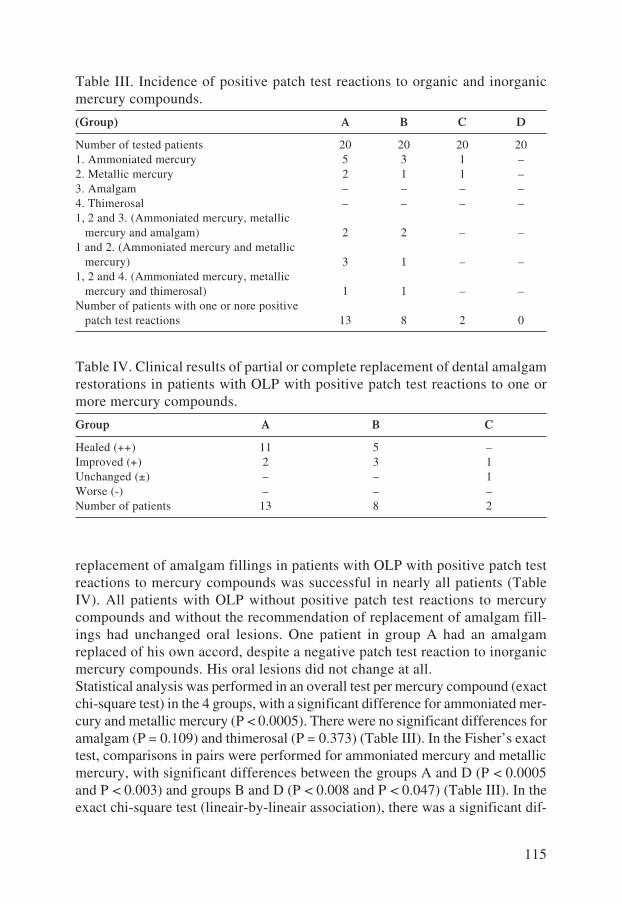
115
replacement of amalgam fillings in patients with OLP with positive patch test reactions to mercury compounds was successful in nearly all patients (Table IV). All patients with OLP without positive patch test reactions to mercury compounds and without the recommendation of replacement of amalgam fill-ings had unchanged oral lesions. One patient in group A had an amalgam replaced of his own accord, despite a negative patch test reaction to inorganic mercury compounds. His oral lesions did not change at all.Statistical analysis was performed in an overall test per mercury compound (exact chi-square test) in the 4 groups, with a significant difference for ammoniated mer-cury and metallic mercury (P < 0.0005). There were no significant differences for amalgam (P = 0.109) and thimerosal (P = 0.373) (Table III). In the Fisher’s exact test, comparisons in pairs were performed for ammoniated mercury and metallic mercury, with significant differences between the groups A and D (P < 0.0005 and P < 0.003) and groups B and D (P < 0.008 and P < 0.047) (Table III). In the exact chi-square test (lineair-by-lineair association), there was a significant dif-
Table III. Incidence of positive patch test reactions to organic and inorganic mercury compounds.
(Group) A B C D
Number of tested patients 20 20 20 201. Ammoniated mercury 5 3 1 –2. Metallic mercury 2 1 1 –3. Amalgam – – – –4. Thimerosal – – – –1, 2 and 3. (Ammoniated mercury, metallic mercury and amalgam) 2 2 – –1 and 2. (Ammoniated mercury and metallic mercury) 3 1 – –1, 2 and 4. (Ammoniated mercury, metallic mercury and thimerosal) 1 1 – –Number of patients with one or nore positive patch test reactions 13 8 2 0
Table IV. Clinical results of partial or complete replacement of dental amalgam restorations in patients with OLP with positive patch test reactions to one or more mercury compounds.
Group A B C
Healed (++) 11 5 –Improved (+) 2 3 1Unchanged (±) – – 1Worse (-) – – –Number of patients 13 8 2

116
ference (P = 0.01) in the tendency of healing whether the amalgam fillings were more associated with OLP and whether there was a positive patch test reaction to ammoniated mercury or metallic mercury (Table IV).Other positive reactions occurred in group A to nickel sulfate (2 patients), fra-grance mix (1 patient), silver nitrate (1 female patient, who also had a positive reaction to ammoniated mercury) and thiuram mix (1 patient). Other positive reactions occurred in group B to nickel sulfate (1 patient), fragrance mix (1 patient), Kathon CG (5-chloro-2-methyl-4-isothiazolin-3-one and 2-methyl-4-isothiazolin-3-one) (1 patient), potassium dichromate (1 patient) and palladium chloride (1 patient). Other positive reactions occurred in group C to nickel sulfate (1 patient), fragrance mix (1 patient), balsam of Peru (1 patient) and neomycin sulfate (1 patient). There were no significant differences between the occurrence of positive reactions and the specific variant of OLP.In group D, positive reactions occurred to nickel sulfate (4 patients), fragrance mix (3 patients), formaldehyde (2 patients), colophony (2 patients), balsam of Peru (2 patients), paraben mix (1 patient), Quaternium-15 (1 patient), cocami-dopropyl betaine (2 patients), benzocaine (1 patient) and cobalt chloride (1 patient). In 8 (35%) of 23 patients, positive patch test reactions to ammoniated mercury, metallic mercury or amalgam only occurred after the regular 3-day evaluation, with a variation of 5 to 18 days (mean, 8 days). Moreover, there were persistent patch test reactions in 6 (26%) of 23 patients, with a variation of 4 to 24 days (mean, 10 days).The effects of replacements of amalgam fillings were mostly seen after 1 to 4 months (mean, 3 months). If there was a positive effect of the replacement of amalgam fillings in patients with OLP, this effect did not change significantly during the follow-up of 2 to 7 years (mean, 4 years). In the patients without replacement of dental amalgam restorations, the lesions remained unchanged during the follow-up. No malig-nant transformation of OLP was observed in this study.
Discussion
Amalgam has been used as a dental restorative material since its inception in 1831 for people all over the world, with few adverse effects.19 It is good for dental use because it is strong, long lasting, well fitting, easy to handle, and cheap. Conventional silver amalgam fillings consist of about 50% mercury and about 50% alloy powder containing silver, tin, copper and zinc.16,32 Mercury and mercury compounds appear to be the most common allergens in amalgam, with the other metals being rarely responsible for allergic reactions.6,31 Contact sensitivity to mercury in amalgam confirmed by patch testing was previously reported by Shovelton.20 Amalgam in the oral cavity is prone to corrosion,

117
and by releasing metal ions, may be responsible for sensitization and allergic reactions (type IV, T-cell dependant).19,25 This process may lead to long-term antigenic stimulation, with mucosal changes, and ultimately to OLP. A less favorable hypothesis is that close contact between dissimilar metals (eg, amal-gam and gold) may produce different potentials and lead to electrochemical reactions (electrogalvanism), corrosion, and increased release of metal ions, also leading to mucosal changes.6,27 Martin et al reported that the presence of amalgam or gold was not associated with increased risk of OLP, but that the corrosion of amalgam and the presence of electrogalvanism from dissimi-lar dental materials in continuous contact (bimetallism) were associated with increased risk of OLP.34 The incidence of positive patch test reactions to mer-cury compounds and OLP varied considerably in several studies and depended largely on whether the mucosal lesions were in direct contact with amalgam (34%-85%) (65% [13/20] in our study) or not (0% -33%) (10% [2/20] in our study) and on the specific allergens and the concentrations that were used to detect allergy to mercury.14,21,26 Mercury is able to penetrate the healthy and the compromised mucosa, but the circumstances in which sensitization occurs are not exactly known. It is reasonable to say that possible involved allergens are dissolved and spread via saliva; therefore, mucosal reactions may extend beyond the contact areas.22,26
From our study, it appeared that there were essential differences in the incidence of contact allergy to mercury compounds between the groups A, B and C based on the topographic relationship of amalgam and OLP (especially if there is an asymmetry). Moreover, no positive patch test reactions to mercury compounds were found in group D. In the case of positive patch test reactions to ammoni-ated mercury, metallic mercury or amalgam, partial or complete replacement of amalgam fillings is beneficial after several months. This suggests that, in these cases, contact allergy is an important etiologic factor in OLP. Moreover, there is frequently more than one positive reaction to mercury compounds, which favors true sensitization and underlines the clinical importance.6,21,30 The most reliable allergen for silver dental amalgam allergy in our study was ammoni-ated mercury. Less reliable allergens are (in diminishing frequency) metallic (elementary) mercury, amalgam, and thimerosal (thiomersal). Mercury chlo-ride was used in patch tests in some studies if there was a suspicion of allergy to mercury.6,23 However, in our opinion, it is not a reliable allergen because the literature also states that it is a strong sensitizer and often produces a non-specific pustular or an irritant reaction even when diluted to a 0.05% aqueous solution. It is wise to test ionized mercury (ammoniated mercury or mercury chloride [0.1% mercury chloride in petrolatum is perhaps better than a 0.05% mercury chloride aqueous solution]) and nonionized mercury (metallic mer-cury and amalgam).23 An important issue is that positive reactions to inorganic mercury compounds may occur after the regular evaluation of 3 days, a finding in 35% (8/23) of the patients in our study. This may be a major cause of the

118
different frequencies of positive reactions in the literature. The same phenom-enon may occur with several other allergens such as gold, palladium chloride, potassium dichromate, neomycin sulfate, and paraphenylenediamine.17,21,23 Histopathological examination of the positive patch test specimens in these cases often shows lichenoid in addition to eczematous changes.21 In some cases, there is also a different time of occlusion of the allergens. In the study by Koch and Bahmer, there was an occlusion only during 1 day, with histo-pathological examination of the oral lesions performed in a small number of patients.17 The clinical relevance of the remaining positive patch test reactions to other allergens in relation to OLP is not clear. Gold, palladium, copper, silver and acrylates may also be responsible in the pathogenesis of OLP.18 We could not confirm the finding by Yiannias et al that allergy to flavorings may also be important in the pathogenesis of OLP. In our experience, this may be relevant in the diagnosis of allergic contact stomatitis.6 In the study by Yiannias et al an important aspect may be that macular erythema as a result of the patch test was regarded as positive.6 Dunsche et al reported that the removal of amalgam fillings should be recommended to all patients with symptomatic OLP associ-ated with amalgam fillings if no concomitant CLP is present, regardless of patch test results to amalgam and other inorganic mercury compounds, because almost all patients (97.1%) benefited from amalgam removal. However, patients with a positive patch test reaction to amalgam showed complete healing more frequently than those with a negative patch test reaction, and in about 8% of the patients, the lesions recurred after a mean period of 14.6 months.13 Wong and Freeman reported that, in patients with a negative patch test result who improve after amalgam replacement, mercury may act as an irritant factor in the pathogenesis of OLP.33 In our opinion, several other aspects may play a role in these cases, such as “ill-fitting” amalgam fillings leading to OLP by the isomorphic response or Koebner’s phenomenon (which is a common feature in lichen planus), “missed” late (after 3 days) positive patch test reactions to inor-ganic mercury compounds, concomitant improvement in oral hygiene during amalgam removal, possible variations in the specific allergens and concentra-tions, and the time of occlusion used in the patch tests. It is reported in the literature that the inorganic mercurials (ammoniated mer-cury, metallic mercury and amalgam) may cross-react with the organic mer-curials (thimerosal and the phenylmercuric salts).5,23 In this study, there was a cross-sensitivity between thimerosal (containing an organic mercury com-pound and a thiosalicylate) which is used as an antiseptic and as a preserva-tive, and the inorganic mercurials, ammoniated mercury and metallic mercury in two patients. The results of this study also indicate that there are several subtypes in the 3 different groups of OLP based on accompanying aspects as CLP, because none of the patients with OLP and CLP had positive reactions to one or more organic or inorganic mercury compounds. In these patients, other factors played a major role in the pathogenesis of OLP. Replacement of amal-

119
gam fillings should be undertaken for good reasons with a proper diagnosis of symptomatic OLP, because it is inconvenient, annoying, time-consuming and often expensive for the patient.10,26
Histopathological examination in OLP is important to exclude other diseases, but in this study, less specific or nonspecific changes were often noted in mucosal lesions irrespective of the clinical variant and the severity. This is in contrast to results of histopathological examination in CLP.24,28
The possible premalignant character of OLP is still debated.24 No malignancy was encountered in this study.
In conclusion, we advise that patch tests should be performed in patients with OLP especially if the lesions are in close contact with amalgam fillings and partial or complete replacement of such fillings should only be recommended if there is a positive patch test reaction to ammoniated mercury, metallic mer-cury, or amalgam and if there are no signs of concomitant CLP. This leads to healing or a significant improvement in the oral lesions in nearly all patients within several months.
References
1. Vincent SD, Fotos PG, Baker KA, Williams TP. Oral lichen planus: the clinical, historical and therapeutic features of 100 cases. Oral Surg Oral Med Oral Pathol 1990; 70: 165-71.
2. Silverman S, Gorsky M, Lozada-Nur F. A prospective follow-up study of 570 patients with oral lichen planus: persistence, remission, and malignant association. Oral Surg Oral Med Oral Pathol 1985; 60: 30-4.
3. Scully C, Beyli M, Ferreiro MC, Ficarra G, Gill Y, Griffiths M, et al. Update on oral lichen planus: etiopathogenesis and management. Crit Rev Oral Biol Med 1998; 9: 86-122.
4. Conklin RJ, Blasberg B. Oral lichen planus. Dermatol Clin 1987; 5: 663-73. 5. Scalf LA, Fowler JF, Morgan KW, Looney SW. Dental metal allergy in patients
with oral, cutaneous, and genital lichenoid reactions. Am J Contact Dermatitis 2001; 12: 146-50.
6. Yiannias JA, el-Azhary RA, Hand JH, Pakzad SY, Rogers RS. Relevant contact sensitivities in patients with the diagnosis of oral lichen planus. J Am Acad Der-matol 2000; 42: 177-82.
7. Camisa C, Taylor JS, Bernat JR, Helm TN. Contact hypersensitivity to mercury in amalgam restorations may mimic oral lichen planus. Cutis 1999; 63: 189-92.
8. Pecegueiro M, Sachse MF, Amaro J, Farinha P, Fonseca I. Oral lichen planus ver-sus oral lichenoid eruption as a manifestation of contact allergy. Contact Dermatitis 1999; 40: 333-4.
9. Thorn JJ, Holmstrup P, Rindum J, Pindborg JJ. Course of various clinical forms of oral lichen planus. A prospective follow-up study of 611 patients. J Oral Pathol 1988; 17: 213-8.
10. Laeijendecker R, Heule F, van Joost Th. [Dutch]. Oral lichen planus: clinical and immunological aspects. Ned Tijdschr Geneeskunde 1992; 136(44): 2167-71.
120
11. Eisen D. The clinical features, malignant potential, and systemic associations of oral lichen planus: a study of 723 patients. J Am Acad Dermatol 2002; 46: 207-14.
12. Boyd AS, Neldner KH. Lichen planus. J Am Acad Dermatol 1991; 25: 593-619.13. Dunsche A, Kastel I, Terheyden H, Springer ING, Cristophers E, Brasch J. Oral
lichenoid reactions associated with amalgam: improvement after amalgam removal. Br J Dermatol 2003; 148(1): 70-6.
14. Hietanen J, Pihlman K, Forstrom L, Linder E, Reunala T. No evidence of hypersen-sitivity to dental restorative metals in oral lichen planus. Scand J Dent Res 1987; 95: 320-7.
15. Laine J, Kalimo K, Forssell H, Happonen RP. Resolution of oral lichenoid lesions after replacement of amalgam restorations in patients allergic to mercury com-pounds. Br J Dermatol 1992; 126: 10-5.
16. Jameson MW, Kardos TB, Kirk EEJ, Ferguson MM. Mucosal reactions to amalgam restorations. J Oral Rehabilitation 1990; 17: 293-301.
17. Koch P, Bahmer FA. Oral lesions and symptoms related to metals used in dental restorations: a clinical, allergological, and histologic study. J Am Acad Dermatol 1999; 41: 422-30.
18. Laeijendecker R, van Joost Th. Oral manifestations of gold allergy. J Am Acad Dermatol 1994; 30: 205-9.
19. Smart ER, Macleod RI, Lawrence CM. Resolution of lichen planus following removal of amalgam restorations in patients with proven allergy to mercury salts: a pilot study. Br Dent J 1995; 178: 108-12.
20. Shovelton DS. Silver amalgam and mercury allergy. Oral Surg Oral Med Oral Path 1968; 25: 29-30.
21. Mollaoglu N. Oral lichen planus: a review. Br J Oral Maxillofac Surg 2000; 38: 370-7.
22. Bolewska J, Holmstrup P, Moller-Madsen B, Kenrad B, Danscher G. Amalgam associated mercury accumulations in normal oral mucosa, oral mucosal lesions of lichen planus and contact lesions associated with amalgam. J Oral Pathol Med 1990; 19: 39-42.
23. Rietschel RL, Fowler JF. Mercury. In: Fisher’s Contact Dermatitis. 5th edn. Lip-pincott Williams & Wilkins, Philadelphia, U.S.A.; 2001: 153-4, 636, 667-9, 740, 779.
24. van der Meij EH, Schepman KP, Smeele LE, van der Wal JE, Bezemer PD, van der Waal I. A review of the recent literature regarding malignant transformation of oral lichen planus. Oral Surg Oral Med Oral Pathol Oral Radiol Endod 1999; 88: 307-10.
25. Ostman PO, Anneroth G, Skoglund A. Oral lichen planus lesions in contact with amalgam fillings: a clinical, histologic, and immunohistochemical study. Scand J Dent Res 1994; 102: 172-9.
26. Laine J, Kalimo K, Happonen RP. Contact allergy to dental restorative materials in patients with oral lichenoid lesions. Contact Dermatitis 1997; 36: 141-6.
27. Lundstrom IMC. Allergy and corrosion of dental materials in patients with oral lichen planus. Int J Oral Surg 1984; 13: 16-24.
28. van der Meij EH, Reibel J, Slootweg PJ, van der Wal JE, de Jong WFB, van der Waal I. Interobserver and intraobserver variability in the histologic assessment of oral lichen planus. J Oral Pathol Med 1999; 28: 274-7.
29. Bolewska J, Hansen HJ, Holmstrup P, Pindborg JJ, Sangerup M. Oral mucosal lesions related to silver amalgam restorations. Oral Surg Oral Med Oral Pathol 1990; 70: 55-8.

121
30. van Joost Th, Laeijendecker R. [Dutch]. Amalgam. VI. Allergy for mercury in den-tal materials; oral and systemic reactions. Ned Tijdschr Tandheelkd 1993; 100(7): 303-7.
31. Pang BK, Freeman S. Oral lichenoid lesions caused by allergy to mercury in amal-gam fillings. Contact Dermatitis 1995; 33: 423-7.
32. Bircher AJ, von Schulthess A, Henning G. Oral lichenoid lesions and mercury sensitivity. Contact Dermatitis 1993: 29: 275-6.
33. Wong L, Freeman S. Oral lichenoid lesions (OLL) and mercury in amalgam fillings. Contact Dermatitis 2003; 48(2): 74-9.
34. Martin MD, Broughton S, Drangsholt M. Oral lichen planus and dental materials: a case-control study. Contact Dermatitis 2003; 48(6): 331-6.


B: LICHENOID CONTACT STOMATITIS.
IS INORGANIC MERCURY THE CULPRIT?
R.S. ROGERS III, MD1
A.J. BRUCE, MD1
ARCHIVES OF DERMATOLOGY 2004; 140: 1524-5.
(EDITORIAL)
Permission granted. Copyright© (2004), American Medical Association. All rights reserved.
Financial disclosure: none.
1 Department of Dermatology, Mayo Clinic, 200 First St SW, Rochester, MN 55905-0002, U.S.A. ([email protected]).


125
The article by Laeijendecker et al1 in this month’s Archives on the relationship between oral lesions of lichen planus and allergy to amalgam dental restora-tions is another attempt to resolve some of the controversies surrounding this topic. The issues are several. First, what should we call these lesions: oral lichenoid lesions (OLL), oral lichenoid reactions, oral lichenoid tissue reac-tions, lichenoid contact stomatitis, lichen planus-like lesions, or oral lichen planus (OLP)?Second, is the clinical benefit from removal related to the removal of a long-term source of antigen stimulation, producing a delayed hypersensitivity reac-tion, or to the removal of a source of trauma such as friction or bimetallism, producing a Koebner phenomenon?Third, what role does patch testing play?
Nomenclature is always fun and often controversial. Some would be rigid and consider that OLP represents only those lesions for which no trigger can be identified and are “idiopathic”. This excludes drug-induced OLP and OLP associated with chronic liver disease, food or flavor allergies, hypertension, and diabetes mellitus, as well as other conditions. The oral mucosa has, like the skin, a limited repertoire of reactions, one of which is a hyperkeratotic, white, thickened, inflammatory reaction said to be “lichenoid”. Therefore, the terms OLL, oral lichenoid reactions, oral lichenoid tissue reactions, lichenoid contact stomatitis, lichen planus-like lesions, and OLP have all been used in describing this reaction.It is difficult, if not impossible, to distinguish these lesions from one another.2-6 Dunsche et al2 concluded that an unambiguous distinction between the lesions of amalgam-associated tissue changes compared with OLP in edentulous patients without dental amalgam was impossible not only clinically but also histologi-cally. Therefore, all these terms are describing the same tissue changes. We prefer the term lichenoid contact stomatitis7 when the allergen and the clinical relevance are identified and OLP as the overarching rubric.Is inorganic mercury, contained in dental amalgams, a sensitizer, and can it cause allergic contact stomatitis or lichenoid contact stomatitis? Fern-strom et al8 were the first to claim a connection between type IV allergy to mercurial compounds released from dental amalgam and OLP. Others have agreed.3,5-17
Koch and Bahmer17 reported that 15 (78.9%) of 19 patients with OLL in close apposition to amalgam dental fillings were sensitized to inorganic mercury compounds. They noted that positive patch test readings may be delayed until days 10 or 17 and that patients often had positive reactions to multiple mercury allergens. Furthermore, they noted that positive patch test reactions were less common in those patients with OLL distant from amalgam (3/25 [12.0%]), patients with other oral diseases (2/29 [6.9%]), and control subjects (2/59 [3.4%]).

126
These authors noted that 13 of 15 patients with positive patch test reactions for inorganic mercury experienced OLL clearance when the dental amalgam was removed.Scalf et al3 studied 51 patients with OLL, of whom 38 had 1 or more positive patch test reactions to a dental metal and 28 had a positive patch test reaction to inorganic mercury. When the dental metals were removed in 9 patients with positive patch test reactions, all experienced clearance, as did 1 patient with negative patch test reactions. On the other hand, it has been proposed that bimetallism or the continuous con-tact of dissimilar dental metals produces electrogalvanism, which might pro-duce OLP by a Koebner phenomenon.10 The restoration may become worn and rough, leading to friction and the Koebner phenomenon. Wong and Freeman18 reported that patients who improve after amalgam dental restoration removal and who have negative patch test reactions may be responding to the removal of an irritant, thereby reducing the element of the Koebner phenomenon.The study by Laeijendecker et al1 concludes that allergic contact stomatitis is important in the pathogenesis of OLP, especially when the lesions of OLP are in apposition to dental amalgam restorations. They note that those patients with positive patch test reactions to inorganic mercury whose amalgams were in close apposition had uniformly excellent outcomes after removal of dental amalgams and replacement with composite resins, glass ionomers, ceramics (porcelain), or gold, with complete clearing after a mean of 3 months in 11 of 13 patients and substantial improvement in the remaining 2 patients. These patients remained clear of lesions in follow-up for 2 to 7 years (mean, 4 years).In addition, patients with the OLP lesions in direct apposition (tongue and buccal mucosal surfaces) to the amalgam dental restorations were more likely to have positive patch test reactions and improvement when the amalgam was removed. Those with the amalgam within 1.0 cm had fewer positive patch test reactions, but among patients with positive reactions, the results were excellent, with 5 of 8 healed and the remaining 3 substantially improved. Notably, all patients with OLP without positive patch test reactions to mercury compounds and who did not have replacement of their amalgam restorations underwent no change in their condition during the long follow-up. Laeijendecker et al1 conclude that, if there is a positive patch test reaction to inorganic mercury, the replacement of dental amalgam restorations is beneficial and the result is noted within 1 to 4 months. The authors state that contact allergy is an important etiological factor in OLP.Dunsche et al2 evaluated 134 patients who had 467 individual oral lichenoid reactions. Biopsy specimens of 159 oral lichenoid reactions were similar to 47 OLP specimens in edentulous patients, as already noted. Of these patients, 119 had patch tests to the inorganic mercury series performed. Among them, 33 (27.7%) had positive patch test reactions. Notably, of the 134 patients, 29 refused patch testing and did not have amalgam dental restorations removed.

127
They served as controls. Only 2 (6.9%) of the 29 “control” patients improved with long-term follow-up. Amalgam dental restorations were removed in 105 patients regardless of patch test results. Restorations were replaced with gold, composite resins, glass ionomers, ceramics (porcelain), metal or ceramic crowns, or titanium. Clinical evaluations were carried out at 6 and 12 months and yearly thereafter.Several important observations were made by Dunsche and coauthors.2 Among patients whose amalgam dental restorations were removed, 97.1% (102/105) improved regardless of patch test results. Those with positive patch test reac-tions improved most. Overall, 31 (29.5%) of 105 had complete clearance. The rest demonstrated partial improvement, with a “subtotal cure” in 8 (7.6%) and “improvement” in 63 (60.0%). Those with tongue lesions (in apposition with amalgam) also experienced greater improvement. After complete remission, 8 patients (7.6%) had a recurrence during a mean of 14.6 months. These authors noted that amalgam dental restoration removal had little effect on patients with oral and cutaneous lichen planus. Because 97.1% of patients benefited from the removal of amalgam dental restorations regardless of patch test results, Dun-sche et al recommend removal of amalgam dental restorations if no cutaneous lichen planus is present. What is the value of patch testing in patients with lesions of OLP in close appo-sition to dental metal restorations? There may be less value if cutaneous lichen planus coexists. If the clinical manifestations are limited to the oral mucosa, patch testing to dental metals serves several functions.First, positive patch test reactions to inorganic mercury compounds identify a subset of OLP patients with lichenoid contact stomatitis who have a high proba-bility of benefiting from amalgam dental restorations removal, particularly if the oral lesions are in close apposition and involve the tongue or buccal mucosa.Second, positive patch test reactions to inorganic mercury compounds identify a subset of OLP patients who are likely to benefit from removal of amalgam dental restorations that are within 1.0 cm of lesions of OLP.Third, patch testing for all dental metals can identify not only amalgam dental metal hypersensitivity but also dental metal hypersensitivity to gold, palladium, beryllium, and other dental metals that can cause a lichenoid contact stomati-tis.3,4,6,7,16,17
Fourth, patch test reactions should be read at 7, 10, or 14 days to identify “late” reactions that occur with dental metals and may be missed otherwise.Finally, because lichenoid contact stomatitis can be caused by other dental metals such as gold and palladium, patch testing can be used to exclude these metals when choosing a dental metal (or other) restoration to replace the offending one.Removal and replacement of dental metal restorations is a costly and time-consuming procedure. Removal should be considered based on objective evi-dence of symptomatic lichenoid tissue changes in apposition to or close to a dental metal restoration. The strong probability of long-term clearing, with a

128
small probability of relapse of the lichenoid tissue changes, can be assessed by performing patch testing to dental metals. Laeijendecker and colleagues1 are to be commended for their careful study and follow-up of patients with OLP associated with amalgam dental restorations.
References
1. Laeijendecker R, Dekker SK, Burger PM, Mulder PGH, van Joost Th, Neumann HAM. Oral lichen planus and allergy to dental amalgam restorations. Arch Der-matol 2004; 140: 1434-8.
2. Dunsche A, Kastel I, Terheyden H, Springer ING, Christophers E, Brasch J. Oral lichenoid reactions associated with amalgam. Br J Dermatol 2003; 148: 70-6.
3. Scalf LA, Fowler JF Jr, Morgan KW, Looney SW. Dental metal allergy in patients with oral, cutaneous, and genital lichenoid reactions. Am J Contact Dermatitis 2001; 12: 146-50.
4. Yiannias JA, el-Azhary RA, Hand JH, Pakzad SY, Rogers RS III. Relevant contact sensitivities in patients with the diagnosis of oral lichen planus. J Am Acad Der-matol 2000; 42: 177-82.
5. Camisa C, Taylor JS, Bernat JR, Helm TN. Contact hypersensitivity to mercury in amalgam restorations may mimic oral lichen planus. Cutis 1999; 63: 189-92.
6. Pecegueiro M, Sachse MF, Amaro J, Farinha P, Fonseca I. Oral lichen planus ver-sus oral lichenoid eruption as a manifestation of contact allergy. Contact Dermatitis 1999; 40: 333-4.
7. Rogers RS III. Lichenoid contact stomatitis. Ann Dermatol Venereol 2002; 129. Abstract. SY0805.
8. Fernstrom AIB, Frykholm KO, Huldt S. Mercury allergy with eczematous derma-titis due to silver-amalgam fillings. Br Dent J 1962; 18: 204-6.
9. Shovelton DS. Silver amalgam and mercury allergy. Oral Surg Oral Med Oral Pathol 1968; 25: 29-30.
10. Lundstrom IM. Allergy and corrosion of dental materials in patients with oral lichen planus. Int J Oral Surg 1984; 13: 16-24.
11. Jameson MW, Kardos TB, Kirk EE, Ferguson MM. Mucosal reactions to amalgam restorations. J Oral Rehabil 1990; 17: 293-301.
12. Bolewska J, Jansen HJ, Holmstrup P et al. Oral mucosal lesions related to silver amalgam restorations. Oral Surg Oral Med Oral Pathol 1990; 70: 55-8.
13. Holmstrup P. Oral mucosa and skin reactions related to amalgam. Adv Dent Res 1992; 6: 120-4.
14. Ostman PO, Anneroth G, Skoglund A. Oral lichen planus lesions in contact with amalgam fillings. Scand J Dent Res 1994; 102: 172-9.
15. Smart ER, Macleod RI, Lawrence CM. Resolution of lichen planus following removal of amalgam restorations in patients with proven allergy to mercury salts: a pilot study. Br Dent J 1995; 178: 108-12.
16. Laine J, Kalimo K, Happonen RP. Contact allergy to dental restorative materials in patients with oral lichenoid lesions. Contact Dermatitis 1997; 36: 141-6.
17 Koch P, Bahmer FA. Oral lesions and symptoms related to metals used in dental restorations. J Am Acad Dermatol 1999; 41: 422-30.
18. Wong L, Freeman S. Oral lichenoid lesions (OLL) and mercury in amalgam fillings. Contact Dermatitis 2003; 48: 74-9.
CHAPTER 4
A. ORAL MANIFESTATIONS OF
GOLD ALLERGY
R. LAEIJENDECKER, MD1 Th. VAN JOOST, MD, PhD1
JOURNAL OF THE AMERICAN ACADEMY OF DERMATOLOGY 1994; 30: 205-9.
1 Department of Dermatology and Venereology, Erasmus University Rotterdam, University Hospital Rotterdam - Dijkzigt, Dr. Molewaterplein 40, 3015 GD Rotterdam, The Nether-lands.

130
ABSTRACT
Background: Sensitization to gold in large populations suspected of clinical allergy to this metal has not been reported.
Objective: Two hundred patients with symptoms of persistent oral muco-sal or cutaneous lesions that were possibly related to allergy to constituents of gold alloys or gold jewelry were patch tested to determine the frequency of sensitization.
Methods: Patch testing was performed with the European standard series and a series of dental materials including three different salts of gold. A persistent papular reaction to gold(tri)chloride was considered to be a positive reaction.
Results: In 17 (8.5%) patients, all women (mean age 50 years), persis-tent papular patch test reactions to both 0.5% and 1% gold- (tri)chloride were observed. In 5 of the 7 patients with oral lichen planus (OLP) and in 1 of the 6 patients with the burning mouth syndrome (BMS), dental gold was replaced. A significant but variable improvement was observed particularly in patients with OLP. The patients were sensitized to 0.5% gold(tri)chloride in all cases in which gold was replaced and the lesions improved. One patient with allergic contact stomatitis and one patient with allergic contact dermatitis healed completely after gold had been replaced.
Conclusions: Sensitization to gold should be considered as a possible cause of allergic contact dermatitis, allergic contact stomatitis and also as a pathogenic or triggering factor in OLP.

131
Introduction
Gold salts are potentially strong sensitizers as shown by the “maximization test”1, but delayed hypersensitivity to metallic gold and to gold salts has been reported infrequently.1-5 One reason could be that an insufficient amount of gold salt leaches from materials resulting in an inadequate concentration of antigen to produce clinical allergy. Patch testing with gold presents problems with irritant and false negative reac-tions. Various gold salts such as gold(tri)chloride (0.5%, 1% and 2% chloro-auric acid), 0.5% aurothiomalate, 0.5% gold sodium thiosulfate, 0.1% sodium chloroaurate, 0.002% potassium dicyanoaurate and 1% gold potassium chloride have been used and recommended to establish “true allergy” to gold in patch test procedures.1,3,4,6,7 It has been suggested that true allergy to gold may only be established when there are definite reactions to at least two gold salts coupled with a relevant clinical history.1 Fowler considered gold sodium thio-sulfate (0.5% in petrolatum) and potassium dicyanoaurate (0.002% in water) as reliable markers for gold allergy.3 Fisher regarded gold(tri)chloride at a con-centration of 1% or lower in water as a reliable test allergen.4 Positive patch test reactions of the eczematous type are possible, but in the majority of cases persistent infiltrated papular reactions have been observed that are considered to be primarily of an immunological origin.4,6,8,10,11 Probably because of accumulation of the allergen in the dermis, gold allergic contact dermatitis may show a marked dermal involvement with little or no changes in the epidermis and may persist for a long period even after exposure to gold is avoided.4,7,8
Gold is mostly alloyed with other metals.1,4 High carat yellow gold contains copper and silver. Low carat yellow gold also contains zinc and a small amount of nickel. White gold usually contains palladium and nickel. Nickel in white gold alloys may be responsible for “white gold dermatitis”.1-4 Dental gold is an alloy that may also contain uncommon, but definite sensitizers such as cop-per, platinum and palladium.5 It is important to establish which particular gold alloy has been used. Therefore, patch testing with a range of metals usually present in gold alloys is essential.5,12-17 Oral lesions attributed to gold allergy are described as stomatitis and lichen planus or lichenoid reactions in close approximation to gold dentures.5,15
The aim of this study was to determine the role of gold as an allergen in 200 selected patients.
Materials and methods
The population studied consisted of 200 patients. The majority was clinically diagnosed as oral lichen planus (OLP), burning mouth syndrome (BMS), aller-

132
gic contact stomatitis (ACS) and less frequently allergic contact dermatitis or contact urticaria. They were suspected to have an allergy to materials contain-ing gold. The patients were patch tested with the European standard series1 and with the series of metals16,17 used in case of a possible allergy to metals other than nickel, cobalt and chromium (Table I). The gold salts used as test allergens were 0.5% and 1% gold(tri)chloride in water, 0.001% potassium dicyanoaurate in water and 0.1% sodium tetrachloroaurate in water. The gold(tri)chloride test solution did not contain nickel as a contaminant and was prepared by dissolving AuCl3 in water resulting in an aqueous solution of HAuCl4 that was highly acidic. Patch tests were performed with Finn-chambers and read after 48 and 72 hours. In addition, the patients were instructed to contact us when positive reactions to allergens developed at a later stage. A persistent papular reaction was considered to be positive.When a clinical correlation was suspected between a positive patch test reac-tion to gold and the oral symptoms, especially when gold was in close con-tact with the oral lesions, the patient was advised to have replaced the dental gold alloy. If patch tests to other allergens in the dental materials were found, patients were advised to have these allergens substituted as well. The replace-ments of gold consisted of composite resin materials, glass ionomer and por-celain. After a follow-up period varying between 2 months and 6 years (mean 2 years) the patients were re-evaluated. Their own subjective assessment by
Table I. Dental material test series.
1 Gold(tri)chloride 0.5% in aqua 2 Gold(tri)chloride 1% in aqua 3 Sodium tetrachloroaurate 0.1% in aqua 4 Potassium dicyanoaurate 0.001% in aqua 5 Nickel sulfate 5% in aqua 6 Cobalt chloride 1% in aqua 7 Magnesium sulfate 5% in aqua 8 Potassium dichromate 0.5% in aqua 9 Palladium sulfate 1.5% in aqua 10 Ferric chloride 2% in aqua 11 Copper sulfate 2% in aqua 12 Palladium chloride 2.5% in aqua 13 Zinc chloride 2.0% in aqua 14 Gallium chloride 10% in aqua 15 Phenylmercuric nitrate 0.05% petrolatum 16 Mercury (metallic) 0.5% petrolatum 17 Thimerosal 0.1% petrolatum 18 Stannous chloride 0.5% in aqua 19 Silver nitrate 1% in aqua 20 Toluene sulfonamide 0.1% alcohol

133
comparing the symptoms before and after gold had been replaced was graded as symptom-free, improved, unchanged or worse. The lesions were objectively evaluated and compared with the pre-treatment condition and graded as healed, improved, unchanged or worse.
Results
In 17 (8.5%) of the 200 patients a positive patch test reaction to 1% and 0.5% gold(tri)chloride was observed (Table II). In all, except one case, the reactions showed a moderate or marked induration with persistent papules varying in duration from about one to several weeks (mean 4 weeks). One positive patch test reaction even persisted for 3 months. In 3 patients, the positive patch tests with 0.5% and 1% gold(tri)chloride developed after the regular 3-day evalua-tion (after 5, 6 and 14 days respectively). Another patient developed a reaction to 1% gold(tri)chloride that was characterized by infiltration and purpura after one month. Histologically, the reaction was classified as a contact allergic reaction with vasculitis. Positive reactions to sodium tetrachloroaurate and potassium dicyanoaurate were not observed in any of the patients.Seven (41.2%) of the 17 patients who were reactive to gold(tri)chloride had positive reactions to other metals including palladium (4 patients), nickel (3 patients), cobalt (2 patients) and mercury (1 patient). Sensitization to gold(tri)chloride without other positive patch tests was observed in 8 (47.1%) patients. All 17 patients were women, aged 28 to 71 years (mean 50 years). Within this group, 7 (41.2 %) patients had OLP and 6 (35.3%) patients had BMS. The remaining 4 (23.5%) patients had allergic contact stomatitis, allergic contact dermatitis, chronic urticaria or a relapsing angioedema. In 7 patients aged 39 to 70 years (mean 49 years) with a persistent papular patch test to gold(tri)chloride, the clinical diagnosis of OLP (5 with erosive OLP, 1 with hyperkeratotic OLP and 1 with a plurimucosal form of lichen planus) was confirmed by histopathological examination (Table II). Five patients with OLP had gold-containing dental alloys corresponding to the site of the oral lesions. Four patients with OLP had also positive patch test reactions to cobalt and mercury (1 patient), nickel (1 patient) and palladium (2 patients). The 7 patients were treated with various agents including topical corticosteroids, topical retinoids and cyclosporin mouthwash with a mean period of about 2 years (range 3 months to 4 years). Three patients were treated with systemic retinoids (mean dose 0.5 mg/kg/day) for about 3 months and 2 patients were treated with cyclosporin mouthwash for 2 months. However, no significant long-term improvement was achieved.Gold-containing dental materials were replaced in 5 patients with OLP (in 2 patients together with replacements of amalgam (cobalt and mercury) and

134
Table II. Clinical characteristics of the patients.
Pat. Age 48 72 >72 Course (Diagnosis) Positive patch tests hrs hrs hrs Therapy Subj/ Remarks Obj
1. 39 goldchloride 0.5% – 2+ Removal of + / + No dental gold.(hyper- goldchloride 1.0% – 2+ dental Complaints ofkeratotic palladium sulfate – 2+ palladium gold earringsOLP) p.toluenediamine – 2+ p.aminoazobenzene 3+ 4+ p.tert.butyl. formal. + 3+ 2. 52 goldchloride 0.5% – 2+ Replacement of + / + No compaints(Plurimuco- goldchloride 1.0% – 2+ gold and nickel of gold jewelrysal LP) nickel sulfate + 2+ containing dental colofonium – 2+ material. p.tert.butyl. formal. – 3+ Vulva: topical + / + woodtar mix – 2+ corticosteroids
3. 45 goldchloride 0.5% – 2+ Topical: cortico- +/- +/- No dental gold.(erosive goldchloride 1.0% – 2+ steroids, cyclo- No complaintsOLP) fragrance mix 2+ 3+ sporin. Systemic: of gold jewelry cinnamaldehyde 2+ 3+ vit A acid, isoniazide
4. 52 goldchloride 0.5% – 2+ Replacement of + / + No complaints(erosive goldchloride 1.0% 2+ 3+ dental gold of gold jewelryOLP) palladium chloride – 2+ (crown)
5. 42 goldchloride 0.5% – – 2+ Replacement of + / + No complaints(erosive goldchloride 1.0% – – 2+ dental gold of gold jewelryOLP) p.aminobenzene – – 2+
6. 70 goldchloride 0.5% – 2+ Replacement of ++/++ Complaints of(erosive goldchloride 1.0% – 2+ dental gold gold earringsOLP) (crowns)
7. 43 goldchloride 0.5% – + 2+ Replacement of + / + No complaints(erosive goldchloride 1.0% – + 2+ dental gold and of gold jewelryOLP) mercury 0.5% – – 2+ amalgam am.mercury 1% – – 2+ cobalt chloride – 2+ 2+
8. 53 goldchloride 0.5% – 2+ Replacement of + / ? No complaints(BMS) goldchloride 1.0% – 2+ . dental gold of gold jewelry (crowns)
9. 28 goldchloride 0.5% – 2+ Replacement of + / ? No dental gold.(BMS) goldchloride 1.0% – 2+ nickel and cobalt Complaints of nickel sulfate + 2+ containing dental gold jewelry cobalt chloride + 2+ material
10. 54 goldchloride 0.5% – – 2+ No replacement +/- ? Complaints of(BMS) goldchloride 1.0% – tox. 2+ gold jewelry vasc ulitis

135
nickel, respectively). In 1 patient with OLP, palladium was replaced. All patients with replacements showed significant subjective and objective improvement after 2 weeks to 6 months (mean 3 months) and remained so afterwards (mean follow-up time 1 year, range 6 months to 2 years). In 1 patient, OLP healed completely after replacement. Two of the 7 OLP patients with a gold allergy, suffered concomitantly from allergic contact dermatitis when they wore gold earrings. In the 5 patients with OLP and dental gold, OLP developed after 3 weeks to 3 years (mean 4 months) after placement of dental gold. None of these patients were on any systemic medication that could be responsible for an oral lichenoid drug erup-tion. One patient had plurimucosal lichen planus and had sensitization to gold
Pat. Age 48 72 >72 Course (Diagnosis) Positive patch tests hrs hrs hrs Therapy Subj/ Remarks Obj
11. 71 goldchloride 0.5% – 2+ No replacement +/- ? No complaints(BMS) goldchloride 1.0% – 2+ of gold jewelry
12. 65 goldchloride 0.5% – 2+ No replacement +/- ? No complaints(BMS) goldchloride 1.0% – 2+ of gold jewelry
13. 58 goldchloride 0.5% – – 2+ Folic acid 10 +/- ? No dental gold.(BMS) goldchloride 1.0% – – 2+ mg/day (minimal No complaints deficiency) of gold jewelry
14. 51 goldchloride 0.5% – 2+ Systemic anti- + / + No dental gold.(angio- goldchloride 1.0% 2+ 3+ histamines. Complaints ofedema) palladium sulfate – 2+ No replacement gold jewelry necessary
15. 53 goldchloride 0.5% – 2+ Replacement of ++/++ No dental gold.(contact goldchloride 1.0% – 2+ gold jewelry dermatitis)
16. 29 goldchloride 0.5% – 2+ Systemic anti- + / + No complaints(chronic goldchloride 1.0% – 2+ histamines. No of gold jewelryurticaria) palladium chloride – 3+ request of dental nickel sulfate 2+ 3+ gold replacement.
17. 48 goldchloride 0.5% – 2+ Replacement of ++/++ No complaints(contact goldchloride 1.0% – 2+ dental gold of gold jewelrystomatitis) (crown)
Evaluation of patch tests after 48, 72 and > 72 hrs (hrs = hours): – = negative, + = erythema (no allergy), 2+ = erythema with induration (= contact allergy), 3+ = erythema with vesicles (= contact allergy).Subj (= subjective, symptoms). Obj (= objective, signs): +/- = unchanged, + = improved++ = healed. ? = uncertain. p = para, Pat = patient number, am.= ammoniated, p-tert.butyl.formal = paratertiary butylphenol formaldehyde resin.
136
and nickel. The oral manifestations improved significantly after both dental gold and nickel were replaced. However, the lesions on the vulva improved only after treatment with topical corticosteroids. Six patients aged 28 to 71 years (mean 55 years) with a positive patch test reaction to gold had a diagnosis of BMS (Table II). In this group, all patients had an isolated positive patch test reaction to gold(tri)chloride, except one who also had a positive patch test reaction to nickel and cobalt. The symptoms had persisted for several years and topical treatment had been uneffective. Four patients with BMS had gold containing dental alloys. Dental gold was replaced in one patient and nickel and cobalt containing dental materials were replaced in another patient, who had no dental gold. In both patients, there was an improvement in the symptoms some weeks after replacement. In 3 patients with BMS, dental gold had not yet been replaced and their symptoms remained unchanged. The 4 patients with dental gold began to complain of BMS after 1 month to 4 years (mean 6 months) after receiving gold alloys in the mouth. One patient was clinically suspected to have allergic contact stomatitis for one year. This patient had an isolated positive patch test reaction to gold- (tri)chloride (1% and 0.5%). Allergic contact stomatitis developed 2 months after a gold crown had been placed. The lesions resolved completely three weeks after the gold crown was replaced. This patient had no complaints when gold jewelry was worn (Table II). A second patient had a relapsing angioedema of the mucous membranes of the mouth and the tongue for 2 years and also had a positive patch test reaction to palladium. She had no dental gold and treatment with systemic antihistamines was effective. She also complained of an itchy contact dermatitis at sites where gold jewelry was worn (Table II). Another patient with gold allergy had chronic urticaria of unknown cause for 5 years. There were also positive patch test reactions to nickel and palladium. Treatment with an antihistamine was effective.A fourth patient without dental gold only had a positive patch test to gold-(tri)chloride and had allergic contact dermatitis on the arms, the hands and the legs for several months even at sites not in close contact with gold jewelry. The eruptions resolved completely within 4 weeks without additional treatment once gold jewelry was avoided (Table II).
Discussion
The objective and subjective improvement or healing in 7 patients (5 with OLP, 1 with BMS and 1 with allergic contact stomatitis) after dental gold alloys were replaced and the resolution of allergic contact dermatitis after avoiding gold jewelry in 1 patient, supported the clinical value of persistent papular patch test reactions as diagnostic markers of true gold sensitization.

137
Metallic gold alone is not a reliable test allergen, because the time required for converting metallic gold into gold salts is longer than 48 hours.3,5 Irritant reaction to 1% gold(tri)chloride is considered to be caused by the acidic nature of the vehicle.1,4,5,13 In a small number of patients, the patch test with 1% gold(tri)chloride resulted in a slight irritant reaction that disappeared within a few days. Irritant reaction to 0.5% gold(tri)chloride was not seen in any of the patients tested. All patients with a positive patch test with 1% gold(tri)chloride also reacted adequately to 0.5% gold(tri)chloride. Therefore, the latter concen-tration should be considered as the concentration of choice for patch testing. Positive patch test reactions to gold(tri)chloride persisted for periods of one to several weeks. We hypothesize that this type of patch test reaction together with a relevant clinical history may be important in the clinical diagnosis of gold allergy. In nearly all patients, clinically relevant sensitization at sites of contact (oral cavity or skin) was obvious. Late positive patch test reactions to gold(tri)chloride may occur and can easily be missed if the last reading is done at 72 hours. In 1 patient, a positive patch test reaction developed after 14 days, whereas in another patient it developed after 1 month. It seems likely that the latter patient was sensitized during the patch test procedure, and in whom we observed the clinical and histological features of a allergic contact reaction with vasculitis. Pseudolymphomatous lesions induced by gold have been reported in a patient in whom a persistent infiltrative patch test reaction to 1% gold(tri)chloride with pseudolymphomatous features was observed.10 Lymphocytoma cutis of the earlobes induced by gold earrings had been reported.18 Only 5 (33.3%) out of the 15 patients with oral symptoms and gold sensitization developed contact dermatitis when they wore gold jewelry. It is likely that the humid environment, pressure and friction in the oral cavity permit slow leach-ing of gold salts capable of provoking an allergic reaction in sensitized indi-viduals.5 This process of leaching may be hastened by galvanic forces induced by different metals in the mouth.5 Penetration into the mucosal membranes is easier than into the skin and this may explain the concomitant absence of allergic reactions to gold jewelry and the presence of only oral symptoms in most patients.1,2 The development of oral mucosal disorders because of contact sensitization to allergic dental materials other than gold (especially amalgam) and the benefit of replacing such materials was reported.14,15,17,19,20 An oral lichenoid drug eruption caused by systemic gold therapy has been described.21-23 Data in favor of T-cell mediated injury in lichen planus have been published.23 The results of the present study indicate that apparently OLP may be related to an allergy to dental gold. In all 8 patients, including the patient with allergic contact dermatitis, in whom metallic gold was replaced, the lesions improved or resolved completely. Oral symptoms also improved significantly in 2 patients in whom palladium, nickel

138
and cobalt containing dental materials were replaced. Only in a few patients out of the group of 183 patients in whom no positive patch test reactions to gold(tri)chloride were observed, sensitization to nickel, cobalt, chromium, pal-ladium, mercury or dental acrylates was observed.17 In 2 patients with OLP, dental replacements included gold and other metals. Therefore, the role of gold replacement could not be established with certainty in those cases. Replacing dentures, crowns or bridges is an expensive intervention and there-fore, the decision to do so must be based on definite reasons. Replacement should only be recommended to patients with chronic disabling oral symptoms, who respond insufficiently to therapy, who have positive patch test reactions to dental metal(s) and in whom there is a relation between the site of the lesions and the site of the specific dental materials.2,5,14,15 However, in spite of these criteria, there is no certainty that BMS will resolve completely after removal of the allergens. Even when gold sensitization is a prime suspect other still unidentified factors may be responsible for perpetuating the disease.17
All our patients with gold allergy were women. It is possible that this may be partly explained by the manner of sensitization (in analogy to nickel), namely early and frequent contact of the skin with jewelry (golden earrings in particular). Sensitization may also be induced by exposure to dental gold (salts), gold injec-tions and gold salts used in certain professions.1,4,10 None of the patients in this study had professional exposure to gold salts. Only one patient with BMS had received several gold injections for arthritis in the past. None of the patients in this study had a positive reaction to sodium tetrachloroaurate 0.1% aq or potas-sium dicyanoaurate 0.001% aq (Table II). Two patients with BMS in whom posi-tive reactions to gold(tri)chloride occured after 14 and 30 days may have been sensitized during patch testing. No replacement was done in these patients.Histologically, allergic gold dermatitis is characterized by marked dermal involvement in the near absence of epidermal changes.4,8 These features, also observed by us in biopsy specimens of positive patch tests, may have been caused by a persistent presence of gold in the dermis and may explain the atypi-cal, non-eczematous appearance of the positive patch tests.
We believe that immune-mediated injury in the oral cavity caused by local hypersensitivity to gold salts is underestimated. In patients with gold dental alloys and mucosal disorders of suspected immune-mediated origin, especially allergic contact stomatitis and OLP, patch testing with gold salts in addition to other metal allergens should be performed.
References
1. Cronin E. Metals. In: Cronin E ed. Contact Dermatitis, Edinburgh, London and New York. Churchill Livingstone; 1980: 328-35.

139
2. Fregert S, Kollander M, Poulsen J. Allergic contact stomatitis from gold dentures. Contact Dermatitis 1979; 5: 63-4.
3. Fowler JF. Selection of patch test materials for gold allergy. Contact Dermatitis 1987; 17: 23-5.
4. Fisher AA. Dermatitis and discolorations from metals. In: Fischer AA. ed. Contact Dermatitis. Philadelphia: Lea & Febiger, 3rd edn, 1986: 729-32.
5. Wiesenfeld D, Ferguson MM, Forsyth A, MacDonald DG. Allergy to dental gold. Oral Surg 1984; 57: 158-60.
6. Shelley WB, Epstein E. Contact-sensitivity to gold as a chronic papular eruption. Arch Dermatol 1963; 87: 388-91.
7. Bowyer A. Epidermal reactions and prolonged dermal reactions to patch testing with goldsalts. Acta Derm Venereol (Stockholm) 1967; 47: 9-14.
8. Petros H, MacMillan AL. Allergic contact sensitivity to gold with unusual features. Br J Dermatol 1973; 88: 505-8.
9. Young E. Contact hypersensitivity to metallic gold. Dermatologica 1974; 149: 294-8.
10. Monti M, Berti E, Cavicchini S, Sala F. Unusual cutaneous reaction after a gold chloride patch test. Contact Dermatitis 1983; 9: 150-1.
11. Silvennoinen-Kassinen S, Niinimäki A. Gold sensitivity blast formation. Contact Dermatitis 1984; 11: 156-8.
12. Elgart M. Allergic contact dermatitis to gold. Arch Dermatol 1972; 106: 254.13. Axell T, Björkner B, Fregert S, Niklasson B. Standard patch test series for screening
of contact allergy to dental materials. Contact Dermatitis 1983; 9: 82-4. 14. Laine J. Kalimo K, Forssell H, Risto-Pekka H. Resolution of oral lichenoid lesions
after replacement of amalgam restorations in patients allergic to mercury com-pounds. Br J Dermatol 1992; 126: 10-5.
15. Jameson MW, Kardos TB, Kirk EEJ, Ferguson MM. Mucosal reactions to amalgam restorations. J Oral Rehabil 1990; 17: 293-301.
16. van Loon LAJ, van Elsar PW, van Joost Th, Davidson CL. Test battery for metal allergy in dentistry. Contact Dermatitis 1986; 14: 158-61.
17. Dutrée-Meulenberg ROGM, Kozel MMA, van Joost Th. Burning Mouth Syn-drome: A possible etiologic role for local contact hypersensitivity. J Am Acad Dermatol 1992; 26: 935-40.
18. Kobayashi Y, Nanko H, Nakamura J, Mizoguchi M. Lymphocytoma cutis induced by gold pierced earrings. J Am Acad Dermatol 1992; 27: 457-8.
19. Bolewska J, Hansen HJ, Holmstrup P, Pindborg JJ, Stangerup M. Oral mucosal lesions related to silver amalgam restorations. Oral Surg Oral Med Oral Pathol 1990; 70: 55-8.
20. Lind PO. Oral lichenoid reactions related to composite restorations. Preliminary report. Acta Odontol Scand 1988; 46: 63-5.
21. Bruinsma W. Lichenoid eruptions In: Bruinsma W. ed. A guide to drug eruptions -the file of side effects in dermatology, 5th edn Oosthuizen: The file of Medicines, 1990: 450.
22. Martindale W. Metals and some metallic salts. In: Wade A. ed. Martindale: The Extra Pharmacopeia. London. The pharmaceutical press, 27th edn, 1979: 896-7.
23. Boyd AS, Neldner KH. Lichen Planus. J Am Acad Dermatol 1991; 25: 593-619.


B. AN UPDATE ON GOLD ALLERGY
APRIL, 2005. R. LAEIJENDECKER.

143
Traditionally, gold allergy has been regarded as a rare entity.1 However, more recent studies have shown that gold allergy is more common than was previ-ously recognized.1-4 There are probably several reasons for this underestima-tion. Gold allergy was rarely tested and poor quality allergens were often used in patch tests.1,16 Moreover, late positive reactions occur frequently and can be easily missed.2,5-7 Gold is metallurgically inert and therefore, it was assumed to be immunologi-cally inert.1 Gold as a metal is inert.7 Gold, like other metals, has to be ion-ized prior to acting as a sensitizer.7 Gold has three oxidation states including metallic gold (Au 0), monovalent (Au I) and trivalent (Au III).8 Ionized gold is chemically very reactive.7 Gold seems to be able to bind directly to the MHC class II molecule. This inflammatory response is indistinguishable from the traditional hapten-peptide conjugate-induced contact hypersensitivity.9,10 Vamnes et al reported the value of the lymphocyte transformation test in the diagnosis of contact hypersensitivity to gold in 8 patients. Gold sodium thio-sulfate and gold chloride were added to lymphocyte cultures and labeled with tritiated (3H)-thymidine.8
Fowler et al reported allergic reactions to gold in 9.5% of more than 4,000 patients with suspected contact dermatitis.11 Björkner et al reported a gold allergy prevalence of 9% and gold was the second most common allergen after nickel.12 Most studies demonstrated a strong female predominance for gold allergy, probably because of gold jewelry, which is commonly used.11,13-15 Moreover, most female patients with gold allergy also have gold-containing dental materials.2,4 The most common sites of dermatitis in patients with posi-tive patch tests to gold were the hands, the neck, and the face (the eyelids and the ears).11,13,14
Exposure to gold may usually occur via dental gold alloys, jewelry and gold therapy.3,4,15 Soluble gold salts may cause occupational dermatitis. Occupa-tional exposure to gold salts may occur via porcelain, gold plating, gilding glass and photographic developers.1,16
In the literature, gold sodium thiosulfate (0.5% in petrolatum), also known as sodium thiosulfatoaurate, is regarded as the most reliable reagent for patch testing in suspected cases of gold allergy.2,4,12,15,17 However, Fisher regarded gold(tri)chloride as a reliable test allergen.18 Since 1994, gold sodium thio-sulfate (0.5% pet and 0.25% pet) has been added to our dental metal series. Furthermore, gold(tri)chloride 0.5% aq and potassium dicyanoaurate 0.002% aq are also tested. We used gold(tri)chloride 0.5% aq for many years with good and reproducible results. However, the acidic nature of the vehicle may predis-pose to irritant reactions.16,17,19 Patch tests for gold allergy should include gold salts because gold leaf, metallic gold, and gold scrapings are poorly soluble and may lead to false negative results in gold-sensitized patients.1 Koch et al
144
reported that gold sodium thiosulfate at 0.5% pet versus 0.25% pet was twice as likely to lead to positive results.20
The gold allergens that have been used in the literature are summarized in Table I.16,17,22 Positive reactions to gold may appear late and may be long- lasting.7,12,21 Active sensitization to gold in patients with late positive reactions cannot always be excluded with certainty.7
Systemic therapy with gold for rheumatic or dermatological diseases, is often complicated by adverse reactions including oral and/or cutaneous lichenoid drug eruptions.23-26 The deposition of gold in the skin, leading to blue-gray pigmentation, is termed chrysiasis.1 Miller et al reported a case of chrysiasis. Gold microparticles were identified in the superficial dermis.27
In 1999, Koch and Bahmer reported allergy to gold in 28 out of 194 patients.2 Eleven (39%) of these 28 patients complained of skin lesions because of gold jewelry. In 2 patiens with oral lichen planus and in 2 patients with gingivo-stomatitis, dental gold was in close contact with the lesions. Patch tests to gold sodium thiosulfate 0.5% pet were positive in 28 patients, but were first observed as positive on day 10 after the initial occlusion in 9 (32%) patients and on day 17 in 4 (14%) patients. Therefore, late positive patch test results occur
Table I. Gold allergens used in patch testing in the literature.
Contactant Concentration and vehicle Comments
Gold sodium thiosulfate 0.5% pet, 0.25% pet Most commonly used(Sodium thiosulfatoaurate) 0.5% aq, 2% aq 0.5% pet
Gold(tri)chloride 0.5% aq, 1% aq, 0.25% aq Reliable allergen 1.0% pet 0.5% aq
Potassium dicyanoaurate 0.002% aq, 0.01% aq Acceptable allergen(Sodium dicyanoaurate) 0.05% alc, 0.001% alc, 0.002% aq 0.002% pet
Potassium bromoaurate 0.1% aq Rarely used
Sodium tetrachloroaurate 0.1% aq Rarely used
Gold potassium chloride 1% aq Rarely used
Gold sodium thiomalate 0.5% aq Rarely used
Gold leaf, metallic gold and as is Not reliablegold scrapings (should not be used in patch tests)
Pet = petrolatumAq = aqua (water)Alc = alcohol 70% (ethanol)
145
frequently to gold sodium thiosulfate and may be easily missed in routine patch testing. In 12 (43%) of the 28 patients, irrespective of the diagnosis, the patch tests showed a persistent positive reaction which occurred varying from 3 to 17 days.2 Histologically, allergic patch test reactions to gold sodium thiosul-fate may not only show eczematous but also lichenoid changes. In addition, a marked dermal reaction with perivascular lymphocytic infiltrate predominantly around the deeper dermal vessels was reported in persistent positive patch tests to gold sodium thiosulfate, indicating a “dermal” contact dermatis.1-3 Bowyer already reported the same reaction for gold chloride.28 Koch and Bahmer con-cluded that sensitization to gold sodium thiosulfate reflected a true gold allergy and should be considered as a cause of oral diseases in some patients.2
Bruze et al reported that only 65% of the positive patch reactions to gold sodium thiosulfate appeared within seven days. They suggested that patch tests in such cases should be read on day 3, day 7 and day 21.5
Gold was formerly used to stain Langerhans cells in histological preparations. The hypothesis that these cells may play a role in the persistent nature of gold patch test reactions by slowing the elimination of the allergen at the site of reaction has been reported.5
In 2003, Lazarov et al described a patient with oro-facial granulomatosis asso-ciated with contact allergy to gold in dental crowns. Marked improvement of oro-facial granulomatosis was noted 3 months after the gold crown had been replaced. Total involution was observed after 5 months.29
References
1. Rietschel RL, Fowler JF. Contact dermatitis and other reactions to metals. In: Fisher’s Contact Dermatitis. 5th edn. Philadelphia. Lippincott Williams & Wilkins; 2001: 631-3.
2. Koch P, Bahmer FA. Oral lesions and symptoms related to metals used in dental restorations. J Am Acad Dermatol 1999; 41(1): 422-30.
3. Sperber BR, Allee J, Elenitsas R et al. Papular dermatitis and a persistent patch test reaction to gold sodium thiosulfate. Contact Dermatitis 2003; 48: 204-8.
4. Bruze M, Edman B, Björkner B et al. Clinical relevance of contact allergy to gold sodium thiosulfate. J Am Acad Dermatol 1994; 31: 579-83.
5. Bruze M, Hedman H, Björkner B et al. The development and course of test reac-tions to gold sodium thiosulfate. Contact Dermatitis 1995; 33: 386-91.
6. Koch P, Pönnighaus J, John S. Patch testing with gold sodium thiosulfate. Case control study and clinical relevance of the patch tests. Dermatosen 1997; 45: 12-6.
7. Bruze M, Andersen KE. Gold – a controversial sensitizer. Contact Dermatitis 1999; 40: 295-9.
8. Vamnes JS, Gjerdet NR, Morken T et al. In vitro lymphocyte reactivity to gold compounds in the diagnosis of contact hypersensitivity. Contact Dermatitis 1999; 41: 156-60.

146
9. Romagnoli P, Spinas GA, Sinigaglia F. Gold-specific T cells in rheumatoid arthri-tis patients treated with gold. J Clin Invest 1992; 89: 254-8.
10. Sinigaglia F. The molecular basis of metal recognition by T cells. J Invest Dermatol 1994; 102: 398-401.
11. Fowler JF, Taylor J, Storss F et al. Gold allergy in North America. Am J Contact Dermatitis 2001; 12: 3-5.
12. Björkner B, Bruze M, Moller H. High frequency of contact allergy to gold sodium thiosulfate. An indication of gold allergy? Contact Dermatitis 1994; 30: 144-51.
13. McKenna KE, Dolan O, Walsh MY et al. Contact allergy to gold sodium thiosul-fate. Contact Dermatitis 1995; 32: 143-6.
14. Fleming C, Forsyth A, MacKie R. Prevalence of gold contact hypersensitivity in the west of Scotland. Contact Dermatitis 1997; 36: 3024.
15. Sabroe RA, Sharp LA, Peachey RDG. Contact allergy to gold sodium thiosulfate. Contact Dermatitis 1996; 34: 345-8.
16. Cronin E. Metals. In: Contact Dermatitis, Edinburgh, London and New York. Churchill Livingstone; 1980; 328-35.
17. Fowler JF. Selection of patch test materials for gold allergy. Contact Dermatitis 1987; 17: 23-5.
18. Fisher AA. Dermatitis and discolorations from metals. In: Contact Dermatitis. 3rd ed. Philadelphia. Lea & Febiger; 1986: 729-32.
19. Goh CL. Occupational dermatitis from gold plating. Contact Dermatitis 1988; 18: 122-3.
20. Koch P, Kiehn M, Frosch P. Epikutane Testung mit Goldsalzen. Zwei Multicenter-studien der Deutschen Kontactallergiegruppe. [German]. Hautarzt 1997; 48: 812-6.
21. Aro T, Kanerva L, Häyrinen-Immonen R et al. Long-lasting allergic patch test reaction caused by gold. Contact Dermatitis 1993; 28: 276-81.
22. Rietschel RL, Fowler JF. Appendix: list of allergens. In: Fisher’s Contact Derma-titis. 5th edn. Philadelphia. Lippincott Williams & Wilkins; 2001: 736-816.
23. van Gestel A, Koopman R, Wijnands M et al. Mucocutaneous reactions to gold: a prospective study of 74 patients with rheumatoid arthritis. J Rheumatol 1994; 21: 1814-9.
24. Swanbeck G, Dahlberg E. Cutaneous drug reactions. An attempt to quantitative estimation. Arch Dermatol Res 1992; 284: 215-8.
25. Zürcher K, Krebs A. In: Cutaneous drug reactions. An integral synopsis of today’s systemic drugs. Kärger, 2nd edn. 1992; 260-1.
26. McCartan BE, McCreary CE. Oral lichenoid drug eruptions. Oral Diseases 1997; 3: 58-63.
27. Miller M, Harford R, Yeager J et al. A case of chrysiasis. Cutis 1997; 59: 256-8.28. Bowyer A. Epidermal reactions and prolonged dermal reactions to patch testing
with goldsalts. Acta Derm Venereol (Stockholm) 1967; 47: 9-14.29. Lazarov A, Kidron D, Tulchinsky Z et al. Contact oro-facial granulomatosis caused
by delayed hypersensitivity to gold and mercury. J Am Acad Dermatol 2003; 49: 1117-20.

CHAPTER 5
PREMALIGNANT NATURE OF
ORAL LICHEN PLANUS.
R. LAEIJENDECKER, MD1 (and formerly2), Th. VAN JOOST, MD, PhD (formerly2),
M.C. KUIZINGA, MD, PhD3, B. TANK, PhD2, and
H.A.M. NEUMANN, MD, PhD2
ACTA DERMATO-VENEREOLOGICA, IN PRESS
1 Department of Dermatology, Albert Schweitzer Hospital, Albert Schweitzerplaats 25, 3318 AT Dordrecht, The Netherlands.
2 Department of Dermatology and Venereology, Erasmus MC, University Medical Center Rotterdam, Dr. Molewaterplein 40, 3015 GD Rotterdam, The Netherlands.
3 Department of Pathology, Albert Schweitzer Hospital, P.O. Box 444, 3300 AK Dordrecht, The Netherlands.

148
ABSTRACT
Background: The issue whether oral lichen planus is a premalignant disorder is still controversial.
Objective: To examine oral malignancies associated with oral lichen planus and to investigate whether oral lichen planus has an intrinsic malignant potential or whether there are also contributing exter-nal risk factors.
Methods: A retrospective cohort study in 200 Caucasian patients with oral lichen planus was conducted between 1991-2003. Aspects such as sex, age, clinical variant, affected anatomical sites, duration of the disease, histopathology, prior immunosuppressive treat-ment, exposure to potential carcinogens and possible concomi-tant diseases were examined. Histopathological examination was repeated during the follow-up if a malignancy was suspected.
Results: Three (1.5%) of the 200 patients developed an oral squamous cell carcinoma at the same site following the initial diagnosis of oral lichen planus after a period of 3-6 years (mean 4.3 years). Contributing external risk factors were also noted in two of these three patients. One male patient had smoked approximately 25 cigarettes a day during 20 years. One female patient had received a systemic immunosuppressive treatment for 2 years for oral lichen planus. There were no contributing external risk factors in another female patient with an oral squamous cell carcinoma.
Conclusions: Our investigations provide some, but not convincing support to the notion that oral lichen planus is a premalignant condi-tion. However, the exact incidence of malignant transformation is difficult to establish, because of the low number of patients and because of the possible contribution of external risk factors, which may be relevant in oral malignancy.
Keywords: Oral lichen planus, Oral malignancy, Oral squamous cell carci-noma, Premalignancy.
149
Introduction
Oral lichen planus (OLP) has a prevalence of about 0.5 to 2% in the general population. It is a disease affecting the middle-aged and the elderly and the female-to-male ratio is about 2: 1. The diagnosis of OLP is based on a com-bination of characteristic clinical findings, history and histopathology.1-3 Oral lichen planus can be categorized into several clinical variants. These are usu-ally an asymptomatic, hyperkeratotic (white) variant: reticular with Wickham’s striae, papular or plaque-like. The atrophic or erythematous (red) variant and the erosive or ulcerative (yellow) variant usually have persistent symptoms of pain or stinging and very often minor signs of the hyperkeratotic (reticular) variant in the surrounding mucosa are also observed.1,2 Generally, it is a dis-ease that persists for many years despite several modes of treatment.3 In Europe, the incidence of malignancies of the oral mucosa is about 4 (0.004%) per 100,000 individuals per year, which represents approximately 1 to 2% of the total number of malignancies. More men than women are affected (ratio 2 to 3 :1). An oral squamous cell carcinoma (OSCC) is encoun-tered in about 80% of the cases.4,7,8 The clinical presentation of an OSCC may vary from indurated, non-healing ulcers to exophytic, hyperkeratotic masses and, less frequently, as red, submucosal and slightly indurated lesions with an apparently intact epithelium.7,13 A verrucous carcinoma is a specific variant of OSCC.20 Histopathological examination generally shows a well-differentiated OSCC. The essential features of OSCC are invasion through the basement membrane and epithelial dysplasia.5,13 Red areas, rather than white areas, should preferentially be biopsied because of the more frequent dysplastic features.23
Tobacco exposure, alcohol abuse (and especially the combination of the two), poor nutrition, leucoplakia and erythroplakia are known to be contributing risk factors in OSCC.6,7 The predilection sites are the lower lip, the lateral parts of the tongue and the floor of the mouth. The risk of metastasis is largely related to the size of the primary tumor. The average 5-year survival rate is about 50%, in spite of surgery and radiotherapy. An early detec-tion of this malignancy favors the prognosis significantly.7,8,23 The possible malignant transformation of OLP still remains a controversial issue in the literature.2,9-12,17,21 The incidence of malignant transformation of OLP into an OSCC is reported to range from 0 to 10%.13 The possible premalignant nature of OLP is very important for the information that should be pro-vided to the patient, the recognition of possible risk factors, the necessity for meticulous clinical and histopathological examination, adequate treatment and the schedule of follow-up. The aim of this study was to determine the incidence of oral malignancies asso-ciated with OLP and to investigate whether OLP is intrinsically premalignant or whether there are also contributing external risk factors.
150
Patients and methods
We conducted a retrospective cohort study in 200 Caucasian patients with a confirmed diagnosis of OLP based on medical history, physical and histopatho-logical examination. Special attention was paid to aspects such as sex, race, age, clinical variant of OLP, involved anatomical site, duration of OLP, histo-pathology, prior treatment (topical and systemic immunosuppressive medica-tion), exposure to tobacco, alcohol abuse, candidosis, concomitant extra-oral lichen planus and associated systemic diseases. This study was conducted from 1991-1993 at the Department of Dermatology, Erasmus MC, University Medical Center, Rotterdam, The Netherlands and continued from 1994-2003 at the Department of Dermatology, Albert Sch-weitzer Hospital, Dordrecht, The Netherlands. Exclusion criteria were an age younger than 18 years, a first histopathological examination with atypical or (lichenoid) dysplastic or even malignant features, a follow-up period of less than 2 years and an oral malignancy in the past. One or more 3-mm diameter punch biopsies were taken from the hyperkera-totic, the atrophic or the erythematous lesions and in case of erosions or ulcers from the edge of the lesions from all patients for histopathological examina-tion. The biopsy specimens were fixed in buffered 4% formalin and sections were stained with hematoxylin and eosin. Sections were also stained with period acid-Schiff (PAS) reagent. If there were obvious erosions or ulcers in OLP, the biopsy specimens were transported in physiological saline for direct immunofluorescence examination to exclude a bullous auto-immune disease or lupus erythematosus. Histopathological examination was repeated if there was a clinical suspicion of malignancy during the follow-up period.Histopathological features of “evident OLP” comprise a varying degree of focal hyperkeratosis or parakeratosis, irregular acanthosis or atrophy, lique-faction degeneration of the basal cell layer and a dense band-like lympho-cytic infiltrate high in the lamina propria. Hyaline (Civatte’s) bodies, which represent degenerated basal cells, are occasionally seen in the epithelium. If the histopathological changes were less pronounced, especially the basal cell layer degeneration and the inflammatory infiltrate, the diagnosis “compatible with OLP” was established. If there were more aspecific changes, then this was diagnosed as “non-specific”, but only after other diagnoses had been excluded. Special attention was paid to atypical and (lichenoid) dysplastic changes and signs of malignancy. If there were signs of cutaneous lichen planus (CLP), histopathological examination of the skin lesions was also undertaken.All patients were followed-up at least once a year and more often if necessary depending on the symptomatology, the extent and the severity of OLP and the possible accompanying external risk factors. The patients were requested to consult us earlier than the regular visit if the oral lesions progressed signifi-cantly. Candidosis superposed on OLP was treated adequately. If there were
151
influenceable external contributory risk factors such as exposure to tobacco or alcohol abuse for malignancy, the patient was strongly urged to discontinue the (bad) habit. Moreover, patients were also provided with information on possible aggravating factors for OLP such as stress, specific foods (citrus and spicy ingredients), mechanical traumata, irritation or allergy to dental restora-tions and poor oral hygiene.In case of an oral malignancy, the patient was referred to the Department of Head & Neck Oncology of the Erasmus MC, University Medical Center, Rot-terdam, for further evaluation and treatment.
Statistical analysis of the results was performed using the exact chi-square test and by assuming a negative exponential distribution of time to the incidence of OSCC in person-years, which is identical to a Poisson distribution for the number of incidences (with a statistical significance set at P < 0.05).
Results
A total of 200 Caucasian patients, 132 (66%) women and 68 (34%) men, aged 25 to 83 years (mean age 53 years), were evaluated in this study. The hyperker-atotic variant of OLP was predominantly seen in 92 (46%) patients (61 women and 31 men), the erosive or ulcerative variant in 67 (33.5%) patients (41 women and 26 men) and the atrophic or erythematous variant in 41 (20.5%) patients (30 women and 11 men). The sites affected by OLP were, in diminishing fre-quency, the buccal mucosa (symmetrical), the lateral margins of the tongue, the gingiva, the labial mucosa and the dorsal part of the tongue. Lesions on the palate and the floor of the mouth were observed only in 5 patients. Histopathological examination showed “evident OLP” in 89 patients, “compat-ible with OLP” in 88 patients and “non-specific changes” in 23 patients without a significant difference between men and women. Based on clinical and histo-pathological examination, 38 (19%) patients also had CLP and 12 (9%) women with OLP had symptomatic vulvar and vaginal lichenoid lesions. The follow-up period ranged from 7 to 13 years (mean 10 years) and an OSCC was encountered at the same site of OLP in 3 of the 200 patients with OLP after a mean period of 4.3 years. The characteristics of these 3 patients are shown in Table I. Histopathological examination was repeated in 4 other patients during the follow-up period, because a malignancy was suspected. However, none of these 4 patients had an OSCC. There was no substantial change in the symptoms of OLP in the 3 patients at the time when an OSCC was detected. All the 3 patients were effectively treated for OSCC. The exact treatment regime is beyond the scope of this study. In the follow-up period of 3 to 5 years (mean 4 years), no indications of tumor recurrences or metas-tases were observed.
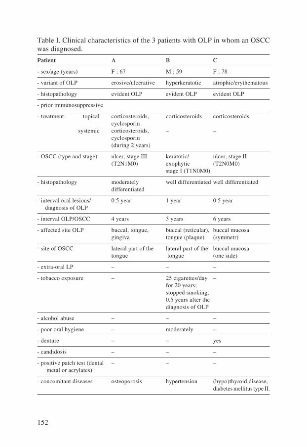
152
Table I. Clinical characteristics of the 3 patients with OLP in whom an OSCC was diagnosed.
Patient A B C
- sex/age (years) F ; 67 M ; 59 F ; 78
- variant of OLP erosive/ulcerative hyperkeratotic atrophic/erythematous
- histopathology evident OLP evident OLP evident OLP
- prior immunosuppressive
- treatment: topical corticosteroids, corticosteroids corticosteroids cyclosporin systemic corticosteroids, – – cyclosporin (during 2 years)
- OSCC (type and stage) ulcer, stage III keratotic/ ulcer, stage II (T2N1M0) exophytic (T2N0M0) stage I (T1N0M0)
- histopathology moderately well differentiated well differentiated differentiated
- interval oral lesions/ 0.5 year 1 year 0.5 year diagnosis of OLP
- interval OLP/OSCC 4 years 3 years 6 years
- affected site OLP buccal, tongue, buccal (reticular), buccal mucosa gingiva tongue (plaque) (symmetr)
- site of OSCC lateral part of the lateral part of the buccal mucosa tongue tongue (one side)
- extra-oral LP – – –
- tobacco exposure – 25 cigarettes/day – for 20 years; stopped smoking, 0.5 years after the diagnosis of OLP
- alcohol abuse – – –
- poor oral hygiene – moderately –
- denture – – yes
- candidosis – – –
- positive patch test (dental – – – metal or acrylates)
- concomitant diseases osteoporosis hypertension (hypo)thyroid disease, diabetes mellitus type II.

153
Statistical analysis was based on the incidence of 4 oral malignancies per 100,000 individuals per year in Europe versus 1, 2 or 3 cases of OSCC of 2000 person-years in our study.The probability of at least one case of OSCC in our cohort study is P = 0.08 according to the Poisson distribution, which could be attributed to chance alone. However, the probability of at least two or three cases is P = 0.003 and P = 0.00008, respectively, which is statistically significant. This means that it is highly improbable that at least two or three cases of OSCC in our study were encountered by chance alone.
Discussion
Hallopeau already reported a case of OLP with malignant degeneration in 1910.14 Krutchkoff et al criticized the literature on the malignant transforma-tion of OLP from the period 1950-1976 and accepted only 15 (7%) of the 223 published cases as adequately documented.15 As shown in Table II, Van der Meij et al used the same criteria from the period 1977-1999 and accepted 33 (34%) of 98 reported cases as adequately documented.11 Their objections were largely based on the uncertainty of the initial diagnosis of OLP on clini-cal and histopathological grounds, the occurrence of oral cancers remote from the anatomic site of OLP and the frequently inadequate information on prior exposure to potentially carcinogenic substances.11,15 Several remarks can be made on these objections.Even if a reliable biopsy is obtained from the patient at the first visit to confirm the initial diagnosis of OLP, there is a significant inter- and intra-observer variation in the interpretation of the criteria for establishing the diagnosis of OLP despite the criteria by the WHO.9,16 Moreover, important aspects such as dysplastic or atypical changes are not always clearly and carefully detailed in the histopathological reports. Therefore, the results reported in different studies are not always easy to compare. If an OSCC occurs at a site remote from OLP, the direct relationship between the two may be disputed, because an OSCC
Legends to Table I:– = none; F = female; M = maleT = primary tumor T1 = tumor size < 2 centimeters T2 = tumor size between 2 and 4 centimeters N = regional lymph nodesN0 = no indication for lymph node metastasesN1 = homolateral lymph node metastases < 3 centimetersM = distant metastasesM0 = no indication for distant metastasessymmetr = symmetrical
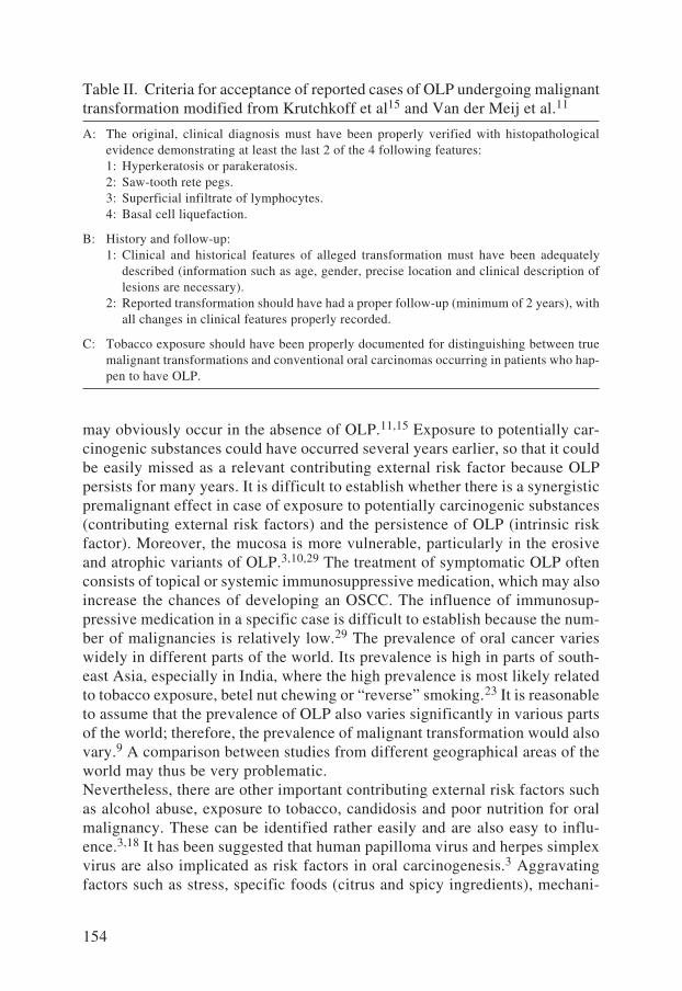
154
may obviously occur in the absence of OLP.11,15 Exposure to potentially car-cinogenic substances could have occurred several years earlier, so that it could be easily missed as a relevant contributing external risk factor because OLP persists for many years. It is difficult to establish whether there is a synergistic premalignant effect in case of exposure to potentially carcinogenic substances (contributing external risk factors) and the persistence of OLP (intrinsic risk factor). Moreover, the mucosa is more vulnerable, particularly in the erosive and atrophic variants of OLP.3,10,29 The treatment of symptomatic OLP often consists of topical or systemic immunosuppressive medication, which may also increase the chances of developing an OSCC. The influence of immunosup-pressive medication in a specific case is difficult to establish because the num-ber of malignancies is relatively low.29 The prevalence of oral cancer varies widely in different parts of the world. Its prevalence is high in parts of south-east Asia, especially in India, where the high prevalence is most likely related to tobacco exposure, betel nut chewing or “reverse” smoking.23 It is reasonable to assume that the prevalence of OLP also varies significantly in various parts of the world; therefore, the prevalence of malignant transformation would also vary.9 A comparison between studies from different geographical areas of the world may thus be very problematic. Nevertheless, there are other important contributing external risk factors such as alcohol abuse, exposure to tobacco, candidosis and poor nutrition for oral malignancy. These can be identified rather easily and are also easy to influ-ence.3,18 It has been suggested that human papilloma virus and herpes simplex virus are also implicated as risk factors in oral carcinogenesis.3 Aggravating factors such as stress, specific foods (citrus and spicy ingredients), mechani-
Table II. Criteria for acceptance of reported cases of OLP undergoing malignant transformation modified from Krutchkoff et al15 and Van der Meij et al.11
A: The original, clinical diagnosis must have been properly verified with histopathological evidence demonstrating at least the last 2 of the 4 following features: 1: Hyperkeratosis or parakeratosis. 2: Saw-tooth rete pegs. 3: Superficial infiltrate of lymphocytes. 4: Basal cell liquefaction.
B: History and follow-up: 1: Clinical and historical features of alleged transformation must have been adequately
described (information such as age, gender, precise location and clinical description of lesions are necessary).
2: Reported transformation should have had a proper follow-up (minimum of 2 years), with all changes in clinical features properly recorded.
C: Tobacco exposure should have been properly documented for distinguishing between true malignant transformations and conventional oral carcinomas occurring in patients who hap-pen to have OLP.
155
cal traumata, irritation or allergy related to dental restorations and poor oral hygiene are also important in OLP.2,3
Zhang et al examined 33 cases of OLP for allelic loss at nine loci located on chromosomes 3p, 9p, and 17p.24 Loss of heterozygosity on these three arms frequently occurs in oral tumors and the presence of these alterations in premalignant lesions suggests that they may play an important role in tumor progression. The lesions in OLP showed minimal genetic deviations, scoring even lower than reactive oral lesions which are not deemed to be precancer-ous. However, epithelial dysplasia and malignant oral lesions had significantly increased genetic alterations. These findings did not support the hypothesis that OLP is a disorder with an increased intrinsic malignant potential.24 Nevertheless, it has also been postulated that prostaglandines play a signifi-cant role in the regulation of the local immune response as well as carcinogen activation and tumor initiation. The inflammatory environment in OLP with cytokines may be particular favorable for tumor promotion. Interleukin-1 leads to an activation of several prostaglandines-mediated events via the action of cyclooxygenase-2 (COX-2) on arachidonic acid.25 Increased prostaglandin E2 levels have been demonstrated in tumor specimens of patients with squamous cell carcinoma, and COX-2 has been shown to be overexpressed nearly 100-fold in squamous cell carcinomas of the head and the neck.26,27 Theoretically, these data could suggest that more aggressive treatment in OLP against the increased inflammatory response in OLP may restore normal immunological surveillance and possibly interrupt the progression into invasive cancer.2 Valente et al reported in a study with 28 patients an overexpression of p53 gene mutations investigated by immunohistochemistry in cases of OLP with a high risk of neoplastic transformation.28
If the criteria by Krutchkoff et al (Table II) are applied in our study, then patient B who smoked heavily should be considered as a drop-out.11,15 In that case, 2 out of the 200 patients investigated in this study developed an OSCC. Patient A had also received systemic immunosuppressive medication (corticosteroids and cyclosporin) during a period of about 2 years, which could have increased the chance of developing a malignancy. In that case, only 1 out of the 200 patients may be regarded as a real intrinsic malignancy. Therefore, it still remains unclear whether OLP has an intrinsic malignant potential because a single OSCC may occur by chance alone. In that case, OLP does not fulfill the WHO criterion of a precancerous condition: “a generalized state associated with a significant increased risk of cancer”.3,22 In our opinion, it is very likely that there is a syn-ergy between intrinsic (chronic inflammatory features in OLP) and contributing external risk factors in possible malignant transformation in OLP. It has been reported that a specific clinical variant of OLP (either hyperkeratotic or erosive) had a higher chance of transformation into an OSCC.3,10,29 The results of other studies including our study fail to support this because of the low number of patients. The follow-up period also varied from 1 to 4 visits per year in other

156
studies.9,19 A more frequent follow-up visit does not necessarily lead to an improved prognosis for OLP patients with an OSCC.9 We recommend a follow-up of at least one or two visits per year as long as OLP persists. A careful physi-cal examination at each visit is imperative and histopathological examination should be repeated if a malignancy is suspected. From a practical point of view, we concur with Voûte et al in that we are also somewhat reluctant to routinely inform each patient with OLP on the possible premalignant character of their lesions, particularly if contributing external risk factors are also involved.19
The results of this study showed that it is mandatory to establish a correct diagnosis of OLP based on history, clinical examination and histopathology. Nonetheless, the results failed to provide an answer to the controversial issue of whether OLP has an intrinsic malignant potential. The exact incidence of OSCC in patients with OLP is difficult to establish, because of the low number of patients involved and because other contributing external risk factors may also be relevant for developing an oral malignancy. Further larger cohort stud-ies are necessary to resolve this issue.
References
1. Vincent SD, Fotos LPG, Baker KA et al. Oral lichen planus: the clinical, historical, and therapeutic features of 100 cases. Oral Surg Oral Med Oral Pathol 1990; 70: 165-71.
2. Eisen D. The clinical features, malignant potential and systemic associations of oral lichen planus: a study of 723 patients. J Am Acad Dermatol 2002; 46: 207-14.
3. Scully C, Beyli M, Ferreiro MC et al. Update on oral lichen planus: etiopathogen-esis and management. Crit Rev Oral Biol Med 1998; 9: 86-122.
4. Parkin DM, Whelan SL, Ferlay J et al. Cancer incidence in five continents, Vol. VII (IARC Scientific publications No. 143). Lyon: IARC. 1997.
5. Pindborg JJ, Reichart PA, Smith CJ et al. International histological classification of tumours. Histological typing of cancer and precancer of the oral mucosa. 2nd
edn. World Health Organization. Berlin Heidelberg New York: Springer-Verlag. 1997.
6. Schlecht NF, Franco EL, Pintos J et al. Interaction between tobacco and alcohol consumption and the risk of cancers of the upper aerodigestive tract in Brazil. Am J Epidemiol 1999; 150: 1129-37.
7. van der Waal I. Tumoren van het mondslijmvlies. [Dutch]. Tumors of the oral mucosa. Ned Tijdschr Derm Venereol 2002; 12: 368-70.
8. van der Meij EH, Bezemer PD, van der Waal I. Cost-effectiveness of screening for the possible development of cancer in patients with oral lichen planus. Comm Oral Dent Epidemiol 2002; 30: 342-51.
9. Mattsson U, Jontell M, Holmstrup P. Oral lichen planus and malignant transforma-tion; Is a recall of patients justified? Crit Rev Oral Biol Med 2002; 13: 390-6.
10. Yaacob HB, Tan PL, Ngeow WC. Malignancy in oral lichen planus: a review of a group from the Malaysian population. J Oral Sci 2002; 44: 65-71.
157
11. van der Meij EH, Schepman KP, Smeele LE et al. A review of the recent literature regarding malignant transformation of oral lichen planus. Oral Surg Oral Med Oral Pathol Oral Radiol Endod 1999; 88: 307-10.
12. Rajentheran R, McLean NR, Kelly CG et al. Malignant transformation of oral lichen planus. Eur J Surg Oncol 1999; 25: 520-3.
13. Mignogna MD, Lo Muzio L, Lo Russo L et al. Clinical guidelines in early detection of oral squamous cell carcinoma arising in oral lichen planus: a 5-year experience. Oral Oncol 2001; 37: 262-7.
14. Hallopeau H. Sur un cas de lichen de Wilson gingival avec neoplasie voisine dans la region maxillaire. [French]. Bull Soc Fr Derm Syph 1910; 17: 33.
15. Krutchkoff DJ, Cutler L, Laskowski S. Oral lichen planus: the evidence regarding potential malignant transformation. J Oral Pathol 1978; 7: 1-7.
16. van der Meij EH, Reibel J, Slootweg PJ et al. Interobserver and intraobserver vari-ability in the histologic assessment of oral lichen planus. J Oral Pathol Med 1999; 28: 274-7.
17. Allen CM. Is lichen planus really premalignant? Oral Surg Oral Med Oral Pathol Oral Radiol Endod 1998; 85: 347.
18. Lozada-Nur F. Oral lichen planus and oral cancer: is there enough epidemiologic evidence? Oral Surg Oral Med Oral Pathol Oral Radiol Endod; 89: 265-6.
19. Voûte ABE, de Jong WFD, Schulten EAJM et al. The possible premalignant character of oral lichen planus; The Amsterdam experience. J Oral Pathol Med 1992; 21: 326-9.
20. Warshaw EM, Templeton SF, Washington CV. Verrucous carcinoma occurring in a lesion of oral lichen planus. Cutis 2000; 65: 219-22.
21. Eisenberg E, Krutchkoff DJ. Lichenoid lesions of the oral mucosa. Oral Surg Oral Med Oral Pathol 1992; 73: 699-704.
22. Holmstrup P, Thorn JJ, Rindum J et al. Malignant development of lichen planus-affected oral mucosa. J Oral Pathol 1988; 17: 219-25.
23. Scully C. The oral cavity. In: Textbook of Dermatology. Champion RH, Burton JL, Burns DA, Breathnach SM. 6th edn, Vol 4. Oxford: Blackwell Science Ltd 1998; 3074-6.
24. Zhang L, Michelsen C, Cheng X et al. Molecular analysis of oral lichen planus: a premalignant lesion? Am J Pathol 1997; 151: 323-7.
25. Noguchi K, Shitashige M, Ishikawa I. Involvement of cyclooxygenase-2 in inter-leukin-1 alpha-induced prostaglandin production by human periodontal ligament cells. J Periodontol 1999; 70: 902-8.
26. Pinto S, Gallo O, Dilaghi M et al. Prostaglandines in peritumor synthesis. Prosta-glandines Leukot Essent Fatty Acids 1990; 39: 53-7.
27. Mestre JR, Chan G, Zhang F et al. Inhibition of cycloocygenase-2 expression: an approach to preventing head and neck cancer. Ann N Y Acad Sci 1999; 889: 62-71.
28. Valente G, Pagano M, Carrozzo M et al. Sequential immunohistochemical p53 expression in biopsies of oral lichen planus undergoing malignant evolution. J Oral Pathol Med 2001; 30: 135-40.
29. van der Meij EH, Schepman KP, van der Waal I. The possible premalignant charac-ter of oral lichen planus and oral lichenoid lesions: a prospective study. Oral Surg Oral Med Oral Pathol Oral Radiol Endod 2003; 96: 164-71.


CHAPTER 6
ORAL LICHEN PLANUS AND
HEPATITIS C VIRUS INFECTION
R. LAEIJENDECKER, MD1 (and formerly 2), Th. VAN JOOST, MD, PhD (formerly2),
B. TANK, PhD2, and H.A.M. NEUMANN, MD, PhD2
ARCHIVES OF DERMATOLOGY 2005; 141: 906-7.
Funding: None.Conflict of interest: None.Corresponding author: R. Laeijendecker, Department of Dermatology, Albert Schweitzer Hospital, P.O. Box 444, 3300 AK Dordrecht, The Netherlands.Tel: 00 31 78 6523330. Fax: 11 31 78 6523866. E-mail: [email protected]
1 Department of Dermatology, Albert Schweitzer Hospital, locations Dordwijk and Sliedrecht, Albert Schweitzerplaats 25, 3318 AT Dordrecht, The Netherlands.
2 Department of Dermatology and Venereology, Erasmus MC, University Medical Center Rot-terdam, Dr. Molewaterplein 40, 3015 GD Rotterdam, The Netherlands.

160
ABSTRACT
Background: The possible association between oral lichen planus and hepa-titis C virus infection is still debated in the literature.
Objectives: To determine the possible relationship between oral lichen pla-nus and hepatitis C virus infection in The Netherlands and to investigate whether routine screening for anti-hepatitis C virus antibodies and liver enzymes is necessary in these patients. Moreover, attempts were made to clarify the discrepant results reported in the literature on the association of oral lichen planus with hepatitis C virus infection.
Methods: A prospective study in a group of 100 patients with oral lichen
planus and a control group of 100 patients with psoriasis vul-garis was carried out from 1995 to 2002. All patients were at least screened for liver enzymes and anti-hepatitis C virus anti-bodies.
Results: In both groups, each with 65 women and 35 men, there was no serological evidence of antibodies against hepatitis C virus at all. In the group of oral lichen planus, there were 2 (2%) patients with significant elevated liver enzyme levels, and in the con-trol group of psoriasis vulgaris, there were also 2 (2%) patients with elevated liver enzyme levels, which was not a statistically significant difference (P > 0.99). In these 4 patients, there was also no evidence of hepatitis A virus and hepatitis B virus infec-tions.
Conclusions: Our study suggests that there is no indication for routinely screening for anti-hepatitis C virus antibodies in patients with oral lichen planus in The Netherlands. Liver enzyme values should be determined only in a relevant, clinical situation. The discrepancies in the literature regarding the possible association of oral lichen planus and hepatitis C virus are largely based on the differences in prevalence of both hepatitis C virus infec-tion and possibly also of oral lichen planus world-wide with a population of different genetic make-up, different environmen-tal conditions and possible variations in hepatitis C virus infec-tion and host immune responses.
Keywords: Oral lichen planus, Hepatitis C virus infection, Liver enzymes.

161
Introduction
Oral lichen planus (OLP) has a prevalence of about 0.5 to 2%. Generally, it is a disease of the middle-aged and the elderly and the female-to-male ratio is about 2: 1.1 The diagnosis of OLP is based on a combination of characteristic clinical findings, history and histopathological examination. Oral lichen planus can be categorized into a hyperkeratotic (white) variant often without symptoms. The atrophic or erythematous (red) variant and the erosive or ulcerative (yellow) variant commonly have persistent symptoms of pain or stinging.1-3 The exact etiopathology of OLP is unknown, but an immune-mediated (T-cell dependant) pathogenesis has been proposed.1-4
Several possible causes, aggravating factors and associations have been sug-gested. These include specific medication, auto-immunity, graft-versus-host disease, trauma, (emotional) stress, infections by plaque-causing micro- organisms, hepatitis B – and especially hepatitis C viruses.3 Hepatitis C virus (HCV) is an RNA virus, which was identified in 1989. It is a major cause of acute and chronic hepatitis which may often be asymptomatic.5,6 Modes of transmission are transfusion of blood, injection with dirty needles and, in lower frequency, sexual or vertical transmission.5,6 Chronic hepatitis C virus infection develops in about 75% of those who are infected and may lead to cirrhosis which may culminate in liver failure or even hepatocellular carcinoma.5 Hepatitis C virus infection seems to be the main cause of liver disease in patients with OLP.7,12 The possible relationship between OLP and HCV infection has been studied extensively but the results are rather discrepant and controversial.7-17
The aim of this study was to determine the possible relationship between OLP and HCV in The Netherlands and to investigate whether screening for anti-HCV antibodies and liver enzymes is necessary in these patients. Moreover, attempts were made to clarify the discrepant results reported in the literature on the association of OLP with HCV.
Patients and methods
A prospective study in group (I) of 100 Caucasian patients with a confirmed diagnosis of OLP based on medical history, physical and histopathological examination according to the diagnostic criteria defined by the WHO was performed. The control group (II) consisted of 100 Caucasian patients with psoriasis vulgaris matched for sex in clusters of 10 patients. The diagnosis of psoriasis vulgaris was made mainly on clinical grounds and if necessary with additional histopathological examination. The study was carried out from 1995 to 2002 at the Department of Dermatology, Albert Schweitzer Hospital, Dor-

162
drecht, The Netherlands. Exclusion criteria in both groups were an age younger than 18 years, the occurrence of OLP and psoriasis vulgaris in the same patient, prior treatment with potentially hepatotoxic agents such as methotrexate and treatment with PUVA, retinoids, cyclosporin or fumaric acid derivatives in the past two years. All patients were screened for liver enzyme levels including total bilirubin (TB) (reference values, < 17 microMol/l or < 1.0 mg/dL), aspartate aminotrans-ferase (ASAT) (reference value,< 30 U/l), alanine aminotransferase (ALAT) (reference value, < 30 U/l), gamma-glutamyl transpeptidase (GGT) (reference values, < 40 U/l (men), < 25 U/l (women)) and alkaline phosphatase (ALP) (reference value, < 100 U/l). Deviations were considered clinically relevant when two or more liver enzyme values were abnormal or if at least one value exceeded the upper limit by more than two times. In case of deviations, the determination of liver enzyme levels was repeated after 3 weeks. These patients were also tested for possible HAV and HBV infections.Anti-HCV antibodies were determined with a third-generation enzyme-linked immunosorbent assay (ELISA), the results of which were confirmed by immu-noblotting technique.
Statistical analysis of the results was performed by means of the exact stratified 2x2 (contingency) table analysis and the Unpaired t-test in which two-sided P-values were calculated, with statistical significance set at P < 0.05.
Results
The OLP group (I) comprised 65 women and 35 men, aged 26 to 77 years (mean 54 years). A hyperkeratotic variant of OLP was diagnosed in 68 patients (42 F and 26 M). The erosive or ulcerative variant was observed in 22 patients (14 F and 8 M) and the atrophic or erythematous variant in 10 patients (9 F and 1M). The control group (II) comprised 65 women and 35 men, aged 19 to 82 years (mean 51 years). The mean ages in the two groups did not differ significantly (P > 0.99). There was no serological evidence of antibodies against HCV in either group. In group I, there were 2 (2%) patients (1 and 2) with the hyperkeratotic variant of OLP with several slightly elevated liver enzyme levels (< 2 times the normal upper limit) (Table I). In group II, there were also 2 (2%) patients with several elevated liver enzyme levels. One patient (3) had slightly elevated liver enzyme levels (< 2 times the normal, upper limit) and another patient (4) had abnormal liver enzyme levels with values of ALAT, ASAT and GGT between 2 and 3 times the normal upper limit (Table I). These abnormalities in liver enzyme values did not change significantly in the second determination after 3 weeks.

163
After consultation with the Department of Internal Medicine and echographic liver examination, no conclusive explanation could be found for the results in patients 1 and 2 in group I or for those in patient 3 in group II. In group II, patient 4 was an excessive alcohol user (about 12 units/day). Moreover, there were also no signs of HAV- and HBV-infections in these 4 patients. The varia-tions in the liver enzyme values between the 2 groups after adjusting for sex were not considered as statistically significant (P > 0.99).
Discussion
Chronic hepatitis C virus infection has been associated with several extra-hepatic diseases, many of which may be seen by dermatologists. The following manifestations have been mentioned: antiphospholipid syndrome, auto-immune thrombocytopenia, autoimmune thyroiditis, B-cell lymphoma, Behçet syn-drome, canities (= graying of hair), glomerulonephritis, leukocytoclastic vas-culitis, LP, MALT lymphoma (mucosal-associated lymphoid tumors), mixed type cryoglobulinemia, Mooren’s corneal ulcer, plasmacytoma, polyarteritis nodosa, porphyria cutanea tarda, prurigo nodularis, sialoadenitis (Sjögren’s-like) and vitiligo.3,5
Table I. Results in 4 patients with elevated liver enzyme levels and determina-tion of serological evidence of hepatitis A, B or C virus infection.
Age TB ASAT ALAT GGT ALP HAV HBV HCV
Group I (OLP) (years) (micro- (U/l) (U/l) (U/l) (U/l) mol/l) (mg/dL)
1-F 48 16 35 35* 40* 80 – – – (0.9)
2-M 66 20* 45* 45* 65* 100 – – – (1.2*)
Group II (psoriasis vulgaris)
3-F 42 30* 50* 52* 45* 110* – – – (1.8*)
4-M 54 25* 65** 65** 110** 120* – – – (1.5*)
– = negative.* = less than 2 times the normal upper limit.** = between 2 and 3 times the normal upper limit.F = FemaleM = Male

164
World-wide, it has been estimated that approximately 170,000,000 individuals are infected with HCV. This is about 3% of the world population.5 Hepatitis C virus infection is prevalent in individuals older than 40 years, but is uncommon in those younger than 20 years.5 The prevalence of both HCV and OLP showed considerable geographic variations.6,7,14,17 In The Netherlands, the prevalence of HCV has been estimated at between 0.1% and 0.7%.18 However, the preva-lence is as high as 62% in some regions in Japan.19 In parts of the world with a high prevalence of HCV, for example in Brazil, Japan, Italy and Spain, a posi-tive correlation between HCV and OLP has been reported.13,15,17,20 In these countries, HCV may play a role in the pathogenesis of OLP. However, the exact etiopathogenic relationship between OLP and liver disease remains unknown. In regions with a low prevalence of HCV, for example the United Kingdom, Scandinavia and The Netherlands, no correlation between OLP and HCV was found, but conflicting results were reported from Germany.7,9,10,16,21,22 It has been suggested that the erosive variant of OLP may have a higher correlation with HCV than the hyperkeratotic variant of OLP.13,23 The lack of a control group was present in certain other studies making the correct interpretation of the results rather difficult.7,23 However, our study has a larger group of patients with OLP with an adequate control group. In the pathogenesis of OLP in rela-tion to HCV, several aspects such as differences in prevalence of OLP and HCV, differences in the genetic make-up and age of the involved population, environmental factors and possibly the variation in the infection with a specific HCV subtype and the host immune responses may play a role.3,5,7,17 In a few studies, HCV RNA was isolated from the oral mucosal tissue in patients with HCV regardless of OLP. No HCV RNA was isolated from patients who were not infected with HCV. These findings indicate that HCV alone is not sufficient for the development of OLP, but that other factors may also play an important role.17,26,27
In our study, there was no correlation between OLP (or a specific variant of OLP) and HCV. Our findings indicate that screening for anti-HCV antibodies in a comparative population is not required in The Netherlands. The prevalence of HCV appears to be too low and the probability that HCV plays an etiopatho-genic role in OLP in The Netherlands is negligible. This advice could possibly be different if more patients with OLP from the Mediterranean region with a probably higher prevalence of HCV are investigated.Two (2%) of 100 patients with OLP and 2 (2%) of the patients in the con-trol group (Table I) had several abnormal asymptomatic liver enzyme values. However, a direct relationship between liver function and the pathogenesis or persistence of OLP is unclear. In our opinion, determination of liver enzyme levels should be performed selectively, especially before starting or evaluating a systemic treatment or for investigation of possible known risk factors such as alcohol abuse, cholestasis, specific infections and prior systemic medication with possible hepatotoxic effects.

165
The conclusions of this study are that screening for anti-HCV antibodies in patients with OLP in The Netherlands is unwarranted and that liver enzyme levels should be determined only in a clinically relevant situation. The discrep-ancies in the literature regarding the possible association of OLP and HCV are largely based on the prominent world-wide differences in the prevalence of both HCV infection and possibly also of OLP among populations with different genetic make-up, different environmental conditions and possible variations in HCV infection and host immune responses.
References
1. Vincent SD, Fotos LPG, Baker KA et al. Oral lichen planus: the clinical, historical, and therapeutic features of 100 cases. Oral Surg Oral Med Oral Pathol 1990; 70: 165-71.
2. Eisen D. The clinical features, malignant potential and systemic associations of oral lichen planus: a study of 723 patients. J Am Acad Dermatol 2002; 46: 207-14.
3. Scully C, Beyli M, Ferreiro MC et al. Update on oral lichen planus: etiopathogen-esis and management. Crit Rev Oral Biol Med 1998; 9: 86-122.
4. Boyd AS, Neldner KH. Lichen planus. J Am Acad Dermatol 1991; 25: 593-619. 5. Bonkovsky HL, Mehta S. Hepatitis C: A review and update. J Am Acad Dermatol
2001; 44: 159-79. 6. Lodi G, Porter SR. Hepatitis C virus infection and lichen planus: a short review.
Oral Diseases 1997; 3: 77-81. 7. van der Meij EH, van der Waal I. Hepatitis C virus infection and oral lichen planus:
a report from the Netherlands. J Oral Pathol Med 2000; 29: 255-8. 8. Bellman B, Reddy RK, Falanga V. Lichen planus associated with hepatitis C (let-
ter). Lancet 1995; 346: 1234. 9. Imhof M, Popal H, Lee JH et al. Prevalence of hepatitis C virus antibodies and
evaluation of hepatitis C virus genotypes in patients with lichen planus. Dermatology 1997; 195(1): 1-5.
10. Grote M, Reichart PA, Berg T et al. Hepatitis C virus (HCV)-infection and oral lichen planus. J Hepatol 1998; 29: 1034-5.
11. Ingafou M, Porter SR, Scully C et al. No evidence of HCV infection or liver disease in British patients with oral lichen planus. Int J Oral Maxillofac Surg 1998; 27: 65-6.
12. Del Olmo JA, Pascual I, Bagan JV et al. Prevalence of hepatitis C virus in patients with lichen planus of the oral cavity and chronic liver disease. Eur J Oral Sci 2000; 108: 378-82.
13. Sanchez-Perez J, Castro M de, Buezo GF et al. Lichen planus and hepatitis C virus: prevalence and clinical presentation of patients with lichen planus and hepatitis C virus infection. Br J Dermatol 1996; 134: 715-9.
14. Mignogna MD, Lo Muzio L, Lo Russo L et al. Oral lichen planus: different clini-cal features in HCV-positive and HCV-negative patients. Int J Dermatol 2000; 39: 134-9.
15. Mignogna MD, Lo Muzio L, Favia G et al. Oral lichen planus and HCV infection: a clinical evaluation of 263 cases. Int J Dermatol 1998; 37: 575-8.
166
16. Roy KM, Dickson EM, Spiteri Staines K et al. Hepatitis C virus and oral lichen planus/ lichenoid reactions: Lack of evidence for an association. Clin Lab 2000; 46: 251-4.
17. Figueiredo LC, Carrilho FJ, de Andrade HF et al. Oral lichen planus and hepatitis C virus infection. Oral Diseases 2002; 8: 42-6.
18. van der Poel CL, Reesink HWQ, Mauser-Bunschoten EP et al. Prevalence of anti-HCV antibodies confirmed by recombinant immunoblot in different population subsets in The Netherlands. Vox Sang 1991; 61: 30-6.
19. Nagao Y, Sata M, Tanikawa K et al. Lichen planus and hepatitis C virus in the Northern Kyushu region of Japan. Euro J Clin Invest 1995; 25: 910-4.
20. Nagao Y, Sata M, Abe K et al. Immunological evaluation in oral lichen planus with chronic hepatitis C. J Gastroenterol 1997; 32: 324-9.
21. Tucker SC, Coulson IH. Lichen planus is not associated with hepatitis C virus infection in patients from northwest England. Acta Derm Venereol 1999; 79: 378-9.
22. Mobacken H, Nilsson LA, Olsson R et al. Incidence of liver disease in chronic lichen planus of the mouth. Acta Derm Venereol 1984; 64: 70-3.
23. Gandolfo S, Carbone M, Carrozzo M et al. Oral lichen planus and hepatitis C virus (HCV) infection: is there a relationship? A report of 10 cases. J Oral Pathol Med 1994; 23: 119-22.
24. Lozada-Nur F, Miranda C. Oral lichen planus: epidemiology, clinical characteris-tics, and associated diseases. Semin Cutan Med Surg 1997; 4: 273-7.
25. Lodi G, Carrozzo M, Hallett R et al. HCV genotypes in Italian patients with HCV-related oral lichen planus. J Oral Pathol Med 1997; 26: 381-4.
26. Arrieta JJ, Rodrigues-Inigo E, Casqueiro M et al. Detection of hepatitis C virus replication by in situ hybridization in epithelial cells of anti-hepatitis C virus-posi-tive patients with and without oral lichen planus. Hepatology 2000; 32: 97-103.
27. Nagao Y, Sata M, Noguchi S et al. Detection of hepatitis C virus RNA in oral lichen planus and oral cancer tissues. J Oral Pathol Med 2000; 29: 259-66.

CHAPTER 7
ORAL LICHEN PLANUS IN CHILDHOOD
R. LAEIJENDECKER, MD1 (and formerly2), Th. VAN JOOST, MD, PhD (formerly2),
B. TANK, PhD2, A.P. ORANJE, MD, PhD2, and
H.A.M. NEUMANN, MD, PhD2
PEDIATRIC DERMATOLOGY 2005; 22(4): 299-304.
Funding: None.Conflict of interest: None.Corresponding author: R. Laeijendecker, Department of Dermatology, Albert Schweitzer Hospital, P.O. Box 444, 3300 AK Dordrecht, The Netherlands.Tel: 00 31 78 6523330. Fax: 11 31 78 6523866.E-mail: [email protected]
1 Department of Dermatology, Albert Schweitzer Hospital, locations Dordwijk and Sliedrecht, Albert Schweitzerplaats 25, 3318 AT Dordrecht, The Netherlands.
2 Department of Dermatology and Venereology, Erasmus MC, University Medical Center Rot-terdam, Dr. Molewaterplein 40, 3015 GD Rotterdam, The Netherlands.

168
ABSTRACT
Background: Oral lichen planus is rare in childhood and there are only scarce reports on this subject in the literature.
Objectives: To report individual cases of oral lichen planus in childhood from our practice and to review the literature on this subject.
Methods: Patients younger than 18 years with oral lichen planus were included. Several clinical aspects, histopathology, patch test reactions and routine blood examination were investigated.
Results: Three patients from about 10,000 dermatological patients younger than 18 years were included from 1994 to 2003. An Asian girl aged 11 years had an asymptomatic reticular variant of oral lichen planus, which disappeared without any treatment after one year. An Asian boy aged 16 years had an erosive vari-ant oral lichen planus with severe pain, which healed after inten-sive topical and systemic treatment in two years. A Caucasian girl aged 14 years had a reticular variant of oral lichen planus with a little soreness, which disappeared with topical treatment after three months.
Conclusions: Oral lichen planus in childhood is rare and therefore, at present it is not possible to draw firm conclusions on this subject. Oral lichen planus in childhood seems to occur preferentially in those of Asian race. The clinical features resemble those of oral lichen planus in adulthood. However, generally, the prognosis of oral lichen planus in childhood seems to be more favorable.
Keywords: Oral lichen planus, Childhood.
169
Introduction
Oral lichen planus (OLP) is a rather common disease in the middle-aged and elderly population and has a prevalence of about 0.5 to 2% with a female-to-male ratio of approximately 2: 1.1,2 In the contrast, oral lichen planus in child-hood (OLPc) is rare and only a few reports are available in the literature.3-8 Oral lichen planus can be categorized into a hyperkeratotic (white) variant commonly without symptoms including reticular with Wickham’s striae, papular or plaque-like forms. The atrophic or erythematous (red) variant and the erosive or ulcerative (yellow) variant often have persistent symptoms of pain or stinging aggravated during eating and drinking.1-3 These variants may occur together in one patient or may transform into each other. The lesions were found (in diminishing frequency) on the buccal mucosa (often symmetri-cal), the lateral margins of the tongue, the gingiva, the lips and the palatum durum.2,3,9 Whereas CLP is self-limiting, OLP is chronic and rarely under-goes spontaneous remission. Oral lichen planus may be a potential source of significant morbidity.3 The family history of LP is more commonly positive in patients with LP in childhood than in adulthood.9 The exact cause of OLP remains unknown, but an immune-mediated (T-cell dependant) pathogenesis has been proposed.1,2,9 Possible causes such as an allergy to dental restorative materials (amalgam, gold), local trauma (Koebner’s phenomenon) and several infections (plaque-causing micro-organisms and hepatitis B or C virus infec-tion) of OLP have been reported. Moreover, genetic factors, lifestyle and (emotional) stress may be contributing factors in the pathogenesis of OLP. An increased association between LP and several auto-immune diseases, such as ulcerative colitis, myasthenia gravis, lupus erythematosus and alopecia areata, and perhaps also with systemic diseases, such as diabetes mellitus and hyper-tension has been reported. Many drugs are capable of producing a lichenoid drug eruption (LDE). Moreover, a lichenoid eruption may occur in graft-ver-sus-host disease.2,3,9-11
In this communication, three new cases of OLPc from our practice are reported together with a review of the current literature. Moreover, possible clinical dif-ferences between OLPc and OLP in adulthood (OLPa) are reported.
Patients and methods
We conducted a retrospective non-randomized study from 1994 to 2003 at the Department of Dermatology, Albert Schweitzer Hospital, Dordrecht, The Netherlands. It is a full-time practice with three dermatologists. One of them is especially interested in OLP. The hospital serves a region of about 500,000 inhabitants with a representative demographical structure. About 1% of the

170
inhabitants are of Asian race. Inclusion criterion was an age younger than 18 years. All patients were referred by a dentist or a physician. The diagnosis of OLP was based on a combination of characteristic clinical findings, history and histopathology. Investigations on possible signs of extra-oral LP, routine blood examination including liver enzymes, hepatitis B- and C-serology, glucose, thyroid-stimulating hormone (TSH) and antinuclear antibodies (ANA’s) were performed. Items such as age, sex, race, oral hygiene, candidosis and family history of LP were also investigated. Patch testing with a standard series (according to the guidelines of the European standard series) and a dental metal series was performed and evaluated at least after three days.One or two 3-mm punch biopsies for histopathological examinations were taken from the hyperkeratotic lesions from each of the three children and if there was an erosion or ulceration at the edge of the lesions. The biopsies were fixed in buffered 4% formalin and stained with hematoxylin and eosin and also with the periodic acid-Schiff (PAS) reagent. If there were obvious erosions in OLP, the biopsies were transported in physiological saline for direct immunofluores-cence to exclude a bullous auto-immune disease or lupus erythematosus. Histopathological criteria of OLP comprised varying degree of focal hyper-keratosis or parakeratosis, irregular acanthosis or atrophy, liquefaction degeneration of the basal cell layer and a dense band-like lymphocytic infil-trate high in the lamina propria. Hyaline (Civatte’s) bodies, which represent degenerated basal cells, were occasionally seen in the epithelium. In case of less pronounced changes in the mucosa, especially the basal cell layer degeneration and the inflammatory infiltrate, the diagnosis “compatible with OLP” was given.
Statistical analysis of the results was performed by a Poisson distribution for the number of incidences of OLPc in the Asian race in the population (with statistical significance set at P-value < 0.05).
Results
In this study of 10 years approximately 10,000 patients younger than 18 years with a boy-to-girl ratio of about 1:1 visited our department and only 3 patients (0.03%) with OLPc could be included. The characteristics of the three patients are detailed in Table I. The patients were born and bred in The Netherlands. Their mean age was 13.7 years and the mean duration of OLPc was about 20 months. The mean follow-up period after the diagnosis of OLPc was 3 years.In patient A, an Asian girl, the duration of OLPc could not be established exactly because there were no symptoms. The regular examination by the den-tist gave an indication of duration. The patient was not treated because of the lack of symptoms (Table I).

171
Table I. Clinical characteristics of the three patients with OLPc.
Patient A B C
Sex/age (years) F/ 11/ Asian M/ 16/ Asian F/ 14/ Caucasian
Predominantly variant of reticular (hyper- erosive/ ulcerative reticular (hyper-OLP keratotic) keratotic)
Histopathology compatible OLP OLP OLP
Duration before diagnosis/ 6 months (?)/ 4 months/ severe oral 12 months/Character of symptoms asymptomatic pain and stinging soreness
Location of OLPc buccal mucosa buccal mucosa, gingiva buccal mucosa and (symmetrical) (asymmetrical) lateral part of tongue (sym.)
Extra-oral LP – – –
Oral hygiene/candidosis normal/ - poor/ - good/ -
Amalgam fillings/ 4/ - 8/ - - / + (withoutorthodontic retainer nickel)
Positive patch test reaction Fragrance mix – Nickel sulfate 5% 8% pet pet
Blood examination – – –deviations
Systemic medication – – regularly cetirizine
Concomitant diseases – – atopic dermatitis, allergic rhinitis
Family history of LP – – –
Duration of OLPc (after 1 year 2 years 3 monthsdiagnosis)
Treatment of OLPc – Topical steroids Topical tretinoin (classes II and IV), 0.05% ointment, Lidocaine gels, Topical corticoste- Tacrolimus ointment. roids (class II) Systemic corticoste- (Triamcinolone roids and Systemic acetonide 0.1% cyclosporin. ointment)
Follow-up period (after 3 years 4 years 2 yearsdiagnosis OLPc)
– = not present. + = present. F = female (girl). M = male (boy). Pet = petrolatum. Sym. = sym-metrical.

172
In patient B, an Asian boy, there were severe persistent symptoms of oral pain and stinging which interfered with eating, talking and functioning for two years. The lesions of the gingiva showed the signs of a desquamative gingivitis. Oral hygiene was improved but did not influence the oral lesions significantly. He was treated with topical corticosteroids (classes II and IV), lidocaine gels and tacrolimus ointment during several months without significant improve-ment. He was subsequently treated with systemic corticosteroids (30 mg per day for 6 weeks) and systemic cyclosporin (4 mg per kilogram per day for 3 months) with remissions and exacerbations.In patients A and B, there was no close contact between the amalgam fillings and the oral lesions. Patient C, a Caucasian girl, had an OLPc with a little soreness. The treatment with topical tretinoin 0.05% ointment and topical corticosteroids (class II) was successful after 3 months. The clinical relevance of an allergy to nickel sulfate could not be demonstrated and a LDE to cetirizine was excluded because the OLPc healed in spite of continued cetirizine therapy. About 6 months after she developed symptoms of OLPc, she was fitted with an orthodontic retainer which remained during two years. Measures were taken to avoid local trauma of the oral mucosa by the orthodontic retainer in order to minimize the risk of aggravating or inducing OLPc via the Koebner’s phenomenon. In this study the girl-to-boy ratio in the patients with OLPc was 2: 1. No relapses of OLPc were seen in these patients during the follow-up period. A malignant transformation of OLPc was also not observed (Table I).The probability of at least one patient of the Asian race with OLPc could be attributable to chance according to the Poisson distribution (P > 0.05). How-ever, the probability of at least two Asian patients equals P = 0.0006 (two-sided) which is statistically significant. This means that it is highly improbable that at least two Asian patients in our study are encountered on the basis of chance. An estimation of the relative risk for an Asian versus a non-Asian is 198, with a 95% confidence interval of 10.3-11678.
Discussion
Oral lichen planus in childhood (OLPc) was first described in 1920 and since then only a few articles in the literature have been published.4-6,12 Predisposing conditions such as GVHD, active hepatitis and hepatitis B immunization are rather frequently mentioned in these reports.13-15 It is suggested that childhood LP is more common in the tropics.16 Handa and Sahoo reported 87 cases of childhood LP in India. Seven patients showed concomitant involvement of the oral mucosa and only one patient had an isolated OLPc.17 Kumar et al reported involvement of the oral mucosa in only one of 25 children with cutaneous lesions.8 However, Sharma and Maheshwari reported 50 children with CLP and

173
there were concomitant oral lesions in 15 of them.18 Generally, the oral mucosa seems to be less commonly involved in children with LP than in adults.7
This study comprised a period of 10 years and also showed that OLPc is rare, because only three cases were encountered. It is not possible to draw firm con-clusions on many aspects of OLPc especially on associated factors, treatment, prognosis, age of preference and the boy-to-girl ratio, because of the low num-bers of OLPc both in this study and those reported in the literature. This study describes the clinical aspects of the patients with OLPc more extensively than any other previously reported study4,5 (Tables I and II). There are not many accompanying factors in our patients, which could contribute in the pathogen-esis of OLPc (Table I).Alam and Hamburger from the United Kingdom reported six cases of boys aged 6 to 14 years with OLPc over a 20-year period in 2001. Four of them were of Asian origin. However, two cases were not histologically proven.4 In contrast, in 1994, Scully et al reported three girls, one of whom was of Asian origin with OLPc.5 In our study 2 of the 3 patients were of Asian origin which is not in line with the proportion of Asians of about 1.0% in the population in our region. This possibly indicates that a specific genetic predisposition (HLA-dependant) in the Asian race, in spite of a negative family history, may be important in the pathogenesis of OLPc.2,4 The very high incidence of hepatitis C virus infection may play a role in OLPa in Japan. Nevertheless, hepatitis C virus infection is rare both in children and in The Netherlands.19-21 Yiannias et al reported that allergy to flavorings may be important in the pathogenesis of OLP.10 However, in our opinion, allergy to flavorings may be relevant in the diagnosis of allergic contact stomatitis.There are also some controversies on the clinical features of OLPc. Some authors reported that the clinical features of OLPc and the affected sites of preference in the oral cavity (the buccal mucosa, the lateral part of the tongue and the gingiva) are essentially the same as those in OLPa.4,5 The affected sites in the oral cavity in this study are also the same. Others suggested that child-hood LP is usually atypical with a positive family history and that OLPc often has asymptomatic lesions.4,6,14,15,17 In our opinion, the prognosis and the effect of treatment in OLPc seems to be more favorable than in OLPa which usually persists for many years in spite of intensive treatment and thorough investiga-tion of associated factors.3,4,22 The treatment in symptomatic OLPc generally consists of topical treatments because of both the general improvement and the possible side effects of systemic therapy.2,4,5 Recently, there was a report on the beneficial effect of topical tacrolimus ointment 0.1% two to three times daily in erosive OLPa during three months.23
The large difference in the prevalence of OLPc (0.03%) versus OLPa (0.5-2%) can only partially be explained by a low number of associated systemic dis-eases, auto-immune phenomena, infections (HBV and HCV), drugs and dental restorations in childhood, which may reduce the risk for developing OLPc.3,4,6

174
Moreover, the diagnosis of OLPc may be missed because of irregular dental check ups, lack of symptoms and ignorance. The pathogenesis of OLP in short is proposed as follows: there is an antigenic reaction stimulated by endogenous and exogenous factors in a genetically predisposed patient whereby, the anti-gen-presenting cells in the epithelium activate CD4+ T-cells with an increased cytokine production (interleukine-1 and interferon-gamma) finally resulting in the activation of CD8+ T-cells, which cause damage to the mucosa featured in OLP.1,2,4 The differential diagnosis of OLPc may be quite extensive and depends on the age of the patient, the clinical variant of OLPc, the severity and the persistence
Patient D E F G H
Sex/ age M/ 8 M/ 6 M/ 7 M/ 14 M/ 14
Race C A A A A
Variant of Erosive/ Ulcerative Atrophic/ Hyperkeratotic HyperkeratoticOLPc ulcerative hyperkeratotic
Histopathology n.i. OLP OLP Comp. OLP OLP
Duration of 6 weeks 4 months ? ? ?OLPc/ character of symptoms pain ? soreness mild pain –
Location of Buccal mucosa Lateral part of Buccal mucosa, Buccal mucosa Buccal mucosaOLPc (a), gingiva the tongue (a) gingiva (s), tongue (a)
CLP – – – n.i. n.i.
Oral hygiene Poor n.i. Poor n.i. n.i.
Blood deviations n.i. – n.i. – –
Medication – – – Inhalers –
Concomitant Congenital – – Asthma –diseases heart disease
Family LP – + – n.i. –
Duration of 2 months 2 years 4 years 2 years (?) ?OLPc
Treatment Improvement ? Chlorhexidine – – of oral hygiene, and Chlorhexidine corticosteroid gel mouthwashes
Table II. Clinical characteristics of the patients with OLPc reported by Alam and Hamburger4 (D, E, F, G, H, I) and by Scully et al5 (J, K, L).

175
of the lesions and includes candidosis, morsicatio buccarum, leucoplakia, lin-gua geographica, bullous auto-immune diseases, lupus erythematosus, several viral infections (Herpes simplex, Epstein-Barr, Coxsackie, HIV), recurrent aphthous stomatitis, (caustic) traumata, erythema multiforme (major), aller-gic gingivostomatitis, gluten sensitivity enteropathy and less commonly M. Crohn, M. Behçet, oral lesions in immunodeficiencies, dyskeratosis follicularis, pachyonychia congenita, dyskeratosis congenita and white sponge naevus.24 The possible malignant transformation of OLPa still remains a controversial issue in the literature.2,3 To our knowledge, no malignant transformation of OLPc has yet been reported.
Table II (continued)
Patient I J K L
Sex/ age M/ 11 F/ 10 F/ 11 F/ 10
Race C A C C
Variant of OLPc Hyperkeratotic Hyperkeratotic Erosive Erosive
Histopathology n.i. n.i. OLP OLP
Duration of OLPc/ 6 months 3 months ? ?
Character of – soreness soreness pain (?)symptoms
Location of OLPc Buccal mucosa Buccal mucosa Tongue Floor of mouth (s), tongue
CLP – – – –
Oral hygiene n.i. n.i. n.i. n.i.
Blood deviations n.i. n.i. n.i. n.i.
Medication – – – –
Concomitant – Vitiligo – –diseases
Family LP – – – –
Duration of OLPc ? ? ? ?
Treatment – Topical Topical Topical corticosteroids corticosteroids corticosteroids, (Beclomethasone) (Beclomethasone) Intralesional Triamcinolone acetonide
– = not present. + = present. ? = unknown/not reported. n.i. = not investigated. a = asymmetrical. s = symmetrical. Age is given in years. F = female (girl). M = male (boy). A = Asian. C = Caucasian. Comp. = compatible with.

176
Nevertheless, in our opinion, the schedule of follow-up of OLPc should be at least one or two visits per year as long as OLPc persists and even more fre-quently in symptomatic OLPc, in line with OLPa.In summary, oral lichen planus in childhood is rare and therefore at present it is not possible to draw firm conclusions on this subject. Oral lichen planus in childhood seems to occur preferentially in those of Asian race. The clinical features resemble those of oral lichen planus in adulthood. However, generally, the prognosis of oral lichen planus in childhood seems to be more favorable.
References
1. Vincent SD, Fotos PG, Baker KA, Williams TP. Oral lichen planus: the clinical, historical and therapeutic features of 100 cases. Oral Surg Oral Med Oral Pathol 1990; 70: 165-71.
2. Scully C, Beyli M, Ferreiro MC, Ficarra G, Gill Y, Griffiths M et al. Update on oral lichen planus: etiopathogenesis and management. Crit Rev Oral Biol Med 1998; 9: 86-122.
3. Eisen D. The clinical features, malignant potential, and systemic associations of oral lichen planus: A study of 723 patients. J Am Acad Dermatol 2002; 46: 207-14.
4. Alam F, Hamburger J. Oral mucosal lichen planus in children. Int J Paed Dent 2001; 11: 209-14.
5. Scully C, de Almeida OP, Wellbury R. Oral lichen planus in childhood. Br J Der-matol 1994; 134: 131-3.
6. Rybojad M, Moraillon I, Laglenne S et al. Lichen plan de l’enfant. [French]. Ann Dermatol Venereol 1998; 125: 679-81.
7. Kanwar AJ, Handa S, Ghosh S et al. Lichen planus in childhood: a report of 17 patients. Pediatr Dermatol 1991; 8(4): 288-91.
8. Kumar FV, Garg BR, Baruah MC et al. Childhood lichen planus. J Dermatol 1993; 20: 175-7.
9. Boyd AS, Neldner KH. Lichen planus. J Am Acad Dermatol 1991; 25: 593-619.10. Yiannias JA, el-Azhary RA, Hand JH, Pakzad SY, Rogers RS. Relevant contact
sensitivities in patients with the diagnosis of oral lichen planus. J Am Acad Der-matol 2000; 42: 177-82.
11. Miles DA, Howard MM. Diagnosis and management of oral lichen planus. Der-matol Clin 1996; 14: 281-90.
12. Culver GDA. Clinical study of lichen planus. Arch Derm Syphilol 1920; 1: 43.13. Nicolatou-Galitis O, Kitra V, van Vliet-Constantinidou C et al. The oral manifesta-
tions of chronic graft-versus-host disease (cGVHD) in paediatric allogeneic bone marrow transplant recipients. J Oral Pathol Med 2000; 30: 148-53.
14. Cottoni F, Ena P, Tedde G et al. Lichen planus in children: a case report. Pediatr Dermatol 1993; 10(2): 132-5.
15. Mérigou D, Léauté-Labrèze C, Louvet S et al. Lichen plan de l’enfant: Rôle des campagnes de vaccination anti hépatite B ? [French]. Ann Dermatol Venerol 1998; 125: 399-403.
16. Ramsay DL, Hurley HJ. Papulosquamous eruptions and exfoliative dermatitis. In: Moschella SL, Hurley HJ. (eds) Dermatology, Vol 1. 2nd edn, Philadelphia: W.B. Saunders, 1985: 529-35.
177
17. Handa S, Sahoo B. Childhood lichen planus: a study of 87 cases. Int J Dermatol 2002; 41: 423-7.
18. Sharma R, Maheshwari V. Childhood lichen planus: a report of fifty cases. Pediatr Dermatol 1999; 16: 345-8.
19. Nagao Y, Sata M, Tanikawa K et al. Lichen planus and hepatitis C virus in the Northern Kyushu region of Japan. Euro J Clin Invest 1995; 25: 910-4.
20. van der Poel CL, Reesink HWQ, Mauser-Bunschoten EP et al. Prevalence of anti-HCV antibodies confirmed by recombinant immunoblot in different population subsets in The Netherlands. Vox Sang 1991; 61: 30-6.
21. Bonkovsky HL, Mehta S. Hepatitis C: A review and update. J Am Acad Dermatol 2001; 2: 159-79.
22. Silverman S, Gorsky M, Lozada-Nur F. A prospective follow-up study of 570 patients with oral lichen planus: persistence, remission, and malignant association. Oral Surg Oral Med Oral Pathol 1985; 60: 30-4.
23. Rozycki TW, Rogers RS, Pittelkow MR et al. Topical tacrolimus in the treatment of symptomatic oral lichen planus: A series of 13 patients. J Am Acad Dermatol 2002; 46: 27-34.
24. Luker J. Diseases of the oral mucosa and tongue. In: Textbook of Pediatric Der-matology (Harper J, Oranje AP, Prose N). Edn 1. Vo1 2. Blackwell Science Ltd, Oxford 2000: 1435-59.

CHAPTER 8
A COMPARISON OF TREATMENT OF
ORAL LICHEN PLANUS WITH TOPICAL
TACROLIMUS AND TRIAMCINOLONE
ACETONIDE OINTMENT
R. LAEIJENDECKER, MD1, B. TANK, PhD2,
S.K. DEKKER, MD, PhD1, andH.A.M. NEUMANN, MD, PhD2
ACCEPTED IN: ACTA DERMATO-VENEREOLOGICA
Funding: noneConflict of interest: noneCorresponding author: R. Laeijendecker, Department of Dermatology, Albert Schweitzer Hospital, P.O. Box 444, 3300 AK Dordrecht, The Netherlands.Tel: 00 31 78 6523330. Fax: 11 31 78 6523866. E-mail: [email protected]
1 Department of Dermatology, Albert Schweitzer Hospital, Albert Schweitzerplaats 25, 3318 AT Dordrecht, The Netherlands.
2 Department of Dermatology and Venereology, Erasmus MC, University Medical Center Rotterdam, Dr. Molewaterplein 40, 3015 GD Rotterdam, The Netherlands.

180
ABSTRACT
Background: Treatment of symptomatic oral lichen planus remains a challeng-ing problem.
Objectives: To compare the efficacy of treatment with topical tacrolimus oint-ment with that of triamcinolone acetonide ointment in patients with symptomatic OLP. The periods of remission after cessation of both the treatments were also compared.
Methods: A prospective randomized study was conducted in 40 patients with the diagnosis of symptomatic OLP. In group I, 20 patients were treated with topical tacrolimus 0.1% ointment, which was applied 4 times a day onto the oral lesions. In group II, 20 patients were treated with triamcinolone acetonide 0.1% ointment in the same way. The clinical effect was graded after 6 weeks. The fol-low-up period was for at least 3 months.
Results: The clinical effects of treatment in group I were significantly better than in group II. In group I, 6 patients healed, 12 patients showed an improvement and 2 patients showed no improve-ment at all. In group II, 2 patients healed, 7 patients showed an improvement and 11 patients showed no improvement at all. The most commonly reported side effect in both groups was temporary burning or stinging at the site of application, but it did not lead to discontinuation of the treatment. Unfortunately, symptomatic oral lesions recurred within 3 to 9 weeks after the cessation of the treatment in 13 of the 18 patients who had ini-tially shown an improvement or were healed in group I, and in 7 of the 9 patients in group II. This difference was not statistically significant.
Conclusions: Topical tacrolimus 0.1% ointment four times daily induced a better initial therapeutic response than triamcinolone acetonide 0.1% ointment in patients with symptomatic OLP.
However, relapses occurred frequently within several weeks after the cessation of both the treatments. The most common side effect was a transient burning or stinging at the site of applica-tion, but did not lead to discontinuation of the treatment.
Keywords: Oral lichen planus, Tacrolimus, Triamcinolone acetonide.

181
Introduction
Oral lichen planus (OLP) is a common benign inflammatory disease mainly affecting the middle-aged and the elderly and has a prevalence of about 0.5 to 2%. The disease has a female-to-male ratio of about 2: 1 and may persist for many years.1 The diagnosis of OLP is based on a combination of characteristic clinical findings, history and histopathological examination. The hyperkeratotic (white) variant of OLP is often symptomless. The atrophic or the erythematous (red) variant and the erosive or the ulcerative (yellow) variant of OLP generally have persistent symptoms.1,5
Oral lichen planus (OLP) may be associated with burning pain or chronic irri-tation. Treatment of symptomatic OLP is challenging and several drugs have been used with varying results.1,3 Specific treatment includes corticosteroids (topical, intralesional, or systemic), retinoids, cyclosporin, PUVA, griseoful-vin, hydroxychloroquine and dapsone.1,3
Recently, topical tacrolimus was reported to be effective in the treatment of patients with OLP in a number of pilot studies.2-6 However, these studies lacked adequate control groups, and relapses of symptomatic OLP were also reported to occur frequently, generally within several weeks after the cessation of topi-cal tacrolimus treatment.2-6
The aim of this prospective randomized study was to compare the efficacy of treatment with topical tacrolimus 0.1% ointment with that of triamcinolone acetonide 0.1% ointment in patients with symptomatic oral lichen planus. The possible side effects of the treatment in each group and the periods of remission after the cessation of therapy were also compared.
Patients and methods
A prospective randomized study was conducted between 2001 and 2004 in 40 Caucasian patients (30 women and 10 men) aged 32 to 82 years (mean 58 years), all with a histopathologically confirmed diagnosis of symptomatic OLP at the Department of Dermatology of the Albert Schweitzer Hospital, Dordrecht, The Netherlands. Exclusion criteria were an age younger than 18 years, histopathological exami-nation with atypical or lichenoid dysplastic features, asymptomatic oral lesions and specific treatment within 4 weeks prior to the study. The extent and the severity of OLP and the prior treatment schedules in the 40 recruited patients were comparable and showed no statistically significant difference (P > 0.99). All patients were treated for 6 weeks. Treatment was discontinued earlier when patients showed a complete healing. The follow-up period was for at least 3 months. Treatments were randomly allocated to patients in order of inclusion

182
according to a predetermined randomisation-list stratified by sex. The patients were divided into 2 groups. Each patient was provided with detailed verbal and written information on the study protocol. In Group I, 20 patients were treated with topical tacrolimus 0.1% ointment (Protopic® 0.1%, Astellas Pharma Netherlands), which was applied 4 times a day onto the symptomatic oral lesions. In Group II, 20 patients were treated with triamcinolone acetonide 0.1% in hypromellose (= hydroxypropylmethyl-cellulose) 20% ointment which was also applied 4 times a day. The reasons for this administration schedule in both groups were to achieve a comparable level of patient compliance and to achieve an effective number of applications of the ointments because topical agents do not easily adhere to the moist mucous membranes.The clinical effect of treatment in the patients was graded after 6 weeks by the treating physician using a ranked score and was recorded as worse, unchanged, improved and healed. The ranked scoring involved assessing the severity and the extent of the disease. An improvement of less than 30% in the extent and the severity of the lesion was scored as unchanged. An improvement of more than 30% in the extent and the severity of the lesions was scored as improved and as healed when the lesion had resolved completely. No blood samples were taken for determining the levels of tacrolimus and cortisol.
Statistical analysis of the results was performed by means of the exact chi-square trend test and the Fisher’s exact test with a statistical significance set at P < 0.05.
Results
The results are shown in Table I. One patient in group I had the vulvovagi-nal-gingival syndrome and one patient in group II also had cutaneous LP. The two groups were similar for characteristics such as the sex, the age, the symptoms and the duration of the disease, the histopathology, the predomi-nant form of OLP and the involved anatomical site. The initial results of the treatment in group I were better than in group II (exact chi-square trend test: P = 0.007). Side effects were temporary and more common in group I, but the difference was not statistically significant (Fisher’s exact test: P = 0.16). The most frequent side effect was transient irritation including burning or stinging at the site of application lasting for about 10 to 30 minutes. It occurred primarily in patients with the erosive or ulcerative form of OLP in both groups, but it did not lead to discontinuation of the treatment in any patient. Moreover, the local irritation after treatment was significantly reduced when the oral lesions became less erosive or ulcerative. Unfortu-nately, oral lesions recurred within 3 to 9 weeks (mean 5 weeks) after the

183
cessation of the treatment in 13 (72%) of the 18 patients in group I and in 7 (78%) of the 9 patients in group II, who initially showed improvement or healing.
Discussion
Tacrolimus is an immunosuppressive macrolide drug produced by Streptomy-ces Tsukubaensis and used to prevent transplant rejection.2,7 In vitro, tacroli-mus exerts an activity that is 10 to 100 times higher than that of cyclosporin.2 Topical tacrolimus (FK 506, Protopic® 0.1% or 0.03% ointment) was approved as a safe treatment for atopic dermatitis.4
Table I. Treatment with topical tacrolimus 0.1% ointment and triamcinolone acetonide 0.1% ointment in 40 patients with symptomatic OLP.
Patients Group I (Tacrolimus) Group II (Triamcinolone acetonide)
F/M 15/5 15/5Age (mean) 32-82 years (57) 36-78 years (58)
Duration of OLP (mean) 0.5-7 years (3) 0.5-8 years (3.5)
Symptoms P(16), B(10) P(15), B(11)
Extra-oral LP 1 vulva 1 cutaneous LP
Histopathology:OLP 13 (65%) 12 (60%)Compatible OLP 7 (35%) 8 (40%)
Predominant form:- erosive/ ulcerative 14 (70%) 15 (75%)- atrophic/erythematous 4 (20%) 3 (15%)- hyperkeratotic 2 (10%) 2 (10%)
Affected site of OLP B*(16), T(8), G(10) B*(17), T(8), G(8)
Results:- healing 6 (30%) 2 (10%)- improvement 12 (60%) 7 (35%)- no improvement 2 (10%) 11 (55%)- worse – –
Side-effects 8 (40%) 3 (15%)
Follow-up 3-24 months 3-24 months(Mean) (15 months) (14 months)
F = female; M = male; LP = lichen planus; B = burning; P = pain; B* = buccal mucosa; G = gingiva; T = tongue.

184
Tacrolimus is a smaller molecule and penetrates better into the skin and the mucosa than cyclosporin.2,3 Generally, no systemic blood levels of tacrolimus were detected in most patients with OLP.2,4 However, systemic absorption of tacrolimus may occur with low, but measurable blood levels through absorption via the oral mucosa or ingestion.6,8 A contributing therapeutic systemic effect cannot be excluded with certainty in such cases.8 Side effects such as burning sensation at the site of application, transient taste disturbance, intermittent headaches, and rarely patchy hyperpigmentation of the oral mucosa as a result of topical tacrolimus treatment in OLP were reported.2,4,5,8,9 Although its exact mechanism of action in OLP remains unknown, topical tacrolimus was shown to inhibit T-lymphocyte activation by inhibiting the phosphatase activity of calcineurin. Without calcineurin to dephosphorylate the nuclear factor of activated T-cells, gene transcription for lymphokines, IL-2, and gamma-IFN is inhibited leading to a decrease in the number of lym-phocytes.3,7 Recently, topical tacrolimus was also reported to be a safe and effective treat-ment in vulvar lichen planus.7 The results of this study allow the conclusion that treatment with topical tacrolimus 0.1% ointment 4 times daily induced a better initial therapeutic response than triamcinolone acetonide 0.1% oint-ment in patients with symptomatic OLP. However, relapses occurred fre-quently in both groups within several weeks after the cessation of both the treatments. Transient irritation at the site of application was common in both groups of patients, but did not lead to discontinuation of the treatment. It is noteworthy that in this study, tacrolimus was applied 4 times daily.6 This was because topical agents adhere poorly to the moist mucous membranes.10
Prolonged or intermittent use of topical tacrolimus 0,1% ointment in patients with symptomatic OLP may be useful, but remains to be clearly established in large, well-designed clinical studies. Nonetheless, at present, topical tacrolimus may be a valuable addition to the already existing therapeutic modalities for treating patients with OLP.
References
1. Setterfield JF, Black MM, Challacombe SJ. The management of oral lichen planus. Clin Exp Dermatol 2000; 25: 176-82.
2. Vente C, Reich K, Rupprecht R, Neumann C. Erosive mucosal lichen planus: response to topical treatment with tacrolimus. Br J Dermatol 1999; 140: 338-42.
3. Rozycki TW, Rogers RS, Pittelkow MR, McEvoy MT, el-Azhary RA, Bruce AJ, Fiore JP, Davis MDP. Topical tacrolimus in the treatment of symptomatic oral lichen planus: A series of 13 patients. J Am Acad Dermatol 2002; 46: 27-34.
4. Kaliakatsou F, Hodgson TA, Lewsey JD, Hegarty AM, Murphy AG, Porter SR. Management of recalcitrant ulcerative oral lichen planus with topical tacrolimus. J Am Acad Dermatol 2002; 46: 35-41.

185
5. Byrd JA, Davis MDP, Bruce AJ, Drage LA, Rogers RS III. Response of oral lichen planus to topical tacrolimus in 37 patients. Arch Dermatol 2004; 140: 1508-12.
6. Morrison L, Kratochvil III FJ, Gorman A. An open trial of topical tacrolimus for erosive oral lichen planus. J Am Acad Dermatol 2002; 47: 617-20.
7. Byrd JA, Davis MDP, Rogers RS. Recalcitrant symptomatic vulvar lichen planus. Arch Dermatol 2004; 140: 715-20.
8. Hodgson TA, Sahni N, Kaliakatsou F, Buchanan JA, Porter SR. Long-term efficacy and safety of topical tacrolimus in the management of ulcerative/erosive oral lichen planus. Eur J Dermatol 2003; 13: 466-70.
9. Shen JT, Pedvis-Leftick A. Mucosal staining after using topical tacrolimus to treat erosive oral lichen planus. J Am Acad Dermatol 2004; 50: 326.
10. Carrozzo M, Gandolfo S. The management of oral lichen planus (review). Oral Diseases 1999; 5: 196-205.

CHAPTER 9
CLINICAL GUIDELINES ON
THE MANAGEMENT OF
ORAL LICHEN PLANUS

188
ALGORITHM FOR THE MANAGEMENT OF ORAL LICHEN PLANUS
HistoryClinical examination (also extra-oral sites)
Histopathological examination (HE, PAS, IF)Drugs Consider oral LDE
General measures:Stop tobacco use and alcohol abuse
Reduce mechanical trauma and excessive stressImprove oral hygiene
Avoid foods aggravating OLPExclude / treat concomitant candidosis
Consider replacementof specificrestorations
Withdrawal of drug
Follow-up
Diagnosis: OLP + variant
Asymptomatic
Reassure
Follow-up
Mild Moderate Severe Patch tests: positive
No contact factor
Symptomatic
Localized to dental (metal) restorations
Circumscribed Extensive Circumscribed Extensive
Topical corticosteroidsTopical tacrolimusTopical retinoids
CorticosteroidmouthwashCyclosporinmouthwash
Intralesionalcorticosteroids
CO2-laserLidocaine gel
Systemic prednisoneSystemic cyclosporin
Systemic retinoidsAzathioprine
Hydroxychloroquine
Monitor treatment and possible side effects
Follow-up: 1 or 2 visits per year. More frequent depending on symptoms and treatment
Repeat histopathological examination if a malignancy is suspected

189
I History
A thorough history should be obtained from the patient. The following aspects are important.
Duration of lesions, oral symptoms, severity and duration of the symptoms, involved sites, aggravating factors, medication, extra-oral LP involvement (e.g. skin, genital region and esophagus), associated diseases, prior treatment, response to treatment, recent dental interventions, oral hygiene, mechanical traumata (lip chewing, cheek biting, friction from dental restorations, ill-fit-ting dentures), tobacco exposure, alcohol abuse, abnormal food pattern, stress factors and lifestyle, affected family members and patient characteristics (sex, age and origin).
II Clinical examination
Meticulous inspection of the oral cavity is necessary. The following aspects are important.
Affected oral sites, variant of OLP, candidosis, dental metal restorations, contact with dissimilar metals, relationship between dental restorations and oral lesions, dentures, other dental restoration materials, mechanical traumata including friction of sharp cusps, rough dental restorations, oral habits (lip chewing, cheek biting), poorly fitting dental prostheses, oral hygiene, extra-oral LP involvement, associated diseases, atypical oral lesions and general health of the patient. The erosive and the atrophic variants of OLP often also have minor signs of the hyperkeratotic (reticular) variant in the surrounding mucosa. This may be an additional clue in the clinical diagnosis of OLP.
III Histopathological examination
Generally, histopathological examination is necessary to establish the diag-nosis of OLP. However, the histopathological features may be less distinct in OLP than in CLP.
Punch biopsies should be obtained from hyperkeratotic or erythematous lesions and from the edge of the lesions in case of erosions or ulcerations to avoid non-specific histopathological features (HE and PAS-reagent staining). In ero-sive, ulcerative, or bullous variants of OLP without concomitant signs of the hyperkeratotic (reticular) variant in the surrounding mucosa, a biopsy for direct immunofluorescence examination should be taken to exclude bullous auto-immune diseases and lupus erythematosus.

190
Direct immunofluorescence examination should also be performed in lesions confined to the gingiva for a correct diagnosis, particularly when clinically a desquamative gingivitis is present. Generally, it is much easier to biopsy non-gingival lesions. Direct immunofluorescence examination may also be valuable in distinguishing the plaque variant of OLP from leucoplakia.In case of concomitant CLP, a 3-mm diameter biopsy of the skin can be easily obtained (HE and PAS). No biopsy is necessary in case of the typical clinical presentation of the reticu-lar variant of OLP with circumscribed lesions in absence of contributing exter-nal risk factors.
IV Diagnosis
The diagnosis of OLP and the specific variant of OLP should be established. However, the different variants of OLP may occur together in one patient or may transform into each other. Pay attention to the differential diagnosis of OLP, probably concomitant candidosis and extra-oral LP.
V Additional investigations
Blood examination may be necessary in case of (possibly) associated diseases or for monitoring specific treatment. Patch tests with dental metal series for contact allergy to dental metal restorations should be performed particularly if there is a close contact between the dental metal restorations and the oral lesions. Be aware of late positive patch test reactions (> 3 days), that occur frequently and can be missed easily. Patch tests with acrylic denture materials or flavorings such as vanillin, cinnamic aldehyde, fragrance mix and balsam of Peru may also be performed if aggravation of symptoms of OLP after exposure to these allergens is suspected.Consultations with other medical specialisms may also be necessary for addi-tional evaluation and treatment of extra-oral LP (e.g. esophageal LP).
VI General measures
The following instructions to the patients with OLP are important.Stop tobacco use and alcohol abuse, reduce mechanical traumata (rough dental restorations, ill-fitting dentures, lip chewing and cheek biting), relieve local irritants, improve oral hygiene, reduce excessive stress, avoid possible aggra-vating foods such as citrus juices, tomatoes, spicy ingredients and crisp foods (corn chips and toasts).

191
Verbal and written general information on OLP including diagnosis, symp-toms, probable aggravating factors such as specific foods, mechanical trau-mata, irritation or allergy to dental restorations, poor oral hygiene, treatment options, contributing risk factors, the possible risk of malignant transformation, possible extra-oral LP involvement and the schedule of follow-up should be provided to the patient.
Written information on OLP of the patient including diagnosis, symptoms, probable aggravating factors, results of further investigations, possible extra-oral LP involvement, recommendations, possible risk of malignant transforma-tion, current treatment and the schedule of follow-up should also be provided to the dentist, the general practitioner and other possibly involved dental and medical specialists.
VII Specific treatment
Withdrawal of a specific drug should be considered if an oral LDE is suspected. In practice, it is generally very difficult to distinguish between OLP and an oral LDE. The clinical and histopathological features of oral LDE may be identical or very similar to those of “idiopathic” OLP.
Topical or systemic antifungal treatment is necessary in case of concomitant candidosis and OLP.
Partial or complete replacement of specific dental metal restorations should be performed when there is a relevant positive patch test reaction and there is close contact between the oral lesions and the dental restorations. Moreover, no concomitant CLP should generally be present in such cases.Replacing dentures should also be considered when the oral lesions are related to poorly fitting dentures or contact allergy to acrylic materials is suspected.
Generally, no specific treatment is necessary in patients with asymptomatic OLP (particularly in the papular and in the reticular variants of OLP).
Oral lichen planus with mild symptoms may be treated with drugs such as topi-cal corticosteroid ointments (classes II, III and IV), fluticason propionate spray, topical tacrolimus, and topical retinoids (or combinations). Topical agents are generally applied 4 times daily for several weeks onto the lesions.
Oral lichen planus with moderate symptoms and extensive lesions may be treated with corticosteroid mouthwash (Betametasone sodium phosphate

192
tablets 0.5 mg dissolved in 10 ml water), cyclosporin mouthwash (5 ml of 100 mg/ml cyclosporin). Both drugs are used 3 to 4 times daily for about 5 minutes for several weeks. (Table I)
Oral lichen planus with severe symptoms and persistent, circumscribed lesions may be treated with intralesional corticosteroids (every 3 to 4 weeks, maximum 3 to 4 times), lidocaine gel (about 4 times daily) and carbon dioxide laser (1 to 2 treatments).
Oral lichen planus with severe symptoms and extensive lesions may be treated with systemic prednisone (30-80 mg once daily or in alternate-day therapy, particularly for short periods), systemic cyclosporin (doses ranging from 3-5 mg/kg/day) and systemic retinoids (acitretin 0.5 mg/day). Systemic treatment may also be combined with topical treatment.When there is an insufficient response to different treatments, other drugs may also be used including azathioprine, hydroxychloroquine, dapsone, oral PUVA, mycophenolate mofetil and thalidomide.
Monitoring of contra-indications, interactions or adverse effects of a specific treatment is very important.
VIII Response to treatment
When there is a complete response to treatment in patients with symptomatic OLP, the specific drug may be reduced and eventually be discontinued.
When there is only a partial response to treatment, several options are avail-able including continuation of current therapy, lower dose of systemic drug or lower application frequency of topical drug or switching to a treatment with less potential adverse effects.
When there is no response to treatment at all, several items should be consid-ered. These include: Is the diagnosis of OLP correct? Is there a superinfection with candida albicans? Is new histopathological investigation necessary? Is the strength or the frequency of the therapy adequate? Is the period of treatment sufficient? How is the treatment compliance ? Are there any other associated disorders? Should a second or expert opinion be recommended?
IX Follow-up
Generally, the schedule of follow-up should be at least 1 or 2 visits per year as long as OLP persists. A careful history and physical examination at each visit
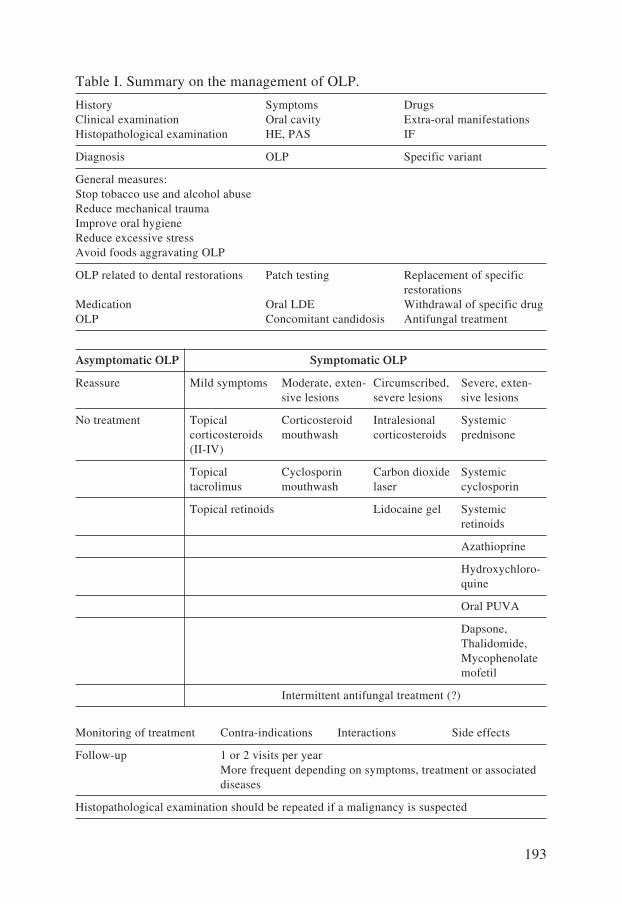
193
Table I. Summary on the management of OLP.
History Symptoms DrugsClinical examination Oral cavity Extra-oral manifestationsHistopathological examination HE, PAS IF
Diagnosis OLP Specific variant
General measures:Stop tobacco use and alcohol abuseReduce mechanical traumaImprove oral hygieneReduce excessive stressAvoid foods aggravating OLP
OLP related to dental restorations Patch testing Replacement of specific restorationsMedication Oral LDE Withdrawal of specific drugOLP Concomitant candidosis Antifungal treatment
Asymptomatic OLP Symptomatic OLP
Reassure Mild symptoms Moderate, exten- Circumscribed, Severe, exten- sive lesions severe lesions sive lesions
No treatment Topical Corticosteroid Intralesional Systemic corticosteroids mouthwash corticosteroids prednisone (II-IV)
Topical Cyclosporin Carbon dioxide Systemic tacrolimus mouthwash laser cyclosporin Topical retinoids Lidocaine gel Systemic retinoids
Azathioprine
Hydroxychloro- quine
Oral PUVA Dapsone, Thalidomide, Mycophenolate mofetil
Intermittent antifungal treatment (?)
Monitoring of treatment Contra-indications Interactions Side effects
Follow-up 1 or 2 visits per year More frequent depending on symptoms, treatment or associated diseases
Histopathological examination should be repeated if a malignancy is suspected

194
in patients with OLP are mandatory. More frequent visits may be necessary depending on the symptomatology, the extent and the severity of OLP, the pos-sible extra-oral involvement of LP, associated diseases, the necessity to moni-tor a specific treatment and the possible accompanying risk factors of an oral malignancy. It is important to give notice of significant changes in the specific variant of OLP which commonly occur in one patient during the follow-up.
Histopathological examination should be repeated if a malignancy is suspected, because an early detection of an OSCC favors the prognosis significantly. The clinical presentation of an OSCC may vary from indurated, non-healing ulcers to exophytic, hyperkeratotic masses and less frequently as red, submucosal and slightly indurated lesions with apparently intact epithelium.The patients are strongly urged to disengage from tobacco use and alcohol abuse. Oral candidosis is a common complication of OLP, particularly when topical or systemic immunosuppressive drugs are used.
Persistent, symptomatic OLP may cause chronic discomfort, nutritional defi-ciencies, reduced oral hygiene, increased anxiety and stress. Moreover, it may cause difficulties with speech and overall functioning and may negatively influ-ence the quality of life. A multi-disciplinary approach may be necessary when there is extra-oral LP involvement.

CHAPTER 10
SUMMARY,
GENERAL DISCUSSION AND
RECOMMENDATIONS
FOR FURTHER RESEARCH


197
Summary and general discussion
In chapter 1, a general introduction on oral lichen planus (OLP), the aim of the thesis, and a short treatise on the history of medicine, dermatology and lichen planus (LP) are described. Moreover, several aspects of the oral cavity includ-ing the histology of the oral mucosa, the immunity of the oral cavity and the dentition are described.Erasmus Wilson (1809-1884) initially coined the term lichen planus (LP) in 1869. In a study of 50 patients with LP, three patients also had involvement of the oral mucosa. Wilson considered it to be the same disease as “leichen ruber” described by F. (von) Hebra in 1860. L.F. Wickham noted the punctations and striae atop the lesions of LP that currently bear his name in 1895. W. Dubreuilh defined the histopathological characteristics of OLP in 1906. H. Hallopeau reported a case of OLP with malignant degeneration in 1910. H. Gougerot and A. Civatte described the presence of colloid or cytoid bodies (Civatte’s bodies) in the microscopical examination of LP in 1953.
A review of the literature on OLP is given in chapter 2. The clinical spectrum of LP may involve the skin, the mucous membranes, the hair and the nails. Oral lichen planus may affect up about 0.5 to 2% of the population. Generally, it is a disease of the middle-aged and the elderly with females being affected more than men. Oral lichen planus can be categorized into different subtypes namely reticular, papular, plaque, erosive, ulcerative or bullous, and atrophic or erythematous. The atrophic, erosive or ulcerative variants generally have persistent symptoms including pain or stinging, which may be extremely distressing and even disabling. The lesions are often found (symmetrical) on the buccal mucosa, the lateral margins of the tongue and the gingiva. Approximately 15 to 35% of the patients with OLP have concomitant cutaneous lichen planus (CLP). Oral lichen planus has been reported to be associated with a variety of disorders and particularly with disorders involving altered or disturbed immunity. However, it may be difficult to determine whether there is a causal or a purely fortuitous association. Generally, the diagnosis of OLP is based on a combination of characteristic clinical findings, history and histopathological examination. The exact cause of OLP is unknown, but an immune-mediated (T-cell dependant) pathogenesis has been proposed. Oral lichen planus may be aggravated by stress, mechanical traumata, specific foods (crisp foods, citrus juices and spicy ingredients), poor oral hygiene, irritation or allergy to dental (metal) restorations. Many drugs may be involved in an oral lichenoid drug eruption (oral LDE), which shows features identical or very similar to those of OLP.Histologically, the principal features of OLP are a dense band-like inflam-matory infiltrate in the lamina propria that mainly consists of T-cells and a liquefaction degeneration of the basal epithelial cells.

198
Oral lichen planus is usually a persistent disorder, despite different kinds of treatment. Asymptomatic reticular (often symmetrical) buccal lesions of OLP do not need treatment in contrast with symptomatic variants of OLP. Topical treatment may include corticosteroids, antifungals, retinoids, tacroli-mus and cyclosporin. Persistent circumscribed lesions may also be treated with analgesics, intralesional corticosteroids, carbon dioxide laser or local PUVA. In severe symptomatic OLP with extensive lesions, systemic therapy with cor-ticosteroids, retinoids, cyclosporin, hydroxychloroquine and azathioprine may be necessary. Combinations of therapeutic agents are also possible. Generally, OLP follows a chronic course, with periods of quiescence and flare ups. Oral candidosis is a common complication of OLP, particularly when topical or systemic immunosuppressive therapeutic agents are used. The possible malig-nant transformation of OLP into an oral squamous cell carcinoma (OSCC) still remains controversial.Generally, the schedule of follow-up of patients with OLP should be at least one or two visits per year as long as OLP persists. A careful physical examina-tion at each visit is imperative and histopathological examination should be repeated if a malignancy is suspected. Other variants of LP such as the vulvovaginal-gingival syndrome (VVGS), genital LP, LP of the lips and LP of the esophagus were also reported. More-over, the clinical aspects of CLP with different variants were described.The classical form of CLP comprises pruritic, faintly erythematous to viola-ceus, flat-topped papules which are usually symmetrically distributed with fine white lines (Wickham’s striae) on the surface of the lesions. The lesions have a predilection for the flexor parts of the wrists and the forearms, the ankles, the lumbar area, the (male) genitals and the shins.Overlap-syndromes including lichen planus-lupus erythematosus (LP-LE) overlap-syndrome and LP pemphigoides were reported. Lichenoid reactions such as graft-versus-host disease (GVHD), lichenoid contact dermatitis, oral and cutaneous lichenoid drug eruptions (LDEs) were also described.
In chapter 3, a prospective non-randomized study in 80 patients with one or more silver amalgam fillings is described. Sixty patients had OLP and were categorized into 3 groups based on the topographic relationship between the oral lesions and the amalgam fillings. In group A (20 patients), the oral lesions were confined to areas in close contact with amalgam fillings. In group B (20 patients), the lesions extended 1 cm beyond the area of contact with amalgam fillings. In group C (20 patients), the oral lesions had no topographic rela-tionship with amalgam fillings. Control group D (20 patients) had an allergic contact dermatitis without any pathologic evidence of OLP. Patch tests were performed with a standard and a dental metal series in all patients. Partial or complete replacement of amalgam fillings was recommended in case of

199
a positive patch test reaction to ammoniated mercury, metallic mercury, or amalgam. In group A, 13 patients had positive patch test reactions to one or more mercury compounds. Partial or complete replacement of dental amalgam resulted in at least a significant improvement in these 13 patients. In group B, 8 patients had positive patch test reactions to one or more mercury compounds. Partial or complete replacement resulted in at least a significant improvement in these 8 patients. In group C, 2 patients had positive patch test reactions to one or more mercury compounds. Replacement of dental amalgam fillings in these 2 patients resulted in improvement in 1 patient. In 8 (35%) of these 23 patients, positive patch test reactions to ammoniated mercury, metallic mercury, or amalgam occurred after the regular 3-day evalu-ation with a variation of 5 to 18 days (mean 8 days). The effect of replacements of amalgam was generally observed after 1 to 4 months (mean 3 months). No positive patch test reactions to mercury compounds were observed in patients with OLP and concomitant CLP.It was concluded that contact allergy to mercury compounds is important in the pathogenesis of OLP, especially if there is a close contact between the oral lesions and the dental amalgam fillings and if no concomitant CLP is present. In case of a positive patch test reaction to ammoniated mercury, metallic mer-cury, or amalgam, partial or complete replacement of amalgam fillings will lead to a significant improvement in nearly all patients with OLP.
In chapter 4, a retrospective non-randomized study on oral manifestations of gold allergy and an update on gold allergy are presented. Generally, gold allergy is more common than was previously recognized. In the literature, gold sodium thiosulfate (0.5% in pet), also known as sodium thiosulfatoaurate, is regarded as the most reliable reagent for patch testing in suspected cases of gold allergy. However, Fisher regarded gold(tri)chloride as a reliable test aller-gen. Positive reactions to gold may appear after the regular 3-day evaluation and may be long-lasting.Two hundred patients with symptoms of persistent oral mucosal or cutaneous lesions that were possibly related to allergy to constituents of dental gold alloys or gold jewelry were patch tested to determine the frequency of sensitization. In 17 (8.5%) patients, all women, with a mean age of 50 years, positive patch test reactions to gold(tri)chloride 0.5% aq and 1.0% aq were observed. Positive reactions to sodium tetrachloroaurate and potassium dicyanoaurate were not observed. In 5 of the 7 patients with OLP and in 1 of the 6 patients with the burning mouth syndrome, dental gold restorations were replaced. Particularly in patients with OLP, a significant but variable improvement was observed. In all cases in which gold was replaced and improvement occurred, the patients were sensitized to gold(tri)chloride 0.5% aq. One patient with allergic contact stomatitis and 1 patient with allergic contact dermatitis healed completely after
200
gold had been replaced. No replacement of gold was recommended in a patient with relapsing angioedema and in another patient with chronic urticaria. In all, except one case, the patch test reactions were persistently positive varying from one to several weeks (mean 4 weeks). In 3 patients, only late positive reactions occurred and were observed after respectively 5, 6 and 14 days. In another patient, a reaction to gold(tri)chloride 1% aq characterized by infiltration and purpura, developed after 1 month.It was concluded that sensitization to gold should be considered as a possible cause of allergic contact dermatitis, allergic contact stomatitis and as a patho-genic or triggering factor in OLP.
A historical cohort study in 200 Caucasian patients with a confirmed diagnosis of OLP is described in chapter 5. The study was conducted to examine oral malignancies associated with OLP and to investigate whether OLP has an intrinsic malignant potential or whether there are also contributing external risk factors.In the literature, the possible malignant transformation of OLP still remains controversial. The transformation of OLP into an oral malignancy ranges from 0 to 10%. Tobacco exposure, alcohol abuse, poor nutrition, leucoplakia and erythroplakia are known risk factors for oral cancer. The follow-up in the study included at least one or two visits per year and ranged from 7 to 13 years (mean 10 years). Histopathological examina-tion was repeated if a malignancy was suspected. Three (1.5%) of the 200 patients developed an oral squamous cell carcinoma (OSCC) after a period of 3 to 6 years (mean 4 years) following the initial diagnosis of OLP. Possible contributing external risk factors were also noted in two of these three patients. One male patient (59 years) with hyperkeratotic OLP (plaque and reticular lesions) had smoked approximately 25 cigarettes a day during 20 years. One female patient (67 years) with erosive OLP had received systemic immunosup-pressive treatment for 2 years for OLP. There were no contributing external risk factors in another female patient (78 years) with the atrophic variant of OLP and an OSCC. The probability of at least one case of OSCC in our study accord-ing to the Poisson distribution could have been by chance alone. However, it is highly improbable (statistically significant) that at least 2 or 3 cases of oral malignancy in our study were encountered by chance alone.The investigations provided some, but not convincing support for the notion that OLP is a premalignant condition. However, the exact incidence of malig-nant transformation is difficult to establish, because of the low number of patients and the possible contribution of external risk factors, which may also be relevant in oral malignancy.
In chapter 6, a prospective study in 100 patients with OLP and a control group of 100 patients with psoriasis vulgaris is described. The study was conducted

201
to determine the possible relationship between OLP and hepatitis C virus infec-tion in The Netherlands and to investigate whether routine screening for anti-hepatitis C virus antibodies and liver enzymes is necessary in these patients. In both groups, there was no serological evidence of antibodies against hepati-tis C virus (HCV) at all. In each group, there were 2 patients with elevated liver enzyme levels, but the difference was not statistically significant.A definite conclusion could not be established in three of these patients. One male patient with psoriasis vulgaris was an alcohol abuser which could explain the abnormal liver enzyme values. In these 4 patients, there was also no evi-dence of either hepatitis A virus infection or hepatitis B virus infection.In the literature, the results of the possible relationship between OLP and HCV infection are rather discrepant and controversial. In parts of the world for exam-ple Brazil, Japan and Spain with a high prevalence of HCV infection a positive relationship has been reported. No correlation between OLP and HCV infection was found in the United Kingdom and Scandinavia where the prevalence of HCV infection is low. However, conflicting results have been reported from Germany. The results of our study suggested that there is no indication for routinely screening for anti-hepatitis C virus antibodies in patients with OLP in The Netherlands. Liver enzyme values should only be determined if warranted. The discrepancies in the literature on the possible association of OLP and hepatitis C virus are largely based on the differences in prevalence of both hepatitis C virus infection and possibly also of OLP world-wide with a population of dif-ferent genetic make-up, different environmental conditions and possible varia-tions in hepatitis C virus infection and host immune responses.
In chapter 7, individual cases of OLP in childhood from our practice together with a review of the literature on this subject are reported. A retrospective non-randomized study was conducted during a period of 10 years. Approximately 10,000 dermatological patients younger than 18 years visited our department. Only three (0.03%) patients with OLP in childhood were encountered.An Asian girl aged 11 years had an asymptomatic reticular variant of OLP, which resolved without any treatment after one year. An Asian boy aged 16 years had an erosive variant of OLP with severe pain, which healed after inten-sive topical and systemic treatment for two years. A Caucasian girl aged 14 years had a reticular variant of OLP with a little soreness, which resolved after topical treatment for three months. Literature on OLP in childhood is scarce and there are only a few reports on this subject. Generally, the oral mucosa seems to be less commonly involved in children than in adults with LP. The large differences in the prevalence of OLP in childhood versus OLP in adulthood can only be explained partially by a low number of associated systemic diseases, auto-immune phenomena,

202
infections, drugs and dental restorations in childhood, which may reduce the risk for developing OLP. Moreover, the diagnosis may be missed because of irregular dental check ups, lack of symptoms and ignorance. In the literature, the prevalence of OLP in childhood seems to be significantly higher in those of the Asian race. It is highly unlikely (statistically significant) according to the Poisson distribution that 2 Asian patients in our study were encountered by chance alone. This indicates that a specific genetic predisposition (HLA-dependant) in the Asian race, despite a negative family history, may possibly be important in the pathogenesis of OLP in childhood.It was concluded that oral lichen planus in childhood is rare. Therefore, at present it is not possible to draw firm conclusions on this subject. Oral lichen planus in childhood seems to occur preferentially in those of the Asian race. The clinical features resemble those of oral lichen planus in adulthood. How-ever, the prognosis of oral lichen planus in childhood generally seems to be more favorable than oral lichen planus in adulthood.
In chapter 8, a prospective randomized study in 40 patients with a confirmed diagnosis of symptomatic OLP is described. The aim of the study was to com-pare the treatment of OLP with topical tacrolimus ointment and triamcino-lone acetonide ointment. The periods of remission after cessation of both the treatments were also compared. Twenty patients (group I) were treated with topical tacrolimus 0.1% ointment and 20 patients (group II) were treated with triamcinolone acetonide 0.1% ointment. The ointments were applied 4 times per day onto the symptomatic oral lesions for a period of 6 weeks. In group I, 6 (30%) patients healed, 12 (60%) patients showed an improvement and 2 (10%) patients showed no improvement at all. In group II, 2 (10%) patients healed, 7 (35%) patients showed an improvement and 11 (55%) patients showed no improvement at all. The results of the treatment in group I were statistically significant better than in group II. Side effects of treatment were temporary and occurred in 8 (40%) patients of group I and in 3 (15%) patients of group II. However, the differences in the side effects were not statistically significant. Local irritation (burning and stinging) at the site of application persisting for about 10 to 30 minutes was the most commonly reported side effect. This side effect particularly occurred in patients with the erosive or ulcerative variants of OLP in both groups, but did not lead to discontinuation of the treatment. Unfortunately, oral lesions recurred within 3 to 9 weeks (mean 5 weeks) after the cessation of treatment in 13 (72%) of the 18 patients in group I and in 7 (78%) of the 9 patients in group II, who initially showed improvement or healing.It was concluded that treatment with topical tacrolimus 0.1% ointment showed a better initial therapeutic response than treatment with triamcinolone acetonide 0.1% ointment in patients with symptomatic OLP. The most common side effect was a transient burning or stinging at the site of application, but did

203
not lead to discontinuation of the treatment. Unfortunately, relapses occurred within several weeks after the cessation of the treatment in both groups.
In chapter 9, clinical guidelines on the management of OLP are described. Sev-eral important aspects such as history, clinical examination, histopathological examination, diagnosis, additional investigations, general measures, specific treatment, response to treatment and follow-up are described.
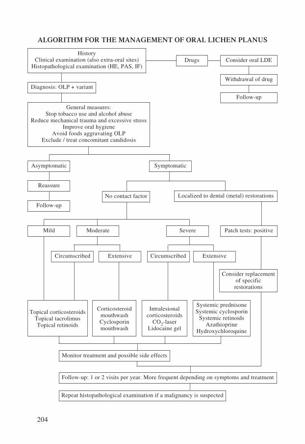
204
ALGORITHM FOR THE MANAGEMENT OF ORAL LICHEN PLANUS
HistoryClinical examination (also extra-oral sites)
Histopathological examination (HE, PAS, IF)Drugs Consider oral LDE
General measures:Stop tobacco use and alcohol abuse
Reduce mechanical trauma and excessive stressImprove oral hygiene
Avoid foods aggravating OLPExclude / treat concomitant candidosis
Consider replacementof specificrestorations
Withdrawal of drug
Follow-up
Diagnosis: OLP + variant
Asymptomatic
Reassure
Follow-up
Mild Moderate Severe Patch tests: positive
No contact factor
Symptomatic
Localized to dental (metal) restorations
Circumscribed Extensive Circumscribed Extensive
Topical corticosteroidsTopical tacrolimusTopical retinoids
CorticosteroidmouthwashCyclosporinmouthwash
Intralesionalcorticosteroids
CO2-laserLidocaine gel
Systemic prednisoneSystemic cyclosporin
Systemic retinoidsAzathioprine
Hydroxychloroquine
Monitor treatment and possible side effects
Follow-up: 1 or 2 visits per year. More frequent depending on symptoms and treatment
Repeat histopathological examination if a malignancy is suspected

205
Recommendations for further research
Well-designed studies in patients with OLP and control groups should be under-taken to elucidate the exact cause of OLP. Contact allergies to other allergens may possibly be relevant in the pathogenesis of OLP. In the literature, there is insufficient consensus on the time of occlusion of patch tests, particularly for metal allergens. Moreover, the optimum reading times of patch tests for these allergens showed significant differences and there is also a significant variation in the allergens and the concentrations that were used in studies in the literature. Therefore, those studies are difficult to compare and the relevant positive patch test reactions may have been missed.Histopathological features in OLP should be described more accurately to reduce intra- and interobserver variation. New immunopathological techniques may be useful for a more accurate diagnosis of OLP. Large studies are necessary to investigate the possible associations between other diseases with altered or disturbed immunity and OLP. At the moment, it may be difficult to determine whether there is a causal or a purely fortuitous association with other diseases. Furthermore, it may be impossible to distin-guish between an oral LDE and OLP on the currently available clinical, histo-pathological and immunological grounds. Many of the current treatments of OLP are empirical, based on anecdotal reports or open clinical trials without adequate control groups. Results of different studies cannot be compared because of the heterogeneity of the publications, the exact definition of OLP, the design of the studies, associated disorders, the therapeutic response and the follow-up. Randomized controlled clinical trials in larger populations are necessary to determine adequate treatment regimes for patients with OLP. The possible malignant transformation of OLP is a very important issue and, although there are many articles on this subject including our study, it still remains highly controversial. Large cohort studies in patients with a confirmed diagnosis of OLP with a long-term follow-up including identification of intrin-sic and contributing external risk factors are necessary. In many studies on OLP, insufficient attention has possibly been paid to the occurrence of extra-oral manifestations of LP, which may lead to inadequate diagnosis and treatment in some patients with LP.


CHAPTER 11
SAMENVATTING,
ALGEMENE DISCUSSIE EN
AANBEVELINGEN
VOOR VERDER ONDERZOEK


209
Samenvatting en algemene discussie
In hoofdstuk 1 worden een algemene inleiding over orale lichen planus (OLP), de doelstellingen van dit proefschrift en een korte verhandeling over de geschie-denis van de geneeskunde, dermatologie en lichen planus (LP) beschreven.
Erasmus Wilson (1809-1884) introduceerde de term “lichen planus” in 1869. In een studie met 50 patiënten met LP hadden 3 patiënten naast huidafwij-kingen ook afwijkingen van het mondslijmvlies passend bij OLP. Wilson dacht dat LP dezelfde ziekte was als “leichen ruber” beschreven door F. (von) Hebra in 1860. L.F. Wickham beschreef in 1895 de witte punten en strepen op de afwijkingen, die ook tegenwoordig nog Wickhamse striae wor-den genoemd. W. Dubreuilh definieerde de histopathologische kenmerken van OLP in 1906. H. Hallopeau rapporteerde in 1910 een casus van OLP met maligne ontaarding. H. Gougerot en A. Civatte beschreven in 1953 de aanwezigheid van colloïd- of cytoïdlichaampjes (Civatte’s bodies) in micros-copische preparaten van LP.
In hoofdstuk 2 wordt een overzicht van de literatuur over OLP gegeven.Het klinische spectrum van LP kan de huid, de aangrenzende slijmvliezen, het haar en de nagels omvatten. De prevalentie van OLP varieert van ongeveer 0,5 tot 2% in de algehele bevol-king. In het algemeen is het een aandoening die vaker gezien wordt bij vrou-wen dan bij mannen en meestal bij mensen van middelbare of oudere leeftijd. Er bestaan verschillende klinische varianten van OLP zoals de reticulaire en papuleuze vorm en de vorm met plaques. Deze varianten geven meestal weinig of geen klachten. Daarnaast bestaan er de erosieve, de ulceratieve of bulleuze en de erythemateuze of atrofische varianten van OLP, die veelal hardnekkige symptomen van pijn of branderigheid in de mondholte geven, hetgeen erg belastend en zelfs invaliderend voor de patiënt kan zijn. De afwijkingen bij OLP zijn vaak (symmetrisch) gelokaliseerd op de binnenzijden van het wang-slijmvlies, de zijkanten van de tong en op het tandvlees. Ongeveer 15 tot 35% van de patiënten met OLP hebben ook afwijkingen op de huid, passend bij LP. Er zijn associaties beschreven tussen OLP en diverse andere ziekten met name aandoeningen die zijn gekenmerkt door een gewijzigde of verstoorde immuni-teit. Het is echter vaak moeilijk te bepalen of er een oorzakelijk verband bestaat of dat er sprake is van een toevallige samenhang. De diagnose “OLP” wordt in het algemeen gesteld op grond van anamnese, lichamelijk onderzoek en histopathologisch onderzoek. De exacte oorzaak van OLP is niet bekend, maar er wordt een immuungemedieerde (T-cel afhanke-lijke) pathogenese verondersteld. Orale lichen planus kan worden verergerd door diverse factoren zoals stress, specifieke voedingsbestanddelen (b.v. sap-pen van citrusvruchten, kruidige ingrediënten en knapperig voedsel), mechani-

210
sche traumata, slechte mondhygiëne en door irritatie of allergie voor (metalen) tandheelkundige materialen. Vele soorten geneesmiddelen kunnen betrokken zijn bij een orale lichenoïde geneesmiddeleruptie. Hierbij worden afwijkingen gezien die niet of nauwelijks te onderscheiden zijn van OLP.De essentiële kenmerken van OLP bij histopathologisch onderzoek zijn een dicht bandvormig ontstekingsinfiltraat in de lamina propria voornamelijk bestaand uit T-lymfocyten en een liquefactiedegeneratie van de basale epithe-liale cellen.Orale lichen planus is gewoonlijk een gedurende vele jaren bestaande aan-doening ondanks diverse vormen van behandeling. De reticulaire variant van OLP met asymptomatische afwijkingen, symmetrisch op de wangslijmvliezen, behoeft geen behandeling in tegenstelling tot de diverse andere, veelal symp-tomatische vormen van OLP. Lokale behandeling kan bestaan uit corticosteroïden, fungistatische of fungi-cide middelen, vitamine-A-zuren, tacrolimus en ciclosporine. Persisterende, omschreven letsels kunnen ook worden behandeld met analgetica, intralesi-onale corticosteroïden, CO2-laser of locale PUVA. Systemische therapie met corticosteroïden, retinoïden, ciclosporine, hydroxychloroquine of azathioprine kan noodzakelijk zijn bij uitgebreide, ernstige symptomatische vormen van OLP. Combinaties van diverse middelen bij de behandeling van OLP zijn ook mogelijk. Orale lichen planus vertoont meestal een chronisch verloop met remissies en exacerbaties. Orale candidiasis is een frequent voorkomende complicatie in OLP, met name wanneer er behandeling plaatsvindt met lokale of systemische immunosuppres-sieve middelen. De mogelijke maligne ontaarding van OLP in een plaveiselcel-carcinoom blijft helaas nog steeds een controversieel onderwerp. Patiënten met OLP dienen in het algemeen een of twee keer per jaar gecontro-leerd te worden zolang er afwijkingen bestaan. Nauwkeurige inspectie van de mondholte is noodzakelijk bij iedere controle. Histopathologisch onderzoek dient te worden herhaald wanneer er een klini-sche verdenking op maligniteit bestaat.Andere varianten van LP zoals het vulvovaginaal-gingivaal syndroom (VVGS), genitale LP, LP van de lippen en LP van de oesophagus worden ook besproken. Daarnaast worden de klinische aspecten van cutane LP (CLP) met de verschil-lende varianten beschreven. De klassieke vorm van CLP wordt gekenmerkt door jeukende, erythemateuze of livide, vlakke papels die meestal symmetrisch zijn gerangschikt met op de oppervlakte fijne witte lijntjes vaak als netwerk aanwezig (Wickhamse striae). De voorkeurslokalisaties zijn de buigzijden van de polsen en onderarmen, de enkels, de onderrug, de (mannelijke) genitaliën en de scheenbenen.Mengbeelden zoals het lichen planus-lupus erythematodes (LP-LE) over-lapsyndroom en LP pemphigoïdes worden beschreven. Lichenoïde reac-

211
ties zoals “graft-versus-host ziekte” (GVHD), lichenoïde contactdermati-tis, orale en cutane lichenoïde geneesmiddelreacties worden eveneens be- schreven.
In hoofdstuk 3 wordt een prospectieve, onwillekeurig verdeelde studie met 80 patiënten met een of meer amalgaamvullingen beschreven. Zestig patiënten met OLP werden verdeeld in 3 groepen met een gelijk aantal patiënten, gebaseerd op de relatie van de slijmvliesafwijkingen en de amalgaamvullingen. Groep A met 20 patiënten had mondafwijkingen beperkt tot gebieden in direct contact met het amalgaam. Groep B met 20 patiënten vertoonde afwijkingen ook bui-ten een gebied van een centimeter van de amalgaamvullingen. In groep C met eveneens 20 patiënten hadden de mondafwijkingen geen directe topografische relatie met de amalgaamvullingen. Twintig patienten in groep D (controlegroep) vertoonden het beeld van een allergisch contacteczeem zonder enige aanwijzing voor OLP. Epicutane tests werden bij alle 80 patiënten uitgevoerd met een standaardreeks en een tand-heelkundige metalenreeks. Bij een positieve uitslag bij plakproeven voor geam-monieerd kwik, elementair kwik of amalgaam werd geadviseerd om de amal-gaamvullingen gedeeltelijk of geheel te vervangen.In groep A vertoonden 13 (65%) patiënten een of meer positieve reacties op de bovengenoemde kwikbestanddelen. Partiële of complete vervanging van de amalgaamvullingen bij deze 13 patiënten leidde tenminste tot een signifi-cante verbetering van de afwijkingen van OLP. In groep B vertoonden 8 (40%) patiënten een of meer positieve reacties op de kwikbestanddelen. Gedeeltelijke of gehele vervanging van het amalgaam bij deze 8 patiënten leidde tenminste tot een significante verbetering van de mondafwijkingen. In groep C hadden 2 (10%) patiënten een of meer positieve reacties op de kwikbestanddelen. Ver-vanging van de amalgaamvullingen leidde slechts bij 1 patiënt tot verbete-ring.In 8 (35%) van de 23 patiënten werden positieve uitslagen voor geammoni-eerd kwik, elementair kwik of amalgaam bij epicutane tests pas gezien na het reguliere aflezen op de derde dag, variërend van 5 tot 18 dagen (gemiddeld 8 dagen). Het positieve resultaat van het vervangen van amalgaamvullingen werd meestal gezien na 1 tot 4 maanden (gemiddeld 3 maanden). Positieve uitslagen bij plakproeven voor kwikbestanddelen werden niet gezien bij patiënten met OLP die ook CLP hadden.Conclusies: een contactallergie voor kwikbestanddelen kan van belang zijn in de pathogenese van OLP, met name wanneer er een nauw contact bestaat tus-sen de mondafwijkingen en de amalgaamvullingen en er geen bijkomende CLP bestaat. Wanneer er een positieve uitslag voor geammonieerd kwik, elementair kwik of amalgaam bestaat bij epicutane tests, zal gedeeltelijke of gehele ver-vanging van de amalgaamvullingen leiden tot een significante verbetering bij nagenoeg alle patiënten met OLP.

212
In hoofdstuk 4 wordt er een recent overzicht gegeven over allergie voor goud en wordt er een studie overwegend over mondafwijkingen in relatie tot allergie voor goud beschreven.Goudallergie blijkt in het algemeen veel meer voor te komen dan men voor-heen dacht. In de literatuur wordt goudnatriumthiosulfaat (0,5% in vaseline), ook bekend onder de naam natriumthiosulfatoauraat, beschouwd als het meest betrouwbare allergeen bij epicutane tests om een goudallergie aan te tonen. Het meest betrouwbare allergeen volgens Fisher was echter goud(tri)chloride. Positieve reacties bij plakproeven op goud kunnen op een later tijdstip dan het reguliere aflezen op de derde dag voorkomen en kunnen langer blijven bestaan. In een retrospectieve, onwillekeurig verdeelde studie werden 200 patiënten met persisterende afwijkingen van de mondholte of van de huid, mogelijk gerela-teerd aan een allergie voor tandheelkundige gouden materialen of voor gouden sieraden, onderzocht met epicutane tests om de frequentie van sensibilisatie vast te stellen.Bij 17 vrouwelijke patiënten (8.5%) met een gemiddelde leeftijd van 50 jaar werden positieve uitslagen gevonden voor goud(tri)chloride 0,5% en 1% in water. Positieve uitslagen voor natriumtetrachloroauraat en kaliumdicyanoau-raat werden niet gezien. Tandheelkundig goud in de mond (met name gouden kronen) werd bij 5 van de 7 patiënten met OLP en bij 1 van de 6 patiënten met het zogenoemde “brandend mondsyndroom” (BMS) vervangen. In het bijzonder bij patiënten met OLP werd een significante maar wisselende verbe-tering geconstateerd. Alle patiënten bij wie het goud werd vervangen en een verbetering werd gezien, vertoonden positieve uitslagen voor goud(tri)chloride 0,5% en 1% in water. Een patiënt met een allergische contactstomatitis en een patiënt met allergisch contacteczeem vertoonden een volledige genezing na het vermijden van het directe contact met goud. Verwijdering van goud was niet noodzakelijk en werd dan ook niet geadviseerd aan een patiënt met recidive-rend angio-oedeem en aan een patiënt met chronische urticaria.Positieve uitslagen voor goud(tri)chloride bij plakproeven waren persisterend positief bij 16 patiënten, variërend van een tot verscheidene weken (gemiddeld 4 weken). Bij 3 patiënten werden slechts laat positieve uitslagen voor goud gezien na respectievelijk 5, 6 en 14 dagen. Bij een andere patiënt werd pas na een maand een reactie op goud(tri)chloride 1% in water gezien, gekenmerkt door infiltratie en purpura. Conclusie: sensibilisatie voor goud kan als mogelijke oorzaak worden beschouwd bij patiënten met allergisch contacteczeem en allergische contact-stomatitis en als een pathogenetische of verergerende factor in OLP.
In hoofdstuk 5 wordt een historische cohortstudie beschreven met 200 patiën-ten met OLP. Hierbij werd onderzocht of orale maligniteiten geassocieerd kun-nen zijn met OLP en of er een intrinsiek maligne potentieel van OLP bestaat
213
of dat er andere, bijkomende externe factoren ook verantwoordelijk kunnen zijn.De mogelijke maligne ontaarding van OLP blijft nog steeds een controversieel onderwerp. De percentages in de literatuur die worden opgegeven voor maligne ontaarding van OLP in een plaveiselcelcarcinoom variëren van 0 tot 10%. Bekende risicofactoren voor orale maligniteiten zijn roken, alcoholmisbruik, ondervoeding, leukoplakie en erythroplakie.De periode van het vervolgen van de patiënten varieerde van 7 tot 13 jaar (gemid-deld 10 jaar). Histopathologisch onderzoek werd herhaald indien er op klinische gronden een maligniteit werd vermoed. Drie (1,5%) patiënten ontwikkelden een plaveiselcelcarcinoom in de mondholte op de aanvankelijke plaats van OLP na een periode van 3 tot 6 jaar (gemiddeld 4 jaar). Twee patiënten hadden aantoon-bare externe risicofactoren. Een patiënt (59 jaar) met de hyperkeratotische vorm (met plaques en reticulaire afwijkingen) van OLP rookte ongeveer 25 sigaretten per dag gedurende 20 jaar. Een patiënte (67 jaar) met de erosieve vorm van OLP had gedurende 2 jaar een systemische immunosuppressieve behandeling voor OLP gehad. Bij een andere patiënte (78 jaar) met de atrofische vorm van OLP konden geen extrinsieke risicofactoren worden vastgesteld.De kans dat er tenminste 1 patiënt met een plaveiselcelcarcinoom van de mond-holte in deze studie aanwezig is, zou op grond van de Poisson-verdeling door het toeval kunnen komen. Het is echter heel onwaarschijnlijk (statistisch sig-nificant), dat er tenminste 2 of 3 patiënten in de studie zouden voorkomen op grond van het toeval.Conclusies: de resultaten van deze studie geven enig, maar geen overtuigend bewijs voor het feit dat OLP een premaligne aandoening is. Het exacte per-centage van eventuele maligne ontaarding van OLP is moeilijk vast te stellen, enerzijds omdat er relatief weinig patiënten aanwezig zijn en anderzijds omdat de mogelijke rol van externe risicofactoren voor orale maligniteiten lastig te bepalen is.
In hoofdstuk 6 wordt een prospectieve studie beschreven met een groep van 100 patiënten met OLP en een controlegroep van 100 patiënten met psoriasis vulgaris. De doelstellingen waren om de mogelijke relatie in Nederland vast te stellen tussen OLP en hepatitis-C-virusinfectie en om te kijken of routinematig onderzoek op hepatitis-C-antilichamen en leverenzymen noodzakelijk is.In beide groepen werden in het serum in het geheel geen antilichamen tegen het hepatitis-C-virus aangetroffen.In elk van beide groepen werden 2 patiënten gezien met enkele verhoogde lever-enzymwaarden, hetgeen niet statistisch significant verschillend is. Een pas-sende verklaring voor de bovengenoemde afwijkingen kon bij 3 patiënten niet worden gegeven. Een patiënt met psoriasis vulgaris gebruikte overmatig veel alcohol, hetgeen de afwijkingen kon verklaren. Bij deze 4 patiënten werden ook geen aanwijzingen gezien voor hepatitis-A- of hepatitis-B-virusinfecties.

214
In de literatuur wordt de mogelijke relatie tussen OLP en hepatitis-C-virusin-fectie nogal verschillend en tegenstrijdig opgegeven. In bepaalde delen van de wereld met een hoge prevalentie van hepatitis-C-virusinfecties, zoals bijvoor-beeld Brazilië, Japan en Spanje, wordt een positief verband beschreven. Geen positief verband tussen OLP en hepatitis-C-virusinfectie wordt vermeld in gebieden met een lage prevalentie van hepatitis-C-virusinfecties zoals bij-voorbeeld het Verenigd Koninkrijk en Scandinavië.In Duitsland zijn er tegenstrijdige berichten over dit onderwerp beschreven.Conclusies: onze studie suggereert dat er geen indicatie is voor routinematig onderzoek op antilichamen tegen hepatitis-C-virus bij patiënten met OLP in Nederland. Leverenzymwaarden dienen alleen te worden bepaald in specifieke, klinisch relevante situaties. De discrepantie in de literatuur omtrent het moge-lijke verband tussen OLP en hepatitis-C-virusinfectie zijn grotendeels te ver-klaren op grond van substantiële verschillen in prevalentie van zowel hepatitis-C-virusinfecties en mogelijk ook van OLP in de wereld. Bovendien spelen een verschillende genetische achtergrond van de diverse volkeren, uiteenlopende omgevingsfactoren en mogelijke verschillen in hepatitis-C-virusinfecties zelf en in de immuunrespons van de gastheer een rol.
In hoofdstuk 7 worden de individuele gevallen van OLP op kinderleeftijd en een beschouwing van de literatuur over dit onderwerp beschreven. Een retros-pectieve studie werd verricht over een periode van 10 jaar. Ongeveer 10.000 patienten jonger dan 18 jaar werden gezien op onze polikliniek dermatologie. In die tijd kon slechts bij 3 patiënten OLP op kinderleeftijd worden vastgesteld. Een 11-jarig Aziatisch meisje had de asymptomatische, reticulaire variant van OLP. De afwijkingen verdwenen zonder behandeling na 1 jaar.Een 16-jarige Aziatische jongen had de erosieve vorm van OLP met veel pijn-klachten. Na intensieve lokale en systemische behandeling trad er in 2 jaar tijd een volledige genezing op. Een 14-jarig meisje van Nederlandse afkomst ver-toonde de reticulaire variant van OLP met geringe pijnklachten. De afwijkingen verdwenen met lokale behandeling na 3 maanden.Ook in de literatuur is OLP op kinderleeftijd zeldzaam en er zijn dan ook maar enkele artikelen over dit onderwerp geschreven. Het mondslijmvlies is bij kin-deren met LP in het algemeen ook minder vaak aangedaan dan bij volwassenen. De grote verschillen in de prevalentie van OLP op kinderleeftijd t.o.v. OLP op volwassen leeftijd kunnen slechts ten dele worden verklaard door een lager aantal geassocieerde aandoeningen, auto-immuun fenomenen, infecties, medi-camenten en tandheelkundige vullingsmaterialen bij kinderen, die het risico op het ontwikkelen van OLP kunnen verminderen. Daarenboven zou de diagnose OLP gemist kunnen worden door onregelmatig bezoek aan de tandarts, het ontbreken van symptomen en door onwetendheid. In de literatuur blijkt OLP op kinderleeftijd meer bij kinderen van Aziatische afkomst voor te komen. Ook in onze studie waren 2 van de 3 kinderen van Aziatische afkomst hetgeen

215
volgens de Poisson-verdeling (statistisch significant) zeer onwaarschijnlijk is op grond van het toeval. Dit geeft wellicht aan dat een specifieke genetische predispositie (afhankelijk van het HLA-systeem) in het Aziatische ras, ondanks een negatieve familieanamnese, belangrijk zou kunnen zijn in de pathogenese van OLP op kinderleeftijd.Conclusies: orale lichen planus op kinderleeftijd is zeldzaam en daarom is het niet goed mogelijk om definitieve uitspraken te doen over diverse aspecten van dit onderwerp. Orale lichen planus op kinderleeftijd blijkt met name voor te komen bij Aziatische kinderen. De klinische presentatie van OLP op kin-derleeftijd lijkt overeen te komen met de presentatie van OLP op volwassen leeftijd. De prognose van OLP op kinderleeftijd lijkt echter in het algemeen gunstiger te zijn dan van OLP op volwassen leeftijd.
In hoofdstuk 8 wordt een prospectieve, willekeurig verdeelde studie beschre-ven met 40 patiënten met een symptomatische vorm van OLP. Het doel van de studie was het vergelijken van de behandelingsresultaten tussen lokale behan-deling met tacrolimuszalf en triamcinolonacetonidezalf en het beoordelen of er verschillen zijn in de perioden van remissie tussen de beide vormen van behan-deling. Groep I met 20 patiënten werd behandeld met 0,1% tacrolimuszalf en groep II met eveneens 20 patiënten werd behandeld met 0,1% triamcinolonace-tonide in 20% hypromellosezalf. De zalven werden gedurende 6 weken 4 keer per dag aangebracht op de afwijkingen van het mondslijmvlies. In groep I genazen 6 (30%) patiënten, vertoonden 12 (60%) patiënten een ver-betering en lieten 2 (10%) patiënten geen verbetering zien. In groep II genazen 2 (10%) patiënten, vertoonden 7 (35%) patiënten een verbetering en lieten 11 (55%) patiënten geen verbetering zien. De (initiële) resultaten van behande-ling in groep I waren significant beter dan die in groep II. Bijwerkingen van de behandeling waren van voorbijgaande aard en traden op in 8 (40%) patiënten van groep I en in 3 (15%) patiënten van groep II. Deze verschillen tussen beide groepen zijn statistisch echter niet significant. Irritatie in de vorm van een bran-derig of prikkelend gevoel op de plaats van de aangebrachte zalf gedurende ongeveer 10 tot 30 minuten was de meest voorkomende bijwerking. Deze bij-werking trad met name op bij patiënten met de erosieve of ulceratieve variant van OLP in beide groepen, maar leidde in geen van de gevallen tot het voortij-dig onderbreken van de behandeling. Helaas recidiveerden de afwijkingen bin-nen 3 tot 9 weken (gemiddeld 5 weken) na het beëindigen van de behandeling in 13 (72%) van de 18 patiënten in groep I en in 7 (78%) van de 9 patiënten van groep II, die aanvankelijk volledig of aanzienlijk waren verbeterd.Conclusies: behandeling met 0,1% tacrolimuszalf is aanvankelijk effectiever dan behandeling met 0,1% triamcinolonacetonidezalf bij patiënten met een symptomatische vorm van OLP. Tijdelijke irritatie op de plaats van applicatie van beide zalven komt regelmatig voor, maar leidt niet tot het voortijdig stop-pen van de behandeling. Helaas treden er frequent recidieven op binnen enkele

216
weken na het beëindigen van de behandeling in beide groepen van patiënten met OLP.
In hoofdstuk 9 worden klinische richtlijnen voor de benadering van patiënten met OLP beschreven. Diverse belangrijke aspecten zoals anamnese, lichame-lijk onderzoek, histopathologie, diagnose, aanvullend onderzoek, algemene adviezen, specifieke behandeling, resultaat van behandeling en het vervolgen van de patiënten met OLP worden in het kort besproken.
Aanbevelingen voor verder onderzoek
De exacte oorzaak van OLP kan alleen worden opgehelderd wanneer er goed opgezette studies met adequate controlegroepen worden verricht. Contactaller-gieën voor andere allergenen dan tot dusver beschreven kunnen mogelijk ook van belang zijn in de pathogenese van OLP. In de literatuur is er onvoldoende overeenstemming over de tijd van occlusie van allergenen bij plakproeven, in het bijzonder van metaalallergenen. De diverse studies in de literatuur kunnen opvallend verschillende tijdstippen van aflezen van de epicutane tests vertonen. Bovendien bestaan er vaak substantiële verschillen in de concentraties en de allergenen die worden gebruikt. Sommige studies zijn daarom moeilijk met elkaar te vergelijken. Bovendien kunnen relevante positieve uitslagen bij plakproeven tamelijk een-voudig worden gemist door het pas laat positief worden van de test. Histopathologische kenmerken van OLP dienen nog nauwkeuriger te worden gedefinieerd om verschillen tussen de beoordeling van dezelfde preparaten tussen specialisten te verminderen. Nieuwe immunopathologische technieken zouden hiertoe wellicht in de toekomst kunnen bijdragen. Studies met vele patiënten zijn nodig om eventueel relevante associaties vast te leggen tussen diverse ziekten, in het bijzonder met een gewijzigde of verstoorde immuniteit, en OLP. Op dit moment is het namelijk veelal moeizaam vast te stellen of er sprake is van een oorzakelijk verband of van een toevallige samenhang tussen OLP en de diverse aandoeningen. Daarnaast is het op dit moment veelal onmogelijk om op grond van klinisch, histopathologisch en immunologisch onderzoek een onderscheid te maken tus-sen OLP en een orale lichenoïde geneesmiddelreactie.Vele huidige behandelingsmethoden van OLP zijn helaas vaak slechts geba-seerd op empirie, anekdotische gevallen of open klinische onderzoeken zonder adequate controlegroepen. Vele gegevens van patiëntengroepen met OLP kun-nen onvoldoende met elkaar worden vergeleken door essentiële verschillen in het type van de publicaties, de exacte definitie van OLP, de opzet van de studies, eventueel bijkomende geassocieerde afwijkingen, de resultaten van behandeling en de periode van vervolgen van de patiënten.

217
Klinische, statistisch verantwoorde onderzoeken met grote groepen patiënten en controlegroepen zijn noodzakelijk om goede behandelingsmethoden te vin-den bij patiënten met symptomatische vormen van OLP. De mogelijke maligne ontaarding van OLP blijft helaas nog steeds een contro-versieel onderwerp, ondanks diverse gepubliceerde artikelen hierover, inclusief een hoofdstuk in dit proefschrift. Grote, langdurige vervolgstudies met patiën-ten met een goed gedocumenteerde diagnose van OLP en een nauwkeurige vaststelling van intrinsieke en extrinsieke risicofactoren zijn noodzakelijk.In vele studies over OLP is er mogelijk onvoldoende aandacht besteed aan het eventueel voorkomen van LP op andere plaatsen van het lichaam, hetgeen bij sommige patiënten de betrouwbaarheid van de diagnose negatief zou kunnen beïnvloeden en een andere behandeling zou kunnen rechtvaardigen.


219
FIGURES AND LEGENDS TO THE FIGURES
Figure 1: Characteristic reticular variant of OLP with Wickham’s striae on the left buccal mucosa, and (less obvious) on the upper gingiva.
Figure A: Statue of Hippocrates (460-377 B.C.).
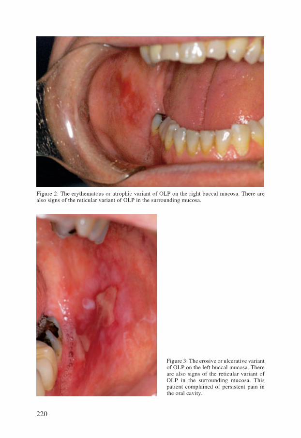
220
Figure 2: The erythematous or atrophic variant of OLP on the right buccal mucosa. There are also signs of the reticular variant of OLP in the surrounding mucosa.
Figure 3: The erosive or ulcerative variant of OLP on the left buccal mucosa. There are also signs of the reticular variant of OLP in the surrounding mucosa. This patient complained of persistent pain in the oral cavity.

221
Figure 4: The reticular variant of OLP on the dorsal side of the tongue, which may lead to taste disturbance.
Figure 5: The erythematous or atrophic variant of OLP on the lateral part of the tongue with reticular OLP lesions in the surrounding mucosa. Oral hygiene is also decreased.

222
Figure 6: Reticular variant of OLP on the vermilion of the lower lip.
Figure 7: An overview of the histopathology in CLP. The epidermis and the dermis may be identified. There is hyperkeratosis, wedge-shaped hypergranulosis, irregular acanthosis with a saw-toothed appearance, and a typical band-like inflammatory infiltrate in the upper part of the dermis. (HE staining, magnification 100×).
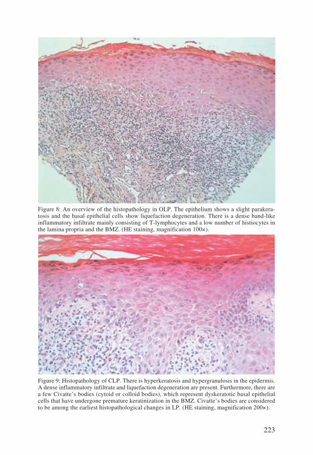
223
Figure 8: An overview of the histopathology in OLP. The epithelium shows a slight parakera-tosis and the basal epithelial cells show liquefaction degeneration. There is a dense band-like inflammatory infiltrate mainly consisting of T-lymphocytes and a low number of histiocytes in the lamina propria and the BMZ. (HE staining, magnification 100×).
Figure 9: Histopathology of CLP. There is hyperkeratosis and hypergranulosis in the epidermis. A dense inflammatory infiltrate and liquefaction degeneration are present. Furthermore, there are a few Civatte’s bodies (cytoid or colloid bodies), which represent dyskeratotic basal epithelial cells that have undergone premature keratinization in the BMZ. Civatte’s bodies are considered to be among the earliest histopathological changes in LP. (HE staining, magnification 200×).
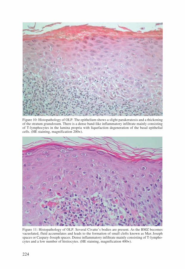
224
Figure 10: Histopathology of OLP. The epithelium shows a slight parakeratosis and a thickening of the stratum granulosum. There is a dense band-like inflammatory infiltrate mainly consisting of T-lymphocytes in the lamina propria with liquefaction degeneration of the basal epithelial cells. (HE staining, magnification 200×).
Figure 11: Histopathology of OLP. Several Civatte’s bodies are present. As the BMZ becomes vacuolated, fluid accumulates and leads to the formation of small clefts known as Max Joseph spaces or Caspary-Joseph spaces. Dense inflammatory infiltrate mainly consisting of T-lympho-cytes and a low number of histiocytes. (HE staining, magnification 400×).

225
Figure 12: A diagrammatic representation of the proposed pathogenesis of OLP (modified from Morhenn and Shiohara et al187,188,198, and Boyd and Neldner26).
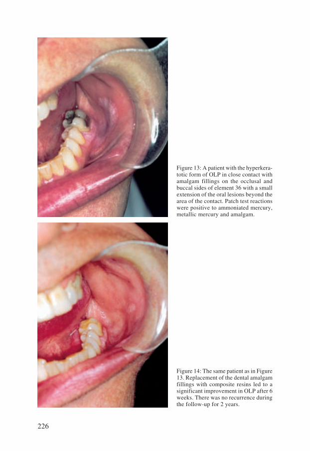
226
Figure 14: The same patient as in Figure 13. Replacement of the dental amalgam fillings with composite resins led to a significant improvement in OLP after 6 weeks. There was no recurrence during the follow-up for 2 years.
Figure 13: A patient with the hyperkera-totic form of OLP in close contact with amalgam fillings on the occlusal and buccal sides of element 36 with a small extension of the oral lesions beyond the area of the contact. Patch test reactions were positive to ammoniated mercury, metallic mercury and amalgam.

227
Figure 15: The ventral part of the lower leg of a patient with CLP. There are many pru-ritic, erythematous to violaceus papules and polygonal plaques present. Wickham’s striae are also visible on the surface of the lesions. The focal increase in thickness of the granular layer and inflammatory infiltrate corresponds to the presence of the Wickham’s striae.
Figure 16: Positive patch test reactions to ammoniated mercury, metallic mercury and amalgam on the back of a patient with OLP. These reactions were not observed at the regular 3-day evalu-ation, but were positive after 1 week and persisted for 2 weeks.
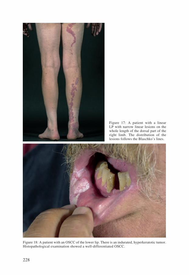
228
Figure 17: A patient with a linear LP with narrow linear lesions on the whole length of the dorsal part of the right limb. The distribution of the lesions follows the Blaschko’s lines.
Figure 18: A patient with an OSCC of the lower lip. There is an indurated, hyperkeratotic tumor. Histopathological examination showed a well-differentiated OSCC.

229
DANKWOORD
Graag wil ik iedereen bedanken die, direct of indirect, heeft bijgedragen aan de totstandkoming van dit proefschrift.
Mijn promotor, Prof.dr. H.A.M. Neumann, wil ik hartelijk danken voor zijn bijdrage aan dit proefschrift. Hoewel het onderwerp niet direct binnen de huidige onderzoekslijn van de afdeling Dermatologie en Venereologie van het Erasmus Medisch Centrum Rotterdam lag, was u direct enthousiast en vol vertrouwen dat het een leuk en goed proefschrift kon worden. Gelukkig werd ik heel vrij gelaten in de onder-werpen die volgens mij behandeld en beschreven moesten worden. Het over-zicht heeft u echter goed behouden met passende adviezen voor een adequate voortgang in het schrijven.Mijn copromotor, dr. B. Tank, wil ik hartelijk danken voor zijn enorme inzet bij het schrijven van dit proefschrift. Het zeer nauwkeurig corrigeren van het “US English” van alle teksten met het bekende rode potlood telkens binnen enkele dagen is een enorm werk geweest. Mijn hiaten in de kennis van woordkeuze, zinsopbouw en grammatica waren helaas veelvuldig aanwezig. Goede adviezen en correcties voor de opbouw en indeling van de artikelen waren nodig om ze klaar te maken voor publicatie in de diverse internationale tijdschriften. Mijn opleider, Prof.dr. Th. van Joost, wil ik hartelijk danken voor het feit dat hij mij in 1989 in Rotterdam in opleiding voor dermatoloog heeft genomen en voor de oprechte vriendschap die er tussen ons is ontstaan. In 1991 raakten we door enkele patiënten op de polikliniek dermatologie geïnteresseerd in de eventuele oorzaak en behandeling van orale lichen planus (OLP). We waren beiden vrij snel overtuigd dat een allergie voor tandheelkundige materialen, in het bijzonder voor amalgaamvullingen en gouden kronen, bij een substantieel deel van de patiënten met OLP klinisch relevant was. Plakproeven met uiteen-lopende tandheelkundige metalen en concentraties van allergenen werden bij vele patiënten uitgevoerd. Gedeeltelijke of volledige vervanging van het betref-fende tandheelkundig materiaal gaf bij meerdere patiënten een aanzienlijke ver-mindering van de klachten. Vanaf die tijd zijn we vele gegevens van patiënten met OLP systematisch gaan verzamelen en werden er diverse onderzoekspro-tocollen opgesteld. Hoewel u al enkele jaren met emeritaat bent, bleef u steeds

230
enthousiast en vol waardevolle adviezen toen ik besloot de vele gegevens te bundelen om een proefschrift te schrijven. Dat niet alles altijd succesvol was tijdens het onderzoek naar OLP, illustreert wel het feit dat we een keer ruim een kwartier zwijgzaam en wat stuurs voor ons uit zaten te kijken in uw kamer in het ziekenhuis met een speciale pleister in onze mond die geschikt zou zijn om slijmvliesafwijkingen te behandelen. Na korte tijd echter voelden we dat de pleister voortijdig los ging laten door de toename van speeksel en door kleine mondbewegingen. Dat die pleister “het niet ging worden” voor de patiënt werd ons spoedig duidelijk en nadien hebben we natuurlijk kostelijk gelachen over het door ons uitgevoerde experiment.
De promotiecommissie bestaande uit Prof.dr. J.W. Oosterhuis, Prof.dr. E.P. Prens en Prof.dr. I. van der Waal wil ik hartelijk danken voor hun gewaardeerde beoordeling van het proefschrift.Mijn directe collegae en maten in het Albert Schweitzer ziekenhuis, Piet Burger en Sybren Dekker, wil ik bedanken voor de loyaliteit, het vertrouwen en de vriendschap in onze maatschap. Goede patiëntenzorg en humor blijken heel goed samen te gaan. Zij hebben in de loop van de jaren vele patiënten met OLP voor mijn onderzoek verzameld, hetgeen noodzakelijk is om tot statistisch verantwoorde aantallen te komen. Daarnaast hebben ze mij ook gestimuleerd om dit proefschrift te schrijven.Dr. P.G.H. Mulder van de afdeling Epidemiologie en Biostatistiek van de Eras-mus Universiteit Rotterdam wil ik hartelijk danken voor zijn hulp voor de noodzakelijke statistische bewerkingen van al het onderzoek. Zoals jij reeds vooraf aankondigde zijn dergelijke statistische handelingen voor een “gewone” medisch specialist veel te ingewikkeld. Dr. Marti C. Kuizinga en andere collegae van het Pathologisch Anatomisch Laboratorium in Dordrecht wil ik hartelijk danken voor het beoordelen van de vele histologische coupes en het beschikbaar stellen van de foto’s met his-topathologische beschrijvingen voor dit proefschrift. Veelvuldig telefonisch overleg over patiënten verliep altijd prima.Esther Teerds-van ’t Hoog, secretaresse van onze maatschap, wil ik hartelijk danken voor haar inzet en het verrichten van het vele werk voor de maatschap. Het bewerken van de artikelen volgens de uiteenlopende richtlijnen van de diverse internationale tijdschriften was bij jou in goede handen.Wil Lokkerbol, voormalig secretaresse van Prof.dr. Th. van Joost en thans secretaresse van Prof.dr. H.A.M. Neumann in het Erasmus Medisch Centrum, wil ik hartelijk danken voor het vele typewerk tijdens mijn opleiding tot der-matoloog en de coördinatie van alle noodzakelijke documenten om te kunnen promoveren. Alle doktersassistenten, verpleegkundigen (en invalkrachten), huidtherapeuten, arts-assistenten en overige medewerksters, in het bijzonder werkzaam op of voor onze polikliniek dermatologie van het Albert Schweitzer ziekenhuis, heb-

231
ben vaak extra werk bij patiënten met OLP verricht, waarvoor dank. De “care” en de “cure” voor de patiënt staan op onze polikliniek dermatologie gelukkig nog steeds centraal. Alfons Jannink, medisch fotograaf, wil ik hartelijk danken voor de vele scherpe foto’s van patiënten met OLP en het verzorgen van de foto’s voor dit proef-schrift. A.P. Djajadiningrat, dermatoloog, wil ik bedanken voor zijn collegialiteit en grote inzet voor onze praktijk op de hem kenmerkende, bescheiden wijze en U.R. Quast, kaakchirurg, voor onze collegiale vriendschap en het verwijzen van vele patiënten met OLP voor mijn onderzoek. Irene van Loon van de medische bibliotheek van het Albert Schweitzer zieken-huis wil ik bedanken voor het verzamelen van de vele artikelen over OLP die soms van ver aangevraagd dienden te worden.Alle dermatologen, kaakchirurgen, huisartsen en tandartsen die patiënten met OLP, ook van buiten het directe verzorgingsgebied van het Albert Schweitzer ziekenhuis, naar mij hebben verwezen, wil ik bedanken voor het vertrouwen dat ze in mij stelden op dit terrein. Mijn paranimfen, Maarten Lequin en Douwe van der Werf, wil ik hartelijk dan-ken voor onze trouwe en plezierige vriendschap, al daterend van de middelbare school en voor het feit dat ze mij vandaag bij willen staan.
Mijn vader en moeder wil ik oprecht bedanken voor de onvoorwaardelijke steun, zorg en liefde gedurende heel mijn leven. Zij hebben het mogelijk gemaakt dat ik geneeskunde kon studeren en mij altijd gemotiveerd om medisch specialist te worden. Trots en dankbaar ben ik dat we samen deze dag mogen vieren. Mijn vader wil ik verder nog bedanken voor de juiste spellingsadviezen volgens het “Groene boekje” in de Nederlandse teksten in dit proefschrift. Mijn broer Harry wil ik bedanken voor zijn continue beschikbaarheid met name op het gebied van de automatisering (het verhelpen en voorkomen van storingen van de laptop en de printer) en mijn zus Helma voor het vroegtijdig in contact brengen met de studie geneeskunde en het studentenleven.
Marjon wil ik intens bedanken voor de onvoorwaardelijke liefde en steun in het dagelijks leven. Ons “thuis” is met name door jou al vele jaren een gelukkige, gastvrije, rustige en stabiele basis. Daarnaast heb je me altijd de benodigde ruimte in het werk gegeven en me gestimuleerd om dit proefschrift te schrijven.Trots ben ik ook op onze lieve kinderen Annelien, Michiel en Esther. Jullie dragen voor een belangrijk deel bij aan het dagelijks geluk in ons gezin. Heel bijzonder vind ik het dat jullie er vandaag bij zijn.


233
CURRICULUM VITAE
Ronald Laeijendecker was born on the 29th of March 1963 in Dordrecht, The Netherlands, and was the youngest of the 4 children in a general practitioner’s family. He attended the Johan de Witt gymnasium in Dordrecht and obtained high school diploma (gymnasium ß) in 1981. He studied Economy at the Eras-mus University Rotterdam (EUR) from 1981 to 1982.
He commenced medical studies at the faculty of Medicine, Erasmus Univer-sity Rotterdam in 1982 and received his medical degree (MD) in 1988. He worked as a resident not in training (AGINO) at the Department of Surgery of the Eudokia Hospital in Rotterdam (head: A. Zwaan) (currently: IJsselland Hos-pital, Capelle aan de IJssel) from 1988 to 1989. He commenced residency in Dermatology at the Academic Hospital Dijkzigt in Rotterdam, The Nether-lands (Heads: Prof.dr. Th. van Joost and Prof.dr. E. Stolz), currently known as the Erasmus MC, University Medical Center Rotterdam (present head of the Department of Dermatology: Prof.dr. H.A.M. Neumann) in 1989 and participated in several investigations on the treatment of psoriasis vulgaris with cyclosporin during the residency. He was the chairman of the National Society for residents in Dermatology and Venereology from 1990 to 1993 and was registered as a Dermatologist in 1993.
Investigations into various clinical aspects of patients with oral lichen planus were initiated at the Department of Dermatology of the Academic Hospital Dijkzigt in Rotterdam in 1991 and continued later at the Albert Schweitzer Hospital in Dordrecht cumulating in the investigations described in this thesis.

234
He has worked as a dermatologist at the Albert Schweitzer Hospital in Dordrecht and Sliedrecht, The Netherlands (previously known as: Merwede Hospital Dordrecht and Sliedrecht) since 1993. Marjon is married to Ronald since 1990. They have three children Annelien (13), Michiel (11) and Esther (9).

235
CURRICULUM VITAE
Ronald Laeijendecker werd geboren op 29 maart 1963 te Dordrecht, als jongste van 4 kinderen in een huisartsengezin. In 1981 werd het eindexamen gymna-sium-ß behaald aan het Johan de Witt-gymnasium te Dordrecht. Van 1981 tot 1982 studeerde hij Economie aan de Erasmus Universiteit Rotterdam (EUR).
In 1982 begon hij met de studie Geneeskunde aan de Erasmus Universiteit Rotterdam. Het artsexamen werd behaald in 1988. Van 1988 tot 1989 was hij arts-assistent chirurgie (niet in opleiding) in het Eudokia ziekenhuis te Rotter-dam (hoofd: A. Zwaan) (thans: IJsselland ziekenhuis, Capelle aan de IJssel). In 1989 werd begonnen met de opleiding Dermatologie in het Academisch ziekenhuis Rotterdam Dijkzigt (opleiders: Prof.dr. Th. van Joost en Prof.dr. E. Stolz) (thans: Erasmus Medisch Centrum Rotterdam. Opleider: Prof.dr. H.A.M. Neumann). Tijdens de studie en opleiding participeerde hij in enkele onderzoeken met name over de behandeling van psoriasis vulgaris met ciclosporine. Van 1990 tot 1993 was hij voorzitter van de landelijke Vereniging van Arts-assistenten Dermatologie en Venereologie. In 1993 werd de opleiding tot dermatoloog voltooid. In 1991 werd begonnen met onderzoek naar diverse klinische aspecten van patiënten met orale lichen planus op de afdeling Dermatologie in het Aca-demisch ziekenhuis Rotterdam en later werd dit voortgezet in het Albert Schweitzer ziekenhuis, hetgeen heeft geresulteerd in dit proefschrift. Sinds 1993 is hij werkzaam als dermatoloog in het Albert Schweitzer zieken-huis in Dordrecht en Sliedrecht. (Voorheen: Merwede ziekenhuis Dordrecht en Sliedrecht). Sinds 1990 is Marjon getrouwd met Ronald. Zij hebben drie kinderen: Annelien (13), Michiel (11) en Esther (9).


237
BIBLIOGRAPHY
Publications:
1. Laeijendecker R, Oranje AP, van Joost Th. Parapemphigus en varianten; klinische en immunologische aspecten. [Dutch]. Bullous pemphigoid: clinical and immuno-logical aspects. Ned Tijdschr Geneesk 1989; 133 (35): 1741-4.
2. Laeijendecker R, Oranje AP, van Joost Th. Ingezonden. Parapemphigus en vari-anten; klinische en immunologische aspecten. [Dutch]. Letter. Bullous pemphigoid; clinical and immunological aspects. Ned Tijdschr Geneesk 1989; 133 (44): 2194.
3. Heule F, Bousema MT, Laeijendecker R, van Joost Th. Three long-term regimens with cyclosporin for psoriasis vulgaris. Acta Derm Venereol. Suppl (Stockh) 1989; 146: 171-5.
4. Laeijendecker R, Heule F, van Joost Th. Orale lichen planus: klinische en immu-nologische aspecten. [Dutch]. Oral lichen planus: clinical and immunological aspects. Ned Tijdschr Geneesk 1992; 136 (44): 2167-71.
5. Heule F, Laeijendecker R, van Joost Th. Topical cyclosporin A treatment in pso-riasis and other dermatogical diseases: theoretical and practical aspects. J Dermatol Treatm 1992; 2: 149-53.
6. van Joost Th, Laeijendecker R. Amalgaam. VI. Allergie voor kwik in tandheelkun-dig materiaal; orale en systemische reacties. [Dutch]. Allergy to mercury in dental materials; oral and systemic reactions. Ned Tijdschr Tandheelk 1993; 100 (7): 303-7.
7. Laeijendecker R, van Joost Th. Oral manifestations of gold allergy. J Am Acad Dermatol 1994; 30: 205-9.
8. Laeijendecker R. Orale lichen planus; kan er sprake zijn van een allergie? [Dutch]. Oral lichen planus: Is there a possible role for allergy? Ned Tijdschr Chron Ziekten 1995; 2: 16-7.
9. Schuttelaar ML, Laeijendecker R, Heinhuis RJ, van Joost Th. Erythema multi-forme and persistent erythema as early cutaneous manifestations of Lyme disease. J Am Acad Dermatol 1997; 37: 873-5.
10. Verwoerd-Verhoef HL, Adriaansen FCPM, Laeijendecker R, van Osch GJ, Ver-woerd CDA. Growth dynamics of the cricoid cartilage and subglottic injury. An autoradiographic and histometric study in the rabbit. Eur Arch Otorhinolaryngol 1997; 254 (1): 101-4.
11. Laeijendecker R. Orale lichen planus. [Dutch]. Oral lichen planus. Ned Tijdschr Dermatol & Venereol 1999; 9: 356-7.

238
12. Nijssen A, Laeijendecker R, Heinhuis RJ, Dekker SK. Polypoid clear cell acan-thoma of unusual size. J Am Acad Dermatol 2001; 44 (2): 314-6.
13. Mulas MW, Dekker SK, Laeijendecker R. CO2-laserbehandeling: effectief in Mor-bus Hailey-Hailey. [Dutch]. CO2 -lasertreatment: effective in Morbus Hailey-Hai-ley. Ned Tijdschr Dermatol & Venereol 2002; 12: 129-31.
14. Laeijendecker R, Dekker SK, Burger PM, Mulder PGH, van Joost Th, Neumann HAM. Oral lichen planus and allergy to dental amalgam restorations. Arch Der-matol 2004; 140: 1434-8.
15. Laeijendecker R, van Joost Th, Tank B, Neumann HAM. Oral lichen planus and hepatitis C virus infection. Arch Dermatol 2005; 141: 906-7.
16. Laeijendecker R, van Joost Th, Tank B, Oranje AP, Neumann HAM. Oral lichen planus in childhood. Pediatr Dermatol 2005; 22(4): 299-304.
Abstracts:
1. Heule F, Bousema MT, Laeijendecker R, van Joost Th. Three long-term regimes with cyclosporin for psoriasis vulgaris. III European Symposium on Psoriasis. September 23-25, 1988. Trieste. Italy.
2. Heule F, Laeijendecker R, van Joost Th. Cyclosporin A (CYA) therapy for pso-riasis masks delayed type hypersensitivity (DTH). International Symposium on Irritant Contact Dermatitis. October 3-5, 1991. Groningen. The Netherlands.
3. Heule F, Bousema MT, Laeijendecker R, van Joost Th. Long-term, continuous and intermittent treatment regimes with cyclosporin for psoriasis compared. Congress: Clinical Dermatology in the year 2000. May 22-25, 1990. London. United King-dom.