
QUIZ Forensic Flow - Denver, Colorado · Forensic Flow Cytometry Jennifer Wilshire, ... STOPPERS...
10
Transcript of QUIZ Forensic Flow - Denver, Colorado · Forensic Flow Cytometry Jennifer Wilshire, ... STOPPERS...

B) Which concentration of antibody is correct?
A) What is the goal of a titration? -particularly with respect to the positive and negative populations?
Titration! Titration! Titration!
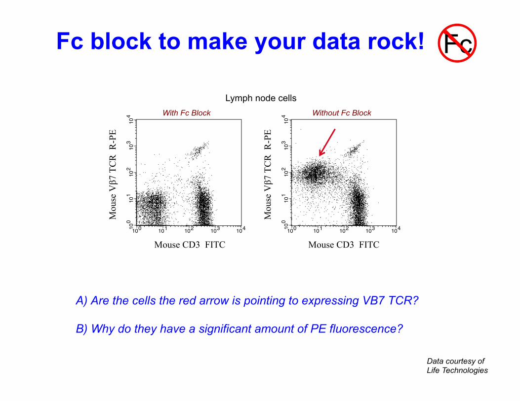
A) Are the cells the red arrow is pointing to expressing VB7 TCR? B) Why do they have a significant amount of PE fluorescence?
Data courtesy of Life Technologies
Lymph node cells
For research use only. CAUTION: Not for human or animal therapeutic or diagnostic use. www.invitrogen.com
Invitrogen Corporation • 542 Flynn Rd • Camarillo • CA 93012 • Tel: 800.955.6288 • E-mail: [email protected] PI: L11554 (Rev 07/10) DCC-10-1870 Important Licensing Information - These products may be covered by one or more Limited Use Label Licenses (see the Invitrogen Catalog or our website, www.invitrogen.com). By use of these products you accept the terms and conditions of all applicable Limited Use Label Licenses. Unless otherwise indicated, these products are for research use only and are not intended for human or animal diagnostic, therapeutic or commercial use.
PRODUCT INSERT
RAT anti-MOUSE CD16/32
Product Code Form Volume Matching Isotype Controls
MFCR00 Purified 0.5 ml MFCR00-4 Purified 2.0 ml MFCR04 R-PE 1.0 ml Rat IgG2b R-PE Code R2b04
PRODUCT DESCRIPTION: Rat monoclonal antibody to mouse CD16/32 Clone: FCR-4G8 Isotype: Rat IgG2b Lot No.: See label Expiration: See label Buffer: Phosphate buffered saline (PBS) Preservatives: 0.1% sodium azide. Sodium azide is an extremely toxic and dangerous compound particularly when combined with acids or metals. Solutions containing sodium azide should be disposed of properly. Stabilizer: For conjugated products only, a highly purified grade of BSA has been added as a stabilizing agent. STORAGE & HANDLING: Store reagents at 2-8°C. Light exposure should be avoided with fluorochrome conjugated reagents. Use dim light during handling, incubation with cells and prior to analysis. It is recommended that cells be analyzed within 18 hours of staining. If the reagent is being diluted, it is recommended that only the quantity to be used within one week be diluted.
PRODUCT CHARACTERIZATION:
Antigen Specificity: The FCR-4G8 monoclonal antibody reacts with mouse CD16/CD32 (FcȖIII/II receptors) which are Fc receptors present on macrophages, NK cells, monocytes, lymphocytes, and dendritic cells. The FCR-4G8 monoclonal antibody can be used to block Fc receptor mediated binding of immunoglobins to the FcȖIII and FcȖII receptors. PRODUCT QUALITY CONTROL: Every lot is tested by flow cytometry using freshly harvested mouse splenocytes. Because conditions may vary, it is recommended that each investigator determine the optimal amount of antibody to be used for each application.
A. Lymph node cells blocked B. Lymph node cells unblocked
Mou
se Vȕ7
TC
R R
-PE
100
101
102
103
104
100 101 102 103 104
Mou
se Vȕ7
TC
R R
-PE
100
101
102
103
104
100 101 102 103 104 Mouse CD3 FITC Mouse CD3 FITC
Decreased background staining of mouse Vȕ7 TCR mAb using purified anti-mouse CD16/32 (Cat.# MFCR00). Briefly, one million cells from a suspension of C57BL/6 lymph node cells were simultaneously stained with 0.5 µg of anti-mouse CD3 conjugated to FITC (Cat.# HM3401) and 0.25 µg of anti-mouse Vȕ7 TCR conjugated to PE (Cat.# RM4404) either with preblocking of Fc receptors for 10 minutes using 0.25 µg of purified anti-mouse CD16/32 (Fig. A) or without preblocking of Fc receptors (Fig. B). Note the decreased staining of Vȕ7 TCR on CD3 negative cells when Fc receptors are blocked. Note: The flow cytometric data shown above may not necessarily have been generated using the enclosed lot of reagent. For this reason, and due to differences in flow cytometers and cytometer settings, results may vary from those illustrated above. It is suggested that investigators titrate reagents to determine optimal conditions for use in their systems.
Explanation of symbols Symbol Description Symbol Description
Catalogue Number Batch code
Research Use Only In vitro diagnostic medical device
Use by Temperature limitation
Manufacturer European Community authorised representative
[-] Without, does not contain [+] With, contains
Protect from light Consult accompanying documents
Directs the user to consult instructions for use (IFU), accompanying the product.
With Fc Block Without Fc Block
Fc block to make your data rock! Fc

1 Experimental Sample CD3 FITC CD4 PE CD8 APC ?
2 Unstained
3 Compensation Controls
4 5 6
7
FMO Gating Controls
8 9
10
A) This should be a 4 color experiment. What is missing? B) Fill in the reagents for the Compensation Controls. C) Fill in the reagents for the FMO Controls.
Controls They make or break it!

A) List 4 possible reasons why a sorted sample has poor purity and/or recovery.
Sorting
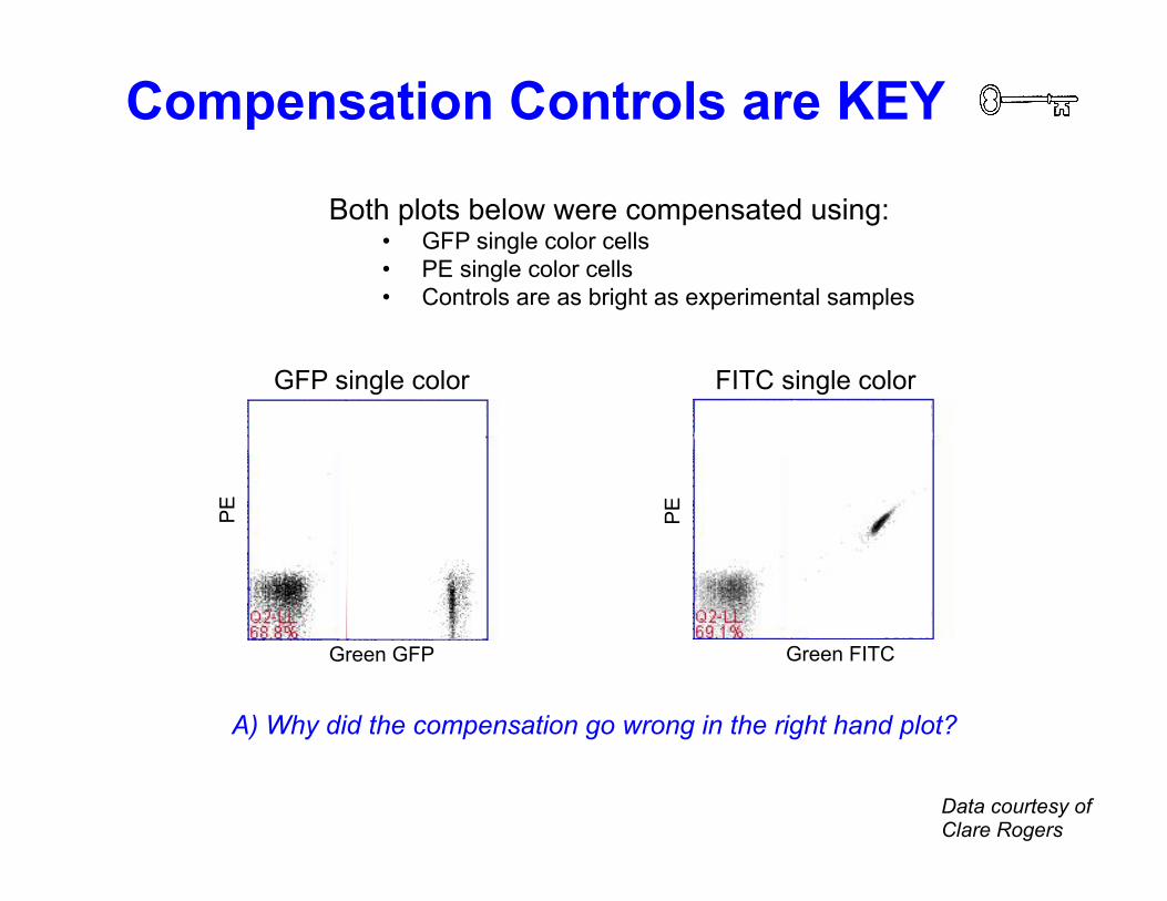
A) Why did the compensation go wrong in the right hand plot?
Data courtesy of Clare Rogers
Green FITC P
E
FITC single color
Both plots below were compensated using: • GFP single color cells • PE single color cells • Controls are as bright as experimental samples
GFP single color
Green GFP
PE
Compensation Controls are KEY

A) List the single color compensation tubes needed to compensate properly.
B) Should you resuspend ALL the controls in media containing DAPI?
Your experiment contains two staining combinations:
Stain 2
CD19 FITC
CD3 PE CD4 Cy7PE
DAPI
Stain 1
GFP
CD3 PE CD8 Cy7PE
DAPI
Compensation Controls are KEY #2

A) Is the purple dotted line correctly gating the Cy7PE+ population?
B) Are some of the CD8+ cells expressing CD20?
C) Where would you draw the gate to define the CD20+ population?
PE: CD8
Cy7
PE
: CD
20
Fully Stained Unstained
PE: CD8
Cy7
PE
: CD
20
Cy7PE FMO
PE: CD8
Cy7
PE
: no
stai
n
Fluorescence Minus One Gate it not debate it!

Cell Sorting
Charged plates
Collection tubes
Laser
Cells
Analysis
Drop 1 (first broken off drop) A) Draw a line between two elements on the picture to identify the drop delay.
B) What can cause the drop delay to change while sorting?
+
+
+
+
+
+
+
-
-
-
-
-
-
-
-
Don’t delay the DROP DELAY

GFP sample
A) The cells in the red circle and the yellow circle have the same amount of Green fluorescence. But the source of that fluorescence is different. - What is the origin of the Green fluorescence of cells in the red circle? - What about of the cells in the yellow circle?
Green
Ora
nge
Mock Transfected
Green
Ora
nge
Histograms Hide Data