QUINT Lung Cancer Staging Update - Scbtmr Lung Cancer... · LUNG CANCER STAGING • TNM...
42
LUNG CANCER LUNG CANCER STAGING UPDATE STAGING UPDATE Leslie E. Quint, M.D. University of Michigan
Transcript of QUINT Lung Cancer Staging Update - Scbtmr Lung Cancer... · LUNG CANCER STAGING • TNM...

LUNG CANCER LUNG CANCER STAGING UPDATESTAGING UPDATE
Leslie E. Quint, M.D.University of Michigan

LUNG CANCER STAGING• Review definitions in new TNM
system and highlight changes• Review imaging features of non-
small cell lung cancers (NSCLC) that indicate tumor stage
• Discuss usefulness/accuracy of imaging findings in predicting stage
• CT, FDG-PET, MRI
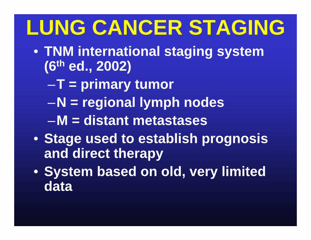
LUNG CANCER STAGING• TNM international staging system
(6th ed., 2002)–T = primary tumor–N = regional lymph nodes–M = distant metastases
• Stage used to establish prognosis and direct therapy
• System based on old, very limited data

LUNG CANCER STAGING• 7th ed. of TNM staging system 2010• International Association for the Study
of Lung Cancer (IASLC): – Initiated international Lung Cancer
Staging project 1996–Data on > 100,000 cases
• Changes to better reflect survival, treatment options
• Rami-Porta R. J Thorac Oncol 2007; 2:593 • Rusch VW. J Thorac Oncol 2007; 2:603• Postmus PE. J Thorac Oncol 2007; 2:686
• Groome PA. J Thorac Oncol 2007; 2:694• Goldstraw P. J Thorac Oncol 2007; 2:706

STAGING: 1O TUMORT1• < 3 cm• Surrounded by
lung or visceral pleura
• Not in mainstem bronchus

STAGING: 1O TUMORT1• < 3 cm• Surrounded by
lung or visceral pleura
• Not in mainstem bronchus
Change• T1a: < 2 cm• T1b: >2-3 cm

T2• > 3 cm• Distal mainstem
bronchus (>2 cm from carina)
• Invades visceral pleura
• Postobstructive atel/pneumonia < entire lung
STAGING: 1O TUMOR

T2• > 3 cm• Distal mainstem
bronchus (>2 cm from carina)
• Invades visceral pleura
• Postobstructive atel/pneumonia < entire lung
STAGING: 1O TUMORChange• T2a: >3-5 cm• T2b: >5-7 cm• > 7 cm T3

STAGING: 1O TUMORT3• Invades chest wall,
diaphragm, phrenicnerve, mediastinal pleura, parietal pericardium
• Proximal mainstem bronchus (< 2 cm from carina)
• Postobstructive atel/pneumonia entire lung

STAGING: 1O TUMORChest wall invasion = T3• Pleural thickening, loss of
extrapleural fat plane• Obtuse angle, >3 cm contact
between mass and chest wall• Soft tissue in chest wall• CT sens 38-87%, spec 40-90%• Only specific sign: bone destruction• Chest wall pain reliable symptom

INDETERMINATE FOR CHEST WALL INVASION
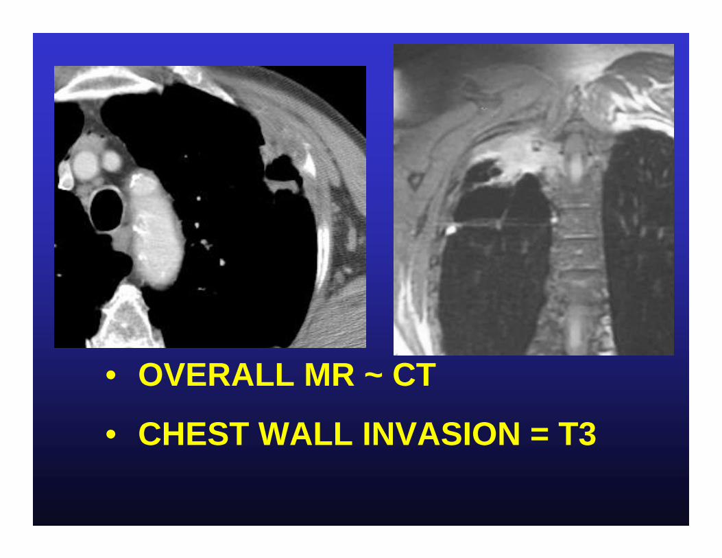
• OVERALL MR ~ CT
• CHEST WALL INVASION = T3

Brachial plexus• MR > CT
STAGING: 1O TUMOR

Chest wall invasion• Does not preclude surgery• En bloc resection and chest wall
reconstruction– ↑ morbidity and mortality
• Contraindicated if mediastinal lymph node metastases due to poor prognosis (7% 5-yr survival)
STAGING: 1O TUMOR

STAGING: 1O TUMORT3• Invades chest wall,
diaphragm, mediastinal pleura, parietal pericardium
• Proximal mainstem bronchus (< 2 cm from carina)
• Postobstructive atel/pneumonia entire lung
Changes• > 7 cm
– previously T2• Separate tumor
nodule(s) in same lobe – previously T4

STAGING: 1O TUMORT4• Invades mediastinum,
heart, great vessels, trachea, esophagus, recurrent laryngeal nerve, vertebral body, carina
• Separate tumor nodule in different ipsilaterallobe (prev M1)

STAGING: 1O TUMORMediastinal fat invasion = T4• CT/MR criteria: extensive
contact with mediastinum, abnormal soft tissue in mediastinal fat, pleural or pericardial thickening
• CT and MR: poor accuracy
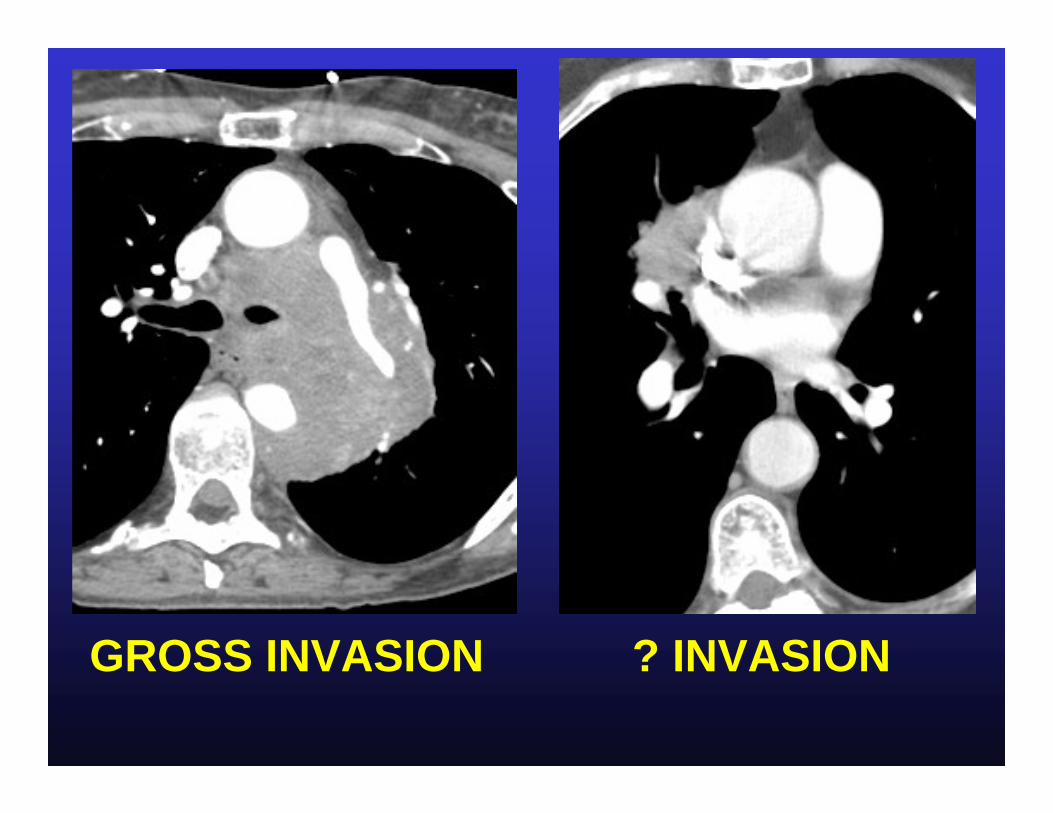
GROSS INVASION ? INVASION

STAGING: 1O TUMOR
• CT criteria: loss of fat plane, mass effect, extensive contact
• MR similar to CT: –Low accuracy–Contiguity ≠ invasion
Invasion of vital structure = T4
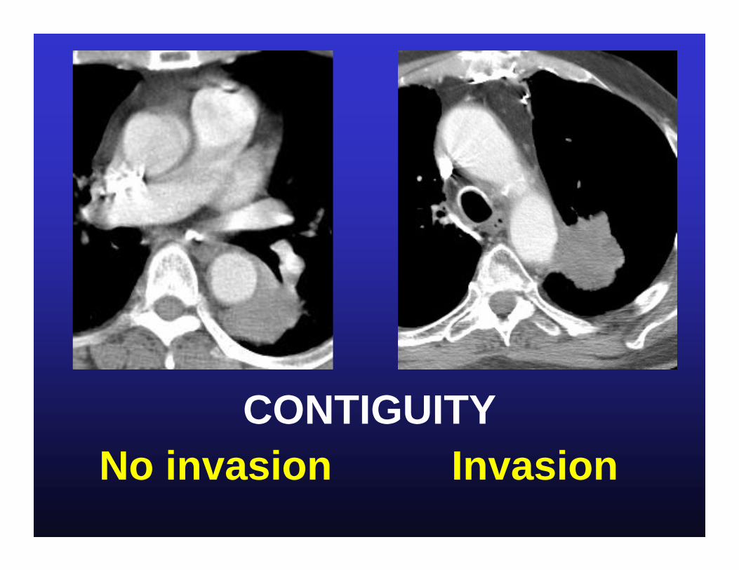
No invasionCONTIGUITY
Invasion
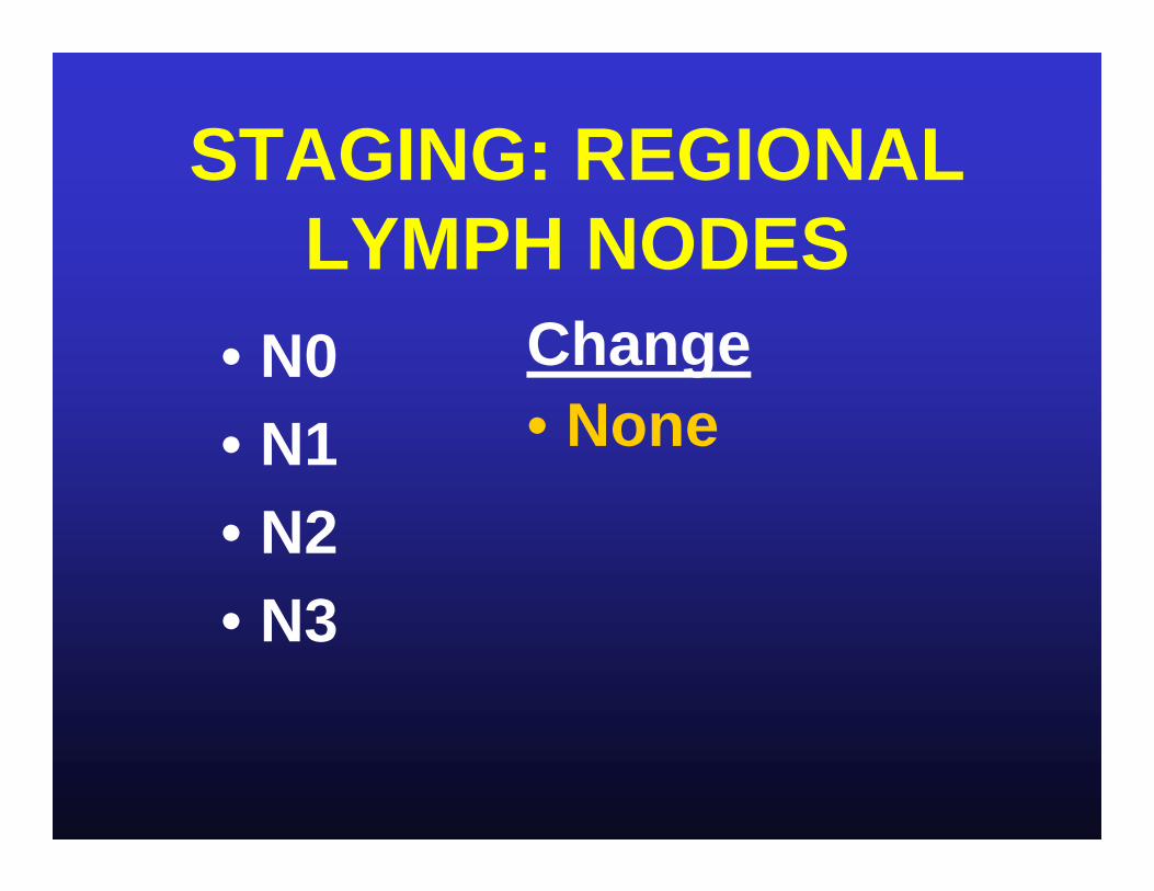
STAGING: REGIONAL LYMPH NODES
• N0• N1• N2• N3
Change• None
STAGING: LYMPH NODESLYMPH NODESN0• No regional
lymph node metastases

Figure 1B
T1 N0

STAGING: LYMPH NODESLYMPH NODESN1• Metastasis in ipsilateral hilar and/or
peribronchial nodes• Intrapulmonary nodes involved by
direct extension from primary tumor
• Affects prognosis, not resectability• Does not predict status of
mediastinal nodes
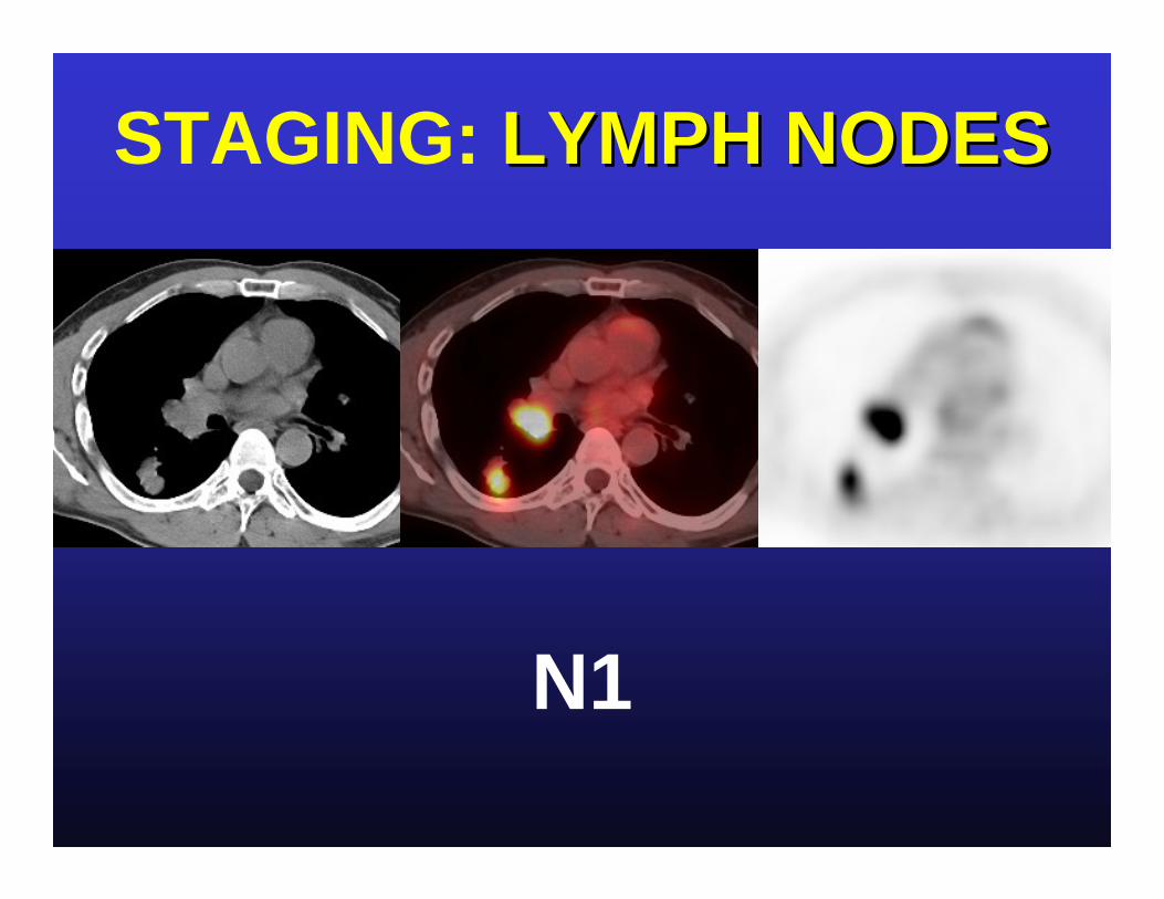
STAGING: LYMPH NODESLYMPH NODES
N1
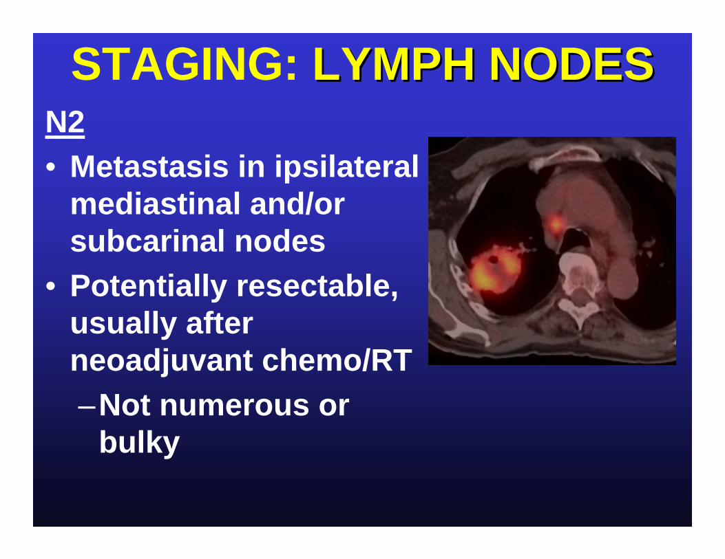
STAGING: LYMPH NODESLYMPH NODESN2• Metastasis in ipsilateral
mediastinal and/or subcarinal nodes
• Potentially resectable, usually after neoadjuvant chemo/RT–Not numerous or
bulky

STAGING: LYMPH NODESLYMPH NODESN3 • Metastasis in contralateral
mediastinal or hilar nodes• Metastasis in any scalene or
supraclavicular nodes• Unresectable

STAGING: LYMPH NODESLYMPH NODES

MEDIASTINALMEDIASTINAL LYMPH NODESLYMPH NODES
Sens SpecCT 60% 80%PET 80% 90%
Birim O. Ann Thor Surg 2005;79:375

MEDIASTINALMEDIASTINAL LYMPH NODESLYMPH NODESPET pitfalls• False positives due to
inflammatory nodes poor specificity in enlarged nodes
• False negatives due to microscopic mets poor sensitivity in small nodes

MEDIASTINALMEDIASTINAL LYMPH NODESLYMPH NODES
use CT to direct best method of lymph node biopsy
Enlarged at CT (no PET or
abnormal PET)
Abnormal at PET(small or enlarged
at CT)
MEDIASTINALMEDIASTINAL LYMPH NODESLYMPH NODES
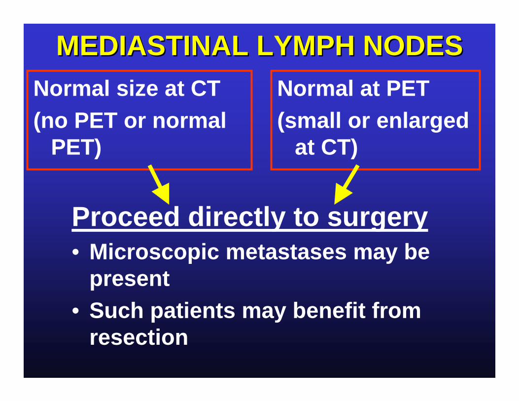
Proceed directly to surgery• Microscopic metastases may be
present• Such patients may benefit from
resection
Normal size at CT (no PET or normal
PET)
Normal at PET(small or enlarged
at CT)

MEDIASTINALMEDIASTINAL LYMPH NODESLYMPH NODES
Proceed directly to surgery• Exception: adenocarcinoma or T3
tumors (including Pancoast)• Presence of mediastinal metastases
poor prognosis, not surgical candidate• Consider mediastinoscopy
Normal size at CT (no PET or normal
PET)
Normal at PET(small or enlarged
at CT)
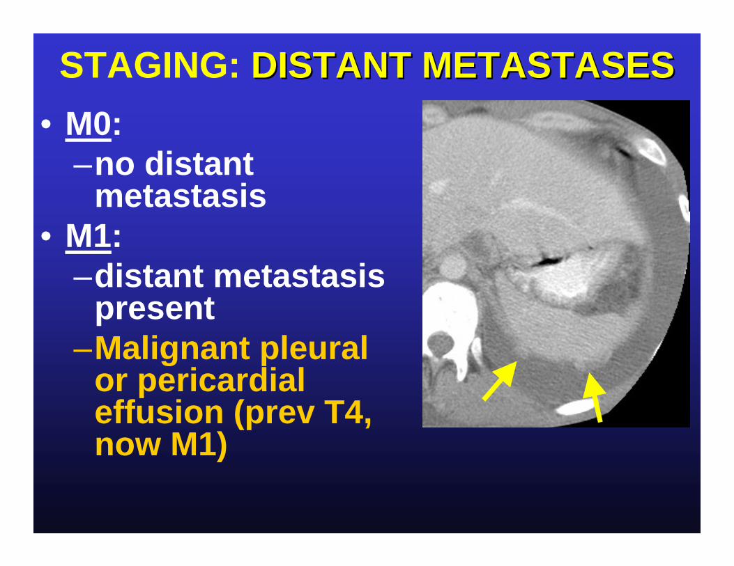
STAGING: DISTANT METASTASESDISTANT METASTASES• M0:
–no distant metastasis
• M1: –distant metastasis
present –Malignant pleural
or pericardial effusion (prev T4, now M1)

• Tiny left pleural effusion
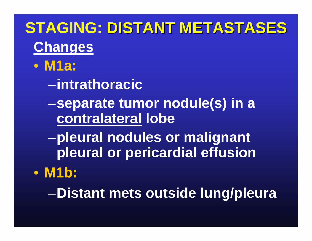
STAGING: DISTANT METASTASESDISTANT METASTASESChanges• M1a:
–intrathoracic –separate tumor nodule(s) in a
contralateral lobe –pleural nodules or malignant
pleural or pericardial effusion• M1b:
–Distant mets outside lung/pleura
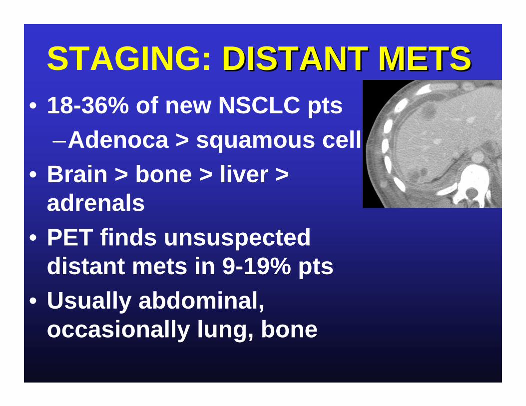
STAGING: DISTANT METSDISTANT METS• 18-36% of new NSCLC pts
–Adenoca > squamous cell• Brain > bone > liver >
adrenals• PET finds unsuspected
distant mets in 9-19% pts• Usually abdominal,
occasionally lung, bone

? N3 disease

M1 disease

LUNG CANCER STAGING
Changes in stage groupings:• Minor
Goldstraw P. J Thorac Oncol 2007; 2:706

Goldstraw P. J Thorac Oncol 2007; 2:706
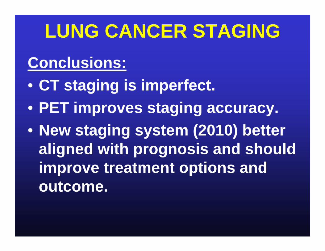
LUNG CANCER STAGINGConclusions:• CT staging is imperfect.• PET improves staging accuracy.• New staging system (2010) better
aligned with prognosis and should improve treatment options and outcome.