![myfaith-data.s3.amazonaws.com · Wedding [Celebrant: Fr. Kevin Murphy] Daniel William Henry & Nicole Margaret Murphy . Reconciliation/Fr. T Castelli : MASS-Saint Nicholas Church/Fr.](https://static.fdocuments.in/doc/165x107/5abff4bf7f8b9a213f8b6ca8/myfaith-datas3-celebrant-fr-kevin-murphy-daniel-william-henry-nicole-margaret.jpg)
Questions for PC Practicum Unit 3 Coding...
35
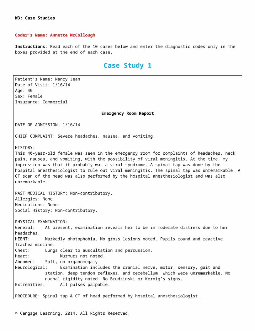
W3: Case Studies Coder’s Name: Annette McCollough Instructions: Read each of the 10 cases below and enter the diagnostic codes only in the boxes provided at the end of each case. Case Study 1 Patient’s Name: Nancy Jean Date of Visit: 1/16/14 Age: 40 Sex: Female Insurance: Commercial Emergency Room Report DATE OF ADMISSION: 1/16/14 CHIEF COMPLAINT: Severe headaches, nausea, and vomiting. HISTORY: This 40-year-old female was seen in the emergency room for complaints of headaches, neck pain, nausea, and vomiting, with the possibility of viral meningitis. At the time, my impression was that it probably was a viral syndrome. A spinal tap was done by the hospital anesthesiologist to rule out viral meningitis. The spinal tap was unremarkable. A CT scan of the head was also performed by the hospital anesthesiologist and was also unremarkable. PAST MEDICAL HISTORY: Non-contributory. Allergies: None. Medications: None. Social History: Non-contributory. PHYSICAL EXAMINATION: General: At present, examination reveals her to be in moderate distress due to her headaches. HEENT: Markedly photophobia. No gross lesions noted. Pupils round and reactive. Trachea midline. Chest: Lungs clear to auscultation and percussion. Heart: Murmurs not noted. Abdomen: Soft, no organomegaly. Neurological: Examination includes the cranial nerve, motor, sensory, gait and station, deep tendon reflexes, and cerebellum, which were unremarkable. No nuchal rigidity noted. No Brudzinski or Kernig’s signs. Extremities: All pulses palpable. PROCEDURE: Spinal tap & CT of head performed by hospital anesthesiologist. © Cengage Learning, 2014. All Rights Reserved.
Transcript of Questions for PC Practicum Unit 3 Coding...
W3: Case Studies
Coder’s Name: Annette McCollough
Instructions: Read each of the 10 cases below and enter the diagnostic codes only in the boxes provided at the end of each case.
Case Study 1Patient’s Name: Nancy JeanDate of Visit: 1/16/14Age: 40Sex: FemaleInsurance: Commercial
Emergency Room Report
DATE OF ADMISSION: 1/16/14
CHIEF COMPLAINT: Severe headaches, nausea, and vomiting.
HISTORY:This 40-year-old female was seen in the emergency room for complaints of headaches, neck pain, nausea, and vomiting, with the possibility of viral meningitis. At the time, my impression was that it probably was a viral syndrome. A spinal tap was done by the hospital anesthesiologist to rule out viral meningitis. The spinal tap was unremarkable. A CT scan of the head was also performed by the hospital anesthesiologist and was also unremarkable.
PAST MEDICAL HISTORY: Non-contributory.Allergies: None.Medications: None.Social History: Non-contributory.
PHYSICAL EXAMINATION:General: At present, examination reveals her to be in moderate distress due to her headaches.HEENT: Markedly photophobia. No gross lesions noted. Pupils round and reactive. Trachea midline.Chest: Lungs clear to auscultation and percussion.Heart: Murmurs not noted.Abdomen: Soft, no organomegaly.Neurological: Examination includes the cranial nerve, motor, sensory, gait and station, deep tendon
reflexes, and cerebellum, which were unremarkable. No nuchal rigidity noted. No Brudzinski or Kernig’s signs.
Extremities: All pulses palpable.
PROCEDURE: Spinal tap & CT of head performed by hospital anesthesiologist.
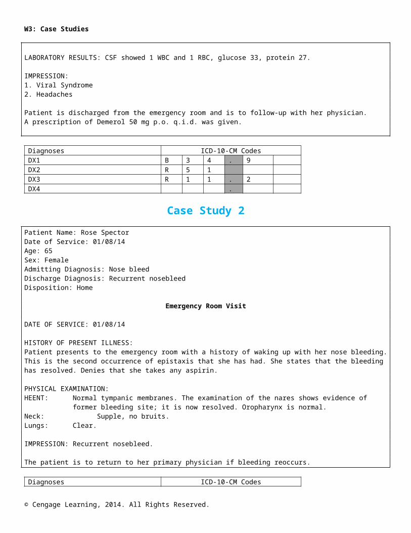
LABORATORY RESULTS: CSF showed 1 WBC and 1 RBC, glucose 33, protein 27.
IMPRESSION:1. Viral Syndrome2. Headaches
Patient is discharged from the emergency room and is to follow-up with her physician.A prescription of Demerol 50 mg p.o. q.i.d. was given.
Diagnoses ICD-10-CM Codes
© Cengage Learning, 2014. All Rights Reserved.
W3: Case Studies
DX1 B 3 4 . 9DX2 R 5 1DX3 R 1 1 . 2DX4 .
Case Study 2Patient Name: Rose SpectorDate of Service: 01/08/14Age: 65Sex: FemaleAdmitting Diagnosis: Nose bleedDischarge Diagnosis: Recurrent nosebleedDisposition: Home
Emergency Room Visit
DATE OF SERVICE: 01/08/14
HISTORY OF PRESENT ILLNESS:Patient presents to the emergency room with a history of waking up with her nose bleeding. This is the second occurrence of epistaxis that she has had. She states that the bleeding has resolved. Denies that she takes any aspirin.
PHYSICAL EXAMINATION:HEENT: Normal tympanic membranes. The examination of the nares shows evidence of former
bleeding site; it is now resolved. Oropharynx is normal.Neck: Supple, no bruits.Lungs: Clear.
IMPRESSION: Recurrent nosebleed.
The patient is to return to her primary physician if bleeding reoccurs.
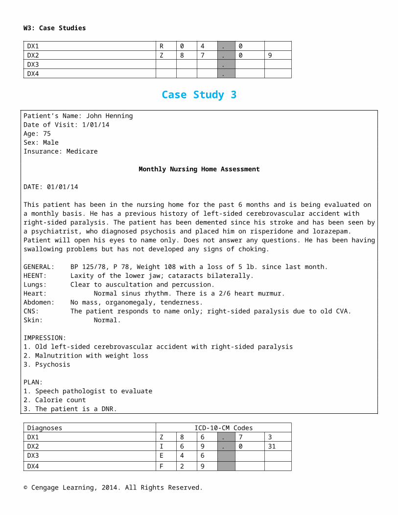
Diagnoses ICD-10-CM CodesDX1 R 0 4 . 0DX2 Z 8 7 . 0 9DX3 .DX4 .
Case Study 3Patient’s Name: John HenningDate of Visit: 1/01/14Age: 75Sex: MaleInsurance: Medicare
Monthly Nursing Home Assessment
DATE: 01/01/14
This patient has been in the nursing home for the past 6 months and is being evaluated on a monthly basis. He has a previous history of left-sided cerebrovascular accident with right-sided paralysis. The patient has been demented since his stroke and has been seen by a psychiatrist, who diagnosed psychosis
© Cengage Learning, 2014. All Rights Reserved.
W3: Case Studies
and placed him on risperidone and lorazepam. Patient will open his eyes to name only. Does not answer any questions. He has been having swallowing problems but has not developed any signs of choking.
GENERAL: BP 125/78, P 78, Weight 108 with a loss of 5 lb. since last month.HEENT: Laxity of the lower jaw; cataracts bilaterally.Lungs: Clear to auscultation and percussion.Heart: Normal sinus rhythm. There is a 2/6 heart murmur.Abdomen: No mass, organomegaly, tenderness.CNS: The patient responds to name only; right-sided paralysis due to old CVA.Skin: Normal.
IMPRESSION:1. Old left-sided cerebrovascular accident with right-sided paralysis2. Malnutrition with weight loss3. Psychosis
PLAN:1. Speech pathologist to evaluate2. Calorie count3. The patient is a DNR.
Diagnoses ICD-10-CM CodesDX1 Z 8 6 . 7 3DX2 I 6 9 . 0 31DX3 E 4 6DX4 F 2 9
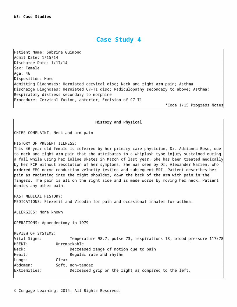
Case Study 4Patient Name: Sabrina GuimondAdmit Date: 1/15/14Discharge Date: 1/17/14Sex: FemaleAge: 46Disposition: HomeAdmitting Diagnoses: Herniated cervical disc; Neck and right arm pain; AsthmaDischarge Diagnoses: Herniated C7-T1 disc; Radiculopathy secondary to above; Asthma; Respiratory distress secondary to morphineProcedure: Cervical fusion, anterior; Excision of C7-T1
*Code 1/15 Progress Notes
History and Physical
CHIEF COMPLAINT: Neck and arm pain
HISTORY OF PRESENT ILLNESS:This 46-year-old female is referred by her primary care physician, Dr. Adrianna Rose, due to neck and right arm pain that she attributes to a whiplash type injury sustained during a fall while using her inline skates in March of last year. She has been treated medically by her PCP without resolution of her symptoms. She was seen by Dr. Alexander Warren, who ordered EMG nerve conduction velocity testing and subsequent MRI. Patient describes her pain as radiating into the right shoulder, down the back of the arm with pain in the fingers. The pain is all on the right side and is made worse by moving her neck. Patient denies any other pain.
© Cengage Learning, 2014. All Rights Reserved.
W3: Case Studies
PAST MEDICAL HISTORY:MEDICATIONS: Flexeril and Vicodin for pain and occasional inhaler for asthma.
ALLERGIES: None known
OPERATIONS: Appendectomy in 1979
REVIEW OF SYSTEMS:Vital Signs: Temperature 98.7, pulse 73, respirations 18, blood pressure 117/78HEENT: UnremarkableNeck: Decreased range of motion due to painHeart: Regular rate and rhythmLungs: ClearAbdomen: Soft, non-tenderExtremities: Decreased grip on the right as compared to the left.
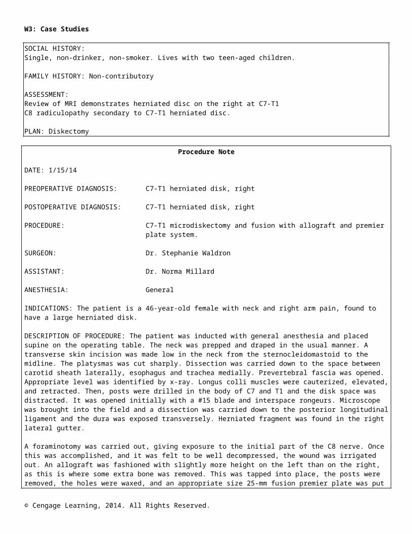
SOCIAL HISTORY:Single, non-drinker, non-smoker. Lives with two teen-aged children.
FAMILY HISTORY: Non-contributory
ASSESSMENT:Review of MRI demonstrates herniated disc on the right at C7-T1C8 radiculopathy secondary to C7-T1 herniated disc.
PLAN: Diskectomy
Procedure Note
DATE: 1/15/14
PREOPERATIVE DIAGNOSIS: C7-T1 herniated disk, right
POSTOPERATIVE DIAGNOSIS: C7-T1 herniated disk, right
PROCEDURE: C7-T1 microdiskectomy and fusion with allograft and premier plate system.
SURGEON: Dr. Stephanie Waldron
ASSISTANT: Dr. Norma Millard
ANESTHESIA: General
INDICATIONS: The patient is a 46-year-old female with neck and right arm pain, found to have a large herniated disk.
DESCRIPTION OF PROCEDURE: The patient was inducted with general anesthesia and placed supine on the operating table. The neck was prepped and draped in the usual manner. A transverse skin incision was made low in the neck from the sternocleidomastoid to the midline. The platysmas was cut sharply. Dissection was carried down to the space between carotid sheath laterally, esophagus and trachea medially. Prevertebral fascia was opened. Appropriate level was identified by x-ray. Longus colli muscles were cauterized, elevated, and retracted. Then, posts were drilled in the body of C7 and T1 and the disk space was distracted. It was opened initially with a #15 blade and interspace rongeurs. Microscope was brought into the field and a dissection was carried down to the posterior longitudinal ligament and the dura was exposed transversely. Herniated fragment was found in the right lateral gutter.
© Cengage Learning, 2014. All Rights Reserved.
W3: Case Studies
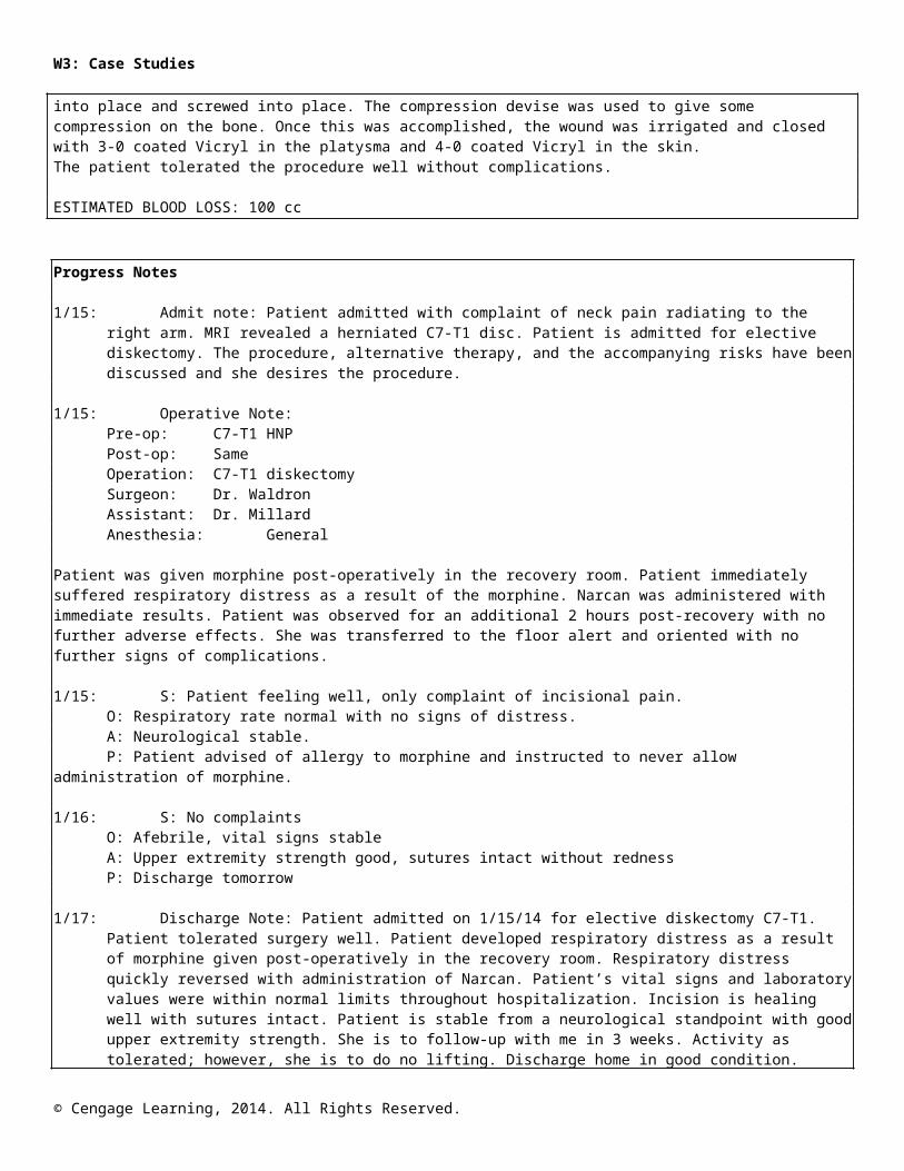
A foraminotomy was carried out, giving exposure to the initial part of the C8 nerve. Once this was accomplished, and it was felt to be well decompressed, the wound was irrigated out. An allograft was fashioned with slightly more height on the left than on the right, as this is where some extra bone was removed. This was tapped into place, the posts were removed, the holes were waxed, and an appropriate size 25-mm fusion premier plate was put into place and screwed into place. The compression devise was used to give some compression on the bone. Once this was accomplished, the wound was irrigated and closed with 3-0 coated Vicryl in the platysma and 4-0 coated Vicryl in the skin.The patient tolerated the procedure well without complications.
ESTIMATED BLOOD LOSS: 100 cc
Progress Notes
1/15: Admit note: Patient admitted with complaint of neck pain radiating to the right arm. MRI revealed a herniated C7-T1 disc. Patient is admitted for elective diskectomy. The procedure, alternative therapy, and the accompanying risks have been discussed and she desires the procedure.
1/15: Operative Note:Pre-op:C7-T1 HNPPost-op: SameOperation: C7-T1 diskectomySurgeon: Dr. WaldronAssistant: Dr. MillardAnesthesia: General
Patient was given morphine post-operatively in the recovery room. Patient immediately suffered respiratory distress as a result of the morphine. Narcan was administered with immediate results. Patient was observed for an additional 2 hours post-recovery with no further adverse effects. She was transferred to the floor alert and oriented with no further signs of complications.
1/15: S: Patient feeling well, only complaint of incisional pain.O: Respiratory rate normal with no signs of distress.A: Neurological stable.P: Patient advised of allergy to morphine and instructed to never allow administration of morphine.
1/16: S: No complaintsO: Afebrile, vital signs stableA: Upper extremity strength good, sutures intact without rednessP: Discharge tomorrow
1/17: Discharge Note: Patient admitted on 1/15/14 for elective diskectomy C7-T1. Patient tolerated surgery well. Patient developed respiratory distress as a result of morphine given post-operatively in the recovery room. Respiratory distress quickly reversed with administration of Narcan. Patient’s vital signs and laboratory values were within normal limits throughout hospitalization. Incision is healing well with sutures intact. Patient is stable from a neurological standpoint with good upper extremity strength. She is to follow-up with me in 3 weeks. Activity as tolerated; however, she is to do no lifting. Discharge home in good condition.
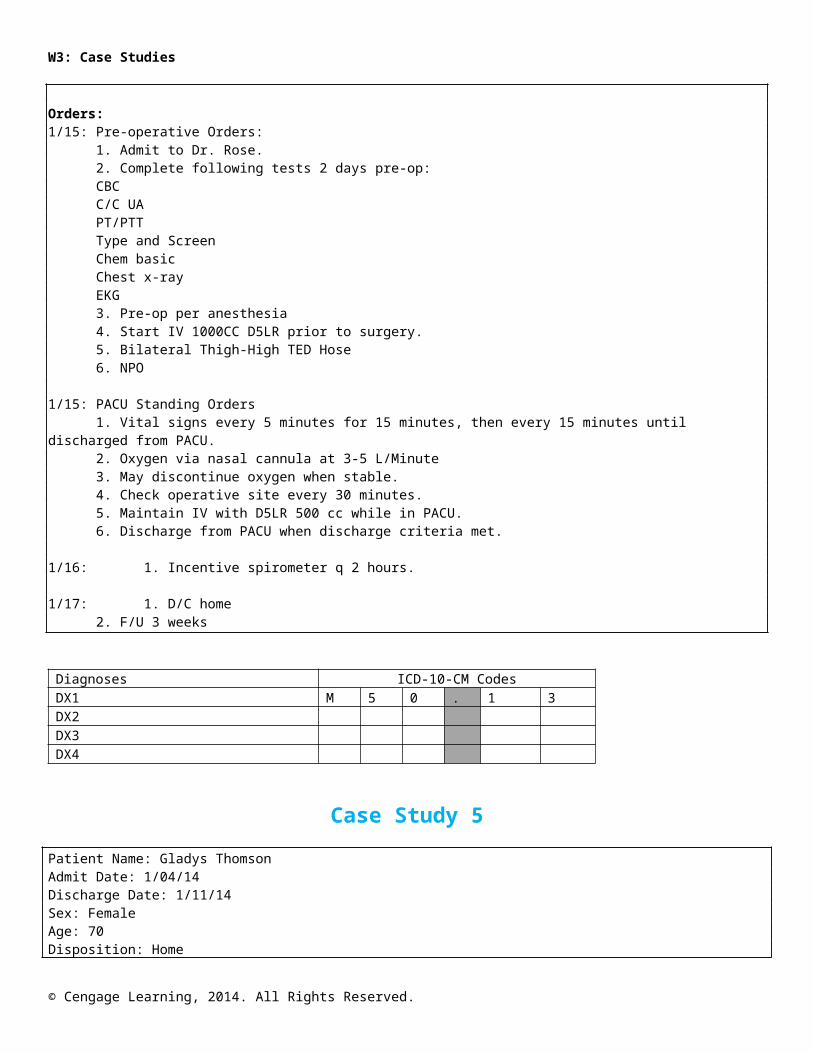
Orders:1/15: Pre-operative Orders:
1. Admit to Dr. Rose.2. Complete following tests 2 days pre-op:CBCC/C UAPT/PTT
© Cengage Learning, 2014. All Rights Reserved.
W3: Case Studies
Type and ScreenChem basicChest x-rayEKG3. Pre-op per anesthesia4. Start IV 1000CC D5LR prior to surgery.5. Bilateral Thigh-High TED Hose6. NPO
1/15: PACU Standing Orders1. Vital signs every 5 minutes for 15 minutes, then every 15 minutes until discharged from PACU.2. Oxygen via nasal cannula at 3-5 L/Minute3. May discontinue oxygen when stable.4. Check operative site every 30 minutes.5. Maintain IV with D5LR 500 cc while in PACU.6. Discharge from PACU when discharge criteria met.
1/16: 1. Incentive spirometer q 2 hours.
1/17: 1. D/C home2. F/U 3 weeks
Diagnoses ICD-10-CM CodesDX1 M 5 0 . 1 3DX2 DX3DX4
Case Study 5Patient Name: Gladys ThomsonAdmit Date: 1/04/14Discharge Date: 1/11/14Sex: FemaleAge: 70Disposition: Home
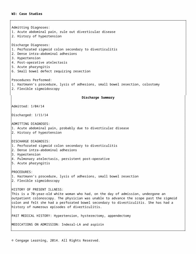
Admitting Diagnoses:1. Acute abdominal pain, rule out diverticular disease2. History of hypertension
Discharge Diagnoses:1. Perforated sigmoid colon secondary to diverticulitis2. Dense intra-abdominal adhesions3. Hypertension4. Post-operative atelectasis5. Acute pharyngitis6. Small bowel defect requiring resection
Procedures Performed:1. Hartmann’s procedure, lysis of adhesions, small bowel resection, colostomy2. Flexible sigmoidoscopy
Discharge Summary
© Cengage Learning, 2014. All Rights Reserved.
W3: Case Studies
Admitted: 1/04/14
Discharged: 1/11/14
ADMITTING DIAGNOSES:1. Acute abdominal pain, probably due to diverticular disease2. History of hypertension
DISCHARGE DIAGNOSIS:1. Perforated sigmoid colon secondary to diverticulitis2. Dense intra-abdominal adhesions3. Hypertension4. Pulmonary atelectasis, persistent post-operative5. Acute pharyngitis
PROCEDURES:1. Hartmann’s procedure, lysis of adhesions, small bowel resection2. Flexible sigmoidoscopy
HISTORY OF PRESENT ILLNESS:This is a 70-year-old white woman who had, on the day of admission, undergone an outpatient colonoscopy. The physician was unable to advance the scope past the sigmoid colon and felt she had a perforated bowel secondary to diverticulitis. She has had a history of numerous episodes of diverticulitis.
PAST MEDICAL HISTORY: Hypertension, hysterectomy, appendectomy
MEDICATIONS ON ADMISSION: Inderal-LA and aspirin
ALLERGIES: None known
PHYSICAL EXAMINATION:Reveals an elderly white woman with tenderness in the left lower quadrant of the abdomen.
LABORATORY DATA:The admission laboratory data were within normal limits, with the exception of low hematocrit of 34, hemoglobin 11.8, and elevated globulin 3.9. Post-operatively, white count was over 19,000, transiently returning to normal in 2 days. The hemoglobin dropped to 10.1 with hematocrit of 29.7.
RADIOLOGY REPORTS:Gastrografin enema findings were suggestive of perforation at the level of the rectosigmoid, at a point where the patient had extensive diverticula.
Chest x-ray revealed pneumomediastinum and subcutaneous emphysema, but no pneumothorax.There was air in the retroperitoneum. The follow-up chest x-rays showed nasogastric tube in satisfactory position with decreased subcutaneous emphysema within the neck and decreased pericardial air collection since the prior study, and development of small bilateral effusions on 1/08/14. The abdominal x-rays showed a large amount of free retroperitoneal air and air in the mediastinum.
HOSPITAL COURSE:The patient was admitted and started on intravenous antibiotics with Unasyn and Tobramycin combination.
The patient was seen in consult by Gastrology, who felt she probably had an acute sigmoid perforation secondary to diverticular disease. He felt she would benefit from Gastrografin enema, especially to determine whether she could be treated conservatively or not. The procedure was completed with the findings as mentioned above. It was felt she would require resection.
© Cengage Learning, 2014. All Rights Reserved.
W3: Case Studies
She was taken to the operating room, where the above procedures were performed. The patient tolerated the procedure well under general anesthesia. The estimated blood loss was less than 100 cc with no replacement. The pathology report revealed diverticulosis with diverticulitis and perforation of the small bowel.
Post-operatively, the wound was healing nicely. The colostomy was noted to be viable. She was transferred to the surgical floor on 1/05/14 She was having the expected amount of abdominal discomfort post-surgery. She also complained of sore throat and was started on Cepacol throat lozenges. The intravenous fluids and medications were continued.
The colostomy was not functioning over the first few days. There was minimal drainage from the nasogastric tube. Reglan was added. Breath sounds were decreased over the right lower lobe on 1/08/14. The chest x-ray revealed atelectasis. CXRs were done daily and the RLL was re-expanded on 1/10/14.
The colostomy began functioning on 1/14/14. Intravenous fluids were discontinued. Intravenous Lasix was given. Her diet was increased with toleration. Activity was increased and medications were changed to p.o.
The patient improved significantly in the next several days and she was stable enough to be discharged. She will be followed in the office in 1 week. She is discharged on Duricef 500-mg b.i.d. and Tylox one q4h prn. Soft diet and activity as tolerated.
History and Physical
CHIEF COMPLAINT: Abdominal pain
HISTORY OF PRESENT ILLNESS:The patient is a 70-year-old white female who has undergone attempted colonoscopy this morning.There was difficulty in negotiating the sigmoid colon and the patient developed tenderness postcolonoscopy. Abdominal x-rays revealed free air as well as mediastinal air.
PAST MEDICAL HISTORY: Hypertension, hysterectomy, appendectomy
MEDICATIONS ON ADMISSION: Inderal-LA and aspirin
ALLERGIES: None known
PHYSICAL EXAMINATION:Well-developed, well-nourished, 70-year-old white female in moderate distress.
REVIEW OF SYSTEMS:Cardiac: Normal sinus rhythmPulmonary: ClearHEENT: Within normal limitsAbdomen: Soft with left lower quadrant pain and some distentionExtremities: Bilateral pedal pulses
Patient will be admitted for conservative management in hopes of avoiding surgery. She will receive intravenous antibiotic and bowel rest.
Procedure Note
DATE OF PROCEDURE: 1/04/14
PROCEDURE: Attempted full colonoscopy
© Cengage Learning, 2014. All Rights Reserved.
W3: Case Studies
SURGEON: Alexander Glenn, MD
INDICATION: Abdominal pain with history of diverticular disease
DESCRIPTION OF PROCEDURE:With the patient in the left lateral position under direct luminal vision, a complete colonoscopy was attempted, but the Olympus colonoscope was only advanced up to 30 cm inside the sigmoid. The findings were as follows:
1. Rectum and anal canal: Internal hemorrhoids are seen in circumferential fashion, not bleeding at the time. No evidence of masses, lesions, angioma, or polyps seen in the rectal area.2. Rectosigmoid junction, sigmoid, and descending colon: Extensive sigmoid diverticulosis with significant peridiverticulitis and spasm appreciated in a highly redundant sigmoid.
In view of the fact that the diverticular process was so extensive and the sigmoid extremely redundant, the advancement of the scope beyond this area was difficult and hence the procedure was terminated.
The patient returned to the floor in stable condition.
FINAL IMPRESSION:1. Extensive sigmoid diverticulosis with diverticulitis and spasm2. Redundant sigmoid3. Internal hemorrhoids
Procedure Note
DATE OF PROCEDURE: 1/04/14
PREOPERATIVE DIAGNOSIS: Perforated sigmoid colon
POSTOPERATIVE DIAGNOSIS: 1. Perforated sigmoid colon 2. Dense intra-abdominal adhesions
PROCEDURE: 1. Hartmann’s procedure 2. Lysis of adhesions 3. Small bowel resection
SURGEON: Eric Worton, MD
FINDINGS: Moderately large perforation of the sigmoid colon, approximately 2 cm in diameter, with pelvic phlegmon. Dense intra-abdominal adhesions requiring extensive lysis, taking 2 hours.
DESCRIPTION OF PROCEDURE:The patient was taken to the operating suite and placed in the supine position. After adequate induction of general anesthesia the patient was prepped and draped in sterile fashion.
A midline incision was made and the abdomen was entered. Dense adhesions were encountered, requiring extensive sharp dissection.
The area of transection in the proximal sigmoid was dissected out circumferentially. The mesentery was taken down between Kelly clamps. Rectosigmoid was mobilized. The distal sigmoid was dissected out circumferentially and a roticulator placed across this. A stapler was fired, the bowel was transected.
Small bowel adhesions were lysed. One area was extremely thin, resulting in a serosal defect, which required resection. An area in the mid jejunum was dissected out circumferentially both proximally and distally. The intervening mesentery was serially cross-clamped between Kelly clamps. The vessels were ligated with 2-0 silks. G.A. was fired across both sides. Functional end-to-end anastomosis was then performed.
© Cengage Learning, 2014. All Rights Reserved.
W3: Case Studies
The abdomen was irrigated with copious amounts of fluid. A Jackson-Pratt drain was placed in the pelvis.
Attention was then directed toward closure. The proximal defect was made in the left lower quadrant. Transected colon was brought out through the colostomy defect without difficulty. Upon adequate sponge, needle, lap, and instrument count, the abdominal wound was closed with running # 2 Prolene suture.
The subcutaneous tissue was irrigated with copious amounts of antibiotic solution. The cautery was utilized for hemostasis. The skin was loosely approximated. Jackson-Pratt drain was secured with 3-0 Nylon. A sterile dressing was applied and attention turned toward maturation of the colostomy.
The colon was secured to the fascia with three interrupted 3-0 Vicryl sutures. The colon was than transected and the colostomy matured in the usual fashion with interrupted 3-0 Vicryl sutures. The colostomy appliance was placed and the patient was taken to the recovery room, having tolerated the procedure well.
PROGRESS NOTES:1/04: Admit Note: Patient admitted following attempted colonoscopy. Physician was unable to advance
the scope past the sigmoid colon and felt she had perforated bowel secondary to diverticulitis. This patient has a long history of diverticulitis. Laboratory data is within normal limits with the exception of H/H of 11.8/34. Patient was taken to surgery for resection of bowel. She tolerated the procedure well with minimal blood loss. The pathology report revealed diverticulitis and bowel perforation.
1/05: S: Incisional painO: H/H dropped to 10.1/29.7, WBC 19,000, vital signs stableA: Good post-operative course so farP: Continue present treatment.
1/06: S: Less surgical painO: WBCs coming down, now at 14,000, vitals stable, incisions clean and dry.A: Continues to improve.P: Begin ambulation, maintain liquid diet.
1/07: S: Complaining of sore throatO: WBCs normal, incisions healing nicely, colostomy still not functioning, but Viable.A: Acute pharyngitisP: Continue all meds; add Cepacol lozenges for sore throat.
1/14: S: Throat still sore, some surgical painO: Minimal NG drainage, decreased breath sounds RLLA: Wound healing nicely, possible atelectasis.P: Get CXR to evaluate atelectasis.
1/14: S: Feels better, throat less sore.O: CXR shows persistent post-op atelectasis RLL, colostomy beginning to function.A: Good post-op courseP: Advance diet; remove NG tube, CXR daily.
1/10: S: More comfortable with NG tube outO: CXR improved, RLL expanded, tolerating diet.A: Continues to improve, colostomy functioning nicely.P: Plan for discharge tomorrow.
1/11: S: Ready to go home.O: Wounds clean and dry, afebrile, tolerating dietA: Good post-op recovery
© Cengage Learning, 2014. All Rights Reserved.
W3: Case Studies
P: Discharge today.
ORDERS:
1/04: 1. Admit patient and prep for bowel resection.2. Transfer to SICU following surgery.3. Follow unit protocol.4. Vitals q4h5. IV Unasyn and Tobramycin
1/05: 1. Transfer to surgical floor.2. Continue all current meds and treatment.3. Cepacol throat lozenges for sore throat
1/06: 1. Portable CXR2. Continue all orders.
1/07: 1. CXR daily
1/14: 1. Advance diet.
1/14: 1. Remove NG tube.
1/10: 1. Discontinue IVs.
1/11: 1. Discharge.
Diagnoses ICD-10-CM CodesDX1 K 6 3 . 1DX2 K 6 6 . 0DX3 DX4
Case Study 6Patient Name: Caroline DelaneyAdmit Date: 1/12/14Discharge Date: 1/18/14Sex: FemaleAge: 75Disposition: Home
Admitting Diagnoses:1. Asthmatic bronchitis2. Pneumonia3. Supraventricular tachycardia4. Chronic diastolic heart failure
Discharge Diagnoses:1. Pneumonia2. Asthmatic bronchitis3. Supraventricular tachycardia4. Chronic diastolic heart failure5. Osteoarthritis
© Cengage Learning, 2014. All Rights Reserved.
W3: Case Studies
Procedures:None
Code E/M Visit for Dr. Magee’s Initial Hospital CareHistory: ComprehensiveExamination: ComprehensiveMedical Decision Making: Moderate Complexity
DISCHARGE SUMMARYADMTTED: 1/12/2014
DISCHARGED: 1/18/2014
ADMITTING DIAGNOSES:1. Asthmatic bronchitis, rule out pneumonia.2. Supraventricular tachycardia
DISCHARGE DIAGNOSES:1. Pneumonia2. Asthmatic bronchitis.3. Supraventricular tachycardia.4. Chronic diastolic heart failure
CHIEF COMPLAINT: Shortness of breath, history of asthma, possible pneumonia, fever.
HISTORY OF PRESENT ILLNESS: A 75-year-old female presented to the emergency room with the above complaints. She had been treated as an outpatient by her primary care physician, Dr. Nicholas Magee. She failed to improve with Zithromax. The patient has a past history of bronchial asthma, supraventricular arrhythmia, and chronic diastolic heart failure. Her cardiologist is Dr. Benjamin William and her pulmonologist is Dr. Victoria Stamper. On 3/23/06, the patient had a cardiac catheterization with findings of minimal coronary artery disease.
PHYSICAL EXAMINATION: On exam today, the patient is short of breath with congestion noted in the head and chest, expiratory wheezing, rales, and rhonchi throughout. No edema. For complete physical details, please see history and physical.
HOSPITAL COURSE: The patient was admitted and given intravenous fluids, placed on telemetry monitoring, started on intravenous steroids, which were subsequently tapered; pan cultured, and had a pulmonary medicine consult. Chest x-ray shows and pneumonia.
Consultation with Dr. Stamper of pulmonary medicine was performed.
RADIOLOGY REPORTS:PA and lateral chest x-ray of January 12, showed left lower lobe pneumonia. Follow-up PA and lateral chest x-ray on 1/15 showed clear, but hyperexpanded lungs compatible with the patient’s clinical history of asthma, no evidence of focal consolidation pneumonia compared to the chest x-ray of 1/13, which showed slight improvement of left lower lobe pneumonia and hiatal hernia.
LABORATORY DATA: On admission glucose 116, sodium 135, potassium 3.5, blood urea nitrogen 11, creatinine 1.4, calcium 8.4. On 1/15 glucose 125, sodium 137, potassium 3.9, blood urea nitrogen 19, creatinine 1.5, calcium 8.5. On admission, white blood cell count was 22,300 with a hemoglobin 13.9, hematocrit 42.0, platelets 210,000. White blood cell count had decreased on 1/15 to 12,900 with hemoglobin of 12.7, hematocrit 37.7, platelets 266,000. Blood culture negative.
HOSPITAL COURSE: The patient presented with shortness of breath, cough and congestion, following outpatient care for respiratory infection. She has a history of asthma. She was admitted and placed on
© Cengage Learning, 2014. All Rights Reserved.
W3: Case Studies
intravenous antibiotics, supplemental oxygen, nebulizer therapy with bronchodilators, and intravenous steroids. She also was found to have sinusitis and pneumonia.
The patient was felt to have reached maximum medical improvement on 1/17 and was cleared for discharge. The patient was given instructions and advice and will go home with the medications and the nebulizer treatments of Albuterol and saline four times a day.
DISCHARGE MEDICATIONS:1. Levaquin 250 mg once a day for 7 days.2. Claritin 10 mg, one tablet daily.3. Nasonex nasal spray, two sprays each nostril daily.4. Potassium chloride spray, three or four times four times a day.5. Lanoxin 0.125 mg daily6. Lasix
History and Physical
ADMISSION DATE: 1/12/2014
CHIEF COMPLAINT: Shortness of breath, history of asthma, possible pneumonia, fever.
HISTORY: This 75-year-old female presented to the emergency room with the above complaints. She was initially seen by her primary care physician, who prescribed Zithromax, which failed to improve her symptoms. The patient was found to have possible infiltrate on chest x-ray, pyrexia, and was admitted for further evaluation and treatment.
The past medical history includes bronchial asthma, history of supraventricular arrhythmia, and palpitation.
PREVIOUS CONSULTANTS: Dr. Benjamin William and Dr. Victoria Stamper
PROCEDURES: On 04/23/2005, the patient had cardiac catheterization with findings of minimal coronary artery disease with recommendation for medical management.
PRESENT MEDICATIONS: The present medications include Lanoxin .25 milligrams daily, Nasonex nasal spray, and Claritin D 24 hour tablet.
Additional medical history includes asthmatic bronchitis, and respiratory allergies.
PAST SURGERY: cataracts
SOCIAL HISTORY: The patient admits to cigarette smoking. She has never been married and has no children.
ALLERGIES: None known.
SYSTEMS REVIEW:INTEGUMENT: Denies rashes, seborrhea, or psoriasis.HEENT: Denies any problems chewing, tasting, hearing, or swallowing.RESPIRATORY: Admits to shortness of breath, asthma, and dyspnea on exertionGASTROINTESTINAL: Unremarkable
FAMILY HISTORY: The family history includes two brothers and two sisters. One brother deceased.One brother has asthma. Parents are deceased. Mother died at age 82 from a stroke and father died atage 85 from a stroke.
© Cengage Learning, 2014. All Rights Reserved.
W3: Case Studies
PHYSICAL EXAMINATION: The physical examination reveals a well-nourished, well-hydrated, Caucasian female with no complaints, alert and cooperative, mild respiratory stridor.
LUNGS: Full aeration, expiratory wheezing bilaterally.
CARDIOVASCULAR: S1 and S2 regular. No palpitations perceived at this time. No irregular rhythm noted on auscultation by this examiner. There is no jugular venous distention.
ABDOMEN: The abdomen is soft, non-tender. No guarding, rebound, or rigidity.
IMPRESSION AT THIS TIME: Asthmatic bronchitis, rule out pneumonia
RECOMMENDATION: Intravenous fluids, intravenous steroids, intravenous Levaquin, panculture, pulmonary medicine consult
CONDITION AT THIS TIME: Guarded.
ConsultationDATE: 1/12/2014
REQUESTING PHYSICIAN: Dr. Magee
CONSULTING PHYSICIAN: Dr. Stamper
This is a 75-year-old female who has a long-standing history of asthma since the age of eight who was not doing well for the past few days with increased shortness of breath, cough, and congestion. She saw Dr. Magee, who gave her Zithromax, which did not help her symptoms. Then she developed increased fever, cough, and congestion, so she came to the emergency room, where she was noted to have questionable pneumonia and bronchitis and bronchospasm. She was admitted to the hospitalfor further treatment.
PAST MEDICAL HISTORY: The past medical history is positive for long-standing asthma, COPD exacerbation requiring recurrent hospitalization, and history of allergic rhinitis. She also has a history of postnasal drip, chronic wheezing, shortness of breath, and tachycardia.
SOCIAL HISTORY: Past history of smoking. Doesn’t abuse alcohol. She lives alone.
FAMILY HISTORY: Parents died of stroke in their eighties. One brother had asthma.
MEDICATIONS PRIOR TO ADMISSION: The patient’s medications prior to admission includedVioxx 25 milligrams once a day, Ablution inhaler, Claritin 10 milligrams once a day, Serevent inhaler,Flonase inhaler, and nasal spray.
ALLERGIES: None knownREVIEW OF SYSTEMS: Otherwise negative.
PHYSICAL EXAMINATION: Alert white female who is in acute distress, tachycardic and tachypneic, she is short of breath. Temperature 100.4, pulse 120, respirations 20, blood pressure 140/98, saturation 88 percent, on admission. Last vitals: temperature 97, pulse 99, respirations 20, blood pressure 150/80.
HEENT: Negative except some sinus tenderness and postnasal drip.NECK: No jugular venous distention.LUNGS: Bilateral diffuse rhonchi and wheezes.HEART: Regular rhythm, tachycardia, no gallops or murmur.ABDOMEN: The abdomen is soft, non-tender.EXTREMITIES: No edema.
© Cengage Learning, 2014. All Rights Reserved.
W3: Case Studies
Chest x-ray was reviewed; there is no definite infiltrate except some basilar atelectasis, early infiltrate cannot be ruled out. Arterial blood gas – pH 7.45, pCO2 30, pO2 73 on two liters nasal cannula. White blood count 18.1, hemoglobin 13.9. Comprehensive metabolic panel essentially unremarkable.Bilirubin 1.67. Digoxin .49.
ASSESSMENT:1. Bronchial asthma.2. Bronchitis.3. No definite pneumonia; however, it cannot be ruled out.4. Basilar atelectasis.5. Tachycardia due to above
RECOMMENDATION: Intravenous Solu-Medrol, intravenous antibiotics, oxygen, nebulizer treatment, sinus x-rays and follow closely.
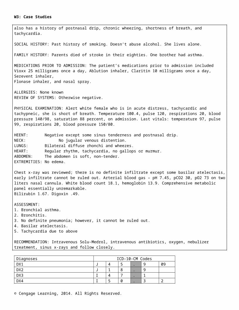
Diagnoses ICD-10-CM CodesDX1 J 4 5 . 9 09DX2 J 1 8 . 9DX3 I 4 7 . 1DX4 I 5 0 . 3 2
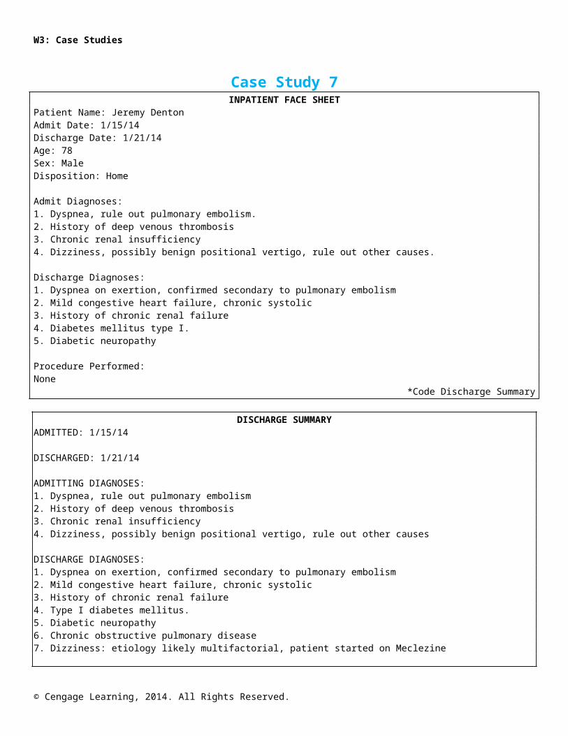
Case Study 7INPATIENT FACE SHEET
Patient Name: Jeremy DentonAdmit Date: 1/15/14Discharge Date: 1/21/14Age: 78Sex: MaleDisposition: Home
Admit Diagnoses:1. Dyspnea, rule out pulmonary embolism.2. History of deep venous thrombosis3. Chronic renal insufficiency4. Dizziness, possibly benign positional vertigo, rule out other causes.
Discharge Diagnoses:1. Dyspnea on exertion, confirmed secondary to pulmonary embolism2. Mild congestive heart failure, chronic systolic3. History of chronic renal failure4. Diabetes mellitus type I.5. Diabetic neuropathy
Procedure Performed:None
*Code Discharge Summary
DISCHARGE SUMMARYADMITTED: 1/15/14
DISCHARGED: 1/21/14
ADMITTING DIAGNOSES:1. Dyspnea, rule out pulmonary embolism2. History of deep venous thrombosis3. Chronic renal insufficiency
© Cengage Learning, 2014. All Rights Reserved.
W3: Case Studies
4. Dizziness, possibly benign positional vertigo, rule out other causes
DISCHARGE DIAGNOSES:1. Dyspnea on exertion, confirmed secondary to pulmonary embolism2. Mild congestive heart failure, chronic systolic3. History of chronic renal failure4. Type I diabetes mellitus.5. Diabetic neuropathy6. Chronic obstructive pulmonary disease7. Dizziness: etiology likely multifactorial, patient started on Meclezine
HISTORY: This is a 78-year-old gentleman with the above-mentioned medical problems who came to the emergency room at this time with light-headedness, dizziness, wooziness, drunk feeling, not sure of his steps. He was having increasing shortness of breath and does have a history of deep venous thrombosis and pulmonary emboli.
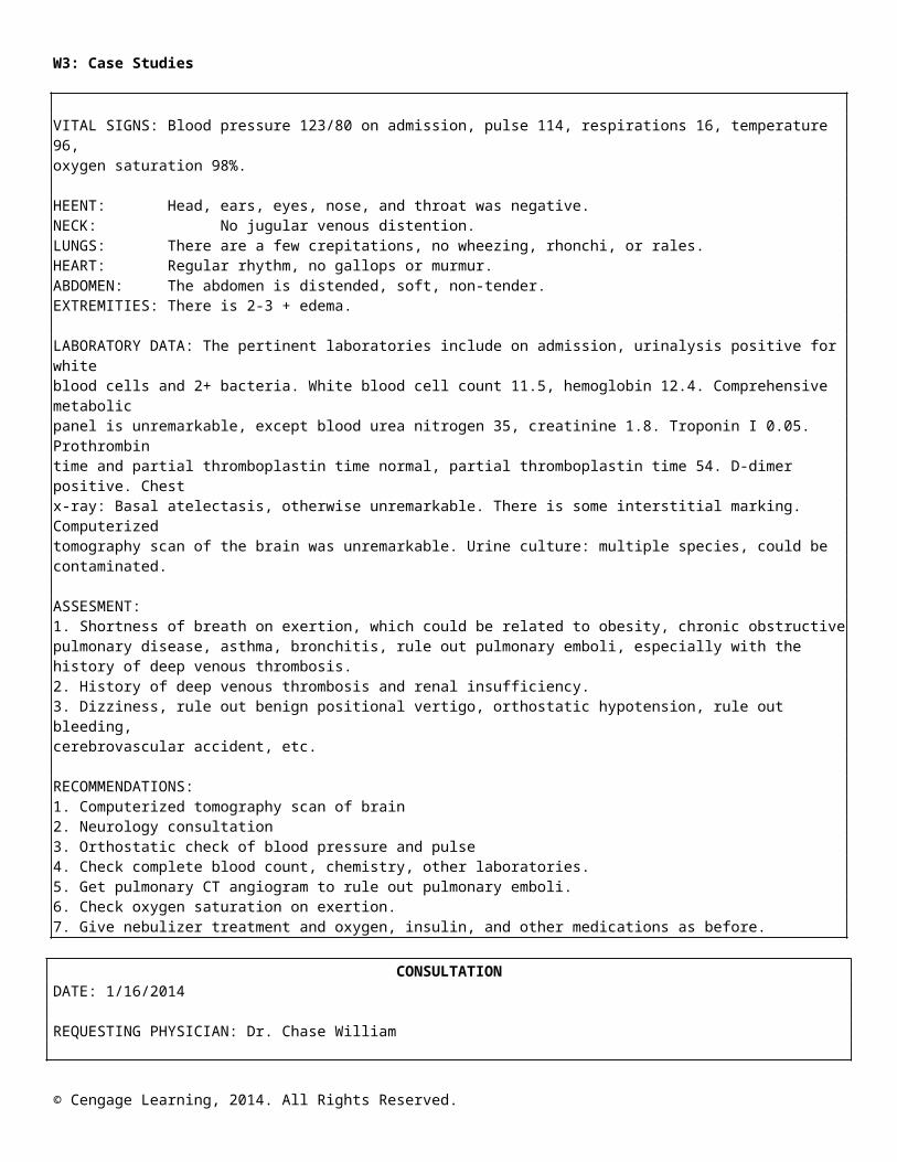
PERTINENT EXAM: Blood pressure 123/80, pulse rate 114, respirations 16, temperature 96.HEENT: Unremarkable.NECK: No jugular venous distention.LUNGS: A few crepitations, no wheezing.HEART: Regular. No murmur.ABDOMEN: Distended, soft, non-tender.EXTREMITIES: 2-3 + edema.
LABORATORY: White blood cell account 11,500, left shift differential. Other counts were normal. Follow-up hemoglobin 11.1, hematocrit 32.9. Baseline INR normal. Chemistries: sodium 135, blood urea nitrogen 35, creatinine 1.8, alkaline phosphatase 80. AST/ALT levels initially normal. Follow-up blood sugar 198-200. Urinalysis: yellow and hazy appearance, microscopic blood noted, protein noted, nitrite negative, a small amount of leukocyte esterase detected. Urine culture suggested a contaminated specimen.
Chest film revealed some prominent interstitial markings and possible congestive heart failure.
Computed tomography brain scan: mild atrophy. Ventilation perfusion lung scan revealed an intermediate probability for pulmonary embolism. Electrocardiogram: sinus rhythm, sinus tachycardia. Arterial blood gases on two liters nasal cannula, pH 7.41, pC02 33.1, p02 98.8, bicarb 20.8, and saturation is 97.5%.
HOSPITAL COURSE: The patient admitted with increasing weakness, dyspnea, and hypoxemia and dizziness at this time as well. There is no evidence of acute cerebrovascular accident. Computed tomography brain scan revealing chronic changes, nothing acute.
Ultimately the patient was found to have a pulmonary embolism and started on subcutaneousLovenox injection, 1 mg/kg subcutaneous every 12 hours and then Coumadin started as well. Dailyprothrombin time, INR evaluations were obtained, and Coumadin doses were titrated accordingly.We were able to confirm the suspicion with a computed tomography scan and pulmonaryangiogram. The patient was continued on anticoagulation, as well as treatment for heart failure withAce inhibitor therapy. He had reached the maximal hospital benefit and was discharged 1/21/14.
MEDICATIONS ON DISCHARGE:1. Humulin 75/25 15 units twice daily2. Xanax .25 mg three times a day3. Celexa 30 mg a daily4. Coumadin 10 mg daily5. Protonex 40 mg a day6. Prinivil 40 mg daily7. Meclezine
DIET: 1,800 ADA, low fat, low salt
© Cengage Learning, 2014. All Rights Reserved.
W3: Case Studies
OTHER THERAPEUTIC MEASURES: The patient reached maximum therapeutic benefit from hishospital admission. He was discharged with his medications and diet as listed.
ACTIVITY: As tolerated.
HISTORY & PHYSICALADMITTED: 1/15/2014
CHIEF COMPLAINT: Dizziness, lightheadedness, and shortness of breath on exertion.
HISTORY OF PRESENT ILLNESS: This is 78-year-old gentleman who has a history of multiple medical problems. He came into the emergency room because he was feeling dizzy, lightheaded, woozy, and kind of drunk. He could not be sure of his steps. He has also been complaining of increased shortness of breath on exertion. He does have chronic dyspnea on exertion due to multiple medical problems including obesity and deep venous thrombosis; however, he feels that his symptoms are worse than before.
PAST MEDICAL HISTORY: His history is positive for multiple medical problems. He has history of hypertension, diabetes mellitus type 1, deep venous thrombosis. He has been on Coumadin for quite some time because of deep venous thrombosis.
SOCIAL HISTORY: He smoked cigarettes, but quit smoking 25 years ago. He does not drink alcohol.He is married and lives with his wife.
ALLERGIES: None.
REVIEW OF SYSTEMS: As mentioned earlier; other than that, is unremarkable. He does not have anychest pain, denies any shortness of breath on exertion. He has no cough, hemoptysis, fever, or chills.No nausea or vomiting, abdominal pain, diarrhea, or urinary burning or hematuria at this time.
MEDICATIONS: Protonix 40 milligrams once a day, and insulin 75/25 Humalog 35 units in the morning and 35 units in the evening.
PHYSICIAL EXAMINATION:
GENERAL APPEARANCE: Elderly white gentleman who is in no respiratory distress, but gets shortof breath on exertion.
VITAL SIGNS: Blood pressure 123/80 on admission, pulse 114, respirations 16, temperature 96,oxygen saturation 98%.
HEENT: Head, ears, eyes, nose, and throat was negative.NECK: No jugular venous distention.LUNGS: There are a few crepitations, no wheezing, rhonchi, or rales.HEART: Regular rhythm, no gallops or murmur.ABDOMEN: The abdomen is distended, soft, non-tender.EXTREMITIES: There is 2-3 + edema.
LABORATORY DATA: The pertinent laboratories include on admission, urinalysis positive for whiteblood cells and 2+ bacteria. White blood cell count 11.5, hemoglobin 12.4. Comprehensive metabolicpanel is unremarkable, except blood urea nitrogen 35, creatinine 1.8. Troponin I 0.05. Prothrombintime and partial thromboplastin time normal, partial thromboplastin time 54. D-dimer positive. Chestx-ray: Basal atelectasis, otherwise unremarkable. There is some interstitial marking. Computerizedtomography scan of the brain was unremarkable. Urine culture: multiple species, could becontaminated.
ASSESMENT:
© Cengage Learning, 2014. All Rights Reserved.
W3: Case Studies
1. Shortness of breath on exertion, which could be related to obesity, chronic obstructive pulmonary disease, asthma, bronchitis, rule out pulmonary emboli, especially with the history of deep venous thrombosis.2. History of deep venous thrombosis and renal insufficiency.3. Dizziness, rule out benign positional vertigo, orthostatic hypotension, rule out bleeding,cerebrovascular accident, etc.
RECOMMENDATIONS:1. Computerized tomography scan of brain2. Neurology consultation3. Orthostatic check of blood pressure and pulse4. Check complete blood count, chemistry, other laboratories.5. Get pulmonary CT angiogram to rule out pulmonary emboli.6. Check oxygen saturation on exertion.7. Give nebulizer treatment and oxygen, insulin, and other medications as before.
CONSULTATIONDATE: 1/16/2014
REQUESTING PHYSICIAN: Dr. Chase William
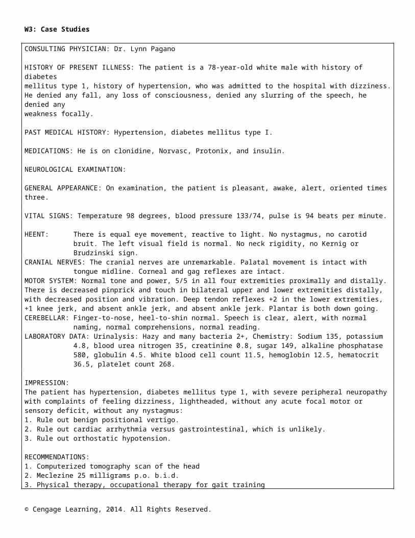
CONSULTING PHYSICIAN: Dr. Lynn Pagano
HISTORY OF PRESENT ILLNESS: The patient is a 78-year-old white male with history of diabetesmellitus type 1, history of hypertension, who was admitted to the hospital with dizziness.He denied any fall, any loss of consciousness, denied any slurring of the speech, he denied anyweakness focally.
PAST MEDICAL HISTORY: Hypertension, diabetes mellitus type I.
MEDICATIONS: He is on clonidine, Norvasc, Protonix, and insulin.
NEUROLOGICAL EXAMINATION:
GENERAL APPEARANCE: On examination, the patient is pleasant, awake, alert, oriented timesthree.
VITAL SIGNS: Temperature 98 degrees, blood pressure 133/74, pulse is 94 beats per minute.
HEENT: There is equal eye movement, reactive to light. No nystagmus, no carotid bruit. The left visual field is normal. No neck rigidity, no Kernig or Brudzinski sign.
CRANIAL NERVES: The cranial nerves are unremarkable. Palatal movement is intact with tongue midline. Corneal and gag reflexes are intact.
MOTOR SYSTEM: Normal tone and power, 5/5 in all four extremities proximally and distally. There is decreased pinprick and touch in bilateral upper and lower extremities distally, with decreased position and vibration. Deep tendon reflexes +2 in the lower extremities, +1 knee jerk, and absent ankle jerk, and absent ankle jerk. Plantar is both down going.CEREBELLAR: Finger-to-nose, heel-to-shin normal. Speech is clear, alert, with normal naming, normal
comprehensions, normal reading.LABORATORY DATA: Urinalysis: Hazy and many bacteria 2+, Chemistry: Sodium 135, potassium 4.8, blood
urea nitrogen 35, creatinine 0.8, sugar 149, alkaline phosphatase 580, globulin 4.5. White blood cell count 11.5, hemoglobin 12.5, hematocrit 36.5, platelet count 268.
IMPRESSION:The patient has hypertension, diabetes mellitus type 1, with severe peripheral neuropathy with complaints of feeling dizziness, lightheaded, without any acute focal motor or sensory deficit, without any nystagmus:1. Rule out benign positional vertigo.
© Cengage Learning, 2014. All Rights Reserved.
W3: Case Studies
2. Rule out cardiac arrhythmia versus gastrointestinal, which is unlikely.3. Rule out orthostatic hypotension.
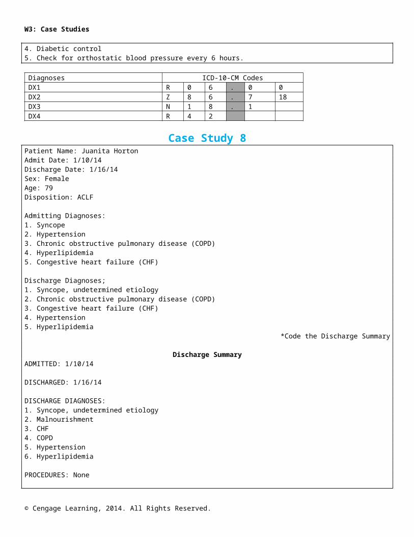
RECOMMENDATIONS:1. Computerized tomography scan of the head2. Meclezine 25 milligrams p.o. b.i.d.3. Physical therapy, occupational therapy for gait training4. Diabetic control5. Check for orthostatic blood pressure every 6 hours.
Diagnoses ICD-10-CM CodesDX1 R 0 6 . 0 0DX2 Z 8 6 . 7 18DX3 N 1 8 . 1DX4 R 4 2
Case Study 8Patient Name: Juanita HortonAdmit Date: 1/10/14Discharge Date: 1/16/14Sex: FemaleAge: 79Disposition: ACLF
Admitting Diagnoses:1. Syncope2. Hypertension3. Chronic obstructive pulmonary disease (COPD)4. Hyperlipidemia5. Congestive heart failure (CHF)
Discharge Diagnoses;1. Syncope, undetermined etiology2. Chronic obstructive pulmonary disease (COPD)3. Congestive heart failure (CHF)4. Hypertension5. Hyperlipidemia
*Code the Discharge Summary
Discharge SummaryADMITTED: 1/10/14
DISCHARGED: 1/16/14
DISCHARGE DIAGNOSES:1. Syncope, undetermined etiology2. Malnourishment3. CHF4. COPD5. Hypertension6. Hyperlipidemia
PROCEDURES: None
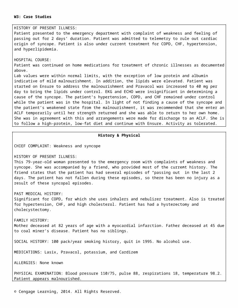
HISTORY OF PRESENT ILLNESS:
© Cengage Learning, 2014. All Rights Reserved.
W3: Case Studies
Patient presented to the emergency department with complaint of weakness and feeling of passing out for 2 days’ duration. Patient was admitted to telemetry to rule out cardiac origin of syncope. Patient is also under current treatment for COPD, CHF, hypertension, and hyperlipidemia.
HOSPITAL COURSE:Patient was continued on home medications for treatment of chronic illnesses as documented above.Lab values were within normal limits, with the exception of low protein and albumin indicative of mild malnourishment. In addition, the lipids were elevated. Patient was started on Ensure to address the malnourishment and Pravacol was increased to 40 mg per day to bring the lipids under control. EKG and ECHO were insignificant in determining a cause of the syncope. The patient’s hypertension, COPD, and CHF remained under control while the patient was in the hospital. In light of not finding a cause of the syncope and the patient’s weakened state from the malnourishment, it was recommended that she enter an ACLF temporarily until her strength returned and she was able to return to her own home. She was in agreement with this and arrangements were made for discharge to an ACLF. She is to follow a high-protein, low-fat diet and continue with Ensure. Activity as tolerated.
History & Physical
CHIEF COMPLAINT: Weakness and syncope
HISTORY OF PRESENT ILLNESS:This 79-year-old woman presented to the emergency room with complaints of weakness and syncope. She was accompanied by a friend, who provided most of the current history. The friend states that the patient has had several episodes of “passing out” in the last 2 days. The patient has not fallen during these episodes, so there has been no injury as a result of these syncopal episodes.
PAST MEDICAL HISTORY:Significant for COPD, for which she uses inhalers and nebulizer treatment. Also is treated for hypertension, CHF, and high cholesterol. Patient has had a hysterectomy and cholecystectomy.
FAMILY HISTORY:Mother deceased at 82 years of age with a myocardial infarction. Father deceased at 45 due to coal miner’s disease. Patient has no siblings.
SOCIAL HISTORY: 100 pack/year smoking history, quit in 1995. No alcohol use.
MEDICATIONS: Lasix, Pravacol, potassium, and Cardizem
ALLERGIES: None known
PHYSICAL EXAMINATION: Blood pressure 110/75, pulse 88, respirations 18, temperature 98.2.Patient appears malnourished.
REVIEW OF SYSTEMS:HEENT: Pupils equal and reactive to light. Pale conjunctiva. Moist mucous membranes.NECK: Supple without masses.LUNGS: Decreased breath sounds.HEART: Regular rate and rhythm.ADOMEN: Soft, non-tender. Bowel sounds present.EXTREMITIES: No cyanosis or clubbing.
IMPRESSION:1. Syncope, etiology to be determined2. Hypertension3. COPD4. Hyperlipidemia5. CHF
© Cengage Learning, 2014. All Rights Reserved.

W3: Case Studies
PLAN:1. Admit to telemetry.2. Continue with home meds.
Progress Notes:
1/10: Admit Note: Patient admitted via emergency room to telemetry for syncope, undetermined etiology. Patient to continue with home meds for CHF, COPD, hypertension, and hyperlipidemia.
1/11: S: No syncope while here.O: EKG showed sinus rhythm with RBBB. ECHO WNL Chest x-ray, no CHF, known COPD.
Hypertension under control. Vital signs good. Chem profile significant for low protein and albumin with HDL of 281.
A: No cardiac reason for syncope. Patient is malnourished.P: Continue with current treatment. Add Ensure to diet as a supplement.
1/12: S: No complaints. No further syncopal episodes.O: Rhythm remains unchanged. Condition stable.A: Repeat labs within normal limits, except for protein and albumin, which are still low.P: Case management to arrange transfer to ACLF until patient can return home.
1/13: Discharge Note: No cardiac or chemical explanation for syncope. Patient to be discharged to an ACLF until she can return to her own home. Continue with dietary supplement in light of mild malnourishment.
Orders:
1/10: 1. Admit patient to service of Dr. James Mitchell, per Dr. Lyle Douglas, emergency room physician.2. Place patient on telemetry.3. Chem profile, CBC4. Continue with patient’s home medications.5. EKG, chest x-ray, ECHO
1/11: 1. Repeat labs.2. Provide patient with Ensure.3. Case management to assist with discharge plans.
1/12: 1. Discontinue telemetry.2. Arrange transfer to ACLF tomorrow.
1/13: 1. Discharge today to ACLF.
Diagnoses ICD-10-CM CodesDX1 E 7 8 . 5DX2 I 1 0DX3 J 4 4DX4 I 5 0 . 9
Case Study 9Patient Name: Elmer MillerAdmit Date: 1/01/14Discharge Date: 1/13/14Age: 79
© Cengage Learning, 2014. All Rights Reserved.
W3: Case Studies
Sex: MaleDisposition: Home with Home Health Care
Admission Diagnoses:1. Acute bronchial asthma in exacerbation2. Hypoxemia, hypercapnia3. Suspected chronic obstructive pulmonary disease4. Rule out pulmonary embolism.5. Hypertension6. Hyperlipidemia
Discharge Diagnoses:1. Acute bronchial asthma with acute exacerbation2. Tracheobronchitis3. Chronic hypoxemia4. Suspect underlying chronic obstructive pulmonary disease5. History of hypertension6. Hyperlipidemia
Procedures Performed:Broncoscopy with biopsies and lavage
*Code Admission History and Physical
DISCHARGE SUMMARYADMITTED: 1/1/14
DISCHARGED: 1/13/14
DISCHARGE DIAGNOSES:1. Acute bronchial asthma with acute exacerbation2. Tracheobronchitis3. Chronic hypoxemia4. Suspect underlying chronic obstructive pulmonary disease5. History of hypertension6. Hyperlipidemia
ADMITTING DIAGNOSES:1. Acute bronchial asthma in exacerbation2. Hypoxemia, hypercapnia3. Suspected chronic obstructive pulmonary disease4. Rule out pulmonary embolism5. Hypertension6. Hyperlipidemia
PROCEDURE:1. Bronchoscopy with lavage and biopsy
HISTORY OF PRESENT ILLNESS:This is a 79-year-old male patient with above-mentioned medical problems who now presents with increasing shortness of breath, cough, wheezing, and respiratory distress. He has been on multiple bronchodilators and inhaled steroids, including Flovent, nebulizer, theophylline, and a small dose of prednisone.
HOSPITAL COURSE:This patient was admitted with increasing shortness of breath. He was not responding to outpatient medical therapy. He was placed on Albuterol nebulized respiratory treatment and started on a short course
© Cengage Learning, 2014. All Rights Reserved.
W3: Case Studies
of pulse intravenous steroids, given mucolytics expectorants and continued on theophylline. He was cultured and placed on intravenous Claforan initially, empirically; he was otherwise continued on treatment for hypertension. A therapeutic bronchoscopy was performed with evidence of thick, purulent mucous plugs widespread on both sides, some narrowing of the left lower lobe bronchus also noted with some inflammation. Biopsies were negative for malignancy. He was continued on Mucomyst at this point, nebulized respiratory treatments, chest physiotherapy, and intravenous Solu-Medrol.
Following several days of aggressive treatment, he started to improve and was less short of breath; bronchospasm was resolving and was discharged on January 13.
PERTINENT EXAM: Audible wheezing, coughed continuously, tachycardic, tachypneic, afebrile.VITAL SIGNS: Blood pressure stable.HEENT: Revealed postnasal drip.NECK: No jugular venous distention.LUNGS: Diffuse wheezing, rhonchi, and rales.HEART: Regular.ABDOMEN: Soft.EXTREMITIES: No edema.
REVIEW OF LABORATORY DATA:The bronchoscopy cytology revealed no malignancy. The arterial blood gases on room air: pH 7.48, pC02 36, p02 56, bicarb 26.4, and saturation 92.3%. Complete blood count: white blood cell count 9,800. Hemoglobin 15, hematocrit 47.1, and platelet count normal. Follow–up complete blood count remained stable. Chemistries: blood urea nitrogen 17, creatinine 1.4. Troponin I normal on a serial basis. Albumin 2.4 to 3.3. Liver functions normal. theophylline level 19.2. Urinalysis: yellow and clear; microscopic blood noted, protein negative, nitrite negative, leukocyte esterase a small amount detected. Sputum revealed Candida. Urine culture: no growth. Sputum for acid-fast bacilli: test still pending at this time although smears were negative. The chest revealed a new hazy opacity of the left mid lung, in the lower lung area, possibly fusion or atelectasis. This persisted on follow-up.
Electrocardiogram: sinus rhythm, sinus tachycardia. While on telemetry the patient was in sinusrhythm.
MEDICATIONS ON DISCARGE:1. Prednisone 30 mg daily for 4 days, then 20 mg daily for 4 days, then 10 mg daily2. Flovent 220 mcg three puffs twice daily3. Albuterol treatments four times daily4. Norvasc 5 mg daily5. Lasix 40 mg half tablet daily6. Potassium 10 mEq daily7. Nasonex spray two puffs daily8. theophylline 200 mg twice daily
ACITIVITY: As tolerated.
HISTORY AND PHYSICIAL
ADMISSION DATE: 1/01/14
CHIEF COMPLAINT: Shortness of breath, wheezing, cough, and chest congestion.
HISTORY OF PRESENT ILLNESS:This is a 79-year-old male with multiple admissions for respiratory problems and exacerbation of asthma. Presented to the emergency room because of increasing shortness of breath, cough, wheezing, and respiratory distress. Apparently he has been on multiple bronchodilators and inhaled steroids, including Flovent, nebulizer, theophylline, and a small dose of Prednisone a day. In spite of the outpatient treatment, his symptoms continued to worsen. He was admitted to the hospital for further treatment.
© Cengage Learning, 2014. All Rights Reserved.
W3: Case Studies
PAST MEDICAL HISTORY:Past medical history is positive for long-standing bronchial asthma with chronic exacerbation, steroid-dependent asthma. He also had history of tracheobronchitis, hypertension, and hyperlipidemia.
MEDICATIONS AT HOME:Flovent 220 micrograms 2 puffs twice a day, but he is not using regularly. Nebulizer 4 times a day, Norvasc 5 milligrams once a day, Lasix 40 milligrams ½ tablet once a day, potassium 10 milliequivalent once a day, calcium t.i.d., theophylline 200 milligrams b.i.d., and Prednisone 5 milligrams once a day.
ALLERGIES: None.
SOCIAL HISTORY: Does not smoke. Never smoked.
REVIEW OF SYSTEMS:HEENT: Revealed postnasal drip.NECK: No jugular venous distension.LUNGS: Bilateral diffuse wheezing, rhonchi, and rales.HEART: Regular rhythm.ABDOMEN: Soft.EXTREMITIES: No edema.GASTROINTESTINAL: No gastritis, gastroesophageal reflux, occasional indigestion; no ulcer.RESPIRATORY: As mentioned in the history.CARDIAC: History of hypertension, but no history of angina, coronary artery disease. Does not
have any chest pain. He had an echocardiogram in 2001, which was normal with ejection fraction of 60 percent.
MUSCULOSKELETAL: NegativeNEUROLOGIC: Negative for stroke, transient ischemic attack, headache, dizziness, or syncope.GENITOURINARY: Negative. The rest of review of systems is negative.
PHYSICAL EXAMINATION:Elderly male who is in acute distress with audible wheeze. He coughs continuously. He is tachycardiac, tachypneic, afebrile. Vitals stable otherwise.
The labs, x-rays, etc., were reviewed.
IMPRESSION:1. Acute bronchial asthma and exacerbation.2. Severe hypoxemia, hypercapnia. Rule out due to chronic obstructive pulmonary disease, bronchospasm. Rule out other causes, like pulmonary embolism.
PLAN: Will give IV steroids, IV antibiotics, nebulizer treatment, and oxygen.Get a spiral CT scan to rule out pulmonary emboli.Continue other treatment, and will follow him closely.
PROCEDURE REPORT
DATE: 1/02/14
ENDOSCOPIST: Dr. Brandon Douglas
PROCEDURE: Bronchoscopy
INDICATION: Left lower lobe collapse, due to mucous plug
ANESTHESIA: Dr. Jeffrey Cottrell
© Cengage Learning, 2014. All Rights Reserved.
W3: Case Studies
DESCRIPTION OF PROCEDURE:After the anesthesiologist anesthetized the patient, the Olympus bronchoscope was introduced through the bite block into the oral cavity. The upper airway was seen and was unremarkable. Cords were sprayed with Xylocaine. The scope was passed through the cords into the trachea, which was free of lesion. Carina was sharp. The scope was passed into the right mid stem bronchus. The upper lobe and middle lobe segments were seen. They are remarkable for thick, purulent mucous plugging of all the bronchial segments. Aggressive lavage with saline and Mucomyst were done. In spite of that, mucous plugs were quite thick and required manual removal through the scope. Subsequently the scope was withdrawn back to the left main stem, and left upper lobe, lingual lobe, and sub segments were seen. They were also remarkable for severe mucous plugs, which were removed manually. After complete evacuation of mucous plugs was done, bronchial segments were visualized again and there was some narrowing of left lower segment with some inflamed, swollen mucosa. Biopsies were done from that segment. Bronchial washings were done and sent for cytology and culture.
The patient tolerated the procedure well.
FINDINGS: Thick, purulent mucous pluggings were widespread on both sides. There was some narrowing of left lower lobe bronchus with inflamed, swollen mucosa with smooth margin.
SPECIMENS: Biopsies were done from left lobe bronchus. Washing was done. Bronchoscopic lavage was done using saline and Mucomyst, and all the mucous plugs were removed.
PATHOLOGY REPORT
DATE: 1/02/14
SPECIMEN TYPE: CYTO
SURGICAL PATHOLOGY / CYTOPATHOLOGY REPORT
PRE – OP DIAGNOSIS: Left lower lobe collapse: inflamed swollen bronchial mucosa left lowerLobe
POST-OP DIAGNOSIS Left lower lobe collapse: inflamed swollen bronchial mucosa left lowerLobe
SPECIMEN(S)
BRONCHIAL WASHING MATERIAL
GROSS DESCRIPTIONReceived 8 cc of hemorrhagic fluid in the laboratory. 2 smears and 1 cell block are prepared for cytological evaluation.
MICROSCOPIC DESCRIPTIONMicroscopic examination of the specimen reveals groups of and single epithelial cells that appear poorly preserved, displaying nuclear enlargement, hyperchromasia, increased N/C bronchial columnar, metaplastic, and reserve cells: pulmonary macrophages, mixed inflammatory cells, necrotic debris, and fungal elements.
CYTOPATHOLOGICAL DIAGNOSIS
BRONCHIAL WASHING: Poorly preserved dysplastic epithelial cells present. Candida species identified.
SURGICAL PATHOLOGY / CYTOPATHOLOGY REPORT
© Cengage Learning, 2014. All Rights Reserved.
W3: Case Studies
PRE–OP Diagnosis: Left lower lobe collapse: inflamed swollen bronchial mucosa left lower lobe
POST-OP Diagnosis: Left lower lobe collapse: inflamed swollen bronchial mucosa left lower lobe
SPECIMEN(S)
LUNG LEFT LOWER LOBE – BIOPSY X4
GROSS DESCRIPTIONThe specimen consists of four fragments of tan tissue ranging from 0.1 up to 0.6 cm in greatest dimension. Entirely submitted.
MICROSCOPIC DIAGNOSIS
BIOPSY OF LEFT LOWER LOBE: Mild non-specific chronic bronchitis with hypertrophy of thesubmucosal glands and thickening of the subepithelial basement membrane.
No tumor present.
FINAL DIAGNOSIS
BIOPSY OF LEFT LOWER LOBE:Minute detached fragment of dysplastic epithelium.
Mild non-specific chronic bronchitis with hypertrophy of submucosal glands and thickened subepithelial basement membrane.
Diagnoses ICD-10-CM CodesDX1 J 4 4 . 1DX2 R 0 9 . 0 2DX3 I 1 0DX4 E 7 8 . 5
Case Study 10Ambulatory Surgery
Face SheetPatient’s Name: Georgia PhillipsDate of Visit: 1/17/14Age: 52Sex: FemaleInsurance: Medicare
*Code Dr. Cheryl Bottom’s pathology report
History and Physical
DATE OF ADMISSION: 1/17/14
HISTORY OF PRESENT ILLNESS:The patient has a history of bilateral breast cysts and in a follow-up mammogram a mass was discovered in the left breast. Patient also has some dimpling in the area, demonstrated on mammogram in the upper outer quadrant.
PAST MEDICAL HISTORY: Patient has a history of mitral valve prolapse.
© Cengage Learning, 2014. All Rights Reserved.
W3: Case Studies
ALLERGIES: Demerol and Biaxin.
MEDICATIONS: V-Tabs prior to procedures for her mitral valve prolapse.
PHYSICAL EXAMINATION:Vital signs: BP 146/80; Respirations 17; Pulse 77; Temperature 99.0Skin: Warm and dry.Eyes: The pupils are equal, round, reactive to light and accommodation. Sclera is clear.Neck: Supple. No masses, scars, or bruits.Lungs: Clear to auscultation and percussion.Heart: Normal sinus rhythm. No murmurs or gallops.Breasts: Breasts are symmetrical. There is an area of slight skin retraction on the upper outer
quadrant of the left breast. There is some thickness in the area. No other masses felt. No axillary lymphadenopathy.
Extremities: Good distal pulses.
Plan:Patient will be brought to the ambulatory surgical center for a left breast biopsy.
Operative ReportDate of Operation: 1/17/14
Preoperative Diagnosis: Left breast mass
Postoperative Diagnosis: Infiltrating ductal cell carcinoma left breast.
Procedure Performed: Excision of left breast mass.
Surgeon: Cheryl Bottom, MD
Assistant: Jon Actor, MD
PROCEDURE:The patient was placed in the supine position where anesthesia was administered. The left breast was prepped and draped in the usual sterile manner.A transverse incision was made along the mass and was carried down through the skin and subcutaneous tissue. A firm mass was identified and was sharply excised from the surrounding breast tissue. Bleeding was controlled with electrocautery.
The wound was closed using interrupted sutures of 3-0 Vicryl for the deep layer. The subcutaneous tissue was closed with interrupted suture of 3-0 Vicryl and the skin was closed 4-0 Monocryl. Dressing was applied. The patient tolerated the procedure well.
Pathology ReportDate: 1/17/14
Physician: Cheryl Bottom, MD
Preoperative Diagnosis: Left breast mass
Surgical Procedure: Excision left breast mass
Postoperative Diagnosis: Infiltrating ductal carcinoma
Specimen(s): Left breast mass
GROSS:
© Cengage Learning, 2014. All Rights Reserved.
W3: Case Studies
Received directly from the operating room is a 4 x 3 x 3 cm ovoid pink-tan to yellow fibroadiposetissue, which is firm.
GROSS AND MICROSCOPIC EXAMINATION:Excisional biopsy of left breast:
Invasive ductal carcinoma, 2.3 cm, histologic grade 2, nuclear grade 2, mitotic grade 1. Focal lymph vascular space invasion is noted.
Diffuse fibrocystic changes are also noted.
PROGRESS NOTES:
Patient has a left breast mass and was taken to the operating room, where an excisional biopsy of themass was performed. Pathology report is pending final determination, but preliminary report isductal carcinoma.
PHYSICIAN ORDERS:Pre-Operative OrdersDiet: NPOConsent to read: Excision left breast massAncef 1 g IV before surgery
Post-Operative OrdersVicodin 1 q6h p.r.n.Liquids as toleratedDischarge when criteria are met.
Diagnoses ICD-10-CM CodesDX1 N 6 5DX2 N 6 0 . 0DX3 DX4
© Cengage Learning, 2014. All Rights Reserved.
![myfaith-data.s3.amazonaws.com€¦ · Instead of taking the glory for ... GOD bless you all! ... [Gunnedah]; Fr. Marian Flis; Fr. Lou Breslan. —Mass Offerings Week:](https://static.fdocuments.in/doc/165x107/5b5bda447f8b9ab8578ed45d/myfaith-datas3-instead-of-taking-the-glory-for-god-bless-you-all-.jpg)

![Durham E-Theses Culture, Tradition and Alternative Justice ...etheses.dur.ac.uk/11265/1/MJur_Thesis_Full.pdf · 1 Otto Kahn-Freund, ‘On Uses and Misuses of Comparative Law’ [1974]](https://static.fdocuments.in/doc/165x107/604658a2cf4cf17d937c339e/durham-e-theses-culture-tradition-and-alternative-justice-1-otto-kahn-freund.jpg)