Questions: Embryos R Us 1. What are the relevant facts of ... · IB Biology 2.3.1 Draw and ......
75
Questions: Embryos R Us 1. What are the relevant facts of this case? 2. What are some ethical questions raised by this situation? 3. Who are the stakeholders in this situation? Who will be affected by decisions that are made? 4. What are the values that play a role in the decision? 5. What are some possible solutions and their consequences? 6. What do you consider to be the best solution and why?
Transcript of Questions: Embryos R Us 1. What are the relevant facts of ... · IB Biology 2.3.1 Draw and ......

Questions: Embryos R Us
1. What are the relevant facts of this case?
2. What are some ethical questions raised by this situation?
3. Who are the stakeholders in this situation? Who will be
affected by decisions that are made?
4. What are the values that play a role in the decision?
5. What are some possible solutions and their consequences?
6. What do you consider to be the best solution and why?
Cells

Eukaryotic Cell

IB Biology
2.3.1 Draw and label a diagram of the ultra structure of a liver cell as an example of a animal
cell.
2.3.2 Annotate the diagram from 2.3.1.with the functions of each named structure.
2.3.3 Identify structures from 2.3.1 in electron micrographs of liver cells.
2.3.4 Compare prokaryotic and eukaryotic cells
2.3.5 State three differences between plant and animal cells.
2.3.6 Outline two roles of extracellular components.

IB Biology
Cell Modeling:
Next Class, you will have 60 minutes to
construct a cell model which features all
of the organelles listed in the cell
component worksheet.
You must provide all of the raw materials
and tools. All construction must be
completed in class during the 60 minute
period.

The Eukaryotic Cells
Cells that contain a nucleus
The Eukaryotic Cell
“Eu” = true “Karyo” = kernal (nucleus)
Protists, Plants, Fungi, and Animals
Internal Membrane System
Has many membranous organelles

The Eukaryotic Cell

Cytoplasm

Cytoplasm
Region inside the plasma membrane excluding the nucleus
Fluid portion of the cytoplasm between the organelles is the
cytosol
Flow of Genetic information and protein Synthesis
Nucleus, Ribosomes, Rough &
Smooth ER

Nucleus
Control Center of the Cell
Genetic material:
•chromatin
•chromosomes
Nucleolus: ribosome synthesis
Double membrane envelope with pores
1st part of Protein synthesis
Nuclear pores
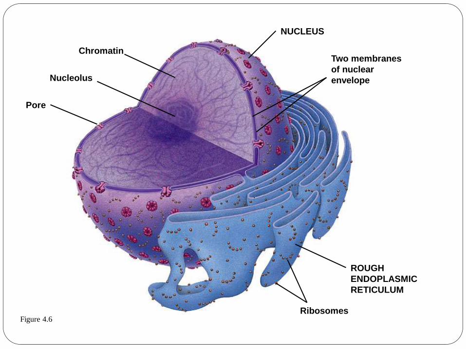
Figure 4.6
Chromatin
Nucleolus
Pore
NUCLEUS
Two membranes
of nuclear
envelope
ROUGH
ENDOPLASMIC
RETICULUM
Ribosomes
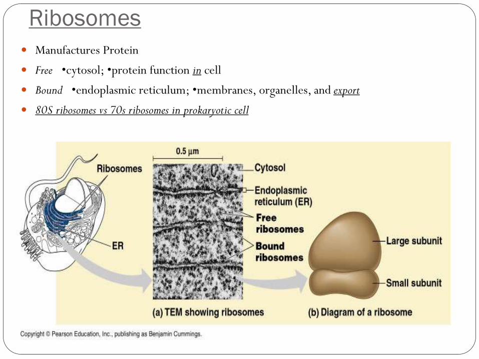
Ribosomes
Manufactures Protein
Free •cytosol; •protein function in cell
Bound •endoplasmic reticulum; •membranes, organelles, and export
80S ribosomes vs 70s ribosomes in prokaryotic cell
Endoplasmic Reticulum
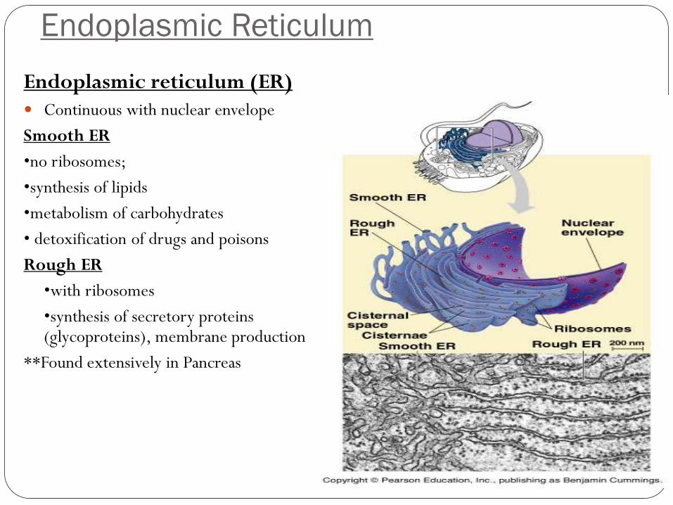
Endoplasmic reticulum (ER)
Continuous with nuclear envelope
Smooth ER
•no ribosomes;
•synthesis of lipids
•metabolism of carbohydrates
• detoxification of drugs and poisons
Rough ER
•with ribosomes
•synthesis of secretory proteins (glycoproteins), membrane production
**Found extensively in Pancreas
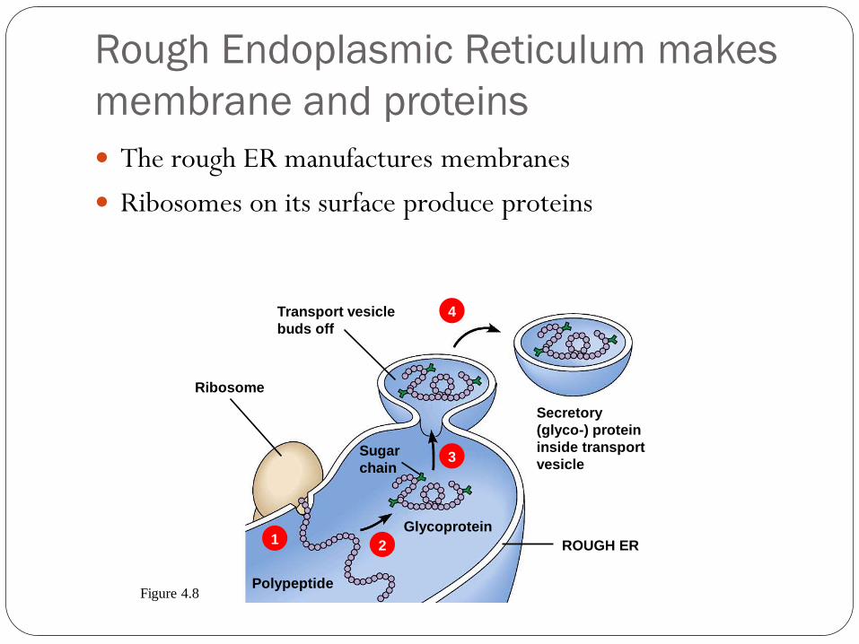
Rough Endoplasmic Reticulum makes
membrane and proteins
The rough ER manufactures membranes
Ribosomes on its surface produce proteins
1 2
3
4 Transport vesicle
buds off
Ribosome
Sugar
chain
Glycoprotein
Secretory
(glyco-) protein
inside transport
vesicle
ROUGH ER
Polypeptide Figure 4.8
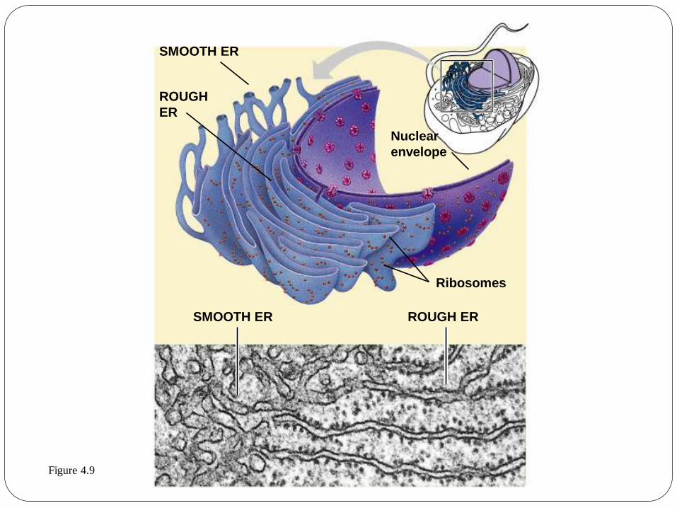
SMOOTH ER
ROUGH
ER
Nuclear
envelope
Ribosomes
SMOOTH ER ROUGH ER
Figure 4.9

Golgi Apparatus (complex)
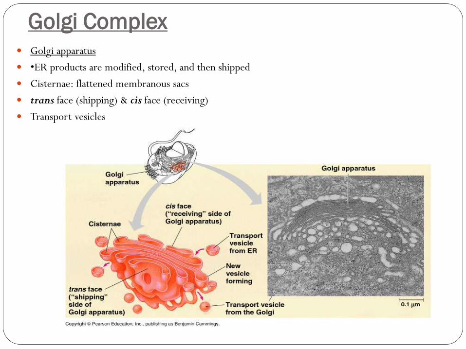
Golgi Complex Golgi apparatus
•ER products are modified, stored, and then shipped
Cisternae: flattened membranous sacs
trans face (shipping) & cis face (receiving)
Transport vesicles
The Golgi apparatus finishes, sorts,
and ships cell products
The Golgi apparatus consists of stacks of membranous sacs
These receive and modify ER products, then send them on to
other organelles or to the cell membrane

The Golgi apparatus
Golgi
apparatus
“Receiving” side of
Golgi apparatus
Transport
vesicle
from ER
New
vesicle
forming
Transport vesicle
from the Golgi
Golgi apparatus
“Shipping”
side of Golgi
apparatus Figure 4.10

Lysosomes & Vacuoles
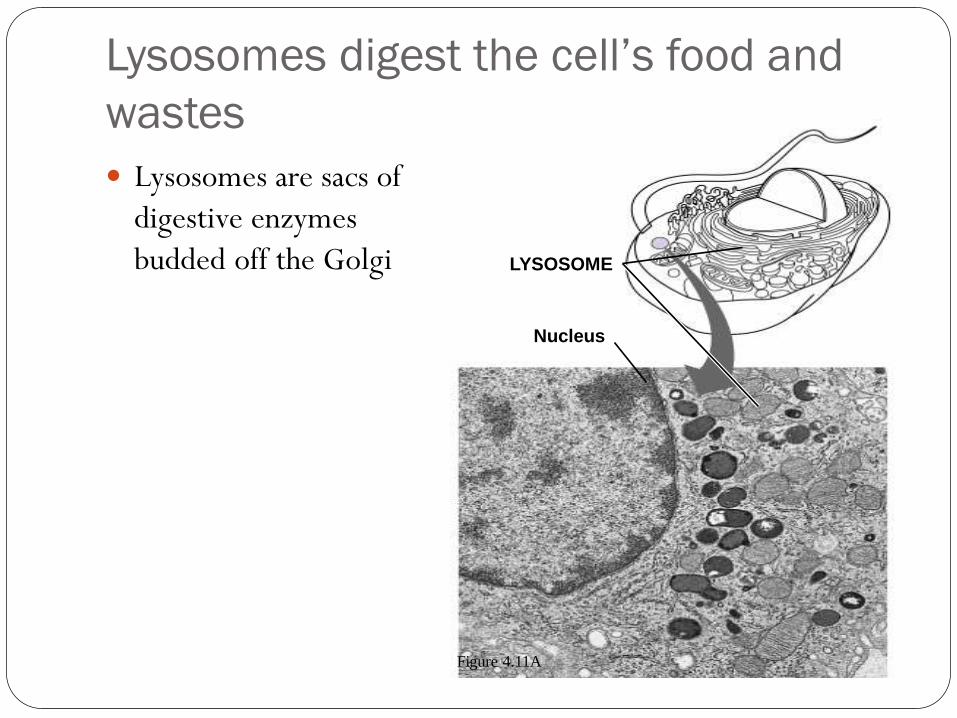
Lysosomes digest the cell’s food and
wastes
Lysosomes are sacs of
digestive enzymes
budded off the Golgi LYSOSOME
Nucleus
Figure 4.11A

Lysosomes

Lysosomes:
Contain lysosomal enzymes (hydrolytic enzymes)
Digests food molecules (macromolecules)
Destroys bacteria
Recycles damaged organelles

Lysosomes:
Function in embryonic development in animals
Undergoes phagocytosis & engulfs material
Recycle cell’s own organic material
**Found extensively in Macrophages (WBC’s)

Figure 4.11B
Rough ER
Transport vesicle
(containing inactive
hydrolytic enzymes)
Golgi
apparatus
Plasma
membrane
LYSOSOMES
“Food”
Engulfment
of particle
Food
vacuole
Digestion
Lysosome
engulfing
damaged
organelle

Lysosomes can cause Fatal Diseases
Lysosomal Storage Diseases are hereditary that interfere with
other cellular functions
*Examples:
Pompe’s disease
Tay-Sachs disease

Vacuoles
Membrane-bound sacs (larger
than vesicles)
Food (phagocytosis)
Contractile (pump excess
water)
Central (storage in plants)
Tonoplast membrane

Vacuoles function in the general
maintenance of the cell
Plant cells contain a large
central vacuole
The vacuole has lysosomal
and storage functions
Central
vacuole
Nucleus
Figure 4.13A

-Energy Harvesting Organelles
Not found in Prokaryotic Cells
Mitochondria & Chloroplasts
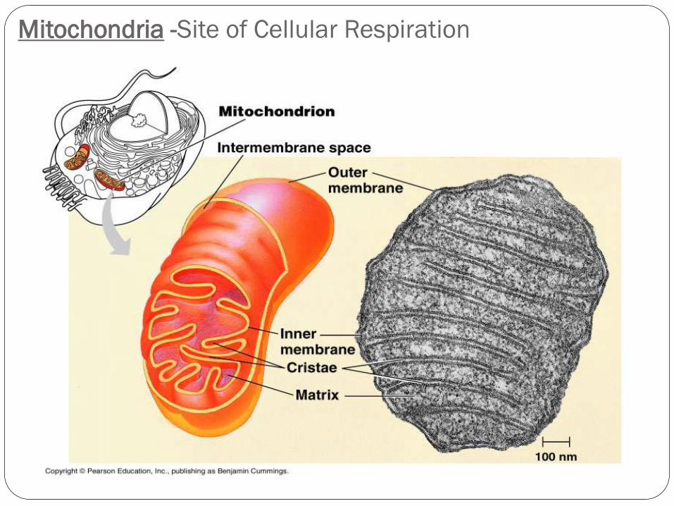
Mitochondria -Site of Cellular Respiration

Mitochondria harvest chemical Energy
from food
Site for Cellular Respiration---Prod. of ATP
Uses O2 to extract energy from sugar, fats, and other
molecules
Found in cells that are motile and contractible
Has a double membrane

Mitochondria harvest chemical Energy
from food
Has Convoluted inner membranes: Cristae
Two spaces: Matrix & intermembrane space
Not part of the endomembrane system
Has its own DNA and rbosomes (able to regenerate &
divide)---Semiautonomous
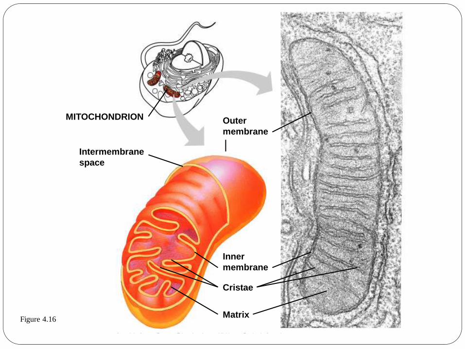
Figure 4.16
Outer
membrane
MITOCHONDRION
Intermembrane
space
Inner
membrane
Cristae
Matrix

Chloroplasts convert solar energy to
chemical energy
Chloroplasts are found in plants and some protists
Chloroplasts convert solar energy to chemical energy in
sugars
Chloroplast Stroma
Inner and outer membranes
Granum
Intermembrane space Figure 4.15

The Chloroplast
Site for Photosysnthesis: combines CO2 & H2O
Converts solar energy into chemical energy (sugar
molecules)
A Type of Plastid
Three types: (Amyloplastid, chromoplast, and chloroplast)
Double membrane w/ thylakoids (flattened disks)

The Chloroplast
Grana (stacked thylakoids)
Three compartments
Stroma
Intermembrane space
Within the thylakoid membranes
Has its own DNA


Cytoskeleton

The Cytoskeleton
Fibrous proteins (actin & tubulin)
Support, cell motility, biochemical regulation, organelle movement
Microtubules: thickest (nm) tubulin protein; shape, support, transport,
Chromosome separation
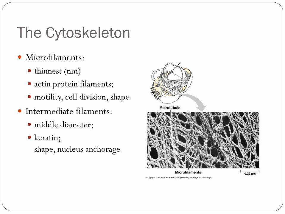
The Cytoskeleton
Microfilaments:
thinnest (nm)
actin protein filaments;
motility, cell division, shape
Intermediate filaments:
middle diameter;
keratin;
shape, nucleus anchorage

The cell’s internal skeleton helps
organize its structure and activities
A network of protein fibers makes up the cytoskeleton
Figure 4.17A

Comparing Cytoskeletal Filaments
Scan image

MICROFILAMENT
Figure 4.17B
INTERMEDIATE
FILAMENT
MICROTUBULE
Actin subunit Fibrous subunits Tubulin
subunit
7 nm 10 nm
25 nm
The Cytoskeleton

Microfilaments of actin enable cells to change shape and move
• Intermediate filaments reinforce the cell and
anchor certain organelles
• Microtubules
– give the cell rigidity
– provide anchors for organelles
– act as tracks for organelle movement
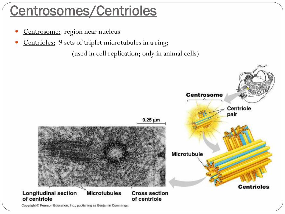
Centrosomes/Centrioles
Centrosome: region near nucleus
Centrioles: 9 sets of triplet microtubules in a ring;
(used in cell replication; only in animal cells)

Internal Structure & Function
Cilia & Flagella-Eukaryotes

Internal Structure & Function
Cilia & Flagella-Eukaryotes

Cilia/Flagella
Locomotive appendages
Ultrastructure: “9+2” (9 doublets of microtubules in a ring)
(2 single microtubules in center)
Connected by radial spoke
Anchored by basal body
(nine triplets of microtubules)
Dynein arm proteins (red)

Cilia/Flagella
Connected by radial spoke
Anchored by basal body
(nine triplets of
microtubules)
Dynein arm proteins (red)
Cilia and flagella move when
microtubules bend
Eukaryotic cilia and flagella are locomotor appendages that
protrude from certain cells
A cilia or flagellum is composed of a core of microtubules
wrapped in an extension of the plasma membrane

Figure 4.18A
FLAGELLUM
Outer microtubule
doublet
Plasma
membrane
Central
microtubules
Outer microtubule
doublet
Plasma
membrane
Electron micrograph
of sections:
Flagellum
Basal body
Basal body
(structurally identical to centriole)

ECM: Extracellular Matrix

ECM Composition
Extracellular matrix (ECM) composed of:
Proteins & Carbohydrate
Specifically:
glycoproteins
glycolipids
integrins
fibronectins
collagen (50% of all protein in the body)
Extracellular Matrix (ECM)
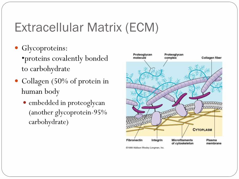
Glycoproteins:
•proteins covalently bonded
to carbohydrate
Collagen (50% of protein in
human body
embedded in proteoglycan
(another glycoprotein-95%
carbohydrate)
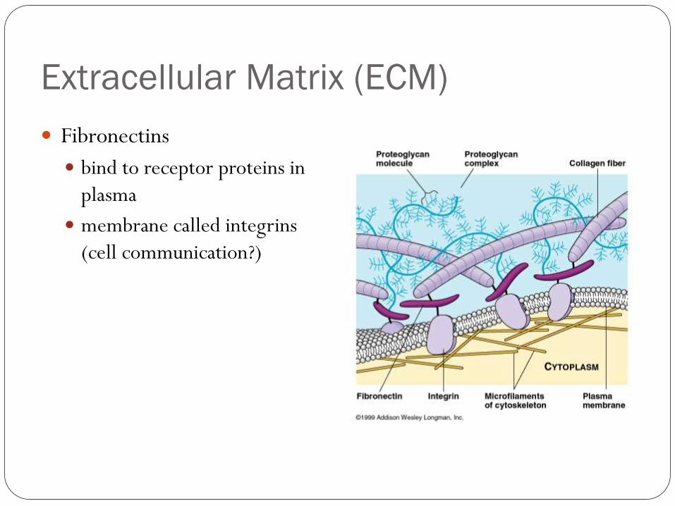
Extracellular Matrix (ECM)
Fibronectins
bind to receptor proteins in
plasma
membrane called integrins
(cell communication?)
Animal cells are embedded in an extracellular matrix
– It is a sticky layer of glycoproteins
– It binds cells together in tissues
– It can also have protective and supportive
functions

Science and Art
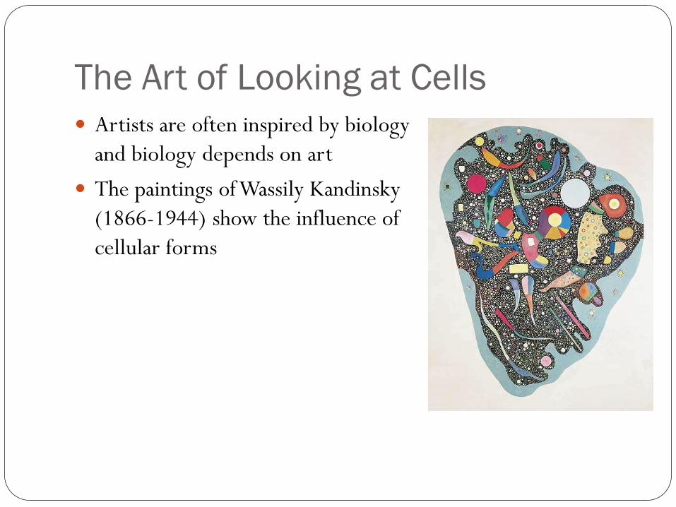
The Art of Looking at Cells
Artists are often inspired by biology
and biology depends on art
The paintings of Wassily Kandinsky
(1866-1944) show the influence of
cellular forms

Illustration is an important way to
represent what scientists see through
microscopes
• The anatomist
Santiago Ramón y
Cajal (1852-1934) was
trained as an artist
– He drew these retina
nerve cells

Samples of Various Types of Cells

Protists may have contractile vacuoles
Figure 4.13B
Nucleus
Contractile
vacuoles
– These pump out excess water
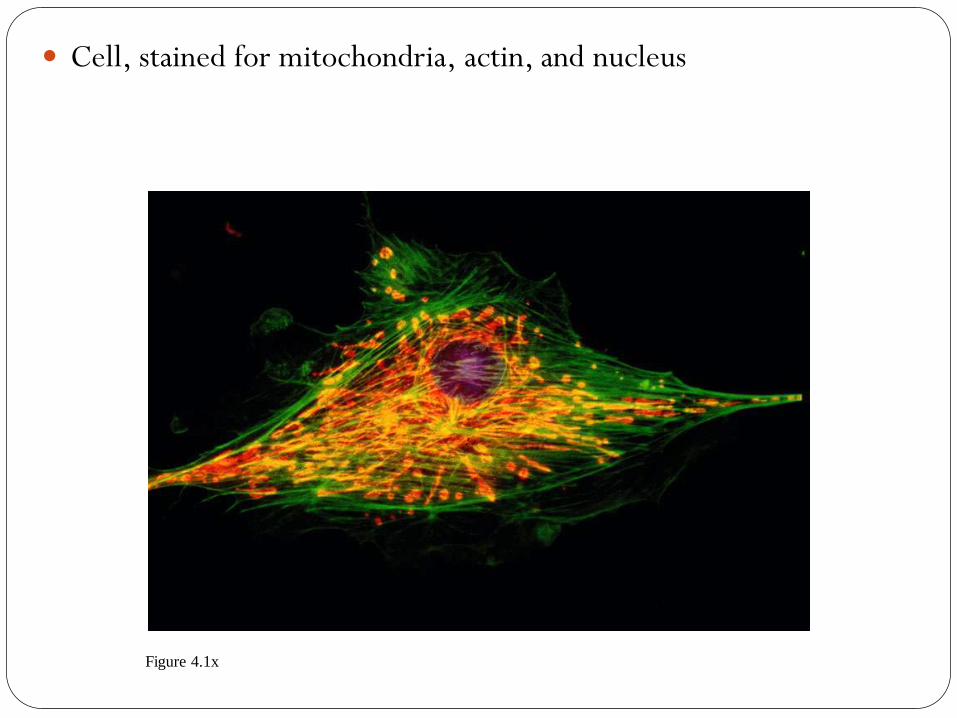
Cell, stained for mitochondria, actin, and nucleus
Figure 4.1x

Prokaryotic cells, Bacillus polymyxa
Figure 4.4x1
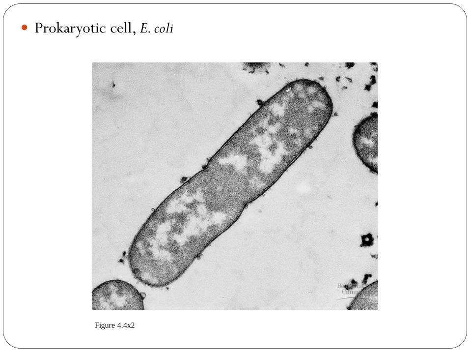
Prokaryotic cell, E. coli
Figure 4.4x2

Pili on a prokaryotic cell
Figure 4.4x3
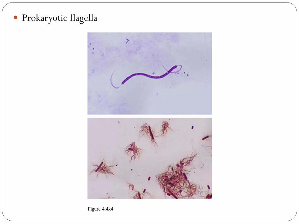
Prokaryotic flagella
Figure 4.4x4
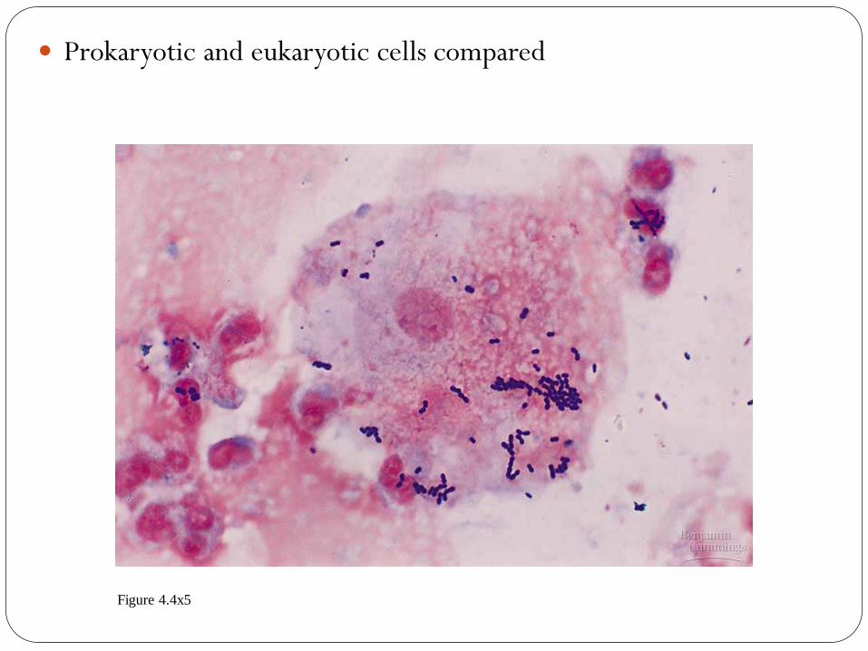
Prokaryotic and eukaryotic cells compared
Figure 4.4x5
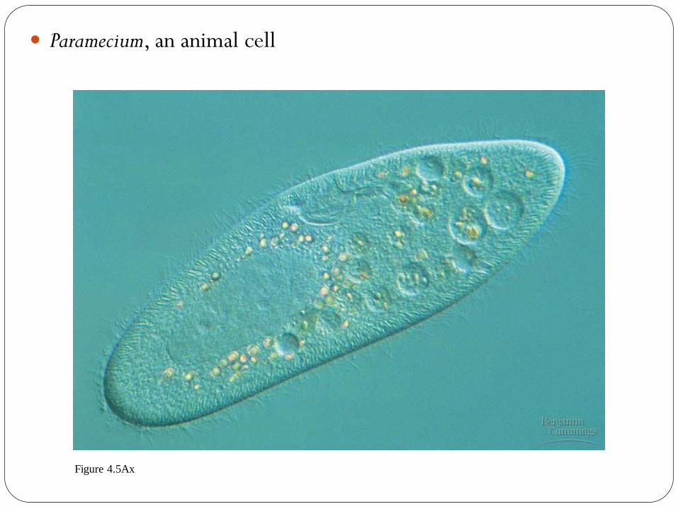
Paramecium, an animal cell
Figure 4.5Ax

Plant cells
Figure 4.5Bx1

Chloroplasts in plant cells
Figure 4.5Bx2

Nuclei (yellow) and actin (red)
Figure 4.6x