Questions Answers on Bioinorganic Chemistry (D Ray)nptel.ac.in/courses/104105031/Bioinorganic...
14
Questions & Answers on Bioinorganic Chemistry (D Ray) 12. Explain the iron binding by transferrin. Ans. Iron, when taken up with the food and processed in the mouth is mostly present in Fe 3+ form and thus gets into the gastrointestinal tract as Fe 3+ . In case of an intact milieu in the small intestines, ferric iron is reduced to its ferrous form (Fe 2+ ). The Fe 3+ ions are then taken up by apotransferrin (H 2 Tf); simultaneously, carbonate is coordinated to iron. Fe 3+ -Tf is the transport form for iron. The iron-loaded transferrin delivers iron to sites of potential use (e.g. incorporation into protoporphyrin IX and generation of haemoglobin), or stored in iron storage proteins (ferritins). The delivery of iron affords reduction from the ferric to the ferrous state; a reductant employed here is ascorbate (vitamin C). Transferrin is a glycoproteid of molecular weight 80 kDa (containing ca. 6% carbohydrate), having available two almost equivalent binding sites for iron(III), in the C- and N-teminal lobes, respectively. The pK (K = stability constant; see inset on p. 5) at pH 7.4 (the pH of blood) is -20.2. Transferrin is also an effective transporter for other tri- and divalent metal cations, and even for anions (e.g. vanadate). Since its loading capacity for iron commonly is only ca. 40%, other ions can be transported simultaneously. 13. Show and explain the iron storage protein ferritin. Ans. Ferritins (Fig. 4) are iron storage proteins, built up of a hollow protein sphere (apo-ferritin, M = 450 kDa, 24 subunits of 163 amino acids each) with an outer diameter of 130 and an inner diameter of 70 Å. The inner surface of this capsule is lined with carboxylate functions, which can coordinate Fe3+. Up to 4500 Fe3+ can be taken up. The various iron centres are connected by bridging oxido and hydroxido groups very much as in the colloidal form of ferric hydroxide. The overall composition of the iron nucleus is 8FeO(OH)·FeO(H2PO4). Channels of threefold symmetry and a width of 10 Å allow for an exchange of Fe3+ between the interior and exterior. For the primary uptake process, iron has to be in the oxidation state +II. Its transport along the channels and built-in into the core is accompanied by oxidation to the +III state.
Transcript of Questions Answers on Bioinorganic Chemistry (D Ray)nptel.ac.in/courses/104105031/Bioinorganic...
Questions & Answers on Bioinorganic Chemistry (D Ray)
12. Explain the iron binding by transferrin.
Ans. Iron, when taken up with the food and processed in the mouth is mostly present in Fe3+ form and thus gets into the gastrointestinal tract as Fe3+. In case of an intact milieu in the small intestines, ferric iron is reduced to its ferrous form (Fe2+). The Fe3+ ions are then taken up by apotransferrin (H2Tf); simultaneously, carbonate is coordinated to iron. Fe3+-Tf is the transport form for iron. The iron-loaded transferrin delivers iron to sites of potential use (e.g. incorporation into protoporphyrin IX and generation of haemoglobin), or stored in iron storage proteins (ferritins). The delivery of iron affords reduction from the ferric to the ferrous state; a reductant employed here is ascorbate (vitamin C). Transferrin is a glycoproteid of molecular weight 80 kDa (containing ca. 6% carbohydrate), having available two almost equivalent binding sites for iron(III), in the C- and N-teminal lobes, respectively. The pK (K = stability constant; see inset on p. 5) at pH 7.4 (the pH of blood) is -20.2. Transferrin is also an effective transporter for other tri- and divalent metal cations, and even for anions (e.g. vanadate). Since its loading capacity for iron commonly is only ca. 40%, other ions can be transported simultaneously.
13. Show and explain the iron storage protein ferritin.
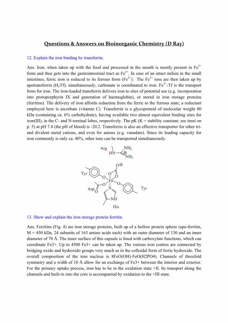
Ans. Ferritins (Fig. 4) are iron storage proteins, built up of a hollow protein sphere (apo-ferritin, M = 450 kDa, 24 subunits of 163 amino acids each) with an outer diameter of 130 and an inner diameter of 70 Å. The inner surface of this capsule is lined with carboxylate functions, which can coordinate Fe3+. Up to 4500 Fe3+ can be taken up. The various iron centres are connected by bridging oxido and hydroxido groups very much as in the colloidal form of ferric hydroxide. The overall composition of the iron nucleus is 8FeO(OH)·FeO(H2PO4). Channels of threefold symmetry and a width of 10 Å allow for an exchange of Fe3+ between the interior and exterior. For the primary uptake process, iron has to be in the oxidation state +II. Its transport along the channels and built-in into the core is accompanied by oxidation to the +III state.
Apoferritin (the inside of the subunit structure and channels one of the subunits hollow sphere is lined with of C2, C3 (for iron exchange carboxylates) with the surroundings) and C4 symmetry 14. Explain with a schematic view the tetrameric structure of the hemoglobin.
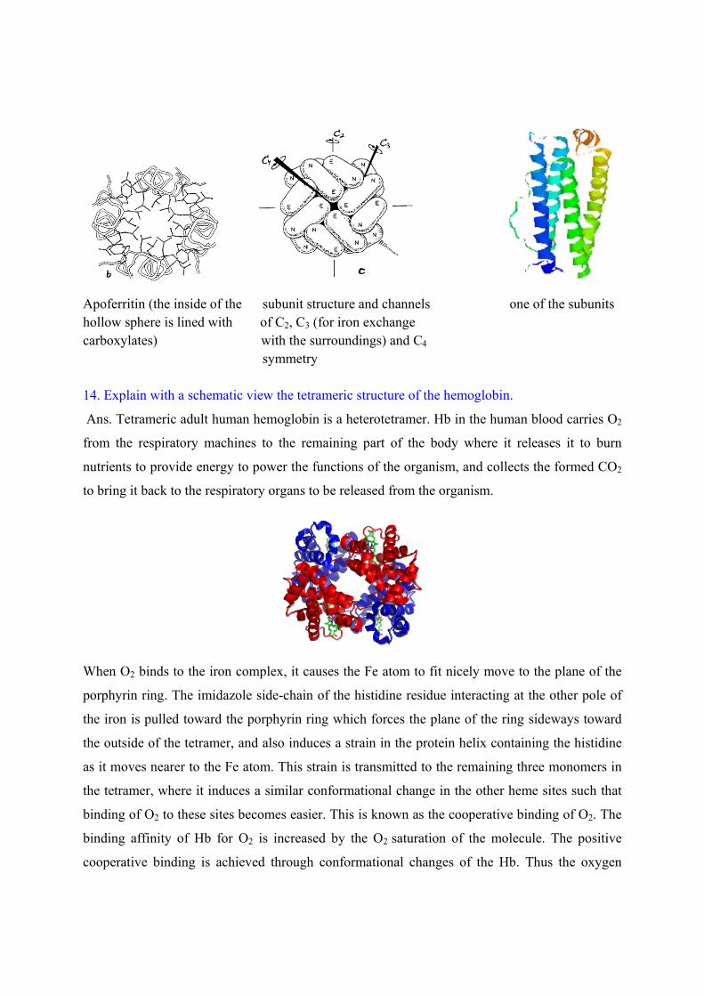
Ans. Tetrameric adult human hemoglobin is a heterotetramer. Hb in the human blood carries O2
from the respiratory machines to the remaining part of the body where it releases it to burn
nutrients to provide energy to power the functions of the organism, and collects the formed CO2
to bring it back to the respiratory organs to be released from the organism.
When O2 binds to the iron complex, it causes the Fe atom to fit nicely move to the plane of the
porphyrin ring. The imidazole side-chain of the histidine residue interacting at the other pole of
the iron is pulled toward the porphyrin ring which forces the plane of the ring sideways toward
the outside of the tetramer, and also induces a strain in the protein helix containing the histidine
as it moves nearer to the Fe atom. This strain is transmitted to the remaining three monomers in
the tetramer, where it induces a similar conformational change in the other heme sites such that
binding of O2 to these sites becomes easier. This is known as the cooperative binding of O2. The
binding affinity of Hb for O2 is increased by the O2 saturation of the molecule. The positive
cooperative binding is achieved through conformational changes of the Hb. Thus the oxygen
binding curve of hemoglobin is sigmoidal as opposed to the hyperbolic one for non-cooperative
binding.
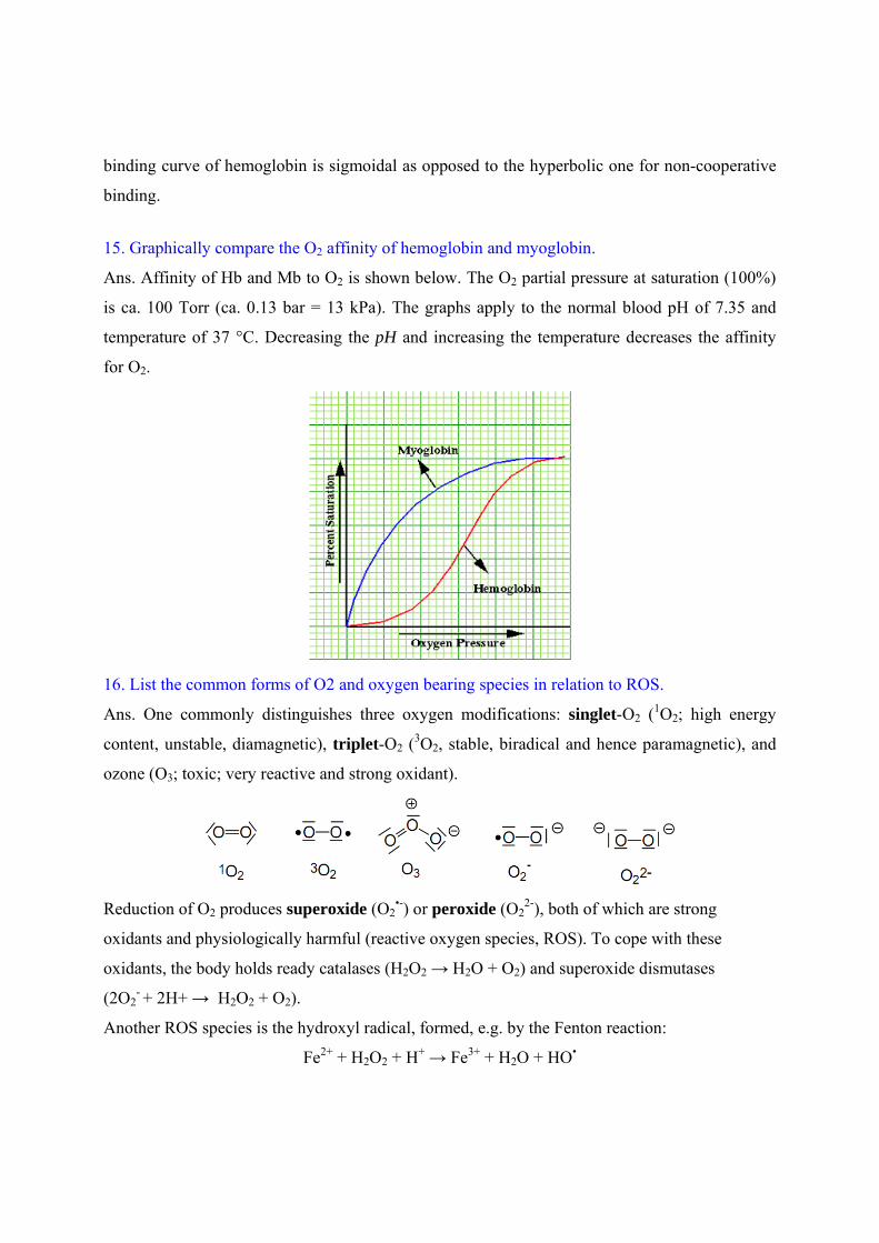
15. Graphically compare the O2 affinity of hemoglobin and myoglobin.
Ans. Affinity of Hb and Mb to O2 is shown below. The O2 partial pressure at saturation (100%)
is ca. 100 Torr (ca. 0.13 bar = 13 kPa). The graphs apply to the normal blood pH of 7.35 and
temperature of 37 °C. Decreasing the pH and increasing the temperature decreases the affinity
for O2.
16. List the common forms of O2 and oxygen bearing species in relation to ROS.
Ans. One commonly distinguishes three oxygen modifications: singlet-O2 (1O2; high energy
content, unstable, diamagnetic), triplet-O2 (3O2, stable, biradical and hence paramagnetic), and
ozone (O3; toxic; very reactive and strong oxidant).
Reduction of O2 produces superoxide (O2•-) or peroxide (O2
2-), both of which are strong
oxidants and physiologically harmful (reactive oxygen species, ROS). To cope with these
oxidants, the body holds ready catalases (H2O2 → H2O + O2) and superoxide dismutases
(2O2- + 2H+ → H2O2 + O2).
Another ROS species is the hydroxyl radical, formed, e.g. by the Fenton reaction:
Fe2+ + H2O2 + H+ → Fe3+ + H2O + HO•
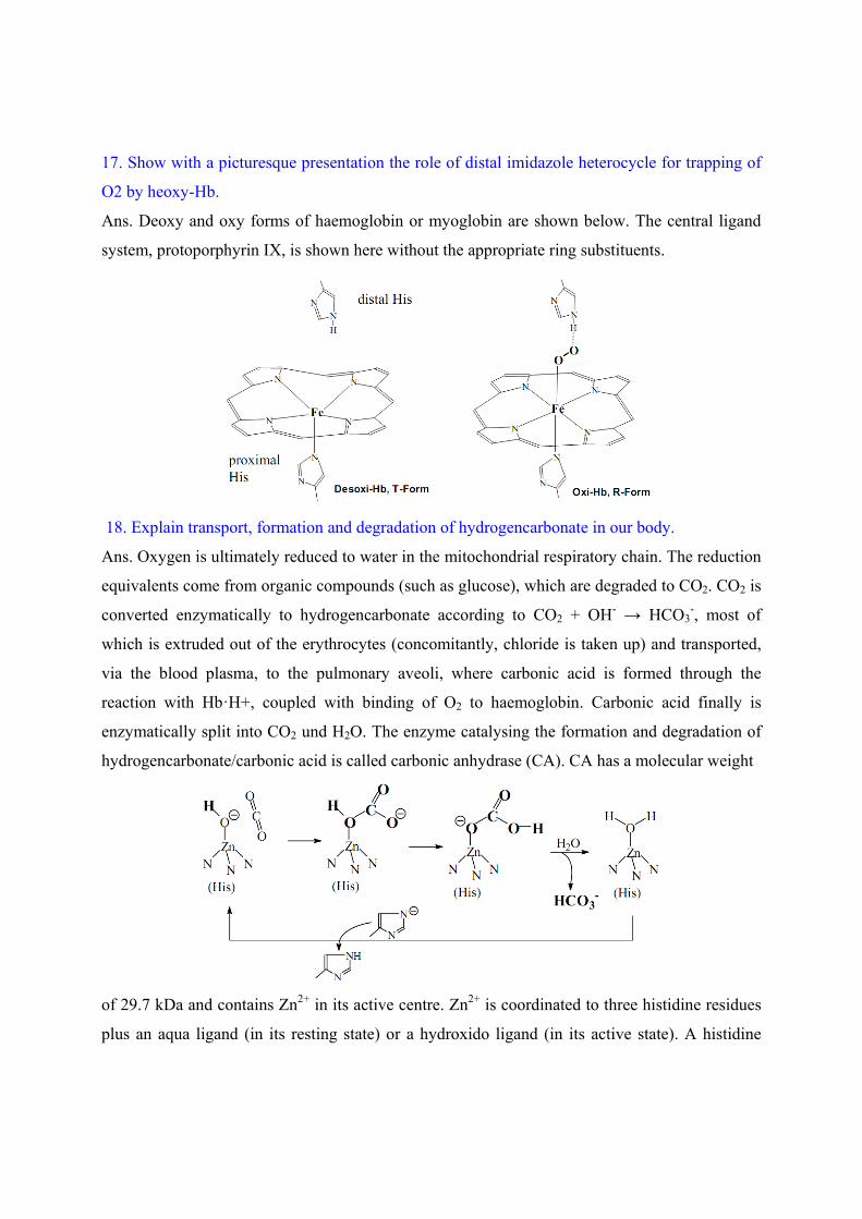
17. Show with a picturesque presentation the role of distal imidazole heterocycle for trapping of
O2 by heoxy-Hb.
Ans. Deoxy and oxy forms of haemoglobin or myoglobin are shown below. The central ligand
system, protoporphyrin IX, is shown here without the appropriate ring substituents.
18. Explain transport, formation and degradation of hydrogencarbonate in our body.
Ans. Oxygen is ultimately reduced to water in the mitochondrial respiratory chain. The reduction
equivalents come from organic compounds (such as glucose), which are degraded to CO2. CO2 is
converted enzymatically to hydrogencarbonate according to CO2 + OH- → HCO3-, most of
which is extruded out of the erythrocytes (concomitantly, chloride is taken up) and transported,
via the blood plasma, to the pulmonary aveoli, where carbonic acid is formed through the
reaction with Hb·H+, coupled with binding of O2 to haemoglobin. Carbonic acid finally is
enzymatically split into CO2 und H2O. The enzyme catalysing the formation and degradation of
hydrogencarbonate/carbonic acid is called carbonic anhydrase (CA). CA has a molecular weight
of 29.7 kDa and contains Zn2+ in its active centre. Zn2+ is coordinated to three histidine residues
plus an aqua ligand (in its resting state) or a hydroxido ligand (in its active state). A histidine
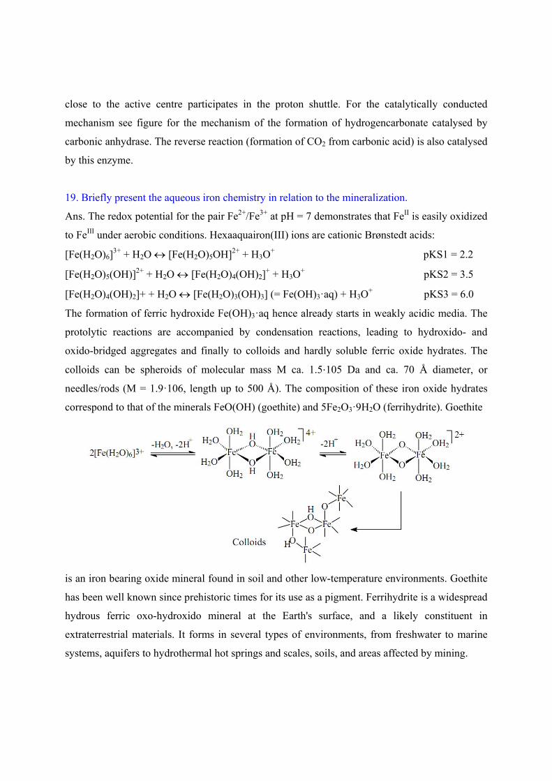
close to the active centre participates in the proton shuttle. For the catalytically conducted
mechanism see figure for the mechanism of the formation of hydrogencarbonate catalysed by
carbonic anhydrase. The reverse reaction (formation of CO2 from carbonic acid) is also catalysed
by this enzyme.
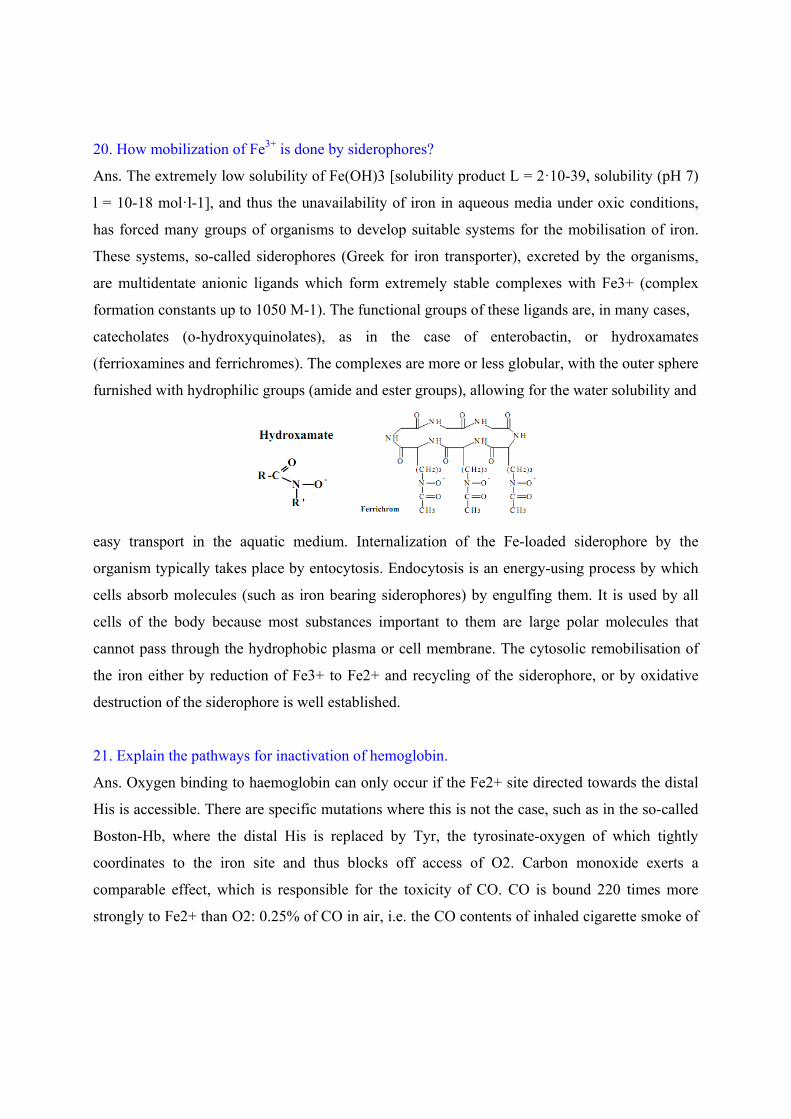
19. Briefly present the aqueous iron chemistry in relation to the mineralization.
Ans. The redox potential for the pair Fe2+/Fe3+ at pH = 7 demonstrates that FeII is easily oxidized
to FeIII under aerobic conditions. Hexaaquairon(III) ions are cationic Brønstedt acids:
[Fe(H2O)6]3+ + H2O [Fe(H2O)5OH]2+ + H3O
+ pKS1 = 2.2
[Fe(H2O)5(OH)]2+ + H2O [Fe(H2O)4(OH)2]+ + H3O
+ pKS2 = 3.5
[Fe(H2O)4(OH)2]+ + H2O [Fe(H2O)3(OH)3] (= Fe(OH)3·aq) + H3O+ pKS3 = 6.0
The formation of ferric hydroxide Fe(OH)3·aq hence already starts in weakly acidic media. The
protolytic reactions are accompanied by condensation reactions, leading to hydroxido- and
oxido-bridged aggregates and finally to colloids and hardly soluble ferric oxide hydrates. The
colloids can be spheroids of molecular mass M ca. 1.5⋅105 Da and ca. 70 Å diameter, or
needles/rods (M = 1.9·106, length up to 500 Å). The composition of these iron oxide hydrates
correspond to that of the minerals FeO(OH) (goethite) and 5Fe2O3·9H2O (ferrihydrite). Goethite
is an iron bearing oxide mineral found in soil and other low-temperature environments. Goethite
has been well known since prehistoric times for its use as a pigment. Ferrihydrite is a widespread
hydrous ferric oxo-hydroxido mineral at the Earth's surface, and a likely constituent in
extraterrestrial materials. It forms in several types of environments, from freshwater to marine
systems, aquifers to hydrothermal hot springs and scales, soils, and areas affected by mining.
20. How mobilization of Fe3+ is done by siderophores?
Ans. The extremely low solubility of Fe(OH)3 [solubility product L = 2·10-39, solubility (pH 7)
l = 10-18 mol·l-1], and thus the unavailability of iron in aqueous media under oxic conditions,
has forced many groups of organisms to develop suitable systems for the mobilisation of iron.
These systems, so-called siderophores (Greek for iron transporter), excreted by the organisms,
are multidentate anionic ligands which form extremely stable complexes with Fe3+ (complex
formation constants up to 1050 M-1). The functional groups of these ligands are, in many cases,
catecholates (o-hydroxyquinolates), as in the case of enterobactin, or hydroxamates
(ferrioxamines and ferrichromes). The complexes are more or less globular, with the outer sphere
furnished with hydrophilic groups (amide and ester groups), allowing for the water solubility and
easy transport in the aquatic medium. Internalization of the Fe-loaded siderophore by the
organism typically takes place by entocytosis. Endocytosis is an energy-using process by which
cells absorb molecules (such as iron bearing siderophores) by engulfing them. It is used by all
cells of the body because most substances important to them are large polar molecules that
cannot pass through the hydrophobic plasma or cell membrane. The cytosolic remobilisation of
the iron either by reduction of Fe3+ to Fe2+ and recycling of the siderophore, or by oxidative
destruction of the siderophore is well established.
21. Explain the pathways for inactivation of hemoglobin.
Ans. Oxygen binding to haemoglobin can only occur if the Fe2+ site directed towards the distal
His is accessible. There are specific mutations where this is not the case, such as in the so-called
Boston-Hb, where the distal His is replaced by Tyr, the tyrosinate-oxygen of which tightly
coordinates to the iron site and thus blocks off access of O2. Carbon monoxide exerts a
comparable effect, which is responsible for the toxicity of CO. CO is bound 220 times more
strongly to Fe2+ than O2: 0.25% of CO in air, i.e. the CO contents of inhaled cigarette smoke of
twenty cigarettes per day, block off about 25% of the oxygen binding capacity of Hb. NO
(formed by reduction of nitrite) has a comparable effect.
The naturally occurring mutant Glu6Ala (glutamate at position 6 exchanged for alanin) causes
sickle cell anaemia, a deformation of the red blood cells by polymerisation of the globin subunits
of Hb. The blood of people suffering from sickle cell anaemia has restricted O2 capacity. These
people are, however, immune against malaria, which has led, by selection, to a high percentage
of anaemic individuals amongst the populations of some tropical African regions.
A certain amount of haemoglobin is consistently oxidised to methaemoglobin (MetHb) by
oxidants such as peroxide, hyperoxide and OH radicals:
Hb(Fe2+) + H2O → MetHb(Fe3+-OH) + e + H+
Met-Hb, in which the second axial position is blocked by OH- is, however, consistently
‘repaired’ by methaemoglobin-reductase (containing NADH as cofactor).
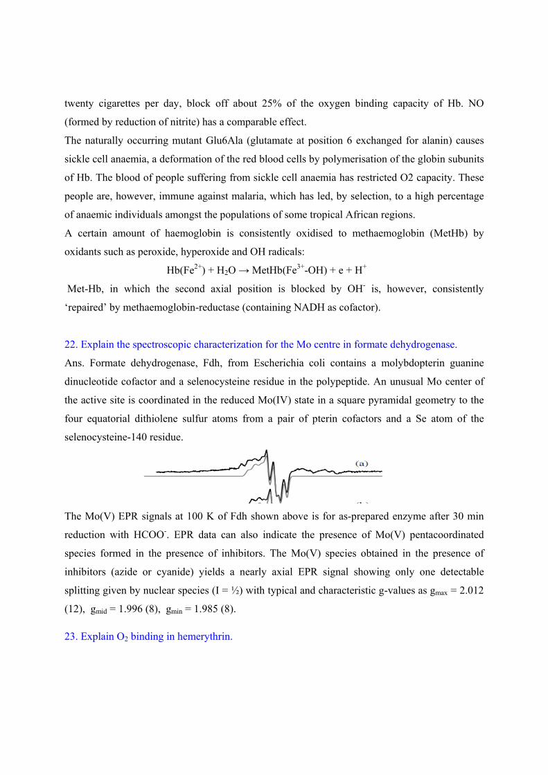
22. Explain the spectroscopic characterization for the Mo centre in formate dehydrogenase.
Ans. Formate dehydrogenase, Fdh, from Escherichia coli contains a molybdopterin guanine
dinucleotide cofactor and a selenocysteine residue in the polypeptide. An unusual Mo center of
the active site is coordinated in the reduced Mo(IV) state in a square pyramidal geometry to the
four equatorial dithiolene sulfur atoms from a pair of pterin cofactors and a Se atom of the
selenocysteine-140 residue.
The Mo(V) EPR signals at 100 K of Fdh shown above is for as-prepared enzyme after 30 min
reduction with HCOO-. EPR data can also indicate the presence of Mo(V) pentacoordinated
species formed in the presence of inhibitors. The Mo(V) species obtained in the presence of
inhibitors (azide or cyanide) yields a nearly axial EPR signal showing only one detectable
splitting given by nuclear species (I = ½) with typical and characteristic g-values as gmax = 2.012
(12), gmid = 1.996 (8), gmin = 1.985 (8).
23. Explain O2 binding in hemerythrin.
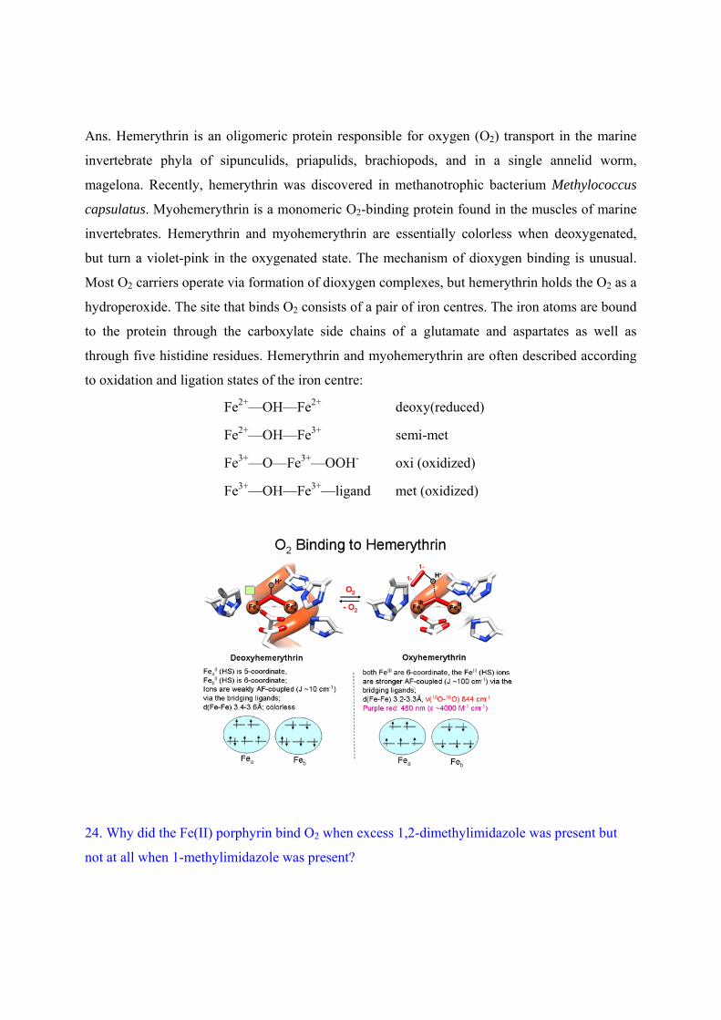
Ans. Hemerythrin is an oligomeric protein responsible for oxygen (O2) transport in the marine
invertebrate phyla of sipunculids, priapulids, brachiopods, and in a single annelid worm,
magelona. Recently, hemerythrin was discovered in methanotrophic bacterium Methylococcus
capsulatus. Myohemerythrin is a monomeric O2-binding protein found in the muscles of marine
invertebrates. Hemerythrin and myohemerythrin are essentially colorless when deoxygenated,
but turn a violet-pink in the oxygenated state. The mechanism of dioxygen binding is unusual.
Most O2 carriers operate via formation of dioxygen complexes, but hemerythrin holds the O2 as a
hydroperoxide. The site that binds O2 consists of a pair of iron centres. The iron atoms are bound
to the protein through the carboxylate side chains of a glutamate and aspartates as well as
through five histidine residues. Hemerythrin and myohemerythrin are often described according
to oxidation and ligation states of the iron centre:
Fe2+—OH—Fe2+ deoxy(reduced)
Fe2+—OH—Fe3+ semi-met
Fe3+—O—Fe3+—OOH- oxi (oxidized)
Fe3+—OH—Fe3+—ligand met (oxidized)
24. Why did the Fe(II) porphyrin bind O2 when excess 1,2-dimethylimidazole was present but
not at all when 1-methylimidazole was present?
Ans. The steric crowding of the bulky dimethyl-Im cause it to be farther below the porphyrin
plane. This pulls the Fe down into the plane, making it unable to bind a second dimethyl-Im on
top (it can, however, bind the smaller O2). MeIm is not so sterically crowded and does not pull
the Fe down far. Therefore the Fe can bind two MeIm’s (one on top and the other on bottom),
leaving no room for O2.
25. Explain the different oxygen binding behavior of the Co(II) prophyrin.
Ans. Co(II) will only bind one axial imidazole, either 1,2-dimethylimidizole or 1-
methylimidizole. Thus there is an open coordination site for O2 in either case.
26. Indicate the oxidation state of the copper ions and of the O2 ligand in oxyhemocyanin.
Ans. Two copper(II) ions and one peroxide ion (O22-).
27. What single method: mass spec, UV-vis, NMR, microwave, or IR/Raman spectroscopy,
would be best suited to verify the oxidation state of the O2 ligand in oxyhemocyanin? Briefly
explain your answer–for example, would oxygen isotopes such as 18O2 be useful?
Ans. a) IR/Raman of O2 stretch would verify the oxidation staste of O2 because more
antibonding electrons have a lower stretching frequency.
b) With 18O2 the frequency shift (isotope effect) can be used to confirm the assignment of the
band.
28. Ferrochelatase is an enzyme which inserts iron into protoporphrrin IX; the iron complex is
then taken up by hemoglobin. Some people are deficient in this enzyme and have some iron free
protoporphyrin IX. Such individuals develop dark lesions on their skin when they are in the sun.
Explain the underlying cause of this disease. Do you expect this disease to be congenital?
Ans. Porphyrins are excellent photosensitizers, exciting oxygen from triplet to singlet O2, using
light energy (hv). Singlet oxygen is very reactive, and will oxidize funcionalities in the person's
cells, etc, leading to the skin lesions.
Paramagnetic metal ions coordinated to the porphyrin rings quench the singlet oxygen. So, the
lack of ferrochelatase, which inserts iron into protoporphyrin IX, is a problem because, without
the iron, the singlet oxygen is not quenched.
This enzymatic defect is congenital. Enzymes are made by coding in the DNA, so this defect is
inherited.
29. Would cytochrome-c be a useful oxygen carrier? Explain your answer.
Ans. No. The monohemeprotein, because it already has two axial ligands, has no vacant
coordination sites to bind O2.
30. What is the function of cytochrome-c?
Ans. It's an electron carrier.
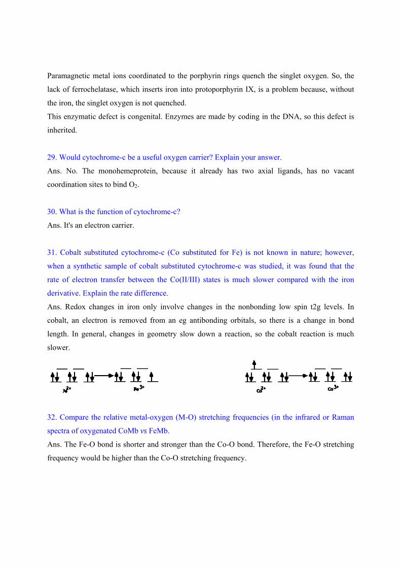
31. Cobalt substituted cytochrome-c (Co substituted for Fe) is not known in nature; however,
when a synthetic sample of cobalt substituted cytochrome-c was studied, it was found that the
rate of electron transfer between the Co(II/III) states is much slower compared with the iron
derivative. Explain the rate difference.
Ans. Redox changes in iron only involve changes in the nonbonding low spin t2g levels. In
cobalt, an electron is removed from an eg antibonding orbitals, so there is a change in bond
length. In general, changes in geometry slow down a reaction, so the cobalt reaction is much
slower.
32. Compare the relative metal-oxygen (M-O) stretching frequencies (in the infrared or Raman
spectra of oxygenated CoMb vs FeMb.
Ans. The Fe-O bond is shorter and stronger than the Co-O bond. Therefore, the Fe-O stretching
frequency would be higher than the Co-O stretching frequency.
33. Explain why cyanide anion inhibits catalase activity for the decomposition of hydrogen
peroxide (H2O2) into water and oxygen but carbon monoxide does not.
Ans. Catalase goes between Fe(III) and the equivalent of Fe(V) (it’s more like Fe(IV) with
porphyrin being 1e- oxidized as well) as H2O2 is oxidized and reduced by two 2e- changes. Thus
the thermodynamically allowed disproportionation is catalysed. The CN- ion binds Fe(III) and
inhibits binding of H2O2 and as a result stop the reaction. The CO molecule would bind Fe(II)
but Fe(II) never occurs in the system resulting no binding.
34. Why does CO bind more tightly to iron(II) porphyrins? Explain in one or two sentences;
comment on the vCO infrared frequency in this complex. Compare this frequency to that of free
CO.
Ans. The Fe(II)-CO bond is strong because there are strong ¼-backbonding interactions which
strengthen the Fe-C bond. This is much less important for Fe(III)-CO because the charge is
higher on the metal. The IR frequency of free CO is higher than the IR frequency of CO in a
porphyrin complex because backbonding added electron density to the p* antibonding orbital of
the CO bond, decreasing its strength.
35. In what way would CO binding change the reduction potential of an iron(III) porphyrin?
Ans. Fe(III) does not bind CO, but Fe(II) binds CO strongly, creating a very stable complex ( a
complex more stable than Fe(II) by itself.) Since the Fe(II)-CO complex is more stable than
Fe(II), the G of forming the complex is lower (more negative) than the G of forming Fe(II).
Because G=-nFE, this also means that the reduction potential for Fe(III) and the complex is
higher than the reduction potential for Fe(III/II).
36. In what way would cyanide alter the Fe(III)/Fe(II) reduction potential of an iron porphyrin
complex?
Ans. Cyanide forms a stable complex with Fe(III) but not Fe(II). Thus, Fe(III) will not be
reduced as easily in the presence of CN−. The reduction potential would be lower.
37. What are oxidoreductases?
Ans. An oxidoreductase is an enzyme that catalyzes the transfer of electrons from one molecule,
the reductant, also called the electron donor, to another the oxidant, also called the electron
acceptor.
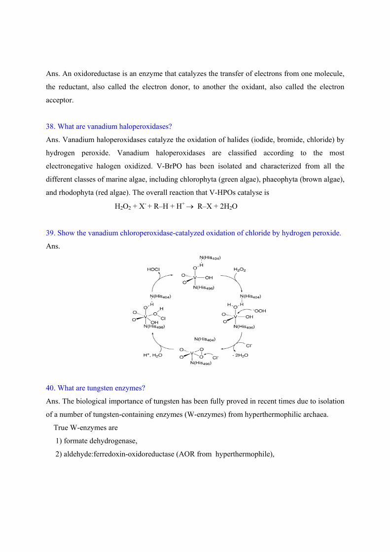
38. What are vanadium haloperoxidases?
Ans. Vanadium haloperoxidases catalyze the oxidation of halides (iodide, bromide, chloride) by
hydrogen peroxide. Vanadium haloperoxidases are classified according to the most
electronegative halogen oxidized. V-BrPO has been isolated and characterized from all the
different classes of marine algae, including chlorophyta (green algae), phaeophyta (brown algae),
and rhodophyta (red algae). The overall reaction that V-HPOs catalyse is
H2O2 + X- + R–H + H+ R–X + 2H2O
39. Show the vanadium chloroperoxidase-catalyzed oxidation of chloride by hydrogen peroxide.
Ans.
40. What are tungsten enzymes?
Ans. The biological importance of tungsten has been fully proved in recent times due to isolation
of a number of tungsten-containing enzymes (W-enzymes) from hyperthermophilic archaea.
True W-enzymes are
1) formate dehydrogenase,
2) aldehyde:ferredoxin-oxidoreductase (AOR from hyperthermophile),
3) formaldehyde:ferredoxin-oxidoreductase, where tungsten cannot be replaced by
molybdenum.
W is able to replace Mo in Mo-enzymes, forming catalytically inactive or possesing very low
activity analogs. Although a tungsten-containing xanthine dehydrogenase from bacteria has been
found to contain tungsten-molydopterin and also non-protein bound selenium, a tungsten-
selenium molybdopterin complex has not been definitively described. Tungsten-using enzymes
typically reduce carboxylic acids to aldehydes. The tungsten oxidoreductases may also catalyse
oxidations. The first tungsten-requiring enzyme to be discovered also requires selenium, and in
this case the tungsten-selenium pair may function analogously to the molybdenum-sulfur pairing
of some molybdenum cofactor-requiring enzymes. One of the enzymes in the oxidoreductase
family which sometimes employ tungsten (bacterial formate dehydrogenase) is known to use a
selenium-molybdenum version of molybdopterin.