QUESTION BANK ILLUSTRATED OBJECTIVE TOXICOLOGY Illustrated objective toxicology... · Professor in...
99
Transcript of QUESTION BANK ILLUSTRATED OBJECTIVE TOXICOLOGY Illustrated objective toxicology... · Professor in...


QUESTION BANK
ILLUSTRATED OBJECTIVE TOXICOLOGY FOR VETERINARY EXAMS AS PER LATEST VCI MSVE 2008 SYLLABUS
Dr. Alpha Raj. M MVSc., PhD.

ABOUT THE AUTHOR
The author is currently working as Assistant
Professor in the department of Veterinary
Pharmacology & Toxicology at College of
Veterinary Science, Proddatur. He has six years of
experience in handling Pharmacology and
Toxicology courses at graduate level. The author
specializes in the fields of toxicology and nano-
medicine and is awarded five young scientist
awards including an international young scientist
award for his research. The author is an alumnus of Tirupati and Rajendranagar campuses. He had an illustrious academic career being a
topper at both graduate and post-graduate study with five academic gold medals.
Price: Free
Copyright Disclaimer
© 2014 Alpha Raj M
This e-book can be reproduced, distributed, or transmitted in any form
and by any means either electronic or print for personal and non-
commercial purposes.
The illustrations used in this e-book belong to creative common license and were
flagged as ‘labeled for reuse’ or ‘labeled for non-commercial use’ in Google search. The
use of any copyrighted illistrations and material complies with fair use for educational
and non-commercial purposes.

DEDICATION
Dedicated to my beloved parents
Late. Sri. M. Deva Rajulu & Smt. N.G.Nirmala Devi
And my doctoral guide
Prof.A.Gopala Reddy
University Head of Pharmacology & Toxicology
Sri Venkateswara Veterinary University

PREFACE
“Freely you have received; freely give”. Mathew 10:8.
Illustrated objective toxicology for veterinary exams is an attempt to provide
comprehensive quick reference for various examinations. However, it should be noted
that this e-book serves only as a supplement and not as a replacement for text book
and class room learning.
This e-book covers all the aspects of veterinary toxicology prescribed by
latest Veterinary Council of India (VCI) minimum standards of veterinary education
(MSVE) 2008. It is a robust question bank comprising of over 777 objective questions
covering 32 lecture outlines. A chapter is devoted on residue toxicology owing to
contemporary importance of the topic. Over 108 illustrations were used at appropriate
places to improve the lucidity and understanding of the student. Appropriate type of
objective questions were used to fit the information being conveyed. Further, the
objective type questions are so framed to convey maximum information possible.
Explanations and additional information are provided where ever deemed necessary.
As this e-book is mainly meant to be read on a computer, laptop, mobile or
tablet, navigation is provided in every page to reach appropriate locations elsewhere in
the book. Any corrections and suggestions for improvement of this e-book are most
welcome.
Dr.M.ALPHA RAJ

TABLE OF CONTENTS
CHAPTER I ................................................................................................ 1
FUNDAMENTALS OF TOXICOLOGY ........................................................ 1
1.1 Introduction ........................................................................................... 1
1.2 Metabolism, Mechanisms and Factors Affecting Toxicity ...................... 6
1.3 Diagnosis & Treatment of Poisoning ..................................................... 9
CHAPTER II ............................................................................................. 13
TOXICOLOGY OF METALS ..................................................................... 13
2.1 Arsenic ................................................................................................ 13
2.2 Mercury ............................................................................................... 15
2.3 Lead .................................................................................................... 17
2.4 Copper ................................................................................................ 20
2.5 Molybdenum ....................................................................................... 22
CHAPTER III............................................................................................. 24
TOXICOLOGY OF NON-METALS ............................................................ 24
3.1 Fluoride ............................................................................................... 24
3.2 Phosphorus ......................................................................................... 26
3.3 Nitrate and Nitrite ................................................................................ 28
3.4 Cyanide .............................................................................................. 31
3.5 Selenium ............................................................................................. 35
3.6 Oxalate ............................................................................................... 38
CHAPTER IV ............................................................................................ 41
TOXICOLOGY OF PLANTS ..................................................................... 41
4.1 Photosensitization ............................................................................... 41
4.2 Thiamine deficiency causing plants ..................................................... 43
4.3 Abrus precatorius (Abrus) and Ricinus communis (Castor) ................. 44
4.4 Datura, Nerium and Ipomea sps ......................................................... 46
4.5 Strychnous nuxvomica and Gossypium sps ........................................ 49

CHAPTER V ............................................................................................. 51
TOXICOLOGY OF AGROCHEMICALS .................................................... 51
5.1 Organochlorine (OC) compounds ....................................................... 51
5.2 Organophosphate (OP) compounds ................................................... 52
5.3 Carbamates ........................................................................................ 55
5.4 Pyrethroids .......................................................................................... 56
5.5 Rodenticides ....................................................................................... 58
5.6 Herbicides, Fungicides & Molluscicides .............................................. 61
CHAPTER VI ............................................................................................ 63
RESIDUE TOXICOLOGY ......................................................................... 63
CHAPTER VII ........................................................................................... 66
VENOUS ANIMALS .................................................................................. 66
CHAPTER VIII .......................................................................................... 73
RADIATION TOXICOLOGY ...................................................................... 73
CHAPTER IX ............................................................................................ 75
TOXICOLOGY OF FOOD AND FEED ADDITIVES .................................. 76
9.1 Food & Feed additives ........................................................................ 76
9.2 Urea and Salt Poisoning ..................................................................... 77
CHAPTER X ............................................................................................. 80
MYCOTOXINS & BACTERIAL TOXINS ................................................... 80
CHAPTER XI ............................................................................................ 86
ENVIRONMENTAL POLLUTANTS ........................................................... 86
REFERENCES ......................................................................................... 89

1 | P a g e
CHAPTER I
FUNDAMENTALS OF TOXICOLOGY
1.1 Introduction
1. The branch of science which deals with the harmful effects of physical and chemical
agents on human and animal life is TOXICOLOGY.
2. In the term toxicology, the word ‘toxicon’ (Greek) means POISON.
3. The branch of toxicology, which deals with diagnosis, treatment and management of
toxic substances, is known as CLINICAL TOXICOLOGY.
4. The branch of toxicology, which is involved in establishing safety limits for chemical
exposure is REGULATORY TOXICOLOGY.
5. Investigating and controlling the toxic effects of various substances on the community
is dealt by TOXICOVIGILANCE.
6. The branch of toxicology, which investigates vetero-legal cases of death, poisoning and
drug abuse is FORENSIC TOXICOLOGY.
7. The study of toxicity produced by substances of plant, animal and microbial origin is
termed as TOXINOLOGY.
8. What is the difference between ‘Poison’, ‘Toxicant’ and ‘Toxin’?
Ans: Poison is a broader term which includes any / every substance causing harmful
effects to living beings. Toxicant is a synonym for poison.Toxins are a sub-category of
poisons, which could be of plant (Phytotoxins), fungal (Mycotoxins), animal (Zootoxins)
or bacteria (Bacteriotoxins – Endo & Exotoxins) origin.
9. A foreign chemical substance, which is not normally produced in the body and which
may form a part of the food, is known as XENOBIOTIC.
10. How ‘Toxicosis’ differs from ‘Toxicity’?
Ans: Toxicosis and Toxicity as related as ‘effect’ and ‘degree/extent of the effect’.
Toxicosis is the disease or condition (effect) which results due to exposure to a poison,
whereas, ‘toxicity’ is the degree of the disease or condition. However, the term toxicity
is also used to mean adverse effects of a poison.
Next Lesson / Chapter / Index

2 | P a g e
11. Which of the following toxicity can occur due to single exposure?
a. Acute toxicity b. Sub-acute toxicity c. Sub-chronic toxicity d. Chronic toxicity
Ans: a. Acute toxicity; Acute toxicity also occurs due to multiple exposures within 24h.
The other types of toxicities definitely need multiple/repeated exposures.
12. Toxicity occurring due to repeated exposure within 30 DAYS or LESS is termed as sub-
acute toxicity, whereas, within 1 TO 3 MONTHS is termed as sub-chronic toxicity.
13. If the period of exposure of a toxicant is more than 3 months, the type of toxicity is
termed as CHRONIC TOXICITY.
14. Repeated oral toxicity of a compound is tested in rats for a period of 28-days. What
kind of toxicity is being studied here? (You need to answer this question!!)
15. The type of toxicity which results due to progressive accumulation of a toxicant in the
body is known as CUMULATIVE TOXICITY.
(Several toxicants such as heavy metals, alcohol, DDT etc cause cumulative toxicity.)
16. The amount of toxicant in food and water, which can be consumed daily over a life time
without any significant health risk, is known as ACCEPTABLE DAILY INTAKE (ADI).
17. What is the difference between Lethal Dose (LD50 or 99) and Lethal Concentration (LC)?
Ans: Both LD and LC causes death in exposed population. However, LD refers to the
‘lowest dose’ of the toxicant administered directly to the animal in any route. LC refers
to the ‘lowest concentration’ of toxicant present in feed and water. (In LD, the
compound is administered directly to the animal on mg/kg bd wt basis, whereas, in LC
the toxicant is present in feed or water (mg/kg of feed or Liter of water).
18. What is the meaning of the subscripts in the terms LD50 or 99 and LC50?
Ans: The subscripts indicate the % of mortality (deaths) in exposed population. In 1927,
J.W.Trevan introduced LD50 to determine the toxic potential of various compounds.
19. For a given compound, between NOEL and NOAEL, which one will be higher in dose?
Ans: NOEL (No observed effect level) is the highest dose, which will not produce any
effect, whereas, NOAEL (No observed adverse effect level) is the highest
concentration, which will not produce any adverse effect. Since, the development of
adverse effect needs higher dose than minor effects, NOAEL is higher than NOEL.
20. Maximum acceptable/ permitted amount of a drug present in feed and foods is known
as MAXIMUM RESIDUE LEVEL (MRL). (For pesticides – MAXIMUM RESIDUE LIMIT).
21. The highest dose of a compound, which produces adverse effects but no mortality is
called MAXIMUM TOLERATED DOSE (MTD).
(MTD is also referred to as LD0 (Zero) as it will cause adverse effects but no mortality).
Next Lesson / Chapter / Index

3 | P a g e
22. For a given compound, between MTD and NOAEL, which one will be higher?
Ans: NOAEL is the highest dose which will not cause any adverse effect whereas MTD
refers to the highest dose which will cause adverse effects. Hence, MTD is higher.
23. What is the relationship between the various toxicity doses with respect to a given
compound?
Ans: LD > LC > MTD > NOAEL > NOEL > ADI > MRL
Fig.1 No-observed adverse effect level (1)
24. The source of adverse effect/ damage is known as HAZARD.
(Eg: Water containing Fluoride; Here fluoride is the hazard and fluorosis is the adverse
effect. Hazard is independent of dose or exposure i.e.,Fluoride is present whether
someone drinks the water or not).
25. The likelihood or probability of adverse effect upon exposure to a hazard is known as
RISK.
(Eg: While drinking water containing fluoride, the chances of getting fluorosis is called
risk. Risk can range from 0 to 100% depending on dose and exposure i.e., one should
be exposed to a hazard to calculate the risk).
26. The statement, “all substances are poisons; the dose differentiates poison from a
remedy” is associate with PARACELSUS.
Fig: 2 Theophrastus Paracelsus Bombastus Von Honheim (1493-1541) (2)
Next Lesson / Chapter / Index

4 | P a g e
27. The scientist, who is referred to as ‘Father of Toxicology’ is M.J.B. ORFILA.
(He established toxicology as a separate discipline; defined the term toxicology;
advocated the use of chemical analysis in forensic toxicology).
Fig:3 Father of Toxicology - Mathieu Joseph Bonaventure Orfila (1787-1853) (3)
28. DDT (Dichloro diphenyl trichloro ethane), which is used to control malaria and typhus
was discovered by PAUL MULLER.
Fig:4 Paul Muller (1899-1969) – Synthesized DDT (4-5)
29. The scientist, who is known as ‘Father of nerve agents’ is GERHARD SCHRADER.
(Nerve gases such as Sarin, Tabun, Soman, Mustard gas etc were used in World War
II and by Iraqi troops on Kurds as a part of chemical warfare).
30. The author of the book ‘Silent Spring’ in which the detrimental effects of DDT and other
pesticides on environment – particularly on birds was documented - is RACHEL
CARSON.
(Springs are pleasant with the noises of birds. But due to the use pesticides, the bird
population is considerably reduced. Hence, the springs are becoming silent. This book
created a silent campaign against the use of pesticides and led to a ban on DDT
worldwide; US Environmental agency was setup in this context.)
Next Lesson / Chapter / Index

5 | P a g e
Fig:5 Rachel Carson (1907-1964)–Author of Silent Spring–Campaign against DDT (6-7)
31. Bhopal gas tragedy, which is considered as the world’s worst industrial disaster, was
caused by the leakage of METHYL ISO-CYANATE (MIC) gas from Union Carbide
fertilizer company.
32. Use of thalidomide in pregnant women for treating morning sickness caused
PHOCOMELIA condition in newborn infants.
(Phocomelia is a condition in which an infant is born with malformation of limbs.
Thalidomide was not properly tested for safety and hence caused thalidomide disaster
worldwide affecting almost 10,000 children)
Fig: 6 Thalidomide tragedy – Phocomelia in children(8)
33. The type of necrosis, which is commonly seen in liver due to various toxicants is
CENTRILOBULAR NECROSIS.
(Centrilobular region around the central vein (zone 3) has poor oxygenation and is rich
in P450 cytochrome (responsible for metabolism of various drugs – needs more
oxygen). Hence, this region is more prone to damage than peri-portal region (around
portal triad - zone 1) which is more oxygenated.)
Next Lesson / Chapter / Index

6 | P a g e
Fig:7 Structure of Liver lobule. Zone 3 around central vein is less oxygenated (9)
1.2 Metabolism, Mechanisms and Factors Affecting Toxicity
1. Unlawful or criminal killing of animals through administration of poisons is known as
MALICIOUS POISONING.
2. Unintentional addition of toxicants or contaminants to feed and water is known as
ACCIDENTAL POISONING.
3. Man-made sources of toxicants are referred to as ANTHROPOGENIC sources.
4. Genetically determined abnormal reactivity of an individual to a chemical is known as
IDIOSYNCRACY.
5. Failure to elicit a response to an ordinary dose of a substance due prior exposure is
known as TOLERANCE.
(Tolerance is generally caused due to the induction of metabolizing enzymes in liver.
However, in case of chronic alcoholism, pseudo-tolerance is observed, due to
thickened GIT mucosa, which decreases absorption).
6. The phenomenon in which toxic substances elicits beneficial effects at low doses is
known as HORMESIS.
7. For which of the following route of exposure, pre-systemic elimination is possible?
a. Oral b. Inhalation c. Intramuscular d. Intravenous
Ans: a. Oral; GIT and liver are responsible for elimination prior to entry into circulation.
Next Lesson / Chapter / Index

7 | P a g e
8. In the event of irreparable injury, the cell undergoes a process of programmed cell
death known as APOPTOSIS.
(Apoptosis is also involved in number of physiological process such as embryogenesis,
ageing, cancer prevention etc)
9. A substance is classified as extremely toxic if the lethal dose (LD) is LESS THAN
1mg/kg and as practically non-toxic if the LD is 5 to 15 g/kg.
(It should be remembered that more the LD of a compound, less is its toxicity. Highly
toxic –1 to 50 mg/kg; Moderately toxic – 50 to 500 mg/kg; Slightly toxic – 0.5 to 5 g/kg).
10. The ability of a substance to induce cancer is known as CARCINOGENESIS.
11. The common process involved in the absorption of xenobiotics across the cell
membrane is PASSIVE DIFFUSION.
12. In body, heavy metals such as mercury, lead, cadmium tend to accumulate in KIDNEY
(organ of the body).
(However, the major site for accumulation of lead in the body is bone and teeth).
13. Organo-chlorine insecticides such as DDT tend to accumulate in ADIPOSE TISSUE
(organ of the body).
14. Arsenic tends to accumulate in HAIR and SKIN (organ of the body).
(Hence, bacterial decomposition of carcass is absent resulting in preserved carcass)
15. Major route of excretion for xenobiotics is RENAL EXCRETION.
16. The process of chemical transformation (conversion from one form to another)
occurring in the body is known as BIOTRANSFORMATION.
17. The major site for biotransformation of xenobiotics in body is LIVER.
(Liver is the major site due to the presence of variety of metabolizing enzymes. Other
important sites for biotransformation include lung, kidney and intestines)
18. In a hepatocyte, metabolism of xenobiotics takes place in ENDOPLASMIC
RETICULUMN (ER) or MICROSOMES.
19. Most important microsomal enzymes involved in biotransformation are
MONOOXYGENASES or MIXED FUNCTION OXIDASES (MFO).
20. Major biotransformation reaction occurring in Phase I is OXIDATION and in Phase II is
CONJUGATION.
21. Phase I Oxidation reactions are mainly catalyzed by MICROSOMAL ENZYMES (MFO).
Next Lesson / Chapter / Index

8 | P a g e
22. All Phase II conjugation reactions are catalyzed by non-microsomal enzymes except
for GLUCURONIDE CONJUGATION, which is catalyzed by microsomal enzymes.
23. The ability of certain substances to increase the activity or synthesis of microsomal
enzymes is known as INDUCTION.
(Non-microsomal enzymes which are present in cytosol are not inducible)
24. The metabolic reaction that is deficient in dogs is ACETYLATION; in cats is
GLUCURONIDE CONJUGATION, and in pigs is SULFATION.
(Hence, sulphonamides which undergo acetylation cause toxicity in dogs and similarly,
paracetamol, which undergoes glucuronide conjugation, causes hepatotoxicity in cats).
25. The process of conversion of non-toxic substance into a toxic metabolite through
biotransformation is known as LETHAL SYNTHESIS.
26. Organo-chlorine insecticides such as DDT, BHC tend to be more toxic in oily vehicles
due to INCREASED ABSORPTION.
27. Elemental mercury is toxic when exposed through INHALATION route.
(Ingested mercury is not likely to cause toxicity; But is toxic through inhalation route.
Guess, which of the following situations is more dangerous: a child chewing on a
thermometer and ingesting mercury or a thermometer falling on to the floor leading to
evaporation of mercury?! ).
28. If the action of one substance opposes or neutralizes the effect of another substance,
the relationship is referred to as ANTAGONISM.
(All antidotes have antagonistic relationship with their respective toxicants)
29. Rodents are preferred for oral toxicity testing as they lack VOMITION reflex.
30. The species of animal that is resistant to the toxic effects of consuming Atropa
belladonna (Belladonna) leaves is RABBIT.
(Rabbits contain the enzyme atropinase, which destroys atropine).
31. The breed of dog that is more susceptible to the toxic effects of ivermectin is COOLIE
BREED.
(In coolies, ivermectin easily crosses blood brain barrier and causes neurological
symptoms).
32. Asprin and sulphonamides cause hemolysis in individuals deficient in the metabolic
enzyme GLUCOSE-6-PHOSPHATE DEHYDROGENASE (G6PD).
Next Lesson / Chapter / Index

9 | P a g e
Fig: 8 Coolie Breed of Dog – More susceptible to ivermectin toxicity (10)
33. Why Greyhounds are more susceptible to the toxic effects of barbiturates (used for
anesthesia)?
Ans: Barbiturates mainly distribute to adipose tissue. Since, Greyhounds, have little
body fat, higher circulating concentration of barbiturates causes toxicity.
Fig: 9 Greyhound Dog Breed – More susceptible to barbiturate toxicity (11)
1.3 Diagnosis & Treatment of Poisoning
1. The type of evidence obtained at the scene of poisoning is referred to as
CIRCUMSTANTIAL EVIDENCE.
2. The most common feed contaminant that can be expected during improper storage is
MYCOTOXINS (Aflatoxin).
3. Pink colouration of urine is suggestive of poisoning with PHENOTHIAZINES.
4. Phenols and cresols produce GREEN colouration of urine.
Next Chapter / Index

10 | P a g e
5. The symptom or lesion that is characteristic to a particular toxicant is known as
PATHOGNOMONIC (symptom or lesion).
6. The evidence that is obtained during postmortem examination is known as
PATHOLOGICAL evidence.
7. Bitter almond smell of ruminal contents is suggestive of CYANIDE POISONING.
(Bitter almond smell
8. Poisoning with phosphorus results in GARLIC-LIKE odour during postmortem
examination.
9. Detection of toxic material in body using laboratory methods constitutes ANALYTICAL
evidence.
10. The evidence that is obtained by feeding suspected material (feed) to healthy animals
to ascertain the presence of a toxicant is known as EXPERIMENTAL EVIDENCE.
11. The aim of treatment during poisoning is to INCREASE the threshold of the toxicant.
(Measures are directed to create a situation where more toxicant is required to produce
the toxicity)
12. The time versus concentration curve of toxicant in the body is BELL OR INVERTED ‘U’
shaped.
13. The ascending phase of the time-concentration curve of the toxicant represents
ABSORPTION.
14. The descending phase of the time-concentration curve of the toxicant represents
EXCRETION.
Fig: 10 Time vs. Concentration curve of toxicant in body during poisoning (12)
Next Chapter / Index

11 | P a g e
15. The following is the aim of treatment in poisoning
a. Increasing the slope of ascending phase (AP) and decreasing slope of
descending phase (DP).
b. Decreasing the slope of AP and increasing slope of DP.
c. Increasing the slope of AP and increasing slope of DP.
d. Decreasing the slope of AP and decreasing slope of DP.
Ans: b. Decreasing the slope of AP and increasing slope of DP. When the slope of AP
is decreased, it takes longer time for the toxicant to be absorbed. And increasing the
slope of DP causes rapid elimination in a short time.(Fig:3)
Fig: 11 Time vs. Concentration curve of toxicant in body during treatment. (13)
16. Emesis is contraindicated in the following poisoning(s)
a. Volatile compounds b. Corrosives c. Convulsants d. CNS stimulants
Ans: All of the above. In addition, emesis is not indicated in unconscious patients, in
cases of respiratory distress or debilitated animals.
17. The safest alternative when emesis is contraindicated is GASTRIC LAVAGE.
(In case of ruminants, rumen lavage or rumenotomy can be performed)
18. The most commonly used adsorbing agent for binding toxicants in GIT is ACTIVATED
CHARCOAL.
(Charcoal should be activated through burning (oxidation) which increases the number
of pores and thus the surface area).
19. The type of diuretics or purgatives that are preferred in cases of poisoning are
OSMOTIC/SALINE TYPE.
(Osmotic or saline type of agents are the only category which actually drag water from
the tissues. However, for prompt action, furosemide, which is a loop diuretic is also
used).
Next Chapter / Index

12 | P a g e
20. When large amounts of toxicant is absorbed or when renal failure ensues, the method
of choice employed for elimination of the toxicant is DIALYSIS.
21. The mechanism involved in the enhanced elimination of acidic agents in alkalized urine
and basic agents in acidified urine is ION TRAPPING.
(Ion trapping for basic drugs occurs in the acidic pH of rumen - due to the production of
volatile fatty acids).
22. The substance that counteracts or neutralizes a toxicant is known as ANTIDOTE.
23. Ethanol acts as an antidote for methanol poisoning through COMPETITIVE
INHIBITION (Mechanism).
24. The non-specific antidote for alkaloids is POTASSIUM PERMANGANATE.
(KMNO4 acts by oxidizing the alkaloids)
25. The antidote for paracetamol (acetaminophen) toxicity is N-ACETYL CYSTEINE.
(Paracetamol toxicity is common worldwide and is leading cause for acute liver failure
in UK and US. Continuous use for a week is likely to cause hepatotoxicity. The toxicity
is due to the depletion of liver stores of glutathione, which is required for conjugating
the metabolite of paracetamol).
26. The agents that are used during CNS depression and respiratory arrest are called as
ANALEPTICS. (Eg. Doxapram).
Next Chapter / Index

13 | P a g e
CHAPTER II
TOXICOLOGY OF METALS
2.1 Arsenic
1. The metallic poison, which is considered as ‘king of poisons and poison of kings’ is
ARSENIC.
(Arsenic (and Thallium) causes severe GIT irritation unparalled by any other poison.
Hence, arsenic is considered ‘King of poisons’. Since, arsenic was extensively used by
kings to eliminate competitors, it is also known as ‘Poison of kings’.
2. Which form of arsenic is more toxic? Why?
Ans: Arsenites (As3+ or trivalent) are 5-10 times more toxic than Arsenates (As5+ or
pentavalent) due to higher solubility.
3. Most toxic gaseous form of arsenic which is released during charging of storage
batteries is ARSINE.
4. Least toxic form of arsenicals are ORGANIC ARSENICALS.
5. Drinking water containing >0.25% of arsenic is potentially toxic to large animals.
6. Arsenic contamination of ground water is endemic in WEST BENGAL state of India.
(Arsenic contamination of ground water is also seen in 70 countries including USA).
7. Which arsenical compound is commonly employed for malicious poisoning?
Ans: Arsenic trioxide
8. The managemental practice that can cause arsenic poisoning in sheep is DIPPING.
(Generally, arsenic containing compounds [Eg. Sodium arsenite; Lead arsenate] are
used for dipping).
9. The species that is more sensitive to arsenic poisoning is CAT.
10. Why arsenic tends to accumulate in keratin rich tissues such as hair and nails?
Ans: Arsenic has high affinity for sulphydryl groups (-SH). Since, hair and nails contain
–SH rich keratin, arsenic accumulates in them.
11. Arsenic undergoes METHYLATION biotransformation reaction in the body.
12. Is arsenic cumulative in animals?
Ans: No; Arsenic is rapidly detoxified and is completely eliminated within few days.
Next Lesson / Chapter / Index

14 | P a g e
13. The mechanism of toxicity of arsenic involves binding to SULPHYDRYL GROUPS
(–SH) (Eg: Lipoic (thioctic) acid).
14. The primary symptom acute arsenic toxicity is GASTROENTERITIS.
15. The nature of diarrhoea in acute arsenic poisoning is described as RICE WATERY.
16. The characteristic coloration of mucosa in hronic arsenic poisoning is BRICK RED.
17. Why arsenic causes abortions but not nervous symptoms?
Ans: Arsenic can cross placental barrier. Hence can cross abortions but it cannot cross
blood brain barrier (BBB), hence, is unable to cause nervous symptoms.
18. In which species, organic arsenicals cause nervous symptoms?
Ans: Swine. Nervous symptoms include ataxia, incoordination etc.
19. Which is the only arsenical that can cause blindness?
a. Arsenic trioxide b. Arsenic pentoxide c. Arsine d. Arsinilic acid
Ans: d. Arsinilic acid.
20. What are the prominent postmortem findings in arsenic poisoning?
Ans: Preserved carcass (absence of bacterial decomposition); Severe gastroenteritis;
21. Samples that should be collected in suspected cases of arsenic toxicity are HAIR and
NAILS.
22. The level of arsenic in visceral organs that is indicative of arsenic poisoning is >3PPM.
23. How is arsenic poisoning differentiated from lead?
Ans: In arsenic poisoning, severe gastroenteritis alone is observed where as in lead
poisoning, nervous symptoms predominate.
24. Specific antidote for arsenic poisoning is BRITISH ANTI-LEWISITE (BAL).
or DIMERCAPROL.
25. Superior water soluble derivatives of BAL are MESO-DIMERCAPRO-SUCCINIC ACID
(MDSA) and DI-MERCAPTO-SUCCINIC ACID (DMSA).
26. In arsenic poisoning, why milk is considered unfit whereas meat is passed for human
consumption?
Ans: Arsenic gets methylated and get rapidly excreted through urine, milk, sweat etc.
Hence, milk is considered unfit. As arsenic tends to accumulate only in visceral organs
and not in muscles, flesh of surviving animal is considered fit for human consumption).
Next Lesson / Chapter / Index
15 | P a g e
2.2 Mercury
1. Give reasons as to why mercury poisoning is not common in animals?
Ans: Hg compounds are completely replaced by better alternatives in medicinal,
agricultural and industrial use. Hence, Hg poisoning is not common in animals.
2. Common source of mercury poisoning in animals is FOOD.
(Predatory animals at the end of food chain are more likely to accumulate Hg).
3. Which of the following forms of Hg is more toxic?
a. Elemental b. Monovalent c. Divalent d. Organic
Ans: d. Organic form of Hg is more toxic than inorganic form due to higher lipid solubility.
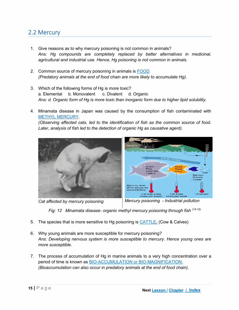
4. Minamata disease in Japan was caused by the consumption of fish contaminated with
METHYL MERCURY.
(Observing affected cats, led to the identification of fish as the common source of food.
Later, analysis of fish led to the detection of organic Hg as causative agent).
Cat affected by mercury poisoning
Mercury poisoning - Industrial pollution
Fig: 12 Minamata disease- organic methyl mercury poisoning through fish (14-15)
5. The species that is more sensitive to Hg poisoning is CATTLE. (Cow & Calves)
6. Why young animals are more susceptible for mercury poisoning?
Ans: Developing nervous system is more susceptible to mercury. Hence young ones are
more susceptible.
7. The process of accumulation of Hg in marine animals to a very high concentration over a
period of time is known as BIO-ACCUMULATION or BIO-MAGNIFICATION.
(Bioaccumulation can also occur in predatory animals at the end of food chain).
Next Lesson / Chapter / Index

16 | P a g e
8. Mercury can cross the following barriers in the body
a. Blood brain barrier (BBB) b. Placental barrier (PB) c. Both d. No barrier
Ans: c. Both. As Hg can cross BBB causing neurological symptoms and crossing PB leads
to accumulation in foetus and abortions.
9. The mechanism of toxicity of mercury involves binding with –SH, THIOL groups of proteins
and enzymes.
10.
Predominant symptoms of organic mercury poisoning are NEUROLOGICAL
(Ataxia, incoordination, convulsions, abnormal behavior etc)
11.
Predominant symptom in inorganic mercury poisoning is GASTROENTERITIS.
12.
Predominant symptom in elemental mercury poisoning is PULMONARY SYMPTOMS.
13.
Elemental mercury (Hg) is toxic only through INHALATION ROUTE of exposure.
14.
The following properties can be attributed to Methyl mercury (Organic Hg)
a. Mutagenic b. Carcinogenic c. Embryotoxic d. Teratogenic
Ans: All of the above.
15.
The sample of choice for detecting inorganic mercury poisoning is URINE.
(Urinary concentration is a reliable indicator of inorganic Hg poisoning).
16.
The sample of choice for detecting organic mercury poisoning is KIDNEY
(Organic Hg tends to accumulate in visceral organs including brain. A concentration of 10
mg/kg in kidney is indicative of Hg poisoning)
17.
Neurological and renal damage caused by mercury are IRREVERSIBLE (even with
treatment).
18.
The following chelating agent(s) that is (are) used for treating mercury poisoning
a. Dimercaprol (BAL) b. D-Penicillamine c. DMSA (Succimer) d. Na-thiosulphate.
Ans: All of the above. It should be noted that all these chelating agents are rich in –SH
groups. As Hg has high affinity for –SH groups, it is easily removed by chelation.
19.
Which of the following nutrient(s) can counteract toxicity of organic mercurial
a. Vitamin A b. Vitamin D c. Vitamin E d. Selenium
Ans: Vitamin E and Selenium. Mercury produces free radicals which are counter acted by
Vit E, which is a free radical scavenger. Selenium and Vit E have interrelationship.
20.
The carcass of animal affected with mercury poisoning is UNFIT for human consumption.
(As HG accumulates in body)
Next Lesson / Chapter / Index

17 | P a g e
2.3 Lead
1. Frequently encountered heavy metal poisoning in veterinary cases is LEAD
POISONING or PLUMBISM
2. Why lead poisoning is more common in veterinary cases?
Ans: Lead is ubiquitous in nature .Most of the animals live close to ground level and
hence get more exposure. Further, habits like frequent digging of soil seen in dogs and
cats increases exposure. Ultimately, increasing vehicular and industrial pollution is the
major reason for lead toxicosis.
3. Animals with depraved appetite (pica) are more commonly affected with lead
poisoning. Why?
Ans: In pica, animals tends to lick walls, chew on dry peelings of paint, eat wall posters
etc. Since, paints are lead based, the animals are affected with lead poisoning. Even
children chewing on toys painted with cheap lead paints are also affected with lead
toxicosis.
4. The lead compound that is added to petrol and gasoline as anti-knocking agent is
TETRA ETHYL LEAD (TEL).
(The tendency for fuels to auto-ignite and damage the engine is knows as knocking.
Addition of TEL to petrol is banned in India from 1996).
5. The lead compound used for sweetening of wine in ancient days was LEAD ACETATE.
(The above process along with usage of lead pipes for water supply, lead to the
downfall of Roman empire due to lead toxicosis, which caused cognitive disorders and
dementia).
6. The species that are most susceptible to lead poisoning are DOG, CATTLE and
HORSES.
(Dogs live close to soil are have the habit of frequent digging of soil; Cattle and horses
tend to lick walls and chew on paints)
7. The species that is considered as indicator for lead in the environment is DOG.
8. The species that is very resistant to lead poisoning is SWINE.
9. The most common route of exposure to lead is ORAL.
10. Why acute lead toxicosis is not common?
Ans: As >90% of ingested lead is eliminated from GIT without absorption and even
after absorption, as only <1% of lead is in free form. Hence, acute lead toxicosis is not
common.
Next Lesson / Chapter / Index

18 | P a g e
11. The organ that is considered as sink for lead is BONE.
(About 95% of body’s lead burden is found in bone. Lead toxicosis is not seen until
bone is saturated).
12. Is it true that lead directly enters bone and get deposited?
Ans: NO. Initially, lead is distributed to various soft tissues and later gets re-distributed
to bone from these soft tissues.
13. Lead can cross BLOOD BRAIN and PLACENTAL barriers in the body.
14. The major route of elimination of lead is BILIARY.
15. Why milk form lead affected animals is dangerous for young ones?
Ans: Considerable amount of lead is excreted in milk (about 5% of blood
concentration). Since, young animals have greater capacity to absorb lead than adults,
milk from lead affected animals is dangerous to young ones.
16. The element with which lead has major interaction in the body is CALCIUM.
17. Lead toxicity is a result of binding with –SH groups of proteins and enzymes.
18. Neurotoxicity in lead poisoning is a result of crossing BLOOD BRAIN barrier.
19. Central neurotransmitters that are affected by lead are GABA and DOPAMINE.
20. The system that is extremely sensitivity to lead toxicity is HAEMOPOETIC SYSTEM.
21. Lead decreases ‘haeme’ synthesis through the inhibition of the enzyme δ-AMINO
LEVULINIC ACID SYNTHETASE (ALA-D synthase).
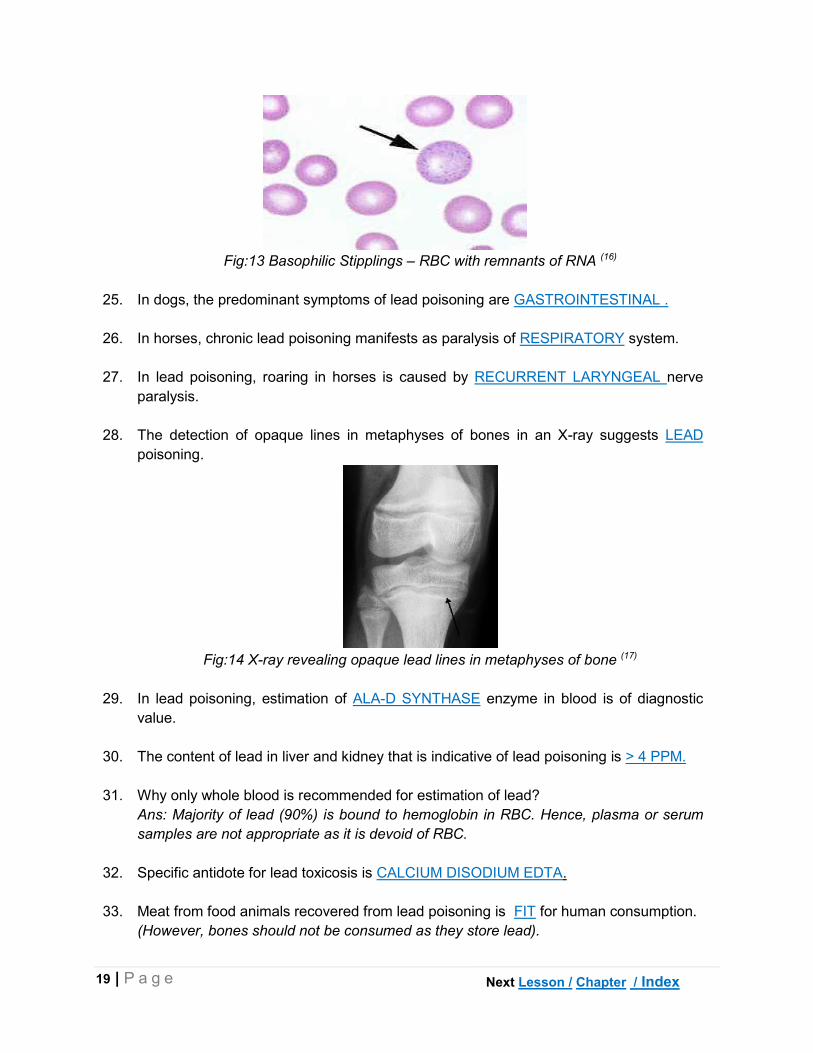
22. In lead poisoning, basophilic stipplings (BS) are commonly seen in this species
a. Cattle b. Sheep c. Dog d. Horse
Ans. c. Dog. BS are remnants of RNA seen in RBC which take up basophilic stain. But
BS are not pathognomonic for lead.
23. The characteristic histological picture of lead poisoning in tubular cells of kidney is
INTRANUCLEAR INCLUSION BODIES. (Eosinophilic)
24. Neurological symptoms accompanied by GIT symptoms possibly indicate LEAD
poisoning.
25. The predominant symptoms of lead poisoning in cattle are NEUROLOGICAL
SYMPTOMS.
Next Lesson / Chapter / Index
19 | P a g e
Fig:13 Basophilic Stipplings – RBC with remnants of RNA (16)
25. In dogs, the predominant symptoms of lead poisoning are GASTROINTESTINAL .
26. In horses, chronic lead poisoning manifests as paralysis of RESPIRATORY system.
27. In lead poisoning, roaring in horses is caused by RECURRENT LARYNGEAL nerve
paralysis.
28. The detection of opaque lines in metaphyses of bones in an X-ray suggests LEAD
poisoning.
Fig:14 X-ray revealing opaque lead lines in metaphyses of bone (17)
29. In lead poisoning, estimation of ALA-D SYNTHASE enzyme in blood is of diagnostic
value.
30. The content of lead in liver and kidney that is indicative of lead poisoning is > 4 PPM.
31. Why only whole blood is recommended for estimation of lead?
Ans: Majority of lead (90%) is bound to hemoglobin in RBC. Hence, plasma or serum
samples are not appropriate as it is devoid of RBC.
32. Specific antidote for lead toxicosis is CALCIUM DISODIUM EDTA.
33. Meat from food animals recovered from lead poisoning is FIT for human consumption.
(However, bones should not be consumed as they store lead).
Next Lesson / Chapter / Index

20 | P a g e
2.4 Copper
1. Copper has inverse inter-relationship with the following element(s)
a. Iron b. Molybdenum c. Sulphur d. Both b & c
Ans: d. Both b & c. Molybdenum and Sulphur.
2. The species that is more susceptible of copper poisoning is SHEEP.
3. The species that is highly resistant to copper poisoning is CHICKEN.
4. The ideal ratio of copper to molybdenum in feeds should be 6:1.
(A ratio of Cu: Mo of 10:1 can cause copper toxicosis).
5. The breed of dog that is highly susceptible to copper toxicosis due to genetic
predisposition is BEDLINGTON TERRIER.
(Autosomal recessive gene causes copper retention in liver as a result of failure of
excretion).
Fig:15 Bedlington Terrier –Genetic predisposition for copper accumulation (18)
6. Deficiency of MOLYBDENUM micro mineral predisposes to copper toxicity.
7. The specific transport proteins for copper in the body are TRANSCUPERIN and
CERULOPLASMIN.
(Transcuperin and albumin transports Cu from blood to liver. However, Transcuperin is
specific but is less abundant. Ceruloplasmin transports from liver to peripheral tissues.
About 90% of Cu in circulation is in bound form with ceruloplasmin)
8. The primary organ for accumulation (storage) of copper is LIVER.
9. The major route of elimination for copper from body is BILIARY.
10. Molybdenum and Sulphur reduce toxicity of copper by enhancing EXCRETION.
Next Lesson / Chapter / Index
21 | P a g e
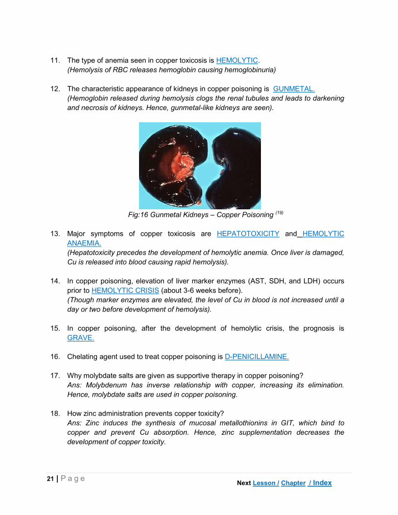
11. The type of anemia seen in copper toxicosis is HEMOLYTIC.
(Hemolysis of RBC releases hemoglobin causing hemoglobinuria)
12. The characteristic appearance of kidneys in copper poisoning is GUNMETAL.
(Hemoglobin released during hemolysis clogs the renal tubules and leads to darkening
and necrosis of kidneys. Hence, gunmetal-like kidneys are seen).
Fig:16 Gunmetal Kidneys – Copper Poisoning (19)
13. Major symptoms of copper toxicosis are HEPATOTOXICITY and HEMOLYTIC
ANAEMIA.
(Hepatotoxicity precedes the development of hemolytic anemia. Once liver is damaged,
Cu is released into blood causing rapid hemolysis).
14. In copper poisoning, elevation of liver marker enzymes (AST, SDH, and LDH) occurs
prior to HEMOLYTIC CRISIS (about 3-6 weeks before).
(Though marker enzymes are elevated, the level of Cu in blood is not increased until a
day or two before development of hemolysis).
15. In copper poisoning, after the development of hemolytic crisis, the prognosis is
GRAVE.
16. Chelating agent used to treat copper poisoning is D-PENICILLAMINE.
17. Why molybdate salts are given as supportive therapy in copper poisoning?
Ans: Molybdenum has inverse relationship with copper, increasing its elimination.
Hence, molybdate salts are used in copper poisoning.
18. How zinc administration prevents copper toxicity?
Ans: Zinc induces the synthesis of mucosal metallothionins in GIT, which bind to
copper and prevent Cu absorption. Hence, zinc supplementation decreases the
development of copper toxicity.
Next Lesson / Chapter / Index

22 | P a g e
2.5 Molybdenum
1. The primary source of molybdenum poisoning in animals is GRAZING
(Molybdenum poisoning is referred to as Molybdenosis or Teart. The term ‘teart’ refers
to watery foul smelling diarrhea which is characteristic in molybdenosis).
2. The species that is more susceptible to molybdenum poisoning is CATTLE.
3. Molybdenum toxicosis occurs primarily in the deficiency of COPPER.
4. The ideal ratio of copper to molybdenum should be 6:1.
(Ratio of <2:1 Cu to Mo can cause molybdenum toxicosis)
5. The level of molybdenum that causes toxicity, irrespective of copper content is >10
PPM.
6. Molybdenum is primarily excreted through URINE.
(Biliary excretion accounts for about 20% of the excretion)
7. In molybdenum poisoning, deficiency of COPPER (mineral) is observed.
(Hence, most of the symptoms in Mo poisoning resemble copper deficiency).
8. Peat scours or teart or shooting diarrhea is a characteristic symptom of
MOLYBDENOSIS.
(Peat scour or teart refers to foul smelling watery faeces with gas bubbles.
Molybdenum complexes with catechols and inactivates them. Hence, the natural
bacteriostatic activity of GIT is lost, which lead to infection and diarrhea).
9. Light coloured hair and depigmentation around eyes in molybdenum poisoning causes
SPECTACLE-EYE appearance.
(Due to Cu deficiency, melanin production is reduced due to decreased in activity of Cu
containing enzyme, tyrosinase, which converts tyrosine to melanin. Spectacle-eye is
prominently visible in buffaloes due to dark colouration of the animal).
Fig: 17 Spectacle-eye appearance – Molybdenum poisoning (20)
Next Chapter / Index

23 | P a g e
10. Molybdenosis in sheep is manifested as ENZOOTIC ATAXIA or SWAY BACK.
(Mo causes Cu deficiency. Cu dependent enzymes like cytochrome oxidase are
necessary for the synthesis of phospholipids of myelin. Hence, in Cu deficiency,
defective nerves are formed causing sway back).
11. The treatment of molybdenum poisoning involves administration of COPPER SALTS
(Eg. Copper sulphate).
Next Chapter / Index

24 | P a g e
CHAPTER III
TOXICOLOGY OF NON-METALS
3.1 Fluoride
1. Why fluorine is not available in free form?
Ans: Fluorine is the most reactive non-metal (due to high electronegativity) and hence
is not available in free form. It is seen in combination with other elements as fluorides.
2. The chronic disease resulting from continuous ingestion of small amounts of fluoride is
FLUOROSIS.
(Fluorosis is endemic in at least 22 countries world-wide and in many areas of Andhra
Pradesh and Telangana states).
Fig: 18 Worldwide distribution of endemic fluorosis – 22 counties are affected (21)
3. The part of the plant that does not accumulates fluoride is
a. Seed b. Stem c. Leaf d. Flower
Ans: Seed or grains.
4. Supplementation with ROCK PHOSPHATE mineral supplements can cause fluorosis.
(The optimum ratio of fluoride to phosphorus in rock phosphates should be 1:100).
5. Fatal toxicosis that can occur from inhaling gases and dust from volcanic eruptions is
FLUORINE INTOXICATION.
6.
Acute fluoride poisoning is common in DOG, whereas, chronic poisoning is common in
HERBIVORES.
(Dogs are poisoned from fluoride containing pesticides whereas herbivores from eating
contaminated pastures)
Next Lesson / Chapter / Index
25 | P a g e
7. Maximum tolerable level of fluoride in forage for herbivorous animals is
40-50 PPM.
(A level of 50 ppm should not be exceeded in the ration of animals )
8. The level of fluoride in drinking water that can cause fluorosis in animals is
>2 PPM.
(Dental defects are see at 5ppm, wear and tear at 10 ppm and systematic effects at 30
ppm).
9. Fluoride accumulates in BONE and TEETH in the body.
(Bone acts as sink for fluoride similar to lead. However, accumulation of fluoride in
teeth occurs only during formative stages i.e., young age only).
10. Fluoride is gradually excreted from the body through URINE.
11. Fluoride interferes with the following element(s) in the body
a. Calcium b. Magnesium c. Manganese d. Phosphorus
Ans: a, b & c: Ca, Mg and Mn. Interaction with calcium leads to hypocalcaemia and
interference with magnesium causes hypomagnesaemia causing seizures.
12. The corrosive effects of fluoride in GIT are due to the formation of HYDROFLUORIC
ACID in the acidic medium of stomach.
13. Hyperkalemia in fluoride poisoning is a result of inhibition of Na+-K+ ATPase enzyme.
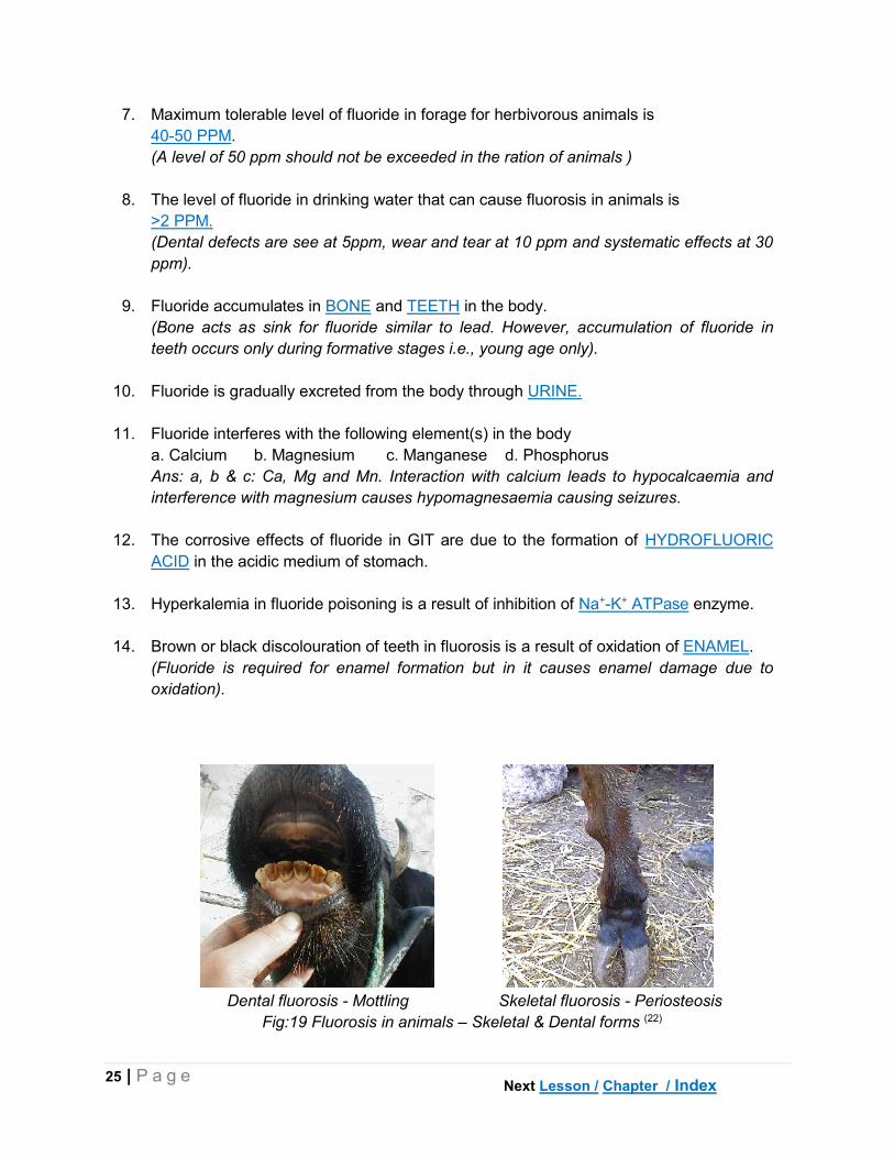
14. Brown or black discolouration of teeth in fluorosis is a result of oxidation of ENAMEL.
(Fluoride is required for enamel formation but in it causes enamel damage due to
oxidation).
Dental fluorosis - Mottling Skeletal fluorosis - Periosteosis
Fig:19 Fluorosis in animals – Skeletal & Dental forms (22)
Next Lesson / Chapter / Index
26 | P a g e
15. In fluorosis, defects in bones are a result of replacement of HYDROXYL groups with
fluoride in hydroxyapatite structure.
16. Chronic fluorosis is manifested as SKELETAL and DENTAL forms.
17. The form of fluorosis observed in young animals exposed to fluoride during early
stages of life is
a. Skeletal b. Dental c. Both d. Not affected
Ans: c. Both. If the animal is exposed in late stages of life, only skeletal form is
observed but if exposure takes place in early stages of life, both forms are seen).
18. The samples of choice to be collected in suspected cases of fluoride poisoning are
BONE and URINE.
(Affected cattle have 3000 ppm and sheep have 5000 ppm of fluoride in bone against a
normal value of 200 – 600 ppm. In urine >15 ppm is suggestive of fluorosis).
19. In fluorosis, the density of bones INCREASES.
(Fluoride binds with Ca by replacing hydroxyl groups in bones causing an increased
mineralization and bone density).
20. Soft tissue that accumulates highest amount of fluoride is PINEAL GLAND.
21. Death in fluoride toxicity is due to the development of HYPERKALEMIA and
HYPOCALCAEMIA.
22. The salts of CALCIUM are employed in treatment of fluorosis.
(Ca salts are mainly used as supportive therapy. However, there is no specific antidote
for fluoride toxicity).
3.2 Phosphorus
1. Which of the following form(s) of phosphorus is(are) toxic?
a. White b. Red c. Yellow d. Black
Ans: a & c. White and yellow phosphorus are soluble and readily absorbed. Hence
cause toxicity. Whereas red phosphorus is insoluble, hence is non-toxic.
2. Excess feeding of wheat bran rich in phosphorus causes BRAN DISEASE in horses.
3. Name the non-metal poisoning that can occur during Diwali and in war-zones?
Ans: Phosphorus. Yellow phosphorus is used in the manufacture of fire crackers and
military ammunition.
Next Lesson / Chapter / Index

27 | P a g e
4. Garlic-like odour of breath and luminous stomach contents suggest PHOSPHORUS
poisoning.
5. The routes of elimination for phosphorus are RENAL and PULMONARY.
(Hence the breath has garlic like odour).
6. Dermal exposure to white or yellow phosphorus leads to SKIN BURNS.
7. Why burns due to phosphorus causes higher mortality than other agents?
Ans: Absorption of phosphorus through raw burnt surface leads to multi-organ failure.
Hence, burns due to phosphorus are more dangerous than other burns.
8. The immediate symptom upon oral ingestion of phosphorus is EMESIS (Hematemesis)
(Phosphorus is a strong irritant with corrosive properties. Hence it causes GIT irritation
leading to vomition).
9. Major organs that are damaged in phosphorus poisoning are LIVER and KIDNEY.
10. Necrosis of jaw that is observed in chronic phosphorus poisoning is called as PHOSSY
JAW.
Fig:20 Phossy Jaw – Phosphorus poisoning – Degeneration of jaw bone (23-24)
11. Ideal sample material for the diagnosis of phosphorus poisoning is VOMITUS or
STOMACH CONTENTS.
12. Why oily purgatives such as mineral oils are contraindicated in phosphorus poisoning?
Ans: Oils increase the absorption of phosphorus. Hence are contraindicated in
phosphorus poisoning.
13. The prognosis in case of phosphorus poisoning is GUARDED to GRAVE.
Next Lesson / Chapter / Index

28 | P a g e
3.3 Nitrate and Nitrite
1. Toxicity of nitrate is due to its conversion into NITRITE by rumen micro flora.
(Nitrites [NO2-] are 10 times more toxic than nitrates [NO3
-]).
2. The use of NITRATE fertilizers increases the concentration of nitrates in plants.
3. The herbicide that increases nitrates in plants is 2, 4-D.
4. The following plant(s) is/ are nitrate accumulators
a. Cereal grasses b. Maize c. Sunflower d. Sorghum
Ans: All.
5. The source of drinking water that is high in nitrates is DEEP-WELL.
(Deep-well water contains around 1700 to 3000 ppm of nitrates due to seepage from
surface soil).
6. The following method of storing forages reduces nitrate content
a. Hay making b. Silage making c. Composting d. Straw making
Ans: b. Silage making. The process of fermentation reduces nitrate content in forages.
7. The species that are more susceptible to nitrate poisoning are RUMINANTS.
8. Why cattle are more susceptible than sheep for nitrate poisoning?
Ans: The rumen of sheep is effective in converting nitrites to ammonia, which is used
for protein synthesis. But in cattle, the rumen is not as effective as sheep. Hence, in
cattle are more susceptible.
9. Why non-ruminants are not affected by nitrates from plant sources?
Ans: The conversion of nitrates to nitrites does not occur in non-ruminants (lack of
rumen micro flora). Hence, non-ruminants are not affected. However, pigs are most
sensitive to ingestion of pre-formed nitrites.
10. Why plants accumulate nitrates in toxic proportions?
Ans: Plants absorbs nitrates from soil as a part of its physiology. However, any change
in environmental conditions that affects the rate of utilization of nitrates leads to nitrate
accumulation in toxic proportions. Eg: Lack of rainfall – no leaching of nitrates from soil;
Low temperature – inhibits nitrate reductase activity; High temperature – excessive
absorption from soil; etc.
11. The concentration of nitrates in plants that is toxic to animals is 1% or 10,000 PPM.
(on dry matter basis, a concentration of 0.5% nitrates is toxic in plant material).
Next Lesson / Chapter / Index

29 | P a g e
12. Drinking water containing more than 1500 PPM of nitrates can cause poisoning.
13. Maximum accumulation of nitrates is seen in the following part of the plant
a. Tip b. Leaves c. Upper 1/3 of stem d. Lower 1/3 of stem
Ans: d. Lower 1/3 of the stem. Parts closer to soil accumulate more nitrates.
14. The nitrate content in young plants is MORE than mature plants.
15. The presence of COLIFORM bacteria in water increases nitrate toxicity.
16. The growth of plants or algal blooms (eutrophication) in ponds leads to DECREASE in
nitrate content of water.
(Excessive growth of plants or algae in water bodies is due to eutrophication i.e.,
addition of nutrients (fertilizers) from run-off land water)
Fig: 21 Algal Bloom in a pond – Plants and algal blooms reduces nitrates in water (25)
17. The deficiency of molybdenum or sulphur or phosphorus in soil causes INCREASED
nitrate accumulation in plants.
18. The deficiency of copper or cobalt or manganese in soil causes DECREASED nitrate
accumulation in plants.
19. Addition of SOLUBLE CARBOHYDRATES (TDN) to feed improves tolerance to nitrate
toxicity.
(Carbohydrates are necessary for rumen micro flora for the conversion of nitrites into
ammonia, which is utilized for protein synthesis)
20. The antibiotic used as feed additive that enhances conversion of nitrates to nitrites
causing poisoning is MONENSIN.
21. Watering of animals immediately after consuming nitrate rich plants DECREASES the
chances of nitrate poisoning.
(As nitrates and nitrites are eliminated through urine. On the contrary, watering after
consumption of cyanogenic plants increases cyanide toxicity).
Next Lesson / Chapter / Index

30 | P a g e
22. Nitrite ion enters erythrocytes in exchange for CHLORIDE ion.
23. Nitrite combines with hemoglobin (in1:2 ratio) to form METHEMOGLOBIN.
(Ferrous (Fe2+) is oxidized to ferric (Fe3+) in met-hemoglobin, which decreases oxygen
carrying capacity of blood).
24. The colour of blood in nitrate poisoning is CHOCOLATE BROWN.
(Due to methemoglobin formation).
Fig:22 Methemoglobin – Brown coloured blood (26)
25. Physiologically formed methemoglobin (1 to 2 %) is converted back to hemoglobin by
the enzymes DIAPHORASE – I & II.
(Diaphorase – I is NAD dependent whereas DIAPHORASE – II is NADP dependent).
26. Formation of 20 to 40% of methemoglobin produces symptoms of nitrate poisoning.
27. Vasodilation and hypotension observed in nitrate poisoning is due to smooth muscle
relaxation caused by NITRIC OXIDE (NO).
28. The respiratiory rate in nitrate poisoning is INCREASED (RAPID).
(Rapid respiration is a prominent clinical sign in nitrate poisoning due to hypoxia).
29. Chronic nitrate poisoning leads to the development of GOITER in sheep.
(Nitrate interferes with iodine metabolism).
30. The preferred ante-mortem sample to be collected in nitrate poisoning is PLASMA.
(Serum cannot be used as nitrate are retained in the blood clot).
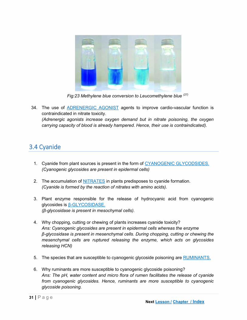
31. Specific treatment for nitrate poisoning is METHYLENE BLUE.
(Reducing agents such as ascorbic acid are also used).
32. Methylene blue is oxidized to LEUCOMETHYLENE BLUE during the conversion of
met-hemoglobin to hemoglobin.
33. Methylene blue mediated conversion of methemoglobin to hemoglobin is dependent on
the availability of NADPH2.
Next Lesson / Chapter / Index
31 | P a g e
Fig:23 Methylene blue conversion to Leucomethylene blue (27)
34. The use of ADRENERGIC AGONIST agents to improve cardio-vascular function is
contraindicated in nitrate toxicity.
(Adrenergic agonists increase oxygen demand but in nitrate poisoning, the oxygen
carrying capacity of blood is already hampered. Hence, their use is contraindicated).
3.4 Cyanide
1. Cyanide from plant sources is present in the form of CYANOGENIC GLYCODSIDES.
(Cyanogenic glycosides are present in epidermal cells)
2. The accumulation of NITRATES in plants predisposes to cyanide formation.
(Cyanide is formed by the reaction of nitrates with amino acids).
3. Plant enzyme responsible for the release of hydrocyanic acid from cyanogenic
glycosides is β-GLYCOSIDASE.
(β-glycosidase is present in mesochymal cells).
4. Why chopping, cutting or chewing of plants increases cyanide toxicity?
Ans: Cyanogenic glycosides are present in epidermal cells whereas the enzyme
β-glycosidase is present in mesenchymal cells. During chopping, cutting or chewing the
mesenchymal cells are ruptured releasing the enzyme, which acts on glycosides
releasing HCN)
5. The species that are susceptible to cyanogenic glycoside poisoning are RUMINANTS.
6. Why ruminants are more susceptible to cyanogenic glycoside poisoning?
Ans: The pH, water content and micro flora of rumen facilitates the release of cyanide
from cyanogenic glycosides. Hence, ruminants are more susceptible to cyanogenic
glycoside poisoning.
Next Lesson / Chapter / Index

32 | P a g e
Fig: 24 Epidermal cells (contains cyanogenic glycosides) and Mesenchymal cells
(contains β-glycosidase enzyme) in a leaf.(28)
7. Among ruminants, the more susceptible species for cyanogenic glycosides is CATTLE.
(Due to large rumen, which has more micro flora releasing more HCN)
8. Why non-ruminants are not affected by cyanogenic glycosides?
Ans: Acidic pH destroys β-glycosidase enzyme, which is responsible for the release of
HCN. Hence, non-ruminants are not affected by cyanogenic glycosides.
9. The cyanogenic glycoside in bitter almond and wild cherry is AMYGDALINE.
10. The cyanogenic glycoside in sorghum and sudan grass is DHURRIN.
11. The cyanogenic glycoside in linseed and wild clover is LINAMARINE.
12. The part of the plant that is rich in cyanogenic glycosides is LEAF.
13. The level of HCN in plants, which can cause cyanide poisoning in animals is 200 PPM
AND ABOVE.
14. The quantity of cyanogenic glycosides in young plants is MORE than mature plants.
15. After a period of drought or cloudy weather, the cyanide content in the plant
a. Increases b. Decreases c. Does not change d. Becomes zero
Ans: a. Increases.
16. The use of NITRATE fertilizers increases cyanide toxicity.
(Nitrate fertilizers increases nitrate content in plants, which is subsequently converted
to cyanide by combining with amino acids).
17. Spraying of the weedicide 2, 4-D can increase cyanide content in plants.
(2,4-D increases nitrate content and consequently the cyanide content).
Next Lesson / Chapter / Index

33 | P a g e
18. Soils that favour cyanide accumulation in plants are rich in NITROGEN and deficient in
PHOSPHORUS content.
19. Watering animals after feeding on cyanogenic plants INCREASES toxicity.
(Water causes hydrolysis of cyanogenic glycosides releasing HCN).
20. The metabolite of cyanide, which is excreted through urine is THIOCYANATE.
21. Cyanide is converted to non-toxic thio-cyanate by the enzyme RHODANESE.
22. Good reserves of SULPHUR in the body reduces the toxic effects of cyanide.
23. Cyanide inhibits cellular respiration by binding with CYTOCHROME OXIDASE (cyta3).
(Cyanide has more affinity towards metallo-porphyrin (Fe) containing enzymes)
Fig: 25 Electron transport chain – Cytochrome Oxidase inhibition by cyanide (29)
24. Why cyanide has more affinity for cytochrome oxidase than hemoglobin?
Ans: Cyanide has more affinity for ferric (Fe3+) form of iron. In hemoglobin, iron is
present in ferrous (Fe2+) form whereas cytochrome oxidase has ferric (Fe3+) iron.
Hence, cyanide prefers cytochrome oxidase.
25. Cyanide has more affinity for the following
a. Hemoglobin b. Cytochrome oxidase c. Met-hemoglobin d. Myoglobin
Ans: c. Met-hemoglobin (Fe3+). Sodium nitrate is used as a therapeutic strategy to
convert hemoglobin to methemoglobin, which removes cyanide from cytochrome
oxidase.
26. The colour of blood in cyanide poisoning is BRIGHT RED.
(Due to non-utilization by tissues, oxygen stays in blood giving bright colour).
Next Lesson / Chapter / Index

34 | P a g e
27. The characteristic smell of rumen contents that is suggestive of cyanide poisoning is
BITTER ALMOND.
(The smell of cyanide is similar to bitter almonds. Consuming of about 18 bitter
almonds can kill a human being. However, the variety used in household purpose is the
non-toxic domesticated sweet version).
28. Chronic form of cyanide toxicity observed in humans due to consumption of cassava
root is called as KONZO.
Fig:26 Cassava Root – causes chronic cyanide poisoning (30)
29. The level of cyanide in rumen contents that is indicative of cyanide poisoning is
10 PPM.
30. Specific treatment for cyanide toxicity is SODIUM NITRATE followed by SODIUM
THIOSULPHATE.
(NaNO3 converts hemoglobin into met-hemoglobin, which dissociates cyanide from
cytochrome oxidase and brings into blood; Na2SO3 helps in the conversion of cyanide
to non-toxic thio-cyanate, which is easily excreted through urine).
Next Lesson / Chapter / Index

35 | P a g e
3.5 Selenium
1. The Italian traveler who first reported the association between sloughing of hooves in
horses with consumption of specific plants is MARCO POLO.
Fig:27 Morco Polo – Italian merchant traveler –Reported selenium accumulator plants (31)
2. The following form(s) of selenium is (are) soluble
a. Elemental (0) b. Selenide (+2) c. Selenite (+2) d. Selenate (+6) e. Organo selenium
Ans: c, d & e. Selenite (+2) and Selenate (+6) are soluble and hence cause toxicity in
animals.
3. The most toxic from of selenium is ORGANO SELENIUM.
(The magnitude of toxicity of selenium is Organo selenium > Selenite=Selenate > Selenide
> Elemental Selenium).
4. Organo selenium is formed due to the replacement of SULPHUR by selenium in amino
acids.
(Sulphur containing amino acids being cysteine and methionine)
5. The level of selenium in plants that is toxic to animals is 5 PPM AND ABOVE.
6. The type of plants that have a physiological requirement for selenium and accumulate high
levels of selenium are called OBLIGATE (PRIMARY) ACCUMULATORS or INDICATOR
PLANTS.
(Eg: Astragalus sps, Stanleya sps, Oonopsis sps etc. Animals avoid these plants as they
are not palatable, but in scarcity they are consumed leading to toxicity)
7. The level of selenium found in obligate accumulators is 100-1500 PPM.
Next Lesson / Chapter / Index

36 | P a g e
Astragalus sps (Milk vetches) Stanleya sps (Princes plumes)
Oonopsis sps (Golden weeds) Xylorrhiza sps (Woody aster)
Fig:28 Obligate selenium accumulators (32-35)
8. The type of plants that dont require selenium but accumulate high levels of selenium, if
present in soil, are called FACULTATIVE ACCUMULATORS.
(Eg: Acacia sps, Aster sps, Astriplex sps, Artemisia sps etc. As these plants are more
palatable, toxicity is often seen in animals.).
9. The level of selenium found in facultative accumulators is 25-100 PPM.
Artimesia sps (Sages) Aster sps (Asters)
Next Lesson / Chapter / Index

37 | P a g e
Astriplex sps (Salt brush) Acacia sps (Acacia)
Fig:29 Facultative selenium accumulators (36-39)
10. The type of plants which do not require selenium but accumulate low levels of selenium, if
it is present in the soil, are called NON- ACCUMULATORS.
(Eg: Maize, Wheat, Barley etc. These plants are more palatable than other accumulators
and hence the chances of toxicity are more).
11. The level of selenium found in non-accumulators is 1-25 PPM.
12. Important natural source of selenium, apart from plants is VOLCANIC GASES.
13. The type of climates that favour selenium accumulation is ARID AND SEMI-ARID.
14. The pH of the soil that favours selenium accumulation is ALKALINE (>7.0).
15. Why selenium toxicity is more commonly seen in arid and semi-arid climatic zones?
Ans: In arid and semi-arid climatic zones, due to less rainfall, selenium accumulates in top
layers of the soil – as it is not leached. Hence, selenium accumulation in plants is common
16. The metabolite of selenium that is excreted through urine is TRIMETHYL-SELENONIUM.
17. Cytotoxicity caused by selenium is a result of FREE RADICAL generation at cellular level.
18. The non-enzymatic antioxidant that is depleted by selenium is GLUTATHIONE (GSH).
19. In selenium poisoning, the characteristic odour is GARLIC-LIKE.
(Recollect, phosphorus also produces garlic-like odour)
20. The vitamin that aggrevates selenium poisoning is VITAMIN E.
(Se and Vit E have inter-relationship)
21. Sub-acute toxicity of selenium is also known as BLIND STAGGERS.
22. Chronic selenium toxicity is also called as ALKALI DISEASE.
Next Lesson / Chapter / Index

38 | P a g e
23. In cattle, cracked and overgrown hooves are suggestive of SELENIUM poisoning.
24. In horses, loss of hair from the mane is the primary symptom of SELENIUM poisoning.
25. Why cracked and overgrown hooves are seen in selenium poisoning?
Ans: Selenium replaces sulphur in sulphur-containing amino acids such as cysteine and
methionine leading to structural abnormalities in proteins. Hence, overgrown and cracked
hooves are seen.
Fig:30 Cracked Hooves – Selenium Poisoning (40-41)
26. In selenium toxicity, depleted glutathione levels can be restored by administering ACETYL
CYSTEINE during treatment.
27. In selenium poisoning, the level of selenium detected in blood is 1-4 PPM and in hooves is
5-20 PPM.
28. The use of DIMERCAPROL (BAL) metal chelator is contraindicated in selenium toxicity.
3.6 Oxalate
1. The common source of oxalate poisoning in animals is PLANTS.
2. Important oxalate containing plants responsible for oxalate poisoning in animals are
HALOGETON GLOMERATUS and OXALIS PESCAPRAE.
3. The oxalate salt of the following element(s) is (are) soluble
a. Sodium b. Potassium c. Magnesium d. Calcium
Ans: a & b. Sodium and Potassium.
Next / Chapter / Index

39 | P a g e
H.glomeratus O.pescaprae
Fig:31 Oxalate accumulators (42-43)
4. Why the oxalates in Halogeton glomeratus and Oxalis pescaprae are the common
cause of oxalate poisoning?
Ans: The oxalates present in the above species are soluble. Hence, these plants cause
oxalate poisoning. In H.glomeratus, both sodium and potassium oxalates are present
whereas in O.pescapre, only potassium oxalates are present.
5. Halogeton species contains 34% of oxalates on dry matter basis.
6. The species of fungus that is rich in oxalates and can cause oxalate poisoning, is
ASPERGILLUS.
7. Why ruminants are less susceptible for oxalate poisoning?
Ans: Rumen has the ability to convert soluble oxalates into insoluble form. Hence,
ruminants are less susceptible for oxalate poisoning. But if the rumen’s ability for
conversion is exceeded, oxalate poisoning is observed.
8. The species that is commonly affected by oxalate poisoning is SHEEP.
9. Pastures containing 2% of oxalates is toxic for sheep.
10. The important mineral with which oxalates interact in the body is CALCIUM.
11. The primary clinical sign in oxalate poisoning is HYPOCALCAEMIA.
12. The deposition of insoluble calcium oxalate crystals in renal tubules causes OXALATE
NEPHROSIS.
13. The common site for urinary obstruction in bulls and rams due to oxalate stones is
SIGMOID FLEXURE.
(In rams, the oxalate crystals also deposit in urethral process).
Next Chapter / Index

40 | P a g e
Fig:32 Oxalate Nephrosis – Deposition of calcium oxalates in kidney (44)
14. Detection of oxalate crystals in KIDNEY and RUMEN EPITHELIUM is indicative of
oxalate poisoning.
15. In oxalate poisoning, CALCIUM salts are used as a part of the treatment.
(Calcium salts are used to correct hypocalcaemia observed in oxalate poisoning)
Next Chapter / Index

41 | P a g e
CHAPTER IV
TOXICOLOGY OF PLANTS
4.1 Photosensitization
1. Abnormal sensitivity of un-pigmented or less pigmented areas of skin to sun light due to
the presence of photodynamic substances in peripheral circulation is known as
PHOTOSENSITIZATION.
2. Substances that absorb UV light and emit energy while coming to ground state are called
PHOTODYNAMIC SUBSTANCES.
3. The type of photosensitivity resulting from direct ingestion of photodynamic substances or
metabolically activated agents is called PRIMARY photosensitization.
(Eg: Plants: Hypericin- Hypericium species; Fagopyrin – Fagopyrum species; Parthenium
sps; Drugs -phenothiazines, tetracyclines, sulphonamides, acridine dyes etc)
Hypericium sps (St. John Wort) Fagopyrum sps
Fig:33 Direct photosensitization causing plants (45-46)
4. The type of photosensitivity resulting from hepatic damage consequent to ingestion of
hepatotoxic plants or substances is called SECONDARY / HEPATOGENOUS
photosensitization.
(Eg. Pyrrolizidine alkaloid containing plants- Senicio sps, Heliotropium sps,; Lantana
camara; Mycotoxins- Sporodesmins; Blue green algae – Microcystis sps)
5. The photodynamic substance formed due to bacterial break down of chlorophyll that is
responsible for secondary photosensitization is PHYLLOERYTHRIN.
6. The species that is more susceptible to secondary photosensitization from pyrrolizidine
alkaloids is PIG.
Next Lesson / Chapter / Index

42 | P a g e
7. Lantana camara causes SECONDARY type of photosensitization.
Senicio sps Lantana camara
Fig:34 Secondary / Hepatogenous photosensitization causing plants (47-48)
8. Secondary photosensitization in Lantana camara is a result of BILE DUCT OCCLUSION.
9. The phyto-constituents of L.camara that are responsible for bile duct occlusion and liver
damage are LANTADENE A & B.
10. Why the lesionsin photosensitization are localized?
Ans: Melanin pigment protects skin from UV light. In less pigmented areas or in areas
devoid of fur/wool, more UV light is absorbed leading to sun burns. Hence, less
pigmented areas and areas devoid of hair/wool like face, eyelids, muzzle, coronary band,
udder etc are more prone for photosensitization.
11. Hepatic lesions such as FIBROSIS and BILIARY HYPERPLASIA are useful for differential
diagnosisis of primary and secondary photosensitization.
12. Visible lesions in photosensitization are SUN BURNS.
Fig:35 Photosensitization – Sun burns (49-50)
13. The prognosis is poor in SECONDARY type of photosensitization.
(The hepatic damage is generally irreversible leading to death).
Next Lesson / Chapter / Index

43 | P a g e
4.2 Thiamine deficiency causing plants
1. Bracken fern poisoning is a result of consuming the plant PTERIDIUM AQUILINUM.
2. The enzyme present in bracken fern that breaks down vitamin B1 is THIAMINASE.
(Other thiaminase containing plants include Horse tail, Australian nardoo fern, rock fern)
Pteridium aquilinum (Bracken fern) Equisetum arvense (Horse tail)
Marsilea drummondii (Austr. Nardoo) Cheilanthes sieberi (Rock fern)
Fig:36 Thiaminase containing plants (51-54)
3. Aplasia of bone marrow due to bracken fern poisoning is observed in RUMINANTS
species.
4. The following part of bracken fern is more toxic
a. Rhizome b. Stem c. Leaves d. Tips
Ans: a. Rhizome. All parts of bracken fern are toxic, however, rhizome is more toxic.
5. Carcinogenic and aplastic anemia inducing factor present in bracken fern is
PTAQUILOSIDE.
6. The active carcinogenic formed from ptaquiloside in alkaline pH is DIENON.
Next Lesson / Chapter / Index

44 | P a g e
7. The co-carcinogen in bracken fern, which causes malignant tumours in mouth,
esophagus and rumen along with papilloma virus is QUERCETIN.
8. The most susceptible species for bracken fern poisoning are HORSE and CATTLE.
9. The species in which anemia due to bracken fern poisoning is absent is HORSE.
(The main symptoms in horses are neurological)
10. The main symptom of bracken fern poisoning in cattle is APLASTIC ANEMIA.
11. Long term consumption of bracken fern in cattle causes tumours of URINARY BLADDER.
12. Why bracken fern causes urinary bladder tumours in cattle?
Ans: Ptaquiloside is converted into an active carcinogen dienon in alkaline medium. As
the urine in cattle is alkaline, the conversion of ptaquiloside to dienon is increased,
leading to urinary bladder tumours.
13. In bracken fern poisoning, enzootic hematuria is seen in CATTLE species.
14. Bracken fern produces permanent blindness in SHEEP species.
15. Diagnosis of bracken fern poisoning involves the estimating THIAMINE vitamin in blood.
(Normal thiamine (B1) content in blood of cattle is 8.5 μg/dL which is reduced to 2.5
μg/dL).
16. Specific treatment for bracken fern induced thiamine deficiency is THIAMINE (vit B1).
(However, there is no specific treatment for bone marrow aplasia and tumours caused by
bracken fern).
4.3 Abrus precatorius (Abrus) and Ricinus communis (Castor)
1. Abrus precatorius is commonly known as ROSARY PEA or RATHI.
2. The toxicity due to seeds of Abrus precatorius is commonly referred to as
SUI/ NEEDLE poisoning.
(Needles prepared from abrus seeds are inserted subcutaneously causing malicious
poisoning. Hence, the name sui or needle poisoning).
3. Chemically, abrin is a
a. Toxalbumin b. Toxglobulin c. Polypeptide d. Carbohydrate
Ans: a.Toxalbumin.
4. The toxic principle in the seeds of Abrus precatorius is ABRIN.
Next Lesson / Chapter / Index

45 | P a g e
Fig:37 Abrus precatorius – seeds contain abrin (55)
4. Ricinus communis is commonly referred to as CASTOR BEAN.
Fig:38 Ricinus communis – seeds contain ricin (56-57)
5. The toxic principle present in the seeds of Ricinus communis is RICIN.
6. Abrin and ricin belongs to the class of glycoproteins known as LECTINS.
7. Why swallowing of seeds of abrus and castor is not toxic?
Ans: Abrus and castor seeds have tough outer coating which resists digestion and hence
are passed through GIT without causing toxicity. However, crushing or chewing prior to
swallowing will produce toxicity.
8. During extraction of oil from castor seeds, the toxic principle ricin is detected only in
a. Oil b. Seed Cake c. Both d. None
Ans: Seed cake. Ricin is not expressed in oil but is retained in seed cake.
9. The toxic principle present in castor bean that causes hemagglutination and hemolysis is
RCA (Ricinus communis agglutinin).
(However, RCA is not absorbed through GIT; it needs parenteral administration)
10. The species that is more susceptible to abrin and ricin poisoning is HORSE.
Next Lesson / Chapter / Index

46 | P a g e
11. At the cellular level, lectins (abrin & ricin) decreases protein synthesis by inhibiting
RIBOSOMES.
(one molecule of abrin inhibits up to 1500 ribosomes per second)
12. Why barley containing lectins is non-toxic where as abrin and castor are?
Ans: Lectins contains A and B chains linked by disulphide bond. The presence of both
chains is necessary for producing toxicity. As barley is non-toxic due to the presence
single A chain.
4.4 Datura, Nerium and Ipomea sps
1. The poisonous plant whose flowers are known as ‘angel’s trumpets’ or ‘moonflowers’ is
DATURA STRAMONIUM (Thorn apple).
2. Major tropane alkaloids present in Datura stramonium are HYOSCINE (Scopolamine) and
ATROPINE (dl-hyoscyamine).
3. The toxic principles in datura are localized in SEEDS and FLOWERS.
Fig:39 Datura stramonium (Thron apple) (58-59)
4. The plant that is known as ‘deadly night shade’ is ATROPA BELLADONNA.
(Italian: Bella = Beautiful; Donna = Lady; Atropine causes dilatation of pupil (mydriasis)
when instilled into eyes. In olden days, women used to beautify their eyes through
atropine induced mydriasis. Hence the name bella-donna. Similar reasons are behind
‘candle light dinners’ of modern times, where dull candle light induces mydriasis,
beautifying ones appearance.
5. The species that is more sensitive to datura poisoning is PIG.
Next Lesson / Chapter / Index

47 | P a g e
Fig:40 Atropa belladonna (Deadly night shade)contains atropine (60-61)
6. The species that is resistant to Atropa belladonna is RABBIT.
(Rabbits contains the enzyme atropinase enzyme, which destroys atropine)
6. Tropane alkaloids (atropine) act by inhibiting MUSCARINIC receptors in the body.
7. Symptoms of datura toxicity are termed as ANTICHOLINERGIC DELIRIUM.
8. Why fever is observed in atropine poisoning?
Ans: ‘Atropine fever’ is a result of decreased secretions like salivation, sweating etc.
9. Urine from datura intoxicated animals produces MYDRIASIS in cat, which is used as
experimental evidence in diagnosis.
10. The competitive inhibitor of acetyl choline esterase, which is used for treating datura
(atropine) intoxication is PHYSOSTIGMINE.
11. Why phenothiazine tranquilizers are contraindicated in datura intoxication?
Ans: In datura intoxication, anticholinergic symptoms are seen. As phenothiazines also
possess anticholinergic activity, they are contraindicated for controlling CNS excitation in
in datura intoxication.
12. The toxic principle in Ipomoea turpethum (Indian jalapa or morning glory) is TURPETHIN.
13. The toxic principle in Ipomoea orizabensis is SCAMMONIN (Jalapin)
14. Major symptom in ipomoea plant poisoning is DIARRHOEA.
(The toxic resins present in ipomoea species produce drastic purgation).
15. Toxic principle in Nerium oleandrum (White oleander) is OLEANDRIN.
Next Lesson / Chapter / Index

48 | P a g e
Fig:41 Ipomea trupethum (Operculina trupethum)- contains drastic purgation causing resin
trupethin(62)
16. Toxic principle in Nerium odorum is NERIN.
17. The toxic glycosides in Cerebra thevetia are THEVETIN and CEREBRIN.
18. Steroidal glycoside in Nerium indicum is ODOROSIDE.
19. Most toxic part of the plants belonging to Nerium species are LEAVES.
(A leaf can kill a human and 2-3 leaves can kill a sheep. Leaf is effective even when dry).
Nerium oleandrum (Oleandrin) Cerebra thevetia (Thevetin)
Fig:42 Cardiac glycoside containing plants (63-64)
20. The species that is most susceptible for oleander poisoning is HORSE.
21. Oleander glycosides act by inhibiting Na+ - K+ ATPase enzyme in cardiac cells.
22. Imbalance of the following electrolyte is observed in oleander poisoning
a. Sodium b. Potassium c. Magnesium d. Calcium
Ans: b.Potassium. Hyperkalemia produced in cardiac glycoside toxicity is fatal.
Next Lesson / Chapter / Index

49 | P a g e
4.5 Strychnous nuxvomica and Gossypium sps
1. The toxic principle in Strychnous nuxvomica is STRYCHNINE.
2. Strychnine is localized in SEEDS part of S.nuxvomica.
3. Inhibition of GLYCINE neurotransmitter causes spinal stimulation in strychnine poisoning
4. The site of action of strychnine in spinal cord is RENSHAW cells.
(Recurrent inhibitory interneuron cells of reflex arc of spinal cord).
5. The characteristic appearance of animal in strychnine poisoning is SAW HORSE.
(Saw horse appearance is due to continuous tetanic seizures, which causes rigidity.
Tetanus similarly produces saw horse appearance)
6. Preferred sedative for treating strychnine poisoning is PENTOBARBITONE (Barbiturate).
7. The following is (are) contraindicated in strychnine poisoning
a. Ketamine b. Morphine c. Emesis d. All
Ans: d. All. Ketamine causes motor stimulation; morphine causes respiratory depression;
emesis causes seizures.
Fig:43 Strychnous nuxvomica – seeds contain strychnine (65)
8. Why strychnine is least toxic to chicken and pigeons?
Ans:Strychnine is least toxic to chicken and pigeons as it is absorbed very slowly.
However, other avians are easily affected (eg. crow).
9. The toxic principle in cotton seeds or seed cake is GOSSYPOL.
10. Gossypol binds with IRON mineral inhibiting heame synthesis and causing anemia.
11. Inhibition of the testicular enzyme LACTATE DEHYDROGENASE (LDH) is responsible
for development of reproductive toxicity in males.
Next Chapter / Index

50 | P a g e
12. The maximum permissible amount of cotton seed cake in cattle ration ONE KG. (total
dose)
(Further, feeding of cotton seed cake should be interrupted after 2-3 months for a period
of 3-4 weeks).
Fig:44 Gossypium hirsutum – seeds contains the toxic principle gossypol (67-68)
13. Gossypol induced infertility is a result of LUTEOLYTIC effect on ovary.
14. Intake of high amounts of PROTEIN is protective against gossypol toxicity.
Next Chapter / Index

51 | P a g e
CHAPTER V
TOXICOLOGY OF AGROCHEMICALS
5.1 Organochlorine (OC) compounds
1. The group of heterogeneous substances that are used to control pests are known as
PESTICIDES.
(Pesticides include insecticides – insects; Herbicides – weeds; Fungicides – Fungi;
Rodenticides – rats & mice)
2. Chlorinated hydrocarbons are known as ORGANOCHLORINE (OC) insecticides.
(Eg. DDT, BHC, Endrin etc)
3. Why OC insecticides are being discouraged/banned?
Ans: OC insecticides are not degradable and are persistent in the environment. And
due to high lipid solubility, they accumulate in the food chain and enter human and
animal bodies. Hence, OC compounds are being discouraged.
4. The following OC insecticides is not persistent in environment
a. DDT b. Aldrin c. Methoxychlor d. Endosulfan
Ans: c & d. Endosulfan and Methoxychlor
5. The most susceptible species for OC insecticide poisoning is CAT.
6. OC insecticides accumulate mainly in ADIPOSE tissue of the body.
7. The only OC insecticide that does not accumulate in adipose tissue is ENDOSULFAN.
(Endosulfan is also biodegradable and not persistent in environment).
8. The OC compound that is bio-degradable by microbes is ENDOSULFAN.
(Endosulfan has a sulphur ring in its structure, which is susceptible to microbial
degradation. However, despite its ban in 2011 worldwide, it is still used in India).
9. The only isomer of benzene hexachloride (BHC) that is biodegradable is LINDANE
(Gamma isomer).
10. OC compounds produce CNS excitation by prolonging DEPOLARIZATION phase of
action potential.
(Depolarization is prolonged by extending the opening of Na+ channels and delaying
the closure of K+ channels. Aliphatic OC compounds like DDT, Methoxychlor etc follow
this mechanism).
Next Lesson / Chapter / Index

52 | P a g e
11. OC compounds act by inhibiting GABA receptors.
(Since, GABA is inhibitory, inhibition of GABA receptors leads to CNS excitation.
Cyclodiene OC compounds like aldrin, endrin, endosulfan etc follow this mechanism).
12. Hyperthermia caused by OC insecticide poisoning is a result of altered metabolism of
SEROTONIN and NORADRENALINE neurotransmitters.
13. The metabolite of DDT (Dichloro-diphenyl-trichloro-ethane) is DDE (Dichloro-Diphenyl-
Dichloro- ethylene).
14. DDT acts as an agonist for ESTROGEN receptors, whereas, DDE acts as an
antagonist for ANDROGEN receptors.
15. DDE, the metabolite of DDT causes thinning of egg shells due to inhibition of
CALCIUM ATPase enzyme.
(DDE is known to cause thinning of egg shells causing decline of birds of prey – [Bald
eagle, Pelican, Falcon] chicken, song birds).
16. The predominate symptoms in OC insecticide poisoning are BEHAVIORAL
accompanied by HYPERTHERMIA.
(Organo phosphorus(OP) compounds do not produce behavioral symptoms and
hyperthermia)
17. The sedatives of choice used in OC insecticide induced CNS excitation is
BENZODIAZEPINES (Eg. Diazepam).
(Benzodiazepines also increases the affinity of GABA to its receptors, which was
previously inhibited by OC insecticides).
18. Death in OC compound poisoning is due to RESPIRATORY failure.
(OC cause initial CNS stimulation followed by depression and coma. Hence, ultimately
death is due to respiratory failure.
5.2 Organophosphate (OP) compounds
1. Organic insecticides which are esters of phosphorus are known as
ORGANOPHOSPHATE (OP) compounds.
2. OP insecticides act as irreversible inhibitors of ACETYL CHOLINE ESTERASE (AChE)
enzyme.
(AChE inhibition causes excess Ach accumulation producing cholinergic symptoms)
Next Lesson / Chapter / Index

53 | P a g e
3. Why OP compounds are preferred insecticides for crops?
Ans: OP compounds are biodegradable and are easily destroyed by sunlight, water,
microbes, alkalis, metals etc. Hence, within 2-4 weeks of application, OP compounds
are destroyed. However, they have considerable toxicity for mammals if consumed
directly.
4. OP compounds, which are capable of inhibiting AChE without requiring metabolic
activation are called DIRECT ACTING OP COMPOUNDS.
(Direct acting compounds have P=O (Oxon) group, hence act without metabolic
activation. Eg. TEPP, Dichlorovas, Sarin, Tabun).
5. Indirect acting OP compounds require metabolic activation to OXON form before
producing toxicity.
(Indirect acting agents have P=S (Thionate bond) which should be converted to P=O
(Oxon) form before inhibiting AChE. Eg. Malathion, Parathion, Chlorpyrifos).
6. What are the metabolic active forms of Malathion and Parathion?
Ans: Maloxon and Paroxon.
7. The toxicity of OP compounds INCREASES during storage.
(Due to development of toxic isomers during storage)
8. The species of animal that is highly sensitive to OP poisoning is CAT.
9. The species of animal that is highly sensitive for delayed neuropathy caused by OP
compounds (OPIDN) is CHICKEN.
(Chicken brain has higher specific activity of NTE, hence is more susceptible)
10. OP compounds bind with AChE at ESTERATIC site causing irreversible inhibition.
11. Phosphorylation of AChE by OP compounds resulting in irreversible bonding due to the
loss of alkyl group is called AGEING.
12. The agents, which are used to reactivate AChE inhibited by OP compounds are called
OXIME REACTIVATORS (Eg. 2-PAM – Pralidoxamine).
13. Oxime reactivators are ineffective in reactivating AChE inhibited by OP compounds
after AGEING stage.
14. OP compounds can inhibit the following enzyme(s)
a. AChE b. BuChE c. NTE d. All
Ans: d. All. AChE - true choline esterase; BuChE – Butryl choline esterase/
Pseudocholine esterase and NTE- Neurotoxic esterase.
Next Lesson / Chapter / Index

54 | P a g e
Fig:45 Ageing of acetylcholine esterase – Phosphorylation and loss of ‘alkyl’ group (69)
15. Pseudocholine esterase (BuChE) is present in PLASMA.
(Inhibition of BuChE is more important in birds than AChE inhibition).
16. OP induced delayed neuropathy (OPIDN) is caused due to the inhibition of NEURO
TOXIC ESTERASE (NTE) enzyme. (also called Neuropathy Target Esterase)
17. The main symptoms in OP poisoning are CHOLINERGIC.
(Salivation, Lacrimation, Urination and Defecation).
18. In OPIDN, the type of paralysis observed is of ASCENDING FLACCID type.
(Hence, OPIDN is also known as dying back axonopathy. Mostly, large myelinated
peripheral nerves are more affected).
19. The following type of toxicity produced by OP compounds is irreversible even with
treatment
a. Acute b. Sub-acute c. Chronic d. OPIDN
Ans: d. OPIDN. There is permanent damage to the peripheral nerves.
20. Diagnosis of OP toxicity is carried out by measuring AChE activity in blood.
(Less than 25% activity of AChE is confirmative of OP poisoning. In birds, estimation of
BuChE is important).
21. The specific antidote for OP poisoning is ATROPINE.
(Atropine inhibits cholinergic symptoms by blocking muscarinic receptors).
22. Oxime reactivators (2-PAM, DAM) should be used within 24-36 h time before ageing of
AChE.
23. Use of atropine is contraindicated in CATS species, as it causes cyanosis.
Next Lesson / Chapter / Index

55 | P a g e
Fig:46 Mechanism of Pralidoxamine reactivation (69)
5.3 Carbamates
1. Why Carbamates are the preferred insecticides in veterinary use?
Ans: Carbamates have broad spectrum of activity, low in mammalian toxicity and
undergo rapid degradation in environment. Hence, carbamates are preferred for
veterinary use. (Eg. Carbaryl, Propoxur, etc)
2. Insecticides that cause reversible inhibition of AChE are CARBAMATES.
3. Carbamates bind with AChE at the following site(s)
a. Anionic b. Esteratic c. Cationic d. Both a & b
Ans: d. Both a & b.
4. Oxime reactivators are contraindicated in CARBAMATE insecticide poisoning.
(Carbamates bind with both sites of AChE leaving no room for oxime reactivators).
5. How carbamates and OP compounds differ interms of their binding to AChE?
Ans: i. Carbamates bind with both anionic and esteratic sites where as OP compounds
binds with esteratic site only
ii. Carbamates cause carbomylation whereas OP compounds cause phosphorylation
iii. Binding of carbamates is reversible whereas that of OP compounds is irreversible.
6. Why binding of carbamates with AChE is reversible?
Ans: Carbamates causes carbomylation of AChE, which is weaker than
phosphorylation caused by OP compounds. Hence, the binding is reversible.
Next Lesson / Chapter / Index

56 | P a g e
7. Why carbamates are unable to produce delayed neuropathy?
Ans; Carbamates cannot bind with neuro-toxic esterase. Hence, delayed neuropathy is
not produced.
8. The least toxic carbamate that is used in veterinary practice against ectoparasites is
CARBARYL.
9. Specific antidote for carbamate poisoning is ATROPINE.
5.4 Pyrethroids
1. Pyrethroids are natural insecticides obtained from CHRYSANTHEMUM flowers.
(The use of marigold flowers is a common folk practice to prevent insects in food
grains. The active ingredient in marigold is pyrethrin. Hence, synthetic compounds
resembling it are called ‘pyrethroids’)
Fig:47 Chrysanthemum flowers – Source of natural pyrethrin (70)
2. The most widely used house-hold insecticides are SYNTHETIC PYRETHROIDS.
(Least toxic and rapidly biodegradable. These agents are used in mosquito repellants -
like good night, all out; Ant and cockroach repellants – Baygon, Cross line etc)
3. Type I pyrethroids resemble NATURAL PYRETHRINS in their structure and activity.
(Eg. Allethrin, Pyrethrin, Permethrin etc)
4. Type II pyrethroids contain α-CYANO group in their structure and are more active and
toxic.
(Eg. Deltamethrin, Cypermenthrin, Fenvalarate etc)
Next Lesson / Chapter / Index

57 | P a g e
5. The first commercial mosquito repellant pyrethroid still in use is ALLETHRIN.
6. Which of the following insecticides are more specific to arthropods?
a. OC compounds b. OP compounds c. Carbamates d. Pyrethroids
Ans: d. Pyrethroids. The LD50 of pyrethroids for insects and mammals is 1:4500, which
means specific action towards insects.
7. The species that is highly susceptibility to pyrethroids toxicity is FISH.
8. The relationship between toxicity of pyrethroids and temperature is INVERSE.
(As ambient temperature decreases the toxicity increases and vice versa).
9. Pyrethroids cause CNS stimulation by inhibiting the closure of VOLTAGE GATED
SODIUM channels.
(This leads to increased influx of Na+ causes prolonged depolarization).
10. Voltage gated sodium channels are more sensitive to II (TWO) type of pyrethroids.
11. In type II pyrethroids produce characteristic ‘TAIL’ appearance in depolarization phase
of action potential.
Fig:48 Characteristic ‘tail’ appearance in type II pyrethroid depolarization (71)
12. Type II pyrethroids acts as inhibitors for the receptors of GABA neurotransmitter.
13. The symptoms in type I pyrethroid toxicity are referred to as T-SYNDROME (Tremors).
14. Type II pyrethroids produces characteristic syndrome known as CS SYNDROME
(Choreoathetosis and Salivation).
15. Tranquilizers that are contraindicated in pyrethroid poisoning are PHENOTHIAZINE.
Next Lesson / Chapter / Index

58 | P a g e
16. The roots of plants belonging to Derris genus are the source of natural insecticide
ROTENONE.
Fig:49 Derris plant – Root is the source of rotenone (72)
17. Rotenone is highly toxic for FISH species.
(Hence, derris is used to control not only insects but also fish (Piscicide)
5.5 Rodenticides
1. The agents which are used to control rodents are known as RODENTICIDES.
2. What is bait shyness?
Ans: A rodent surviving the exposure of a particular rodenticide will avoid the same
rodenticide in the future. This phenomenon is called bait shyness.
3. What is secondary poisoning?
Ans: The poisoning that is seen in dogs and cats as a result of consuming rodenticide
poisoned rodents.
4. Toxicity of Zinc phosphide (Zn3P2) is due to the release of PHOSPHINE (PH3) gas.
5. The characteristic smell of phosphine gas, which can be used for diagnosis of zinc
phosphide toxicity, is FISH LIKE or ACETYLENE.
6. Zinc phosphide releases phosphine gas in the following pH
a. Acidic b. Basic c. Neutral d. All
Ans: a. Acidic pH.
7. Why zinc phosphide preparations contain antimony potassium tartrate?
Ans: Antimony potassium tartrate is an emetic, which prevents accidentally ingested zinc
phosphide through emesis in non-target species.
Next Lesson / Chapter / Index

59 | P a g e
8. Why zinc phosphide is more toxic on full stomach than empty ?
Ans: On full stomach, the acid production is increased. As zinc phosphide releases
phosphine gas in acidic medium, full stomach increases the toxicity.
9. The metabolite of zinc phosphide is HYPOPHOSPHITE.
(Hypophosphite is excreted in urine).
10. Phosphine gas acts as PROTOPLASMIC poison. (It is also highly irritant to GIT).
11. Bait shyness is not observed with ANTICOAGULANT rodenticides.
(Anticoagulants are odourless and tasteless, and act after a lag period. Hence, the rodent
forgets the exposure).
12. Warfarin inhibits clotting factors that are dependent on VITAMIN K for synthesis.
(Vitamin K dependent clotting factors are II (Prothrombin), VII, IX and X).
13. Anticoagulant rodenticides decrease vitamin K synthesis through the inhibition of VITAMIN
K EPOXIDE REDUCTASE enzyme.
14. Capillary damage seen in warfarin rodenticides is due to the presence of
BENZALACTONE chemical moiety.
(Mere reduction in clotting factors by anticoagulants is not fatal. Hence, benzolactone
induced capillary damage is necessary to cause fatal hemorrhage)
15. The hematological tests used to confirm poisoning from anticoagulants are CLOTTING
TIME and PROTHROMBIN TIME.
(An increase of 2 to 6 times in clotting and prothrombin times are noted).
16. The specific treatment for anticoagulant poisoning is PHYTOMENADIONE (Vitamin K1).
(Menadione is a precursor of vitamin K. Menadione from synthetic sources is K3;
Menaquinone – K2 I from bacteria; Phytomenadione – K1 is from plants).
17. Anticoagulant rodenticides effective against warfarin-resistant rats are SECOND
GENERATION anticoagulants.
(Hence, they are also called super-warfarins. Eg. Bromadiolone, Brodifacoum etc)
18. Risk of secondary poisoning is very high with 2ND GENERATION ANTICOAGULANT
rodenticides.
19. Fluoroacetate lowers energy production in the body by inhibiting TCA or KREBS CYCLE.
20. Why fluoroacetate rodenticide is devoid of bait shyness?
Ans: Fluoroacetate develops toxicity after a lag period. Hence, the rodent forgets the
exposure.
Next Lesson / Chapter / Index
60 | P a g e
21. The prominent symptoms in fluoroacetate poisoning are NEUROLOGICAL.
(Brain is severely affected in the event of depletion of energy in body).
22. The competitive antagonist that is used in the treatment of fluoroacetate poisoning is
GLYCERYL MONOACETATE.
(However, glyceryl monoacetate is not a specific antidote of fluoroacetate)
23. Why the use of fluoroacetate is restricted?
Ans: Fluoroacetate is highly toxic and non-specific affecting other species.
24. The rodenticide effective against warfarin-resistant rats is VITAMIN D (cholecalciferol).
25. The mechanism of toxicity of vitamin D compounds (cholecalciferol) rodenticides through
the induction of HYPERCALCEMIA.
(Hypercalcemia causes calcification of visceral organs like kidney, blood vessels, heart
and lungs).
26. Treatment for vitamin D rodenticide poisoning is CORTICOSTEROIDS and CALCITONIN.
(Both corticosteroids and calcitonin reduces hypercalcemia)
27. The phrase ‘drowning in one’s own fluids’ is associated with ANTU (alpha-napthyl-thio-
urea) rodenticide poisoning.
(ANTU causes leakage of fluid into lungs leading to froth formation and death)
28. Despite being very specific to rats, why ANTU is banned?
Ans: Due to carcinogenic potential of alpha napthylamine impurities found in ANTU.
29. The breed of rat, which is very sensitive for ANTU is BROWN or NORWAY rat.
29. Bait shyness and tolerance develops very rapidly for ANTU rodenticide.
30. The main symptom in ANTU poisoning is PULMONARY EDEMA.
31. What is the difference between pulmonary toxicity caused by ANTU (rodenticide) and
Paraquat (herbicide)?
Ans: ANTU causes pulmonary edema which is fatal, whereas, paraquat causes pulmonary
fibrosis which is not fatal.
32. Red squill, which is used as a rodenticide is obtained from the plant URGENIA
MARITIMA.
(In south India, toxicity due to Indian squill (Urgenia indica) is more common)
33. Toxic glycosidic principle present in red squill is SCILLIROSIDE.
34. The concentration of scillirosides is highest in BULB part of the plant.
Next Lesson / Chapter / Index
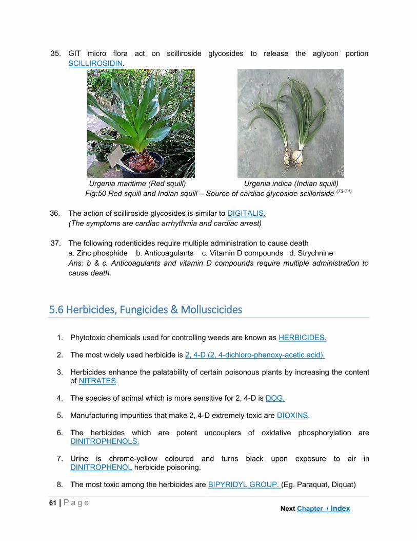
61 | P a g e
35. GIT micro flora act on scilliroside glycosides to release the aglycon portion
SCILLIROSIDIN.
Urgenia maritime (Red squill) Urgenia indica (Indian squill)
Fig:50 Red squill and Indian squill – Source of cardiac glycoside scilloriside (73-74)
36. The action of scilliroside glycosides is similar to DIGITALIS.
(The symptoms are cardiac arrhythmia and cardiac arrest)
37. The following rodenticides require multiple administration to cause death
a. Zinc phosphide b. Anticoagulants c. Vitamin D compounds d. Strychnine
Ans: b & c. Anticoagulants and vitamin D compounds require multiple administration to
cause death.
5.6 Herbicides, Fungicides & Molluscicides
1. Phytotoxic chemicals used for controlling weeds are known as HERBICIDES.
2. The most widely used herbicide is 2, 4-D (2, 4-dichloro-phenoxy-acetic acid).
3. Herbicides enhance the palatability of certain poisonous plants by increasing the content of NITRATES.
4. The species of animal which is more sensitive for 2, 4-D is DOG.
5. Manufacturing impurities that make 2, 4-D extremely toxic are DIOXINS.
6. The herbicides which are potent uncouplers of oxidative phosphorylation are DINITROPHENOLS.
7. Urine is chrome-yellow coloured and turns black upon exposure to air in DINITROPHENOL herbicide poisoning.
8. The most toxic among the herbicides are BIPYRIDYL GROUP. (Eg. Paraquat, Diquat)
Next Chapter / Index

62 | P a g e
9. Paraquat selectively accumulates in LUNGS organ of the body.
10. Despite being highly toxic, bipyridyl herbicides are harmless after being sprayed on plants? Ans: i. Bipyridyl herbicides are used in very low doses, which are not toxic ii. They are inactivated immediately upon contact with soil.
11. The mechanism of toxicity of paraquat is through generation of SUPEROXIDE free radical, which affects unsaturated membrane lipids in lungs.
12. Why lung tissue is primarily affected by paraquat? Ans: i. Paraquat accumulates up to 10 times in lungs. ii. Lung tissue is deficient in superoxide dismutase (SOD) enzyme, which neutralizes superoxide radical. Hence, lungs are primarily affected.
13. The bipyridyl herbicide that can cause pulmonary fibrosis is PARAQUAT.
14. The agents which are used to prevent fungal infestation of plants or seeds are known as FUNGICIDES.
15. Fungicides that uncouple oxidative phosphorylation are PENTA-CHLORO-PHENOLS (PCP).
16. The mechanism of uncoupling of oxidative phosphorylation of PCP is due to PROTON INOPHORE activity. (PCP being a lipid soluble weak acid carries protons (H+) across mitochondrial membrane leading to depletion of proton gradient required for ATP synthesis. Hence, ATP synthesis is inhibited).
17. Dithio-carbamates such as ziram, thiram are derivatives of CARBAMATE insecticides.
18. Agents which are used to control snails and slugs are known as MOLLUSCICIDES.
19. The most commonly used molluscicide is METALDEHYDE.
20. Metaldehyde is extremely toxic though INHALATION route of exposure.
21. The active metabolite of metaldehyde is ACETALDEHYDE.
22. Metaldehyde inhibits GABA neurotransmitter in CNS, which results in excitation.
Next Chapter / Index
63 | P a g e
CHAPTER VI
RESIDUE TOXICOLOGY
1. What are Drug Residues?
Ans: The presence of veterinary drugs and their metabolites along with associated impurities
in any edible portion of the animal products is known as Drug Residue. (Codex Alimentarius
Commission, 2004)
2. What is Withdrawal time?
Ans: ‘Withdrawal time’ is the time between treatment and slaughter or milking in which, the
drug/ pesticide residues deplete to a safe level (Tolerance/ Maximum residue level).
3. Who sets withdrawal times and levels for drug residues in animal products?
Ans: Centre for Veterinary Medicine (CVM) of Food and Drug Administration (FDA) sets the
withdrawal times and limits for approved food animal drugs.
4. Who sets the withdrawal times and limits for pesticide residues in animal products?
Ans: Environment Protection Agency (EPA) sets the withdrawal times and limits for pesticide
residues in plant and animal products.
5. How are with drawl times set for veterinary drugs?
Ans: The following steps are involved in setting with drawl times
NOEL ADI Safety
concentration
Tolerance/
MRL
Withdrawal
time
No observable effect level (NOEL) is established in animals for the drug in question
Average daily intake (ADI) values are determined
o ADI = (NOAEL / 1000) x Ave. Body weight
(ADI = Total dose of a drug that an average human can ingest on daily basis for entire
life with no adverse effect)
Safe concentration is arrived by diving ADI with the amount of edible organ which is
generally consumed in a day (Eg. Muscle 300 g; Liver 100g). Safe concentration
includes all metabolites of a drug.
o Safe concentration = ADI/300 (for muscle) ADI/100 (for liver)
A marker residue which is sensitive and easy for detection is chosen (Parent
compound or any of its metabolite).
Tolerance level/ maximum residue level is ascertained through estimation of marker
Next Chapter / Index

64 | P a g e
residue. Tolerance (MRL) = the % of safe concentration represented by the marker
residue.
Studies are performed to know the time at which the target tissue is below tolerance,
which is called withdrawal time.
Withdrawal times are aplicable only when the drug is used according to label
instructions. The tolerance for extra label use of a drug is Zero (i.e., detection of
extalabel drug at any level leads to rejection of the product).
Fig: 51 Withdrawal time and Maximum residue levels (Tolerance) determination (75-76)
6. How withdrawal time can be extrapolated form pharmacokinetic parameters?
Ans: Withdrawal time = 10 x T1/2 (T1/2 = half-life of the drug)
7. What is Tolerance / Maximum residue level?
Ans: Maximum residue level is the maximum concentration of the administered veterinary
drug, which is legally permitted or accepted in the food product.
8. What is the difference between ‘Maximum residue level’ and ‘Maximum residue limit’?
Ans: ‘Maximum residue level’ is used for drugs whereas ‘maximum residue limit is used for
pesticides.
9. What are the techniques used for detection of residues in foods?
Ans: Techniques such as ELISA, HPLC, LC, GC, Paper chromatography.
10. What are the general categories of veterinary drugs found as residues in animal products?
Ans: The general classes of veterinary drugs found in animal products are
Antimicrobials, Anti-inflammatory, Growth promoters, Anti-parasitic and insecticides and
Tranquilizers.
Next Chapter / Index

65 | P a g e
11. Which is the most common type of drug residue in animal products?
Ans: Antibiotics are the most frequently found drug residues (44%) followed by anti-
inflammatory drugs.
12. In which type of animals drug residues are common?
Ans: Drug residues are most commonly found in Dairy cattle (43%) followed by beef cattle
(17%).
13. What are the hazards of drug residues in animal products?
Ans: The hazards of drug residues in animal products are as follows
Aesthetic issues: The presence of drugs in animal is not appealing for consumer.
Allergic reactions: Certain drug residues like Penicillins can cause allergic reaction in
sensitive individuals at a level of 10 IU (0.6 μg)
Development of antibiotic resistance in microorganisms
Direct toxic effects – cancer, reproductive and developmental effects.
Eg: i. Clenbutarol residues were reported to have caused tachycardia,
muscle tremor, headache, nausea and fever and chills in humans
ii. Furazolidone and its metabolites are banned by FDA as they
were reported to cause cancer in humans.
Deleterious effects of hormone residues in humans
Eg: Diethyl stilbesterol (DES) – Vaginal clear cell adenocarcinoma in
female off springs exposed in utero; Structural abnormalities of uterus
International export barriers: Export of animal products need to comply with
international standards for drug residues.
14. What are the measures to be taken to prevent residues in animal products?
Ans: Measures to prevent drug residues include
Education of veterinary personnel, organizations and government agencies involved
in animal production.
Development of rapid screening methods for field use.
Avoiding irrational use of drugs in animals
Processing of animal products
Eg: Refrigeration completely destroys penicillins and pasteurization
causes loss of activity of most of the antibiotics.
Nationwide monitoring and surveillance for drug residues.
Next Chapter / Index

66 | P a g e
CHAPTER VII
VENOUS ANIMALS
1. What is the difference between venom and poison (toxin)?
Ans: Venom is produced by a specialized gland and is delivered either by biting or
stinging. Poison (toxin) is contained in the tissues and is delivered through food. Eg:
Snake – Venom; Puffer fish – Tetradotoxin (poison)
2. The order under the class insect that includes greatest number of poisonous insect
species, is HYMENOPTERA.
(The important families are Apidae [Honey bees]; Vespidae [Wasps] and Formicidae
[Ants].Stinging from wasps is more common as they dwell in human and animal
settlements)
Polistes annularis (Red paper wasp) Apis mellifera (Honey bee)
Fig:52 Important stinging insects in the order Hymenoptera (77-78)
3. Why wasps can deliver multiple stings whereas honey bees only once?
Ans: The stinger in honey bees is barbed, which is caught in the skin of the victim durig
stinging. This results in the loss of stinging apparatus along with venom sac and death of
the insect. In wasps, the stinger is plain and can be withdraw from victims skin. Hence,
wasps sting multiple times.
Stinger of Wasp (Unbarbed) Stinger of Honey bee (Barbed)
Fig: 53 Stinging apparatus of Wasp (Unbarbed) and Honey bee (Barbed) (79-80)
4. The antigenic component in honey bee venom that causes allergies or anaphylaxis is
MELLITIN.
Next Chapter / Index

67 | P a g e
5. The drug of choice for treating systemic reactions from bee or wasp stings is
EPINEPHRINE (ADRENALINE).
6. The potent cytotoxin present in ant venom is FORMIC ACID.
7. The piperidine alkaloid component of fire ant venom is SOLENOPSIN.
(Piperidine alkaloids are also known as hemolytic factors and induce histamine release)
8. Focal necrotic ulcers of cornea and conjunctiva in calves are caused by the bite of FIRE
ANTS.(Soloenopsis invicta) insect.
Fire ant (Solenopsis invicta) Sterile pustule – fire ant bite
Fig:54 Fire ant and bite of fire ant – sterile pustule (81-82)
9. The most potent neurotoxin in the venom of black widow spider (Lactrodectus mactans) is
α-LACROTOXIN.
10. The most dangerous species of scorpion is LEIURUS QUINQUESTRIATUS.
(The venom contains potent neurotoxins)
Fig: 55 Leirus quinquestriatus – Dangerous scorpion (83)
11. The most common tick species responsible for tick paralysis are RIPHICEPHALUS and
DERMACENTOR.
(Other species of ticks which can cause tick paralysis include Ixodes, Amblyomma, and
Ornithodorus).
Next Chapter / Index

68 | P a g e
12. Tick paralysis is caused by the injection of neurotoxic SALIVA.
13. The most susceptible species for tick paralysis is DOG.
14. The type of paralysis seen induced by ticks is ASCENDING FLACCID.
Riphicephalus sps Dermacenter sps
Fig:56 Important ticks causing tick paralysis (84-85)
15. The toxins from dermacentor species acts by blocking SODIUM channels, whereas, that
of Ixodidae species by blocking the release of ACETYLCHOLINE in motor nerves.
16. Diagnosis of tick toxicity is made by observing FEMALE sex ticks on animal body along
with paralysis.
17. Bufotixins (venom) in toads is produced primarily by PAROTOID glands.
(Skin also contains small glands, which produces bufotoxins but parotid glands are larger
and are located dorsal and posterior to eyes [fig 57])
18. The most toxic toad is BUFO MARINUS.
19. The principal component of toad venom is BUFODIENOLIDES (Cardiac glycosides).
(The symptoms involve cardiac arrhythmia, ventricular fibrillation and heart failure)
20. The species in which frequent toad poisoning is observed is DOG.
21. Treatment of toad poisoning involves the use of PROPRANOLOL drug to control
symptoms of cardiac arrhythmia and fibrillation.
22. Snake bite is commonly observed in DOGS and HORSES species of animals.
23. The type of toxins present in the venom of elapidae snakes (Cobra, Krait, Mamba, Coral
snakes) are NEUROTOXINS.
24. The type of toxins present in the venom of viperidae snakes (Viper, Rattle snake, Adder)
are HAEMOTOXINS.
Next Chapter / Index

69 | P a g e
Fig:57 Bufo marinus – Skin contains bufodienolide cardiac glycosides (86)
25. The neurotoxin present in the venom of kraits is α-BUNGAROTOXIN.
26. In snakes, the venomous glands are homologues to PAROTID glands in other animals.
27. Why snake bite in dogs is fatal compared to other animals?
Ans: Dogs are relatively smaller in size compared to other large animals like horses and
cattle. Hence, the bite is fatal in dogs as the proportion of venom injected to body weight
is very high.
28. The following snake bite results in prominent local reaction
a. Cobra b. Krait c. Sea snake d. Rattle snake
Ans: d. Rattle snake. The venom in viperidae (Vipers, rattle snake etc) snakes is
hemotoxic and contains enzymes such as hyaluronidase. Hence, local reactions
predominate.
29. The symptoms in elapine snake bite are predominantly NEUROLOGICAL.
30. Diagnosis of snake bite is possible by observing FANG marks on the body.
31. The main treatment for snake bite is MONOVALENT /POLYVALENT ANTIVENIN.
(If snake is identified, monovalent antivenin is sufficient otherwise polyvalent antivenin is
necessary. Further, during treatment with antivenin, the development of allergic reactions
should be controlled using epinephrine)
32. Why the use of alcohol is contraindicated for cleaning snake bite injuries?
Ans: Alcohol causes vasodilation promoting the spread of the venom in the body.
33. The only poisonous species of lizard is HELODERMA SUSPECTUM (Gila monster).
34. The inclusion of FISH MEAL component in feeds exposes animals to marine toxins.
Next Chapter / Index

70 | P a g e
Fig:58 Gila monster – Poisonous lizard (87)
35. The most potent among all marine toxins is TETRADOTOXIN (TTX).
(TTX is 10 times more toxic than snake venom; 100 times more toxic than black widow
spider; 10000 times more toxic than cyanide)
36. Consuming TETRAODON (Puffer) /FUGU fish is commonly associated with tetradotoxin
poisoning.
Fig:59 Puffer fish – Source of tetradotoxins (88-89)
37. In puffer fish, tetradotoxin (TTX) primarily accumulates in LIVER and OVARY.
Fig:60 Pseudoaltermonas tetradonis – produces tetradotoxin (90)
38. Tetradotoxin is produced by the symbiotic bacteria PSEUDOALTEROMONAS
TETRADONIS in puffer fish.
Next Chapter / Index

71 | P a g e
39. Why puffer fish are resistant to tetradotoxin?
Ans: Puffer fish have mutation altered sodium channels that are resistant to tetradotoxin.
39. Tetradotoxin acts as a potent neurotoxin by blocking of SODIUM channel in central and
peripheral nervous system.
40. Ciguatera is a fish-borne poisoning resulting from the consumption of REEF fish.
(Reef fish such as barracuda, eel etc).
Fig:61 Barracuda fish – Source of ciguatoxins (91)
41. The marine toxin responsible for ciguatera poisoning is CIGUATOXIN (CTX).
42. Ciguatoxin is produced by the dinoflagellate GAMBIERDISCUS TOXICUS present in reef
fish.
(Similar to TTX, reef fish do not produce CTX, they only possess the toxin)
Gamberdiscus toxicus Gamberdiscus toxicus (Ultrastructure)
Fig:62 Microalgae (dinoflagellate) Gamberdiscus toxicans – produces ciguatoxin(92-93)
43. The most heat resistant marine toxin is CIGUATOXIN.
44. Why avoiding reef fish consumption is the only way to avoid ciguatera?
Ans: The detection of ciguatoxin is difficult as there are no perceptible organoleptic
changes in fish. Further, CTX is not destroyed by temperature or gastric acid. Hence,
avoiding reef fish consumption is the only way to prevent ciguatera.
Next Chapter / Index

72 | P a g e
45. Consumption of tuna and mackerel fish is associated with SCROMBROID poisoning.
46. Scrombroid poisoning is caused by HISTAMINE, which is produced from histidine through
bacterial action.
(Other compounds such as puterscien and cadaverine are also involved in triggering
histamine toxicity)
Tuna Fish Mackerel Fish
Fig:63 Tuna and Mackerel fish contains scrombroid toxins (94-95)
Next Chapter / Index

73 | P a g e
CHAPTER VIII
RADIATION TOXICOLOGY
1. The process of transmission of electromagnetic waves through a medium or vacuum is
known as RADIATION.
2. Ionizing radiations produce ions by knocking out ELECTRONS from atoms.
3. Ionizing radiations – Particles type (α-particles, β-particles, neutrons) and
Electromagnetic waves (X-rays, γ-rays, UV-rays).
4. The ionizing radiation that has least penetrating capacity is ALPHA (α)-PARTICLES.
(α-particles are large in size (2 protons+2 neutrons = α-particle). Hence, they have
least penetration ability. However, due to their high mass, they have high linear energy
transfer (LET) ability.
5. The ionizing radiation that has highest penetrating capacity is GAMMA (γ) RAYS.
(Since γ-rays are non-particulate electromagnetic radiation. Hence, they have the
highest penetration ability. However, as the mass is least, LET is very low).
6. The type of UV rays, which reach earth are UV-A TYPE.
(UV-B&C are more powerful than UV-A rays, however, they are filtered by ozone layer
(up to 99%).A sunscreen with both UV-A&B sun protection factor (SPF) of at least 25 is
recommended in Indian conditions.
7. Non-ionizing radiations include– Visible light, Infrared rays, microwaves, radio waves.
8. The SI unit of radiation is GRAY (Gy) and the CGS unit is RAD (rad).
9. One Gray is equal to 100 rad. (or 1 rad = 0.01 Gy).
10. The instrument used to measure radiation is GEIGER-MULLER COUNTER.
11. What is the difference between radiation exposure and irradiation?
Ans: Both radiation exposure and irradiation, exposure to radiation occurs. However, in
irradiation, post exposure contamination with radioactive material is absent (Eg. γ-
irradiation of syringes; irradiation used for treating cancers etc).
12. Which two elements are responsible for natural background radiation in earth?
Ans: Radon (from uranium) and Thoron (from thorium) are responsible for 54% of
natural background radiation in earth.
Next Chapter / Index

74 | P a g e
13. During nuclear explosion, the ascent and subsequent descent of radioactive material
either in the vicinity or away from site of explosion is known as FALL OUT.
(Fall out can occur within few minutes to months and either in the vicinity or far away).
14. Among the radioactive material produced in nuclear explosions, the elements with
biological significance are STRONTIUM (ST90) and CESIUM (CS137).
(Due to long half-lives)
15. The environmental hazard from nuclear power plants is due to the release of
COOLANT, which is radioactive.
(Light water, Heavy water, liquid sodium etc are used as coolants).
16. Cells are more susceptible for radiation in the following stage(s) of cell cycle
a. M-Phase (mitosis) b. Early G-Phase c. Late G-Phase d. S-phase
Ans: a & b. M-phase and early G-phase. S-phase is resistant.
17. The group of symptoms that appear within 48h of exposure to radiation are known as
PRODROMAL SYNDROME.
(The symptoms include nausea, vomition, anorexia, fatigue, diarrhea, sweating etc).
18. The sensitivity of rapidly proliferating cells to radiation is HIGHER than non-
proliferative cells.
19. The most sensitive system for radiation exposure is HAEMOPOETIC SYSTEM.
20. The lethal dose of radiation in animals is expressed as LD50/30
(Lethal dose required to causes 50% mortality in 30 days; In humans it is LD50/60).
21. The cellular component, which is affected during radiation damage is DNA.
22. Molecules with unpaired electrons in the outer shells are known as FREE RADICALS.
23. Most common free radicals generated in the body are REACTIVE OXYGEN SPECIES
(ROS).
(ROS include – Hydroxyl (OH-), Superoxide (O-2), Singlet oxygen (O-), Hydrogen
peroxide (H2O2) etc; Reactive nitrogen species (RNS) – Nitric oxide (NO), peroxynitrate)
24. The most potent among the free radical is HYDROXYL (OH-).
(OH- is produced through Fenton’s reaction where hydrogen peroxide acts on iron).
25. Why the presence of heavy metals generates more free radicals?
Ans; Heavy metals (like Fe, Pb, Cd etc) have more number of electrons in their outer
shell. Hence, when a free radical attacks a heavy metal, there will be shower of
electrons, which in turn produces more free radicals.
Next Chapter / Index

75 | P a g e
26. Damaging effect of free radicals on protein is PROTEIN OXIDATION and on DNA is
DNA FRAGMENTATION.
27. The most sensitive hematological test for detecting radiation damage is ABSOLUTE
LYMPHOCYTE COUNT.
(Lymphocyte count (LC) <1000 cells/cc within 24h or <500 cells/cc within 48h indicates
severe exposure. If LC is >1500/cc after 48h post exposure, rules out radiation).
28. The most common type of cancer induced by radiation is LEUKAEMIA.
29. Grazing animals on fall-out pastures primarily affects GASTROINTESTINAL system.
30. The following organ is resistant to radiation
a. Endocrine glands b. Kidney c. Bone marrow d. Germinal cells.
Ans: a. Endocrine glands; as they have very low proliferation rate. The other organs
are very sensitive due to high proliferative rate.
31. The amount of radio frequency radiation absorbed by human body while using mobile
phone is measured by SPECIFIC ABSORPTION RATE (SAR).
(In India and US, permissible SAR is set at 1.6 Watt/Kg user mass; In Europe, SAR is
set at 2.0 W/Kg).
32. The part of the head, which is sensitive to the heating (diathermic) effects of mobile
phone is CORNEA.
(Cornea of the eye lacks temperature regulating mechanism and hence could be
affected by dielectric heating from mobile phones during usage)
Next Chapter / Index

76 | P a g e
CHAPTER IX
TOXICOLOGY OF FOOD AND FEED ADDITIVES
9.1 Food & Feed additives
1. Non-nutritive substances added to food to improve the physical, organoleptic, nutritive
properties or shelf life are called as FOOD ADDITIVES.
2. Standards relating to food production and safety are covered by CODEX
ALIMENTARIUS COMMITTEE,
3. Agents which prevent oxidative damage of food are called as ANTIOXIDANTS.
4. The most toxic and suspected carcinogens among the food additives are COLOURING
AGENTS.
5. The rare adverse effect of the colouring agent Tartrazine in humans is ALLERGY OR
ANAPHYLAXIS.
6. The flavouring agent that produces characteristic ‘candy-shop’ aroma banned by FDA
due to its carcinogenic potential is SAFROLE.
7. The flavor enhancing agent that produces ‘umami’ type taste in foods is
MONOSODIUM GLUTAMATE (MSG).
8. ‘Chinese restaurant syndrome’ is caused by the food additive MONOSODIUM
GLUMATE (MSG).
(Symptoms include headache, flushing, tingling, numbness around mouth, palpitations
etc. However, as exact association with adverse effects have not be found, MSG is still
used. The use of MSG needs to be listed in food label).
9. The oldest artificial sweetener used in foods is SACCHARIN.
(Saccharin was reported to produce epithelia hyperplasia of urinary bladder in male
rats. However, due to lack of association between saccharin and cancer in humans at
normal doses, WHO approved saccharine for human use ).
10. The most common artificial sweetener approved by FDA for use in pharmaceutical
products and foods is ASPARTAME.
(Breakdown of aspartame produces phenylalanine and hence should be avoided by
persons with ‘Phenyl Ketonuria. Diet coke and other sugar-free products contain
aspartame)
Next Lesson / Chapter / Index

77 | P a g e
11. Vitamin C can reacts with benzoic acid – used as preservative in foods - to form
BENZENE.
12. The substances which are added to animal feeds form improving quality of feeds or
improving animal performance are called as FEED ADDITIVES.
13. Feed additives which increases growth rate and feed conversion in animals are called
GROWTH PROMOTERS.
14. Antibiotics approved for use as growth promoters in food producing animals are
IONOPHORE ANTIBIOTICS.
(Monensin, lasalocid and salinomycin are the commonly used ionophore antibiotics).
15. The most sensitive species for ionophores toxicity is HORSE.
(Horses are 10 times more sensitive than cattle).
16. The most common symptom in chronic ionophore toxicity is CARDIOMYOPATHY.
17. Antibiotic fed additive that should not be used simultaneously with other feed additives
or growth promoters is ZINC BACITRACIN.
18. The use of Zinc bacitracin is contraindicated in LACTATING type of animals.
9.2 Urea and Salt Poisoning
1. Apart from dietary sources, animals get exposure to urea from FERTILIZERS
.
2. Excessive use of ammonium nitrate and urea fertilizers for crops can result in
NITRATE poisoning in animals.
3. Urea is added to the diets of ruminants as a source of NITROGEN.
(Urea contains 46.7% nitrogen and 1 g of urea is equivalent to 2.92 g of protein)
4. The lethal dose of urea in cattle is 1 to 1.5 g /day/animal.
(4g/day/animal in horses)
5. Urea is permitted in the diets of ruminants and horses due to the presence of UREASE
microbial enzyme in rumen and caecum respectively.
(Urease converts urea in to ammonia which is used for microbial amino acid synthesis).
Next Chapter / Index

78 | P a g e
6. The following reason(s) increases the susceptibility of ruminants to urea poisoning
a. Urease present in plants b. Alkaline pH of rumen
c. Acidic pH of stomach d. Urease activity of rumen
Ans: a & b. Urease converts urea to ammonia. Both plants and rumen microbes
contain urease enzyme.Further, alkaline pH of rumen facilitates this conversion. Henc,
ruminants are more susceptible. In non-ruminants, the pH of stomach is acidic and the
diet is not primarily from plants, hence they are least susceptible.
7. Which of the following age group is more resistant to urea toxicity?
a. Calf b. Heifer c. Bull d. Cow
Ans: a. Calf. In young ruminants the rumen microbial is not well developed hence less
urease activity is seen.
8. The feed component that is required for efficient utilization of urea is
CARBOHYDRATES or TOTAL DIGESTIBLE NUTRIENTS (TDN).
(Carbohydrates (CHO) provide carbon skeleton for the production of amino acids. In
cases of CHO deficiency, urea toxicity occurs due to unutilized ammonia).
9. The recommended ratio of urea to molasses for straws and other high fiber diets is 1:5.
10. In horses, the site of conversion of urea to ammonia is CAECUM.
(The urease activity in horses is about 25% that of cattle)
11. Excessive ammonia inhibits TCA/ KREBS cycle in body decreasing energy production.
12. Ruminal pH above 7.5 is diagnostic of urea / non-protein nitrogen (NPN) poisoning.
(Normal ruminal pH is 6.0 to 6.2, which increases due to release of ammonia)
13. Why the pH of the blood is acidic in urea poisoning?
Ans: In liver detoxification of ammonia to urea requires bi-carbonate (HCO3-), which
depletes blood HCO3- buffer leading to acidosis. Blood pH changes from 7.4 to 7.0
contrary to the assumption that released ammonia alkalizesblood.
13. The treatment in urea poisoning includes infusion of 5% ACETIC ACID (VINEGAR) and
COLD WATER into rumen.
(Acetic acid reduces ruminal pH and decreases conversion of urea to ammonia. Cold
water reduces rumen temperature there by decreasing urease activity).
14. Intravenous infusions containing GLUCOSE/ DEXTROSE are contraindicated in urea
poisoning as they cause hyperglycemia.
15. The toxic principle produced from the interaction of ammonia with reducing sugars in
feeds is 4-METHYL IMMIDAZOLE (4-MI).
( Similar to urea, ammonia treatment is used to improve the N2 content of roughages)
Next Chapter / Index

79 | P a g e
16. 4-Methyl imidazole (4-MI) in ammoniated feeds produces CNS symptoms in cattle
known as BOVINE BONKER’S SYNDROME.
17. Salt poisoning is commonly associated with deprivation of WATER.
(Hence, salt poisoning is termed aptly as water-deprivation-induced salt poisoning).
18. The most susceptible species for salt poisoning are POULTRY and PIGS.
(Poultry have poor sense of taste and indiscriminate feeding behavior).
19. Salt toxicity is consequent to the development of HYPEROSMOLALITY or
HYPERTONICITY in blood and CSF.
20. Major symptoms evident in salt poisoning are NERVOUS SYMPTOMS.
21. Nervous symptoms in salt poisoning are a result of edem of BRAIN.
22. Dragging of hind limbs and knuckling of fetlock joints is characteristic feature of SALT
poisoning in cattle.
23. In salt poisoning, pathognomonic histological appearance in brain is EOSINOPHILIC
MENINGIOENCEPHALITIS.
(Perivascular cuffing of eosinophils is observed in cerebral cortex and meninges).
Fig:64 Eosinophilic Perivascular Cuffing – Salt poisoning (96)
24. The level of sodium in plasma and CSF that is diagnostic of salt poisoning is
> 1mEq/L.
(Further, the level of sodium in CSF should be more than plasma).
25. Prognosis in case of salt poisoning is GRAVE.
(Less than 50% of affected animals survive with treatment; recumbent and convulsing
animals definitely die irrespective of treatment).
Next Chapter / Index

80 | P a g e
CHAPTER X
MYCOTOXINS & BACTERIAL TOXINS
1. Secondary metabolites of fungus, which cause deleterious effects to animal and human
life are called as MYCOTOXINS.
2. Molds generally grow in stored fed stuffs containing a moisture content more than 15%.
(The other factors being a relative humidity of >85% and optimum temperature of
25-30o C i.e., without refrigeration).
3. Mycotoxins are classified based on ORGAN SYSTEM affected.
(Hepatotoxic – Aflatoxins, Rubra toxins; Nephrotoxic – Ochratoxin, Citrinin;
Estrogenic – Zearalenone (F-2); Cytotoxic – Trichothecenes (T-2); Neurotoxic –
Tremorgens).
4. Turkey X disease is caused by AFLATOXIN mycotoxins.
5. Aflatoxins are produced by ASPERGILLUS FLAVUS and A.PARASITICUS.
Fig:65 Aspergillus flavus – Source of aflatoxins (97-98)
6. The most potent among the aflatoxins is AFB1.
(The other being AFB2, AFG1 and AFG2. The letters B (blue) and G (green) represent the
type of light under which the respective toxin exhibits fluorescence.
7. Aflatoxins are heat resistant but are unstable in UV LIGHT.
8. Aspergillus species grows in stored feed stuffs containing moisture content more than
15%.
9. The following feed stuff(s) supports growth of aflatoxins
a. Groundnut cake b. Soybean cake c. Cotton seed meal d. All
Ans: All
Next Chapter / Index
81 | P a g e
10. The domestic species that are highly susceptible to aflatoxicosis are DOG and
DUCKLINGS.
11. Aflatoxin has the following characteristic (s)
a. Carcinogenic b. Mutagenic c. Teratogenic d. Immunosuppressive
Ans: All of the above. Hence aflatoxins are considered more dangerous than other
mycotoxins.
12. The metabolite of aflatoxins excreted through milk and urine is AFM1.
13. Aflatoxin content in cattle feeds should not exceed 20 PPB.
14. The carcinogenic metabolite of aflatoxins is AFLATOXIN 8, 9-EPOXIDE.
(Aflatoxins need biological activation to produce their toxic effects i.e., lethal synthesis)
15. Aflatoxins causes defective protein synthesis by binding with N-7 GUANINE residue of
DNA causing mis-pairing of nucleotides.
16. Aflatoxin epoxide causes carcinogenic and mutagenic effect by causing ALKYLATION of
the strands of DNA.
(Alkylation forms cross bridges between DNA strands)
17. Hemorrhage in aflatoxicosis is due to the decrease in PROTHROMBIN and VITAMIN K
18. Which of the following forms of aflatoxicosis is most common
a. Per acute b. Acute c. Sub acute d. Chronic
Ans: d. Chronic. Chronic form occurs due to continuous intake of low level of aflatoxins.
19. The type of carcinoma caused by aflatoxins is HEPATOCELLULAR CARCINOMA.
20. Alfatoxins can be detected by THIN LAYER CHROMATOGRAPHY method.
21. Hemorrhagic syndrome in poultry is caused by RUBRATOXINS mycotoxins.
22. Rubratoxins are produced by PENICILLIUM RUBRUM and P.PURPUROGENUM.
23. The most toxic metabolite of rubratoxins is RUBRATOXIN B.
(Rubratoxin B is hepatotoxic, mutagenic and teratogenic)
24. Rubratoxins are destroyed at the following temperature
a. Freezing b. Room temperature c. 50 to 60oC d. 85 to 100oC
Ans: d. 85 to 100oC for two hours can destroy rubratoxins
Next Chapter / Index

82 | P a g e
Fig:66 Penicillium rubrum (painting brush appearance) – source of rubratoxins (99-100)
25. Ruminant’s facial eczema is caused by SPORIDESMIN mycotoxicosis.
26. Nephrotoxic mycotoxins responsible for mold nephrosis or mycotoxic nephropathy are
OCHRATOXINS.
27. Ochratoxins are produced by ASPERGILLUS OCHRACEUS and PENICILLIUM
VIRIDICATUM.
28. The most toxic among the ochratoxins is OCHRATOXIN A.
29. The most susceptible species for ochratoxicosis are BIRDS and PIG.
30. The site of action of ochratoxins in the nephron is
a. Proximal Convoluted Tubule b. Loop of henle
c. Distal Convoluted Tubule d. Collecting duct
Ans: a.Proximal convoluted tubule. Inhibits anion transport and damages renal brush
border.
31. The level of ochratoxin in feed should not exceed 10 PPB.
32. Estrogenic mycotoxin responsible for reproductive disorders is ZEARALENONE (F-2).
33. Zearalenone (F-2) mycotoxins are produced by FUSARIUM ROSEUM mold.
34. The most susceptible species for zearalenone toxicosis is PIG.
35. Vulvo-vaginitis or hyper-estrogenic syndrome in pigs is caused by ZEARALENONE
mycotoxin.
36. The maximum permitted level of zearalenone in feed is 10 PPB.
Next Chapter / Index

83 | P a g e
Fig:67 Fusarium roseum – Source of Zearalenone toxins (101-102)
37. Why pigs and cattle differ in their susceptibility to zearalenone?
Ans: In pigs, the major metabolite of Zearalenone is α-zearalenol, which has greater
affinity for estrogen receptors than β-zearalenol, which is formed in cattle. Hence, pigs are
more susceptible than cattle.
38. Name the hormone involved in ovarian follicle maturation that is inhibited by Zearalenone.
Ans: Follicle Stimulating hormone (FSH).Henc, the characteristic symptom of follicular
atresia is observed in ovaries.
39. Histological picture of uterus and vaginal in zearalenone toxicosis is METAPLASIA.
40. Alimentary toxic aleukia (ATA) in human being is caused by TRICHOTHECENE
mycotoxins.
(Trichothecenes commonly includes T-2 toxin, DON and DAS. DON -Deoxy-nevalenol
[vomition] and DAS -Diacetoxy-scirpenol).
41. The mycotoxins that were used as biological warfare agents are TRICHOTHECENES.
(Alleged ‘yellow rain’ attacks of Vietnam on local tribes, who supported US during
Vietnam war)
42. The species that is more susceptible to trichothecenes is CAT.
43. An example for neurotoxic mycotoxin is TREMORGENS.
(The name ‘tremorgens’ indicates muscle tremors produced by the toxins due to CNS
effects).
44. The most potent among the tremorgens is PENITREM A.
45. The most susceptible species for tremorgen mycotoxins are CATTLE and DOG.
46. Staggers syndrome in cattle is produced by TREMORGEN mycotoxins.
47. Ergotoxins are produced by the mold CLAVICEPS PURPUREA.
(Important ergot alkaloids are ergotamine and ergometrine)
Next Chapter / Index

84 | P a g e
Fig: Rye affected with C.purpurea Sclerotia of C.purpurea around grains
Fig:68 Claviceps purpurea – source of ergot alkaloids – Ergotism (103-104)
48. Most common cause of ergotism in cattle is feeding of RYE grass..
(Claviceps purpurea grows mainly on rye grass. The fungi forms a dense sclerotia around
the seeds)
49. Ergot alkaloids are partial agonist for ALPHA receptors.
(Hence, vasoconstriction is seen in ergotism leading to gangrene in extremities )
50. The ergot alkaloid which has oxytocic effect on uterus is ERGOMETRINE or
ERGONOVINE.
51. Acute egotism is manifested as NERVOUS form while chronic ergotism is manifested as
GANGRENOUS form.
52. The type of ergotism commonly found in cattle is of GANGRENOUS form.
Fig:69 Gangrenous ergotism – due to vasoconstrction in extremities (105-106)
53. In buffaloes, fusarium mycotoxins cause DEGNALA disease, which is similar to chronic
ergot posioing.
(In 2012, an epidemic of degnala disease was reported in south east Asia due to feeding
of rice straw contaminated with fusarium species)
54. Botulin exo-toxins are produced by CLOSTRIDIUM BOTULINUM microorganism.
55. Botulism is most commonly seen in CHICKEN species.
Next Chapter / Index

85 | P a g e
56. Which of the following serotype(s) of botulinum is most commonly implicated in animals
and poultry?
a. A-type b. B-type c. C-type d. D-type
Ans: c & d. Type C and D botulinum toxins are mostly implicated in animals and birds.
57. The food borne toxicity resulting from pre-formed toxins is BOTULISM.
(Anaerobic environment favours the production of botulinum toxin. Hence, tinned foods
are more likely to contain botulinum toxins).
58. In birds, limber neck - due to paralysis of neck muscles - is caused by BOTULINUM
toxins.
59. The practice of using chicken manure as cattle feed or fertilizer can cause BOTULISM
food borne toxicosis.
60. Botulinum acts as a neurotoxin by inhibiting the release of ACETYL CHOLINE
neurotransmitter.
(Due to inhibition of ACh release, flaccid paralysis is seen).
61. Specific treatment for botulism is POLYVALENT BOTULINUM ANTITOXIN.
62. Tetanus toxin (also called tetanospasmin) is produced by CLOSTRIDIUM TETANI.
63. Which of the following toxicity is infectious?
a. Botulism b. Tetanus
Ans: b. Tetanus. Tetanus can spread from one animal to another whereas botulism is a
food-borne toxicity.
64. The most susceptible species for tetanus is HORSE.
65. ‘Saw-horse’ or ‘Wooden-horse’ condition in horses is caused by TETANUS.
(Due to spasms and stiffness in neck, back and leg muscles, the animal assumes wooden
horse appearance. Prolapse of third eyelid is also observed)
66. Spinal stimulation in botulism is due to the inhibition of GLYCINE neurotransmitter.
67. Skeletal muscle contractions seen in tetanus are of the following type
a. Clonic b. Tonic c. Both d. Twitching
Ans: b. Tonic. Tonic muscle contraction without twitching seen in tetanus is called Tetany.
68. The specific treatment for tetanus is TETANUS ANTITOXIN.
69. The vaccine used against tetanus consists of TETANUS TOXOID.
(All toxins inactivated with formaldehyde are termed as toxoids)
Next Chapter / Index

86 | P a g e
CHAPTER XI
ENVIRONMENTAL POLLUTANTS
1. The process of contamination of environment (air, water, soil) through the discharge of
harmful substances is known as POLLUTION.
2. The size of the particles that can reach alveoli of lungs is <1 µm.
(Particles of 5 µm or more are deposited in upper respiratory tract and between 1-5 µm
in the terminal airways)
3. Pollutants released directly into atmosphere either through natural or anthropogenic
sources are called PRIMARY pollutants.
(Eg. Carbon monoxide, carbon dioxide etc)
4. Pollutants synthesized as a result of interaction of primary pollutants in the atmosphere
are known as SECONDARY pollutants.
(Eg. Ozone, Peroxyacetyl nitrates [PAN])
5. Man-made sources or anthropogenic sources constitute 98% percent of pollution.
6. The major air pollutant is CARBON MONOXIDE (CO).
(CO – 52%, Sulphur oxide – 18%, hydrocarbons – 12% are the top three air pollutants).
7. The pollutant that is produced due to incomplete combustion is CO.
8. The lethal concentration of CO in air is 400 PPM.
9. The major source of CO in urban setting is AUTOMOBILES.
10. CO binds with hemoglobin forming CARBOXY HAEMOGLOBIN.
(Since CO has 200 times more affinity for HB than oxygen, the oxygen carrying
capacity of blood is severely hampered).
11. The colour of blood in CO poisoning is CHERRY RED.
(Carboxy hemoglobin gives characteristic cherry red colour to blood. Hence, despite of
asphyxia, cyanosis is absent. The skin and mucosa are red .)
12. The main symptom in CO poisoning is HYPOXIA.
(Brain and heart are very sensitive for hypoxia as they have high oxygen requirement)
13. Specific therapy for CO poisoning is OXYGEN.
Index

87 | P a g e
14. The normal concentration of carbon dioxide in atmosphere is 0.5% (5000 PPM)
15. Symptoms of CO2 poisoning are evident at a concentration of 5% in air.
16. The process of elevation of earth’s temperature due to increase in green-house gases
in the atmosphere is called GLOBAL WARMING.
(Important green-house gases are CO2, Methane, water vapour etc)
17. Name the green-house gas of animal origin, which is implicated in global warming?
Ans: Methane; It is produced in ruminants due to bacterial fermentation in rumen.
18. Primary pollutants responsible for acid-rains are SULPHUR & NITROGEN OXIDES.
(Sulphur oxides combine with water vapour either in air or body to form sulphurous or
sulphuric acid. The other component is nitrogen oxides, which similarly form nitric acid
upon contact with moisture)
19. The level of SO2 in air which is considered dangerous is 100 PPM.
20. Why SO2 is used in canned meat products?
Ans: SO2 acts as a preservative; further, it masks the odour of the meat and improves
colour.
21. ‘Silo-fillers disease’ or ‘Silage gas’ poisoning is caused by NITROGEN DIOXIDE (NO2).
22. The level of nitrogen oxides (NO2 and N2O4) in air harmful to life is 100 PPM.
23. The major system affected by NO2 and SO2 poisoning is RESPIRATORY system.
24. Painter’s syndrome is caused due to continuous exposure to SOLVENT vapours.
25. Encephalopathy is a result of chronic exposure to SOLVENTS.
(Endocrine disruption is also reported for organic solvents. Organic solvents include
benzene, toluene, gasoline, petrol, kerosene etc. Bad news for those who like the smell
of petrol, kerosene etc)
26. The pollutant which can form ozone in the atmosphere by absorbing UV light is NO2.
27. The pollutant that can cause permanent damage to lungs even with short-term
exposure at low concentrations is OZONE (O3).
28. The combination of smoke and fog results in the formation of SMOG which
considerably reduces visibility.
Index

88 | P a g e
Fig:70 Smog – Smoke + Fog – Reduces visibility (107)
29. Important contributor for development of photochemical smog is PEROXY ACETYL
NITRATE (PAN).
(PAN is mutagenic and can cause skin cancer).
30. Safe level of total dissolved solids (TDS) in drinking water for animals is <1000 PPM.
31. The species that is highly sensitive for polychlorinated biphenyls (PCB) toxicity is
MINK.
Fig:71 Mink – Most susceptible to PCB posioning (108)
32. PCB inhibits the synthesis of the central neurotransmitter DOPAMINE in brain.
33. Feminization of male foetus is caused by PCB pollutants.
(PCB are endocrine disruptors possessing estrogenic activity that causes feminization
of male foetus).
34. ‘Cola-coloured babies’ are born when mothers are exposed to PCB pollutants during
pregnancy.
35. ‘Chick oedema’ is a characteristic clinical condition produced by PCB pollutants.
36. Water-treatment processes such as chlorination leads to the production of
TRIHALOMETHANES (THM) due to reaction of chlorine with organic matter.
-END-
Index

89 | P a g e
REFERENCES Sources for Illustrations
1. https://lookfordiagnosis.com/mesh_info.php?term=no-observed-adverse-effect+level&lang=1
2. http://commons.wikimedia.org/wiki/File:Paracelsus-um-1540.jpg
3. http://en.wikipedia.org/wiki/Mathieu_Orfila
4. http://en.wikipedia.org/wiki/Paul_Hermann_M%C3%BCller
5. http://commons.wikimedia.org/wiki/File:DDT_name_explanation.svg
6. http://en.wikipedia.org/wiki/Rachel_Carson
7. http://www.geneticliteracyproject.org/2013/10/10/the-battle-lines-on-food-labeling/
8. http://vioxxlawyer.org/thalidomide-vs-vioxx/
9. http://www.studyblue.com/notes/note/n/microanatomy/deck/1448274
10. http://en.wikipedia.org/wiki/Collie
11. http://en.wikipedia.org/wiki/Italian_Greyhound
12. Custom made illustration
13. Custom made illustration
14. http://en.wikipedia.org/wiki/Mercury_in_fish
15. http://foodandfoodiesinjapan.wordpress.com/2012/03/06/oed-definition-of-etiology/
Filmed by Prof. Tokuomi, University of Kumamoto School of Medicine
16. http://womansgirls.com/fine-basophilic-stippling/
17. http://en.wikipedia.org/wiki/Lead_poisoning
18. http://en.wikipedia.org/wiki/Bedlington_Terrier
19. http://ocw.tufts.edu/Content/72/imagegallery/1362318/1368971/1377327
20. http://colliefarm.wordpress.com/2009/11/24/cu-for-ewes/
21. http://en.wikipedia.org/wiki/Water_fluoridation
22. http://en.wikipedia.org/wiki/Skeletal_fluorosis
23. http://kidlabor.wordpress.com/
24. http://prehistoricict.blogspot.in/2013/06/on-origins-of-lifes-phosphorus.html
25. http://en.wikipedia.org/wiki/Fish_kill
26. Shihana, F., Dissanayake, D. M., Buckley, N. A., & Dawson, A. H. (2010). A simple quantitative
bedside test to determine methemoglobin. Annals of emergency medicine, 55(2), 184-189.
27. http://pubs.rsc.org/en/content/articlelanding/2009/pp/b821222h
28. http://www.ext.colostate.edu/mg/gardennotes/134.html
29. http://commons.wikimedia.org/wiki/File:Complex_III.png
30. http://en.wikipedia.org/wiki/Tapioca
Index

90 | P a g e
31. http://es.wikipedia.org/wiki/Marco_Polo
32. http://commons.wikimedia.org/wiki/File:Astragalus_bisulcatus_(4001371036).jpg
33. http://commons.wikimedia.org/wiki/File:Stanleya_pinnata_USDA.jpg
34. http://www.wildflower.org/gallery/result.php?id_image=4597
35. http://en.wikipedia.org/wiki/Xylorhiza
36. http://en.wikipedia.org/wiki/Sagebrush
37. http://commons.wikimedia.org/wiki/File:Asters_FR_2012.jpg
38. http://commons.wikimedia.org/wiki/File:Atriplex_confertifolia_(5063218662).jpg
39. http://en.wikipedia.org/wiki/List_of_Acacia_species
40. http://fashioncentris.com/65692-additional-resources
41. http://farmprogress.com/library.aspx/hoof/cracking/common/with/dairy/cattle/73/74/2144
42. http://www.anpc.ab.ca/wiki/index.php/Halogeton_glomeratus
Photo by: Don Eastman; Photo: XID services
43. http://en.wikipedia.org/wiki/Oxalis
44. http://www.zoossa.com.au/conservation-ark/research/past-
projects?project=Kidney%20Failure%20in%20Koalas%20in%20the%20Adelaide%20Hills:%20t
he%20Significance%20of%20Oxalate%20Nephrosis
45. http://pixabay.com/en/flower-st-john-s-wort-374511/
46. http://es.wikipedia.org/wiki/Fagopyrum_esculentum
47. http://hu.wikipedia.org/wiki/Agg%C3%B3f%C5%B1fajok_list%C3%A1ja
48. http://pixabay.com/en/lantana-lantana-camara-9910/
49. http://www.redwings.org.uk/welfare/advice-and-information/ragwort/
50. http://extension.udel.edu/equine/
51. https://nhgardensolutions.wordpress.com/tag/bracken-fern/
http://en.wikipedia.org/wiki/Fern
52. http://en.wikipedia.org/wiki/Equisetum_arvense
53. http://en.wikipedia.org/wiki/Marsilea
54. http://commons.wikimedia.org/wiki/File:Cheilanthes_sieberi.jpg
55. http://en.wikipedia.org/wiki/Abrus_precatorius
56. http://www.plant-world-
seeds.com/store/view_seed_item/2480?itemname=RICINUS+COMMUNIS+%27RED+GIANT%
27
57. http://en.wikipedia.org/wiki/Castor_oil_plant
Index

91 | P a g e
58. http://fr.wikipedia.org/wiki/Datura_stramonium
59. http://es.wikipedia.org/wiki/Datura_stramonium
60. http://www.botanicalgarden.ubc.ca/potd/2005/10/atropa_belladonna.php
61. https://auntiedogmasgardenspot.wordpress.com/tag/belladonna/
62. http://convolvulaceae.myspecies.info/content/convolvulaceae-family-0
63. http://www.wellgrowhorti.com/Page/LandscapePlants/ShrubImageList/Shrub%20Images%20N.
htm
64. http://commons.wikimedia.org/wiki/File:Thevetia_peruviana.jpg
65. http://commons.wikimedia.org/wiki/File:Strychnos_nux-vomica_L..jpg
66. http://en.wikipedia.org/wiki/Strychnos_nux-vomica
67. http://en.wikipedia.org/wiki/Gossypium_arboreum
68. http://commons.wikimedia.org/wiki/File:A_bag_of_cotton_seeds.JPG
69. http://www.atsdr.cdc.gov/csem/csem.asp?csem=11&po=23
70. http://ko.fotopedia.com/items/flickr-4120295359
71. http://www.felixmay.com/nit/pages/cellularmechanisms.html
72. http://commons.wikimedia.org/wiki/File:Derris_scandens_in_Hyderabad,_AP_W_IMG_4289.jpg
73. http://commons.wikimedia.org/wiki/File:Urginea_maritima_1.jpg
74. http://www.mpbd.info/plants/urginea-indica.php
75. https://lookfordiagnosis.com/mesh_info.php?term=no-observed-adverse-effect+level&lang=1
76. Moller, G. Animal drug residues in food. Food Toxicology, University of Idaho. Available at:
http://www.google.co.in/url?sa=t&rct=j&q=&esrc=s&source=web&cd=3&cad=rja&uact=8&ved=0
CCkQFjAC&url=http%3A%2F%2Fwww.webpages.uidaho.edu%2Ffoodtox%2Flectures%2Flect
ure22%2FL22-Animal%2520Drug%2520Residues%2520in%
2520Food.pdf&ei=RELOU869Cs798QXDzYD4Cg&usg=AFQjCNGvNcrF45KPphGknPx5sjdQC
PEJng&bvm=bv.71198958,d.dGc
77. http://en.wikipedia.org/wiki/Italian_bee
78. http://www.austinbug.com/larvalbug/beast/archbeast3-05.html
79. http://en.wikipedia.org/wiki/Bee_sting
80. http://www.raymondhuber.co.nz/honey-bees-free-book-life-cycle-honey-stings-crisis-myths/bee-
book-download/stings/
81. http://www.slugabug.com/fire-ant-control/
82. http://msucares.com/insects/fireants/sting.html
83. http://www.spidy.goliathus.com/english/leiurus-quinquestriatus-id330.html Photo courtesy:
Ondrej Rehak
Index

92 | P a g e
Cover page: http://upload.wikimedia.org/wikipedia/commons/c/c6/Blue_tiles_%28283113377%29.jpg
Books Referred
Garg, S.K. (2010). Veterinary Toxicology. 1st Edition. CBS Publishers and Distributors PVT, Ltd, New Delhi, India.
Kahn, C.M. and Line, S. (Eds.) (2010). The Merck Veterinary Manual. 10th Edition. Merck Publishing Group, New Jersey.
Klaassen, C.D. (Ed) (2001). Casarett and Doulls Toxicology : The basic science of poisons. 6th Edition. McGraw-Hill Medical Publishing Division, New york, USA.
Sandhu, H.S. and Brar, R.S. (2012). Text book of veterinary toxicology. 2nd Edition. Kalyani Publishers, Ludhiana, India.
84. http://en.wikipedia.org/wiki/Rhipicephalus
85. http://tr.wikipedia.org/wiki/Dermacentor
86. http://commons.wikimedia.org/wiki/File:Bufo_marinus_1_(1).jpg
87. http://en.wikipedia.org/wiki/Gila_monster
88. http://www.foodsafetynews.com/2014/01/fda-investigating-puffer-fish-poisoning-in-virginia/
89. http://ztona.org/puffer/
90. https://microbewiki.kenyon.edu/index.php/Pseudoalteromonas
91. http://flowergarden.noaa.gov/about/fishlist.html
92. http://www.whoi.edu/redtide/photos/cfp?tid=542&cid=83388&c=3&idx=2&slideshow=30812
93. http://fitopasion.blogspot.in/2012/01/una-especie-nueva-en-canarias.html
94. http://wesettle.wordpress.com/tag/tuna-fish/
95. http://www.edibleblog.com/mackerel-delicious-fish/
96. http://www.studyblue.com/notes/note/n/final-ear-and-nervous/deck/3295216
97. http://www.mycology.adelaide.edu.au/Fungal_Descriptions/Hyphomycetes_(hyaline)/Aspergillus
/flavus.html
98. http://de.wikipedia.org/wiki/Schimmelpilz
99. http://thunderhouse4-yuri.blogspot.in/2012/12/penicillium-species.html
100. http://en.wikipedia.org/wiki/Penicillium
101. http://www.studyblue.com/notes/note/n/lab/deck/2399138
102. http://scarab.msu.montana.edu/Disease/DiseaseGuidehtml/webFunghead.htm
103. http://flickrhivemind.net/Tags/clavicepspurpurea/Recent
104. http://www.shroomery.org/forums/showflat.php/Number/17836616
105. http://www.albertafarmexpress.ca/2014/07/17/were-those-frozen-ears-and-sore-feet-caused-by-
a-cold-winter/
106. http://quizlet.com/2624672/necrosis1-flash-cards/
107. http://www.theguardian.com/world/2013/jan/14/beijing-smog-continues-media-action
108. https://www.flickr.com/photos/peter-trimming/5357651712/?rb=1
Index


![[Toxicology] toxicology introduction](https://static.fdocuments.in/doc/165x107/55c46616bb61ebb3478b4643/toxicology-toxicology-introduction.jpg)