Question 2: The Best Diagnosis Is Pruritus= … L Fox syllabus...Question 2: The Best Diagnosis Is...
26
10/31/2014 1 Common Dermatologic Disorders: Tips for Diagnosis and Management Lindy P. Fox, MD Associate Professor Director, Hospital Consultation Service Department of Dermatology University of California, San Francisco 1 Outline • Approach to the itchy patient • How to really treat eczema • Psoriasis as a systemic disease • Acne in the adult • The red leg • Drug eruptions • Skin cancer • Sunscreens 2 Approach to the itchy patient 3 Question 1 • 57 M with 3 months of itch • started on his lower extremities • No response to antifungal creams and OTC hydrocortisone cream • He showers 2 x/day with hot water, uses an antibacterial soap, and does not moisturize 4
Transcript of Question 2: The Best Diagnosis Is Pruritus= … L Fox syllabus...Question 2: The Best Diagnosis Is...

10/31/2014
1
Common Dermatologic Disorders: Tips for Diagnosis and Management
Lindy P. Fox, MDAssociate Professor
Director, Hospital Consultation Service Department of Dermatology
University of California, San Francisco
1
Outline
• Approach to the itchy patient• How to really treat eczema• Psoriasis as a systemic disease• Acne in the adult• The red leg• Drug eruptions• Skin cancer• Sunscreens
2
Approach to the itchy patient
3
Question 1
• 57 M with 3 months of itch• started on his lower
extremities• No response to antifungal
creams and OTC hydrocortisone cream
• He showers 2 x/day with hot water, uses an antibacterial soap, and does not moisturize
4

10/31/2014
2
Question 1: The Best Diagnosis Is
1. Asteatotic dermatitis
2. Prutitus of renal failure
3. Nummular dermatitis
4. Tinea corporis
5. Neuropathic pruritus
Question 268M with ESRD complains of generalized itch
6
Question 2: The Best Diagnosis Is
1. Asteatotic dermatitis
2. Prutitus of renal failure
3. Nummular dermatitis
4. Tinea corporis
5. Neuropathic pruritus
Pruritus = the sensation of itch
• Itch can be divided into four categories:1. Pruritoceptive
• Generated within the skin• Itchy rashes: scabies, eczema, bullous pemphigoid
2. Neurogenic • Due to a systemic disease or circulating pruritogens• Itch “without a rash”
3. Neuropathic• Due to anatomical lesion in the peripheral or central
nervous system• Notalgia paresthetica, brachioradial pruritus
4. Psychogenic itch
8

10/31/2014
3
Pruritus- History
• Suggest cutaneous cause of itch:– Acute onset (days)
– Related exposure or recent travel
– Household members affected
– Localized itch
• Itch is almost always worse at night– does not help identify cause of pruritus
• Aquagenic pruritus suggests polycythemia vera
• Dry skin itches
9
Pruritus- Physical Exam
10
Are there primary lesions present?
noyes
Pruritoceptive Neurogenic,Neuropathic, or Psychogenic
Question 1
• 57 M with 3 months of itch• started on his lower
extremities• No response to antifungal
creams and OTC hydrocortisone cream
• He showers 2 x/day with hot water, uses an antibacterial soap, and does not moisturize
11
Nummular dermatitis
Case 268M with ESRD complains of generalized itch
12
Linear Erosions in “Butterfly” DistributionPruritus “Without Rash”

10/31/2014
4
Causes of Neurogenic Pruritus (Pruritus Without Rash)
• 40% will have an underlying cause:• Dry Skin• Liver diseases, especially cholestatic• Renal Failure• Iron Deficiency• Thyroid Disease• Low or High Calcium• HIV• Medications• Cancer, especially lymphoma (Hodgkin’s)
13Linear erosions due to pruritus in patient with cholestatic liver disease
14
Workup of “Pruritus Without Rash”
• CBC with differential• Serum iron level, ferritin, total iron binding capacity• Thyroid stimulating hormone and free T4• Renal function (blood urea nitrogen and creatinine)• Calcium• Liver function tests
– total and direct bilirubin, AST, ALT, alkaline phosphatase, GGT, fasting total plasma bile acids
• HIV test• Chest X‐ray• Age‐appropriate malignancy screening, with more
advanced testing as indicated by symptoms
15
Neuropathic Pruritus
• Notalgia paresthetica• Brachioradial Pruritus
– Localized and persistent area of pruritus, without associated primary skin lesions, usually on the back or forearms
• Workup= MRI!!– Cervical and/or thoracic spine disease in ~100% of
patients with brachioradial pruritus and 60% of patients with notalgia paresthetica
• Treatment‐ capsaicin cream TID, neurontin– Surgical intervention when appropriate
16

10/31/2014
5
Notalgia Paresthetica
17
Treatment of Pruritus
• Treat the underlying cause if there is one• Dry skin care
– Short, lukewarm showers with Dove or soap‐free cleanser
– Moisturize with a cream or ointment BID• Cetaphil, eucerin, vanicream, vaseline, aquaphor
• Sarna lotion (menthol/phenol)• Topical corticosteroids to inflamed areas
– Face‐ low potency (desonide ointment)– Body‐mid to high potency (triamcinolone acetonide
0.1% oint)
18
Antihistamines for Pruritus
• Work best for histamine‐induced pruritus, but may also be effective for other types of pruritus
• First generation H1 antihistamines
– hydroxyzine 25 mg QHS, titrate up to QID if tolerated
• Second generation H1 antihistamines
– longer duration of action, less somnolence
– cetirizine, loratidine, desloratidine, fexofenadine
19
Systemic Treatments for Pruritus
• Doxepin - 10mg QHS, titrate up to 50 mg QHS
– Tricyclic antidepressant with potent H1 and H2 antihistamine properties
– Good for pruritus associated with anxiety or depression
– Anticholinergic side effects
• Paroxetine (SSRI)- 25- 50 mg QD
• Mirtazepine- 15-30 mg QHS
– H1 antihistamine properties
– Good for cholestatic pruritus, pruritus of renal failure
• Gabapentin- 300 mg QHS, increase as tolerated
– Best for neuropathic pruritus, pruritus of renal failure20
10/31/2014
6
Eczemas
Eczema (=dermatitis)• Group of disorders
characterized by:1. Itching
2. Intraepidermal vesicles (= spongiosis)– Macroscopic (you can see)
– Microscopic (seen histologically on biopsy)
3. Perturbations in the skin’s water barrier
4. Response to steroids
22
Eczemas
• Atopic Dermatitis
• Hand and Foot Eczemas
• Asteatotic Dermatitis (Xerotic Eczema)
• Nummular Dermatitis
• Contact Dermatitis (allergic or irritant)
• Stasis Dermatitis
• Lichen Simplex Chronicus
23 24
Asteatotic Dermatitis

10/31/2014
7
25
Nummular Dermatitis EczemaGood Skin Care Regimen
• Soap to armpits, groin, scalp only (no soap on the rash)
• Short cool showers or tub soak for 15‐20 minutes
• Apply medications and moisturizer within 3 minutes of bathing or swimming
26
Eczema Topical Therapy
• Choose agent by body site, age, type of lesion (weeping or not), surface area
• For Face: – Hydrocortisone 2.5% Ointment BID – If fails, aclometasone (Aclovate), desonide ointment
• For Body: – Triamcinolone acetonide 0.1% Ointment BID– If fails, fluocinonide ointment
• For weepy sites: – soak 15 min BID with dilute Burrow’s solution (aluminum acetate) (1:20) for 3 days
27
Eczema Oral Antipruritics
• Suppress itching with nightly oral sedating antihistamine
• If it is not sedating it doesn’t help
– i.e. Claritin, Allegra, Zyrtec not useful
• Diphenhydramine, Hydroxyzine 25‐50mg, Doxepin 10‐25mg
28

10/31/2014
8
Eczema Severe Cases
• Refer to dermatologist
• Do not give systemic steroids
• We might use phototherapy, hospitalization, immunotherapy
• Beware of making the diagnosis of atopic dermatitis in an adult‐ this can be cutaneous T cell lymphoma!
29
Psoriasis pearls for the internist
Psoriasis
• 2‐3% of the US population has psoriasis
31
Psoriasis Aggravators
• Medications
– Systemic steroids (withdrawal)
– Beta blockers
– Lithium
– Hydroxychloroquine
• Infections
– Strep‐ children and young adults
– Candida (balanitis)
• Trauma
• Sunburn
• Severe life stress
• HIV
– 6% of AIDS patients develop psoriasis
• Alcohol for some
• Smoking for some32

10/31/2014
9
33
Psoriasis and Comorbidities
• Psoriasis is linked with:– Arthritis
– Cardiovascular disease (including myocardial infarction)
– Hypertension
– Obesity
– Diabetes
– Metabolic syndrome
– Malignancies• Lymphomas, SCCs, ? Solid organ malignancies
– Higher mortality
• Psoriasis patients more likely to– Be depressed
– Drink alcohol
– Smoke
34
•Psoriasis - independent risk factor for MI•Risk for MI -
•Greatest in young patients with severe psoriasis
•Attenuated with age•Remains increased after controlling
for other CV risk factors•Magnitude of association is equivalent to
other established CV risk factors
Psoriasis and Comorbidities
• In patients with psoriasis, important to1. Recognize these associations
2. Screen for and treat the comorbidities according to American Heart Association, American Cancer Society, and other accepted guidelines
36

10/31/2014
10
Pustular Psoriasis
• Pustular and erythrodermic variants of psoriasis can be life‐threatening
• Most commonly seen in patients who carry a diagnosis of psoriasis who have been given systemic steroids and now are rebounding
• High cardiac output state with risk of high output failure
• Electrolyte imbalance (hypo Ca2+), respiratory distress, temperature dysregulation
• Best treated with hospitalization and cyclosporine or acitretin
37 38
39 40
10/31/2014
11
Approach to the Adult Acne Patient
41
Acne Treatment Options‐ Topical
• Benzoyl peroxide• Antibiotics- clindamycin, erythromycin,
combination benzoyl peroxide and either of above
• Sulfur based preparations• Azelaic acid• Retinoids
42
Acne Treatment Options‐ Systemic
• Antibiotics– Doxycycline 100 mg po BID
– Minocycline 50-100 mg po BID
– Tetracycline 500 mg po BID
• Oral contraceptives
• Spironolactone
• Isotretinoin
43
Pathogenesis and Clinical Features of Acne
• Clinical features– Non-inflammatory
open and closed comedones (“blackheads and whiteheads”)
– Inflammatory papules and pustules
– Cystic nodules
44
• Pathogenesis (treatment targets)
– Excess sebum
– Abnormal follicular keratinization
– Inflammation from Propionibacterium acnes

10/31/2014
12
Acne Treatment
• Mild inflammatory acne- benzoyl peroxide + topical antibiotic (clindamycin, erythromycin)
• Moderate inflammatory acne- oral antibiotic (tetracyclines) (with or without topicals)
• Comedonal acne - topical retinoid• Acne with hyperpigmentation- azelaic acid
• Acne/rosacea overlap or if also has seborrheic dermatitis- sulfur based preparations
• Hormonal component- oral contraceptive, spironolactone• Cystic, scarring- isotretinoin
– Teratogenic, hypertriglyceridemia, transaminitis, cheilitis, xerosis, alopecia (telogen effluvium)
45
Topical Retinoids
• Side effects– Irritating- redness, flaking/dryness
– May flare acne early in course
– Photosensitizing
– Tazarotene is category X in pregnancy !!!
46
Topical Retinoids‐ How to Use Them
Warn patients of side effects
Start with a low dose: tretinoin 0.025% cream
Wait 20‐30 minutes after washing face to apply
Use 1‐2 pea‐sized amount to cover the whole face
Start BIW or TIW
Moisturize 30 minutes after applying
If using another topical acne therapy, use on alternate days
Sunscreen daily
47 48

10/31/2014
13
49
Acne in Adult Women
• Often related to excess androgen or excess androgen effect on hair follicles
• Other features of PCOD are often not present—irregular menses, etc.
• Serum testosterone can be normal
• Spironolactone 50 mg-100mg daily with or without OCP’s can be very effective, especially in women with lower facial acne
50
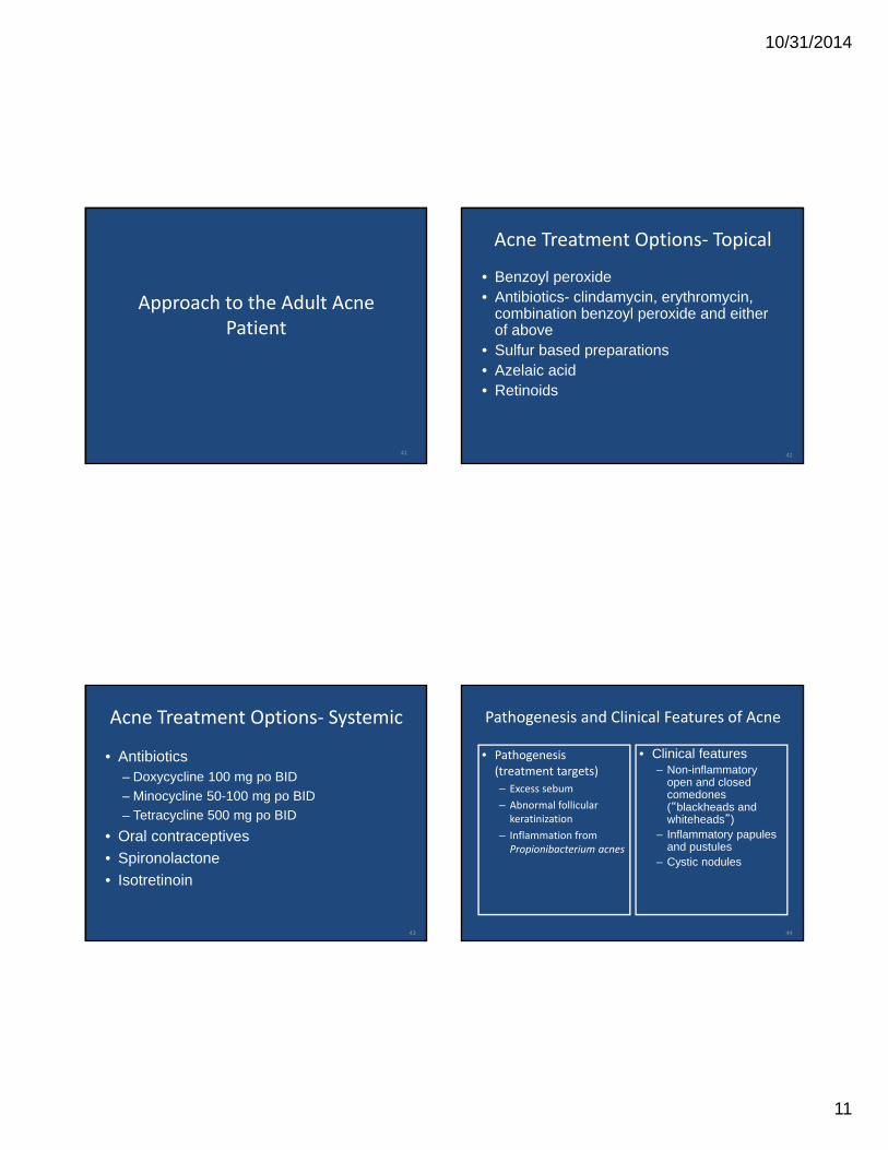
The red leg:Cellulitis and its (common) mimics
• Cellulitis/erysipelas
• Stasis dermatitis
• Contact dermatitis
Cellulitis• Infection of the dermis
• Gp A beta hemolytic strep and Staph aureus
• Rapidly spreading
• Erythematous, tender plaque, not fluctuant
• Patient often toxic
• WBC, LAD, streaking
• Rarely bilateral
• Treat tinea pedis
10/31/2014
14
Stasis Dermatitis
• Often bilateral, L>R
• Itchy and/or painful
• Red, hot, swollen leg
• No fever, elevated WBC, LAD, streaking
• Look for: varicosities, edema, venous ulceration, hemosiderin deposition
• Superimposed contact dermatitis common

10/31/2014
15
Contact Dermatitis
• Itch (no pain)
• Patient is non‐toxic
• Erythema and edema can be severe
• Look for sharp cutoff
• Treat with topical steroids
Contact Dermatitis• Common causes
– Applied antibiotics (Neomycin, Bacitracin)
– Topical anesthetics (benzocaine)
– Other (Vitamin E, topical benadryl)
• Avoid topical antibiotics to leg ulcers – Metronidazole OK (prevents odor)
The Red Leg: Key features of the physical exam:
Fever Pain Warmth Bilateral Streaking Lymphad-enopathy
Elevated WBC
Cellulitis Yes Yes Yes Almost never
Yes Yes Yes
Consider another diagnosis
No +/- +/- often No No No

10/31/2014
16
Drug Eruptions
61
Drug reactions:3 things you need to know
1. Type of drug reaction
2. Statistics:
– Which drugs are most likely to cause that type of reaction?
3. Timing:
– How long after the drug started did the reaction begin?
Case• 46 year old HIV+ man man admitted to ICU for r/o sepsis
• Severely hypotensive IV fluids, norepinephrine
• Sepsis? antibiotics are started
• At home has been taking trimethoprim/sulfamethoxazole for UTI
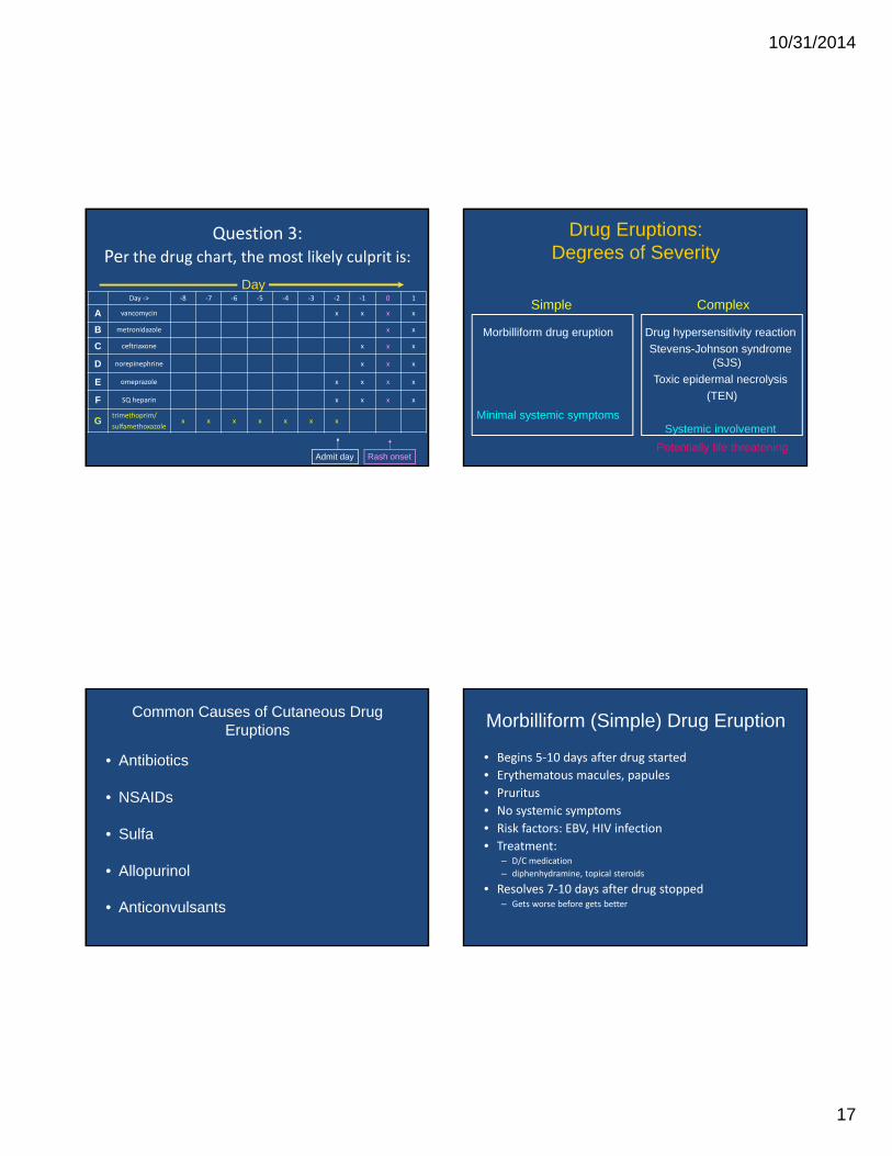
Question 3:Per the drug chart, the most likely culprit is:
DayDay ‐> ‐8 ‐7 ‐6 ‐5 ‐4 ‐3 ‐2 ‐1 0 1
A vancomycin x x x x
B metronidazole x x
C ceftriaxone x x x
D norepinephrine x x x
E omeprazole x x x x
F SQ heparin x x x x
Gtrimethoprim/
sulfamethoxazolex x x x x x x
Rash onsetAdmit day
10/31/2014
17
Question 3:Per the drug chart, the most likely culprit is:
DayDay ‐> ‐8 ‐7 ‐6 ‐5 ‐4 ‐3 ‐2 ‐1 0 1
A vancomycin x x x x
B metronidazole x x
C ceftriaxone x x x
D norepinephrine x x x
E omeprazole x x x x
F SQ heparin x x x x
Gtrimethoprim/
sulfamethoxazolex x x x x x x
Rash onsetAdmit day
Drug Eruptions: Degrees of Severity
Potentially life threatening
Morbilliform drug eruption
Minimal systemic symptoms
Drug hypersensitivity reaction
Stevens-Johnson syndrome (SJS)
Toxic epidermal necrolysis
(TEN)
Systemic involvement
Simple Complex
Common Causes of Cutaneous Drug Eruptions
• Antibiotics
• NSAIDs
• Sulfa
• Allopurinol
• Anticonvulsants
Morbilliform (Simple) Drug Eruption
• Begins 5‐10 days after drug started
• Erythematous macules, papules
• Pruritus
• No systemic symptoms
• Risk factors: EBV, HIV infection
• Treatment: – D/C medication
– diphenhydramine, topical steroids
• Resolves 7‐10 days after drug stopped– Gets worse before gets better

10/31/2014
18
Simple drug eruption‐ day 1 Simple drug eruption‐ day 3
Simple drug eruption‐ day 7 Hypersensitivity Reactions
• Skin eruption associated with systemic symptoms and alteration of internal organs
• “DRESS”‐ Drug reaction w/ eosinophilia and systemic symptoms
• “DIHS”= Drug induced hypersensitivity syndrome
• Begins 2‐ 6 weeks after medication started
– time to abnormally metabolize the medication
• May be role for HHV6
• Mortality 10‐25%

10/31/2014
19
Hypersensitivity Reactions
Drugs • Aromatic anticonvulsants
– phenobarbital, carbamazepine, phenytoin– THESE CROSS‐REACT
• Sulfonamides• Lamotrigine• Dapsone• Allopurinol (HLA‐B*5801)• NSAIDs• Other
– Abacavir (HLA‐ B*5701)– Nevirapine (HLA‐DRB1*0101)– Minocycline, metronidazole, azathioprine, gold salts
• Each class of drug causes a slightly different clinical picture
Hypersensitivity ReactionsClinical features
• Rash
• Fever (precedes eruption by day or more)
• Pharyngitis
• Hepatitis
• Arthralgias
• Lymphadenopathy
• Hematologic abnormalities– eosinophilia
– atypical lymphocytosis
• Other organs involved– myocarditis, interstitial pneumonitis, interstitial nephritis,
thyroiditis
10/31/2014
20
Hypersensitivity Reactions Treatment• Stop the medication• Follow CBC with diff, LFT’s, BUN/Cr• Avoid cross reacting medications!!!!
– Aromatic anticonvulsants cross react (70%)• Phenobarbital, Phenytoin, Carbamazepine• Valproic acid and Keppra generally safe
• Systemic steroids (Prednisone 1.5‐2mg/kg)– Taper slowly‐ 1‐3 months
• Allopurinol hypersensitivity may require steroid sparing agent
• NOT azathioprine (also metabolized by xanthine oxidase)
• Completely recover, IF the hepatitis resolves • Check TSH monthly for 6 months• Watch for later cardiac involvement (low EF)
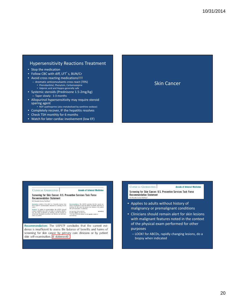
Skin Cancer
• Applies to adults without history of malignancy or premalignant conditions
• Clinicians should remain alert for skin lesions with malignant features noted in the context of the physical exam performed for other purposes
– LOOK! for ABCDs, rapidly changing lesions, do a biopsy when indicated
10/31/2014
21
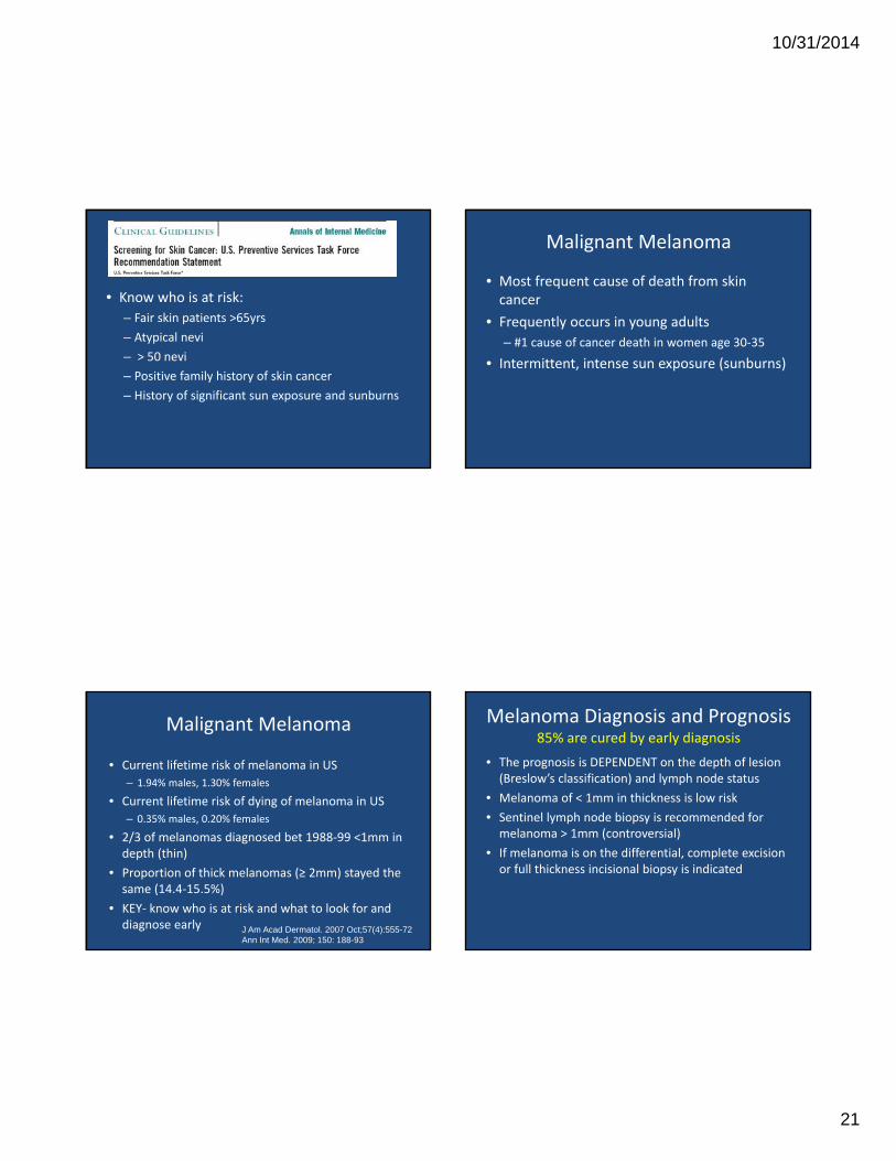
• Know who is at risk:
– Fair skin patients >65yrs
– Atypical nevi
– > 50 nevi
– Positive family history of skin cancer
– History of significant sun exposure and sunburns
Malignant Melanoma
• Most frequent cause of death from skin cancer
• Frequently occurs in young adults
– #1 cause of cancer death in women age 30‐35
• Intermittent, intense sun exposure (sunburns)
Malignant Melanoma
• Current lifetime risk of melanoma in US
– 1.94% males, 1.30% females
• Current lifetime risk of dying of melanoma in US
– 0.35% males, 0.20% females
• 2/3 of melanomas diagnosed bet 1988‐99 <1mm in depth (thin)
• Proportion of thick melanomas (≥ 2mm) stayed the same (14.4‐15.5%)
• KEY‐ know who is at risk and what to look for and diagnose early J Am Acad Dermatol. 2007 Oct;57(4):555-72
Ann Int Med. 2009; 150: 188-93
Melanoma Diagnosis and Prognosis85% are cured by early diagnosis
• The prognosis is DEPENDENT on the depth of lesion (Breslow’s classification) and lymph node status
• Melanoma of < 1mm in thickness is low risk
• Sentinel lymph node biopsy is recommended for melanoma > 1mm (controversial)
• If melanoma is on the differential, complete excision or full thickness incisional biopsy is indicated

10/31/2014
22
Malignant Melanoma
• Asymmetry
• Border
• Color
• Diameter
• Evolution

10/31/2014
23
Acral Melanoma
• Suspect in African American, Latino, Asian patients
Skin Cancers:What to Refer to Dermatology
• ANY suspicious pigmented lesion
• Any bleeding skin lesion
• Any red spot that doesn’t clear in 6‐8 weeks
• Any non‐healing erosion or ulceration
• Persons with greater than 50 moles, atypical moles, or family history of melanoma
• Fair‐skinned organ transplant recipients with prior sun exposure
10/31/2014
24
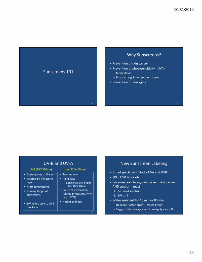
Sunscreens 101
93
Why Sunscreens?
• Prevention of skin cancer
• Prevention of photosensitivity (UVA)
– Medications
– Diseases: e.g. lupus erythematosus
• Prevention of skin aging
94
UV‐B and UV‐A
• Burning rays of the sun
• Filtered by the ozone layer
• Most carcinogenic
• Primary target of sunscreens
• SPF refers only to UVB blockade
• Tanning rays
• Aging rays
– a complete UVA blocker = anti‐aging cream
• Cause of medication related photosensitivity (e.g. HCTZ)
• Harder to block
UVB (290‐320nm) UVA (320‐400nm)
95
New Sunscreen Labeling
• Broad spectrum = blocks UVA and UVB
• SPF= UVB blockade
• For sunscreen to say can prevent skin cancer AND sunburn, must
1. be broad spectrum
2. SPF ≥ 15
• Water resistant for 40 min or 80 min
– No more “water proof”, “sweat proof”
– Suggests that always need to re‐apply every 2h96

10/31/2014
25
Chemical vs Physical Sunscreens
• Chemical sunscreens have UV absorbing chemicals– Benzophenone, Parsol 1789, Mexoryl, etc– Chemical UVA blockers are photo‐unstable (degrade)
• Stabilizers are now common (e.g. Helioplex)
• Physical sunscreens scatter or block UV rays– Zinc and titanium are physical blockers– More photostable – Block UVA well– Inelegant (white film)
97
How to Apply Sunscreen
• Put it on every morning before leaving the house– at least 20 min before sun exposure
• For heavy sun exposure: reapply 20 minutes after exposure begins
• Reapply every 2 hours or after swimming/sweating/towel‐drying
• Apply liberally – 1oz application= shot glass = covers the body
98
What to Tell Your Patients
• Use sunscreen, SPF ≥ 30 EVERYDAY• Avoid mid‐day sun/Short Shadow Seek Shade• Wear protective clothing (hats)• Put sunscreen on your children • Ask your doctor to check your skin lesions (most persons with melanoma have been seeing doctors regularly for years)
• Vitamin D Supplement for those at risk for osteoporosis who obey stringent sun‐protections practices • E.g. organ transplant patients
99
• The American Academy of Dermatology recommends that an adequate amount of vitamin D should be obtained from a healthy diet that includes foods naturally rich in vitamin D, foods/beverages fortified with vitamin D, and/or vitamin D supplements. Vitamin D should not be obtained from unprotected exposure to ultraviolet (UV) radiation.
• Unprotected UV exposure to the sun or indoor tanning devices is a known risk factor for the development of skin cancer.
• There is no scientifically validated, safe threshold level of UV exposure from the sun or indoor tanning devices that allows for maximal vitamin D synthesis without increasing skin cancer risk.
• To protect against skin cancer, a comprehensive photoprotective regimen, including the regular use and proper use of a broad-spectrum sunscreen, is recommended
100Taken from: American Academy of Dermatology website, 1/25/11

10/31/2014
26
A few simple rules to live by:
• Don’t use lotrisone!
• Never give systemic steroids for psoriasis or atopic dermatitis
• Do an excisional biopsy to diagnose melanoma
• Cellulitis is almost never bilateral
• Drug eruptions are usually due to medications started 7-10 prior to onset of the rash