Quantum Mechanical/Molecular Mechanical and Density...
9
Quantum Mechanical/Molecular Mechanical and Density Functional Theory Studies of a Prototypical Zinc Peptidase (Carboxypeptidase A) Suggest a General Acid-General Base Mechanism Dingguo Xu* ,† and Hua Guo* ,‡ MOE Key Laboratory of Green Chemistry & Technology, College of Chemistry, Sichuan UniVersity, Chengdu, Sichuan 610064, P. R. China, and Department of Chemistry and Chemical Biology, UniVersity of New Mexico, Albuquerque, New Mexico 87131 Received April 7, 2009; E-mail: [email protected]; [email protected] Abstract: Carboxypeptidase A is a zinc-containing enzyme that cleaves the C-terminal residue in a polypeptide substrate. Despite much experimental work, there is still a significant controversy concerning its catalytic mechanism. In this study, the carboxypeptidase A-catalyzed hydrolysis of the hippuryl-L-Phe molecule (k cat ) 17.7 ( 0.7 s -1 ) is investigated using both density functional theory and a hybrid quantum mechanical/molecular mechanical approach. The enzymatic reaction was found to proceed via a promoted- water pathway with Glu270 serving as the general base and general acid. Free-energy calculations indicate that the first nucleophilic addition step is rate-limiting, with a barrier of 17.9 kcal/mol. Besides activating the zinc-bound water nucleophile, the zinc cofactor also serves as an electrophilic catalyst that stabilizes the substrate carbonyl oxygen during the formation of the tetrahedral intermediate. In the Michaelis complex, Arg127, rather than Zn(II), is responsible for the polarization of the substrate carbonyl and it also serves as the oxyanion hole. As a result, its mutation leads to a higher free-energy barrier, in agreement with experimental observations. 1. Introduction Carboxypeptidase A (CPA), which contains a divalent zinc ion in its active site, is an important exopeptidase secreted by the pancreas for digesting intake proteins in the metabolism cycle. It catalyzes the elimination of the C-terminal amino acid via hydrolysis, with a preference toward residues with hydro- phobic side chains. 1 Although peptides are the intended substrates, CPA can also hydrolyze other unnatural substrates, including esters. CPA is a representative member of the zinc hydrolase superfamily and has served as a prototype for understanding binding and catalysis of such enzymes. 2,3 As a result, the corresponding enzymatic reactions have been exten- sively studied. Historically, much of the earlier work on CPA was motivated by its similarities in sequence and activity to the clinically important angiotensin-converting enzyme (ACE), 4 which plays an important role in the blood-pressure control mechanism, and its structure was not available until 2003. 5 Numerous high-resolution structures of CPA have been determined in its apo form and in complexes with various substrates, substrate analogues, and transition-state analogues. 6-21 The binding of ligands has been observed to induce a large conformational change in Tyr248, resulting in a hydrogen bond between the phenolic hydroxyl group and the terminal carboxy- late of the peptidyl substrate. 13 In addition, three CPA residues, namely Arg127, Asn144, and Arg145, also interact strongly with the same substrate carboxylate group as well as the carbonyl group in the peptide backbone. These interactions are believed to play an important role in both substrate binding and catalysis. † Sichuan University. ‡ University of New Mexico. (1) Vallee, B. L.; Galdes, A.; Auld, D. S.; Riordan, J. F. In Zinc Enzymes; Spiro, T. G., Ed.; Wiley: New York, 1983. (2) Lipscomb, W. N. Proc. Natl. Acad. Sci. U.S.A. 1980, 77, 3875–3878. (3) Christianson, D. W.; Lipscomb, W. N. Acc. Chem. Res. 1989, 22, 62–69. (4) Ondetti, M. A.; Cushman, D. W. Annu. ReV. Biochem. 1982, 51, 283. (5) Natesh, R.; Schwager, S. L. U.; Sturrock, E. D.; Acharya, K. R. Nature 2003, 421, 551–554. (6) Rees, D. C.; Lipscomb, W. N. J. Mol. Biol. 1982, 160, 475–498. (7) Rees, D. C.; Lipscomb, W. N. Proc. Natl. Acad. Sci. U.S.A. 1983, 80, 7151–7154. (8) Rees, D. C.; Lipscomb, W. N. J. Mol. Biol. 1983, 168, 367–387. (9) Christianson, D. W.; Lipscomb, W. N. Proc. Natl. Acad. Sci. U.S.A. 1985, 82, 6840–6844. (10) Christianson, D. W.; Kuo, L. C.; Lipscomb, W. N. J. Am. Chem. Soc. 1985, 107, 8281–8283. (11) Christianson, D. W.; Lipscomb, W. N. J. Am. Chem. Soc. 1986, 108, 545–546. (12) Christianson, D. W.; Lipscomb, W. N. J. Am. Chem. Soc. 1986, 108, 4998–5003. (13) Christianson, D. W.; Lipscomb, W. N. Proc. Natl. Acad. Sci. U.S.A. 1986, 83, 7568–7572. (14) Christianson, D. W.; Lipscomb, W. N. J. Am. Chem. Soc. 1987, 109, 5536–5538. (15) Christianson, D. W.; Lipscomb, W. N. J. Am. Chem. Soc. 1988, 110, 5560–5565. (16) Kim, H.; Lipscomb, W. N. Biochemistry 1990, 29, 5546–5555. (17) Mangani, S.; Carloni, P.; Orioli, P. J. Mol. Biol. 1992, 223, 573–578. (18) Teplyakov, A.; Wilson, K. S. Acta Crystallogr. 1993, D49, 534–540. (19) Greenblatt, H. M.; Feinberg, H.; Tucker, P. A.; Shoham, G. Acta Crystallogr. 1998, D54, 289–305. (20) Cho, J. H.; Kim, D. H.; Chung, S. J.; Ha, N.-C.; Oh, B.-H.; Choi, K. Y. Bioorg. Med. Chem. 2002, 10, 2015–2022. (21) Kilshtain-Vardi, A.; Glick, M.; Greenblatt, H. M.; Goldblum, A.; Shoham, G. Acta Crystallogr. 2003, D59, 323–333. Published on Web 06/24/2009 10.1021/ja9027988 CCC: $40.75 2009 American Chemical Society 9780 9 J. AM. CHEM. SOC. 2009, 131, 9780–9788
Transcript of Quantum Mechanical/Molecular Mechanical and Density...

Quantum Mechanical/Molecular Mechanical and DensityFunctional Theory Studies of a Prototypical Zinc Peptidase
(Carboxypeptidase A) Suggest a General Acid-General BaseMechanism
Dingguo Xu*,† and Hua Guo*,‡
MOE Key Laboratory of Green Chemistry & Technology, College of Chemistry, SichuanUniVersity, Chengdu, Sichuan 610064, P. R. China, and Department of Chemistry and
Chemical Biology, UniVersity of New Mexico, Albuquerque, New Mexico 87131
Received April 7, 2009; E-mail: [email protected]; [email protected]
Abstract: Carboxypeptidase A is a zinc-containing enzyme that cleaves the C-terminal residue in apolypeptide substrate. Despite much experimental work, there is still a significant controversy concerningits catalytic mechanism. In this study, the carboxypeptidase A-catalyzed hydrolysis of the hippuryl-L-Phemolecule (kcat ) 17.7 ( 0.7 s-1) is investigated using both density functional theory and a hybrid quantummechanical/molecular mechanical approach. The enzymatic reaction was found to proceed via a promoted-water pathway with Glu270 serving as the general base and general acid. Free-energy calculations indicatethat the first nucleophilic addition step is rate-limiting, with a barrier of 17.9 kcal/mol. Besides activating thezinc-bound water nucleophile, the zinc cofactor also serves as an electrophilic catalyst that stabilizes thesubstrate carbonyl oxygen during the formation of the tetrahedral intermediate. In the Michaelis complex,Arg127, rather than Zn(II), is responsible for the polarization of the substrate carbonyl and it also servesas the oxyanion hole. As a result, its mutation leads to a higher free-energy barrier, in agreement withexperimental observations.
1. Introduction
Carboxypeptidase A (CPA), which contains a divalent zincion in its active site, is an important exopeptidase secreted bythe pancreas for digesting intake proteins in the metabolismcycle. It catalyzes the elimination of the C-terminal amino acidvia hydrolysis, with a preference toward residues with hydro-phobic side chains.1 Although peptides are the intendedsubstrates, CPA can also hydrolyze other unnatural substrates,including esters. CPA is a representative member of the zinchydrolase superfamily and has served as a prototype forunderstanding binding and catalysis of such enzymes.2,3 As aresult, the corresponding enzymatic reactions have been exten-sively studied. Historically, much of the earlier work on CPAwas motivated by its similarities in sequence and activity tothe clinically important angiotensin-converting enzyme (ACE),4
which plays an important role in the blood-pressure controlmechanism, and its structure was not available until 2003.5
Numerous high-resolution structures of CPA have beendetermined in its apo form and in complexes with varioussubstrates, substrate analogues, and transition-state analogues.6-21
The binding of ligands has been observed to induce a largeconformational change in Tyr248, resulting in a hydrogen bondbetween the phenolic hydroxyl group and the terminal carboxy-late of the peptidyl substrate.13 In addition, three CPA residues,namely Arg127, Asn144, and Arg145, also interact strongly withthe same substrate carboxylate group as well as the carbonylgroup in the peptide backbone. These interactions are believedto play an important role in both substrate binding and catalysis.
† Sichuan University.‡ University of New Mexico.
(1) Vallee, B. L.; Galdes, A.; Auld, D. S.; Riordan, J. F. In Zinc Enzymes;Spiro, T. G., Ed.; Wiley: New York, 1983.
(2) Lipscomb, W. N. Proc. Natl. Acad. Sci. U.S.A. 1980, 77, 3875–3878.(3) Christianson, D. W.; Lipscomb, W. N. Acc. Chem. Res. 1989, 22,
62–69.(4) Ondetti, M. A.; Cushman, D. W. Annu. ReV. Biochem. 1982, 51, 283.(5) Natesh, R.; Schwager, S. L. U.; Sturrock, E. D.; Acharya, K. R. Nature
2003, 421, 551–554.
(6) Rees, D. C.; Lipscomb, W. N. J. Mol. Biol. 1982, 160, 475–498.(7) Rees, D. C.; Lipscomb, W. N. Proc. Natl. Acad. Sci. U.S.A. 1983, 80,
7151–7154.(8) Rees, D. C.; Lipscomb, W. N. J. Mol. Biol. 1983, 168, 367–387.(9) Christianson, D. W.; Lipscomb, W. N. Proc. Natl. Acad. Sci. U.S.A.
1985, 82, 6840–6844.(10) Christianson, D. W.; Kuo, L. C.; Lipscomb, W. N. J. Am. Chem. Soc.
1985, 107, 8281–8283.(11) Christianson, D. W.; Lipscomb, W. N. J. Am. Chem. Soc. 1986, 108,
545–546.(12) Christianson, D. W.; Lipscomb, W. N. J. Am. Chem. Soc. 1986, 108,
4998–5003.(13) Christianson, D. W.; Lipscomb, W. N. Proc. Natl. Acad. Sci. U.S.A.
1986, 83, 7568–7572.(14) Christianson, D. W.; Lipscomb, W. N. J. Am. Chem. Soc. 1987, 109,
5536–5538.(15) Christianson, D. W.; Lipscomb, W. N. J. Am. Chem. Soc. 1988, 110,
5560–5565.(16) Kim, H.; Lipscomb, W. N. Biochemistry 1990, 29, 5546–5555.(17) Mangani, S.; Carloni, P.; Orioli, P. J. Mol. Biol. 1992, 223, 573–578.(18) Teplyakov, A.; Wilson, K. S. Acta Crystallogr. 1993, D49, 534–540.(19) Greenblatt, H. M.; Feinberg, H.; Tucker, P. A.; Shoham, G. Acta
Crystallogr. 1998, D54, 289–305.(20) Cho, J. H.; Kim, D. H.; Chung, S. J.; Ha, N.-C.; Oh, B.-H.; Choi,
K. Y. Bioorg. Med. Chem. 2002, 10, 2015–2022.(21) Kilshtain-Vardi, A.; Glick, M.; Greenblatt, H. M.; Goldblum, A.;
Shoham, G. Acta Crystallogr. 2003, D59, 323–333.
Published on Web 06/24/2009
10.1021/ja9027988 CCC: $40.75 2009 American Chemical Society9780 9 J. AM. CHEM. SOC. 2009, 131, 9780–9788

In particular, Arg127 is hydrogen-bonded with the scissilecarbonyl oxygen, serving as the oxyanion hole to stabilize thetetrahedral intermediate.15,22,23 Furthermore, the terminal sidechain of the substrate is surrounded by a hydrophobic pocketin the so-called S1′ subsite, which is presumably responsiblefor the preference of CPA for aromatic terminal residues. Inthe catalytic S1 subsite, the zinc cofactor is coordinated by threeprotein ligands, namely His196, Glu72, and His69, as well asa water molecule. A more recently determined high-resolutionstructure of CPA indicates that the zinc-bound water is mostlikely in its neutral state,21 presumably due to its hydrogen bondwith Glu270.
Despite extensive experimental studies, there is still noconsensus on the catalytic mechanism of CPA.2,3 Two majorclasses of mechanisms have been proposed, and they aredepicted in Scheme 1. The anhydride mechanism envisages anacyl-enzyme intermediate resulting from direct nucleophilicattack of the scissile carbonyl carbon by the carboxylate sidechain of Glu270, which is subsequently hydrolyzed by waterto form the product. The alternative promoted-water mechanismstipulates instead a tetrahedral intermediate formed by nucleo-philic attack of the zinc-bound water, activated by proton transferto Glu270. The tetrahedral intermediate subsequently collapsesupon protonation of the nitrogen leaving group by the Glu270side chain. In a classical example of electrostatic catalysis,24
Glu270 serves as a general base in the nucleophilic additionstep and a general acid in the elimination step. Apparently, theroles played by the metal cofactor and by the key residue(Glu270) are quite different in the two mechanisms.
Experimental evidence supporting both mechanisms has beenpresented by several groups. In support of the promoted-water
mechanism, Breslow and Wernick have shown in an 18O isotopelabeling experiment that the acyl-enzyme intermediate isunlikely to form in the hydrolysis of peptide substrates.25,26
Later, cryospectrokinetic studies by Auld and co-workers27,28
ruled out the existence of the acyl-enzyme intermediate inhydrolysis of both peptides and esters. However, there have alsobeen reports of accumulating acyl-enzyme intermediates at lowtemperatures in ester hydrolysis catalyzed by CPA,29-33 al-though some have questioned the correct identity of theintermediate.34 It has been suggested that the hydrolysismechanism might be different for ester and peptide substrates,because the former substrates typically bind with direct metalcoordination by the scissile carbonyl, while such an interactionis considered nonproductive for the latter.3 In 1998, Lee et al.35
reported the detection of an anhydride intermediate in the CPA-catalyzed hydrolysis of glycyl-L-tyrosine by low-temperaturesolid-state NMR. However, it is worth noting that this substratehas a very slow hydrolysis rate.36 In addition, the X-ray structureof the Michaelis complex indicates the absence of zinc-bound
(22) Phillips, M. A.; Fletterick, R.; Rutter, W. J. J. Biol. Chem. 1990, 265,20692–20698.
(23) Phillips, M. A.; Rutter, W. J. Protein Sci. 1992, 1, 517–521.(24) Jencks, W. P. Catalysis in Chemistry and Enzymology; Dover: New
York, 1986.
(25) Breslow, R.; Wernick, D. L. J. Am. Chem. Soc. 1976, 98, 259–261.(26) Breslow, R.; Wernick, D. L. Proc. Natl. Acad. Sci. U.S.A. 1977, 74,
1303–1307.(27) Auld, D. S.; Galdes, A.; Geoghegan, K. F.; Holmquit, B.; Martinelli,
R. A.; Vallee, B. L. Proc. Natl. Acad. Sci. U.S.A. 1984, 81, 5041–5045.
(28) Geoghegan, K. F.; Galdes, A.; Hanson, G.; Holmquit, B.; Auld, D. S.;Vallee, B. L. Biochemistry 1986, 25, 4669–4674.
(29) Makinen, M. W.; Kuo, L. C.; Dymowski, J. J.; Jaffer, S. J. Biol. Chem.1979, 254, 356–366.
(30) Kuo, L. C.; Makinen, M. W. J. Biol. Chem. 1982, 257, 24–27.(31) Suh, J.; Cho, W.; Chung, S. J. Am. Chem. Soc. 1985, 107, 4530–
4535.(32) Sander, M. E.; Witzel, H. Biochem. Biophys. Res. Commun. 1985,
132, 681.(33) Britt, B. M.; Peticolas, W. L. J. Am. Chem. Soc. 1992, 114, 5295–
5303.(34) Hoffman, S. J.; Chu, S. S.-T.; Lee, H.-H.; Kaiser, E. T.; Carey, P. R.
J. Am. Chem. Soc. 1983, 105, 6971–6973.(35) Lee, H. C.; Ko, Y. H.; Baek, S. B.; Kim, D. H. Bioorg. Med. Chem.
Lett. 1998, 8, 3379–3384.
Scheme 1. Two Putative Mechanisms for CPA Catalysis
J. AM. CHEM. SOC. 9 VOL. 131, NO. 28, 2009 9781
QM/MM and DFT Studies of Catalytic Mechanism of Carboxypeptidase A A R T I C L E S
water, apparently displaced by the substrate.13 This controversyis still far from fully resolved.
Given the experimental difficulties in distinguishing the twomechanisms, it is desirable to address the mechanistic issuestheoretically. Indeed, attempts have been made in the pastin this direction. For instance, molecular dynamics (MD)simulations of CPA have been reported by Banci et al.,37 whoshowed that the active-site structure does not exclude eithermechanism. Quantum chemical investigations on biological zincand its coordination can be traced back to the earlier work ofPullman38 and of Allen.39 For CPA catalysis, most quantumchemical studies have been performed with semiempiricalmethods.40-44 These theoretical studies focused mostly on thepromoted-water mechanism, but Vardi-Kilshtain et al.44 havealso presented some evidence against the anhydride mechanism.Although these pioneering studies provided valuable insights,they suffer from two major shortcomings. First, the validity ofsemiempirical models requires careful validation by higher-leveltheory, particularly when a transition metal is involved. Second,the quantum chemical studies did not take into considerationthe protein environment, essential for catalysis. The enzymaticreaction is perhaps best modeled using a hybrid quantummechanical and molecular mechanical (QM/MM) method,45-49
in which the enzyme active site is treated quantum mechanicallywhile the remainder of the protein is modeled with a MM forcefield. Indeed, such QM/MM studies have been reported for CPA,but only for its inhibition.50,51
In this publication, we examine the two mechanistic proposalsusing a hybrid QM/MM approach. In particular, the QM regionis described by an approximate density functional approach,namely the self-consistent charge-density functional tight bind-ing (SCC-DFTB) method,52 while the MM region is describedby the CHARMM force field. The SCC-DFTB Hamiltonian hasbeen parametrized for biological zinc ions,53 and the combinedSCC-DFTB/CHARMM approach54 has been shown to give areasonably accurate description of several zinc enzymes.55-61
In addition to the QM/MM studies, we also investigate thereaction pathways in truncated active-site models with high-level density functional theory (DFT) calculations in order toverify the validity of the conclusions reached by the QM/MMmodels.
2. Methods
2.1. QM/MM Models. The hybrid quantum mechanical andmolecular mechanical approach62 has been extensively applied tostudy enzymatic reaction mechanisms.45-49 Such a method has theadvantage that a very large system such as an enzyme can beinvestigated with manageable computation costs. The basic ideaof the QM/MM scheme is to divide the system into two parts: thesmaller QM region, where the chemical bond breaking and formingtake place, while the surrounding region is described by a MMforce field.
In the work reported here, we used the self-consistent charge-density functional tight binding (SCC-DFTB) model52 to describethe QM region and the CHARMM all-atom force field63 tocharacterize the MM region. The SCC-DFTB method is based ona second-order expansion of the total DFT energy with respect tothe charge-density variation. It is much more efficient than ab initioQM/MM approaches and consequently amenable to free-energycalculations. The efficiency is essential for metallo-enzymes becausethe active site is often much too large for an accurate ab initioQM/MM free-energy simulation. The SCC-DFTB model has beenextensively tested for enzyme systems,48,54,64,65 including activesites with zinc cofactors.53 The QM/MM approach based on theSCC-DFTB method has been shown to give a satisfactory descrip-tion of several zinc enzymes, including carbonic anhydrase55,56 and�-lactamases.57-61
The starting point of the simulation was selected from anenzyme-inhibitor complex structure (PDB code 6CPA), whichis the wild-type (WT) bovine CPA complexed with the O-[[(1R)-[N-phenylmethoxycarbonyl)-L-alanyl]amino]ethyl]hydroxyphos-phinyl]-L-3-phenyllactate (ZAAP(O)F) inhibitor.16 This structurewas chosen because of the similarities between the inhibitor and abona fide substrate of CPA, namely hippuryl-L-Phe (HPA, Scheme2).66 The HPA substrate was recovered manually by modifyingthe ZAAp(O)F inhibitor in the X-ray structure. A water moleculewas added in the active site for studying the promoted-waterpathway. Hydrogen atoms were added using the HBUILD utilityin CHARMM, and the titratable residues in the enzymes wereassigned the appropriate ionization states at pH ) 7. In addition,the disulfide bond between Cys138 and Cys161 was enforced. Allsimulations were performed with the CHARMM suite of simulationcodes.67
The resulting structure was then solvated by a pre-equilibratedsphere of TIP3P waters68 with 25 Å radius centered at the zinc
(36) Quiocho, F. A.; Lipscomb, W. M. AdV. Protein Chem. 1971, 25, 1–78.
(37) Banci, L.; Bertini, I.; La Penna, G. Proteins 1994, 18, 186–197.(38) Demoulin, D.; Pullman, A. Theo. Chim. Acta 1978, 49, 161.(39) Kitchen, D. B.; Allen, L. C. J. Phys. Chem. 1989, 93, 7265.(40) Alex, A.; Clark, T. J. Comput. Chem. 1992, 13, 704–717.(41) Alvarez-Santos, S.; Gonzalez-Lafont, A.; Lluch, J. M. Can. J. Chem.
1994, 72, 2077.(42) Alvarez-Santos, S.; Gonzalez-Lafont, A.; Lluch, J. M.; Oliva, B.;
Aviles, F. X. New J. Chem. 1998, 319–326.(43) Kilshtain-Vardi, A.; Shoham, G.; Goldblum, A. Int. J. Quantum Chem.
2002, 88, 87–98.(44) Vardi-Kilshtain, A.; Shoham, G.; Goldblum, A. Mol. Phys. 2003, 101,
2715–2724.(45) Gao, J. In ReViews in Computational Chemistry; Lipkowitz, K. B.,
Boyd, D. B., Eds.; VCH: New York, 1996; Vol. 7, pp 119-185.(46) Warshel, A. Annu. ReV. Biophys. Biomol. Struct. 2003, 32, 425–443.(47) Zhang, Y. Theor. Chem. Acc. 2006, 116, 43–50.(48) Riccardi, D.; Schaefer, P.; Yang, Y.; Yu, H.; Ghosh, N.; Prat-Resina,
X.; Konig, P.; Li, G.; Xu, D.; Guo, H.; Elstener, M.; Cui, Q. J. Phys.Chem. B 2006, 110, 6458–6469.
(49) Hu, H.; Yang, W. Annu. ReV. Phys. Chem. 2008, 59, 573–601.(50) Phoon, L.; Burton, N. A. J. Mol. Graph. Model. 2005, 24, 94–101.(51) Cross, J. B.; Vreven, T.; Meroueh, S. O.; Mobashery, S.; Schlegel,
H. B. J. Phys. Chem. B 2005, 109, 4761–4769.(52) Elstner, M.; Porezag, D.; Jungnickel, G.; Elsner, J.; Haugk, M.;
Frauenheim, T.; Suhai, S.; Seigert, G. Phys. ReV. 1998, B58, 7260–7268.
(53) Elstner, M.; Cui, Q.; Munih, P.; Kaxiras, E.; Frauenheim, T.; Karplus,M. J. Comput. Chem. 2003, 24, 565–581.
(54) Cui, Q.; Elstner, M.; Kaxiras, E.; Frauenheim, T.; Karplus, M. J. Phys.Chem. B 2001, 105, 569–585.
(55) Riccardi, D.; Cui, Q. J. Phys. Chem. A 2007, 111, 5703–5711.
(56) Riccardi, D.; Konig, P.; Guo, H.; Cui, Q. Biochemistry 2008, 47, 2369–2378.
(57) Xu, D.; Zhou, Y.; Xie, D.; Guo, H. J. Med. Chem. 2005, 48, 6679–6689.
(58) Xu, D.; Xie, D.; Guo, H. J. Biol. Chem. 2006, 281, 8740–8747.(59) Xu, D.; Guo, H.; Cui, Q. J. Phys. Chem. A 2007, 111, 5630–5636.(60) Xu, D.; Guo, H.; Cui, Q. J. Am. Chem. Soc. 2007, 129, 10814.(61) Wang, C.; Guo, H. J. Phys. Chem. B 2007, 111, 9986–9992.(62) Warshel, A.; Levitt, M. J. Mol. Biol. 1976, 103, 227–249.(63) MacKerell, A. D., Jr.; et al. J. Phys. Chem. B 1998, 102, 3586–3616.(64) Sattelmeyer, K. W.; Tirado-Rives, J.; Jorgensen, W. L. J. Phys. Chem.
A 2006, 110, 13551–13559.(65) Otte, N.; Scholten, M.; Thiel, W. J. Phys. Chem. A 2007, 111, 5751–
5755.(66) Gardell, S. J.; Craik, C. S.; Hilvert, D.; Urdea, M. S.; Rutter, W. J.
Nature 1985, 317, 551–555.(67) Brooks, B. R.; Bruccoleri, R. E.; Olafson, B. D.; States, D. J.;
Swaminathan, S.; Karplus, M. J. Comput. Chem. 1983, 4, 187–217.(68) Jorgensen, W. L.; Chandrasekhar, J.; Madura, J. D.; Impey, R. W.;
Klein, M. L. J. Chem. Phys. 1983, 79, 926–935.
9782 J. AM. CHEM. SOC. 9 VOL. 131, NO. 28, 2009
A R T I C L E S Xu and Guo

ion, followed by a 30 ps MD simulation with all protein andsubstrate atoms fixed. This process was repeated several times withrandomly rotated water spheres to ensure uniform solvation.Subsequently, stochastic boundary conditions69 were applied to thesolvated enzyme system. In particular, atoms that are 25 Å awayfrom the origin (Zn) were removed, while atoms in the buffer zone(22 < r < 25 Å) were subjected to Langevin dynamics with frictionand random forces, as well as additional harmonic restraining forces.In the inner reaction zone (r < 22 Å), the atoms follow Newtoniandynamics on the hybrid QM/MM potential energy hypersurface.A group-based switching scheme was used for nonbonded interac-tions.70
The QM region includes the zinc ion, the side-chain atoms ofits three protein ligands His69, Glu72, and His196, the zinc-boundwater, the side-chain atoms of Arg127 and Glu270, and the entiresubstrate molecule. The QM boundaries were treated with the linkatom approach,71 in which a H atom is added to each C� of therelevant amino acid. The QM region has a total of 125 atoms. Inthe R127A mutant calculations, the side chain of Arg127 is replacedby a methyl group for Ala. Furthermore, the water nucleophile wasremoved in studying the anhydride pathway.
To gain insight about the enzyme-substrate (ES) complex, wehave carried out extensive MD simulations of the hybrid QM/MMsystem. The starting system was brought to room temperature (300K) in 30 ps, followed by room temperature MD for another 70 psto ensure the system is sufficiently equilibrated. The final 500 psMD trajectory was used to for analysis. The integration step had a1.0 fs time interval, and the SHAKE algorithm72 was applied tomaintain all covalent bonds-involved hydrogen atoms.
Several random snapshots were picked from the MD trajectoryas the initial configurations for minimal energy path calculations.For the promoted-water pathway, two putative reaction coordinatesare defined as follows. The reaction coordinate for the nucleophilicaddition (NA) of the water nucleophile is given by the distancebetween the water oxygen (Ow) and the substrate carbonyl carbon(C6): r1 ) dOw · · ·C6
. The corresponding reaction coordinate for theelimination (E) of the leaving group is given by the antisymmetricstretching coordinate of the proton (H1) between Oε2 of the generalbase (Glu270) and the substrate backbone amide nitrogen (N9): r2
) dOε2 · · ·H1- dN9 · · ·H1
. For the anhydride pathway, the NA reactioncoordinate is given by the distance between a Glu270 oxygen andthe substrate carbonyl carbon: r′1 ) dOε2 · · ·C6
. The reaction pathswere determined by adiabatic mapping along the putative reactioncoordinates.
To include the fluctuation and reorganization of the enzymaticsystem, we further computed potentials of mean force (PMFs) forthe two mechanistic pathways. The minimum energy structuresalong a putative reaction coordinate were used as the initial pointsfor the PMF calculations. The PMF calculations used umbrellasampling73 with harmonic constraints of 100-200 kcal/mol ·Å2.For the NA and E steps of the promoted-water pathway, 12 and 9windows were used, respectively. On the other hand, the anhydridePMF was obtained with 17 simulation windows.
In the PMF calculations, 60 ps equilibration simulations werefirst performed to bring the system to 300 K. The distributionfunction in the reaction coordinate was then collected in thesubsequent 40 ps. The final PMF was obtained using the weightedhistogram analysis method (WHAM).74 During the PMF simula-tions, the SHAKE module was applied to all hydrogen atoms excepthydrogen atoms on the zinc-bound water molecule.
2.2. DFT and Truncated Active-Site Models. Given thesemiempirical nature of the SCC-DFTB Hamiltonian, it is importantto provide an independent check of the results obtained by theQM/MM approach. In this work, we examined the reaction pathsin several truncated active-site models using high-level DFT. Thetruncated active-site models consist of the zinc ion, the nucleophilicwater for the promoted-water pathway, analogues of Glu270 andthe three protein ligands of Zn(II), and the substrate analogue. Inaddition, we have included the side chains of Arg127 and Arg145in our model to simulate the binding mode of C-terminal carboxylategroup. For the substrate analogue, the phenol groups were removedto reduce computational costs. The total number of atoms is 98 forthe promoted-water pathway and 74 for the anhydride pathway.
The B3LYP exchange-correlation functional75,76 and a standardbasis set of 6-31G(d) were used in fully geometrical optimizationof stationary points, followed by harmonic vibrational frequenciescalculations for confirmation. These stationary points were thenconnected by intrinsic reaction coordinate (IRC) method.77 Thesolvent effects were studied with the PCM model78 with dielectricconstants of water (ε ) 80) and protein (ε ) 5). All of thecomputations were carried out using the Gaussian 03 suite ofprograms.79
3. Results
3.1. The Promoted-Water Pathway. 3.1.1. Enzyme-Substrate Complex. A 600 ps MD simulation was carried out forthe ES complex using the hybrid QM/MM Hamiltonian. Figure 1displays the root-mean-square deviation (rmsd) for the backboneatoms, which increases in the first 100 ps and then becomesrelatively stable for the remaining 500 ps of simulation, with the
(69) Brooks, C. L., III; Karplus, M. J. Mol. Biol. 1989, 208, 159–181.(70) Steinbach, P. J.; Brooks, B. R. J. Comput. Chem. 1994, 15, 667.(71) Field, M. J.; Bash, P. A.; Karplus, M. J. Comput. Chem. 1990, 11,
700–733.(72) Ryckaert, J. P.; Ciccotti, G.; Berendsen, H. J. J. Comput. Phys. 1977,
23, 327–341.
(73) Torrie, G. M.; Valleau, J. P. J. Comput. Phys. 1977, 23, 187–199.(74) Kumar, S.; Bouzida, D.; Swendsen, R. H.; Kollman, P. A.; Rosenberg,
J. M. J. Comput. Chem. 1992, 13, 1011–1021.(75) Becke, A. D. J. Chem. Phys. 1993, 98, 5648–5652.(76) Lee, C.; Yang, W.; Parr, R. G. Phys. ReV. B 1988, 37, 785–789.(77) Gonzalez, C.; Schlegel, H. B. J. Phys. Chem. 1990, 94, 5523.(78) Tomasi, J.; Persico, M. Chem. ReV. 1994, 94, 2027–2094.(79) Frisch, M. J.; et al. Gaussian 03; Gaussian, Inc.: Pittsburgh, PA, 2003.
Scheme 2. Atom Definition and Active-Site Interaction Pattern forthe ES Complex of CPA
Figure 1. Rmsd for the QM/MM MD simulation of the ES complex ofCPA.
J. AM. CHEM. SOC. 9 VOL. 131, NO. 28, 2009 9783
QM/MM and DFT Studies of Catalytic Mechanism of Carboxypeptidase A A R T I C L E S
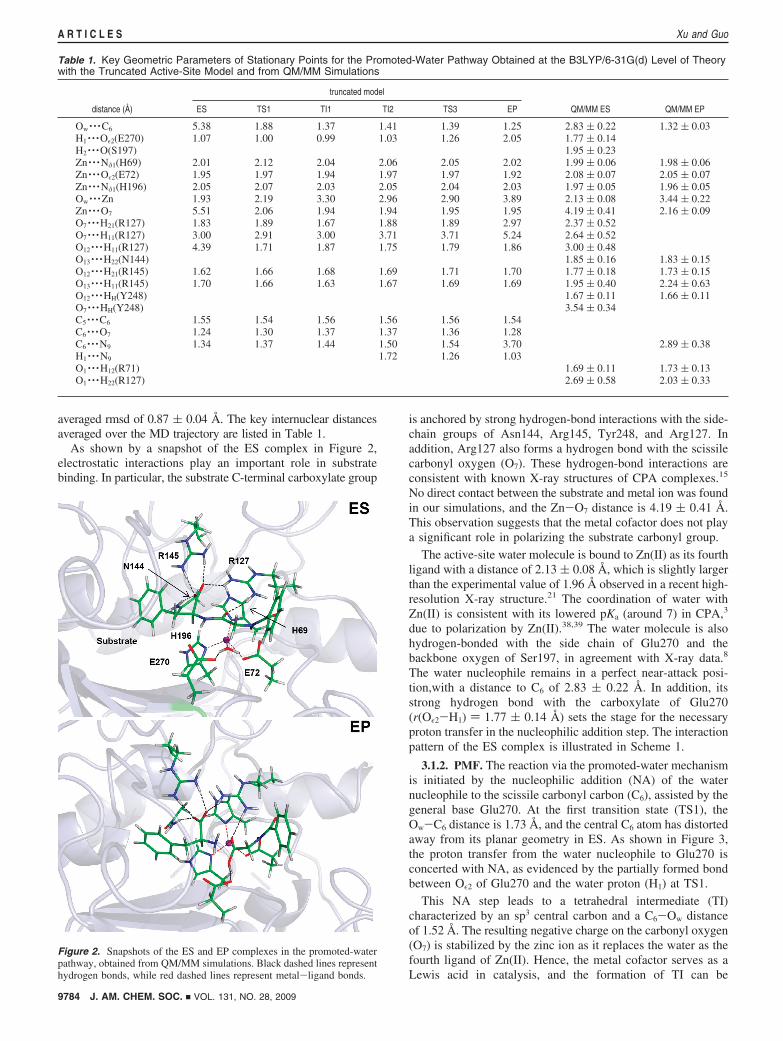
averaged rmsd of 0.87 ( 0.04 Å. The key internuclear distancesaveraged over the MD trajectory are listed in Table 1.
As shown by a snapshot of the ES complex in Figure 2,electrostatic interactions play an important role in substratebinding. In particular, the substrate C-terminal carboxylate group
is anchored by strong hydrogen-bond interactions with the side-chain groups of Asn144, Arg145, Tyr248, and Arg127. Inaddition, Arg127 also forms a hydrogen bond with the scissilecarbonyl oxygen (O7). These hydrogen-bond interactions areconsistent with known X-ray structures of CPA complexes.15
No direct contact between the substrate and metal ion was foundin our simulations, and the Zn-O7 distance is 4.19 ( 0.41 Å.This observation suggests that the metal cofactor does not playa significant role in polarizing the substrate carbonyl group.
The active-site water molecule is bound to Zn(II) as its fourthligand with a distance of 2.13 ( 0.08 Å, which is slightly largerthan the experimental value of 1.96 Å observed in a recent high-resolution X-ray structure.21 The coordination of water withZn(II) is consistent with its lowered pKa (around 7) in CPA,3
due to polarization by Zn(II).38,39 The water molecule is alsohydrogen-bonded with the side chain of Glu270 and thebackbone oxygen of Ser197, in agreement with X-ray data.8
The water nucleophile remains in a perfect near-attack posi-tion,with a distance to C6 of 2.83 ( 0.22 Å. In addition, itsstrong hydrogen bond with the carboxylate of Glu270(r(Oε2-H1) ) 1.77 ( 0.14 Å) sets the stage for the necessaryproton transfer in the nucleophilic addition step. The interactionpattern of the ES complex is illustrated in Scheme 1.
3.1.2. PMF. The reaction via the promoted-water mechanismis initiated by the nucleophilic addition (NA) of the waternucleophile to the scissile carbonyl carbon (C6), assisted by thegeneral base Glu270. At the first transition state (TS1), theOw-C6 distance is 1.73 Å, and the central C6 atom has distortedaway from its planar geometry in ES. As shown in Figure 3,the proton transfer from the water nucleophile to Glu270 isconcerted with NA, as evidenced by the partially formed bondbetween Oε2 of Glu270 and the water proton (H1) at TS1.
This NA step leads to a tetrahedral intermediate (TI)characterized by an sp3 central carbon and a C6-Ow distanceof 1.52 Å. The resulting negative charge on the carbonyl oxygen(O7) is stabilized by the zinc ion as it replaces the water as thefourth ligand of Zn(II). Hence, the metal cofactor serves as aLewis acid in catalysis, and the formation of TI can be
Table 1. Key Geometric Parameters of Stationary Points for the Promoted-Water Pathway Obtained at the B3LYP/6-31G(d) Level of Theorywith the Truncated Active-Site Model and from QM/MM Simulations
truncated model
distance (Å) ES TS1 TI1 TI2 TS3 EP QM/MM ES QM/MM EP
Ow · · ·C6 5.38 1.88 1.37 1.41 1.39 1.25 2.83 ( 0.22 1.32 ( 0.03H1 · · ·Oε2(E270) 1.07 1.00 0.99 1.03 1.26 2.05 1.77 ( 0.14H2 · · ·O(S197) 1.95 ( 0.23Zn · · ·Nδ1(H69) 2.01 2.12 2.04 2.06 2.05 2.02 1.99 ( 0.06 1.98 ( 0.06Zn · · ·Oε2(E72) 1.95 1.97 1.94 1.97 1.97 1.92 2.08 ( 0.07 2.05 ( 0.07Zn · · ·Nδ1(H196) 2.05 2.07 2.03 2.05 2.04 2.03 1.97 ( 0.05 1.96 ( 0.05Ow · · ·Zn 1.93 2.19 3.30 2.96 2.90 3.89 2.13 ( 0.08 3.44 ( 0.22Zn · · ·O7 5.51 2.06 1.94 1.94 1.95 1.95 4.19 ( 0.41 2.16 ( 0.09O7 · · ·H21(R127) 1.83 1.89 1.67 1.88 1.89 2.97 2.37 ( 0.52O7 · · ·H11(R127) 3.00 2.91 3.00 3.71 3.71 5.24 2.64 ( 0.52O12 · · ·H11(R127) 4.39 1.71 1.87 1.75 1.79 1.86 3.00 ( 0.48O13 · · ·H22(N144) 1.85 ( 0.16 1.83 ( 0.15O12 · · ·H21(R145) 1.62 1.66 1.68 1.69 1.71 1.70 1.77 ( 0.18 1.73 ( 0.15O13 · · ·H11(R145) 1.70 1.66 1.63 1.67 1.69 1.69 1.95 ( 0.40 2.24 ( 0.63O12 · · ·HH(Y248) 1.67 ( 0.11 1.66 ( 0.11O7 · · ·HH(Y248) 3.54 ( 0.34C5 · · ·C6 1.55 1.54 1.56 1.56 1.56 1.54C6 · · ·O7 1.24 1.30 1.37 1.37 1.36 1.28C6 · · ·N9 1.34 1.37 1.44 1.50 1.54 3.70 2.89 ( 0.38H1 · · ·N9 1.72 1.26 1.03O1 · · ·H12(R71) 1.69 ( 0.11 1.73 ( 0.13O1 · · ·H22(R127) 2.69 ( 0.58 2.03 ( 0.33
Figure 2. Snapshots of the ES and EP complexes in the promoted-waterpathway, obtained from QM/MM simulations. Black dashed lines representhydrogen bonds, while red dashed lines represent metal-ligand bonds.
9784 J. AM. CHEM. SOC. 9 VOL. 131, NO. 28, 2009
A R T I C L E S Xu and Guo

considered as a ligand exchange reaction. In addition, O7 is alsohydrogen-bonded with the guanidinium group of Arg127, whichserves with Zn(II) as the oxyanion hole in assisting the catalysis.The role of Arg127 in catalysis will be discussed further below.Interestingly, the protonated carboxylate of Glu270 flips andforms a hydrogen bond with the nitrogen atom (N9) in thesubstrate, setting the stage for the second elimination step andits accompanying proton transfer. On the other hand, the otherhydrogen of the nucleophile (H2) maintains its hydrogen-bondinteraction with the Ser197 backbone oxygen.
From the TI, the proton in the general acid (Glu270) istransferred from its Oε2 site to the backbone amide nitrogenatom (N9) of the substrate. As shown in Figure 3, the secondtransition state (TS2) is characterized by a N9-C6 distance of2.05 Å. So the elimination (E) step is concerted with the proton-transfer process. At TS2, the Oε1 atom of Glu270 is hydrogen-bonded with the H2-Ow group, which breaks its hydrogen-bondinteraction with Ser196.
During the entire reaction process, the coordination bondsbetween the zinc ion and its protein ligands are essentiallyunperturbed. So are the hydrogen-bond interactions between thesubstrate and the Asn144, Arg145, and Tys248 residues.
The promoted-water pathway described above is depicted inFigure 3 with active-site arrangements of the key stationarypoints. The corresponding PMF in Figure 4 indicates that thisreaction has a typical nucleophilic substitution character, featur-ing a TI flanked by two transition states for the NA and E steps,respectively. The first NA step limits the rate with a barrier of17.9 kcal/mol, while the second E step has a much smallerbarrier of 2.4 kcal/mol. These data are in fairly good agreementwith the experimental activation free energy of 15.7 kcal/molfor the catalyzed reaction estimated from kcat ) 17.7 ( 0.7 s-1,66
using transition-state theory with the assumption of a unittransmission coefficient.
As discussed above, Arg127 appears to play an importantrole in catalysis. In addition to its role in substrate binding, itserves, along with the zinc ion, as the oxyanion hole. Experi-mental studies have found that Arg127 mutations led tosignificant reduction of the catalytic activity of CPA,22 and the
activity of the mutants can be rescued by guanidine derivatives.23
To achieve a more quantitative understanding of its role incatalysis, we have carried out PMF calculations for the R127Amutant using essentially the same protocol used in the WTcalculations. The replacement of the guanidinium group by amethyl group forfeits the possibility of hydrogen bonding forthe carbonyl oxygen (O7), thus reducing the efficiency of thecatalytic machinery. Figure 4 shows that the PMF for the R127Amutant has a higher barrier than that of the WT by about 3.0kcal/mol. This is in reasonable agreement with the experimental∆∆G ) 3.4-6.0 kcal/mol, depending on the substrate.22 Exceptfor the higher barrier, the reaction mechanism of the mutantremains the same.
3.1.3. Enzyme-Product Complex. As shown in Figure 2, theenzyme-product (EP) complex features a cleaved amide bondbetween the benzoyl-Gly carboxylate carbon (C6) and thebackbone amide nitrogen (N9) of Phe in the substrate. We havecarried out a 600 ps MD study of the EP complex withinthe same QM/MM framework, and the results are listed in Table1. The C6-N9 distance is about 2.89 ( 0.38 Å in the EPcomplex. The newly formed carboxylate oxygen (O7) replacesthe water as the fourth ligand of the zinc cofactor, while theother three ligand-metal bonds are largely unchanged. Thecarboxylate group of Phe is hydrogen-bonded with Asn144,Arg145, and Tyr248. Arg127 moves away from the Glycarboxylate and forms a strong hydrogen bond to the benzoylcarbonyl oxygen, to help in positioning the benzoyl-Gly species.The carboxylate of Glu270 is now engaged in hydrogen bondingwith both the amide group and the carboxylate group of thetwo products. On the other hand, Tyr248 maintains the hydrogenbond with one of the oxygen atoms of the C-terminal carboxy-late group, but it does not seem to participate in the reaction inany significant way, an observation consistent with experimentaldata.66,80 The major function of Tyr248 appears to assist thesubstrate binding. However, these data do not exclude thepossibility that Tyr248 plays some role in CPA catalysis.81
3.1.4. Truncated Active-Site Model. The DFT investigationsof the truncated active-site model at the B3LYP/6-31G(d) levelidentified six stationary points along the reaction path. Thegeometries and energies of these stationary points are listed inTables 1 and 2, respectively. From Table 2, it can be seen thatthe inclusion of diffuse functions and polarization functions inthe basis set did not qualitatively change the picture of thereaction path. Solvation effects are quite significant, whichimplies an important role of the local solvation environment inthe enzyme. Finally, the SCC-DFTB method yields resultsqualitatively similar to those obtained with DFT, which providesfurther support for the validity of our QM/MM model.
The geometry of the ES complex in the truncated active-sitemodel is very similar to that seen in our QM/MM simulations,featuring a tetra-coordinated Zn(II) and an intact peptide amideC6-N9 bond in the substrate. The zinc-bound water shares oneof its protons with the carboxylate group of Glu270. The productEP complex, on the other hand, has a cleaved peptide amidebond. In addition, the water ligand of the Zn(II) cofactor is
Figure 3. Active-site arrangements of the stationary points along the promoted-water reaction pathway obtained from QM/MM simulations.
Figure 4. Potentials of mean force for the promoted-water pathway of theHPA hydrolysis catalyzed by the wild-type and R127A mutant of CPA.
J. AM. CHEM. SOC. 9 VOL. 131, NO. 28, 2009 9785
QM/MM and DFT Studies of Catalytic Mechanism of Carboxypeptidase A A R T I C L E S
replaced in EP by the substrate carbonyl oxygen (O7), whilethe Glu270 carboxylate forms hydrogen bonds with the newlyformed amide and the carboxylate group in the products.
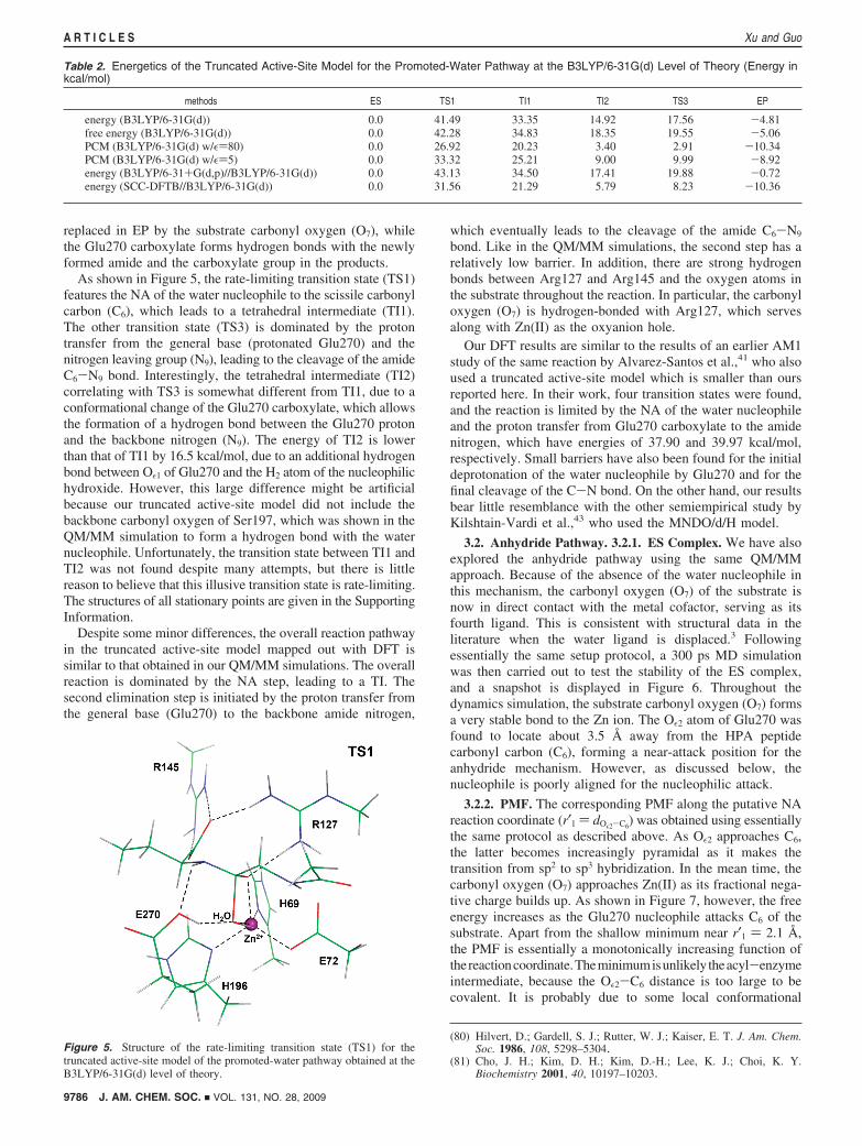
As shown in Figure 5, the rate-limiting transition state (TS1)features the NA of the water nucleophile to the scissile carbonylcarbon (C6), which leads to a tetrahedral intermediate (TI1).The other transition state (TS3) is dominated by the protontransfer from the general base (protonated Glu270) and thenitrogen leaving group (N9), leading to the cleavage of the amideC6-N9 bond. Interestingly, the tetrahedral intermediate (TI2)correlating with TS3 is somewhat different from TI1, due to aconformational change of the Glu270 carboxylate, which allowsthe formation of a hydrogen bond between the Glu270 protonand the backbone nitrogen (N9). The energy of TI2 is lowerthan that of TI1 by 16.5 kcal/mol, due to an additional hydrogenbond between Oε1 of Glu270 and the H2 atom of the nucleophilichydroxide. However, this large difference might be artificialbecause our truncated active-site model did not include thebackbone carbonyl oxygen of Ser197, which was shown in theQM/MM simulation to form a hydrogen bond with the waternucleophile. Unfortunately, the transition state between TI1 andTI2 was not found despite many attempts, but there is littlereason to believe that this illusive transition state is rate-limiting.The structures of all stationary points are given in the SupportingInformation.
Despite some minor differences, the overall reaction pathwayin the truncated active-site model mapped out with DFT issimilar to that obtained in our QM/MM simulations. The overallreaction is dominated by the NA step, leading to a TI. Thesecond elimination step is initiated by the proton transfer fromthe general base (Glu270) to the backbone amide nitrogen,
which eventually leads to the cleavage of the amide C6-N9
bond. Like in the QM/MM simulations, the second step has arelatively low barrier. In addition, there are strong hydrogenbonds between Arg127 and Arg145 and the oxygen atoms inthe substrate throughout the reaction. In particular, the carbonyloxygen (O7) is hydrogen-bonded with Arg127, which servesalong with Zn(II) as the oxyanion hole.
Our DFT results are similar to the results of an earlier AM1study of the same reaction by Alvarez-Santos et al.,41 who alsoused a truncated active-site model which is smaller than oursreported here. In their work, four transition states were found,and the reaction is limited by the NA of the water nucleophileand the proton transfer from Glu270 carboxylate to the amidenitrogen, which have energies of 37.90 and 39.97 kcal/mol,respectively. Small barriers have also been found for the initialdeprotonation of the water nucleophile by Glu270 and for thefinal cleavage of the C-N bond. On the other hand, our resultsbear little resemblance with the other semiempirical study byKilshtain-Vardi et al.,43 who used the MNDO/d/H model.
3.2. Anhydride Pathway. 3.2.1. ES Complex. We have alsoexplored the anhydride pathway using the same QM/MMapproach. Because of the absence of the water nucleophile inthis mechanism, the carbonyl oxygen (O7) of the substrate isnow in direct contact with the metal cofactor, serving as itsfourth ligand. This is consistent with structural data in theliterature when the water ligand is displaced.3 Followingessentially the same setup protocol, a 300 ps MD simulationwas then carried out to test the stability of the ES complex,and a snapshot is displayed in Figure 6. Throughout thedynamics simulation, the substrate carbonyl oxygen (O7) formsa very stable bond to the Zn ion. The Oε2 atom of Glu270 wasfound to locate about 3.5 Å away from the HPA peptidecarbonyl carbon (C6), forming a near-attack position for theanhydride mechanism. However, as discussed below, thenucleophile is poorly aligned for the nucleophilic attack.
3.2.2. PMF. The corresponding PMF along the putative NAreaction coordinate (r′1 ) dOε2-C6
) was obtained using essentiallythe same protocol as described above. As Oε2 approaches C6,the latter becomes increasingly pyramidal as it makes thetransition from sp2 to sp3 hybridization. In the mean time, thecarbonyl oxygen (O7) approaches Zn(II) as its fractional nega-tive charge builds up. As shown in Figure 7, however, the freeenergy increases as the Glu270 nucleophile attacks C6 of thesubstrate. Apart from the shallow minimum near r′1 ) 2.1 Å,the PMF is essentially a monotonically increasing function ofthereactioncoordinate.Theminimumisunlikely theacyl-enzymeintermediate, because the Oε2-C6 distance is too large to becovalent. It is probably due to some local conformational
(80) Hilvert, D.; Gardell, S. J.; Rutter, W. J.; Kaiser, E. T. J. Am. Chem.Soc. 1986, 108, 5298–5304.
(81) Cho, J. H.; Kim, D. H.; Kim, D.-H.; Lee, K. J.; Choi, K. Y.Biochemistry 2001, 40, 10197–10203.
Table 2. Energetics of the Truncated Active-Site Model for the Promoted-Water Pathway at the B3LYP/6-31G(d) Level of Theory (Energy inkcal/mol)
methods ES TS1 TI1 TI2 TS3 EP
energy (B3LYP/6-31G(d)) 0.0 41.49 33.35 14.92 17.56 -4.81free energy (B3LYP/6-31G(d)) 0.0 42.28 34.83 18.35 19.55 -5.06PCM (B3LYP/6-31G(d) w/ε)80) 0.0 26.92 20.23 3.40 2.91 -10.34PCM (B3LYP/6-31G(d) w/ε)5) 0.0 33.32 25.21 9.00 9.99 -8.92energy (B3LYP/6-31+G(d,p)//B3LYP/6-31G(d)) 0.0 43.13 34.50 17.41 19.88 -0.72energy (SCC-DFTB//B3LYP/6-31G(d)) 0.0 31.56 21.29 5.79 8.23 -10.36
Figure 5. Structure of the rate-limiting transition state (TS1) for thetruncated active-site model of the promoted-water pathway obtained at theB3LYP/6-31G(d) level of theory.
9786 J. AM. CHEM. SOC. 9 VOL. 131, NO. 28, 2009
A R T I C L E S Xu and Guo

minimum in the active site. The absence of a PMF minimumcorresponding to the acyl-enzyme intermediate indicates thatthe anhydride pathway is not viable, at least for the HPAsubstrate.
3.2.3. Truncated Active-Site Model. To confirm the QM/MMresults for the anhydride pathway, we have carried out high-level DFT (B3LYP/6-31G(d)) studies with a truncated active-site model, which is similar to that used for the promoted-watercase. All attempts to locate the transition state for the NA stepand the acyl-enzyme intermediate failed. Figure 8 displays theminimal energy path along the Oε2-C6 reaction coordinate, andit clearly shows a monotonic increase of energy without abarrier, consistent with the QM/MM PMF. The structures ofthe ES minimum and the high-energy complex correspondingto the Oε2-C6 distance of 1.47 Å were also included in thefigure. The sp2 and sp3 geometries of C6 in the two structuresare apparent.
4. Discussion
CPA bears many similarities to thermolysin, which is also apeptidase with a zinc cofactor. Unlike CPA, thermolysin is anendopeptidase that cleaves peptide bonds containing hydropho-
bic residues.82 In thermolysin, the zinc ion is also coordinatedby two His residues and a Glu residue, as well as a watermolecule. However, the two enzymes differ in many otheraspects, including their structures. As in the CPA case, thecatalytic mechanism of thermolysin was also uncertain.82 Thefavored mechanism involves Glu143, which serves as a generalbase to activate the zinc-bound water. The NA of the hydroxidegroup to the substrate carbonyl carbon results in a tetrahedralintermediate, which eventually collapses to form the products.In the E step, a proton is transferred from the Glu143 generalacid to the nitrogen leaving group in the substrate. Thismechanism is essentially the same as the promoted-waterpathways discussed here for CPA.
In a recent ab initio QM/MM free-energy simulation, Blum-berger et al. have convincingly demonstrated that the generalbase-general acid mechanism is indeed the preferred mecha-nism for thermolysin.83 These authors found that the PMF hastwo transition states, with the first barrier for NA slightly higherthan the second one for the E step. The overall free-energybarrier height is 14.8 kcal/mol, very close to what we have foundfor CPA. The zinc cofactor was also found to stabilize thenegatively charged carbonyl oxygen of the scissile peptide bond,although it has weak interactions with the substrate in the EScomplex. Since CPA and thermolysin have similar active-sitearrangements, it is not surprising that they have the samecatalytic mechanism.
The lack of direct interaction between Zn(II) and the scissilecarbonyl in the ES complex is an important feature shared bythermolysin and CPA. Christianson and Lipscomb argued thata direct metal-carbonyl interaction found in several CPA-inhibitor complexes represents a nonproductive binding modefor CPA, because the zinc ion in such an configuration couldnot activate the water nucleophile.3 Our simulation resultsconcerning this key interaction, along with the work ofBlumberger et al. on thermolysin,83 provided strong evidencein support of this argument. It can thus be concluded that themetal cofactor in zinc hydrolases does not play a role inpolarizing the substrate carbonyl. Rather, it serves as a Lewisacid to activate the water nucleophile and to stabilize thetransition state. The similarities between the two quite differentenzymes underscore convergent evolution of the two catalysts.
(82) Lipscomb, W. M.; Strater, N. Chem. ReV. 1996, 96, 2375–2433.(83) Blumberger, J.; Lamoureux, G.; Klein, M. L. J. Chem. Theor. Comput.
2007, 3, 1837–1850.
Figure 6. Snapshot of the QM/MM ES complex for the anhydride reactionpathway. Black dashed lines represent hydrogen bonds, while red dashedlines represent metal-ligand bonds.
Figure 7. Potential of mean force for the anhydride pathway of the HPAhydrolysis.
Figure 8. Energy profile of a truncated active-site model for the anhydridepathway at the B3LYP/6-31G(d) level of theory. The structure of the EScomplex and that corresponding to the TI complex are shown as well.
J. AM. CHEM. SOC. 9 VOL. 131, NO. 28, 2009 9787
QM/MM and DFT Studies of Catalytic Mechanism of Carboxypeptidase A A R T I C L E S

The general base-general acid mechanism for both CPA andthermolysin has also been implicated in several other monozinchydrolases. For example, members of the B2 subclass of�-lactamases such as CphA,84 which has one Zn(II) cofactor,catalyze the breaking of the amide bond in �-lactam antibioticsusing an active-site water promoted by a zinc-bound Aspresidue.58 It was also found recently in a DFT QM/MM study85
that class I and II histone deacetylases, another type of monozinchydrolases, use an active-site His residue to promote a waternucleophile in a general base-general acid mechanism.
Although uncommon in enzymatic systems, a carboxylatenucleophile is not without precedent. For example, a Meisen-heimer complex is known to exist in the catalysis of dechlori-nation of 4-chlorobenzoyl-CoA by 4-chlorobenzoyl-CoA de-halogenase, in which an Asp residue serves as the nucleophile.86,87
An anhydride intermediate has also been found in the hydrolysisof a thioester (3-hydroxyisobutyryl-CoA) to 3-hydroxyisobu-tyrate catalyzed by 3-hydroxyisobutyryl-CoA hydrolase.88 Toour best knowledge, however, there has been essentially noclear-cut evidence for an anhydride intermediate in amidehydrolysis. Our results, from both QM/MM and DFT calcula-tions, clearly discount the anhydride mechanism, at least forthe HPA substrate. It is somewhat surprising that no acyl-enzymeintermediate can be found for the anhydride pathway in boththe QM/MM and DFT calculations, given the fact that the zincion provides significant stabilization of the oxyanion. Theinability to form an acyl-enzyme intermediate is presumablydue to a number of factors. First, the carboxylate group is arather poor nucleophile, with a Swain-Scott value of 2.72.24
Its nucleophilicity is further eroded by the hydrogen bondformed between Oε1 and the NH moiety in the substratebackbone. [Interestingly, an acyl-enzyme intermediate has beenobserved to form in our DFT calculations when the amide isreplaced by an ester, which has no hydrogen bond with thenucleophile (see Supporting Information). The preference ofester substrates in the anhydride mechanism has been extensivelydiscussed before,3 and we plan to investigate this possibility inmore detail in the future.] Second, the conjugation between thecarbonyl group and the adjacent amide group reduces theelectrophilicity of the carbonyl carbon. Finally, the Glu270carboxylate is not well aligned in the ES complex for NA ofthe substrate carbonyl carbon. The Oε2-C6-O7, Oε2-C6-N9,and Oε2-C6-C7 angles are 140°, 79°, and 54°, respectively,far from the ideal value of 90°. The misalignment results insubstantial stress in the system when the Glu270 carboxylateattacks the scissile carbonyl carbon. These factors make theformation of the acyl-enzyme intermediate very unfavorable.
5. Conclusions
Despite the fact that CPA is one of the most extensivelystudied zinc enzymes, its catalytic mechanism has not beencompletely settled. In this work, we reported the first hybridQM/MM and DFT studies on the hydrolysis reaction catalyzedby CPA using HPA as the substrate molecule. Both QM/MMsimulations and DFT calculations support the hypothesis thatthe CPA-catalyzed hydrolysis reaction for peptide substrates isthrough the promoted-water pathway. In particular, Glu270serves as the general base to facilitate the nucleophilic attackof the substrate carbonyl carbon by the deprotonated waternucleophile. Subsequently, the protonated carboxylate group ofGlu270 acts as a general acid by donating the proton to theamide nitrogen leaving group, leading to the cleavage of thepeptide C-N bond in the substrate. Free-energy calculationsindicated that the first nucleophilic addition is the rate-limitingstep, with a barrier of 17.9 kcal/mol, in reasonable agreementwith experimental data. On the other hand, the free-energy wellof the tetrahedral intermediate is relatively shallow toward thecleavage of the peptide CN bond.
The catalysis is assisted by the zinc cofactor, which servesas the Lewis acid. Its role is twofold. First, it activates the waternucleophile bound to Zn(II), and second, along with Arg127, itstabilizes the fractionally charged carbonyl oxygen of thesubstrate in the tetrahedral intermediate. The first role has beenrecognized since the work of Demoulin and Pullman,38 and thereare numerous examples of Zn(II) serving as an oxyanion holein enzyme catalysis.82 However, our calculations indicate thatthe zinc ion in CPA does not play a significant role in polarizingthe substrate carbonyl. Rather, the polarization is largely dueto Arg127, which is hydrogen-bonded with the carbonyl oxygenof the substrate. Free-energy simulations concluded that thecontribution of this residue leads to about 3 kcal/mol reductionof the rate-limiting barrier, in agreement with experiment.
Our results also provided strong evidence against the anhy-dride mechanism in CPA catalysis, at least for the HPAsubstrate. The free-energy profile suggested that no acyl-enzymeintermediate can be formed. However, preliminary resultsindicate that such a mechanism is possible for ester substrates.
Acknowledgment. This work was supported by the NationalNatural Science Foundation of China (No. 20803048 to D.X.)and by the National Institutes of Health (R03-AI068672 and R03-AI071992 to H.G.). Some of the calculations have beenperformed at the National Centers for Supercomputing Applica-tions. We also acknowledge Q. Cui for helping with the SCC-DFTB simulations and D. Dunaway-Mariano for some stimu-lating discussions.
Supporting Information Available: DFT stationary pointstructures along the promoted-water pathway for the peptidesubstrate and those for the anhydride pathway for the estersubstrate; complete refs 63 and 79. This material is availablefree of charge via the Internet at http://pubs.acs.org.
JA9027988
(84) Garau, G.; Bebrone, C.; Anne, C.; Galleni, M.; Frere, J.-M.; Dideberg,O. J. Mol. Biol. 2005, 345, 785–795.
(85) Corminboeuf, C.; Hu, P.; Tuckerman, M. E.; Zhang, Y. J. Am. Chem.Soc. 2006, 128, 4530–4531.
(86) Yang, G.; Liu, R.-Q.; Taylor, K. L.; Xiang, H.; Price, J.; Dunaway-Mariano, D. Biochemistry 1996, 35, 10879–10885.
(87) Xu, D.; Wei, Y.; Wu, J.; Dunaway-Mariano, D.; Guo, H.; Cui, Q.;Gao, J. J. Am. Chem. Soc. 2004, 126, 13649–13658.
(88) Wong, B. J.; Gerlt, J. A. J. Am. Chem. Soc. 2003, 125, 12076–12077.
9788 J. AM. CHEM. SOC. 9 VOL. 131, NO. 28, 2009
A R T I C L E S Xu and Guo