Structural, Photocatalytic and Antibacterial Activity of ...

QUANTITATIVE STRUCTURAL ACTIVITY RELATIONSHIP OF FLAVONOIDS: STUDIES OF
ANTIOXIDANT PROPERTIES AND HUMAN INTESTINAL PERMEABILITY
by
HAMIN HWANG
(Under the Direction of William Kerr)
ABSTRACT
Flavonoids are common antioxidants and are found in many plant species. However,
only a few studies are based on quantification of structural information and bioavailability.
Therefore, both QSAR (quantitative structural‐activity relationship) and QSPR (quantitative
structural‐permeability relationship) study on flavonoids were conducted. In the QSAR study,
flavonoid structures were obtained from the Cambridge Molecular Structure Database,
minimized, and studied using Grid and Volsurf. The generated data was compared to
experimental TEAC (Trolox equivalent antioxidant capacity) data from a previous study. The
QSAR study found that antioxidant potential of flavonoids increases with hydrophobicity,
smaller molecular weight, lack of rugosity, and increasing number of hydroxy groups. The PCA
(principal components analysis) 7 component model explained 88.7% and PLS 6 component
model gave an R2 value of 0.8626, showing high correlation between antioxidant potential and
23 flavonoid molecules. In the next portion of the study, a QSPR study was performed using
structures from the Cambridge Software Molecular Database. After energy minimization, QSPR
software, known as Volsurf, was used to generate the permeability data. This data was
correlated and compared to the experimental Caco‐2 data from another study. In the QSPR

study, the computational study matched with 94.1% of data determining permeability of 17
structures. In addition, smaller molecular weight and hydrophobic flavonoids showed much
higher permeability than larger and hydrophilic flavonoid molecules.
INDEX WORDS: Flavonoids, QSAR, Structure relationship, Computational study, Volsurf, Caco‐2
cell monolayer, Digestion, Absorption, Antioxidant.

QUANTITATIVE STRUCTURAL ACTIVITY RELATIONSHIP OF FLAVONOIDS: STUDIES OF
ANTIOXIDANT PROPERTIES AND HUMAN INTESTINAL PERMEABILITY
By
HAMIN HWANG
B.S., The University of Georgia, 2006
A Thesis Submitted to the Graduate Faculty of The University of Georgia in Partial Fulfillment of
the Requirements for the Degree
MASTER OF SCIENCE
ATHENS, GEORGIA
2008

©2008
HAMIN HWANG
All Rights Reserved

QUANTITATIVE STRUCTURAL ACTIVITY RELATIONSHIP OF FLAVONOIDS: STUDIES OF
ANTIOXIDANT PROPERTIES AND HUMAN INTESTINAL PERMEABILITY
by
HAMIN HWANG
Major Professor: William Kerr
Committee: Ron Pegg Robert Woods
Electronic Version Approved:
Maureen Grasso Dean of the Graduate School The University of Georgia August 2008

iv
DEDICATION
This dissertation is dedicated to my family‐‐Ju Sang Hwang (father), Myoung Sook Yoon
(mother), Ha Kyung Hwang (brother), and Ye Kang (grandmother), who recently passed away at
the age of 92.
Special appreciation goes to Polly Cleveland, from whom I’ve learned much computer
knowledge.
v
ACKNOWLEDGEMENTS
I would like to express my gratitude to the following people:
My major professor, Dr. William Kerr, has accepted me for whom I am, shown me the necessary
steps to further advance myself, being my friend and family. Thanks to my committee
members, Dr. Ron Pegg and Dr. Robert Woods for their guidance during this research and the
pursuit of higher education. I show my special thanks to Dr. Robert Shewfelt and Dr. Yao‐wen
Huang for introducing food science to my life. I am thankful to Dr. Romeo Toledo for his tough
yet mind‐opening engineering exams. Special thanks to Carl Ruiz for his companionship,
twisted humor, and religious guidance. My lab mates Mark Corey, Laura Brindle, George
Cavender, Katherine Acosta, and Jinhee Yi for their help all these years. Dr. Ramsey Bakali, Dr.
Joe Mouldin, Dr. Gene Pesti, Dr. Hardy Edwards, Dr. Sammy Aggry for their lifelong lessons in
life and teachings that will carry my heart throughout my life.

vi
TABLE OF CONTENTS
Page
ACKNOWLEDGEMENTS................................................................................................................v
CHAPTERS
1. INTRODUCTION ...................................................................................................................1
2. LITERATURE REVIEW ...........................................................................................................9
3. QUANTITATIVE STRUCRUAL‐ACTIVITY RELATIONSHIP OF FLAVONOIDS AND
ANTIOXIDANT POTENTIAL ……………………………………………………………………………………………..34
4. QUANTITATIVE STRUCTURE‐PERMEABILITY RELATIONSHIP OF FLAVONOIDS
USING CACO‐2 CELLS: INDEPTH STUDY OF HUMAN INTESTINAL PERMEATION OF
LAVONOIDS ….…………………...............................................................................................52
5. SUMMARY AND CONCLUSIONS ........................................................................................76

1
CHAPTER 1
INTRODUCTION
In the face of the new world and scientific knowledge that drives forth such machine,
few forces can reshape how people think, believe, and prosper. Even more so than ever,
consumers and mankind thrive to discover new ideas, understand mechanisms. In the paper to
follow, I present my dedication and works that hopefully change the spectacle of sands or the
path that light bends when it hits that little dust particle‐‐all with a hope that it is a small step
into the light of knowledge.
Flavonoids are phenolic compounds that are available in plants, and they represent
more than half of the 8000 known phenolics found in nature (Harborne, Baxter et al.
1999)(Harborne, Baxter et al. 1999). They are derived from amino acids, tyrosine and
phenylalanine and play significant roles in the diet. It is common knowledge that diets rich in
vegetables and fruits are beneficial to human health. Therefore, it is reasonable to state that
consumption of fruits and vegetables, containing flavonoids, are related to positive health
benefits. How much of such benefits are specifically due to flavonoids is indeterminant, but
flavonoids are believed to be strong antioxidants and are shown to help prevent many diseases.
Antioxidants quench reactive oxygen and nitrogen species such as the superoxide anion
radicals, hydroxyl radicals, and peroxyl radicals. These reactive oxygen species reduce oxygen
(Williams and Jeffrey 2000)(Williams and Jeffrey 2000) and can produce harm in the biological
system. Such harm includes protein denaturation, membrane activity, DNA alterations, and

2
lipid peroxidation (Kinsella, Frankel et al. 1993)(Kinsella, Frankel et al. 1993). Therefore,
antioxidants like flavonoids can provide health benefits such as prevention of atherosclerosis
(Hertog, Hollman et al. 1992; Keys 1995)(Hertog, Hollman et al. 1992; Keys 1995), anti‐
inflammation (Middleton, Kandaswami et al. 2000)(Middleton, Kandaswami et al. 2000), anti‐
aging, prevention of coronary heart disease, prevention of diabetes mellitus (Slater 1984;
Cheng, Lin et al. 2003)(Slater 1984; Cheng, Lin et al. 2003), Alzheimer’s disease (Smith,
Rottkamp et al. 2000)(Smith, Rottkamp et al. 2000), and anti‐microbial effects (Harborne and
Williams 2000)(Harborne and Williams 2000).
Due to the many benefits flavonoids offer to man, quantification methodologies are an
important part of the science and further understanding of flavonoids. Currently, popular
methods include the Trolox equivalent antioxidant capacity (TEAC) assay (Miller, Rice‐Evans et
al. 1993; Rice‐Evans and Miller 1994; Pellegrini, Proteggente et al. 1999)(Miller, Rice‐Evans et al.
1993; Rice‐Evans and Miller 1994; Pellegrini, Proteggente et al. 1999), DPPH∙ method (Brand‐
Williams, Cuvelier et al. 1995; Sánchez‐Moreno, Larrauri et al. 1998)(Brand‐Williams, Cuvelier et
al. 1995; Sánchez‐Moreno, Larrauri et al. 1998), ferric reducing ability of plasma (FRAP) assay
(Benzie and Strain 1999)(Benzie and Strain 1999), oxygen radical absorbance capacity (ORAC)
assay (Glazer 1990)(Glazer 1990), total radical trapping parameter (TRAP) (Wayner, Burton et
al. 1985)(Wayner, Burton et al. 1985), dichlorofluorescin diacetate (DCFH‐DA) based assay
(Valkonen and Kuusi 1997; Amado, Jaramillo et al. 2007)(Valkonen and Kuusi 1997; Amado,
Jaramillo et al. 2007), cyclic voltammetry method (Kohen, Beit‐Yannai et al. 1999)(Kohen, Beit‐
Yannai et al. 1999), total oxyradical scavenging capacity (TOSC) assay (Winston, Regoli et al.
1998)(Winston, Regoli et al. 1998), photochemiluminescence (PCL) assay (Popov, Lewin et al.

3
1987; Popov and Lewin 1994; Popov and Lewin 1996)(Popov, Lewin et al. 1987; Popov and
Lewin 1994; Popov and Lewin 1996). Among the many, the TEAC assay was selected due to its
ability to study both hydrophilic and lipophilic compounds as well as successful recent studies
performed (Dastmalchi, Damien Dorman et al. 2007; Srinivasan, Chandrasekar et al.
2007)(Dastmalchi, Damien Dorman et al. 2007; Srinivasan, Chandrasekar et al. 2007).
When new tools and technology are introduced, keen scientists take advantage of the
tools and application that can be performed. In this day and age, there have been dramatical
advances in computer technology and related fields. For example, robots and computer
programs are present in many things that surround life, society, and work. To an extent, they
are being used from sending a man into the moon or used for garbage disposal units. With that
in mind, there have been an increasing number of computational studies to perform scientific
experiments. In this case, application of computational study was used to study the
quantitative structure–activity relationships (QSAR) of flavonoids. In this study, linkage
between antioxidant potential to the molecular structures will be examined. This was
examined using Volsurf (Volsurf version 3.0 software by Molecular Discovery Ltd)(Volsurf
version 3.0 software by Molecular Discovery Ltd) program.
In the next portion of the study, a similar computational study is performed to examine
the properties of molecules that can predict and relate human intestinal absorption of
flavonoids. Human digestion studies are important due to the new scientific evidence they can
provide. Much like yester‐age of vitamins, antioxidants, like flavonoids, are considered the new
generation nutrients, providing something extra on top of basic nutrition. Therefore, RDIs or
4
AIs would be interesting to observe in the future. However, for RDI or AI to exist, absorption
studies must be performed and more so, quantitative studies are absolutely necessary.
Recently, a few different methods were employed to predict or measure the absorption of
flavonoids. These include computational studies (Ekins, Durst et al. 2001; Ponce, Perez et al.
2004)(Ekins, Durst et al. 2001; Ponce, Perez et al. 2004), in vitro studies (Kuo 1998; Murota,
Shimizu et al. 2000; Masataka Oitate 2001)(Kuo 1998; Murota, Shimizu et al. 2000; Masataka
Oitate 2001), and in vivo studies (Hollman, Vries et al. 1995; Hollman, Trijp et al. 1997)(Hollman,
Vries et al. 1995; Hollman, Trijp et al. 1997). Among the many however, Caco‐2 monolayer cells
are widely used and accepted in the scientific community as a human intestinal absorption
model (Artursson, Palm et al. 2001)(Artursson, Palm et al. 2001). Therefore, combination of
Caco‐2 absorption methods and computational methods can be very powerful tool in predicting
and determining molecular characteristics that are linked with absorption data. The reason
behind such combinatory research lies in few factors. As more data is obtained from in vivo
and in vitro experiments, these data can be compiled into a large database. This can be used to
predict different types of compounds. As a matter of fact, the Volsurf (Volsurf version 3.0
software by Molecular Discovery Ltd)(Volsurf version 3.0 software by Molecular Discovery Ltd)
program has a Caco‐2 database of 751 chemical compounds. Moreover, the time consumption,
economics of experimental equipments, reagents, and differences in lab methods and materials
can be detrimental to Caco‐2 studies, which do not seem to be an issue in computational
studies. Furthermore, in vivo and in vitro experiments only show quantifying data for Caco‐2
permeable samples, leaving all impermeable samples as zeros in the permeation

5
measurements. On the other hand, computation studies can provide a magnitude for such
impermeable samples, further providing data that can explain the degree of impermeability.
By using state‐of‐the‐art equipment and methodologies, combining traditional
chemistry and experiments to newest computation studies, an area never explored can be
researched, and further deliver light into the darkness that exists in the world. For such reason,
and being one of the earlier pioneers of the food science to combine and pursue computational
study, incredible new findings of flavonoids, antioxidant potential, and human intestinal
absorption have been found and reported throughout this manuscript.
6
References
Amado, L. L., M. D. Jaramillo, et al. (2007). "16.P20. A new method to evaluate total antioxidant capacity against reactive oxygen and nitrogen species (RONS) in aquatic organisms." Comparative Biochemistry and Physiology ‐ Part A: Molecular & Integrative Physiology 148(Supplement 1): S75‐S76. Artursson, P., K. Palm, et al. (2001). "Caco‐2 monolayers in experimental and theoretical predictions of drug transport." Advanced Drug Delivery Reviews 46: 27‐43. Benzie, I. F. F. and J. J. Strain (1999). "reducing antioxidant power assay: Direct measure of total antioxidant activity of biological fluids and modified version for simultaneous measurement of total antioxidant power and ascorbic acid concentration." Methods in Enzymology 299: 15‐27. Brand‐Williams, W., M. E. Cuvelier, et al. (1995). "Use of a free radical method to evaluate antioxidant activity." Food Science and Technology 28: 25‐30. Cheng, H. Y., T. C. Lin, et al. (2003). "Antioxidant and free radical scavenging activities of Terminalia chebula." Biological & Pharmaceutical Bulletin 26: 1331‐1335. Dastmalchi, K., H. J. Damien Dorman, et al. (2007). "Chemical composition and antioxidative activity of Moldavian balm (Dracocephalum moldavica L.) extracts." LWT ‐ Food Science and Technology 40(9): 1655‐1663. Ekins, S., G. L. Durst, et al. (2001). "Three‐Dimensional Quantitative Structure‐Permeability Relationship Analysis for a Series of Inhibitors of Rhinovirus Replication." J. Chem. Inf. Comput. Sci. 41(6): 1578‐1586. Glazer, A. N. (1990). "Phycoerythrin fluorescence‐based assay for reactive oxygen species." Methodes in Enzymology 186: 161‐168. Harborne, J. B., H. Baxter, et al. (1999). Phytochemical Dictionary, Handbook of bioactive compounds from plants. London, Taylor and Francis. Harborne, J. B. and C. A. Williams (2000). "Advances in flavonoid research since 1992." Phytochemistry 55(6): 481‐504. Hertog, M. G. L., P. C. H. Hollman, et al. (1992). "Content of potentially anticarcinogenic flavonoids of 28 vegetables and 9 fruits commonly consumed in Netherlands." Journal of Agricultural and Food Chemistry (40): 2379–2383. Hollman, P. C. H., J. M. P. Trijp, et al. (1997). FEBS Lett. 418: 152‐516.

7
Hollman, P. C. H., J. H. M. Vries, et al. (1995). American Journal of Clinical Nutrition 62: 1276‐1282. Keys, A. (1995). "Mediterranean diet and public health: Personal reflections." American Journal of Clinical Nutrition (61): 1321–1323. Kinsella, J. E., E. Frankel, et al. (1993). "Possible mechanisms for the protective role of antioxidants in wine and plant foods." Food Technology(47): 85‐89. Kohen, R., E. Beit‐Yannai, et al. (1999). "Overall low molecular weight antioxidant activity of biological fluids and tissues by cyclic voltammetry." Methodes in Enzymology 300: 285‐296. Kuo, S.‐M. (1998). "Transepithelial transport and accumulation of flavone in human intestinal CACO‐2 cells." Life Sciences 63(26): 2323‐2331. Masataka Oitate, R. N. N. K. H. T. H. M. H. O. Y. S. (2001). "Transcellular transport of genistein, a soybean‐derived isoflavone, across human colon carcinoma cell line (Caco‐2)." Biopharmaceutics & Drug Disposition 22(1): 23‐29. Middleton, E. J., C. Kandaswami, et al. (2000). "The effects of plant flavonoids on mammalian cells: implications for inflammation, heart disease, and cancer." Pharmacol Rev 52: 673‐751. Miller, N. J., C. A. Rice‐Evans, et al. (1993). "A novel method for measuring antioxidant capacity and its application to monitoring the antioxidant status in premature neonates." Clinical Science 84: 407‐412. Murota, K., S. Shimizu, et al. (2000). "Efficiency of Absorption and Metabolic Conversion of Quercetin and Its Glucosides in Human Intestinal Cell Line Caco‐2." Archives of Biochemistry and Biophysics 384(2): 391‐397. Pellegrini, N., A. Proteggente, et al. (1999). "activity applying an improved ABTS radical cation decolorization assay." Free Radicle Biologyl and Medicine 26: 1231‐1237. Ponce, Y. M., M. A. C. Perez, et al. (2004). J. Pharm. Pharmaceut. Sci.: 186‐199. Popov, I., G. Lewin, et al. (1987). "detection of antiradical activity. I. Assay of superoxide dismutase." Biomed Biochim Acta 46: 775‐779. Popov, I. N. and G. Lewin (1994). "Photochemiluminescent detection of antiradical activity: II. Testing of nonenzymic water‐soluble antioxidants." Free Radical Biology and Medicine 17(3): 267‐271.

8
Popov, I. N. and G. Lewin (1996). "Photochemiluminescent detection of antiradical activity; IV: testing of lipid‐soluble antioxidants." Journal of Biochemical and Biophysical Methods 31(1‐2): 1‐8. Rice‐Evans, C. A. and N. J. Miller (1994). "Total antioxidant status in plasma and body fluids." Methodes in Enzymology 234: 279‐293. Sánchez‐Moreno, J. A., Larrauri, et al. (1998). "A procedure to measure the antiradical efficiency of polyphenols." Journal of the Science of Food and Agriculture 76: 270‐276. Slater, T. F. (1984). "Free‐radical mechanisms in tissue injury." Biochemical Journal 222: 1‐15. Smith, M. A., C. A. Rottkamp, et al. (2000). "Oxidative stress in Alzheimer's disease." Biochimica et Biophysica Acta (BBA) ‐ Molecular Basis of Disease 1502(1): 139‐144. Srinivasan, R., M. J. N. Chandrasekar, et al. (2007). "Antioxidant activity of Caesalpinia digyna root." Journal of Ethnopharmacology 113(2): 284‐291. Valkonen, M. and T. Kuusi (1997). "Spectrophotometric assay for total peroxyl radical‐trapping antioxidant potential in human serum." Journal of Lipid Research 38: 823‐833. Volsurf version 3.0 software by Molecular Discovery Ltd. Wayner, D. D. M., G. W. Burton, et al. (1985). "Quantitative measurement of the total, peroxyl radical‐trapping antioxidant capability of human blood plasma by controlled peroxidation : The important contribution made by plasma proteins." FEBS Letters 187(1): 33‐37. Williams, G. M. and A. M. Jeffrey (2000). "Oxidative DNA Damage: Endogenous and Chemically Induced." Regulatory Toxicology and Pharmacology 32(3): 283‐292. Winston, G. W., F. Regoli, et al. (1998). "A Rapid Gas Chromatographic Assay for Determining Oxyradical Scavenging Capacity of Antioxidants and Biological Fluids." Free Radical Biology and Medicine 24(3): 480‐493.

9
CHAPTER 2
LITERATURE REVIEW
2.1 Basic Flavonoid Structure
Flavonoids are derived from amino acids, tyrosine and phenylalanine, and has three
aromatic ring structure. As it loses a hydrogen atom to quench a free radical, resonance occurs
and stabilizes the flavonoid molecule after a hydrogen atom is donated. Additionally, previous
studies (Vrielynck, Cornard et al. 1993)(Vrielynck, Cornard et al. 1993) noted that energy
differences between the planar and twisted structure is minimal. However, flavones should
exist as a planar structure due to their lower energy and more favorable condition to packing or
rugosity. The presence of hydroxyl groups at the C‐3 and C‐4 position on the B ring of the
flavonoids increases the antioxidant activity. Moreover, the basic structure of flavonoids is
important as well. This can be seen in the example of kaempferol (Silva, Santos et al.
2002)(Silva, Santos et al. 2002), where the number of hydroxyl groups and their location on the
aromatic ring determines antioxidant properties. Quantitative Structure‐Activity Relationship
study of 42 different flavonoids was performed. Using a beta carotene linoleate system
oxidation to test antioxidant and antiradical activities of flavonoids (Burda and Oleszek
2001)(Burda and Oleszek 2001) found that the presence of hydroxyl groups at the C‐3 position
is associated with good antioxidant activity. Additionally, they also found that antiradical
activity is primarily connected with free hydroxyl groups at the C‐4 position.
10
A study conducted by (Lien, Ren et al. 1999)(Lien, Ren et al. 1999) shows the presence of a 3‐
OH, 4‐oxo, and 0‐dihydroxy in the B ring is needed in addition to a 2,3 double bond for the
highest antioxidant potential. Additionally, higher antioxidant potentials were correlated with
increasing numbers of hydroxyl groups. Besides structural placement of hydroxy groups,
flavonoids show varying degrees of antioxidant behavior depending on the food system where
being used (Chen, Chan et al. 1996)(Chen, Chan et al. 1996). According to an earlier study,
ascorbic acid is stable under acidic conditions; however, when exposed to neutral or alkaline
solution conditions, oxidation can rapidly occur. Thus, flavonoids can be used to preserve
ascorbic acid, while providing antioxidants for food items (Thompson, Williams et al.
1976)(Thompson, Williams et al. 1976).
2.2 Existing Computational Studies
Ab initio molecular orbital calculation has been performed to fullly optimize structure
geometry. In this study, an explanation of catechin and taxifolin has been conducted and they
have used Log P values to study lipophilicity of the compounds. Moreover, researchers used
acidity values from a computation study and in vivo data (Teixeira, Siquet et al. 2005)(Teixeira,
Siquet et al. 2005) to examine relationship between them. In another study of melatonin and
related indoles, semiempirical AM1 (Austin Model 1) and DFT (Density Functional Theory) were
used to measure changes in Gibbs free energy to test molecules for their antioxidant potential.
In their experiment design, both vacuum and aqueous settings were employed to find out that
no significant difference existed between the two settings. Experimenters concluded that this
is due to a lack of charged species used in the computational methodology. The obtained data

11
was compared to other established data and found some qualitative similarity (Turjanski,
Rosenstein et al. 1998)(Turjanski, Rosenstein et al. 1998). Similar studies conducted using DFT
was conducted. B3LYP/6‐31+G(d) density functional theoretical level was used to study QSAR.
They used heat of formation as their primary measurement to observe antioxidant potential.
They were able to find a close correlation between experimental data and calculated data
(Vafiadis and Bakalbassis 2003)(Vafiadis and Bakalbassis 2003). Another research found that
the dissociation enthalpy parameter of O‐H bonds provides the best antiradical activity of
chalcones. This study was conducted using the B3P86 theory level of quantum calculations
(Kozlowski, Trouillas et al. 2007)(Kozlowski, Trouillas et al. 2007). Additionally, another study
examined the length of the C‐O bond from phenol molecules and discovered the correlation
between proton affinities and electron transfer enthalpies (Klein and Lukes 2006)(Klein and
Lukes 2006).
Predictions of thermodynamic properties such as enthalpy of formation, bond
dissociation energy, and ionization potential of flavonoids were studied and compared to
experimental values obtained from x‐ray crystallography and calorimetric techniques. The
researchers found good correlations between thermodynamic properties and experiment data
(Mendoza‐Wilson, Lardizabal‐Gutierrez et al. 2007)(Mendoza‐Wilson, Lardizabal‐Gutierrez et al.
2007). The free energy relationship can be used to predict antioxidant potential of target
compounds given the proportionality between Gibbs free energy of activation and the overall
Gibbs free energy of reaction. Thus, this concept will allow numeric measurement of the target
antioxidant (Rhodes, Tran et al. 2004)(Rhodes, Tran et al. 2004). In 2004, scientists used the
Dragon program package (Todeschini, Consonni et al. 2002)(Todeschini, Consonni et al. 2002)

12
and PLS method to study flavonoids and to correlate antioxidant potential. In another study
(Burda and Oleszek 2001)(Burda and Oleszek 2001), 36 flavonoids and their 2D descriptors
were calculated. Then, the descriptors and antioxidant values were compared to
experimentally measured data. More recent studies shows acceptable model between
predicted antioxidant data and experimental data (Farkas, Jakus et al. 2004)(Farkas, Jakus et al.
2004). This study shows that antioxidant activity and structure can be assessed using
computational study and the PLS statistical analysis.
2.3 Measurement of Antioxidant Potential
It is extremely important to quantitatively measure the antioxidant potential of
flavonoids in both foods and model systems. Therefore, numerous methods have been
developed, each having their own advantages and disadvantages. One of the more widely used
methods is called the Trolox equivalent antioxidant capacity (TEAC) assay (Miller, Rice‐Evans et
al. 1993; Rice‐Evans and Miller 1994; Pellegrini, Proteggente et al. 1999)(Miller, Rice‐Evans et al.
1993; Rice‐Evans and Miller 1994; Pellegrini, Proteggente et al. 1999). In this assay, inhibition
of free radicals by ABTS or 2,2’‐azinobis (3‐ethylbenzothiazoline 6‐sulfonate) is used and
absorbance is measured. Then, absorbance is compared to the standards or Trolox equivalent.
The advantage of this method is that it can be used to measure antioxidant potential for both
lipophilic and hydrophilic compounds.
The TEAC assay can be used to measure the antioxidant activity of a compound.
However, the TEAC does not always relate to the antioxidant potential because it measures
how much radical is scavenged over time, where both reaction compounds and reacted

13
compounds play a role. Meanwhile, antioxidant activity measures the rate of radical scavenged
and it is associated with only the initial reaction compound (Arts, Sebastiaan Dallinga et al.
2003)(Arts, Sebastiaan Dallinga et al. 2003). The oxygen radical absorbance capacity (ORAC)
assay (Glazer 1990)(Glazer 1990) is another popular assay to determine antioxidant activities in
samples. In this assay, an fluorescenin and a peroxyl radical generator called 2,2’‐azobis (2‐
amidinopropane) dihydrochloride are used to measure antioxidant potential. The strength of
this assay lies in the combination of both inhibition time and percentage into a quantitative
data.
Following ORAC, the DPPH∙ method (Brand‐Williams, Cuvelier et al. 1995; Sánchez‐
Moreno, Larrauri et al. 1998)(Brand‐Williams, Cuvelier et al. 1995; Sánchez‐Moreno, Larrauri et
al. 1998) is a well recognized method to determine antioxidant potential, especially for plant
samples. In this assay, reaction of the stable 1,1‐diphenyl‐2‐picrylhydrazyl radical and donor
sample and color change can be measured. When an antioxidant donates a hydrogen atom, a
reduced form of DPPH loses color, which can be measured by using spectrophotometer at
520nm.
Others methods of measuring antioxidant potential include Ferric reducing ability of
plasma (FRAP) assay (Benzie and Strain 1999)(Benzie and Strain 1999), total radical trapping
parameter (TRAP) (Wayner, Burton et al. 1985)(Wayner, Burton et al. 1985), dichlorofluorescin‐
diacetate (DCFH‐DA) based assay (Valkonen and Kuusi 1997; Amado, Jaramillo et al.
2007)(Valkonen and Kuusi 1997; Amado, Jaramillo et al. 2007), cyclic voltammetry method
(Kohen, Beit‐Yannai et al. 1999)(Kohen, Beit‐Yannai et al. 1999), total oxyradical scavenging
14
capacity (TOSC) assay (Winston, Regoli et al. 1998)(Winston, Regoli et al. 1998), and
photochemiluminescence (PCL) assay (Popov, Lewin et al. 1987; Popov and Lewin 1994; Popov
and Lewin 1996)(Popov, Lewin et al. 1987; Popov and Lewin 1994; Popov and Lewin 1996).
Each of these methods has its own pros and cons that is specific to the sample, methods, cost,
and easy of performing, accuracy, precision, and time issues.
2.4 Absorption Studies using Caco‐2 Cells
For many compounds, including both drugs and nutrients, human absorption
information is highly sought after. This absorption data can determine the proper dosage and
recommendation intake levels for humans and other animals. Presently, antioxidants are
considered as healthy components in foods, and the consumer‐driven market is increasing at a
fast pace. This is the driving force behind the formulation of new health foods containing
flavonoids and other antioxidants. Therefore, quantification plays another level in food
formulations and in the supplement sector of the world. Among many different methods to
model human gastrointestinal absorption, Caco‐2 cells are widely accepted and used world‐
wide (Murota, Shimizu et al. 2000; Murota, Shimizu et al. 2002)(Murota, Shimizu et al. 2000;
Murota, Shimizu et al. 2002). The materials for Caco‐2 studies include Caco‐2 cells, buffer, and
plates as shown in Figure 2.41. If the antioxidant of interest can pass through the medium, it
will slowly move through the sample block. Likewise, if the sample cannot pass through the
medium, no movement through the sample block will occur. The measurement usually takes 6
days to conclude and distance over time of sample traveled is recorded. It is at this point in the
15
experiment that quantification can be used, as greater the distance traveled associates with
better intestinal absorption.
Currently, there have been many different experiments using Caco‐2 cell studies to
mimic or model human digestion (Umeda, Yano et al.; Delgado‐Andrade, Seiquer et al. 2008;
Kobayashi and Konishi 2008; Laparra, Tako et al. 2008; Lv, Wang et al. 2008; Waltenberger,
Avula et al. 2008; Weerachayaphorn and Pajor 2008; Zhang, Yu et al. 2008)(Umeda, Yano et al.;
Delgado‐Andrade, Seiquer et al. 2008; Kobayashi and Konishi 2008; Laparra, Tako et al. 2008;
Lv, Wang et al. 2008; Waltenberger, Avula et al. 2008; Weerachayaphorn and Pajor 2008;
Zhang, Yu et al. 2008). These studies include permeation of special nutrients, toxins, and drugs.
However, like all methods, Caco‐2 studies have a few flaws. Not only does it take a long time to
perform the experiment, many different types of Caco‐2 cell cultures exist, temperature can
affect permeability, it is impossible to use with gas and colorless samples or unstable
compounds, and it requires complicated steps for proper data collection. However, the biggest
flaw is the exclusion of quantity measurement for impermeable samples. In a Caco‐2 study,
impermeable samples are all given a value of 0 cm, making it difficult to determine the degree
of impermeability.
In recent years, many companies and researchers already started to collect permeability
data for vast number of samples. This is exactly why computational studies are so well
accepted by the scientific community and is given prestigious welcome to provide further
understanding of the complex working knowledge of intestinal permeation (Ungell
2004)(Ungell 2004). The basic concept lies in building a large database of real life experiment

16
Caco‐2 values, obtaining 3D molecular structures, and a tool or software that can transform 3D
molecular structures into many descriptors for statistical analysis. Therefore, using a large
database of different molecules, Caco‐2 data can be calculated from the molecular structure
and related descriptors (Volsurf 2000‐2004)(Volsurf 2000‐2004). In order to study molecules
and structures, it is critical to understand basic molecular structure and how energies affect
conformers as they exist in nature. In the next section, energy minimization will be discussed in
detail.
2.5 Energy minimization
Molecules are dynamic in nature as they are not in a fixed state. Often, they exist in the
lowest energy state possible. In order to capture such molecular movement and energies,
different models have been developed. They include classical force fields such as AMBER,
CHARMM, CVFF, GROMACS, GROMOS, ENZYMIX, ECEPP/2, and QCFF/PI as well as second
generation or modified force fields like CFF, MMFF, MM2, MM3, and MM4. Each of these force
fields has its own advantages and disadvantages. Therefore, careful selection of force fields to
minimize molecules is a vital step in accuracy of a computational study.
The Merck Molecular Force Field (MMFF) has been developed and updated by a
chemical company called the Merck & Co., Inc. The MMFF is largely based on the MM3 force
field, created by Norman Allinger (Allinger, Yuh et al. 1989)(Allinger, Yuh et al. 1989). It is
especially valuable to analyze hydrocarbons and smaller molecular structures containing carbon
atoms; however, it is not optimized for larger molecules and non‐protein molecules. Because
of these limitations, MMFF has been derived and is commonly used for wider range of

17
molecules. MMFF94 is designed to combine the advantages of MM3, OPLS, AMBER, and
CHARMM to develop a force field that is not only great for smaller molecules, but can also be
used for larger molecules (Halgren 1996)(Halgren 1996). Due to a lack of high quality
experiment data, MMFF94 has been computationally derived, tested, and validated by
numerous experiments. Its parameters include structural optimization of 500 structures at
HF/6‐31G*, 475 structures at MP2/6‐31G*, 380 structures at MP4SDQ/TZP level at MP2/6‐
31G*, 1450 structures at MP2/TZP, and has been expanded using 2800 additional structures
from the Cambridge Structural Database. The parameters employed are algorithms that are
used in the computational studies. They are both basis set of mathematical tools and theories
developed for specific functions. Therefore, MMFF94 can be effectively used for many
different molecules, including alcohols and phenols groups present in the flavonoids. In
summary, the main focuses of MMF94 are conformational and intermolecular‐interaction
energies as well as inclusion of molecular geometries, torsional barriers, and intermolecular‐
interaction geometries, making itself a great tool for energy minimization (Halgren 1996;
Halgren 1996; Halgren 1996; Halgren 1996; Halgren 1996)(Halgren 1996; Halgren 1996; Halgren
1996; Halgren 1996; Halgren 1996).
2.6 Grid
The Grid program (Goodford 1985)(Goodford 1985) is used to find interaction sites
between a target molecule and one or more probes. Probes include water, hydrophobic,
amphipatic, sp2 carboxy oxygen atom, neutral flat NH, sp2 N with a lone pair, sp3 amine NH3
cation, and sp2 phenolate oxygen. The program can be used to study many different arrays of

18
molecules and more than one molecule can be processed in a fairly short time period. It is
popularly used in the protein and ligand binding simulations due to its efficiency in finding
energetically favorable regions (Leach 2001)(Leach 2001). Moreover, it is widely employed for
its simple systematic search, often called a grid search. The grid search is comprised of many
steps. In the beginning, identification of all rotatable bonds occurs as bond angles and lengths
are held fixed to a stationary position without movement. Then, each and all rotatable bonds
are rotated 360 degrees in very small fixed increments that generates numerous conformations
of the structure. This step continues until all rotatable bonds and associated structures are
generated and minimized (Baroni, Costantino et al. 1993)(Baroni, Costantino et al. 1993). Due
to such bond rotation, if a molecule has many rotatable bonds, the number of conformations
generated can increase drastically, a phenomenon known as the combinatorial explosion (Leach
2001)(Leach 2001). However, flavonoids do not contain many rotatable bonds and the Grid can
handle basic flavonoids structure quickly, especially after the MMFF94 energy minimization.
2.7 QSAR: Converting 3D information to Volsurf descriptors
The Volsurf software package is used to quantify the 3D structure of molecules into
many 2D quantitative descriptors. These descriptors can be used to calculate and build
multivariate models dealing with biological responses (Cruciani, Crivori et al. 2000)(Cruciani,
Crivori et al. 2000). In details, there are several steps in which Volsurf turns 3D structure into
2D descriptors. First, 3D molecular field maps are converted to simple and easy to understand
quantification. Normally, water and hydrophobic probes are used; however, other probes or
grid maps produced by semi‐empirical or different molecular mechanics could be used. In

19
many other computational software, molecular surface is always partitioned into small section
of tesserae or polyhedral for further rendering and graphic effects. Conversely, Volsurf has a
unique method where it starts with many tesserae containing the same information and builds
a large framework that incorporates volume and or surface of relative information for a specific
molecule. In 2D image, pixels with different color and pattern describe the image. In the 3D
molecular field maps, information is contained in many small regular boxes called voxels.
Voxels is related to attractive and repulsive forces between the two molecules and it is defined
by volume, surface, and interacting energy. Volsurf uses these images to compute volume and
surfaces. In the beginning, Voxels are given 1 if within the energy range, and 0 is out of the
energy. Then they are separated and grouped according to their energy. However, when
energy level changes, the size and shape changes accordingly, ultimately changing the
information contained within. Therefore, Volsurf used many energy levels to calculate volume
and surfaces of desired molecule. To summarize, Volsurf converts 3D information into simple,
easy to understand surfaces and volumes where molecular recognition is achieved by image
analysis software. Also, image compression is done by adding external chemical information.
Many might argue that Volsurf does not translate 100% of 3D information into its description;
however, practical examples exist where most of the relevant information has been extracted
(Mannhold, Cruciani et al. 1999)(Mannhold, Cruciani et al. 1999).
2.8 Chemical interpretation
Flavonoids and biological interaction is a complicated to calculate, where many variables
must be included to yield an accurate model. Variables include shape, electrostatic forces,

20
hydrogen bonds, and hydrophobicity. In this step, the GRID force field was selected to
transform flavonoids into quantitative descriptors. Simply, volume and surface of the
interaction contours are calculated. In more detail, water and hydrophobic probe interactions
with target molecule provides 3D molecular description desired. Then, Volsurf descriptors,
such as size, shape, hydrophilic, and hydrophobic regions are calculated by Volsurf. The
software takes interaction between probes and flavonoids and makes a large list of descriptors.
Then, these descriptors are used in conjunction with PCA and PLS to develop a mathematical
model.
2.9 Volsurf descriptors: Size and Shape
The size and shape of molecules play a major role in chemistry due to its importance in
assessing similarities, regularities, and correlation of drug design optimization. In Volsurf, four
important size and shape categories are used to generate further data—molecular volume,
molecular surface, ratio volume/surface, and molecular globularity. The molecular volume
represents all molecular volume excluding water. Molecular surface represents accessible
surface by water probe at +0.20kcal/mol during interaction. Ratio volume/surface refers to the
degree of wrinkled surface, known as rugosity. Finally, molecular globularity refers to
“sphereness” of a molecule is important measurement of molecular flexibility.
2.10 Volsurf descriptors: Hydrophilic regions
Hydrophilic regions are where molecular envelope attracts water molecule. In Volsurf,
hydrophilic descriptors are defined in the molecular fields of ‐0.2 to ‐1.0 kcal/mol, including
polarisability. This means that interaction between the probe and the molecule is assessed in a

21
few different energy levels. When considering capacity factors, ratio of the hydrophilic surface
and total molecular surface, eight different energy levels are used to calculate, ensuring
flexible, yet accurate data.
2.11 Volsurf descriptors: Hydrophobic regions
Hydrophobicity quantifies degree of resistance to polar solvents. In Volsurf, Grid uses
dry probe, or 3D lipophilic regions to determine such. Likewise, Volsurf uses 0.0 to ‐2.0
kcal/mol energy to distinct such variable and eight different energy levels are used to ensure
flexible and accurate data.
2.12 Volsurf descriptors: Interaction Energy Moments and others
Interaction energy moments are known as integy moments in the Volsurf. It is used to
describe concentration of either hydrophilic or hydrophobic areas of the molecule.
Additionally, Volsurf uses many other descriptors, such as local interaction of energy minima,
energy minima distance, hydrophilic‐hydrophobic balance, amphiphilic moments, critical
packing parameters, hydrogen bonding, and polarisability to assess the molecule to generate
accurate 2D descriptors. They are tools that allows conversion of structural information into
easy to understand 2D numeric descriptors.
2.13 Volsurf statistics
According to Volsurf manual (Volsurf 2000‐2004)(Volsurf 2000‐2004), there are two
major statistical tool that Volsurf uses to convert complicated descriptors into presentable
mathematical models—PCA and PLS. PCA or principal components analysis summarizes
22
complex information into two parts of the X‐matrix that can be easily be understood. These
two parts include loading and scoring matrices.
Equation 1. X = TP' + E
According to the Eq. 1, loading matrices or P is composed of vectors called principal
components and it is the linear combination of the original X‐variables. Likewise, score
matrices or T contains information that is a projection of principal components. Finally, E
stands for unexpected variance that is not covered by either loading or score matrices. PCA is
used to group those that are similar from those that are different. The first principal
component explains the greatest variance in the data, and consequent principal component
explains the next greatest variance in the data excluding the variance covered in the previous
principal components. Therefore, first few principal components explain most of the data;
therefore, lesser the principal components necessary to represent the data, more accurate the
data. The actual calculation of the PC depends on matrix techniques explained by (Chatfield
and Collins 1980)(Chatfield and Collins 1980).
PLS or partial least square is a statistical tool that is based on similar concept as PCA, but
it relates to the Y variable based on X variables as shown in Equation 2. It can explain Y
variables based on linear combination of X or independent variables. Just like PCA, first few
latent variables or components explains the most of the variation in the data.
Equation 2. Y = f(X) + E

23
However, due to predicting of Y by X variable, error can be substantial if X matrix contains more
variables than objects. Due to this reason, 3:1 ratio of molecules to variable is necessary to
validate multiple linear regression (Volsurf 2000‐2004)(Volsurf 2000‐2004).
Extensive use of components in both PCA and PLS is associated with lesser accurate data
as each added components yield higher R‐squared value, providing a false sense of explanation
of data. In fact, there is a certain point where adding more components does not increase
explaining power, but just adds noise to the model. Therefore, SPRESS, SDEP, and bootstrapping
methods can be used to determine number of components necessary for a proper model,
which is explained in the detail (Leach 2001)(Leach 2001).

24
Tables and Figures
Figure 2.41. Shows Caco‐2 monolayer cells in a typical setup.
25
References
"VolSurf manual." Retrieved 4/15, 2008, from http://www.moldiscovery.com/docs/volsurf/intro.html. Adams, D. J. (1983). Introduction to Monte Carlo Simulation Techniques. New York, Plenum. Allinger, N. L., Y. H. Yuh, et al. (1989). "Molecular Mechanics. The MM3 Force Field for Hydrocarbons." J. Am. Chem. Soc(111): 8551‐8565. Amado, L. L., M. D. Jaramillo, et al. (2007). "16.P20. A new method to evaluate total antioxidant capacity against reactive oxygen and nitrogen species (RONS) in aquatic organisms." Comparative Biochemistry and Physiology ‐ Part A: Molecular & Integrative Physiology 148(Supplement 1): S75‐S76. Ames, B. N., M. K. Shigenaga, et al. (1993). "Oxidants, Antioxidants, and the Degenerative Diseases of Aging." Proceedings of the National Academy of Sciences 90(17): 7915‐7922. Arts, M. J. T. J., J. Sebastiaan Dallinga, et al. (2003). "A critical appraisal of the use of the antioxidant capacity (TEAC) assay in defining optimal antioxidant structures." Food Chemistry 80(3): 409‐414. Artursson, P., K. Palm, et al. (2001). "Caco‐2 monolayers in experimental and theoretical predictions of drug transport." Advanced Drug Delivery Reviews 46: 27‐43. Baroni, M., G. Costantino, et al. (1993). "Generating Optimal Linear PLS Estimations (GOLPE): an Advanced Chemometric Tool for Handling 3D‐QSAR Problems. ." Quant.Struct.‐Act.Relat., 12: 9‐20. Benzie, I. F. F. and J. J. Strain (1999). "reducing antioxidant power assay: Direct measure of total antioxidant activity of biological fluids and modified version for simultaneous measurement of total antioxidant power and ascorbic acid concentration." Methods in Enzymology 299: 15‐27. Bobbyer, D. N. A., P. J. Goodford, et al. (1989). "New Hydrogen‐Bond Potential for Use in Determining Energetically Favourable Binding Sites of Molecules of Known Structure." J. Med. Chem.(32): 1083‐1094. Brand‐Williams, W., M. E. Cuvelier, et al. (1995). "Use of a free radical method to evaluate antioxidant activity." Food Science and Technology 28: 25‐30. Burda, S. and W. Oleszek (2001). "Antioxidant and Antiradical Activities of Flavonoids." Journal of Agricultural and Food Chemistry 49: 2774‐2779.

26
Chatfield, C. and A. J. Collins (1980). Introduction to Multivariate Analysis. London, Chapman & Hall. Chen, Z. Y., P. T. Chan, et al. (1996). "Antioxidant activity of natural flavonoids is governed by number and location of their aromatic hydroxyl groups." Chemistry and Physics of Lipids 79(2): 157‐163. Cheng, H. Y., T. C. Lin, et al. (2003). "Antioxidant and free radical scavenging activities of Terminalia chebula." Biological & Pharmaceutical Bulletin 26: 1331‐1335. Chun, O. K., D. O. Kim, et al. (2003). "Superoxide Radical Scavenging Activity of the Major Polyphenols in Fresh Plums." J. Agric. Food Chem. 51(27): 8067‐8072. Crivori, P., G. Cruciani, et al. (2000). J. Med. Chem. 43(11): 2204‐2216. Crivori, P., I. Zamora, et al. (2004). J. comput. ‐Aided Mol. Cruciani, G., P. Crivori, et al. (2000). "Molecular fields in quantitative structure‐permeation relationships: the VolSurf approach." Journal of Molecular Structure: THEOCHEM 503(1‐2): 17‐30. Cruciani, G. and M. Meniconi (2003). Drug Bioavailability. Dastmalchi, K., H. J. Damien Dorman, et al. (2007). "Chemical composition and antioxidative activity of Moldavian balm (Dracocephalum moldavica L.) extracts." LWT ‐ Food Science and Technology 40(9): 1655‐1663. Delgado‐Andrade, C., I. Seiquer, et al. (2008). "Estimation of hydroxymethylfurfural availability in breakfast cereals. Studies in Caco‐2 cells." Food and Chemical Toxicology 46(5): 1600‐1607. Doll, R. (1990). "An overview of the epidemiological evidence linking diet and cancer." Proceedings of Nutrition and Society 49(1): 119‐131. Ejaz, A., S. Ejaz, et al. (2006). "Limonoids as cancer chemopreventive agents." Journal of the Science of Food and Agriculture (86): 339‐345. Ekins, S., G. L. Durst, et al. (2001). "Three‐Dimensional Quantitative Structure‐Permeability Relationship Analysis for a Series of Inhibitors of Rhinovirus Replication." J. Chem. Inf. Comput. Sci. 41(6): 1578‐1586. Farkas, O., J. Jakus, et al. (2004). "Quantitative Structure – Antioxidant Activity Relationships of Flavonoid Compounds." Molecules 9: 1079‐1088.

27
Glazer, A. N. (1990). "Phycoerythrin fluorescence‐based assay for reactive oxygen species." Methodes in Enzymology 186: 161‐168. Gonthier, M.‐P., J. L. Donovan, et al. (2003). "Metabolism of dietary procyanidins in rats." Free Radical Biology and Medicine 35(8): 837‐844. Goodford, P. J. (1985). "Computational Procedure for Determining Energetically Favourable Binding Sites on Biologically Important Macromolecules." J. Med. Chem.(28): 849‐857. Halgren, T. A. (1996). "A Broadly Parameterized, Computationally Derived Force Field for Organic and Bio‐organic Systems." Retrieved 4/10, 2008. Halgren, T. J. (1996). "Merck Molecular Force Field. I. Basis, Form, Scope, Parameterization, and Performance of MMFF94." J. Comput. Chem 17: 490‐519 Halgren, T. J. (1996). "Merck Molecular Force Field. II. MMFF94 van der Waals and Electrostatic Parameters for Intermolecular Interactions." J. Comput. Chem 17: 520‐552. Halgren, T. J. (1996). "Merck Molecular Force Field. III. Molecular Geometries and Vibrational Frequencies for MMFF94." J. Comput. Chem 17: 553‐586 Halgren, T. J. (1996). "Merck Molecular Force Field. IV. Conformational Energies and Geometries for MMFF94," Thomas A. Halgren and Robert B. Nachbar." J. Comput. Chem 17: 587‐615. Halgren, T. J. (1996). "Merck Molecular Force Field. V. Extension of MMFF94 Using Experimental Data, Additonal Computational Data, and Empirical Rules." J. Comput. Chem 17: 616‐641 Hansch, C., A. Leo, et al. (2004). "QSAR and ADME." Bioorganic & Medicinal Chemistry 12(12): 3391‐3400. Harborne, J. B., H. Baxter, et al. (1999). Phytochemical Dictionary, Handbook of bioactive compounds from plants. London, Taylor and Francis. Harborne, J. B. and C. A. Williams (2000). "Advances in flavonoid research since 1992." Phytochemistry 55(6): 481‐504. Hertog, M. G. L., E. J. M. Feskens, et al. (1993). "Dietary antioxidant flavonoids and risk of coronary heart disease: the Zutphen Elderly Study." The Lancet 342(8878): 1007‐1011. Hertog, M. G. L., P. C. H. Hollman, et al. (1992). "Content of potentially anticarcinogenic flavonoids of 28 vegetables and 9 fruits commonly consumed in Netherlands." Journal of Agricultural and Food Chemistry (40): 2379–2383.
28
Hidalgo, I. J., T. J. Raub, et al. (1989). Gastroenterology 96: 736. Hollman, P. C. H., J. M. P. Trijp, et al. (1997). FEBS Lett. 418: 152‐516. Hollman, P. C. H., J. H. M. Vries, et al. (1995). American Journal of Clinical Nutrition 62: 1276‐1282. Keys, A. (1995). "Mediterranean diet and public health: Personal reflections." American Journal of Clinical Nutrition (61): 1321–1323. Kim, D.‐O., S. W. Jeong, et al. (2003). "Antioxidant capacity of phenolic phytochemicals from various cultivars of plums." Food Chemistry 81(3): 321‐326. Kinsella, J. E., E. Frankel, et al. (1993). "Possible mechanisms for the protective role of antioxidants in wine and plant foods." Food Technology(47): 85‐89. Klein, E. and V. Lukes (2006). "DFT/B3LYP study of the substituent effect on the reaction enthalpies of the individual steps of sequential proton loss electron transfer mechanism of phenols antioxidant action: Correlation with phenolic C‐O bond length." Journal of Molecular Structure 805: 153‐160. Kobayashi, S. and Y. Konishi (2008). "Transepithelial transport of flavanone in intestinal Caco‐2 cell monolayers." Biochemical and Biophysical Research Communications 368(1): 23‐29. Kohen, R., E. Beit‐Yannai, et al. (1999). "Overall low molecular weight antioxidant activity of biological fluids and tissues by cyclic voltammetry." Methodes in Enzymology 300: 285‐296. Kozlowski, D., P. Trouillas, et al. (2007). "Density Functional Theory study of the conformational, electronic, and antioxidant properties of natural chalcones." Journal of Phys. Chem. A(111): 1138‐1145. Kuo, S.‐M. (1998). "Transepithelial transport and accumulation of flavone in human intestinal CACO‐2 cells." Life Sciences 63(26): 2323‐2331. Kwon, K. H., A. Murakami, et al. (2005). "Dietary rutin, but not its aglycone quercetin, ameliorates dextran sulfate sodium‐induced experimental colitis in mice: attenuation of pro‐inflammatory gene expression." Biochemical Pharmacology 69(3): 395‐406. Laparra, J. M., E. Tako, et al. (2008). "Supplemental inulin does not enhance iron bioavailability to Caco‐2 cells from milk‐ or soy‐based, probiotic‐containing, yogurts but incubation at 37 °C does." Food Chemistry 109(1): 122‐128. Leach, A. R. (2001). Molecular Modelling: Principles and Applications. Harlow, Pearson Prentice Hall.
29
Lien, E. J., S. Ren, et al. (1999). "Quantitative structure‐activity relationship analysis of phenolic antioxidants." Free Radical Biology and Medicine 26(3‐4): 285‐294. Lombardo, F., R. S. Obach, et al. (2002). J. Med. Chem. 45(13): 2867‐2876. Lv, H., G. Wang, et al. (2008). "Transport characteristics of ginkgolide B by Caco‐2 cells and examination of ginkgolide B oral absorption potential using rat in situ intestinal loop method." International Journal of Pharmaceutics 351(1‐2): 31‐35. Manach, C., A. Mazur, et al. (2005). "Polyphenols and prevention of cardiovascular diseases, Current Opinion in Lipidology " Current Opinion in Lipidology 16: 77‐84. Mannhold, R., G. Cruciani, et al. Journal of Med. Chem 42: 981. Masataka Oitate, R. N. N. K. H. T. H. M. H. O. Y. S. (2001). "Transcellular transport of genistein, a soybean‐derived isoflavone, across human colon carcinoma cell line (Caco‐2)." Biopharmaceutics & Drug Disposition 22(1): 23‐29. Mendoza‐Wilson, A. M. a., D. Lardizabal‐Gutierrez, et al. "Optimized structure and thermochemical properties of flavonoids determined by the CHIH(medium)‐DFT model chemistry versus experimental techniques." Journal of Molecular Structure In Press, Corrected Proof. Middleton, E. J. and C. Kandaswami (1992). "Effects of flavonoids on immune and inflammatory cell functions." Biochem Pharmacol 43: 1167‐1179. Middleton, E. J., C. Kandaswami, et al. (2000). "The effects of plant flavonoids on mammalian cells: implications for inflammation, heart disease, and cancer." Pharmacol Rev 52: 673‐751. Miller, N. J., C. A. Rice‐Evans, et al. (1993). "A novel method for measuring antioxidant capacity and its application to monitoring the antioxidant status in premature neonates." Clinical Science 84: 407‐412. Moreira, A. J., C. Fraga, et al. (2004). "Quercetin prevents oxidative stress and NF‐[kappa]B activation in gastric mucosa of portal hypertensive rats." Biochemical Pharmacology 68(10): 1939‐1946. Murota, K., S. Shimizu, et al. (2000). "Efficiency of Absorption and Metabolic Conversion of Quercetin and Its Glucosides in Human Intestinal Cell Line Caco‐2." Archives of Biochemistry and Biophysics 384(2): 391‐397. Murota, K., S. Shimizu, et al. (2000). Biochem. Biophys.(384): 391‐397.

30
Murota, K., S. Shimizu, et al. (2002). Journal of Nutrition 132: 1956‐1961. Murota, K. and J. Terao (2003). "Antioxidative flavonoid quercetin: implication of its intestinal absorption and metabolism." Biochemistry and Biophysics 417: 12‐17. Oprea, T. I., I. Zamora, et al. (2002). Comb. Chem. 4(4): 258‐266. Packer, L., M. Hiramatsu, et al. (1999). Antioxidant food supplements in human health. San Diego, Academic Press. Pearlstein, R., R. Vaz, et al. (2003). J. Med. Chem. 46(11): 2017‐2022. Pellegrini, N., A. Proteggente, et al. (1999). "activity applying an improved ABTS radical cation decolorization assay." Free Radicle Biologyl and Medicine 26: 1231‐1237. Ponce, Y. M., M. A. C. Perez, et al. (2004). J. Pharm. Pharmaceut. Sci.: 186‐199. Popov, I., G. Lewin, et al. (1987). "detection of antiradical activity. I. Assay of superoxide dismutase." Biomed Biochim Acta 46: 775‐779. Popov, I. N. and G. Lewin (1994). "Photochemiluminescent detection of antiradical activity: II. Testing of nonenzymic water‐soluble antioxidants." Free Radical Biology and Medicine 17(3): 267‐271. Popov, I. N. and G. Lewin (1996). "Photochemiluminescent detection of antiradical activity; IV: testing of lipid‐soluble antioxidants." Journal of Biochemical and Biophysical Methods 31(1‐2): 1‐8. Rhodes, C., T. Tran, et al. (2004). "A determination of antioxidant efficiencies using ESR and computational methods." Molecular and Biomolecular Spectroscopy 60(6): 1401‐1410. Rice‐Evans, C. A. and N. J. Miller (1994). "Total antioxidant status in plasma and body fluids." Methodes in Enzymology 234: 279‐293. Rice‐Evans, C. A. and N. J. Miller (1998). Structure‐antioxidant activity relationships of flavonoids and isoflavonoids. New York, Marcel Dekker, Inc. Rubinstein, R. Y. (1981). Simulation and Monte Carlo Methods. New York, John Wiley & Sons. Salucci, M., L. A. Stivala, et al. (2002). "Flavonoids uptake and their effect on cell cycle of human colon adenocarcinoma cells." British Journal of Cancer 86: 1645‐1651. Sánchez‐Moreno, J. A., Larrauri, et al. (1998). "A procedure to measure the antiradical efficiency of polyphenols." Journal of the Science of Food and Agriculture 76: 270‐276.

31
Serra, H., T. Mendes, et al. "Prediction of intestinal absorption and metabolism of pharmacologically active flavones and flavanones." Bioorganic & Medicinal Chemistry In Press, Corrected Proof. Shils, M. E. and R. S. Goodhart (1956.). The Flavonoids in Biology and Medicine: a Critical Review. New York,, New York National Vitamin Foundation. Silva, M., M. Santos, et al. (2002). "Strucrual‐antioxidant Activity Relationships of Flavonoids: A Re‐examination." Free Radical Research 36(11): 1219‐1227. Slater, T. F. (1984). "Free‐radical mechanisms in tissue injury." Biochemical Journal 222: 1‐15. Smith, M. A., C. A. Rottkamp, et al. (2000). "Oxidative stress in Alzheimer's disease." Biochimica et Biophysica Acta (BBA) ‐ Molecular Basis of Disease 1502(1): 139‐144. Srinivasan, R., M. J. N. Chandrasekar, et al. (2007). "Antioxidant activity of Caesalpinia digyna root." Journal of Ethnopharmacology 113(2): 284‐291. Sun, J., Y. F. Chu, et al. (2002). "Antioxidant and Antiproliferative Activities of Common Fruits." J. Agric. Food Chem. 50(25): 7449‐7454. Tammela, P., L. Laitinen, et al. (2004). "Permeability characteristics and membrane affinity of flavonoids and alkyl gallates in Caco‐2 cells and in phospholipid vesicles." Archives of Biochemistry and Biophysics 425(2): 193‐199. Teixeira, S., C. Siquet, et al. (2005). "Structure‐property studies on the antioxidant activity of flavonoids present in diet." Free Radical Biology and Medicine 39(8): 1099‐1108. Thomas, A. and J. Halgren (1996). "Merck Molecular Force Field. I. Basis, Form, Scope, Parameterization, and Performance of MMFF94." J. Comput. Chem 17: 490‐519 Thompson, M., C. R. Williams, et al. (1976). "Stability of flavonoid complexes of copper(II) and flavonoid antioxidant activity." Analytica Chimica Acta 85(2): 375‐381. Todeschini, R., V. Consonni, et al. (2002). Dragon Software version 2.1. Tsang, C., C. Auger, et al. (2005). British Journal of Nutrition 94: 170‐181. Turjanski, A. G., R. E. Rosenstein, et al. (1998). "Reactions of Melatonin and Related Indoles with Free Radicals: A Computational Study." J. Med. Chem. 41(19): 3684‐3689.

32
Umeda, D., S. Yano, et al. "Involvement of 67‐kDa laminin receptor‐mediated myosin phosphatase activation in antiproliferative effect of epigallocatechin‐3‐O‐gallate at a physiological concentration on Caco‐2 colon cancer cells." Biochemical and Biophysical Research Communications In Press, Uncorrected Proof. Ungell, A. L. (2004). Caco‐2 replace or refine? Drug Discovery Today: Technologies. 1. Vafiadis, A. and E. Bakalbassis (2003). "A computational study of the structure‐activity relationships of some p‐hydroxybenzoic acid antioxidants." Journal of the American Oil Chemists' Society 80(12): 1217‐1223. Valkonen, M. and T. Kuusi (1997). "Spectrophotometric assay for total peroxyl radical‐trapping antioxidant potential in human serum." Journal of Lipid Research 38: 823‐833. Volsurf (2000‐2004). Volsurf, Molecular Discovery Ltd. Volsurf version 3.0 software by Molecular Discovery Ltd. Vrielynck, L., J. P. Cornard, et al. (1993). "Conformational analysis of flavone: vibrational and quantum mechanical studies." Journal of Molecular Structure 297: 227‐234. Waltenberger, B., B. Avula, et al. (2008). "Transport of sennosides and sennidines from Cassia angustifolia and Cassia senna across Caco‐2 monolayers ‐ an in vitro model for intestinal absorption." Phytomedicine 15(5): 373‐377. Wayner, D. D. M., G. W. Burton, et al. (1985). "Quantitative measurement of the total, peroxyl radical‐trapping antioxidant capability of human blood plasma by controlled peroxidation : The important contribution made by plasma proteins." FEBS Letters 187(1): 33‐37. Weerachayaphorn, J. and A. M. Pajor (2008). "Identification of transport pathways for citric acid cycle intermediates in the human colon carcinoma cell line, Caco‐2." Biochimica et Biophysica Acta (BBA) ‐ Biomembranes 1778(4): 1051‐1059. Williams, G. M. and A. M. Jeffrey (2000). "Oxidative DNA Damage: Endogenous and Chemically Induced." Regulatory Toxicology and Pharmacology 32(3): 283‐292. Winston, G. W., F. Regoli, et al. (1998). "A Rapid Gas Chromatographic Assay for Determining Oxyradical Scavenging Capacity of Antioxidants and Biological Fluids." Free Radical Biology and Medicine 24(3): 480‐493. Zhang, L., H. Yu, et al. "Preclinical characterization of intestinal absorption and metabolism of promising anti‐Alzheimer's dimer bis(7)‐tacrine." International Journal of Pharmaceutics In Press, Corrected Proof.

33
Zuo, Z., L. Zhang, et al. (2006). "Intestinal absorption of hawthorn flavonoids ‐ in vitro, in situ and in vivo correlations." Life Sciences 79(26): 2455‐2462.
34
Chapter 3
QUANTITATIVE STRUCRUAL‐ACTIVITY RELATIONSHIP OF FLAVONOIDS
AND ANTIOXIDANT POTENTIAL

35
ABSTRACT
Flavonoids are powerful antioxidants that are commonly available in many fruits and
vegetables. They are linked with many positive health benefits and thus receiving increased
attention in the foods and diets. There have been many studies available, but many are
qualitative and lack good quantitative research. In this study, 23 common flavonoids were
selected and their structural activity relationships have been compared to existing TEAC
studies. The QSAR found that flavonoids antioxidant potential increase with increasing
hydrophobicity, smaller molecular weight, lack of rugosity, and presence of hydroxyl groups.
The PCA 7 component model explained 88.68% and PLS 6 component model showed R2 value
of 0.8626, indicating a good correlation between structures and antioxidant potential measured
by TEAC.

36
Introduction
Flavonoids are polyphenolic compounds that are often found in nature (Shils and
Goodhart 1956.)(Shils and Goodhart 1956.), and they are widely distributed throughout the
plant world, especially in fruits and vegetables that are popular in the human diet. Increasing
public awareness of flavonoids and their positive health roles fuel the need for further research
into antioxidants. Such increase in attention is given to flavonoids due to their health benefits,
including antioxidant activity and various cure or prevention of diseases (Packer, Hiramatsu et
al. 1999; Teixeira, Siquet et al. 2005)(Packer, Hiramatsu et al. 1999; Teixeira, Siquet et al. 2005) .
These diseases include cancer, atherosclerosis (Hertog, Hollman et al. 1992; Keys 1995)(Hertog,
Hollman et al. 1992; Keys 1995), cardiovascular diseases (Hertog, Feskens et al. 1993)(Hertog,
Feskens et al. 1993), and degenerative diseases (Ejaz, Ejaz et al. 2006)(Ejaz, Ejaz et al. 2006).
Moreover, flavonoids are shown to have anti‐tumor effect (Middleton and Kandaswami
1992)(Middleton and Kandaswami 1992), anti‐inflammatory (Middleton, Kandaswami et al.
2000)(Middleton, Kandaswami et al. 2000), and anti‐microbial effects (Harborne and Williams
2000)(Harborne and Williams 2000). Moreover, flavonoids are powerful antioxidant that can
quench free radicals and super oxide anion radical that are associated with many chronic and
degenerative diseases. In order to assess flavonoids for antioxidant potential, many lab
techniques exist. However, Quantitative Structural‐Activity Relationship is a powerful method
to further study the flavonoids.
QSAR can be used to predict ADME or absorption, distribution, metabolism, excretion of
drugs, nutrients, and various molecules. It does this by calculating relevant descriptors for

37
ADME models, optimizing drug properties, and screening compound databases. Volsurf is an
important QSAR tool that can translate 3D molecular structure into simple 2D descriptors to
study flavonoids. The Volsurf can quickly convert structural information into simple descriptors
that can be analyzed by statistical methods. The program uses different probes to calculate
various molecular characteristics, such as size, shape, hydrophilic, and hydrophobic regions of
the molecule (Volsurf version 3.0 software by Molecular Discovery Ltd)(Volsurf version 3.0
software by Molecular Discovery Ltd). However, the greatest advantage of this software is the
quickness of calculation and its ability to calculate multiple molecules in a single run. Volsurf
can operate in two major ways. It can be used to generate simple descriptors and probes to
determine activities of molecular structures or it can be used with pre‐existing models to
compare and analyze new molecules to the database. Database includes models like blood
brain barrier permeation, termodinamic solubility, Caco‐2 permeation, biopharmaceutical
classification, protein binding, volume of distribution, hERG, water and DMSO solubility, and
CYP3A4 metabolic stability (Crivori, Cruciani et al. 2000; Lombardo, Obach et al. 2002; Oprea,
Zamora et al. 2002; Cruciani and Meniconi 2003; Pearlstein, Vaz et al. 2003; Crivori, Zamora et
al. 2004)(Crivori, Cruciani et al. 2000; Lombardo, Obach et al. 2002; Oprea, Zamora et al. 2002;
Cruciani and Meniconi 2003; Pearlstein, Vaz et al. 2003; Crivori, Zamora et al. 2004).
Among many classes of antioxidants, flavonoids were studied due to their abundant
nature in popular fruits such as apples, peaches, pears, plums, and cherries (Chun, Kim et al.
2003; Kim, Jeong et al. 2003)(Chun, Kim et al. 2003; Kim, Jeong et al. 2003). Many antioxidant
data available study flavonoids in groups, as analyzed by ORAC. Therefore, additional work in
the sub‐class or individual flavonoids is needed to further understand physicochemical

38
characteristics that contribute to antioxidant potential based on TEAC experimental finding.
This will not only reveal detailed structural characteristics of flavonoids, but also, show
characteristics that dictate the level of antioxidant potential in flavonoid molecules.
Furthermore, Volsurf has many great advantages. Compared to traditional wet chemistry
methods, QSAR is much quicker, less costly, and can be used for unstable samples. Therefore,
QSAR approach was used to study flavonoids and antioxidant potential in this study.
Materials and Methods
3.1 Structures and Energy Minimization
Twenty three molecular structure of different class of flavonoids were selected from the
established work of (Rice‐Evans and Miller 1998)(Rice‐Evans and Miller 1998). Then, these
molecules were searched and obtained from the chemacx.com, a structure database that is
linked with ChemBio3D Ultra software package. Energy minimization was conducted for all 23
molecules using MMFF94 (Merck Molecular field 94) (Thomas and Halgren 1996)(Thomas and
Halgren 1996). The maximum iteration was set at 5000 and minimum RMS gradient set at
0.0010 to ensure good minimized structure for further analysis. This means either molecules
reach 5000 steps or RMS gradient of less than 0.0010 should be sufficient to determine that the
molecular structure has been minimized to its energy minima. This method of energy
minimization is called the Monte Carlo energy minimization. This method is chosen for the

39
experiment due to its strength dealing with conformational changes. The details and further
working result can be refer to (Rubinstein 1981; Adams 1983)(Rubinstein 1981; Adams 1983)
3.2 Probes
After energy minimization, molecules were complied and loaded in the Volsurf program.
The criteria used for Volsurf program includes, keep kont option, using OH2, Dry, and O probes.
The OH2 probe is water, and it is designed to finds hydrophilic regions around the molecule.
Moreover, it has sp3 tetrahedral geometry, or sp2 flat trigonal geometry that is designed to
donate two hydrogen bonds and accept two. The dry probe is the hydrophobic probe that finds
hydrophobic regions when both probe and molecule of interest is immersed in water. The O
probe is the carbonyl oxygen atom that accepts two hydrogen bonds in the direction of its loan
pairs. It is especially useful for many other atoms that share similar geometry, such as
aldehyde, amide, nitro, nitroso, phenolate, sulphonamide, and sulphoxide oxygens (Volsurf
version 3.0 software by Molecular Discovery Ltd)(Volsurf version 3.0 software by Molecular
Discovery Ltd). It is this step that the Grid program (Goodford 1985; Bobbyer, Goodford et al.
1989)(Goodford 1985; Bobbyer, Goodford et al. 1989) was used to calculate 3D molecular
interaction fields. Afterwards, the 3D molecular interaction field is converted or calculated to
simple molecular descriptors by the Volsurf program. This is followed by comparing descriptors
to the TEAC data from a previous study (Rice‐Evans and Miller 1998)(Rice‐Evans and Miller
1998) as shown in Table 3.1.

40
Result and Discussion
The first part of the study was intended to look for the relationship between molecular
structure and Volsurf descriptors. At this time, no biological activity input was performed. In
the analysis, we found that the PCA (Principle Component Analysis) of 7 components is 88.68%.
This is the percent of variance explained by the X‐matrix or Volsurf descriptors. In the search of
the best model, R‐squared vs. Q‐squared statistics tools were used to determine that the 7
components model showed the best correlation with the flavonoid structures (Leach
2001)(Leach 2001).
According to Figure 3.3, chrysin is an outlier among the group. This is explained by the
fact that it is the only molecule lacking a hydroxy group in the B ring and that it has the least
number of hydroxy groups. Additionally, molecules rutin, hesperidin, and narirutin are located
far away from the rest of the molecules, representing themselves as outliers to the others.
Although the difference is not readily visible from the molecular structure, energy minima
revealed something much more valuable where these molecules have much higher total energy
at 242.5, 213.0, and 237.8 K cal/mol in comparison to average of 66.7 K cal/mol for all
molecules in the data set.
Following PCA analysis, we tried to predict properties of flavonoids using antioxidant
activity as obtained from earlier work of Dr. Rice‐Evan (Rice‐Evans and Miller 1998)(Rice‐Evans
and Miller 1998) that shows TEAC values of numerous flavonoids, using PLS (Partial Least
Square). In the PLS analysis, 6 components model has been utilized with R‐square value of
0.8626, explaining a good correlation of TEAC values and the flavonoids structures. In order to

41
select the ideal number of components, SDEC (standard deviation of error of correaltion) vs.
SDEP (standard errors of prediction) and R‐square vs. Q‐square plots were examined (Volsurf
version 3.0 software by Molecular Discovery Ltd; Leach 2001)(Volsurf version 3.0 software by
Molecular Discovery Ltd; Leach 2001).
Figure 3.4 shows the PLS coefficients plot that correlates antioxidant activity of the
flavonoids to the Volsurf descriptors. Antioxidant activity increases with capacity factors (Cw1 –
Cw4) measured at ‐0.2 ‐0.5 ‐1.0 ‐2.0 kcal/mol energy level. Capacity factors show the ratio of
hydrophilic volume to the molecular surface. Moreover, antioxidant properties increase with
best volumes (BV21 BV22) or measurement of three best local hydrophilic volumes. Finally,
hydrophobic regions (D1 ‐ D8) in the molecule have a direct relationship to increasing
antioxidant properties. Log P derived from Volsurf descriptors Vs. experimentally observed
water/octanol partition coefficient, are positively related to increasing antioxidant activity.
Conversely, antioxidant activity decreases with polar interaction sites, ratio of volume to
surface (R), rugosity or wrinkles in the molecules, hydrophilic regions (W1 – W8). Moreover,
best volumes (BV11 BV21 BV31 BV12 BV22 BV32) that measures three best local hydrophilic
volumes, integy moments (Iw1 ‐ Iw8) that shows hydrated regions are clustered together in one
part of the molecule decreases antioxidant potential. Furthermore, capacity factors (Cw6 ‐
Cw8) measured at ‐4.0 ‐5.0 ‐6.0 kcal/mol energy levels shows the ratio of hydrophilic region and
molecular surface, hydrophobic integy moments (ID1 ‐ ID8) that shows unequal distribution of
hydrophobic regions throughout the molecule, and hydrogen bonding (HB1 ‐ HB8) all
contribute to decreasing antioxidant potential. The findings are similar to the findings of
(Farkas, Jakus et al. 2004)(Farkas, Jakus et al. 2004) that double bonds between 2 and 3 position
42
of the C ring contributes to superior antiradical activities of flavonoids, creating less rugosity as
molecules with less folds can donate hydrogen atoms much easier. Moreover, double bonds
represent more hydrophilic activity in the ring, increasing antioxidant potential. The works of
(Lien, Ren et al. 1999)(Lien, Ren et al. 1999) has shown that antioxidant potential and number
of hydroxyl groups has been clearly established. Therefore, strong hydrogen bond will make
hydrogen atom donation more difficult, lowering antioxidant potential.
43
Conclusion
Volsurf computational study of 23 flavonoids was conducted to determine Quantitative
Structural Activity Relationship. In this study, Volsurf converted 3D molecular structure into 2D
descriptors and analyzed using PCA and PLS statistical analysis. The 7 components PCA explains
88.68% of the data and 6 components PLS has R‐square value of 0.8626. Both of these
statistical tools show that there is definitely a correlation between the molecular structure and
TEAC value. Overall, the study showed that antioxidant properties of flavonoids heavily
depended on hydrophobic regions, lack of rugosity, lack of strong hydrogen bonds. Although
simple and much time consuming that many other computational study, Volsurf computational
study has provided good descriptors and factors that determines both positive and negative
effects of each descriptors to the antioxidant potential.
44
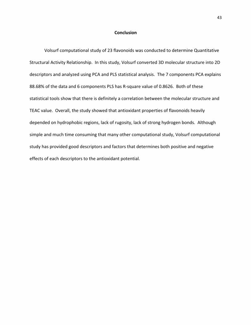
Tables and Figures
Subclass Molecule
ID Compound TEAC value Structure
Flavanol 2 epigallocatechin gallate (EGCG) 4.80
4 epicatechin (EC) 2.50
5 taxifolin 1.90
6 catechin 2.40
Flavonol 7 quercetin 4.70
8 myricetin 3.10
9 morin 2.55
10 rutin 2.40

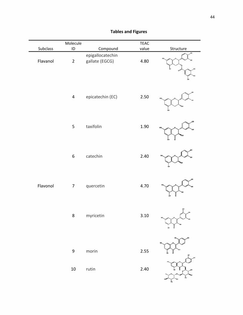
45
11 kaempferol 1.34
Flavone 12 luteolin 2.10
14 apigenin 1.45
15 chrysin 1.43
Flavanone 17 naringenin 1.53
18 hesperetin 1.37
19 hesperidin 1.08
20 narirutin 0.76
21 dihydrokaempferol 1.39
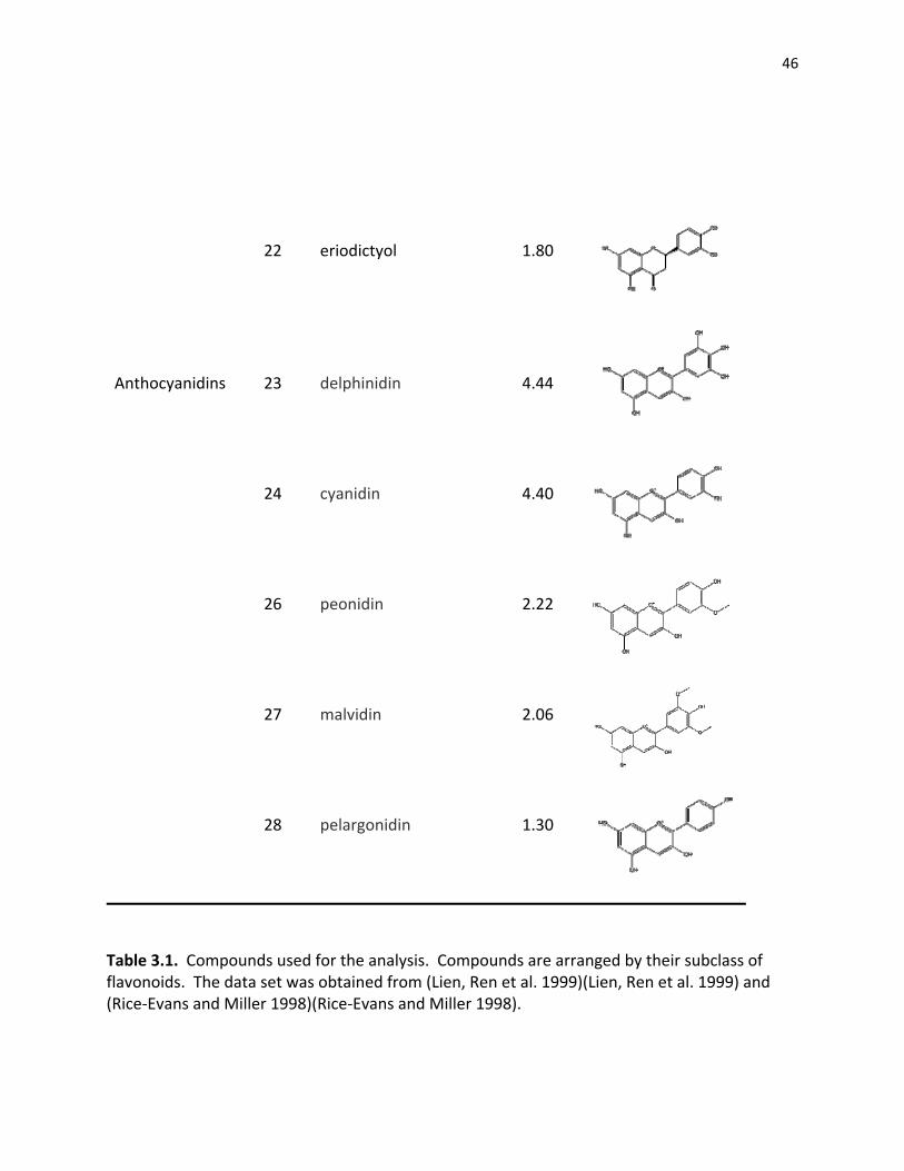
46
22 eriodictyol 1.80
Anthocyanidins 23 delphinidin 4.44
24 cyanidin 4.40
26 peonidin 2.22
27 malvidin 2.06
28 pelargonidin 1.30
Table 3.1. Compounds used for the analysis. Compounds are arranged by their subclass of flavonoids. The data set was obtained from (Lien, Ren et al. 1999)(Lien, Ren et al. 1999) and (Rice‐Evans and Miller 1998)(Rice‐Evans and Miller 1998).

47
Table 3.2. MMFF94 Energy Minimized structures.
Molecule ID Total Energy (kcal/mol) 2 34.8 4 51.2 5 70.0 6 43.1 7 43.4 8 51.4 9 54.5 10 242.5 11 57.5 12 10.0 14 34.7 15 46.0 17 35.5 18 44.9 19 237.8 20 213.0 21 21.3 22 21.3 23 31.4 24 31.3 26 52.0 27 63.9 28 42.9

48
Figure 3.3. 2D PCA score plot
2D PCA score plot shows relative position of molecules in comparison with others. It is used to find outliers and cluster of related molecules.
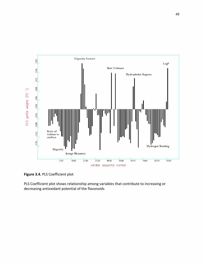
49
Figure 3.4. PLS Coefficient plot
PLS Coefficient plot shows relationship among variables that contribute to increasing or decreasing antioxidant potential of the flavonoids

50
Reference
Adams, D. J. (1983). Introduction to Monte Carlo Simulation Techniques. New York, Plenum. Bobbyer, D. N. A., P. J. Goodford, et al. (1989). "New Hydrogen‐Bond Potential for Use in Determining Energetically Favourable Binding Sites of Molecules of Known Structure." J. Med. Chem.(32): 1083‐1094. Chun, O. K., D. O. Kim, et al. (2003). "Superoxide Radical Scavenging Activity of the Major Polyphenols in Fresh Plums." J. Agric. Food Chem. 51(27): 8067‐8072. Crivori, P., G. Cruciani, et al. (2000). J. Med. Chem. 43(11): 2204‐2216. Crivori, P., I. Zamora, et al. (2004). J. comput. ‐Aided Mol. Cruciani, G. and M. Meniconi (2003). Drug Bioavailability. Ejaz, A., S. Ejaz, et al. (2006). "Limonoids as cancer chemopreventive agents." Journal of the Science of Food and Agriculture (86): 339‐345. Farkas, O., J. Jakus, et al. (2004). "Quantitative Structure – Antioxidant Activity Relationships of Flavonoid Compounds." Molecules 9: 1079‐1088. Goodford, P. J. (1985). "Computational Procedure for Determining Energetically Favourable Binding Sites on Biologically Important Macromolecules." J. Med. Chem.(28): 849‐857. Harborne, J. B. and C. A. Williams (2000). "Advances in flavonoid research since 1992." Phytochemistry 55(6): 481‐504. Hertog, M. G. L., E. J. M. Feskens, et al. (1993). "Dietary antioxidant flavonoids and risk of coronary heart disease: the Zutphen Elderly Study." The Lancet 342(8878): 1007‐1011. Hertog, M. G. L., P. C. H. Hollman, et al. (1992). "Content of potentially anticarcinogenic flavonoids of 28 vegetables and 9 fruits commonly consumed in Netherlands." Journal of Agricultural and Food Chemistry (40): 2379–2383. Jackson, J. E. (1991). A Users Guide to Principal Components. New York. Joliffe, I. T. (1986). Principal Component Analysis. New York. Keys, A. (1995). "Mediterranean diet and public health: Personal reflections." American Journal of Clinical Nutrition (61): 1321–1323.

51
Kim, D.‐O., S. W. Jeong, et al. (2003). "Antioxidant capacity of phenolic phytochemicals from various cultivars of plums." Food Chemistry 81(3): 321‐326. Leach, A. R. (2001). Molecular Modelling: Principles and Applications. Harlow, Pearson Prentice Hall. Lien, E. J., S. Ren, et al. (1999). "Quantitative structure‐activity relationship analysis of phenolic antioxidants." Free Radical Biology and Medicine 26(3‐4): 285‐294. Lombardo, F., R. S. Obach, et al. (2002). J. Med. Chem. 45(13): 2867‐2876. Middleton, E. J. and C. Kandaswami (1992). "Effects of flavonoids on immune and inflammatory cell functions." Biochem Pharmacol 43: 1167‐1179. Middleton, E. J., C. Kandaswami, et al. (2000). "The effects of plant flavonoids on mammalian cells: implications for inflammation, heart disease, and cancer." Pharmacol Rev 52: 673‐751. Oprea, T. I., I. Zamora, et al. (2002). Comb. Chem. 4(4): 258‐266. Packer, L., M. Hiramatsu, et al. (1999). Antioxidant food supplements in human health. San Diego, Academic Press. Pearlstein, R., R. Vaz, et al. (2003). J. Med. Chem. 46(11): 2017‐2022. Rice‐Evans, C. A. and N. J. Miller (1998). Structure‐antioxidant activity relationships of flavonoids and isoflavonoids. New York, Marcel Dekker, Inc. Rubinstein, R. Y. (1981). Simulation and Monte Carlo Methods. New York, John Wiley & Sons. Shils, M. E. and R. S. Goodhart (1956.). The Flavonoids in Biology and Medicine: a Critical Review. New York,, New York National Vitamin Foundation. Teixeira, S., C. Siquet, et al. (2005). "Structure‐property studies on the antioxidant activity of flavonoids present in diet." Free Radical Biology and Medicine 39(8): 1099‐1108. Thomas, A. and J. Halgren (1996). "Merck Molecular Force Field. I. Basis, Form, Scope, Parameterization, and Performance of MMFF94." J. Comput. Chem 17: 490‐519 Volsurf version 3.0 software by Molecular Discovery Ltd.
52
Chapter 4
QUANTITATIVE STRUCTURE‐PERMEABILITY RELATIONSHIP OF FLAVONOIDS USING CACO‐2
CELLS: INDEPTH STUDY OF HUMAN INTESTINAL PERMEATION OF LAVONOIDS

53
ABSTRACT
Flavonoids are widely available compounds that are often present in human diet. Many
studies of flavonoids exist, but not many studies focus on bioavailability. As interest in
flavonoids grow stronger due to health benefits of antioxidant, further bioavailability study is
essential. Over the years, Caco‐2 cells are used to model human intestinal absorption of many
compounds. In recent years, computational studies have been developed in conjunction with
experimental data to develop a highly sophisticated and accurate Caco‐2 model. In this study,
QSPR or Quantitative Structural‐Permeability Relationship study of 17 molecules was
conducted for Caco‐2 study. All of the molecules were obtained from the Cambridge Software
Molecular Database. The structures were energy minimized using MMFF94 and converted into
2D descriptors by the Grid and Volsurf Program. Then, descriptors were compared to the 751
existing Caco‐2 database. 94.1% of QSAR structures match permeability data of experimentally
derived data. Moreover, we were able to conclude hydrophobic and smaller molecular weight
flavonoids show much higher permeability.

54
Introduction
A diet rich in fruits and vegetables has been related to various positive health benefits.
These include many chronic conditions that affect numerous people world‐wide (Doll 1990;
Ames, Shigenaga et al. 1993; Sun, Chu et al. 2002)(Doll 1990; Ames, Shigenaga et al. 1993; Sun,
Chu et al. 2002). These protective effects are related to antioxidants (Moreira, Fraga et al.
2004; Kwon, Murakami et al. 2005)(Moreira, Fraga et al. 2004; Kwon, Murakami et al. 2005).
Flavonoids are widely present in the plant kingdom and account for approximately half of more
than 8000 phenolic compounds found in nature (Harborne, Baxter et al. 1999)(Harborne, Baxter
et al. 1999). Additionally, they are associated with health benefits of protection against
atherosclerosis (Hertog, Hollman et al. 1992; Keys 1995)(Hertog, Hollman et al. 1992; Keys
1995), inflammatory conditions (Middleton, Kandaswami et al. 2000)(Middleton, Kandaswami
et al. 2000), allergenic conditions, artherogenic conditions, microbial infections, thrombotic
problems, and provide cardio protective effects beyond basic nutrition (Manach, Mazur et al.
2005)(Manach, Mazur et al. 2005). These medicinal effects are believed to be the antioxidant
activity of the flavonoids which quench free radicals, free radical anions, and peroxy radicals.
However, proper dosage of flavonoids in the human diets is unknown due to limited studies on
bioavailability and bioassesibility. Therefore, digestion and absorption of flavonoids in the
human gastrointestinal system is important, and related studies have been conducted using
different methods, such as in vivo (Hollman, Vries et al. 1995; Hollman, Trijp et al.
1997)(Hollman, Vries et al. 1995; Hollman, Trijp et al. 1997), in vitro (Kuo 1998; Murota, Shimizu
et al. 2000; Masataka Oitate 2001)(Kuo 1998; Murota, Shimizu et al. 2000; Masataka Oitate
2001), and computational studies (Ekins, Durst et al. 2001; Ponce, Perez et al. 2004)(Ekins,

55
Durst et al. 2001; Ponce, Perez et al. 2004). Diffusion mechanism and permeation study is
divided into two main types—passive diffusion and active transport. For the passive diffusion,
2/4/A1 cell, PAMPA, chromatography, and LogP are used. For active transport, Caco‐2, MDCK‐
MDR1, and transformed cells with transporters are used (Ungell 2004)(Ungell 2004). Among
these however, Caco‐2 cells, (Hidalgo, Raub et al. 1989)(Hidalgo, Raub et al. 1989) or
differentiated monolayer cells of human colon adenocarcinoma, are selected due to their wide
acceptance as a model for human intestinal absorption. The Caco‐2 cell method is active
transport method that can determine permeability of samples through the medium. Onto this
medium, sample and buffer mix is loaded. The samples that need specific molecules or
chemicals, HPLC or other separation methods are necessary to obtain more pure forms. After a
certain time period, ranging from few minutes to days, the distance of permeation is measured.
Although the distance permeable is different from sample to sample, many experiments use 10‐
6 cm/sec or permeation length over time (Tammela, Laitinen et al. 2004; Zuo, Zhang et al. 2006;
Serra, Mendes et al. 2008)(Tammela, Laitinen et al. 2004; Zuo, Zhang et al. 2006; Serra, Mendes
et al. 2008).
In terms of faster and newer scientific methods, computational studies and models are
widely accepted for drug lead optimization and knowledge that can benefit the community.
Computational methods, such as quantitative structure‐permeability relationships, are
important and needed to predict intestinal absorptivity of flavonoids. In QSPR, molecular
structures of compounds are turned into descriptors and matched with preexisting database for
the purpose of finding structural‐permeability relationship. The end result is quantitative
permeation data based on structure (Hansch, Leo et al. 2004)(Hansch, Leo et al. 2004). Not

56
only is this method faster, economically efficient, and has less variability due to the nature of
the software, but also it can be used for molecular structures that are difficult to isolate,
unstable, and can use different solvents to develop a model that can predict human digestion
and absorption model.
In the experiment, Caco‐2 cells were chosen because previous studies by (Murota,
Shimizu et al. 2000; Murota, Shimizu et al. 2002)(Murota, Shimizu et al. 2000; Murota, Shimizu
et al. 2002) have shown that flavonoids are ideal candidates for Caco‐2 studies due to their
lipophilic nature. Although Caco‐2 cells have been used to study bioavailability of many
compounds, many flaws do exist. It is costly, time consuming, lack of precision among labs, and
requires expertise to handle. Therefore, many recent studies used QSPR have to study
permeability. One of the tools for QSPR is Volsurf and it contains 751 Caco‐2 permeation data
and their structural descriptors.
Volsurf is a tool that can predict properties various molecules and serve important to
drug‐lead optimization and bioavailability of numerous bioflavonoids. It works by converting
3D molecular information into 2D descriptors, such as hydrophobic regions, hydrophilic regions,
strength of hydrogen bonds, log cp values, size, and other characteristics of molecule using
various different probes (Volsurf 2000‐2004)(Volsurf 2000‐2004). The converted descriptors
can be matched with database of 751 molecules where the descriptors and Caco‐2 permeability
has been integrated. In order to generate descriptors, Volsurf uses probes, designed
specifically to examine molecules via measuring interaction energies between the probes and
molecules of interest. However, one of the most powerful advantages is its accuracy and short

57
amount of time it takes to convert 3D molecules into 2D descriptors. Furthermore, existing cell
culture studies can be used to correlate and build a database that can relate molecules and
their properties to selected variable in study (Ungell 2004)(Ungell 2004). Therefore, library of
flavonoids was selected to run in the Volsurf program in order to compare Caco‐2 properties of
experimentally found data to the database exist in the Volsurf. Not only has this method
required less time requirements, the models exist in the Volsurf database has been proven to
predict various properties of array of different drugs. Moreover, there has been an increase in
molecular databases that incorporate structural and experimental Caco‐2 data and prediction
and accuracy of Caco‐2 permeability increases as the database grows (Ungell 2004)(Ungell
2004). Furthermore, experimental Caco‐2 studies is expensive, time consuming, lacks
quantitative measurement of impermeable molecules, and yields lower quality data due to
different materials and methods being used. In a computation study, even the impermeable
molecules are assigned a quantitative value, faster to run, and is consistent world‐wide. In
order to study QSPR, Volsurf (Volsurf version 3.0 software by Molecular Discovery Ltd)(Volsurf
version 3.0 software by Molecular Discovery Ltd) is used to predict permeability of flavonoids
using its library of 751 related, yet diverse chemical compounds and their experimental Caco‐2
permeability data. Additionally, non flavonoid, phenolic compounds were selected to test the
Volsurf model using external compounds.

58
Methods
A Total of 17 structures of 12 flavonoids and 5 non‐flavonoids phenolic compounds and
their experimental Caco‐2 permeabilities were selected from existing experiments (Tammela,
Laitinen et al. 2004; Serra, Mendes et al. 2008)(Tammela, Laitinen et al. 2004; Serra, Mendes et
al. 2008). These are shown in Figure 4.1. The selected molecules were obtained from the
Cambridge Structural Database, a large database that works with ChemBio3D Ultra program
(Halgren 1996)(Halgren 1996).
Energy Minimization
All structures were minimized using MMFF94 (Thomas and Halgren 1996)(Thomas and Halgren
1996). The details of this energy minimization called for maximum iteration at 5000 with
minimum RMS gradient of 0.0010. This was conducted to ensure that the energies of
molecules were closely accurate to its true energy minima. Energy minimization step was
carefully conducted with maximum precision as improper energy minimization would not
generate accurate data. MMFF94 was selected due to its powerful ability to minimize both
small organic and large structures (Halgren 1996)(Halgren 1996).
QSPR Modeling
After MMFF94 energy minimization, all molecules were compiled and submitted to the Volsurf
program. During this step, all 3D molecules were turned to 2D descriptors. In this step, OH2,
Dry and O probes were used. The probes work to determine important descriptors for
molecules. For example, OH2 probe is especially useful in determination of hydrophilic regions,

59
dry probe looks for hydrophobic regions, and O looks for hydrogen bonds in general. More
detail of different probes and their functions are listed in the Volsurf manual (Volsurf version
3.0 software by Molecular Discovery Ltd)(Volsurf version 3.0 software by Molecular Discovery
Ltd) and available online http://www.moldiscovery.com/docs/volsurf/intro.html. For the Caco‐
2 study, 751 different molecules, their related 2D descriptors, and experimentally found Caco‐2
values were incorporated into the database. In this experiment, molecular structures of the
flavonoids are imposed onto the Volsurf database to determine human intestinal permeability.
This is possible by comparing 2D descriptors from flavonoids and comparing to the existing
Volsurf library, thus predicting Caco‐2 permeation of flavonoid to high degree of accuracy.
Result and Discussion
Figure 4.2 shows Caco‐2 permeation of all 17 molecules. The three linear lines in the
middle of the graph is the dividing line that determines whether a molecule can permeate
through the Caco‐2 monolayer or not. Two lines around the center line is the confidence
interval for the permeation separation line. Molecules further away from the division line to
the right shows increasing Caco‐2 permeation. Likewise, molecules further away from the left
shows decreasing Caco‐2 permeation.
The Volsurf model predicted at 94.1% accuracy for all molecules whether they are
permeable or impermeable. In detail, all structures were categorized into either permeable or
impermeable groups. The relationship between the experimental and the computational data
indicate 94.1 % accuracy that QSRP study can determine permeability of flavonoids.
Methylgallate and morin are close to the dividing line, indicating increasing chance of error in
60
true representation of their Caco‐2 permeability. However, previous studies show that morin is
not permeable, while methylgallate has very slightly permeable (Tammela, Laitinen et al.
2004)(Tammela, Laitinen et al. 2004); therefore, computational study by Volsurf shows its high
degree of sensitivity for the Caco‐2 permeation. Naringin, diosmin, and hesperidin show high
impermeability. This is due to their large molecular size with addition of two gallate groups to
basic flavonoid structure. This also correlates to data found in the previous experiment that
related increase in molecular size decreases caco‐2 permeability (Gonthier, Donovan et al.
2003; Tsang, Auger et al. 2005)(Gonthier, Donovan et al. 2003; Tsang, Auger et al. 2005). In
contrast, flavone has the highest permeability due to lacks of hydroxy groups in the molecule,
increasing hydrophobicity. In another study, similar result was found (Murota and Terao
2003)(Murota and Terao 2003). They concluded that flavonoids permeability increase as
hydrophobicity increases. Paracetamol shows the next highest permeation data, and is largely
due to its small molecular weight and size.
In the following portion of the study, we cross examined the Volsurf using phenolic
compounds that are non‐flavonoids. This was conducted to test the Volsurf and its processing
abilities for molecular size, hydroxy groups, and reaction to the hydrophobicity. According to
earlier works by (Salucci, Stivala et al. 2002)(Salucci, Stivala et al. 2002), smaller molecules like
gallic acid have good permeability. Further examination shows increasing permeability of gallic
acid as hydrocarbon tail length increases. Longer tail increases hydrophobic nature of the
molecule, increasing permeability through the Caco‐2 cells.

61
Finally, a model was created using data of Caco‐2 permeable molecules. The closest
distance between the permeation separation line and the flavonoids were measured. The
predicted QSPR values were tested via comparison with experimental Caco‐2 data as shown in
Figure 4.3. Methylgallate has the higest correlation, followed by diosmetin and hesperetin. On
the other hand, paracetamol showed the least correlation. Among all the molecules, flavonoid
molecules composed of flavones, naringenin, diosmetin, and hesperatin. All of the flavonoids
molecules showed great relationship between experimental and QSPR data. More importantly,
Figure 4.3 shows that Volsurf is fairly accurate in representation of Caco‐2 permeability for both
phenolic compounds and flavonoids.
Conclusion
Quantitative structure‐permeability relationship using Volsurf and Caco‐2 monolayer
cells has been conducted. As with the general understanding in the beginning, impermeable
molecules were associated with magnitude, that explains how much less likely the permeation
through the Caco‐2 cells occur. This could be very useful during modification of compounds
during drug‐lead optimization. Overall, Volsurf direct prediction was able to determine with
94.1% of accuracy whether a molecule will be permeable or not. However, prediction of
permeable data proved to be more difficult. But with more data points in the database, the
model can become very accurate in the years to come.

62
Tables and Figures
Molecule ID Total Energy (kcal/mol) 1 43.1 2 51.2 3 52.4 4 10.0 5 12.1 6 48.2 7 35.5 8 224.8 9 12.9 10 14.0 11 43.4 12 71.5 13 ‐11.9 14 233.0 15 237.7 16 39.4 17 44.9
Table 4.1. MMFF94 Energy Minimized structures.
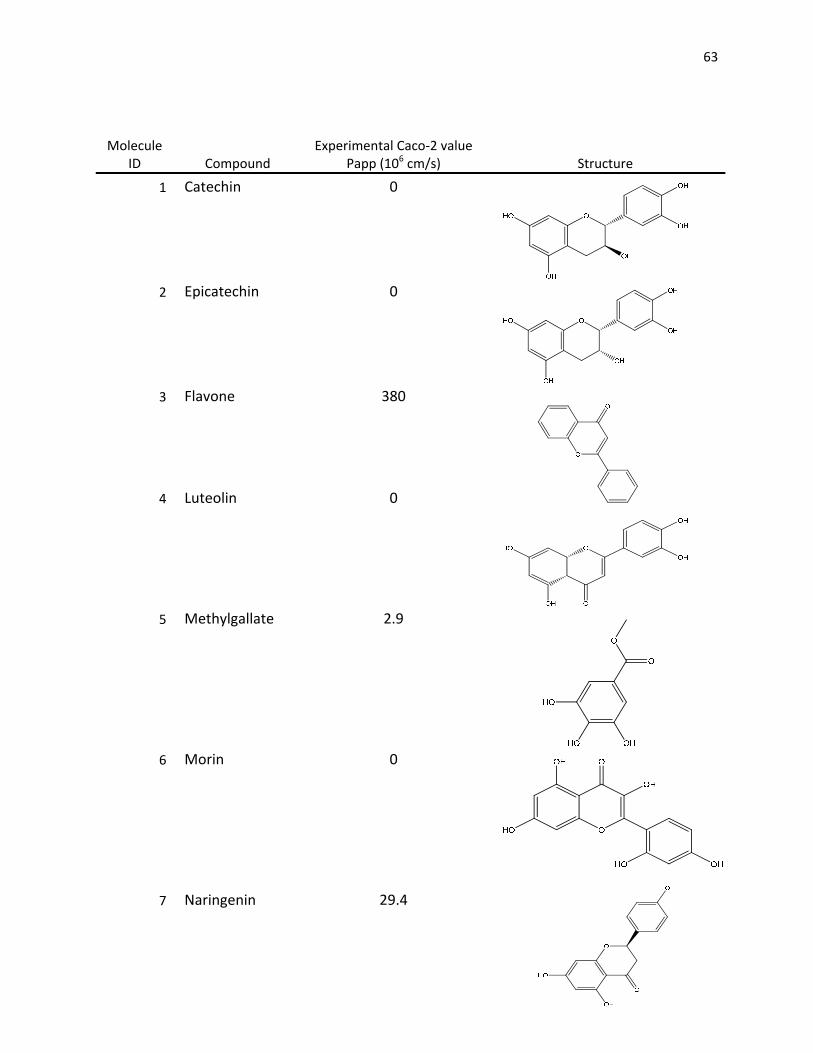
63
Molecule ID Compound
Experimental Caco‐2 value Papp (106 cm/s) Structure
1 Catechin 0
2 Epicatechin 0
3 Flavone 380
4 Luteolin 0
5 Methylgallate 2.9
6 Morin 0
7 Naringenin 29.4

64
8 Naringin 0
9 Octylgallate 0
10 Propylgallate 7.3
11 Quercetin 0
12 Ketoprofen 24.6
13 Paracetamol 22
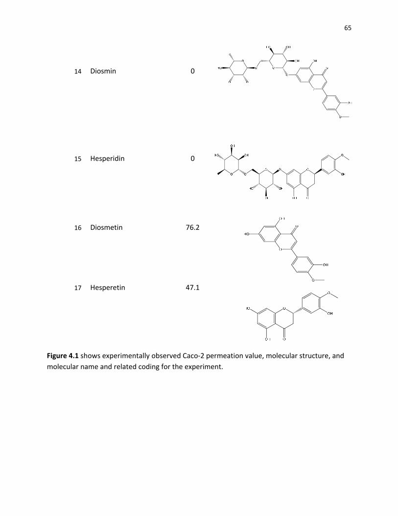
65
14 Diosmin 0
15 Hesperidin
0
16 Diosmetin 76.2
17 Hesperetin 47.1
Figure 4.1 shows experimentally observed Caco‐2 permeation value, molecular structure, and molecular name and related coding for the experiment.
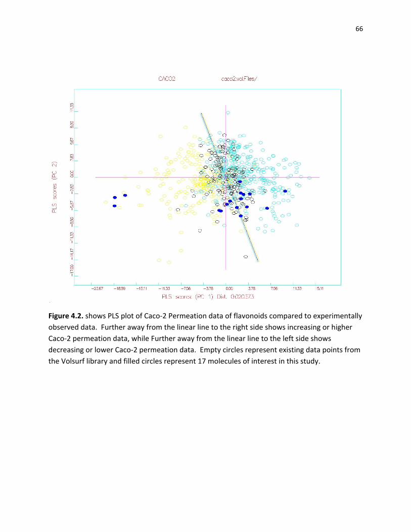
66
Figure 4.2. shows PLS plot of Caco‐2 Permeation data of flavonoids compared to experimentally observed data. Further away from the linear line to the right side shows increasing or higher Caco‐2 permeation data, while Further away from the linear line to the left side shows decreasing or lower Caco‐2 permeation data. Empty circles represent existing data points from the Volsurf library and filled circles represent 17 molecules of interest in this study.

67
Figure 4.3. Caco‐2 shows permeation relationship between experimental and QSPR data. Paracetamol has the largest gap, while methylgallate has the lowest gap.
00.51
1.52
2.53
3.54
4.55
Caco‐2 Permeation (106 cm/s)Experimental Computational Caco‐2 / 100

68
References
. "VolSurf manual." Retrieved 4/15, 2008, from http://www.moldiscovery.com/docs/volsurf/intro.html. Adams, D. J. (1983). Introduction to Monte Carlo Simulation Techniques. New York, Plenum. Allinger, N. L., Y. H. Yuh, et al. (1989). "Molecular Mechanics. The MM3 Force Field for Hydrocarbons." Journal of the American Chemical Society(111): 8551‐8565. Amado, L. L., M. D. Jaramillo, et al. (2007). "A New Method to Evaluate Total Antioxidant Capacity Against Reactive Oxygen and Nitrogen Species (RONS) in Aquatic Organisms." Comparative Biochemistry and Physiology ‐ Part A: Molecular & Integrative Physiology 148(Supplement 1): S75‐S76. Ames, B. N., M. K. Shigenaga, et al. (1993). "Oxidants, Antioxidants, and the Degenerative Diseases of Aging." Proceedings of the National Academy of Sciences 90(17): 7915‐7922. Arts, M. J. T. J., J. Sebastiaan Dallinga, et al. (2003). "A Critical Appraisal of the Use of the Antioxidant Capacity (TEAC) Assay in Defining Optimal Antioxidant Structures." Food Chemistry 80(3): 409‐414. Artursson, P., K. Palm, et al. (2001). "Caco‐2 Monolayers in Experimental and Theoretical Predictions of Drug Transport." Advanced Drug Delivery Reviews 46: 27‐43. Baroni, M., G. Costantino, et al. (1993). "Generating Optimal Linear PLS Estimations (GOLPE): an Advanced Chemometric Tool for Handling 3D‐QSAR Problems. ." Quantitative Structure‐Activity Relationships 12: 9‐20. Benzie, I. F. F. and J. J. Strain (1999). "Reducing Antioxidant Power Assay: Direct Measure of Total Antioxidant Activity of Biological Fluids and Modified Version for Simultaneous Measurement of Total Antioxidant Power and Sscorbic Acid Concentration." Methods in Enzymology 299: 15‐27. Bobbyer, D. N. A., P. J. Goodford, et al. (1989). "New Hydrogen‐Bond Potential for Use in Determining Energetically Favourable Binding Sites of Molecules of Known Structure." Journal of Medicinal Chemistry (32): 1083‐1094. Brand‐Williams, W., M. E. Cuvelier, et al. (1995). "Use of a Free Radical Method to Evaluate Antioxidant Activity." Food Science and Technology 28: 25‐30. Burda, S. and W. Oleszek (2001). "Antioxidant and Antiradical Activities of Flavonoids." Journal of Agricultural and Food Chemistry 49: 2774‐2779. Chatfield, C. and A. J. Collins (1980). Introduction to Multivariate Analysis. London, Chapman & Hall. Chen, Z. Y., P. T. Chan, et al. (1996). "Antioxidant Activity of Natural Flavonoids is Governed by Number and Location of Their Aromatic Hydroxyl Groups." Chemistry and Physics of Lipids 79(2): 157‐163. Cheng, H. Y., T. C. Lin, et al. (2003). "Antioxidant and Free Radical Scavenging Activities of Terminalia Chebula." Biological & Pharmaceutical Bulletin 26: 1331‐1335.
69
Chun, O. K., D. O. Kim, et al. (2003). "Superoxide Radical Scavenging Activity of the Major Polyphenols in Fresh Plums." Journal of Agricultural and Food Chemistry 51(27): 8067‐8072. Crivori, P., G. Cruciani, et al. (2000). "Molecular Fields in Quantitative Structurepermeation Relationships: The VolSurf Approach." Journal of Medicinal Chemistry 43(11): 2204‐2216. Crivori, P., I. Zamora, et al. (2004). J. comput. ‐Aided Mol. Cruciani, G., P. Crivori, et al. (2000). "Molecular Fields in Quantitative Structure‐Permeation Relationships: the VolSurf Approach." Journal of Molecular Structure 503(1‐2): 17‐30. Cruciani, G. and M. Meniconi (2003). Drug Bioavailability. New York, Wiley‐VCH. Dastmalchi, K., H. J. Damien Dorman, et al. (2007). "Chemical Composition and Antioxidative Activity of Moldavian Balm (Dracocephalum moldavica L.) Extracts." Food Science and Technology 40(9): 1655‐1663. Delgado‐Andrade, C., I. Seiquer, et al. (2008). "Estimation of Hydroxymethylfurfural Availability in Breakfast Cereals. Studies in Caco‐2 Cells." Food and Chemical Toxicology 46(5): 1600‐1607. Doll, R. (1990). "An Overview of the Epidemiological Evidence Linking Diet and Cancer." Proceedings of Nutrition and Society 49(1): 119‐131. Ejaz, A., S. Ejaz, et al. (2006). "Limonoids as Cancer Chemopreventive Agents." Journal of the Science of Food and Agriculture (86): 339‐345. Ekins, S., G. L. Durst, et al. (2001). "Three‐Dimensional Quantitative Structure‐Permeability Relationship Analysis for a Series of Inhibitors of Rhinovirus Replication." Journal of Chemical Information and Computer Sciences 41(6): 1578‐1586. Farkas, O., J. Jakus, et al. (2004). "Quantitative Structure – Antioxidant Activity Relationships of Flavonoid Compounds." Molecules 9: 1079‐1088. Glazer, A. N. (1990). "Phycoerythrin Fluorescence‐Based Assay for Reactive Oxygen Species." Methods in Enzymology 186: 161‐168. Gonthier, M.‐P., J. L. Donovan, et al. (2003). "Metabolism of Dietary Procyanidins in Rats." Free Radical Biology and Medicine 35(8): 837‐844. Goodford, P. J. (1985). "Computational Procedure for Determining Energetically Favourable Binding Sites on Biologically Important Macromolecules." Journal of Medicinal Chemistry(28): 849‐857. Halgren, T. A. (1996). "A Broadly Parameterized, Computationally Derived Force Field for Organic and Bio‐organic Systems." Retrieved 4/10, 2008. Halgren, T. J. (1996). "Merck Molecular Force Field. I. Basis, Form, Scope, Parameterization, and Performance of MMFF94." Journal of Computational Chemistry 17: 490‐519

70
Halgren, T. J. (1996). "Merck Molecular Force Field. II. MMFF94 van der Waals and Electrostatic Parameters for Intermolecular Interactions." Journal of Computational Chemistry 17: 520‐552. Halgren, T. J. (1996). "Merck Molecular Force Field. III. Molecular Geometries and Vibrational Frequencies for MMFF94." Journal of Computational Chemistry 17: 553‐586 Halgren, T. J. (1996). "Merck Molecular Force Field. IV. Conformational Energies and Geometries for MMFF94," Thomas A. Halgren and Robert B. Nachbar." Journal of Computational Chemistry 17: 587‐615. Halgren, T. J. (1996). "Merck Molecular Force Field. V. Extension of MMFF94 Using Experimental Data, Additonal Computational Data, and Empirical Rules." Journal of Computational Chemistry 17: 616‐641 Hansch, C., A. Leo, et al. (2004). "QSAR and ADME." Bioorganic & Medicinal Chemistry 12(12): 3391‐3400. Harborne, J. B., H. Baxter, et al. (1999). Phytochemical Dictionary, Handbook of Bioactive Compounds from Plants. London, Taylor and Francis. Harborne, J. B. and C. A. Williams (2000). "Advances in Flavonoid Research since 1992." Phytochemistry 55(6): 481‐504. Hertog, M. G. L., E. J. M. Feskens, et al. (1993). "Dietary Antioxidant Flavonoids and Risk of Coronary Heart Disease: the Zutphen Elderly Study." The Lancet 342(8878): 1007‐1011. Hertog, M. G. L., P. C. H. Hollman, et al. (1992). "Content of Potentially Anticarcinogenic Flavonoids of 28 Vegetables and 9 Fruits Commonly Consumed in Netherlands." Journal of Agricultural and Food Chemistry (40): 2379–2383. Hidalgo, I. J., T. J. Raub, et al. (1989). "D‐cycloserine Uses an Active Transport Mechanism in the Human Intestinal Cell Line Caco 2." Gastroenterology 96: 736. Hollman, P. C. H., J. M. P. Trijp, et al. (1997). "Bioavailability of the Antioxidant Flavonoid Quercetin from Various Foods in Man." Federation of Biochemical Societies 418: 152‐516. Hollman, P. C. H., J. H. M. Vries, et al. (1995). "Absorption of Dietary Quercetin Glycosides and Quercetin in Healthy Ileostomy Volunteers." American Journal of Clinical Nutrition 62: 1276‐1282. Keys, A. (1995). "Mediterranean Diet and Public Health: Personal Reflections." American Journal of Clinical Nutrition (61): 1321–1323. Kim, D.‐O., S. W. Jeong, et al. (2003). "Antioxidant Capacity of Phenolic Phytochemicals from Various Cultivars of Plums." Food Chemistry 81(3): 321‐326. Kinsella, J. E., E. Frankel, et al. (1993). "Possible Mechanisms for the Protective Role of Antioxidants in Wine and Plant Foods." Food Technology(47): 85‐89.
71
Klein, E. and V. Lukes (2006). "DFT/B3LYP Study of the Substituent Effect on the Reaction Enthalpies of the Individual Steps of Sequential Proton Loss Electron Transfer Mechanism of Phenols Antioxidant Action: Correlation with Phenolic C‐O Bond Length." Journal of Molecular Structure 805: 153‐160. Kobayashi, S. and Y. Konishi (2008). "Transepithelial Transport of Flavanone in Intestinal Caco‐2 Cell Monolayers." Biochemical and Biophysical Research Communications 368(1): 23‐29. Kohen, R., E. Beit‐Yannai, et al. (1999). "Overall Low Molecular Weight Antioxidant Activity of Biological Fluids and Tissues by Cyclic Voltammetry." Methods in Enzymology 300: 285‐296. Kozlowski, D., P. Trouillas, et al. (2007). "Density Functional Theory study of the Conformational, Electronic, and Antioxidant Properties of Natural Chalcones." Journal of Physical Chemistry A(111): 1138‐1145. Kuo, S.‐M. (1998). "Transepithelial Transport and Accumulation of Flavone in Human Intestinal CACO‐2 Cells." Life Sciences 63(26): 2323‐2331. Kwon, K. H., A. Murakami, et al. (2005). "Dietary Rutin, but not Its Aglycone Quercetin, Ameliorates Dextran Sulfate Sodium‐Induced Experimental Colitis in Mice: Attenuation of Pro‐Inflammatory Gene Expression." Biochemical Pharmacology 69(3): 395‐406. Laparra, J. M., E. Tako, et al. (2008). "Supplemental Inulin does not Enhance Iron Bioavailability to Caco‐2 Cells from Milk or Soy‐based, Probiotic‐Containing, Yogurts but Incubation at 37 °C does." Food Chemistry 109(1): 122‐128. Leach, A. R. (2001). Molecular Modelling: Principles and Applications. Harlow, Pearson Prentice Hall. Lien, E. J., S. Ren, et al. (1999). "Quantitative Structure‐Activity Relationship Analysis of Phenolic Antioxidants." Free Radical Biology and Medicine 26(3‐4): 285‐294. Lombardo, F., R. S. Obach, et al. (2002). "Prediction of Volume of Distribution Values in Humans for Neutral and Basic Drugs Using Physicochemical Measurements and Plasma Protein Binding Data." Journal of Medicinal Chemistry 45(13): 2867‐2876. Lv, H., G. Wang, et al. (2008). "Transport Characteristics of Ginkgolide B by Caco‐2 Cells and Examination of Ginkgolide B Oral Absorption Potential Using Rat in Situ Intestinal Loop Method." International Journal of Pharmaceutics 351(1‐2): 31‐35. Manach, C., A. Mazur, et al. (2005). "Polyphenols and Prevention of Cardiovascular Diseases." Current Opinion in Lipidology 16: 77‐84. Mannhold, R., G. Cruciani, et al. (1999). "6‐varied Benzopyrans as Potassium Channel Activators: Synthesis, Vasodilator Properties and Multivariate Analysis." Journal of Medicinal Chemistry 42: 981. Masataka Oitate, R. N. N. K. H. T. H. M. H. O. Y. S. (2001). "Transcellular Transport of Genistein, a Soybean‐Derived Isoflavone, Across Human Colon Carcinoma Cell Line (Caco‐2)." Biopharmaceutics & Drug Disposition 22(1): 23‐29.

72
Mendoza‐Wilson, A. M. a., D. Lardizabal‐Gutierrez, et al. (2007). "Optimized Structure and Thermochemical Properties of Flavonoids Determined by the CHIH(medium)‐DFT Model Chemistry Versus Experimental Techniques." Journal of Molecular Structure. Middleton, E. J. and C. Kandaswami (1992). "Effects of Flavonoids on Immune and Inflammatory Cell Functions." Biochemical Pharmacology 43: 1167‐1179. Middleton, E. J., C. Kandaswami, et al. (2000). "The Effects of Plant Flavonoids on Mammalian Cells: Implications for Inflammation, Heart Disease, and Cancer." Pharmacological Reviews 52: 673‐751. Miller, N. J., C. A. Rice‐Evans, et al. (1993). "A Novel Method for Measuring Antioxidant Capacity and Its Application to Monitoring the Antioxidant Status in Premature Neonates." Clinical Science 84: 407‐412. Moreira, A. J., C. Fraga, et al. (2004). "Quercetin Prevents Oxidative Stress and NF‐[kappa]B Activation in Gastric Mucosa of Portal Hypertensive Rats." Biochemical Pharmacology 68(10): 1939‐1946. Murota, K., S. Shimizu, et al. (2000). "Efficiency of Absorption and Metabolic Conversion of Quercetin and Its Glucosides in Human Intestinal Cell Line Caco‐2." Archives of Biochemistry and Biophysics 384(2): 391‐397. Murota, K., S. Shimizu, et al. (2000). "Efficiency of Absorption and Metabolic Conversion of Quercetin and Its Glucosides in Human Intestinal Cell Line " Biochemical and Biophysical Research Communications (384): 391‐397. Murota, K., S. Shimizu, et al. (2002). "Unique Uptake and Transport of Isoflavone Aglycones by Human Intestinal Caco‐2 Cells: Comparison of Isoflavonoids and Flavonoids." Journal of Nutrition 132: 1956‐1961. Murota, K. and J. Terao (2003). "Antioxidative Flavonoid Quercetin: Implication of Its Intestinal Absorption and Metabolism." Biochemistry and Biophysics 417: 12‐17. Oprea, T. I., I. Zamora, et al. (2002). "Pharmacokinetically Based Mapping Device for Chemical Space Navigation." Combinatorial Chemistry And High Throughput Screening 4(4): 258‐266. Packer, L., M. Hiramatsu, et al. (1999). Antioxidant Food Supplements in Human Health. San Diego, Academic Press. Pearlstein, R., R. Vaz, et al. (2003). "Understanding the Structure‐Activity Relationship of the Human Ether‐a‐go‐go‐Related Gene Cardiac K+ Channel. A Model for Bad Behavior." Journal of Medicinal Chemistry 46(11): 2017‐2022. Pellegrini, N., A. Proteggente, et al. (1999). "Activity Applying an Improved ABTS Radical Cation Decolorization Assay." Free Radical Biology and Medicine 26: 1231‐1237. Ponce, Y. M., M. A. C. Perez, et al. (2004). "A New Topological Descriptors Based Model for Predicting Intestinal Epithelial Transport of Drugs in Caco‐2 Cell Culture." Journal of Pharmacy and Pharmaceutical Sciences(7): 186‐199.

73
Popov, I., G. Lewin, et al. (1987). "Detection of Antiradical Activity. Assay of Superoxide Dismutase." Biomedica Biochimica Acta 46: 775‐779. Popov, I. N. and G. Lewin (1994). "Photochemiluminescent Detection of Antiradical Activity: II. Testing of Nonenzymic Water‐Soluble Antioxidants." Free Radical Biology and Medicine 17(3): 267‐271. Popov, I. N. and G. Lewin (1996). "Photochemiluminescent Detection of Antiradical Activity; IV: Testing of Lipid‐Soluble Antioxidants." Journal of Biochemical and Biophysical Methods 31(1): 1‐8. Rhodes, C., T. Tran, et al. (2004). "A Determination of Antioxidant Efficiencies Using ESR and Computational Methods." Molecular and Biomolecular Spectroscopy 60(6): 1401‐1410. Rice‐Evans, C. A. and N. J. Miller (1994). "Total Antioxidant Status in Plasma and Body Fluids." Methods in Enzymology 234: 279‐293. Rice‐Evans, C. A. and N. J. Miller (1998). Structure‐Antioxidant Activity Relationships of Flavonoids and Isoflavonoids. New York, Marcel Dekker, Inc. Rubinstein, R. Y. (1981). Simulation and Monte Carlo Methods. New York, John Wiley & Sons. Salucci, M., L. A. Stivala, et al. (2002). "Flavonoids Uptake and Their Effect on Cell Cycle of Human Colon Adenocarcinoma Cells." British Journal of Cancer 86: 1645‐1651. Sánchez‐Moreno, J. A., Larrauri, et al. (1998). "A Procedure to Measure the Antiradical Efficiency of Polyphenols." Journal of the Science of Food and Agriculture 76: 270‐276. Serra, H., T. Mendes, et al. (2008). "Prediction of Intestinal Absorption and Metabolism of Pharmacologically Active Flavones and Flavanones." Bioorganic & Medicinal Chemistry In Press, Corrected Proof(16): 4009‐4018. Shils, M. E. and R. S. Goodhart (1956.). The Flavonoids in Biology and Medicine: a Critical Review. New York,, New York National Vitamin Foundation. Silva, M., M. Santos, et al. (2002). "Strucrual‐antioxidant Activity Relationships of Flavonoids: A Re‐examination." Free Radical Research 36(11): 1219‐1227. Slater, T. F. (1984). "Free‐Radical Mechanisms in Tissue Injury." Biochemical Journal 222: 1‐15. Smith, M. A., C. A. Rottkamp, et al. (2000). "Oxidative Stress in Alzheimer's Disease." Biochimica et Biophysica Acta (BBA) ‐ Molecular Basis of Disease 1502(1): 139‐144. Srinivasan, R., M. J. N. Chandrasekar, et al. (2007). "Antioxidant Activity of Caesalpinia Digyna Root." Journal of Ethnopharmacology 113(2): 284‐291. Sun, J., Y. F. Chu, et al. (2002). "Antioxidant and Antiproliferative Activities of Common Fruits." Journal of Agricultural and Food Chemistry 50(25): 7449‐7454.

74
Tammela, P., L. Laitinen, et al. (2004). "Permeability Characteristics and Membrane Affinity of Flavonoids and Alkyl Gallates in Caco‐2 Cells and in Phospholipid Vesicles." Archives of Biochemistry and Biophysics 425(2): 193‐199. Teixeira, S., C. Siquet, et al. (2005). "Structure‐Property Studies on the Antioxidant Activity of Flavonoids Present in Diet." Free Radical Biology and Medicine 39(8): 1099‐1108. Thomas, A. and J. Halgren (1996). "Merck Molecular Force Field. I. Basis, Form, Scope, Parameterization, and Performance of MMFF94." Journal of Computational Chemistry 17: 490‐519 Thompson, M., C. R. Williams, et al. (1976). "Stability of Flavonoid Complexes of Copper(II) and Flavonoid Antioxidant Activity." Analytica Chimica Acta 85(2): 375‐381. Todeschini, R., V. Consonni, et al. (2002). Dragon Software version 2.1. Tsang, C., C. Auger, et al. (2005). "The Absorption, Mmetabolism and Excretion of Flavan‐3‐ols and Orocyanidins Following the Ingestion of a Grape Seed Extract by Rats." British Journal of Nutrition 94: 170‐181. Turjanski, A. G., R. E. Rosenstein, et al. (1998). "Reactions of Melatonin and Related Indoles with Free Radicals: A Computational Study." Journal of Medicinal Chemistry 41(19): 3684‐3689. Umeda, D., S. Yano, et al. "Involvement of 67‐kDa Laminin Receptor‐Mediated Myosin Phosphatase Activation in Antiproliferative Effect of Epigallocatechin‐3‐O‐Gallate at a Physiological Concentration on Caco‐2 Colon Cancer Cells." The Journal of Biological Chemistry I283(6): 3050‐3058. Ungell, A. L. (2004). Caco‐2 Replace or Refine? Drug Discovery Today: Technologies. 1. Vafiadis, A. and E. Bakalbassis (2003). "A Computational Study of the Structure‐Activity Relationships of Some p‐hydroxybenzoic Acid Antioxidants." Journal of the American Oil Chemists' Society 80(12): 1217‐1223. Valkonen, M. and T. Kuusi (1997). "Spectrophotometric Assay for Total Peroxyl Radical‐Trapping Antioxidant Potential in Human Serum." Journal of Lipid Research 38: 823‐833. Volsurf (2000‐2004). Volsurf, Molecular Discovery Ltd. Volsurf version 3.0 software by Molecular Discovery Ltd. Vrielynck, L., J. P. Cornard, et al. (1993). "Conformational Analysis of Flavone: Vibrational and Quantum Mechanical Studies." Journal of Molecular Structure 297: 227‐234. Waltenberger, B., B. Avula, et al. (2008). "Transport of Sennosides and Sennidines from Cassia angustifolia and Cassia senna Across Caco‐2 Monolayers ‐ an in Vitro Model for Intestinal Absorption." Phytomedicine 15(5): 373‐377.

75
Wayner, D. D. M., G. W. Burton, et al. (1985). "Quantitative Measurement of the Total, Peroxyl Radical‐Trapping Antioxidant Capability of Human Blood Plasma by Controlled Peroxidation : The Important Contribution Made by Plasma Proteins." FEBS Letters 187(1): 33‐37. Weerachayaphorn, J. and A. M. Pajor (2008). "Identification of Transport Pathways for Citric Acid Cycle Intermediates in the Human Colon Carcinoma Cell Line, Caco‐2." Biochimica et Biophysica Acta 1778(4): 1051‐1059. Williams, G. M. and A. M. Jeffrey (2000). "Oxidative DNA Damage: Endogenous and Chemically Induced." Regulatory Toxicology and Pharmacology 32(3): 283‐292. Winston, G. W., F. Regoli, et al. (1998). "A Rapid Gas Chromatographic Assay for Determining Oxyradical Scavenging Capacity of Antioxidants and Biological Fluids." Free Radical Biology and Medicine 24(3): 480‐493. Zhang, L., H. Yu, et al. (2008). "Preclinical Characterization of Intestinal Absorption and Metabolism of Promising Anti‐Alzheimer's Dimer bis(7)‐tacrine." International Journal of Pharmaceutics In Press, Corrected Proof. Zuo, Z., L. Zhang, et al. (2006). "Intestinal Absorption of Hawthorn Flavonoids ‐ in vitro, in situ and in vivo Correlations." Life Sciences 79(26): 2455‐2462.

76
Chapter 5
SUMMARY AND CONCLUSION
In spite of many studies of flavonoids, good quantitative studies of flavonoids are
scarce. With that in mind and that these flavonoids and antioxidants are taking a large fame in
the food and nutrition market, these two studies were conducted. In the QSAR study, high
antioxidant potential of flavonoids correlates with small molecular structures, number of
hydroxy groups on the structure, lack of rugosity, and hydrophobicity. In the QSPR study, the
flavonoids that are smaller and more hydrophobic have higher permeability through the Caco‐2
cells. In both cases, computational study using Volsurf and Grid show that they are fast,
dependable, and can be expanded in the future to generate even more accurate data than ever
before.