Quantitative Flow Cytometric Analysis of Kinases and .... Quantitative Flow Cytometric Analysis of...
29
1 Quantitative Flow Cytometric Analysis of Kinases and Transcription Factors Involved in Maintaining Pluripotency in Human Embryonic Stem Cells Stephanie Widmann BD Biosciences 23-13004-00
Transcript of Quantitative Flow Cytometric Analysis of Kinases and .... Quantitative Flow Cytometric Analysis of...

1
Quantitative Flow Cytometric Analysis of Kinases and Transcription Factors Involved in Maintaining
Pluripotency in Human Embryonic Stem Cells
Stephanie WidmannBD Biosciences
23-13004-00

2

3
Growth factors and signaling pathways
This pathway was generated using Ingenuity Pathways Analysis (IPA®) (Ingenuity® Systems, ingenuity.com).
4
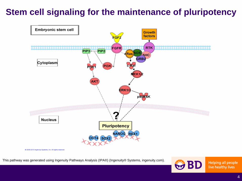
Stem cell signaling for the maintenance of pluripotency
This pathway was generated using Ingenuity Pathways Analysis (IPA®) (Ingenuity® Systems, ingenuity.com).
5
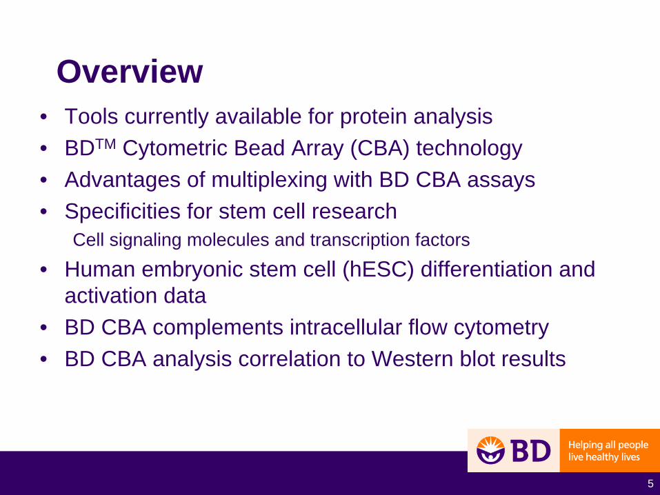
Overview• Tools currently available for protein analysis• BDTM Cytometric Bead Array (CBA) technology• Advantages of multiplexing with BD CBA assays• Specificities for stem cell research
Cell signaling molecules and transcription factors
• Human embryonic stem cell (hESC) differentiation and activation data
• BD CBA complements intracellular flow cytometry • BD CBA analysis correlation to Western blot results
6
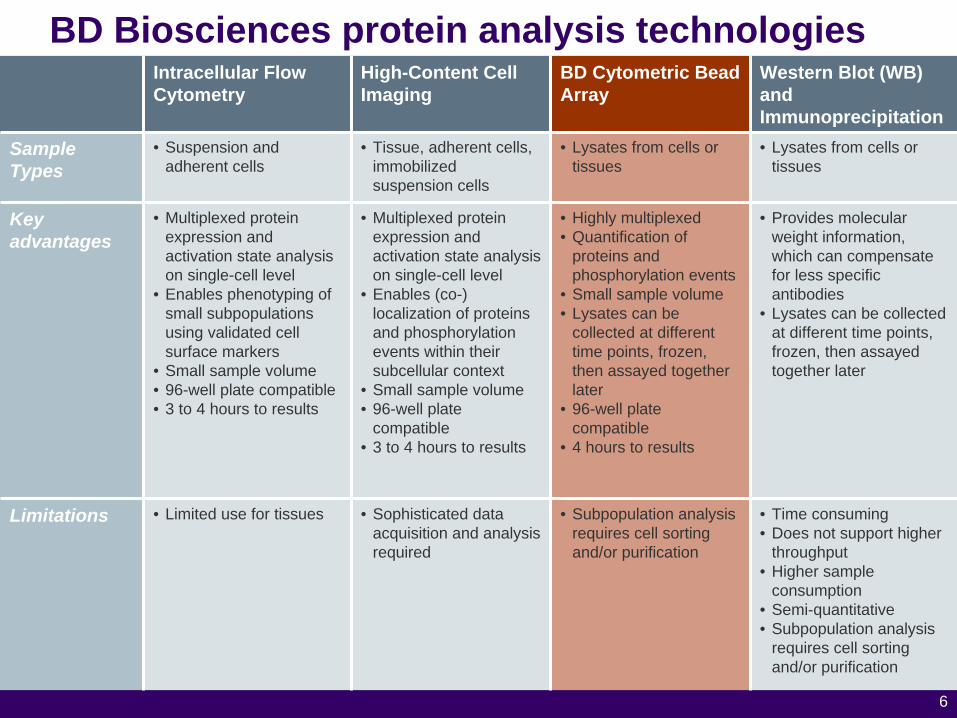
BD Biosciences protein analysis technologiesIntracellular Flow Cytometry
High-Content Cell Imaging
BD Cytometric Bead Array
Western Blot (WB) and Immunoprecipitation
Sample Types
• Suspension and adherent cells
• Tissue, adherent cells, immobilized suspension cells
• Lysates from cells or tissues
• Lysates from cells or tissues
Key advantages
• Multiplexed protein expression and activation state analysis on single-cell level
• Enables phenotyping of small subpopulations using validated cell surface markers
• Small sample volume• 96-well plate compatible• 3 to 4 hours to results
• Multiplexed protein expression and activation state analysis on single-cell level
• Enables (co-) localization of proteins and phosphorylation events within their subcellular context
• Small sample volume• 96-well plate
compatible• 3 to 4 hours to results
• Highly multiplexed• Quantification of
proteins and phosphorylation events
• Small sample volume• Lysates can be
collected at different time points, frozen, then assayed together later
• 96-well plate compatible
• 4 hours to results
• Provides molecular weight information, which can compensate for less specific antibodies
• Lysates can be collected at different time points, frozen, then assayed together later
Limitations • Limited use for tissues • Sophisticated data acquisition and analysis required
• Subpopulation analysis requires cell sorting and/or purification
• Time consuming• Does not support higher
throughput• Higher sample
consumption• Semi-quantitative• Subpopulation analysis
requires cell sorting and/or purification

7
BD Cytometric Bead Array (CBA)

8
BD CBA assay protocol
Y
YYY
Y
YY
Y
Y
YYY
Y
YY
Y
Y
YYY
Y
YY
Y
Y
YYY
Y
YY
Y
Y
Y
YY
Y
YY
Y DETECTORANTIBODIES
LYSATE, SERUM, orSUPERNATE (50 μL)
BEADS
YY
YY
Y
YY
YY
YY
Y
YY
YY
YY+
Y
Y
YY
Y
YY
Y
Y Y
Y
YYY
Y
YY
Y
Y
Y
Y
Y
YYY
YYY
Y
Y
YY
Y Y
YYY
Y
YY
Y Y
Y
YY
YY
Y
YY
Y
Y
Y
Y
Y
Y
YYY
Y
YY
Y Y
Y
Y
Y
READ ON FLOW
CYTOMETERY
YYY
Y
YY
Y
9
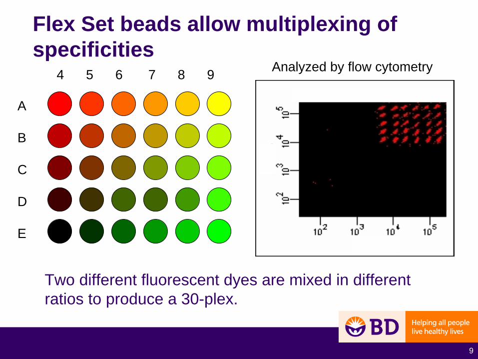
Flex Set beads allow multiplexing of specificities
Two different fluorescent dyes are mixed in different ratios to produce a 30-plex.
A
B
C
D
E
4 5 6 7 8 9 Analyzed by flow cytometry

10
Nanog standard curve generated from recombinant protein
11
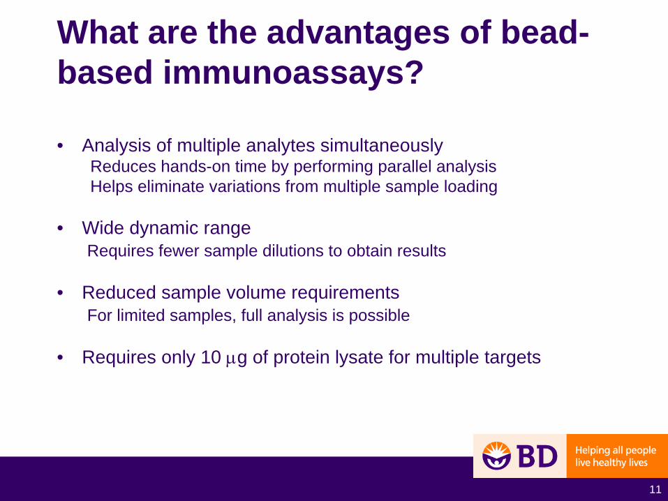
What are the advantages of bead- based immunoassays?
• Analysis of multiple analytes simultaneouslyReduces hands-on time by performing parallel analysisHelps eliminate variations from multiple sample loading
• Wide dynamic range Requires fewer sample dilutions to obtain results
• Reduced sample volume requirements For limited samples, full analysis is possible
• Requires only 10 μg of protein lysate for multiple targets

12
Stem cell signaling and the maintenance of pluripotency
This pathway was generated using Ingenuity Pathways Analysis (IPA®) (Ingenuity® Systems, ingenuity.com).

13
PhosphoAKT1 S473 and
AKT2 S474
Stem cell signaling and the maintenance of pluripotency
JNK1/2
p38
Phospho MEK1/2 S222
Phospho JNK1/2
T183/Y185
Phospho p38 T180/Y182
Phospho RSK T473
Phospho ERK1/2
T202/Y204
TX factorsOct4, Sox2, Nanog and
Rex1
This pathway was generated using Ingenuity Pathways Analysis (IPA®) (Ingenuity® Systems, ingenuity.com).
14
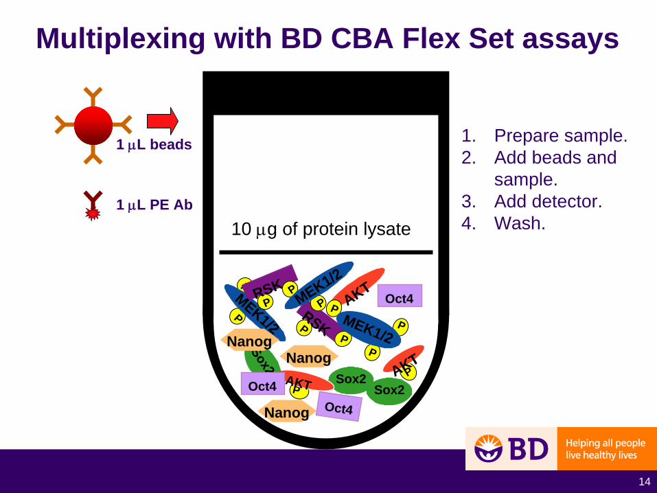
Multiplexing with BD CBA Flex Set assays
Sox2
PRSK
AKT
PAKT
P
P
Sox2
Nanog
PAKT
P
PP
PMEK1/2
PRSK
Oct4PPMEK1/2
MEK1/2
Oct4
NanogNanog
Sox2Oct4
1. Prepare sample.2. Add beads and
sample.3. Add detector.4. Wash.
1 μL beads
1 μL PE Ab
10 μg of protein lysate

Analyze the complexes by flow cytometry
AKT
AKT
AKT
MEK
MEK
MEKIdentify bead populations for data analysis with FCAP Array TM software
P
P
P
P
P
P
P
P
15

16
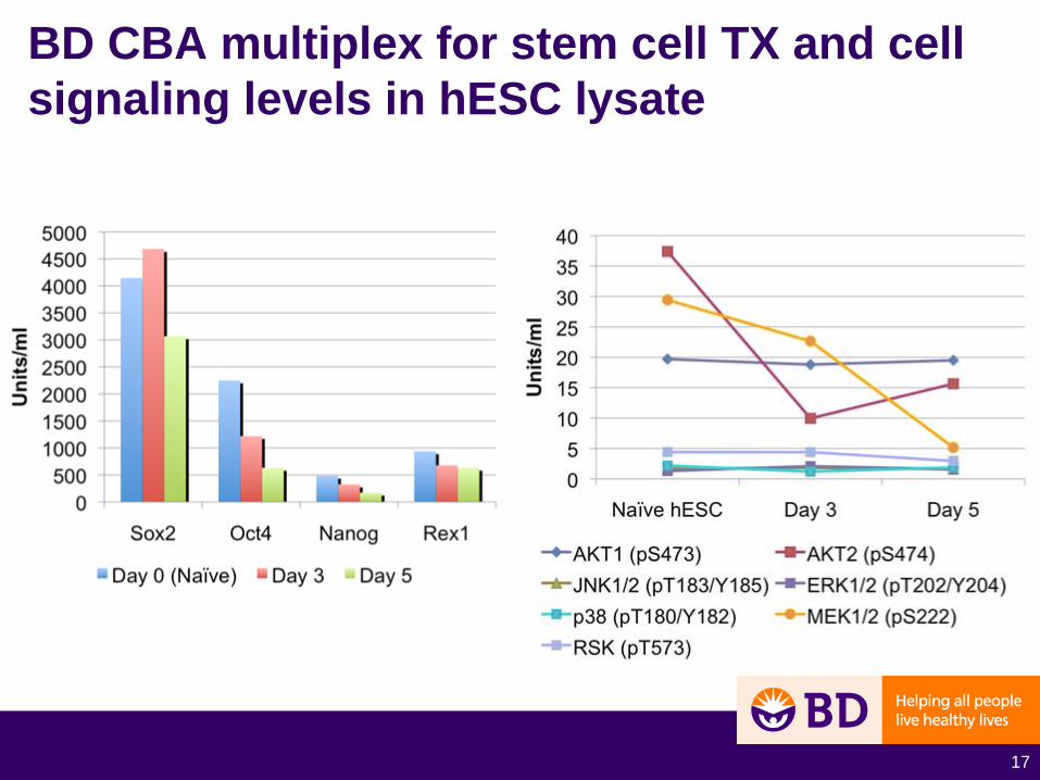
Spontaneous differentiation in human ESC line H9
• Cells were maintained on BD Matrigel™ hESC-qualified matrix and mTeSR®1*.
• Cells were then cultured in hESC media without bFGF.• Cells were lysed for BD CBA testing on days 0, 3, and 5.• Cell lysate protein concentrations were acquired by
bicinchoninic acid assay. • Lysates were tested using BD CBA for 4 stem cell
transcription factors and 7 phospho-signaling proteins.
*Registered trademark of STEMCELL Technologies Inc.
17
BD CBA multiplex for stem cell TX and cell signaling levels in hESC lysate
18
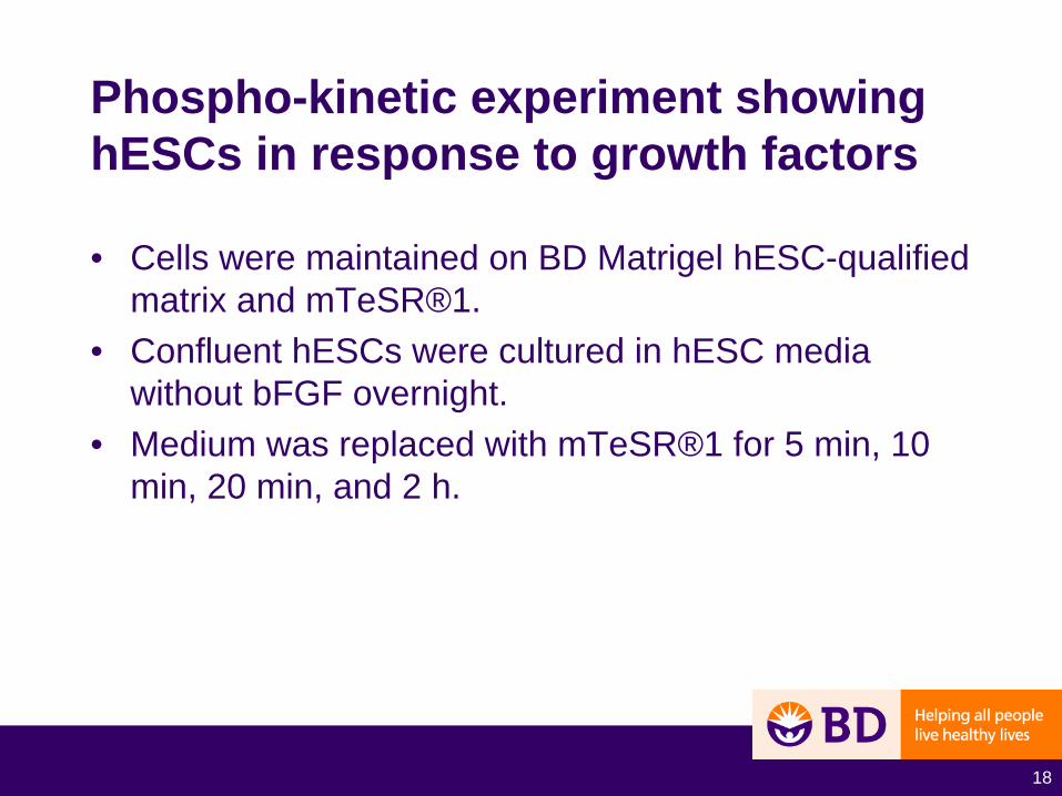
Phospho-kinetic experiment showing hESCs in response to growth factors
• Cells were maintained on BD Matrigel hESC-qualified matrix and mTeSR®1.
• Confluent hESCs were cultured in hESC media without bFGF overnight.
• Medium was replaced with mTeSR®1 for 5 min, 10 min, 20 min, and 2 h.
19
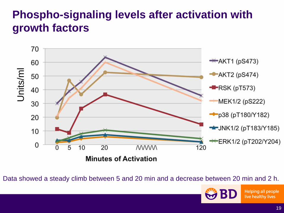
Phospho-signaling levels after activation with growth factors
Data showed a steady climb between 5 and 20 min and a decrease between 20 min and 2 h.
Uni
ts/m
l

20
Effects of PI3K inhibitor LY294002 on transcription factors involved in maintaining pluripotency
• Naïve hESCs were cultured in 10 mM of the PI3K/AKT inhibitor, L7394002.
• Cell lysates were acquired on days 0, 3, 6, and 7.
• Lysates were tested for the transcription factors Sox2, Oct4, Nanog, and Rex1.

21
Effects of PI3K inhibitor LY294002 on transcription factors involved in maintaining pluripotency
Uni
ts/m
l
22
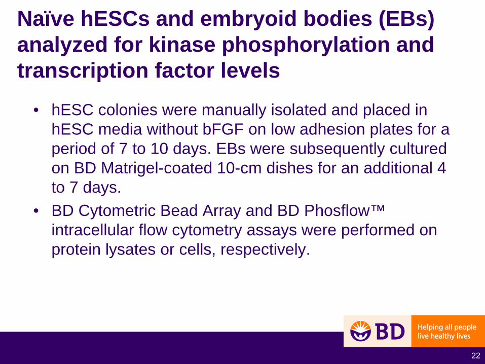
Naïve hESCs and embryoid bodies (EBs) analyzed for kinase phosphorylation and transcription factor levels
• hESC colonies were manually isolated and placed in hESC media without bFGF on low adhesion plates for a period of 7 to 10 days. EBs were subsequently cultured on BD Matrigel-coated 10-cm dishes for an additional 4 to 7 days.
• BD Cytometric Bead Array and BD Phosflow™ intracellular flow cytometry assays were performed on protein lysates or cells, respectively.

23
Multiplexing of BD CBA cell signaling Flex Sets and BD CBA stem cell Flex Sets permits the simultaneous quantitation of hESC and EB protein lysates
24
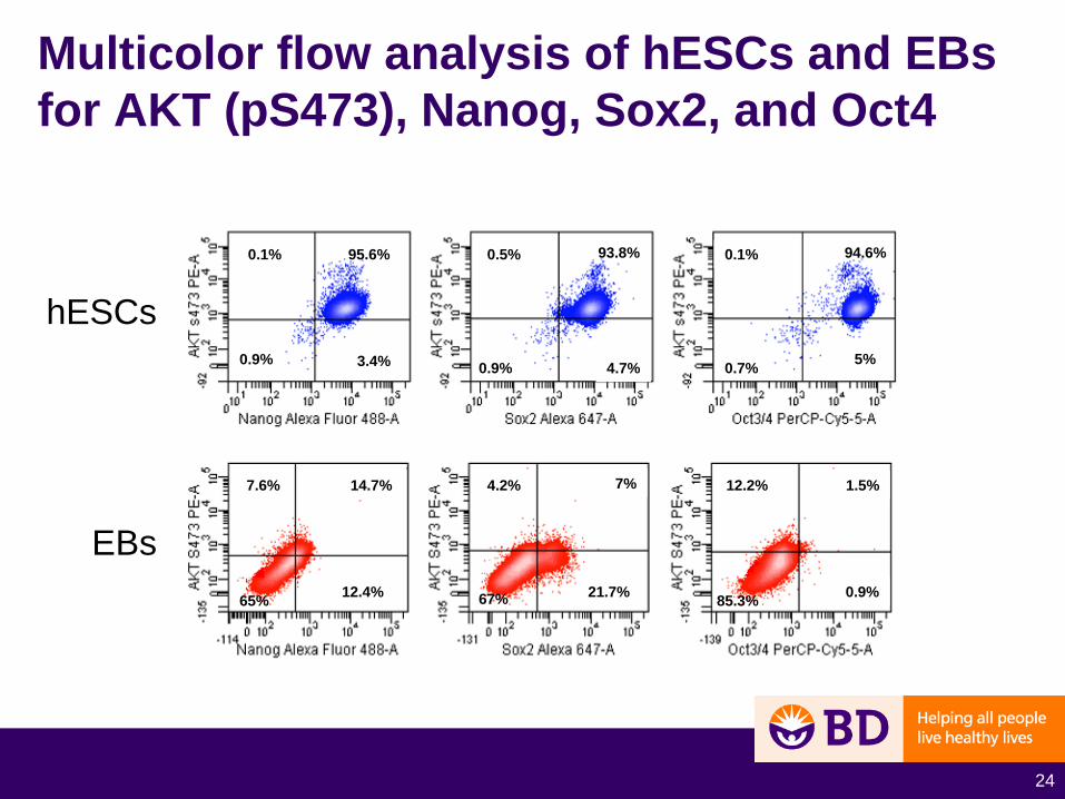
Multicolor flow analysis of hESCs and EBs for AKT (pS473), Nanog, Sox2, and Oct4
hESCs
EBs
3.4%0.9%
0.1% 93.8%0.5%
0.9% 4.7% 5%
0.1%
0.7%
95.6%
7.6%
65%
14.7%
12.4%
4.2% 7%
21.7%67%
12.2%
85.3% 0.9%
1.5%
94.6%

25
Flow cytometric measurement of intracellular signal transduction in mesenchymal stem cells
Patrick C. Baer1, PhD and Ralf Schubert2, PhD1Division of Nephrology, Department of Internal Medicine, and 2Children's Hospital, Goethe University, Frankfurt am Main, Germany
26
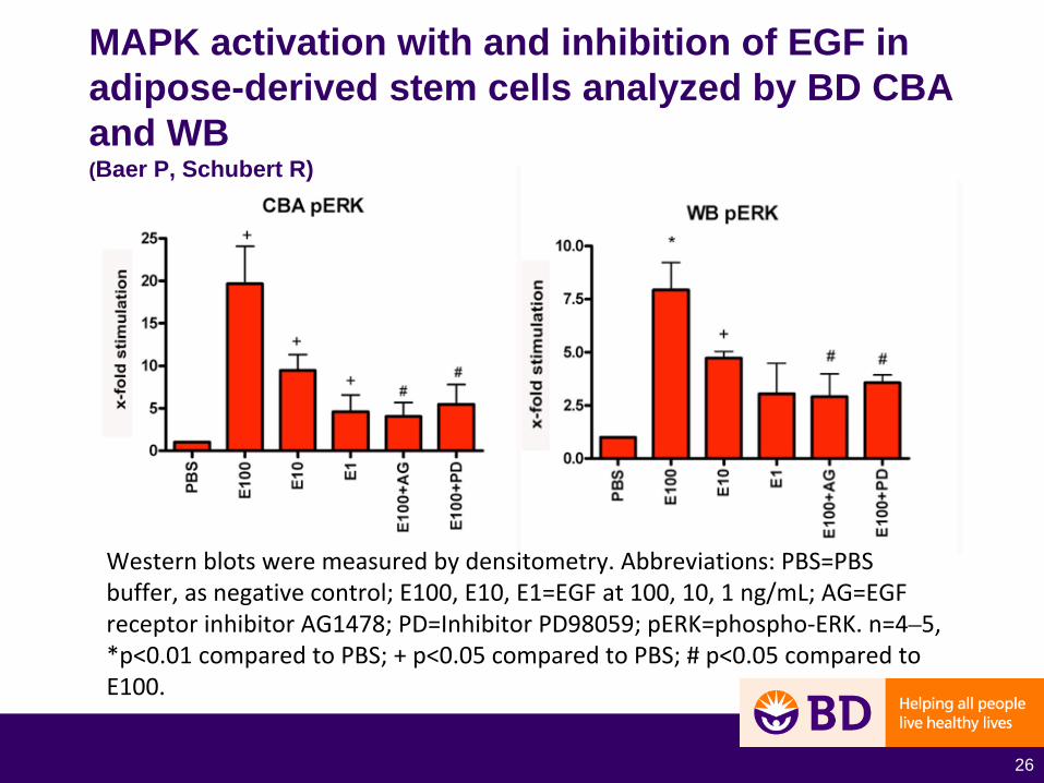
Western blots were measured by densitometry. Abbreviations: PBS=PBS
buffer, as negative control; E100, E10, E1=EGF at 100, 10, 1 ng/mL; AG=EGF
receptor inhibitor AG1478; PD=Inhibitor PD98059; pERK=phospho‐ERK. n=4–5,
*p<0.01 compared to PBS; + p<0.05 compared to PBS; # p<0.05 compared to
E100.
MAPK activation with and inhibition of EGF in adipose-derived stem cells analyzed by BD CBA and WB (Baer P, Schubert R)
27
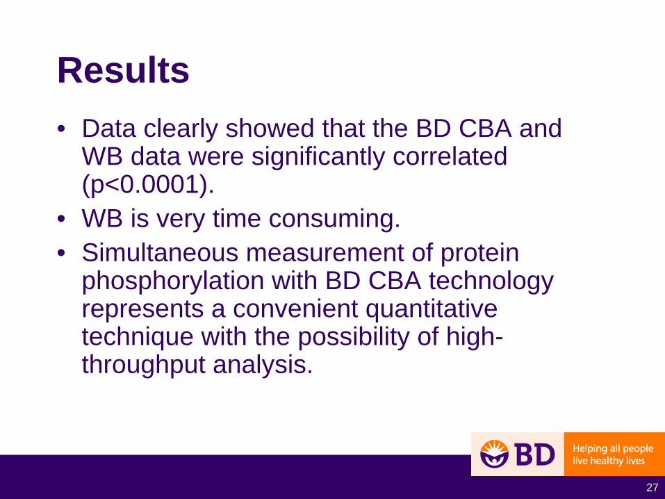
Results• Data clearly showed that the BD CBA and
WB data were significantly correlated (p<0.0001).
• WB is very time consuming.• Simultaneous measurement of protein
phosphorylation with BD CBA technology represents a convenient quantitative technique with the possibility of high- throughput analysis.
28
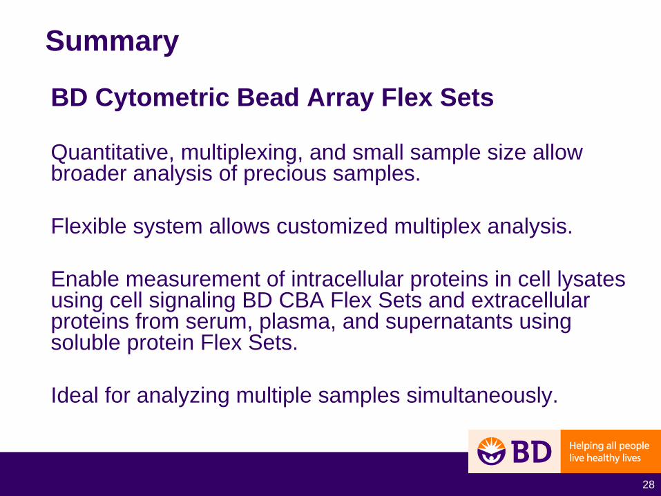
Summary
BD Cytometric Bead Array Flex Sets
Quantitative, multiplexing, and small sample size allow broader analysis of precious samples.
Flexible system allows customized multiplex analysis.
Enable measurement of intracellular proteins in cell lysates using cell signaling BD CBA Flex Sets and extracellular proteins from serum, plasma, and supernatants using soluble protein Flex Sets.
Ideal for analyzing multiple samples simultaneously.

29
Acknowledgments
Christian CarsonJohn ApgarHomero SepulvedaJulia LizondoTeresa GiagoRosanto ParambanNil Emre
For Research Use Only. Not for use in diagnostic or therapeutic procedures.
Alexa Fluor® is a registered trademark of Molecular Probes, Inc.