Culinary FoundationsSession Nine: Eggs CHRM 1030 Culinary Foundations Eggs.
Ital. J. Food Sci., vol. 32, 2020 - 540
PAPER
QUALITY AND MYCOBIOTA COMPOSITION OF STORED EGGS
I. REGECOVÁ, M. PIPOVÁ*, P. JEVINOVÁ, S. DEMJANOVÁ and B. SEMJON Department of Food Hygiene and Technology, University of Veterinary Medicine and Pharmacy,
Komenského 73, 041 81 Košice, Slovak Republic *Corresponding author: [email protected]
ABSTRACT The aim of this study was to monitor the quality and mycobiota composition of table eggs during storage period. The most significant changes in the egg weight and water activity were observed on Day 7. To identify the mycobiota present on the eggshell by PCR method, a newly designed procedure for the extraction of fungal DNA based on a combination of commercial isolation kit, proteinase K and ultrasound was implemented. Identified mold genera included Penicillium spp., Cladosporium spp., Fusarium spp. and Alternaria alternata group. Their ratio varied considerably during storage with the dominance of Penicillium spp. on Day 14.
Keywords: egg quality, Haugh units, micromycetes, PCR, storage, table eggs

Ital. J. Food Sci., vol. 32, 2020 - 541
1. INTRODUCTION Egg is one of the most nutritious low-energy foods possessing all the proteins, vitamins and minerals needed for human health (TOLIK et al., 2014). Because of its beneficial composition, it may also be used as nutraceuticals and protein ingredients for food applications (ZAMBROWICZ et al., 2015). However, during egg collection, there is significant risk of contamination of the shell surface with microscopic filamentous fungi. By laying out, an egg gets to the external environment, which becomes its source of contamination. The majority of egg microbiological contamination studies deal primarily with microorganisms of bacterial origin (BAHOBAIL et al., 2012; JONES et al., 2015; ERKMEN and BOZOGLU, 2016; KARIMIAZAR et al., 2019), secondarily with fungal contamination (TOMCZYK et al., 2019). Micromycetes are able to grow under conditions which are unsuitable for bacteria (extreme pH, low aw, wide range of enzymatic activity). Spores of various mold genera are regularly found on the shell surface and can furher penetrate into the egg contents (TOMCZYK et al., 2018). Contamination of the eggshell may be initiated by ambient conditions during egg collection and storage, which may be as a result of the quality of litter and feed, the presence of dust, air temperature and humidity. Therefore, the presence of numerous fungal genera (Aspergillus, Cladosporium, Drechslera, Penicillium, Stemphylium and Fusarium) on the eggshell has been reported in many studies (KOKKONEN et al., 2010; ROHWEDER et al., 2011; TOMCZYK et al., 2019). Fungi not only degrade the substrate on which they occur, they also cause numerous diseases due to the presence of spores in the air (SKÓRA et al., 2015). Mycotoxins, which are secondary metabolites produced by some fungal strains, are of the greatest risk for consumers (SYPECKA et al., 2004). Freshness, the characteristic most commonly related to egg quality, declines with time and temperature after laying (HIDALGO et al., 2006). Several chemical and physical changes occur during egg storage such as increase in albumen pH, thinning of the thick albumen, water evaporation through the shell (LUCISANO et al., 1996), enlargement of air cell, development of Maillard reaction (HIDALGO et al., 2006). The most common indices used to evaluated egg freshness are air cell height (EU, 2007) and thick albumen height, expressed as Haugh units (USDA, 1995). Storage conditions (especially the temperature) also have a great impact on mycological contamination and the production of fungal exoproducts on the shell surface. Temperatures below 5°C slow the aging process, but primarily reduce the development of microorganisms. If the refrigerator temperature decreases below ‒2°C, the egg contents would start to freeze. According to the European legislation (EC, 2008), shelleggs must not be exposed to refrigeration at temperatures lower than 5°C. Another significant parameter in egg storage is the relative air humidity which is adjusted to reduce as much as possible the losses caused by water evaporation. This results in a weight decrease and changes in individual egg quality indicators. In general, the lower the storage temperature, the higher the relative humidity (STEINHAUSEROVÁ et al., 2003). Within the storage period, diversified mycoflora, including potentially toxinogenic and toxinogenic species of micromycetes, is found in the eggs. Therefore, the identification of micromycetes is necessary (THRANE et al., 2004). Current routine methods used for the detection and identification of microscopic filamentous fungi often require culture isolation and further morphological and physiological characterisation (SIMMONS, 2007). However, it does not provide sufficient distinction among species. The limitation of these techniques is unstable micromycete cell morphology, which may vary within the species and also dependent on ambient conditions (RAINIERI et al., 2003). The development of molecular techniques has

Ital. J. Food Sci., vol. 32, 2020 - 542
enabled the identification of not only the genus, but also the individual species of micromycetes. Currently, numerous identification methods are generally in use. DNA reassociation techniques are used to detect DNA complementarity. The disavantage of these methods is inability to distinguish closely related species. Gene sequence comparison no longer has this limitation. However, detection of rRNA genes, such as the internal transcriptional region (ITS), is the preferred method (KURTZMAN et al., 2011). For proper identification of micromycetes by PCR methods, sufficient amount and high purity of the DNA are necessary. The aim of this study was to detect changes in quality of table eggs during the storage period of 28 days and to identify mold genera present on the eggshell using the PCR method. For correct and accurate identification it is necessary to obtain DNA which is often very complex in micromycetes. To extract the fungal DNA, the cell wall, cytoplasm, and nuclear membrane must be first effectively lysed. However, the structure of fungal cell wall itself prevents lysis and sufficient nucleic acid isolation (Čmoková et al., 2014). Therefore, a new effective procedure for lysing fungal cells and further DNA extraction has been designed and implemented in this study. 2. MATERIALS AND METHODS Micromycetes were isolated from 120 brown-shell table eggs, laid by the Lohmann Brown crossbreed laying hens in cages, weight category M (EC, 2008). The eggs were graded, labeled and stored in the breeding stock at an average temperature of 14.3°C and a relative humidity of 61%. Sets of 30 sample units were taken every 7 days. Among them, water activity (aw) of the eggshell was determined in 10 eggs, another 10 eggs were checked for selected egg quality indicators and the enumeration of microscopic filamentous fungi according to STN ISO 21527-1 (2010) and STN ISO 21527-2 (2010) was carried out using the last 10 eggs. Since molds are aerobic organisms and colonize mainly the shell, enumeration of molds was focused on the eggshell surface. 2.1. Determination of water activity (aw) of eggshell and egg quality indicators Water activity of the eggshell was determined by the LabMASTER-aw (Novasina AG, Lachen, Switzerland) at a constant temperature (25°C) for 20 min±2.48 min. Measurements were repeated three times for individual egg. Eggshell breaking force was measured in accordance with the manufacturer's instructions by the Egg Force Reader (Orka Food Technology Ltd., Ramat HaSharon, Israel) - a compact system for automatic measuring of eggshell rupture point. The unit of strength measurement (kilogram-force, kgf) is calculated as gently applied force on the eggshell until it cracks. Egg Analyzer™ (Orka Food Technology Ltd., Ramat HaSharon, Israel) was used to determine the egg weight, Haugh units (HU), quality grade and yolk color. The device is able to measure egg weight (g), height of the thick albumen (mm), and color of the yolk. The first two measurements were used for calculation of Haugh units that indicate egg quality. The equation for working out the rating is shown below:
HU = 100 log (h − 1.7w0.37 + 7.6),

Ital. J. Food Sci., vol. 32, 2020 - 543
where: HU = egg quality in Haugh units w = egg weight in grams h = height of the thick albumen in mm (NAGY et al., 2011) Yolk color intensity was compared to the Roche Yolk Color Fan (RCF) ranging from 1 to 15 (1 means pale yellow and 15 dark yellow). After evaluation, the egg contents were transferred to large Petri dishes with diameter of 200 mm each, they were then placed on a dark flat surface and both the height and the width of the egg yolk as well as those of the thick albumen were measured using a micrometer gauger and a ruler. Subsequently, the yolk index (YI) and albumen index (AI) were calculated according to NAGY et al. (2011) as follows:
Albumen index = Height of the thick albumen (mm) x 100/ [Length of the thick albumen (mm) + Width of the thick albumen (mm)/2]
Yolk index = Height of the yolk (mm)/Yolk diameter (mm) x 100
2.2. Fungal strains and culture condition Microscopic filamentous fungi were removed from the eggshell surface as described by CUPÁKOVÁ et al. (2010). For this purpose, ten eggs of each experimental group were tested every week. Individual egg was transferred aseptically into a sterile plastic bag and sterile peptone water (0.1 wt%) in a volume of 100 mL was added. The sample was then shaken for 15 minutes using the Orbi-ShakerTMJR (Benchmark Scientific Inc., Sayreville, USA). Further decimal dilutions were prepared in accordance with STN EN ISO 6887-1 (2017). As described by the standard procedure, the first two decimal dilutions were spread in parallel on the surface of Dichloran Glycerol agar medium (DG-18; OXOID, Basingstoke, UK) and Dichloran-Rose Bengal Chloramphenicol agar medium (DRBC; OXOID, Basingstoke, UK) in a volume of 0.1 mL and incubated for 5 days at 25°C (STN ISO 21527-1 and STN ISO 21527-2, 2010). Colonies were further subcultured individually on the surface of Czapek agar medium (OXOID, Basingstoke, UK) and incubated at 25°C for another 7 days. After incubation, 1 to 5 colonies were subjected to macroscopic and microscopic identification. Macroscopic evaluation included the growth rate, color, texture and topography of fungal colonies. Microscopic study of different mold genera was carried out by preparing slides stained with lacto phenol cotton blue and observed under light microscope. 2.3. DNA isolation Because the cell wall of micromycetes is difficult to break, there was a challenge to obtain pure and concentrated fungal DNA. Therefore, the following three pre-isolation techniques were designed: 1. Mycelium in a quantity of 10-50 mg was taken with a sterile scalpel from the surface of Czapek agar medium, transferred into 1.5 mL eppendorf, exposed to liquid nitrogen for 5 min and then heated to 95°C for 5 min. Freezing and heating was repeated three times. The DNA isolation was then performed with the help of commercially available E.Z.N.A.® Fungal DNA Mini Kit (Omega Bio-Tek, Norcross, USA).

Ital. J. Food Sci., vol. 32, 2020 - 544
2. Proteinase K (Macherey-Nagel, Düren, Germany) in a volume of 10 µL was added to eppendorfs with 10-50 mg mycelium and incubated for 30 min at 37°C. After that, the DNA isolation procedure was carried out in accordance with instructions of the commercially available E.Z.N.A.® Fungal DNA Mini Kit manufacturer (Omega Bio-Tek, Norcross, USA). 3. Both zircon and glass beads (1:1) in a volume of 0.2 mL and Proteinase K (Macherey-Nagel, Düren, Germany) in a volume of 10 μL were added to fungal mycelium (10-50 mg) in the eppendorf. Samples were incubated at 37°C for 30 min. After that, 800 μL of lysing solution FG1, which was a part of the E.Z.N.A.® Fungal DNA Mini Kit (Omega Bio-Tek, Norcross, USA) was added. Samples were then incubated at 65°C for 10 min with ultrasound waves of 500 Hz. Further DNA isolation was performed according to recommended procedure for the commercially available E.Z.N.A.® Fungal DNA Mini Kit (Omega Bio-Tek, USA, Norcross, USA). The purity and concentration of DNA was measured using spectrophotometer (SHIMADZU, Wien, Austria). The obtained supernatant was used as a source of DNA in PCR reactions. 2.4. Primer design The forward primer ITS 212d and the reverse primer ITS 549 amplified a specific DNA fragment for Penicillium spp. (336 bp; Table 1). Table 1. DNA sequences of primers used in this study.
Primer name Primer sequence (5′-3′)
Annealing temperature
(°C)
PCR product
GenBank-EMBL accession
number Reference
Penicillium spp. ITS 212d AAATATAAATTATTTAAAACTTTC
46 336 bp LC 317718.1 Pedersen et al., 1997 ITS 549 CTGGATAAAAATTTGGGTTG
A. alternata group 18S‒F AGGATCCATTGGAGGGCAAGT
61 99 bp
AY 563301 Pavón et al., 2010
18S‒R TCCAACTACGAGCTTTTTAACTGCA Altsp‒F GAGAACAGCTTCATGGACTTCTCTTT
61 195 bp Altsp‒R CGCGGCAGTAGTTGGGAA Alt A–F CGCATCCTGCCCTGTCA
60 118 bp Alt A–R GTTGGTAGCCTTGATGTTGAAGC
Cladosporium spp. MS1 CAGCAGTCAAGAATATTAGTCAATG
51 370 bp AY 291273 Zeng
et al., 2006 MS2 GCGGATTATCGAATTAAATAAC
Clado‒PF TACTCCAATGGTTCTAATATTTTCCTCTC 51 87 bp
Clado‒PR GGGTACCTAGACAGTATTTCTAGCCT Fusarium spp.
ITS‒Fu‒f CAACTCCCAAACCCCTGTGA 55 410 MK 156681.1
Abd-Elsalam
et al., 2003 ITS‒Fu‒r GCGACGATTACCAGTAACGA
Ital. J. Food Sci., vol. 32, 2020 - 545
These primers were designed based on ITS region and 5.8S rRNA sequences from Penicillium spp. available in the GenBank-European Molecular Biology Laboratory database (GenBank-EMBL; PEDERSEN et al., 1997). Alternaria-specific and Alternaria alternata-group-specific primer pairs were designed based on Alt a 1 gene sequences from Alternaria spp. available in the GenBank-EMBL database (Table 1; PAVÓN et al., 2010). Both the forward primer AltspF and the reverse primer AltspR amplified a DNA fragment of 195 bp. in all Alternaria spp. The primer pair AltAF and AltAR amplified a specific DNA fragment (118 bp) for Alternaria alternata group. Finaly, the primer pair 18SF and 18SR, based on conserved 18S rRNA gene, was used as positive amplification control of the assay (Table 1; MARTÍN et al., 2009; PAVÓN et al., 2010). Mitochondrial (mt) small subunit rRNA (SSU rRNA) of Cladosporium was amplified by PCR using the universal fungal mitochondrial primers MS1 and MS2. Two specific primers Clado‒PF and Clado‒PR were designed for multiplex PCR assay (Table 1). The expected amplicon size for primer pair Clado-PF/R was 87 bp (ZENG et al., 2006). Two primers were designed based on sequence information obtained to amplify specific fragments within the ITS regions of Fusarium spp. The initial tests for specificity revealed that the primer pair ITS-Fu-f and ITS-Fu-r were highly specific for Fusarium genus (Table 1; ABD-ELSALAM et al., 2003). All the primers were synthesized by the same commercial company (Ecoli s.r.o., Bratislava, Slovakia). 2.5. PCR amplifcation The amplification was done in a volume of 20 μL containing 1 ng to 10 ng of DNA, 0.5 µL of each primer (concentration 10 pmol/µL), 4.0 μL HOT Firepol® Blend Master Mix (Solis BioDyne, Tartu, Estonia) in the thermal cycler (TC-512, Techne UK, Staffordshire, United Kingdom) using the same cycling conditions for each primer sets, with an initial cycling step at 95°C for 12 min, followed by DNA denaturation at 95°C for 20 s, annealing at diferent temperatures depending on the type of primer used (Table 1) for 60 s, and elongation at 72°C for 2 min. The amplification was terminated by cooling to 6°C. The following reference strains were used as positive controls in this study: Penicillium chrysogenum CCM F-362, Fusarium sporotrichioides CCM F-352, Alternaria alternata CCM F-397, Cladosporium cladosporioides CCM F-348 (Czech Collection of Microorganisms, Brno, Czech Republic). 2.6. Sensitivity of specific detection assays To determine the minimum detectable amount of fungal DNA in three established PCR assays, variable quantities of fungal genomic DNA ranging from 10 ng to 100 ng were used as DNA template. The PCR products were size fractionated in agarose gels (1.5%) and visualized using GelRed™Nucleic Acid gel stain (Biotium INC., Fremont, USA). Amplicons were visualized by UV transillumination using the Mini Bis Pro® (DNR Bio-Imaging Systems Ltd., Neve Yamin, Izrael). The identity of PCR products with the selected primers was confirmed by a commercial company (GATC Biotech AG, Cologne, Germany). The DNA sequences obtained from fungal strains were searched for homology to those available at the GenBank-EMBL database using the BLAST program (NCBI software package).

Ital. J. Food Sci., vol. 32, 2020 - 546
2.7. Statisical analysis Two-way analysis of variance (ANOVA) and Tukey test for multiple comparison of means with a confidence interval set at 95% was conducted with R - statistics software (R CORE TEAM, 2018). The storage period was set as the main factor. Multiple factor analysis was conducted in R - statistics software (R CORE TEAM, 2018) with “FactomineR” (SEBASTIEN et al., 2008) and “factoextra” package (KASSAMBARA and MUNDT, 2007) according to SEMJON et al. (2018). 3. RESULTS AND DISCUSSION Pre-market table eggs must be sorted, packed and stored under appropriate conditions to maintain temporary shelf life. According to Commission Regulation (EC) No. 589/2008, the minimum shelf life of fresh eggs is 28 days from the day it is laid. Storage conditions are limited by storage temperature where the lower limit is 5°C. However, the upper limit is not defined, it only requires maintaining the optimum egg quality. 3.1. Changes in egg quality indicators Quality parameters of table eggs change during storage. Within the entire storage period, the average values of egg weight ranged from 60.47±2.838 to 57.83±3.368. The most significant egg weight loss (2.64 g±0.87) in this study was recorded on Day 7. The decrease in egg weight was accompanied by a decrease in water activity (aw) in the eggshell. Similarly, the most significant decrease in water activity was recorded on Day 7. On the same day, the average shell firmness achieved its maximum value. Shell firmness is related to water evaporation and subsequent drying of the eggs. The rate of water evaporation is influenced by the permeability of the shell and the number of pores in the shell (SIMEONOVOVÁ and MÍKOVÁ, 2003). As already reported, the above-mentioned three egg indicators are interrelated. Due to egg drying, the increased water evaporation has a negative effect on egg weight (MATUŠOVIČOVÁ et al., 1986), which has also been confirmed in this study. Similar results were published by DE LEO et al. (2018). The authors reported an average weight loss of 1.67% after 35 days of egg torage. According to SIMEONOVOVÁ et al. (1999), evaporation of water from egg contents depends on the environment, in particular temperature and humidity, as well as on the storage period. ALLEONI and ANTUNES (2004) also reported a reduction in egg weight during storage. The HU indicates egg quality and the calculation is based on both the height of the thick albumen and the egg weight (CANER and YÜCEER, 2015). The initial value of HU represents the main marker for evaluating egg quality, and its expression provides an indication of the egg shelf-life as well as the storage conditions (FIGUEIREDO et al., 2014). As seen in Table 2, mean Haugh unit values ranged between 53.74 and 59.80 during egg storage in this study. DE LEO et al. (2018) also confirmed a HU decrease during the storage period of 42 days, which corresponds to similar earlier studies (CANER, 2005; CANER and YÜCEER, 2015; MORSY et al., 2015). In general, Haugh units decrease during egg storage. This can be explained by changes in protein structure resulting in albumen thinning (SIMEONOVOVÁ et al., 1999). Results of JIN et al. (2011) were consistent with those obtained in this study. In contrast, lower values

Ital. J. Food Sci., vol. 32, 2020 - 547
within the entire storage period were reported by SAMLI et al. (2005) and AKYUREK and OKUR (2009). All of the cited authors stated that Haugh units were decreasing over time. As also reported by TOMCZYK et al. (2019), longer egg storage period resulted in a more noticeable decline in Haugh unit values. A similar decrease was observed in the albumen index, which is based on both the width and height of the thick albumen. The values ranged from 1.370 to 10.500, depending on the storage period. Albumen index was decreasing with prolonged egg storage. The most significant decline occurred on Day 7, when the maximum weight loss and shell firmness, as well as minimum eggshell breaking point, water activity and Haugh units were all determined. Similarly, LAZAR (1990) also reported a decrease in albumen index value with increase in storage time and MÍKOVÁ and DAVÍDEK (2000) ranked the albumen index among the main indicators of egg freshness. Table 2. Changes in physico-chemical parameters during egg storage.
Storage period (days) 1 7 14 21 28
Water activity (aw) 0.91±0.00a 0.91±0.00a 0.91±0.01a 0.91±0.00a 0.91±0.00a Weight (g) 60.47±2.84a 58.20±3.71a 58.23±2.60a 59.00±2.14a 58.23±2.26a
Color 8.33±1.63a 9.17±2.23a 9.50±1.64a 9.33±0.52a 9.00±1.26a Haugh units 59.80±23.03a 57.85±11.91a 58.33±14.28a 56.15±13.83a 54.57±9.76a
Albumen index (%) 10.50±1.30a 6.86±3.49b 2.25±1.44c 1.23±0.26c 1.42±0.77c Yolk index (%) 36.70±2.90a 35.51±2.59a 32.92±2.63ab 34.85±1.67ab 31.02±2.99b
Eggshell breaking force (kgf) 3.55±1.06a 4.97±1.08a 4.69±0.73a 4.21±1.25a 3.75±1.53a
Egg quality grade AA‒Ba AA‒Ba AA‒Ba AA‒Ba Ba a-c Different superscripts in each row indicate significant differences between the mean values (Tukey´s, p<0.05). The yolk index is also an excellent indicator of egg freshness, which is based on the measurements of the yolk height and width (YÜCEER and CANER, 2014). Yolk index ranged from 30.74% to 36.90% depending on the length of storage. Yolk width increased during storage as the vitelline membrane lost its elasticity due to aging. Similarly, in a study by DE LEO et al. (2018), the yolk index ranged from 30.99% to 39.96% during 42 days of egg storage. As reported by NEDOMOVÁ (2012), the yolk index decreases with prolonged egg storage. During storage, the structure of vitelline membrane is changed, its strenght decreases, which may, in a combination with water gained from the egg albumen, lead to the enlargement and deformation of the yolk (SIMEONOVOVÁ et al.,1999). Similar decrease in yolk index during egg storage was also observed in other studies (SAMLI et al., 2005; AKYUREK and OKURK, 2009; NEDOMOVÁ and SIMEONOVOVÁ, 2010), where the storage period was identified as the limiting factor. Another indicator of egg quality is the yolk color. The yolk color is a consumer factor without any nutritional significance. In this study, the yolk color ranged from 8 to 10 La Roche scale and became more intense as the egg aged. Similar to other studies (MÍKOVÁ and DAVÍDEK, 2000; JINN et al., 2011), it becomes more intense during egg storage.
Ital. J. Food Sci., vol. 32, 2020 - 548
Changes in egg quality parameters during storage may also be accompanied by egg contamination with microscopic filamentous fungi. Therefore, not only the temperature, but also the relative humidity is of great importance during egg storage. Microscopic filamentous fungi need sufficient moisture to grow. Thus, eggs are more frequently contaminated in high relative humidity environments. DE REU et al. (2006) and MESSENS et al. (2007) reported that a higher level of eggshell contamination led to a better possibility for penetration of micromycetes into the egg contents. Shell pores allow a potential route of entry for fungi and this can lead to both health hazards and a foul smell and taste of an egg. Therefore, detecting egg contamination is an important aspect of public health concern. In addition, volatile organic compounds (VOCs) are produced by micromycetes as they proliferate.These chemicals are emitted back into the environment through the eggshell. Currently, 69 fungi are known as volatile emitters (LEMFACK et al., 2014). The fact that table eggs are often contaminated with fungal spores was also confirmed by TOMCZYK et al. (2019) during three weeks of egg storage under various storage conditions. NEAMATALLAH et al. (2009) also reported the presence of microscopic filamentous fungi in 38% of eggs in their study with an average count of 3.53 log CFU/mL. The authors have identified the following mold genera: Aspergillus (14%), Penicillium (9%), Fusarium (1%), Mucor (6%), Rhizopus (4%), and Cladosporium (5%). In this study, micromycetes were isolated from the shell surface using DG−18 and DRBC selective culture media. According to STN ISO 21527-1, DRBC culture medium is used for enumeration of yeasts and molds in food products with water activity greater than 0,95 (e. i. egg contents). However, the measurements in this study gave a value of aw ≤ 0.95 in the eggshell which requires the use of DG−18 medium (STN ISO 21527-2). Some mold genera and species are xerophilic and need lower aw to grow. Therefore, mycological examination of the eggshell was performed using both the above-mentioned culture media in this study. For genus identification, macroscopic evaluation of colony size and characteristics on special culture media is of great importance. Czapek agar medium often provides useful information with regards to growth rates on low water activity media. However, experience has shown that the brand of agar medium used may affect the appearance of colonies that grow on it (SAMSON and PITT, 2000). Therefore, microscopic evaluation of fungal structure is used for more accurate genus identification. Phenotypic key identification is difficult, because these characteristics are unstable and micromycetes sometimes appear to be atypical with slow spore formation and aberrant production of conidiophores (CHEŁKOWSKI and VISCONTI, 1992). As seen in Table 3, fungal isolates in this study belonged to Penicilium spp., Fusarium spp., Alternanria spp. and Cladosporium spp. After inclusion of mold isolates into individual genera, the ability of both culture media to capture these genera was evaluated (Tables 2 and 3). Despite the fact that STN ISO 21527 recommends the use of two selective culture media depending on water activity of particular food samples, different capture ability has been confirmed for both media. DG-18 medium was more suitable for isolation of Cladosporium spp. and Alternaria spp. On the contrary, capture of Fusarium spp. and to a lesser extent Penicillium spp., was significantly higher on DRBC medium. Similar results have been published by WEIDENBORNER et al. (1995) and PEREIRA et al. (2010). The authors reported that DRBC medium permited the isolation of a wider range of fungal genera/species, regardless of the type of food under study. In this study, the counts and composition of mycoflora on the eggshell varied during the storage period. Similar to changes in egg quality indicators, changes in numbers of micromycetes were also confirmed between 7 and 28 days of egg storage. However, these
Ital. J. Food Sci., vol. 32, 2020 - 549
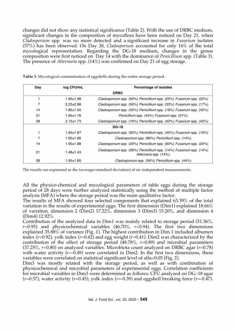
changes did not show any statistical significance (Table 2). With the use of DRBC medium, significant changes in the composition of mycoflora have been noticed on Day 21, when Cladosporium spp. was no more detected and a significant increase in Fusarium isolates (57%) has been observed. On Day 28, Cladosporium accounted for only 16% of the total mycological representation. Regarding the DG-18 medium, changes in the genus composition were first noticed on Day 14 with the dominance of Penicillium spp. (Table 3). The presence of Alternaria spp. (14%) was confirmed on Day 21 of egg storage. Table 3. Mycological contamination of eggshells during the entire storage period.
Day log CFU/mL Percentage of isolates DRBC
1 1.95±1.98 Cladosporium spp. (50%) Penicillium spp. (25%) Fusarium spp. (25%) 7 2.23±2.88 Cladosporium spp. (50%) Penicillium spp. (33%) Fusarium spp. (17%) 14 1.85±1.65 Cladosporium spp. (42%) Penicillium spp. (16%) Fusarium spp. (42%) 21 1.85±1.76 Penicillium spp. (43%) Fusarium spp. (57%) 28 2.15±1.73 Cladosporium spp. (16%) Penicillium spp. (42%) Fusarium spp. (42%)
DG‒18 1 1.95±1.87 Cladosporium spp. (50%) Penicillium spp. (40%) Fusarium spp. (10%) 7 1.90±1.96 Cladosporium spp. (86%) Penicillium spp. (14%) 14 1.90±1.88 Cladosporium spp. (20%) Penicillium spp. (60%) Fusarium spp. (20%)
21 1.48±1.43 Cladosporium spp. (58%) Penicillium spp. (14%) Fusarium spp. (14%) Alternaria spp. (14%)
28 1.95±1.85 Cladosporium spp. (56%) Penicillium spp. (44%) The results are expressed as the (average±standard deviation) of six independent measurements. All the physico-chemical and mycological parameters of table eggs during the storage period of 28 days were further analyzed statistically using the method of multiple factor analysis (MFA) where the storage period was the main qualitative factor. The results of MFA showed four selected components that explained 63.39% of the total variation in the results of experimental eggs. The first dimension (Dim1) explained 18.66% of variation, dimension 2 (Dim2) 17.22%, dimension 3 (Dim3) 15.20%, and dimension 4 (Dim4) 12.92%. Contribution of the analyzed data in Dim1 was mainly related to storage period (51.36%, r=0.95) and physicochemical variables (46.70%, r=0.94). The first two dimensions explained 35.88% of variance (Fig. 1). The highest contribution in Dim 1 included albumen index (r=0.92), yolk index (r=0.62) and egg weight (r=0.41). Dim2 was characterized by the contribution of the effect of storage period (48.78%, r=0.89) and microbial parameters (37.25%, r=0.80) on analyzed variables. Microbiota count analyzed on DRBC agar (r=0.78) with water activity (r=-0.49) were correlated in Dim2. In the first two dimensions, these variables were correlated on statistical significant level of alfa<0.05 (Fig. 2). Dim3 was mostly related with the storage period, as well as with combination of physicochemical and microbial parameters of experimental eggs. Correlation coefficients for microbial variables in Dim3 were determined as follows: CFU analyzed on DG‒18 agar (r=0.57), water activity (r=0.45), yolk index (r=-0.39) and eggshell breaking force (r=-0.47).

Ital. J. Food Sci., vol. 32, 2020 - 550
The highest contribution on Dim4 was related with physicochemical variables (43.38%). However in Dim4 only the correlated CFU analysed on DG‒18 agar (r=0.39) was statistically significant.
Figure 1. Plot of individuals in the first and second extracted dimensions during egg storage period. From the results obtained by the MFA method, it can be concluded that the effect of storage conditions on experimental eggs was significant. The MFA method showed differences between experimental egg groups during the storage period. Analyzed variables of experimental eggs were more similar to each other on Days 14, 21 and 28 than on other days during the storage period. On experimental days 1 and 7, the eggs were significantly different from each other as well as from experimental eggs analyzed on Days 14, 21 and 28 in all monitored variables. 3.2. Isolation of fungal DNA To overcome the poor diagnostic sensitivities and long turnaround times associated with the detection and identification of fungal pathogens in samples by cultivation, non-cultivation methods including the polymerase chain reaction (PCR) are increasingly being used for exact confirmation and more accurate identification of micromycetes. The

Ital. J. Food Sci., vol. 32, 2020 - 551
ultimate sensitivity of any PCR assay for the detection of fungal pathogens depends on the efficient lysis of fungal cells in the tissue sample and the purification of DNA that is free of PCR inhibitors. Fungi have cell walls that impede lysis and the recovery of nucleic acids. Furthermore, highly sensitive and specific nucleic acid-based methods for the detection of fungi necessitate the use of DNA extraction reagents that are free of contaminating fungal nucleic acids (FREDRICKS et al., 2005).
Figure 2. Correlation plot of variables in the first and second extracted dimensions during egg storage period. In the case of the direct detection of molds in food samples (especially eggshell), DNA yield and purity depends primarily on the quantity and quality of the material taken. To isolate fungal DNA, it is first necessary to effectively disrupt the cell wall, lyse the cytoplasm and nucleus membrane, precipitate the proteins and remove the DNA from a number of inhibitors that can reduce the effectiveness of PCR. The individual steps can be executed using chemicals by known methodological procedures, or using commercial kits to facilitate isolation. In this case, however, it may be a problem to optimize the methodological process, because some chemicals in diagnostic kits are subject to corporate secrecy (ČMOKOVÁ et al., 2014). The cell walls can be disrupted by homogenising of frozen sample in mortar using liquid nitrogen (GARG et al., 2009; UCHIDA et al., 2009), by

Ital. J. Food Sci., vol. 32, 2020 - 552
vortexing with glass or zircon beads (EBIHARA et al., 2009; SATO et al., 2010), or by repeated freezing (LITZ and CAVAGNOLO, 2010). Membrane lysis is made with detergents (e.g. Triton X-100, sodium laurysulfate) followed by nucleic acids release into a buffered solution which contains chelators, most commonly EDTA and bonding calcium cations serving as a nucleases cofactor to prevent cleavage of the released DNA. To increase the nucleic acid purity, proteinase K is sometimes added into the lysis solution to cleave proteins including DNA-bound histones. A commonly used method is the incubation with proteinase K and subsequent completion of isolation using a commercial kit (BERGMANS et al., 2010; ALEXANDER et al., 2011; BEIFUSS et al., 2011; WISSELINK et al., 2011). In this study, three isolation procedures of fungal DNA were used and compared: 1. Combination of liquid nitrogen and heat 95°C; 2. Proteinase K and FG1 lysis solution; and 3. Zircon and glass bead isolation with simultaneous effects of proteinase K and ultrasonication of 50 Hz. Measurements of DNA concentration (Table 4) provided useful information on which of the three test procedures is the most effective for cell wall lysis and DNA extraction. DNA samples with the highest concentration and purity were used for further analysis. After PCR identification, the effectiveness of isolation procedures in relation to individual mold genera was re-evaluated. Table 4. Comparison of DNA concentrations (ng/µL) yielded by three extraction procedures (average±SD).
Procedure 1 Procedure 2 Procedure 3 One-Way ANOVA P value
Penicillium spp. 36.372±17.272C 49.146±26.657B 97.669±12.225A <0.001 Cladosporium spp. 12.462±4.807B 35.585±11.433A 38.175±20.410A <0.001
Fusarium spp. 22.335±9.134C 64.222±18.859B 71.554±17.674A <0.001 Alternaria alternata group 10.867±0.103C 29.017±0.618B 49.650±0.812A <0.001
A-C Different superscripts in each row indicate significant differences between the mean values (Tukey´s, p<0.05). As can be seen in Table 4, the lowest average values for all identified genes were obtained by isolation procedure using a combination of thermal shock and liquid nitrogen. Among four mold genera, this procedure was most effective for Penicilium spp. isolates, where the average DNA concentration achieved a value of (36.372±17.272 ng/μL). The first procedure proved to be the worst for isolates of the genus Cladosporium. The second isolation procedure involving the use of proteinase K and a lysing solution FG1 appeared to be sufficient for isolates of the genus Fusarium (average DNA concentration was 64.222±18.859 ng/μL). The third method of isolation with a combination of zirconium and glass beads, proteinase K and ultrasonic waves, appeared to be the most effective for all four mold genera tested (Figs. 3-5). However, the highest average DNA concentration was recorded in Penicillium spp. (97.669±12.225 ng/μL). The last procedure was also sufficient for Cladosporium spp. (38.175±20.410 ng/μL) where the other isolation methods did not yield satisfactory results. In general, the purity of DNA obtained by all the three isolation procedures was very high (1.75-1.91).

Ital. J. Food Sci., vol. 32, 2020 - 553
A B
Figure 3. Identification of Penicillium spp. (A) and Fusarium spp. (B) - comparison of three DNA isolation procedures (1 - 3). Figure A: Line 1 - 100 bp ladder; Lines 4,7,10 - reference strain Penicillium chrysogenum CCM F-362; Lines 2,3,5,6,8,9 - isolates of Penicillium spp. (336 bp). Lines 2,3,4 - Procedure 1; Lines 5,6,7 - Procedure 2; Lines 8,9,10 - Procedure 3. Figure B: Line 1 - 100 bp ladder; Lines 2,4,6 - isolates of Fusarium spp.; Lines 3,5,7 - reference strain Fusarium sporotrichioides CCM F-352 (410 bp). Lines 2,3 - Procedure 1; Lines 4,5 - Procedure 2; Lines 6,7 - Procedure 3. A B
Figure 4. Identification of Alternaria alternata - comparison of three DNA isolation procedures (1 - 3). Figure A: Line 1 - 100 bp ladder; Lines 2,5 - reference strain Alternaria alternata CCM F-397; Lines 3,4,6,7 - isolates of Alternaria alternata (118 bp). Lines 2,3,4 - Procedure 2; Lines 5,6,7 - Procedure 1. Figure B: Line 1 - 100 bp ladder; Line 2 - negative control; Lines 3,4,5 - isolates of Alternaria alternata; Line 5 - reference strain Alternaria alternata CCM F-397 (118 bp). Lines 3,4,5 -Procedure 3. Statistically significant differences (p<0.001) in the DNA concentrations obtained by three isolation procedures were observed in Penicillium spp., Fusarium spp. and Alternaria alternata group (Table 4). In Cladosporium spp., statistically significant difference was only recorded for the first isolation procedure where the minimum average concentration of DNA was obtained. The effectiveness of fungal DNA isolation using a combination of mechanical and chemical actions was also confirmed by LIU et al. (2000). However, other authors have reported various methods of cell wall destruction. In the most common

Ital. J. Food Sci., vol. 32, 2020 - 554
method, fungal mycelium is milled with liquid nitrogen or glass beads (LEE et al., 1988, WU et al., 2001). Some researchers also used dry ice (GRIFFIN et al., 2002), glass or magnetic beads (FAGGI et al., 2005), enzymatic cleavage (LI et al., 2002), or benzyl chloride (XUE et al., 2006). Although these techniques generally provide DNA of satisfactory quantity and quality, the greater problem arises with the Cladosporium DNA. Melanized cell walls contain complex of polysaccharides and various secondary metabolites, including complex phenolic compounds, which hamper successful isolation of DNA (ADAMS et al., 1994). Due to the complexity of the fungal cell wall, conventional methods are often not appropriate for DNA extraction (KARAKOUSIS et al., 2006) and, compared to mammalian cells (WONG et al., 2007), the use of rigorous techniques is required (YANO et al., 2003).
Figure 5. Isolation of Cladosporium spp. - comparison of three DNA isolation procedures (1 - 3). Line 1 - 100 bp ladder; Lines 2,3,4,5,7,8,9,10,12,13,14,15,16 - isolates of Cladosporium spp.; Lines 6,11,17 - reference strain Cladosporium cladosporioides CCM F-348 (87 bp). Lines 2,3,4,5,6 - Procedure 1; Lines 7,8,9,10,11 - Procedure 3; Lines 12,13,14,15,16 - Procedure 2. In this study, the combination of glass and zircon beads, proteinase K, ultrasound, and a commercially distributed insulating kit, yielded DNA in sufficient concentration and quality to identify the genus Cladosporium by PCR method. The PCR products obtained in this study were further separated by agarose gel electrophoresis. This procedure verified the DNA integrity required for reliable identification of fungal isolates at the genus level by the PCR method. 3.3. Identification of micromycetes Identification of fungal isolates from table eggs was performed with the help of PCR method. The main advantage of this method lies in its high sensitivity and detection rate (MEHLIG et al., 2014) ranging from several hours to 2-3 days. Methods that allow the detection of genus without futher specification (pan-dermatophyte PCR) are focused on conservative DNA segments (KANO et al., 2003). Most current methods used to identify major fungal species are targeted at the ribosomal DNA domains (including 18S, ITS1, 5,8S, ITS2 and 28S). Specifically, the ITS region provides sufficient differentation among micromycete species. Methods based on classical PCR are most frequently used to detect fungal genus as a whole without more precise determination (BRILLOWSKA et al., 2007,

Ital. J. Food Sci., vol. 32, 2020 - 555
2010a,b; KONDORI et al., 2010; KIM et al., 2011). By this method, 30 isolates of Fusarium spp. (Fig. 6A) and 27 isolates of Penicillium spp. (Fig. 6B) were identified in this study, where the ITS region and 5.8S rRNA were used as the target site. A B
Figure 6. Identification of Fusarium spp. (A) and Penicilium spp. (B) by PCR method. Figure A: Line 1 - 100 bp ladder; Lines 2,3,4,5,6,7,8,9 - isolates of Fusarium spp. (410 bp). Figure B: Line 1 - 100 bp ladder; Lines 2,3,4,5,6,7 - isolates of Penicillium spp. (336 bp). Unlike classical PCR, multiplex PCR offers identification of major mold species (LI et al., 2002). Multiplex PCR was used to identify the prevalent species within Alternaria alternata group and Cladosporium spp. (Figs. 7A and 7B). Alt a 1 gene sequence was selected as the target site for identification of Alternaria alternata group. Strains of Cladosporium spp. (35 isolates) were identified by specific sequence in mitochondrial (mt) small subunit rRNA (ZENG et al., 2006). A B
Figure 7. Identification of Alternaria alternata group (A) and Cladosporium spp. (B) by mutliplex PCR method. Figure A: Line 1 - 50 bp ladder; Lines 2,3 - isolates of Alternaria alternata group (400 bp, 195 bp, 118 bp), Lines 4,5 - unidentified isolates. Figure B: Line 1 - 100 bp ladder; Lines 2,3,4,5,6,7,8,9,10 - isolates of Cladosporium spp. (370 bp, 87 bp).

Ital. J. Food Sci., vol. 32, 2020 - 556
The PCR products obtained in this study were further sequenced and the homologues of the amplified DNA sequences were searched for in the GenBank-EMBL database. PCR genus identification correlated perfectly with the results of both macroscopic and microscopic identifications. Based on these results, fungal isolates from the eggshells were identified as Cladosporium spp. (35 isolates), Fusarium spp. (30 isolates), Penicilium spp. (27 isolates), and Alternaria alternata group (1 isolate). With the exception of Cladosporium spp., the remaining three fungal genera are potentially toxinogenic common pathogens of food crops. Their increasing occurrence in table eggs was reported by NEAMATALLAH et. al. (2009). The authors found that 38% of the examined eggs were contaminated by potentially toxinogenic micromycetes of the genus Aspergillus (14%), Penicillium (9%), Fusarium (1%), Mucor (6%), Rhizopus (4%) and Cladosporium (5%). GILARDI et al. (2015) confirmed the presence of the same species on the eggshell as found in this study (Alternaria alternata group), which was manifested as black spots on the inner shell membrane. Based on these findings, there is a high probability that the contaminating toxinogenic micromycetes on the eggshell may serve as a potential source of mycotoxins found in the egg contents. This theory has already been confirmed by EL MALT (2015). Mycological analysis of stored eggs also indicated the presence of micromycetes on the shell surface in other studies. In Nigeria, micromycetes of the genus Penicillium were found in 82.5% of the examined egg samples (OBI and IGBOKWE, 2009). GRECO et al. (2014) reported that Penicillium is the second most common genus of microscopic filamentous fungi that contributes to the contamination of eggs in Buenos Aires, La Pampa and the province of Rio Negro. This genus was also placed at the forefront by EL MALT (2015). The prevalence of Penicillium spp. has also been demonstrated in this study. Problems with the occurrence of microscopic filamentous fungi in stored eggs concern the entire world, but especially African countries which account for around 5% of the world egg production (MOTTET and TEMPIO, 2017). In these countries, environmental conditions are combined with very poor hygiene, resulting in the survival and proliferation of microorganisms (SALIHU, 2015). 4. CONCLUSION The results of this study confirmed that storage period has significant impact on egg quality. Therefore, commercial freshness of table eggs can be extended by maintaining appropriate storage period and adequate storage conditions. In this study, changes in egg quality and the counts of micromycetes were observed during egg storage at an average temperature of 14.3°C and a relative humidity of 61%. The composition of eggshell mycoflora indicates a risk araising from the presence of potentially toxinogenic micromycetes belonging to Fusarium spp., Penicillium spp. and Alternaria alternata group. Rapid and reliable identification of mold genera by specific PCR methods requires high quality and purity of fungal DNA. For that purpose, a new effective method of DNA extraction from fungal cells based on the combination of a commercial isolation kit, proteinase K and ultrasound was designed and implemented in this study. ACKNOWLEDGEMENTS This work was supported by IGA UVLF 01/2017 „Identification of toxigenic species micromycetes isolated from eggs“, Scientific Grant Agency of the Ministry of Education, Science, Research and Sport of the Slovak Republic and the Slovak Academy of Sciences (VEGA 1/0705/16) and The Slovak Research and Development Agency (APVV-18-0039).
Ital. J. Food Sci., vol. 32, 2020 - 557
REFERENCES Abd-Elsalam K.A., Aly I.N., Abdel-Satar M.A., Khalil, M.S. and Verreet J.A. 2003. PCR identification of Fusarium genus based on nuclear ribosomal-DNA sequence data. African J. Biotechnol. 2(4):82. Adams R.P., Miller J.S., Golenberg E.M. and Adams J.E. (Ed.). 1994. “Conservation of Plant Genes II: Utilization of Ancient and Modern DNA“ 2nd ed. Missouri Botanical Garden, Missouri, USA. Akyurek H. and Okur A.A. 2009. Effect of storage time, temperature and hen age on egg quality in free-range layer hens. J. Anim. Vet. Advances. 8(10):1953. Alexander C., Shankland G., Carman W. and Williams C. 2011. Introduction of a dermatophyte polymerase chain reaction assay to the diagnostic mycology service in Scotland. Br. J. Dermatol. 164(5):966. Alleoni A.C.C. and Antunes A.J. 2004. Internal quality of eggs coated with whey protein concentrate. Scientia Agricola. 61(3):276. Bahobail A.A.S., Hassan S.A. and El-Deeb B.A. 2012. Microbial quality and content aflatoxins of commercially available eggs in Taif, Saudi Arabia. Afr. J. Microbiol. Res. 6(13):3337. Beifuss B., Bezold G., Gottlöber P., Borelli C., Wagener J., Schaller M. and Korting H.C. 2011. Direct detection of five common dermatophyte species in clinical samples using a rapid and sensitive 24-h PCR-ELISA technique open to protocol transfer. Mycoses. 54(2):137. Bergmans A., van der Ent M., Klaassen N., Böhm G., Andriesse I. and Wintermans, R.G. 2010. Evaluation of a single-tube real-time PCR for detection and identification of 11 dermatophyte species in clinical material. Clin. Microbiol. Infect. 16(6):704. Brillowska-Dąbrowska A., Saunte D.M. and Arendrup M.C. 2007. Five-hour diagnosis of dermatophyte nail infections with specific detection of Trichophyton rubrum. J. Clin. Microbiol. 45(4):1200. Brillowska-Dabrowska A., Nielsen S.S., Nielsen H.V. and Arendrup M.C. 2010a. Optimized 5-hour multiplex PCR test for the detection of tinea unguium: performance in a routine PCR laboratory. Med. Mycol. 48(6):828. Brillowska-Dabrowska A., Swierkowska A., Lindhardt Saunte D.M. and Arendrup M.C. 2010b. Diagnostic PCR tests for Microsporum audouinii, M. canis and Trichophyton infections. Med. Mycol. 48(3):486. Caner C. 2005. The effect of edible eggshell coatings on egg quality and consumer perception. J. Sci. Food Agric. 85:1897. Caner C. and Yüceer M. 2015. Efficacy of various protein‐based coating on enhancing the shelf life of fresh eggs during storage. Poult. Sci. 94(7):1665. Chełkowski J. and Visconti A. (Ed.). 1992. “Alternaria: Biology, Plant Diseases and Metabolites“ 1st ed. Elsevier, Amsterdam, The Netherlands. Cupáková Š., Karpíšková R. and Necidová L. (Ed.). 2010. “Mikrobiologie potravin“ 1st ed. University of Veterinary and Pharmaceutical Sciences, Brno, Czech Republic. Čmoková A., Hamal P., Svobodová L. and Hubka V. 2014. Detection, identification and typing of dermatophytes by molecular genetic methods. Cesk. Dermatol. 89(4):175. De Leo R., Quartieri A., Haghighi H., Gigliano S., Bedin E. and Pulvirenti A. 2018. Application of pectin-alginate and pectin-alginate-laurolyl arginate ethyl coatings to eliminate Salmonella enteritidis cross contamination in egg shells. J. Food Saf. 38(6):e12567. De Reu K., Gryspeerdt K., Messens W., Heyndrickx M., Uyttendaele M., Debevere J. and Herman L. 2006. Eggshell factors influencing eggshell penetration and whole egg contamination by different bacteria, including Salmonella enteritidis. Int. J. Food Microbiol. 112(3):253. Ebihara M., Makimura K., Sato K., Abe S. and Tsuboi R. 2009. Molecular detection of dermatophytes and nondermatophytes in onychomycosis by nested polymerase chain reaction based on 28S ribosomal RNA gene sequences. Br. J. Dermatol. 161(5):1038. EC (European Communities). 2003. Commission Regulation (EC) No 2295/2003, 23 December, 2003. Council. Off. J. Eur. Union, L340, 16-34.

Ital. J. Food Sci., vol. 32, 2020 - 558
EC (European Communities). 2008. Commission Regulation (EC) No 589/2008, June 23, 2008. Council. Off. J. Eur. Union, L163, 6-23. El Malt L.M. 2015. Assesment of the microbial quality and aflatoxins content in poultry farms eggs sold in Qena city- Upper Egypt. Assiut. Vet. Med. J. 145(61):144. Erkme O. and Bozoglu T.F. (Ed). 2016. “Food Microbiology: Principles into Practice. Spoilage of Eggs and Egg Products“ 1st ed. John Wiley & Sons, Ltd., Manhattane, USA. Faggi E., Pini G. and Campisi E. 2005. Use of magnetic beads to extract fungal DNA. Mycoses. 48(1):3. Figueiredo T.C., Assis D.C.S., Menezes L.D.M., Oliveira D.D., Lima A.L., Souza M.R. and Cançado S.V. 2014. Effects of packaging, mineral oil coating, and storage time on biogenic amine levels and internal quality of eggs. Poult. Sci. 93(12):3171. Fredricks D.N., Smith C. and Meier A. 2005. Comparison of six DNA extraction methods for recovery of fungal DNA as assessed by quantitative PCR. J. Clin. Microbiol. 43(10):5122. Garg J., Tilak R., Garg A., Prakash P., Gulati A.K. and Nath G. 2009. Rapid detection of dermatophytes from skin and hair. BMC Res. Notes. 2(1):60. Gilardi G., Demarchi S., Ortu G., Gullino M.L. and Garibaldi A. 2015. Occurrence of Alternaria japonica on seeds of wild and cultivated rocket. J. Phytophatol. 163(5):419. Greco M.V., Franchi M.L., Golba S.L.R., Pardo A.G. and Pose G.N. 2014. Mycotoxins and mycotoxigenic fungi in poultry feed for food-producing animals. Scient World J. 2014:9. Griffin D.W., Kellogg C.A., Peak K.K. and Shinn E.A. 2002. A rapid and efficient assay for extracting DNA from fungi. Lett. Appl. Microbiol. 34(3):210. Hidalgo A., Rossi M. and Pompei C. 2006. Estimation of equivalent egg age through furosine analysis. Food Chem. 94:608. Jin Y.H., Lee K.T. Lee W.I. and Han Y.K. 2011. Effects of storage temperature and time on the quality of eggs from laying hens at peak production. Asian-Australas. J. Anim. Sci. 24(2):279. Jones D.R., Cox N.A., Guard J., Fedorka-Cray P.J., Buhr R.J., Gast R.K., Abdo Z., Rigsby L.L., Plumblee J.R., Karcher D.M., Robison C.I., Blatchford R.A. and Makagon M.M. 2015. Microbiological impact of three commercial laying hen housing systems. Poult. Sci. 94(3):544. Kano R., Hirai A., Muramatsu M., Watari T. and Hasegawa A. 2003. Direct detection of dermatophytes in skin samples based on sequences of the chitin synthase 1 (CHS1) gene. J. Vet. Med. Sci. 65(2):267. Karakousis A., Tan L., Ellis D., Alexiou H. and Wormald P.J. 2006. An assessment of the efficiency of fungal DNA extraction methods for maximizing the detection of medically important fungi using PCR. J. Microbiol. Methods. 65(1):38. Karimiazar F., Soltanpour M.S., Aminzare M. and Hassanzadazar H. 2019. Prevalence, genotyping, serotyping, and antibiotic resistance of isolated Salmonella strains from industrial and local eggs in Iran. J. Food Saf. 39(1):e12585. Kassambara A. and Mundt, F. 2007. Factoextra: Extract and Visualize the Results of Multivariate Data Analyses. R package version 1.0.5. Kim J.Y., Choe Y.B., Ahn K.J. and Lee Y.W. 2011. Identification of dermatophytes using multiplex polymerase chain reaction. Ann. Dermatol. 23(3):304. Kokkonen M., Ojala L., Parikka P. and Jestoi M. 2010. Mycotoxin production of selected Fusarium species at different culture conditions. Int. J. Food Microbiol. 143(1-2):17. Kondori N., Abrahamsson A.L., Ataollahy N. and Wennerās C. 2010. Comparison of a new commercial test, Dermatophyte-PCR kit, with conventional methods for rapid detection and identification of Trichophyton rubrum in nail specimens. Med. Mycol. 48(7):1005. Kurtzman C.P., Fell J.W. and Boekhout. T. (Ed.). 2011. “The Yeasts, a Taxonomic Study“ 5th ed. Elsevier, Amsterdam, Netherlands.

Ital. J. Food Sci., vol. 32, 2020 - 559
Lazar V. (Ed.). 1990. “Chov drůbeže“ 1st ed. Mendel University, Brno, Czech Republic. Lee S.B., Milgroom M.G. and Taylor J.W. 1988. A rapid, high yield mini-prep method for isolation of total genomic DNA from fungi. Fungal Genet. Rep. 35(1):23. Lemfack M., Nickel J., Dunkel M., Preissner R. and Piechulla B. 2014. mVOC: a database of microbial volatiles. Nucleic Acids Res. 46(D1):D1261. Li S. L., Zhou B., Yang L.Y. Li, Z.Y., Zhang Q. and Chen Y.W. 2002. An improved method for extracting fungal DNA. Journal of Yunnan University. 24(6):471. Litz C. and Cavagnolo R. 2010. Polymerase chain reaction in the diagnosis of onychomycosis: a large, single-institute study. Br. J. Dermatol. 163(3):511. Liu D., Coloe S., Baird R. and Pedersen J. 2000. Rapid mini-preparation of fungal DNA for PCR. J. Clin. Microbiol. 38(1):471. Lucisano M., Hidalgo A., Comelli E.M. and Rossi M. 1996. Evolution of chemical and physical albumen characteristics during the storage of shell eggs. J. Agric. Food Chem. 44:1235. Martín I., García T., Fajardo V., Rojas M., Pegels N., Hernández P.E. and Martín I.G. 2009. SYBR-Green real-time PCR approach for the detection and quantification of pig DNA in feedstuffs. Meat Sci. 82(2):252. Matušovičová E., Žillová M. and Dekastellová L. (Ed.) 1986. “Technológia hydinárskeho priemyslu“ 1st ed. Príroda, Bratislava, Slovak Republic. Mehlig L., Garve C., Ritschel A., Zeiler A., Brabetz W., Weber C. and Bauer A. 2014. Clinical evaluation of a novel commercial multiplex-based PCR diagnostic test for differential diagnosis of dermatomycoses. Mycoses 57(1):27. Messens W., Grijspeerdt K., De Reu K., De Ketelaere B., Mertens K., Bamelis F., Kemps B., De Baerdemaeker J., Decuypere E. and Herman L. 2007. Eggshell penetration of various types of hens’ eggs by Salmonella enterica serovar Enteritidis. J. Food Prot. 70(3):623. Míková K. and Davídek J. 2000. Criteria of quality and freshness of hen eggs. Czech J. Food Sci.18(6):250. Morsy M.K., Sharoba A.M., Khalaf H.H., El‐Tanahy H.H. and Cutter C.N. 2015. Efficacy of antimicrobial pullulan‐based coating to improve internal quality and shelf‐life of chicken eggs during storage. J. Food Sci. 80(5):M1066. Mottet A. and Tempio G., 2017. Global poultry production: current state and future outlook and challenges. Worlds Poult. Sci. J. 73(2):245. Nagy J., Jevinová P., Korenéková B., Kožárová I., Pipová M., Popelka P. and Turek P. (Ed.) 2011. “Technológia výroby a kvalita produktov hydiny, rýb, zveriny a medu“ 1st ed. Publishing Center of the University of Veterinary Medicine, Košice, Slovak Republic. Neamatallah A.A., El-Leboudy A., Amer A.A. and El-Shenawy N.M. 2009. Biosafety against fungal contamination of hen's eggs and mycotoxins producing species. JKAU: Met., Env. & Arid Land Agric. Sci. 20(2):63. Nedomová Š. 2012. Stanovení jakostních znaků skořápkových slepičích vajec. Thesis, Mendel University, Brno, Czech Republic. Nedomová Š. and Simeonovová J. 2010. Effect of length and temperature of storage on the egg quality parameters. Slovak J. Food Sci. 4:196. Obi C.N. and Igbokwe A.J. 2009. Microbiological analyses of freshly laid and stored domestic poultry eggs in selected poultry farms in Umuahia, Abia State, Nigeria. Res. J. Biol. Sci. 4(12):1297. Pavón M.Á., González I., Pegels N., Martín R. and García T. 2010. PCR detection and identification of Alternaria species-groups in processed foods based on the genetic marker Alt a 1. Food Control. 21(12):1745. Pedersen L.H., Skouboe P., Boysen M., Souleb J. and Rossen L. 1997. Detection of Penicillium species in complex food samples using the polymerase chain reaction. Int. J. Food Microbiol. 35(2):169.
Ital. J. Food Sci., vol. 32, 2020 - 560
Pereira V.J., Fernandes D., Carvalho G., Benoliel M.J., San Romão M.V. and Barreto Crespo M.T. 2010. Assessment of the presence and dynamics of fungi in drinking water sources using cultural and molecular methods. Water Res. 44(17):4850. R Core Team. 2018. R (Version 3.5.1.). Vienna, Austria: A Language and Environment for Statistical Computing. R Foundation for Statistical Computing. Retrieved from www.r-project.org/ Rainieri S., Zambonelli C. and Kaneko Y. 2003. Saccharomyces sensu stricto: systematics, genetic diversity and evolution. J. Biosci. Bioeng. 96(1):1. Rohweder D., Valenta H., Sondermann S., Schollenberger M., Drochner W., Pahlow G., Döll S and Dänicke S. 2011. Effect of different storage conditions on the mycotoxin contamination of Fusarium culmorum-infected and non-infected wheat straw. Mycotoxin Res. 27(2):145. Salihu M.D., Garba B. and Isah Y. 2015. Evaluation of microbial contents of table eggs at retail outlets in Sokoto metropolis, Nigeria. Sokoto J. Vet. Sci. 13(1):22. Samli H.E., Agma A. and Senkoylu N. 2005. Effects of storage time and temperature on egg quality in old laying hens. J. Appl. Poult. Res. 14(3):548. Samson R.A. and Pitt J.I. (Ed.). 2000. “Integration of modern taxonomic methods for Penicillium and Aspergillus classification“ 1st ed. Harwood Academic Publishers, Reading, UK. Sato T., Takayanagi A., Nagao K., Tomatsu N., Fukui T., Kawaguchi M., Kudoh J., Amagai M., Yamamoto N. and Shimizu N. 2010. Simple PCR-based DNA microarray system to identify human pathogenic fungi in skin. J. Clin. Microbiol. 48(7):2357. Sebastien L., Josse J. and Husson F. 2008. FactoMineR: An R package for multivariate analysis. J. Stat. Softw. 25(1):1. Semjon B., Král M., Pospiech M., Reitznerová A., Maľová J., Tremlová B. and Dudriková E. 2018. Application of multiple factor analysis for the descriptive sensory evaluation and instrumental measurements of bryndza cheese as affected by vacuum packaging. Int. J. Food Prop. 21(1):1508. Simeonovová J. and Míková K. (Ed.). 2003. “Technologie drůbeže, vajec a minoritních živočišných produktů“ 1st ed. Mendel University, Brno, Czech Republic. Simeonovová J., Míková K., Kubišová S. and Ingr I. (Ed.). 1999. “Technologie drůbeže, vajec a minoritních živočišných produktů“ 1st ed. Mendel University, Brno, Czech Republic. Simmons E.G. (Ed.). 2007. “Alternaria: An identification manual“ 6th ed. CBS Fungal Biodiversity Centre, Utrecht, The Netherlands. Skóra J., Gutarowska B., Pielech-Przybylska K., Stepień Ł., Pietrzak K., Piotrowska M. and Pietrowski P. 2015 Assessment of microbiological contamination in the work environments of museums, archives and libraries. Aerobiologia. 31(3):389. Steinhauserová I., Simeonovová J., Nápravníková E. and Tremlová B. (Ed.). 2003. “Produkce a zpracování drůbeže, vajec a medu“ 1st ed. University of Veterinary and Pharmaceutical Sciences, Brno, Czech Republic. STN ISO 21527-1. 2010. Microbiology of food and animal feeding stuffs.Horizontal method for the enumeration of yeasts and molds. Part 1: Colony count technique in products with water activity greater than 0,95 (ISO 21527-1:2008). Slovak Standards Institute, Bratislava, Slovak Republic. STN ISO 21527-2. 2010. Microbiology of food and animal feeding stuffs.Horizontal method for the enumeration of yeasts and molds. Part 2: Colony count technique in products with water activity less than or equal to 0,95 (ISO 21527-2:2008). Slovak Standards Institute, Bratislava, Slovak Republic. STN EN ISO 6887-1. 2017. Microbiology of the food chain - Preparation of test samples, initial suspension and decimal dilutions for microbiological examination. Part 1: General rules for the preparation of the initial suspension and decimal dilutions (ISO 6887-1:2017). Slovak Standards Institute, Bratislava, Slovak Republic. Sypecka Z., Kelly M. and Brereton P. 2004. Deoxynivalenol and zearalenone residues in eggs of laying hens fed with a naturally contaminated diet, effects on egg production and estimation of transmission rates from feed to eggs. J. Agric. Food Chem. 52(17):5463.
Ital. J. Food Sci., vol. 32, 2020 - 561
Thrane U., Adler A., Clase P.E., Galvano, F., Langseth W., Lew H., Logrieco A., Nielsen K.F. and Ritieni A. 2004. Diversity in metabolite production by Fusarium langsethiae, Fusarium poae and Fusarium sporotrichioides. Int. J. Food Microbiol. 95(3):257. Tolik D., Poławska E., Charuta A., Nowaczewski S. and Cooper R. 2014. Characteristics of egg parts, chemical composition and nutritive value of Japanese quail eggs-a review. Folia Biol. (Krakow). 62(4):287. Tomczyk Ł., Stepień Ł., Urbaniak M., Szablewski T., Cegielska-Radziejewska R. and Stuper-Szablewska K. 2018. Characterisation of the mycobiota on the shell surface of table eggs acquired from different egg-laying hen breeding systems. Toxins. 10(7):293. Tomczyk Ł., Szablewski T., Stuper-Szablewska K., Nowaczewski S. and Cegielska-Radziejewska R. 2019. The influence of the conditions of acquisition and storage of table eggs on changes in their quality and the presence of mycobiota and Fusarium mycotoxins. Poult. Sci. 98(7):2964. Uchida T., Makimura K., Ishihara K., Goto H., Tajiri Y., Okuma M., Fujisaki R., Uchida K., Abe S. and Iijima M. 2009. Comparative study of direct polymerase chain reaction, microscopic examination and culture-based morphological methods for detection and identification of dermatophytes in nail and skin samples. J. Dermatol. 36(4):202. U.S. Department of Agriculture (USDA). U.S. Standards, Grades, and Weight Classes for Shell Eggs, 56.200 et seq. USDA Agricultural Marketing Service: Washington, DC, 1995. Weidenborner M., Wieczorek C. and Kunz B. 1995. Mold spectra of various foods in relation to plating medium. J. Food Prot. 58(6):661. Wisselink G., van Zanten E. and Kooistra-Smid A. 2011. Trapped in keratin; a comparison of dermatophyte detection in nail, skin and hair samples directly from clinical samples using culture and real-time PCR. J. Microbiol. Methods. 85(1):62. Wong S.F., Mak J.W. and Pook P.C. 2007. New mechanical disruption method for extraction of whole cell protein from Candida albicans. Southeast Asian J. Trop. Med. Public Health 38(3):512. Wu Z.H., Wang T.H., Huang W. and Qu Y.B. 2001. A simplified method for chromosome DNA preparation from filamentous fungi. Mycosystema. 20(4):575. Xue S.J., Yue T.L., Guan J., Yuan Y.H. and Gao Z.P. 2006. An improved method for extracting fungal DNA. Food Res. Develop. 4:39. Yano S., Koyabashi K. and Kato K. 2003. Intrabronchial lesion due to Cladosporium sphaerospermum in a healthy, non-asthmatic woman. Mycoses. 46(8):348. Yüceer M. and Caner C. 2014. Antimicrobial lysozyme‐chitosan coatings affect functional properties and shelf life of chicken eggs during storage. J. Sci. Food Agric. 94(1):153. Zambrowicz A., Eckert E., Pokora M., Dąbrowska A., Szołtysik M., Bobak Ł., Trziszka T. and Chrzanowska J. 2015. Biological activity of egg-yolk protein by-product hydrolysates obtained with the use of non-commercial plant protease. Ital. J. Food Sci. 27(4):450. Zeng Q.Y., Westermark S.O., Lestander R. and Wang X.R. 2006. Detection and quantificaion of Cladosporium in aerosols by real-time PCR. J. Environ. Monit. 8(1):153.
Paper Received September 24, 2019 Accepted January 31, 2020