QIBA Profile: CT Tumor Volume Change for Advanced 5 ...
44
QIBA Profile: CT Tumor Volume Change for Advanced Disease (CTV-AD) - 2016 Page: 1 QIBA Profile: CT Tumor Volume Change for Advanced Disease (CTV-AD) 5 Stage: Consensus When referencing this document, please use the following format: QIBA CT Volumetry Technical Committee. CT Tumor Volume Change Profile - 2016, Consensus Profile. Quantitative Imaging Biomarkers Alliance, November 21, 2016. Available at: http://qibawiki.rsna.org/index.php/Profiles
Transcript of QIBA Profile: CT Tumor Volume Change for Advanced 5 ...

QIBA Profile: CT Tumor Volume Change for Advanced Disease (CTV-AD) - 2016
Page: 1
QIBA Profile:
CT Tumor Volume Change for Advanced Disease (CTV-AD) 5
Stage: Consensus
When referencing this document, please use the following format: QIBA CT Volumetry Technical Committee. CT Tumor Volume Change Profile - 2016, Consensus Profile. Quantitative Imaging Biomarkers Alliance, November 21, 2016. Available at: http://qibawiki.rsna.org/index.php/Profiles

QIBA Profile: CT Tumor Volume Change for Advanced Disease (CTV-AD) - 2016
Page: 2
Table of Contents 1. Executive Summary ........................................................................................................................................ 4 10 2. Clinical Context and Claim(s) .......................................................................................................................... 5
3. Profile Requirements ...................................................................................................................................... 8
3.0. Site Conformance .................................................................................................................................. 10
3.0.1 DISCUSSION ......................................................................................................................................... 10
3.0.2 SPECIFICATION...................................................................................................................................... 10 15 3.1. Product Validation ................................................................................................................................. 11
3.1.1 Discussion ........................................................................................................................................ 11
3.1.2 Specification .................................................................................................................................... 13
3.2. Staff Qualification .................................................................................................................................. 15
3.2.1 Discussion ........................................................................................................................................ 16 20 3.2.2 Specification .................................................................................................................................... 16
3.3. Periodic QA ............................................................................................................................................ 16
3.3.1 Discussion ........................................................................................................................................ 16
3.3.2 Specification .................................................................................................................................... 16
3.4. Protocol Design ...................................................................................................................................... 16 25 3.4.1 Discussion ........................................................................................................................................ 17
3.4.2 Specification .................................................................................................................................... 19
3.5. Subject Handling .................................................................................................................................... 20
3.5.1 DISCUSSION ......................................................................................................................................... 20
3.5.2 SPECIFICATION...................................................................................................................................... 22 30 3.6. Image Data Acquisition .......................................................................................................................... 23
3.6.1 DISCUSSION ......................................................................................................................................... 24
3.6.2 SPECIFICATION...................................................................................................................................... 25
3.7. Image Data Reconstruction ................................................................................................................... 25
3.7.1 DISCUSSION ......................................................................................................................................... 26 35 3.7.2 SPECIFICATION...................................................................................................................................... 26
3.8. Image QA ............................................................................................................................................... 27
3.8.1 DISCUSSION ......................................................................................................................................... 27
3.8.2 SPECIFICATION...................................................................................................................................... 29
3.9. Image Analysis ....................................................................................................................................... 29 40 3.9.1 DISCUSSION ......................................................................................................................................... 29
3.9.2 SPECIFICATION...................................................................................................................................... 30
4. Assessment Procedures ................................................................................................................................ 30
4.1. Assessment Procedure: In-plane Spatial Resolution ............................................................................. 30
4.2. Assessment Procedure: Voxel Noise ..................................................................................................... 31 45 4.3. Assessment Procedure: Tumor Volume Computation .......................................................................... 32
4.4. Assessment Procedure: Tumor Volume Change Repeatability ............................................................. 32
4.4.1 OBTAIN TEST IMAGE SET ......................................................................................................................... 33
4.4.2 DETERMINE VOLUME CHANGE ................................................................................................................. 33
4.4.3 CALCULATE STATISTICAL METRICS OF PERFORMANCE .................................................................................... 34 50 4.5. Assessment Procedure: Tumor Volume Bias and Linearity ................................................................... 34
4.5.1 OBTAIN TEST IMAGE SET ......................................................................................................................... 34
4.5.2 DETERMINE VOLUME ............................................................................................................................. 35
4.5.3 CALCULATE STATISTICAL METRICS OF PERFORMANCE .................................................................................... 36

QIBA Profile: CT Tumor Volume Change for Advanced Disease (CTV-AD) - 2016
Page: 3
4.6. Assessment Procedure: Imaging Site Performance .............................................................................. 36 55 4.6.1 ACQUISITION VALIDATION ...................................................................................................................... 37
4.6.2 TEST IMAGE SET ................................................................................................................................... 37
Closed Issues:.................................................................................................................................................... 38
Appendices ....................................................................................................................................................... 41
Appendix A: Acknowledgements and Attributions ...................................................................................... 41 60 Appendix B: Conventions and Definitions .................................................................................................... 44
65

QIBA Profile: CT Tumor Volume Change for Advanced Disease (CTV-AD) - 2016
Page: 4
1. Executive Summary
The goal of a QIBA Profile is to help achieve a useful level of performance for a given biomarker.
Profile development is an evolutionary, phased process; this Profile is in the Consensus stage. The performance claims represent expert consensus and will be empirically demonstrated at a subsequent stage. Users of this Profile are encouraged to refer to the following site to understand the document’s 70 context: http://qibawiki.rsna.org/index.php/QIBA_Profile_Stages.
The Claim (Section 2) describes the biomarker performance. The Activities (Section 3) contribute to generating the biomarker. Requirements are placed on the Actors that participate in those activities as necessary to achieve the Claim. Assessment Procedures (Section 4) for evaluating specific requirements are defined as needed. 75
This QIBA Profile (CT Tumor Volume Change for Advanced Disease) addresses tumor volume change which is often used as a biomarker of disease progression or response to treatment. It places requirements on actors (Acquisition Devices, Technologists, Physicists, Radiologists, Reconstruction Software and Image Analysis Tools) involved in activities (Periodic QA, Subject Handling, Image Data Acquisition, Image Data Reconstruction, Image QA and Image Analysis). 80
The requirements are primarily focused on achieving sufficient accuracy and avoiding unnecessary variability of the tumor volume measurements. The biomarker performance target is that:
A true change in a tumor volume has occurred with 95% confidence if the measured change is larger than 24%, 29% or 39% when the longest in-plane diameter is initially 50-100mm, 35-49mm or 10-34mm, respectively. 85
This document is intended to help clinicians basing decisions on this biomarker, imaging staff generating this biomarker, vendor staff developing related products, purchasers of such products and investigators designing trials with imaging endpoints.
For convenience, the QIBA website also provides a "checklist" document which has re-grouped the 90 requirements from Section 3 for each Actor to more easily communicate and confirm conformance of sites, staff and equipment to this Profile.
Note that this Profile document only states requirements to achieve the claim, not “requirements on standard of care.” Further, meeting the goals of this Profile is secondary to properly caring for the patient.
QIBA Profiles addressing other imaging biomarkers using CT, MRI, PET and Ultrasound can be found at 95 qibawiki.rsna.org.

QIBA Profile: CT Tumor Volume Change for Advanced Disease (CTV-AD) - 2016
Page: 5
2. Clinical Context and Claim(s)
Clinical Context 100
Quantifying the volumes of thoracic tumors and measuring tumor longitudinal changes within subjects (i.e. evaluating growth or regression with image processing of CT scans acquired at different timepoints).
Conformance with this Profile by all relevant staff and equipment supports the following claims (see Disclaimer in Discussion below): 105
Claim 1: A true change in a tumor volume has occurred with 95% confidence if the measured change is larger than 24% and the longest in-plane diameter is initially 50-100mm. Claim 2: A true change in a tumor volume has occurred with 95% confidence if the measured change is larger than 29% and the longest in-plane diameter is initially 35-49mm. 110
Claim 3: A true change in a tumor volume has occurred with 95% confidence if the measured change is larger than 39% and the longest in-plane diameter is initially 10-34mm. Claim 4: The tumor volume measurement performance, expressed as within-tumor coefficient 115
of variation (wCV), is 0.085, 0.103, and 0.141 respectively for tumors with diameters of 50-100mm, 35-49mm, and 10-34mm. The resulting 95% confidence interval for the true change in volume for several example measured tumors is:
Baseline Diameter ( Volume)
Subsequent Diameter (Volume)
Volume Change Confidence Interval Calculation
95% Confidence Interval of True Volume Change
100mm (524 cm3)
50mm (65 cm3)
-459 cm3 ± 88 cm3 [-547 cm3, -371 cm3]
40mm (34 cm3)
80mm (268 cm3)
234 cm3 ± 45 cm3 [189 cm3, 279 cm3]
10mm (0.5 cm3)
20mm (4.2 cm3)
3.7 cm3 ± 1.2 cm3 [2.5 cm3, 4.9 cm3]
computed as (𝒀𝟐 − 𝒀𝟏) ± 𝟏. 𝟗𝟔 × √(𝒀𝟏 × 𝒘𝑪𝑽𝟏)𝟐 + (𝒀𝟐 × 𝒘𝑪𝑽𝟐)𝟐, where 𝒀𝟏 and 𝒀𝟐 are the
volume measurements at baseline and the subsequent timepoint, and 𝒘𝑪𝑽𝟏 and 𝒘𝑪𝑽𝟐 are the 120
wCV estimates corresponding to these measurements. These claims hold when:
the tumor is measurable at both timepoints (i.e., tumor margins are sufficiently conspicuous and geometrically simple enough to be recognized on all images in both scans; the tumor is 125 unattached to other structures of equal density)
the tumor longest in-plane diameter is between 10 mm (volume 0.5 cm3) and 100 mm (volume 524 cm3) at both timepoints

QIBA Profile: CT Tumor Volume Change for Advanced Disease (CTV-AD) - 2016
Page: 6
Discussion 130
Disclaimer: While this profile is written to be applicable to thoracic tumors, the quantitative performance values were derived from analysis of tumor volumetry consisting solely of lung data. The claims assert that this performance holds for tumors throughout the thorax based on the expert opinion of key contributors to this profile who anticipate that performance for segmentation and volumetry of tumors in the liver, lymph nodes and elsewhere will meet or exceed performance in the lung. 135
Confidence Thresholds: The 95% confidence thresholds (±24%, ±29%, ±39%) in Claims 1, 2 and 3 can be thought of as “error bars” or “noise” around the measurement of volume change. If you measure change within this range, you cannot be certain that there has really been a change. However, if a tumor changes size beyond these limits, you can be 95% confident there has been a true change in the size of the tumor, and the perceived 140 change is not just measurement variability. Note that this does not address the biological significance of the change, just the likelihood that the measured change is real.
Clinical interpretation (progression/response): The existence of a true change is described in Claims 1, 2 and 3 in terms of the minimum measured change required to be 95% confident a change has occurred. So, to be 95% confident there has been a true 145 increase or decrease in tumor volume, the measured change should be at least 24% for a tumor that had a longest in-plane diameter of between 50mm and 100mm at baseline (and at least 29% or 39% for the next two size categories respectively).
Clinical interpretation (magnitude of change): The magnitude of the true change is described in Claim 4 in terms of the 95% Confidence Interval of the 150 measured volume change value. (See Confidence Interval of Result in section 3.1.2 below). If you measured the volume to be 34 cm3 at baseline and 268 cm3 at follow-up (corresponding to a diameter change from 40mm to 80mm), then the 95% confidence interval for the true change is an increase in volume of 234 cm3 ± 45. A confidence interval that contains zero indicates one should not conclude a true change has occurred. 155
Whether a change in tumor volume constitutes clinically meaningful disease progression or response is a distinct decision that requires a clinician’s judgment. There are currently no validated response criteria based on volume. The most commonly used response criteria in solid tumors, RECIST 1.1, uses unidimensional measurements. For comparison, RECIST 1.1 specifies that progression has occurred when there has been a 20% increase in tumor diameter, which corresponds to a 73% increase in volume for a 160 spherical tumor, and favorable treatment response has occurred when there has been a 30% decrease in diameter, which corresponds to a 66% decrease in volume.
The lower bound of 10mm on the tumor longest in-plane diameter is set to limit the variability introduced when approaching the resolution of the dataset, e.g. partial volume. The upper bound of 100mm is set to 165 limit the variability introduced by more complex tumor morphology and organ involvement, and also to keep performance assessment procedures manageable.

QIBA Profile: CT Tumor Volume Change for Advanced Disease (CTV-AD) - 2016
Page: 7
While the claims have been informed by an extensive review of the literature and expert consensus that has not yet been fully substantiated by studies that strictly conform to the specifications given here. The expectation is that during field test, data on the actual field performance will be collected and appropriate 170 revisions will be made to the claim or the details of the Profile. At that point, this caveat may be removed or re-stated.
The performance values in Claims 1, 2, 3 and 4 reflect the likely impact of variations permitted by this Profile. The Profile requires that for a given tumor the same conformant radiologist actor and image analysis tool actor must make the measurement at both timepoints. If a different radiologist and/or image 175 analysis tool was used at the baseline, this means the current radiologist and image analysis tool must repeat the baseline measurement for the result to be conformant with this profile. The profile permits the other actors (acquisition device, technologist, physicist, etc) to differ at the two timepoints, i.e. it is not required that the same scanner be used for both exams of a patient. If one or more of the actors that are permitted to differ are the same, the implementation is still conformant with this Profile and it is expected 180 that the measurement performance will be improved. To give a sense of the possible improvement, the following table presents expected precision for alternate scenarios; however, except for the bolded column, these precision values are not Claims of this Profile. If the radiologist or image analysis tool are different (or any other requirement of the profile is not met), the measurement might still be clinically useful, but the measurement is no longer conformant with the Profile and the measurement claims should 185 not be presumed.
Table 2-1: Minimum Detectable Differences for Tumor Volume Changes (Informative)
Tumor Diameter
Different Acquisition Device
Same Acquisition Device
Different Radiologist
Same Radiologist
Different Radiologist
Same Radiologist
Different Analysis
Tool
Same Analysis
Tool
Different Analysis
Tool
Same Analysis
Tool
Different Analysis
Tool
Same Analysis
Tool
Different Analysis
Tool
Same Analysis
Tool
>50mm 43% 24% 43% 24% 37% 10% 37% 8%
35-49mm 67% 33% 65% 29% 62% 22% 60% 14%
10-34mm 139% 120% 80% 39% 136% 117% 75% 28%
Notes: 1. Acquisition Device actors being different means the scanner used at the two timepoints were different models (from 190 the same or different vendors). Two scanners with different serial numbers but of the same model are considered to be the same Acquisition Device actor. 2. Precision is expressed here as the repeatability or reproducibility coefficient, depending on the column. 3. A measured change in tumor volume that exceeds the relevant precision value in the table indicates 95% confidence in the presence of a true change. 195 4. Minimum detectable differences can be calculated from the following formula: 1.96 x sqrt(2 x wCV2), where wCV is estimated from the square root of the sum of the variances from the applicable sources of uncertainty (which makes the assumption that the variance components are additive, an assumption that has not yet been tested). 5. The estimates of the sources of variation were derived from several groundwork studies, some of which were performed on phantoms and some of which were performed on human subjects. 200

QIBA Profile: CT Tumor Volume Change for Advanced Disease (CTV-AD) - 2016
Page: 8
3. Profile Requirements
The Profile is documented in terms of “Actors” performing “Activities”. Equipment, software, staff or sites 205 may claim conformance to this Profile as one or more of the “Actors” in the following table.
Conformant Actors shall support the listed Activities by conforming to all requirements in the referenced Section.
Table 3-1: Actors and Required Activities
Actor Activity Section
Site Site Conformance 3.0
Acquisition Device Product Validation 3.1
Reconstruction Software Product Validation 3.1
Image Analysis Tool Product Validation 3.1
Radiologist
Staff Qualification 3.2
Protocol Design 3.4
Subject Handling 3.5
Image QA 3.8
Image Analysis 3.9
Physicist Periodic QA 3.3
Protocol Design 3.4
Technologist
Subject Handling 3.5
Image Data Acquisition 3.6
Image Data Reconstruction 3.7
210
Formal claims of conformance by the organization responsible for an Actor shall be in the form of a published QIBA Conformance Statement. QIBA Conformance Statements for Acquisition Devices, Reconstruction Software and Image Analysis Tools shall describe configuration settings or “Model-specific Parameters” (e.g. protocols) used to achieve conformance. 215 The requirements in this Profile do not codify a Standard of Care; they only provide guidance intended to achieve the stated Claim. Failing to conform to a “shall” in this Profile is a protocol deviation. Although deviations invalidate the Profile Claim, such deviations may be reasonable and unavoidable and the

QIBA Profile: CT Tumor Volume Change for Advanced Disease (CTV-AD) - 2016
Page: 9
radiologist or supervising physician is expected to do so when required by the best interest of the patient or research subject. How study sponsors and others decide to handle deviations for their own purposes is 220 entirely up to them.
For the Acquisition Device, Reconstruction Software and Image Analysis Tool actors, while it will typically be the manufacturer who claims the actor is conformant, it is certainly possible for a site to run the necessary tests/checks to confirm conformance and make a corresponding claim. This might happen in the case of an older model device which the manufacturer is no longer promoting, but which a site needs a conformance 225 claim to participate in a clinical trial.
The Physicist actor represents the person at the site responsible for managing the equipment performance related specifications. At some sites this will be a staff physicist, and at other sites it may be a person who manages a contractor or a service provided by a vendor.
The sequencing of the Activities specified in this Profile are shown in Figure 1: 230
Figure 1: CT Tumor Volumetry - Activity Sequence
The method for measuring change in tumor volume may be described as a pipeline. Subjects are prepared for scanning, raw image data is acquired, and images are reconstructed and evaluated. Such images are obtained at two (or more) time points. Image analysis assesses the degree of change between two time 235 points for each evaluable target tumor by calculating absolute volume at each time point and subtracting. When expressed as a percentage, volume change is the difference in volume between the two time points divided by the volume at time point 1. Although this introduces some asymmetry (volume measurements of 50cm3 and 100cm3 represent either a 100% increase or a 50% decrease depending on which was measured first), it is more familiar to clinicians than using the average of the two timepoints as the 240 denominator.
The change may be interpreted according to a variety of different response criteria. These response criteria are beyond the scope of this document. Detection and classification of tumors as target is also beyond the scope of this document.
The Profile does not intend to discourage innovation, although it strives to ensure that methods permitted 245 by the profile requirements will result in performance that meets the Profile Claim. The above pipeline provides a reference model. Algorithms which achieve the same result as the reference model but use
Acq.
Subtract volumes
Subject Handling Recon
Obtain images per timepoint (2)
Imaging Agent ( if any )
images
Measure change per target lesion
Measure change in target lesion volume
Calculate volume
Calculate volume
volume changes
volumes
...
QA Image Analysis

QIBA Profile: CT Tumor Volume Change for Advanced Disease (CTV-AD) - 2016
Page: 10
different methods may be permitted, for example by directly measuring the change between two image sets rather than measuring the absolute volumes separately. Developers of such algorithms are encouraged to work with the appropriate QIBA committee to conduct any groundwork and assessment 250 procedure revisions needed to demonstrate the requisite performance.
The requirements included herein are intended to establish a baseline level of capabilities. Providing higher performance or advanced capabilities is both allowed and encouraged. The Profile does not intend to limit how equipment suppliers meet these requirements. 255
This Profile is “lesion-oriented”. The Profile requires that images of a given tumor be acquired and processed the same way each time. It does not require that images of tumor A be acquired and processed the same way as images of tumor B; for example, tumors in different anatomic regions may be imaged or processed differently, or some tumors might be examined at one contrast phase and other tumors at another phase. 260
Since much of this Profile emphasizes performing subsequent scans consistent with the baseline scan of the subject, the parameter values chosen for the baseline scan are particularly significant and should be considered carefully.
In some scenarios, the “baseline” might be defined as a reference point that is not necessarily the first scan of the patient. 265
3.0. Site Conformance
This activity involves establishing the overall conformance of an imaging site to this Profile. It includes criteria to confirm the conformance of each of the participating Actors at the site.
3.0.1 DISCUSSION
A site conforms to the Profile if each relevant actor conforms to each requirement assigned to them in the 270 Activities of the Profile. Activities represent steps in the chain of preparing for and generating biomarker values (e.g. product validation, system calibration, patient preparation, image acquisition, image analysis, etc.).
Since a site may assess conformance actor by actor, a checklist document is available which extracts, for convenient reference, all the requirements in this Profile and regroups the requirements by Actor. 275
Sites may be able to obtain a QIBA Conformance Statement for some actors (e.g. Acquisition Devices) attesting to their conformance to this Profile, rather than the site having to confirm conformance themselves.
3.0.2 SPECIFICATION
Parameter Actor Specification
Acquisition Devices
Site Shall confirm all participating acquisition devices conform to this Profile.

QIBA Profile: CT Tumor Volume Change for Advanced Disease (CTV-AD) - 2016
Page: 11
Parameter Actor Specification
Reconstruction Software
Site Shall confirm all participating reconstruction software conforms to this Profile.
Image Analysis Tool
Site Shall confirm all participating image analysis tools conform to this Profile.
Radiologist Site Shall confirm all participating radiologists conform to this Profile.
Physicist Site Shall confirm all participating physicists conform to this Profile.
Technologist Site Shall confirm all participating technologists conform to this Profile.
280
3.1. Product Validation
This activity involves evaluating the product Actors (Acquisition Device, Reconstruction Software, and Image Analysis Tool) prior to their use in the Profile (e.g. at the factory). It includes validations and performance assessments that are necessary to reliably meet the Profile Claim.
3.1.1 DISCUSSION 285
Performance measurements of specific protocols are not addressed here. Those are included in section 3.4.2.
Volume Calculation values from a segmentation may or may not correspond to the total of all the segmented voxels. The algorithm may consider partial volumes, do surface smoothing, tumor or organ modeling, or interpolation of user sculpting of the volume. The algorithm may also pre-process the images 290 prior to segmentation.
Segmentation may be performed automatically by a software algorithm, manually by a human observer, or semi-automatically by an algorithm with human guidance/intervention, for example to identify a starting seed point, stroke, or region, or to edit boundaries.
If a human observer participates in the segmentation, either by determining while looking at the images 295 the proper settings for an automated process, or by manually editing boundaries, the settings for conversion of density into display levels (window and level) should either be fixed during the segmentation process or documented so that observers can apply consistent display settings at future scans (or a different observer for the same scan, if multiple readers will read each scan, as for a clinical trial).
Tumor Volume Computation is assessed to confirm that the software is computing the volume correctly 300 and confirm there is a reasonable lack of bias at individual timepoints.
Tumor Volume Change Repeatability is assessed to confirm that the software produces sufficiently consistent results over a set of test data. Recall that repeatability considers multiple measurements taken under the same conditions (same equipment, parameters, reader, algorithm, etc.) but different subjects, while reproducibility considers multiple measurements taken where one or more conditions have changed. 305 So while the Profile Claim is addresses reproducibility, this particular requirement is limited to repeatability.

QIBA Profile: CT Tumor Volume Change for Advanced Disease (CTV-AD) - 2016
Page: 12
The target repeatability values were chosen based on the work referenced here: Athelogou M, Kim HJ, Dima A, et al., Algorithm Variability in the Estimation of Lung Nodule Volume From Phantom CT
Scans: Results of the QIBA 3A Public Challenge. Acad Radiol 2016.
Buckler AJ, Danagoulian J, Johnson K, et al., Inter-Method Performance Study of Tumor Volumetry Assessment on 310 Computed Tomography Test-Retest Data. Acad Radiol 2015; 22:1–16.
Fenimore C, Lu ZQ, McNitt-Gray MF, et al., Clinician sizing of synthetic nodules to evaluate CT interscanner effects. RSNA 2012.
McNitt-Gray MF, Kim GH, Zhao B, et al., Determining the Variability of Lesion Size Measurements from CT Patient Datasets Acquired Under "No Change" Conditions. Transl Oncol 2015 Feb; 8(1):55-64. 315
Petrick NP, PhD, Kim HJ, Clunie DA, et al., Comparison of 1D, 2D, and 3D Nodule Sizing Methods by Radiologists for Spherical and Complex Nodules on Thoracic CT Phantom Images. Acad Radiol 2014; 21:30–40.
Methods that calculate volume changes directly without calculating volumes at individual time points are acceptable so long as the results are conformant with the specifications set out by this Profile.
The Image Analysis Tool should be prepared to process both the current data and previous data at the 320 same time and support matching up the appearance of each tumor in both data sets in order to derive volume change values. Although it is conceivable that they could be processed separately and the results of prior processing could be imported and a method of automated tagging and matching of the tumors could be implemented, such interoperability mechanisms are not defined or mandated here and cannot be depended on to be present or used. 325
Reading Paradigms (such as the “sequential locked” paradigm described here) can reduce variability from inconsistent judgments (such as where to separate an attached tumor) but also have the potential to introduce subconscious biases. The current edition of the profile does not prohibit the Image Analysis Tool from displaying the actual volume value from the previous timepoint since that might unnecessarily disqualify existing products. If it is determined to be the source of problems, it might be prohibited in 330 future editions. Also, note that while the Image Analysis Tool is required to be capable of displaying the image from the previous timepoint, the radiologist is not required to look at it for every case. It is up to their judgment when to use that capability.
Storing segmentations and measurement results that can be loaded by an Image Analysis Tool analyzing data collected at a later date is certainly a useful practice as it can save time and cost. For this to happen 335 reliably, the stored format must be compatible and the data must be stored and conveyed. Although DICOM Segmentation objects are appropriate to store tumor segmentations, and DICOM SR objects are appropriate to store measurement results, these standards are not yet widely enough deployed to make support for them mandatory in this Profile. Similarly, conveying the segmentations and measurements from baseline (and other time points prior to the current exam) is not done consistently enough to 340 mandate that it happen and to require their import into the Image Analysis Tool. Managing and forwarding the data files may exceed the practical capabilities of the participating sites.
Medical Devices such as the Image Analysis Tool are typically made up of multiple components (the hardware, the operating system, the application software, and various function libraries within those). Changes in any of the components can affect the behavior of the device. In this specification, the “device 345 version” should reflect the total set of components and any changes to components should result in a change in the recorded device version. This device version may thus be different than the product release version that appears in manufacturer documentation.

QIBA Profile: CT Tumor Volume Change for Advanced Disease (CTV-AD) - 2016
Page: 13
For analysis methods that involve an operator (e.g. to draw or edit boundaries, set seed points or adjust parameters), the operator is effectively a component of the system, with an impact on the reproducibility 350 of the measurements, and it is important to record the operator’s identify as well. Fully automated analysis software removes that source of variation; although even then, since a human is generally responsible for the final results, they retain the power to approve or reject measurements so their identity should be recorded.
The Tumor Volume Change performance specification below includes the operator performance and is 355 intended to be evaluated based on a typical operator (i.e. without extraordinary training or ability). This should be kept in mind by manufacturers measuring the performance of their tools and sites validating the performance of their installation. Although the performance of some methods may depend on the judgment and skill of the operator, it is beyond this Profile to specify the qualifications or experience of the operator. 360
Determination of which tumors should be measured is out of scope for this Profile. Such determination may be specified within a protocol or specified by formal response criteria standards, or may be determined by clinical requirements. Tumors to be measured may be designated by the oncologist or clinical investigator, by a radiologist at a clinical site, by a reader at a central reading facility, or they may be designated automatically by a software analysis tool. 365 Confidence Interval of Result provides a range of plausible values for the change in tumor volume. The 95% confidence interval (CI) can be interpreted as follows: If the change in a tumor's volume over two timepoints is measured repeatedly and the 95% CI constructed for each measurement, then 95% of those CIs would contain the true volume of the tumor. 370 A reference implementation of a calculator that uses the specified equation is available at the following location: http://www.accumetra.com/NoduleCalculator.html Recording various details can be helpful when auditing the performance of the biomarker and the site using it. For example, it is helpful for the system to record the set-up and configuration parameters used, 375 or to be capable of recording the tumor segmentation as a DICOM Segmentation. Systems based on models should be capable of recording the model and parameters.
It is up to products that do not use contours to propose a method for verification by the radiologist.
3.1.2 SPECIFICATION
Parameter Actor Requirement
Acquisition Protocol
Acquisition Device
Shall be capable of storing protocols and performing scans with all the parameters set as specified in section 3.4.2 "Protocol Design Specification".
Acquisition Device
Shall prepare a protocol conformant with section 3.4.2 "Protocol Design Specification" and validate that protocol as described in section 3.4.2.
Image Header Acquisition
Device Shall record in the DICOM image header the actual values for the tags listed in the DICOM Tag column in sections 3.4.2 "Protocol Design Specification".
Image Header Acquisition
Device Shall record actual timing and triggers in the image header by including the Contrast/Bolus Agent Sequence (0018,0012).

QIBA Profile: CT Tumor Volume Change for Advanced Disease (CTV-AD) - 2016
Page: 14
Parameter Actor Requirement
Image Header Acquisition
Device
Shall support recording in the image header (Image Comments (0020,4000) or Patient Comments (0010,4000)) information entered by the Technologist about the acquisition.
Reconstruction Protocol
Reconstruction Software
Shall be capable of performing reconstructions and producing images with all the parameters set as specified in section 3.4.2 "Protocol Design Specification".
Image Header Reconstruction
Software
Shall record in the DICOM image header the actual values for the tags listed in the DICOM Tag column in section 3.4.2 "Protocol Design Specification" as well as the model-specific Reconstruction Software parameters utilized to achieve conformance.
Multiple Tumors
Image Analysis Tool
Shall allow multiple tumors to be measured. Shall either correlate each measured tumor across time points or support the radiologist to unambiguously correlate them.
Reading Paradigm
Image Analysis Tool
Shall be able to present the reader with both timepoints side-by-side for comparison when processing the second timepoint. Shall re-process the first time point if it was processed by a different Image Analysis Tool or Radiologist.
Tumor Volume Computation
Image Analysis Tool
Shall be validated to compute tumor volume with accuracy within 3% of the true volume. See section 4.3 Assessment Procedure: Tumor Volume Computation.
Tumor Volume Change Repeatability
Image Analysis Tool
Shall be validated to achieve tumor volume change repeatability with:
an overall repeatability coefficient of less than or equal to 16%.
a small subgroup repeatability coefficient of less than 21%
a large subgroup repeatability coefficient of less than 21% See section 4.4. Assessment Procedure: Tumor Volume Change Repeatability.
Tumor Volume Bias & Linearity
Image Analysis Tool
Shall be validated to achieve:
an overall tumor volume %bias of less than the Allowable Overall %Bias
a tumor volume %bias for each shape subgroup (spherical, ovoid, lobulated) of less than the Allowable Shape Subgroup %Bias
slope (β̂1) between 0.98 and 1.02 The Allowable Overall %Bias and the Allowable Shape Subgroup %Bias are taken from Table 3.1.2-2 based on the overall repeatability coefficient achieved by the Image Analysis Tool using the assessment procedure in section 4.4. See section 4.5 Assessment Procedure: Tumor Volume Bias and Linearity.

QIBA Profile: CT Tumor Volume Change for Advanced Disease (CTV-AD) - 2016
Page: 15
Parameter Actor Requirement
Confidence Interval of Result
Image Analysis Tool
Shall calculate and make available to the operator the 95% confidence interval for tumor volume change based on the equation:
(𝑌2 − 𝑌1) ± 1.96 × √(𝑌1 × 𝑤𝐶𝑉1)2 + (𝑌2 × 𝑤𝐶𝑉2)2 Where Y1 and Y2 is the volume measurement at timepoint 1 and 2, wCV1 and wCV2 is the within-nodule coefficient of variation for Y1 and Y2 as taken from the following table, D1 and D2 is the longest in-plane diameter of the volume at timepoint 1 and 2:
D1, D2 10-34mm 35-49mm 50-100mm
wCV1, wCV2
0.141 0.103 0.085
Result Recording
Image Analysis Tool
Shall record percentage volume change relative to baseline for each tumor. Shall record the confidence interval of result for each change measurement. Shall record the image analysis tool version.
380
Table 3.1.2-2: Allowable Tumor Volume %Bias based on Repeatability Coefficient
Overall Repeatability Coefficient
𝑹�̂�p
Allowable Overall %Bias
(RMSE Target: 7.1%)
Allowable Shape Subgroup %Bias
(RMSE Target: 7.8%)
5% <6.7% <7.4%
6% <6.5% <7.3%
7% <6.3% <7.1%
8% <6.1% <6.8%
9% <5.8% <6.6%
10% <5.5% <6.3%
11% <5.1% <5.9%
12% <4.6% <5.6%
13% <4.1% <5.1%
14% <3.4% <4.6%
15% <2.6% <4.0%
16% <1.1% <3.2%
17% n/a (failed repeatability) n/a (failed repeatability)
3.2. Staff Qualification 385

QIBA Profile: CT Tumor Volume Change for Advanced Disease (CTV-AD) - 2016
Page: 16
This activity involves evaluating the human Actors (Radiologist, Physicist, and Technologist) prior to their participation in the Profile. It includes training, qualification or performance assessments that are necessary to reliably meet the Profile Claim.
3.2.1 DISCUSSION These requirements, as with any QIBA Profile requirements, are focused on achieving the Profile Claim. 390 Evaluating the medical or professional qualifications of participating actors is beyond the scope of this profile.
3.2.2 SPECIFICATION
Parameter Actor Specification
Tumor Volume Change Repeatability
Radiologist
Shall, if operator interaction is required by the Image Analysis Tool to perform measurements, be validated to achieve tumor volume change repeatability with:
an overall repeatability coefficient of less than or equal to 16%.
a small subgroup repeatability coefficient of less than 21%
a large subgroup repeatability coefficient of less than 21% See section 4.4. Assessment Procedure: Tumor Volume Change Repeatability.
3.3. Periodic QA 395
This activity involves periodic quality assurance of the imaging devices that is not directly associated with a specific subject. It includes calibrations, phantom imaging, performance assessments or validations that are necessary to reliably meet the Profile Claim.
3.3.1 DISCUSSION This activity is focused on ensuring that the acquisition device is aligned/calibrated/functioning normally. 400 Performance measurements of specific protocols are not addressed here. Those are included in section 3.4.
3.3.2 SPECIFICATION
Parameter Actor Requirement
QC Physicist Shall perform relevant quality control procedures as recommended by the manufacturer. Shall record the date/time of QC procedures for auditing.
3.4. Protocol Design 405
This activity involves designing acquisition and reconstruction protocols for use in the Profile. It includes

QIBA Profile: CT Tumor Volume Change for Advanced Disease (CTV-AD) - 2016
Page: 17
constraints on protocol acquisition and reconstruction parameters that are necessary to reliably meet the Profile Claim.
3.4.1 DISCUSSION
The Profile considers Protocol Design to take place at the imaging site, however, sites may choose to make 410 use of protocols developed elsewhere.
The approach of the specifications here is to focus as much as possible on the characteristics of the resulting dataset, rather than one particular technique for achieving those characteristics. This is intended to allow as much flexibility as possible for product innovation and reasonable adjustments for patient size (such as increasing acquisition mAs and reconstruction DFOV for larger patients), while reaching the 415 performance targets. Again, the technique parameter sets in the Conformance Statements for Acquisition Devices and Reconstruction Software may be helpful for those looking for more guidance.
The purpose of the minimum scan duration requirement is to permit acquisition of an anatomic region in a single breath-hold, thereby preventing respiratory motion artifacts or anatomic gaps between breath-holds. This requirement is applicable to scanning of the chest and upper abdomen, the regions subject to 420 these artifacts, and is not required for imaging of the head, neck, pelvis, spine, or extremities.
Pitch is chosen so as to allow completion of the scan in a single breath hold.
Total Collimation Width (defined as the total nominal beam width, NxT, for example 64x1.25mm) is often not directly visible in the scanner interface. Manufacturer reference materials typically explain how to determine this for a particular scanner make, model and operating mode. Wider collimation widths can 425 increase coverage and shorten acquisition, but can introduce cone beam artifacts which may degrade image quality. Imaging protocols will seek to strike a balance to preserve image quality while providing sufficient coverage to keep acquisition times short.
Nominal Tomographic Section Thickness (T), the term preferred by the IEC, is sometimes also called the Single Collimation Width. It affects the spatial resolution along the subject z-axis. 430
Smaller voxels are preferable to reduce partial volume effects and provide higher accuracy due to higher spatial resolution. The resolution/voxel size that reaches the analysis software is affected by both acquisition parameters and reconstruction parameters.
X-ray CT uses ionizing radiation. Exposure to radiation can pose risks; however as the radiation dose is reduced, image quality can be degraded. It is expected that health care professionals will balance the need 435 for good image quality with the risks of radiation exposure on a case-by-case basis. It is not within the scope of this document to describe how these trade-offs should be resolved.
The acquisition parameter constraints here have been selected with scans of the chest, abdomen and pelvis in mind.
Image reconstruction is modeled as a separate Activity in the QIBA Profile. Although it is closely related to 440 image acquisition, and is usually performed on the Acquisition Device, reconstruction may be performed, or re-performed, separate from the acquisition. Many reconstruction parameters will be influenced or

QIBA Profile: CT Tumor Volume Change for Advanced Disease (CTV-AD) - 2016
Page: 18
constrained by related acquisition parameters. This specification is the result of discussions to allow a degree of separation in their consideration without suggesting they are totally independent.
Many reconstruction parameters can have direct or indirect effects on identifying, segmenting and 445 measuring tumors. To reduce this potential source of variance, all efforts should be made to have as many of the parameters as possible consistent with the baseline.
Spatial Resolution quantifies the ability to resolve spatial details and scales the impact of partial volume effects. Lower spatial resolution can make it difficult to accurately determine the borders of tumors, and as a consequence, decreases the precision of volume measurements. Increased spatial resolution typically 450 comes with an increase in noise which may degrade segmentation. If the spatial resolution is significantly different between the two timepoints, these impacts will change which can affect repeatability. So both balance and consistency is desirable. Maximum spatial resolution is mostly determined by the scanner geometry (which is not usually under user control) and the reconstruction kernel (over which the user has some choice). 455
Resolution is assessed (See section 4.1) in terms of the f50 value of the modulation transfer function (MTF) measured in a scan of a resolution phantom (such as module 1 of the CT Accreditation Program (CTAP) phantom from the American College of Radiology). An implication of using the ACR phantom is that the resolution is assessed at only one distance from the isocenter. Although spatial resolution may vary with distance from the isocenter and tumors can be expected at various distances from the isocenter, it is 460 considered fair to assume that resolution does not degrade drastically relative to the acceptable range of the resolution specification here.
Voxel Noise Metrics quantify the magnitude of the random variation in reconstructed CT numbers. Increased levels of noise can make it difficult to identify the boundary of tumors by humans and automated algorithms. If algorithms become uniformly more "noise tolerant", the maximum threshold may be raised. 465 Decreased image noise is not always beneficial, if achieved through undesirable image manipulation (e.g. extreme amounts of image smoothing), or scanning technique (e.g. increases in radiation dose or decreases in resolution). The profile does not currently define a minimum threshold, although it could be introduced as a means of forcing a balance between the goal of noise reduction, and other priorities.
The preferred metric for voxel noise is the standard deviation of reconstructed CT numbers over a uniform 470 region in a phantom. The use of standard deviation has limitations since it can vary with different reconstruction kernels, which will also impact the spatial resolution. While the Noise-Power Spectrum would be a more comprehensive metric, it is not practical to calculate (and interpret) at this time.
Voxel noise (pixel standard deviation in a region of interest) can be reduced by reconstructing images with greater thickness for a given mAs. It is not expected that the Voxel Noise be measured for each subject 475 scan, but rather the Acquisition Device and Reconstruction Software be qualified for the expected acquisition and reconstruction parameters.
Note that specific constraints are not placed on most of the acquisition and reconstruction parameters in a protocol. It is presumed that significant changes to those parameters would result in non-conformant changes in Noise and Resolution. Changes that do not affect the Noise and Resolution are considered 480 insignificant.

QIBA Profile: CT Tumor Volume Change for Advanced Disease (CTV-AD) - 2016
Page: 19
Reconstructed Image Thickness is the nominal width of the reconstructed image along the z-axis (reconstructed image thickness) since the thickness is not technically the same at the middle and at the edges.
Reconstructed Image Interval is the distance between two consecutive reconstructed images. An interval 485 that results in discontiguous data is unacceptable as it may “truncate” the spatial extent of the tumor, degrade the identification of tumor boundaries, confound the precision of measurement for total tumor volumes, etc. Decisions about overlap (having an interval that is less than the nominal reconstructed slice thickness) need to consider the technical requirements of the clinical trial, including effects on measurement, throughput, image analysis time, and storage requirements. 490
Reconstructing datasets with overlap will increase the number of images and may slow down throughput, increase reading time and increase storage requirements. For multi-detector row CT (MDCT) scanners, creating overlapping image data sets has NO effect on radiation exposure; this is true because multiple reconstructions having different kernel, slice thickness and intervals can be reconstructed from the same acquisition (raw projection data) and therefore no additional radiation exposure is needed. 495
Reconstruction Characteristics influence the texture and the appearance of tumors in the reconstructed images, which may influence measurements. A softer kernel can reduce noise at the expense of spatial resolution. An enhancing kernel can improve resolving power at the expense of increased noise. Kernel characteristics also interact with acquisition parameters and reconstruction algorithm types; a sharper kernel in a low-dose scan might make a greater difference with an FBP Algorithm than with an Iterative 500 Algorithm. The characteristics of different tissues (e.g. lung) may call for the use of different kernels, and implementers are encouraged to use kernels suitable for the anatomic region and tissue imaged. The use of multiple kernels in a single study is not prohibited by the specification below, but any given tumor must be measured on images reconstructed using consistent kernels at each time point.
The stability of HU between time points and its effect on volume measurements is not fully understood as 505 of the writing of this version of the Profile.
3.4.2 SPECIFICATION
Note: The Radiologist is responsible for the protocol parameters, although they may choose to use a protocol provided by the vendor of the acquisition device. The Radiologist is also responsible for ensuring that the protocol has been validated, although the Physicist actor is responsible for performing the 510 validation. The role of the Physicist actor may be played by an in-house medical physicist, a physics consultant or other staff (such as vendor service or specialists) qualified to perform the validations described.
Parameter Actor Specification DICOM Tag
Acquisition Protocol
Radiologist
Shall prepare a protocol to meet the specifications in this table. Shall ensure technologists have been trained on the requirements of this profile.
Total Collimation
Radiologist Shall set to Greater than or equal to 16mm. Total Collimation

QIBA Profile: CT Tumor Volume Change for Advanced Disease (CTV-AD) - 2016
Page: 20
Parameter Actor Specification DICOM Tag
Width Width (0018,9307)
IEC Pitch Radiologist Shall set to Less than 1.5. Spiral Pitch Factor (0018,9311)
Nominal Tomographic Section Thickness (T)
Radiologist Shall set to Less than or equal to 1.5mm.
Single Collimation Width (0018,9306)
Scan Duration for Thorax
Radiologist Shall achieve a table speed of at least 4cm per second, if table motion is necessary to cover the required anatomy.
Table Speed (0018,9309)
Reconstruction Protocol
Radiologist
Shall prepare a protocol to meet the specifications in this table. Shall ensure technologists have been trained on the requirements of this profile.
Reconstructed Image Thickness
Radiologist Shall set to between 1.0mm and 2.5mm (inclusive). Slice Thickness (0018,0050)
Reconstructed Image Interval
Radiologist Shall set to less than or equal to the Reconstructed Image Thickness (i.e. no gap, may have overlap).
Spacing Between Slices (0018,0088)
In-plane Spatial Resolution
Physicist
Shall validate that the protocol achieves an f50 value that is between 0.3 mm-1 and 0.75 mm-1. See section 4.1. Assessment Procedure: In-plane Spatial Resolution
Voxel Noise Physicist
Shall validate that the protocol achieves:
a standard deviation that is < 60HU. See section 4.2. Assessment Procedure: Voxel Noise
515
3.5. Subject Handling
This activity involves handling each imaging subject at each time point. It includes subject handling details that are necessary to reliably meet the Profile Claim.
3.5.1 DISCUSSION This Profile will refer primarily to “subjects”, keeping in mind that the requirements and recommendations 520 apply to patients in general, and subjects are often patients too.

QIBA Profile: CT Tumor Volume Change for Advanced Disease (CTV-AD) - 2016
Page: 21
Timing Relative to Index Intervention Activity
When the Profile is being used in the context of a clinical trial, refer to relevant clinical trial protocol for further guidance or requirements on timing relative to index intervention activity. 525
Timing Relative to Confounding Activities
This document does not presume any timing relative to other activities.
Fasting prior to a contemporaneous FDG PET scan or the administration of oral contrast for abdominal CT is not expected to have any adverse impact on this Profile.
Contrast Preparation and Administration 530
Contrast characteristics influence the appearance, conspicuity, and quantification of tumor volumes. Non-contrast CT might not permit an accurate characterization of the malignant visceral/nodal/soft-tissue tumors and assessment of tumor boundaries.
However, the use of contrast material (intravenous or oral) may not be medically indicated in defined clinical settings or may be contra-indicated for some subjects. It is up to Radiologists and supervising 535 physicians to determine if the contrast protocol is appropriate for the subject. They may omit intravenous contrast or vary administration parameters when required by the best interest of patients or research subjects, in which case tumors may still be measured but the measurements will not be subject to the Profile claims.
It is important that the Contrast Protocol achieves a consistent phase and degree of enhancement. Bolus 540 tracking is a good tool if available, but is not required. When using bolus tracking, be consistent between scans with where the ROI used for triggering is placed and the threshold used to trigger the scan. When bolus tracking is not available, be consistent between the scans with the contrast volume, rate, scan timing after injection, and use (or lack) of a saline flush. The use of oral contrast material should be consistent for all abdominal imaging timepoints. 545 Recording the use and type of contrast, actual dose administered, injection rate, and delay in the image header by the Acquisition Device is recommended. This may be by automatic interface with contrast administration devices in combination with text entry fields filled in by the Technologist. Alternatively, the technologist may enter this information manually on a form that is scanned and included with the image 550 data as a DICOM Secondary Capture image. Subject Positioning
Positioning the subject Supine/Arms Up/Feet First has the advantage of promoting consistency (if it’s always the same, then it’s always consistent with baseline), and reducing cases where intravenous lines go 555 through the gantry, which could introduce artifacts. Consistent positioning avoids unnecessary changes in attenuation, changes in gravity induced shape and fluid distribution, or changes in anatomical shape due to posture, contortion, etc. Significant details of subject positioning include the position of their arms, the anterior-to-posterior curvature of their spines as determined by pillows under their backs or knees, the lateral straightness of their spines. Prone positioning is not recommended. 560

QIBA Profile: CT Tumor Volume Change for Advanced Disease (CTV-AD) - 2016
Page: 22
When the patient is supine, the use of positioning wedges under the knees and head is recommended so that the lumbar lordosis is straightened and the scapulae are both in contact with the table. However, the exact size, shape, etc. of the pillows is not expected to significantly impact the Profile Claim. It is expected that clinical trial documentation or local clinical practice will specify their preferred patient positioning.
Recording the Subject Positioning and Table Heights in the image header is helpful for auditing and 565 repeating baseline characteristics.
Artifact sources, in particular metal and other high density materials, can degrade the reconstructed volume data such that it is difficult to determine the true boundary of a tumor. Due to the various scan geometries, artifacts can be induced some distance from the artifact source. The simplest way to ensure no degradation of the volume data is to remove the artifact sources completely from the patient during the 570 scan, if feasible. Although artifacts from residual oral contrast in the esophagus could affect the measurement of small tumors near the esophagus, this is not addressed here.
Consistent centering of the patient avoids unnecessary variation in the behavior of dose modulation algorithms during scan.
Instructions to Subject During Acquisition 575
Breath holding reduces motion that might degrade the image. Full inspiration inflates the lungs, which separates structures and makes tumors more conspicuous.
Since some motion may occur due to diaphragmatic relaxation in the first few seconds following full inspiration, a proper breath hold will include instructions like "Lie still, breathe in fully, hold your breath, and relax”, allowing 5 seconds after achieving full inspiration before initiating the acquisition. 580
Although performing the acquisition in several segments (each of which has an appropriate breath hold state) is possible, performing the acquisition in a single breath hold is likely to be more easily repeatable and does not depend on the Technologist knowing where the tumors are located.
Timing/Triggers
The amount and distribution of contrast at the time of acquisition can affect the appearance and 585 conspicuity of tumors.
3.5.2 SPECIFICATION
Parameter Actor Specification
Contrast Protocol
Radiologist Shall prescribe a contrast protocol that achieves enhancement consistent with baseline.
Use of intravenous contrast
Radiologist Shall determine whether the selected contrast protocol, if any, will achieve sufficient tumor conspicuity.
Technologist Shall use the prescribed intravenous contrast parameters.

QIBA Profile: CT Tumor Volume Change for Advanced Disease (CTV-AD) - 2016
Page: 23
Parameter Actor Specification
Shall document the total volume of contrast administered, the concentration, the injection rate, and whether a saline flush was used.
Use of oral contrast
Radiologist Shall determine whether the selected contrast protocol, if any, will achieve sufficient tumor conspicuity.
Technologist
Shall use the prescribed oral contrast parameters. Shall document the total volume of contrast administered and the type of contrast.
Subject Positioning
Technologist Shall position the subject consistent with baseline. If baseline positioning is unknown, position the subject Supine if possible, with devices such as positioning wedges placed as described above.
Artifact Sources
Technologist Shall remove or position potential sources of artifacts (specifically including breast shields, metal-containing clothing, EKG leads and other metal equipment) such that they will not degrade the reconstructed CT volumes.
Table Height & Centering
Technologist
Shall adjust the table height for the mid-axillary plane to pass through the isocenter. Shall position the patient such that the “sagittal laser line” lies along the sternum (e.g. from the suprasternal notch to the xiphoid process).
Breath hold Technologist
Shall instruct the subject in proper breath-hold and start image acquisition shortly after full inspiration, taking into account the lag time between full inspiration and diaphragmatic relaxation. Shall ensure that for each tumor the breath hold state is consistent with baseline.
Image Header Technologist
Shall record factors that adversely influence subject positioning or limit their ability to cooperate (e.g., breath hold, remaining motionless, agitation in subjects with decreased levels of consciousness, subjects with chronic pain syndromes, etc.).
Contrast-based Acquisition Timing
Technologist
Shall ensure that the time-interval between the administration of intravenous contrast (or the detection of bolus arrival) and the start of the image acquisition is consistent with baseline (i.e. obtained in the same phase; arterial, venous, or delayed). Shall ensure that the time-interval between the administration of oral contrast and the start of the image acquisition is consistent with baseline. (Note that the tolerances for oral timing are larger than for intravenous).
590
3.6. Image Data Acquisition
This activity involves the acquisition of image data for a subject at either time point. It includes details of

QIBA Profile: CT Tumor Volume Change for Advanced Disease (CTV-AD) - 2016
Page: 24
data acquisition that are necessary to reliably meet the Profile Claim.
3.6.1 DISCUSSION
CT scans for tumor volumetric analysis can be performed on any equipment that complies with the 595 specifications set out in this Profile. However, we strongly encourage performing all CT scans for an individual subject on the same platform (manufacturer, model and version) which we expect will further reduce variation.
Many scan parameters can have direct or indirect effects on identifying, segmenting and measuring tumors. To reduce this potential source of variance, all efforts should be made to have as many of the scan 600 parameters as possible consistent with the baseline.
Consistency with the baseline implies a need for a method to record and communicate the baseline settings and make that information available at the time and place that subsequent scans are performed. Although it is conceivable that the scanner could retrieve prior/baseline images and extract acquisition parameters to encourage consistency, such interoperability mechanisms are not defined or mandated here 605 beyond requiring that certain fields be populated in the image header. Similarly, managing and forwarding the data files when multiple sites are involved may exceed the practical capabilities of the participating sites. Sites should be prepared to use manual methods instead.
Image Header recordings of the key parameter values facilitate meeting and confirming the requirements to be consistent with the baseline scan. 610
The goal of parameter consistency is to achieve consistent performance. Parameter consistency when using the same scanner make/model generally means using the same values. Parameter consistency when the baseline was acquired on a different make/model may require some “interpretation” to achieve consistent performance since the same values may produce different behavior on different models. See Section 3.4 "Protocol Design". 615
Coverage of additional required anatomic regions (e.g. to monitor for metastases in areas of likely disease) depends on the requirements of the clinical trial or local clinical practice. In baseline scans, the tumor locations are unknown and may result in a tumor not being fully within a single breath-hold, making it “unmeasurable” in the sense of this Profile.
For subjects needing two or more breath-holds to fully cover an anatomic region, different tumors may be 620 acquired on different breath-holds. It is still necessary that each tumor be fully included in images acquired within a single breath-hold to avoid discontinuities or gaps that would affect the measurement.
Scan Plane (transaxial is preferred) may differ between subjects due to the need to position for physical deformities or external hardware. For an individual subject, a consistent scan plane will reduce unnecessary differences in the appearance of the tumor. 625
Recording of Anatomic Coverage by the Acquisition Device may or may not depend on attention and interaction by the Technologist.

QIBA Profile: CT Tumor Volume Change for Advanced Disease (CTV-AD) - 2016
Page: 25
3.6.2 SPECIFICATION
Parameter Actor Specification DICOM Tag
Acquisition Protocol
Technologist
Shall select a protocol that has been previously prepared and validated for this purpose (See section 3.4.2 "Protocol Design Specification"). Shall report if any parameters are modified beyond the specifications in section 3.4.2 "Protocol Design Specification".
Scan Plane (Image Orientation)
Technologist Shall set Consistent with baseline. Gantry/Detector Tilt (0018,1120)
Tube Potential (kVp)
Technologist Shall set Consistent with baseline (i.e. the same kVp setting if available, otherwise as similar as possible).
KVP (0018,0060)
Scanogram Technologist Shall confirm on the scanogram the absence of artifact sources that could affect the planned volume acquisitions.
Scan Duration for Thorax
Technologist Shall achieve a table speed of at least 4cm per second, if table motion is necessary to cover the required anatomy.
Table Speed (0018,9309)
Anatomic Coverage
Technologist
Shall ensure the tumors to be measured and additional required anatomic regions are fully covered. Shall, if multiple breath-holds are required, obtain image sets with sufficient overlap to avoid gaps within the required anatomic region(s), and shall ensure that each tumor lies wholly within a single breath-hold.
Anatomic Region Sequence (0008,2218)
Image Header
Technologist
Shall enter on the console any factors that adversely influenced subject positioning or limited their ability to cooperate (e.g., breath hold, remaining motionless, agitation in subjects with decreased levels of consciousness, subjects with chronic pain syndromes, etc.).
Image Comments (0020,4000) or Patient Comments (0010,4000
Acquisition Field of View (FOV)
Technologist Shall set Consistent with baseline. Data Collection Diameter (0018,0090)
3.7. Image Data Reconstruction
This activity involves the reconstruction of image data for a subject at either time point. It includes criteria 630 and procedures related to producing images from the acquired data that are necessary to reliably meet the Profile Claim.

QIBA Profile: CT Tumor Volume Change for Advanced Disease (CTV-AD) - 2016
Page: 26
3.7.1 DISCUSSION
Note that the requirement to "select a protocol that has been prepared and validated for this purpose" is not asking the technologist to scan phantoms before every patient, or to validate the protocol themselves. 635 Sites are required in section 3.4.2 to have validated the protocols that the technologist will be using and conformance with the protocol depends on the technologist selecting those protocols.
Reconstruction Field of View affects reconstructed pixel size because the fixed image matrix size of most reconstruction algorithms is 512x512. If it is necessary to expand the field of view to encompass more anatomy, the resulting larger pixels may be insufficient to achieve the claim. A targeted reconstruction with 640 a smaller field of view may be necessary, but a reconstruction with that field of view would need to be performed for every time point. Pixel Size directly affects voxel size along the subject x-axis and y-axis. Smaller voxels are preferable to reduce partial volume effects and provide higher measurement precision. Pixel size in each dimension is not the same as spatial resolution in each dimension. The spatial resolution of the reconstructed image depends on a number of additional factors including a strong dependence on 645 the reconstruction kernel.
3.7.2 SPECIFICATION
Parameter Actor Specification DICOM Tag
Reconstruction Protocol
Technologist
Shall select a protocol that has been previously prepared and validated for this purpose (See section 3.4.2 "Protocol Design Specification"). Shall report if any parameters are modified beyond those specifications.
In-plane Spatial Resolution
Technologist
Shall either
select the same protocol as used for the baseline scan, or
select a protocol with a recorded f50 value within 0.2 mm-1 of the f50 value recorded for the baseline scan protocol.
See section 3.4.2 for further details.
Voxel Noise Technologist
Shall either
select the same protocol as used for the baseline scan, or
select a protocol with a recorded standard deviation within 5HU of the standard deviation recorded for the baseline scan protocol.
See section 3.4.2 for further details.
Reconstructed Image Thickness
Technologist Shall set to between 1.0mm and 2.5mm (inclusive) and consistent (i.e. within 0.5mm) with baseline.

QIBA Profile: CT Tumor Volume Change for Advanced Disease (CTV-AD) - 2016
Page: 27
Parameter Actor Specification DICOM Tag
Reconstructed Image Interval
Technologist Shall set to less than or equal to the Reconstructed Image Thickness (i.e. no gap, may have overlap) and consistent with baseline.
Reconstruction Characteristics
Technologist
Shall set the reconstruction kernel and parameters consistent with baseline (i.e. the same kernel and parameters if available, otherwise the kernel most closely matching the kernel response of the baseline).
Convolution Kernel Group (0018,9316), Convolution Kernel (0018,1210)
Reconstruction Field of View
Technologist
Shall ensure the Field of View spans at least the full extent of the thoracic and abdominal cavity, but not substantially greater than that, and is consistent with baseline.
Reconstruction Field of View (0018,9317)
3.8. Image QA 650
This activity involves evaluating the reconstructed images prior to image analysis. It includes image criteria that are necessary to reliably meet the Profile Claim.
3.8.1 DISCUSSION This Image QA activity represents the portion of QA performed between image generation and analysis where characteristics of the content of the image are checked for conformance with the profile. The Image 655 QA details listed here are the ones QIBA has chosen to highlight in relation to achieving the Profile claim. It is expected that sites will perform many other QA procedures as part of good imaging practices. The Radiologist is identified here as ultimately responsible for this activity; however sites may find it beneficial for technologists to review these details at the time of imaging and identify cases which might 660 require repeating acquisition and/or reconstruction to address issues with patient motion or artifacts. Similarly, some or all of these checks may be performed at reporting time and as a result some or all of the tumor measurements may then be identified as not falling within the performance Claim of the Profile. 665 Patient motion artifacts can manifest in a variety of ways, such as a perceptible tram tracking appearance of the bronchioles or blurring of the lung architectural contours with lung windows. Dense object artifacts (both internal and external to the patient) can variably degrade the ability to assess tumor boundaries as discussed in section 3.5, resulting in poor change measures and repeatability. 670 Clinical conditions can also degrade the ability to assess tumor boundaries, or influence the structure of the tumor itself. For example, atelectasis, pleural effusion, pneumonia and/or pneumothorax can result in architectural changes to the lung surrounding a nodule. Necrosis may complicate decisions on the tumor extent. 675

QIBA Profile: CT Tumor Volume Change for Advanced Disease (CTV-AD) - 2016
Page: 28
Tumor Size can affect the accuracy of measurements. Both theoretical considerations and the groundwork projects done by QIBA indicate that for tumors that are small, errors in measurement represent a greater percentage of the measured size. For tumors that are smaller than the limits defined in this profile, please see the profile produced by the QIBA Small Nodule group for more information on imaging 680 recommendations and performance claims. For tumors that are extremely large, the limitations on measurement are based less on imaging physics and more on anatomy. Such tumors are likely to cross anatomical boundaries and abut structures that make consistent segmentation difficult. Tumor Margin Conspicuity refers to the clarity with which the boundary of the tumor can be discerned 685 from the surroundings. Conspicuity can directly impact the ability to segment the tumor to properly determine its volume. Conspicuity problems can derive from poor contrast enhancement, from the inherent texture, homogeneity or structure of the tumor, or from attachment of the tumor to other structures. 690 Tumor Measurability is a general evaluation that is essentially left to the judgement of the radiologist, and it is their responsibility to oversee segmentation and disqualify tumors with poor measurability or inconsistent segmentation between the two timepoints. If the tumor has varying margin conspicuity on different slices, or is conspicuous but has complex geometry, or the segmentation software is visibly failing, or the background didn't respond to contrast the same way in the two timepoints, the radiologist should 695 disqualify the tumor. Conversely, if the tumor is attached to another structure but the radiologist is confident they can get consistent segmentation over the two timepoints, they may allow a tumor that would be otherwise disqualified. Tumor Shape is not explicitly identified as a specification parameter. No specific tumor shapes are 700 considered a priori unsuitable for measurement. Although groundwork has shown that consistent measurements are more readily achieved with simple shapes than with complex shapes (such as spiculated tumors), the parameters for tumor size, tumor margin conspicuity and tumor measurability are felt to be sufficient. Moreover, complex shapes are even more difficult to assess accurately using simple linear measurements, increasing the relative added value of volumetry. 705 Keep in mind that this Profile is “lesion-oriented”. If one tumor in a study is excluded from the Profile Claim because the tumor does not conform with the specifications in this section, that does not affect other tumors in the same study which do conform with these specifications at both timepoints. Further, if a future study results in the excluded tumor being conformant at two timepoints, then the claim holds across 710 those two timepoints. While the radiologist is responsible for confirming case conformance with the Image QA specifications in Section 3.8.2, it is left to individual sites to determine the best approach in their work environment for capturing this audit data. Possible approaches include the use of a QIBA worksheet that captures this 715 information, or asking the radiologist to dictate each parameter into the clinical report (e.g. the scan is free of motion or dense object artifacts, contrast enhancement is consistent with baseline, the tumor margins are sufficiently conspicuous").

QIBA Profile: CT Tumor Volume Change for Advanced Disease (CTV-AD) - 2016
Page: 29
3.8.2 SPECIFICATION 720
The Radiologist shall ensure that the following specifications have been evaluated for each tumor being measured.
Parameter Actor Specification
Patient Motion Artifacts
Radiologist Shall confirm the images containing the tumor are free from artifact due to patient motion.
Dense Object Artifacts
Radiologist Shall confirm the images containing the tumor are free from artifact due to dense objects, materials or anatomic positioning.
Clinical Conditions Radiologist Shall confirm that there are no clinical conditions affecting the measurability of the tumor.
Tumor Size Radiologist
Shall confirm (now or during measurement) that tumor longest in-plane diameter is between 10 mm and 100 mm. (For a spherical tumor this would roughly correspond to a volume between 0.5 cm3 and 524 cm3.)
Tumor Margin Conspicuity
Radiologist Shall confirm the tumor margins are sufficiently conspicuous and unattached to other structures of equal density to distinguish the volume of the tumor.
Contrast Enhancement
Radiologist Shall confirm that the phase of enhancement and degree of enhancement of appropriate reference structures (vascular or tissue) are consistent with baseline.
Tumor Measurability
Radiologist
Shall disqualify any tumor they feel might reasonably degrade the consistency and accuracy of the measurement. Conversely, if artifacts or attachments are present but the radiologist is confident and prepared to edit the contour to eliminate the impact, then the tumor need not be judged non-conformant to the Profile.
Consistency with Baseline
Radiologist Shall confirm that the tumor is similar in both timepoints in terms of all the above parameters.
3.9. Image Analysis 725
This activity involves measuring the volume change for subjects over one or more timepoints. It includes criteria and procedures related to producing quantitative measurements from the images that are necessary to reliably meet the Profile Claim.
3.9.1 DISCUSSION This Profile characterizes each designated tumor by its volume change relative to prior image sets. 730
This is typically done by determining the boundary of the tumor (referred to as segmentation), computing the volume of the segmented tumor and calculating the difference of the tumor volume in the current scan and in the baseline scan.

QIBA Profile: CT Tumor Volume Change for Advanced Disease (CTV-AD) - 2016
Page: 30
The profile requires that the same Image Analysis Tool and the same Radiologist measure both timepoints of a given tumor. This requirement is due to the variability introduced when a different Image Analysis Tool 735 and/or Radiologist is used between the two timepoints. See Table 2-1 and the related Discussion for more details.
The Analysis Tool is required (See section 3.1.2) to present to the Radiologist for each volume change the Confidence Interval of Result which indicates a range of plausible values for the change in tumor volume. The 95% confidence interval (CI) can be interpreted as follows: If the change in a tumor's volume over two 740 timepoints is measured repeatedly and the 95% CI constructed for each measurement, then 95% of those CIs would contain the true volume of the tumor.
3.9.2 SPECIFICATION
Parameter Actor Specification
Reading Paradigm
Radiologist Shall re-process the first timepoint if it was processed by a different Image Analysis Tool or Radiologist.
Result Verification
Radiologist Shall review & approve margin contours produced by the tool.
745
4. Assessment Procedures
To conform to this Profile, participating staff and equipment (“Actors”) shall support each activity assigned to them in Table 3-1. To support an activity, the actor shall conform to the checklist of requirements (indicated by “shall 750 language”) listed in the Specification table of that activity in Section 3.
Although most of the requirements described in Section 3 can be assessed for conformance by direct observation, some of the performance-oriented requirements cannot, in which case the requirement references an Assessment Procedure subsection here in Section 4.
4.1. Assessment Procedure: In-plane Spatial Resolution 755
This procedure can be used by a manufacturer or an imaging site to assess the In-plane Spatial Resolution of reconstructed images. Resolution is assessed in terms of the f50 value (in mm-1) of the modulation transfer function (MTF). Loosely speaking, the MTF represents the blur of an infinitely small feature of interest, f50 represents the spatial frequency at which the contrast of the feature has decreased by 50%, and the inverse of the f50 value represents the size of a feature that would be degraded 50%. So for an f50 760 value of 0.4 mm-1, features that are 2.5mm (or smaller) would have their contrast degraded by 50% (or more). The assessor shall first warm up the scanner’s x-ray tube and perform calibration scans (often called air-calibration scans) according to scanner manufacturer recommendations. 765 The assessor shall scan a spatial resolution phantom, such as the ACR CT Accreditation Program (CTAP)

QIBA Profile: CT Tumor Volume Change for Advanced Disease (CTV-AD) - 2016
Page: 31
Phantom’s module 1, which has a series of HU-value cylindrical inserts including one with soft-tissue equivalence. The acquisition protocol and reconstruction parameters shall conform to this Profile (See Section 3.4.2, 3.6.2 and 3.7.2). The same protocol and parameters shall be used when performing the 770 assessments in section 4.1 and 4.2, i.e., the noise level during resolution assessment should correspond to that measured during noise assessment. The phantom shall be positioned with the center of the phantom at isocenter and properly aligned along the z-axis as described in the ACR CTAP documentation about alignment of the beads. 775 When the scan is performed, the assessor shall generate an MTF curve, measured as an average of the MTF in the x-y plane along the edge of a target soft-tissue equivalent insert using AAPM TG233 methodology as implemented in manufacturer analysis software, AAPM TG233 software or equivalent. The assessor shall then determine and record the f50 value, defined as the spatial frequency (in mm-1 units) 780 corresponding to 0.5 MTF on the MTF curve. The procedure described above is provided as a reference method. This reference method and the method used by the scanner manufacturer for FDA submission of MTF values are accepted methods for this assessment procedure. Note that for iterative reconstruction, the manufacturer may have specific test 785 methodologies appropriate for the given algorithm. Sites may submit to QIBA a proposed alternative method and evidence that the results produced by the proposed method are equivalent to this reference method or to the manufacturer method. Upon review and approval by QIBA, the alternative method will also become an accepted assessment procedure in this 790 Profile. The test procedure described here may be applied to both conventional filtered backprojection reconstruction methods and iterative reconstruction methods. 795
4.2. Assessment Procedure: Voxel Noise
This procedure can be used by a manufacturer or an imaging site to assess the voxel noise of reconstructed images. Voxel noise is assessed in terms of the standard deviation of pixel values when imaging a material with uniform density. 800 The assessor shall first warm up the scanner’s x-ray tube and perform calibration scans (often called air-calibration scans) according to scanner manufacturer recommendations. The assessor shall then scan a phantom of uniform density, such as the ACR CT Accreditation Program (CTAP) Phantom’s module 3, which is a 20 cm diameter cylinder of water equivalent material. The phantom shall be placed at the isocenter of the scanner. The acquisition protocol and reconstruction parameters shall be conformant with this Profile 805 (See Section 3.4.2, 3.6.2 and 3.7.2). The same protocol and parameters shall be used when performing the assessments in section 4.1 and 4.2. When the scan is performed, the assessor shall select a single representative slice from the uniformity portion of the phantom. 810

QIBA Profile: CT Tumor Volume Change for Advanced Disease (CTV-AD) - 2016
Page: 32
An approximately circular region of interest (ROI) of at least 400 mm2 shall be placed near the center of the phantom. The assessor shall record the values reported for the ROI mean and standard deviation. The procedure described above is provided as a reference method. Sites may submit to QIBA a proposed 815 alternative method (such as using the water phantom portion of a manufacturer’s QA phantom) and evidence that the results produced by the proposed method are equivalent to this reference method or manufacturer methodology. Upon review and approval by QIBA, the alternative method will also become an accepted assessment procedure in this Profile. 820 The test procedure described here is intended to be a simple phantom measurement that can be used to set a reasonable ceiling on the noise which is considered sufficient to avoid degrading segmentation performance. The procedure may be used for both conventional filtered backprojection and iterative reconstruction methods. It is noted that when characterizing reconstruction methods, voxel noise is a limited representation of image noise when noise texture is varied. 825
4.3. Assessment Procedure: Tumor Volume Computation
This procedure can be used by a manufacturer or an imaging site to assess whether an Image Analysis Tool computes the volume of a single tumor correctly. Accuracy is assessed in terms of the percentage error when segmenting and calculating the volume of a tumor with known truth. 830 The assessor shall obtain the test files in DICOM format from the QIDW. They can be found by searching for the CT volumetry digital reference object (DRO) DICOM image set. The test files represent a digital test object with z-axis resolution of 1.5mm. A test nodule with -10 HU radio-density is placed within a flat -1000 HU region of the phantom to make the segmentation intentionally easy since the test is not intended to 835 stress the segmentation tool but to instead evaluate any bias in the volume computation after the lesion is segmented. The assessor shall use the Image Analysis Tool to segment and calculate the volume of the single tumor present in the test images. 840 The assessor shall record the percentage difference between the reported volume and the true value. The true value is provided in the description of the test files on QIDW.
4.4. Assessment Procedure: Tumor Volume Change Repeatability
This procedure can be used by a manufacturer or an imaging site to assess the repeatability with which the 845 volume of a single tumor is measured. Repeatability is assessed in terms of the repeatability coefficient when segmenting and calculating the volume of a tumor with known truth. The procedure assesses an Image Analysis Tool and a Radiologist operating the tool as a paired system. The assessment procedure has the following steps: 850
Obtain a designated test image set (see section 4.4.1).
Determine the volume change for designated tumors (see section 4.4.2).
Calculate statistical metrics of performance (see section 4.4.3).

QIBA Profile: CT Tumor Volume Change for Advanced Disease (CTV-AD) - 2016
Page: 33
Note that tumor detection is not evaluated by this procedure since the locations of the target lesions are 855 provided.
4.4.1 OBTAIN TEST IMAGE SET The test image set consists of multiple target tumors in the lung in multiple subjects which is representative of the stated scope of the Profile. 860 The assessor shall obtain the test files in DICOM format from the CT Volumetry Profile Conformance section of the Quantitative Imaging Data Warehouse (QIDW http://qidw.rsna.org/) by selecting the test-retest subset of the RIDER Lung CT Dataset. 865 The test files represent 31 cases, with two time points per case, each with one target tumor to segment. The target tumor is identified in terms of its x/y/z coordinates in the dataset. The list of target tumors and coordinates are provided in a .csv file associated with each study in the Dataset download package. Note that for some of the cases the two timepoints are in different series in the same study and for some of the cases the two timepoints are in different studies. 870 Future editions of the Profile may address a larger number of body parts (e.g., metastases in the mediastinum, liver, adrenal glands, neck, retroperitoneum, pelvis, etc.) by including such tumors in the test data, and may test boundary condition performance by including test data that is marginally conformant (e.g. maximum permitted slice thickness, maximum permitted noise, etc.) to confirm conformant 875 performance is still achieved. The target tumors have been selected to be measureable (as defined in the Profile) and have a range of volumes, shapes and types to be representative of the scope of the Profile. 880 The test image set has been acquired according to the requirements of this Profile (e.g. patient handling, acquisition protocol, reconstruction). If the algorithm has been developed using the specified test files, that shall be reported by the assessor. It is undesirable to test using training data, but until more datasets are available it may be unavoidable. 885
4.4.2 DETERMINE VOLUME CHANGE The assessor shall segment each target tumor at each timepoint as described in the Image Analysis Activity (See section 3.9). The assessor is permitted to edit the tumor segmentation or seed point if that is part of the normal operation of the tool. If segmentation edits are performed, results shall be reported both with 890 and without editing. When evaluating an Image Analysis Tool, a single reader shall be used for this entire assessment procedure. When evaluating a Radiologist, a single tool shall be used for this entire assessment procedure. 895 The assessor shall calculate the volume (Y) of each target tumor at time point 1 (denoted Yi1) and at time point 2 (Yi2) where i denotes the i-th target tumor.

QIBA Profile: CT Tumor Volume Change for Advanced Disease (CTV-AD) - 2016
Page: 34
The assessor shall calculate the resulting % volume change (d) for each target tumor as 𝑑𝑖 = log (𝑌𝑖2) − log (𝑌𝑖1). 900
4.4.3 CALCULATE STATISTICAL METRICS OF PERFORMANCE The assessor shall calculate the within-subject Coefficient of Variation (wCV), where N=31 and
𝑤𝐶𝑉 = √∑ 𝑑𝑖2 /(2 × 𝑁)𝑁
𝑖=1
905 The assessor shall estimate the Repeatability Coefficient (RC) as
𝑅�̂� = 2.77 × 𝑤𝐶𝑉 The assessor shall convert the Repeatability Coefficient (RC) estimate to a percentage as
𝑅�̂�𝑝 = (exp (𝑅�̂�) − 1) ∗ 100%. 910
The assessor shall divide the target tumors into a small subgroup (containing the 15 target tumors with the smallest measured volumes) and a large subgroup (containing the 16 tumors with the largest measured volumes). The assessor shall repeat the above calculations on both subgroups to estimate a small subgroup repeatability coefficient and a large subgroup repeatability coefficient. 915 The assessor is recommended to also compute Bland-Altman plots of the volume estimates as part of the assessment record. For further discussion/rationale, see Annex E.2 Considerations for Performance Assessment of Tumor 920 Volume Change.
4.5. Assessment Procedure: Tumor Volume Bias and Linearity
This procedure can be used by a manufacturer or an imaging site to assess the bias and linearity with which the volume of a single tumor is measured. Bias is assessed in terms of the percentage population bias 925 when segmenting and calculating the volume of a number of tumors with known truth. Linearity is assessed in terms of the slope of an ordinary least squares (OLS) regression fit to the volume data.
4.5.1 OBTAIN TEST IMAGE SET The test image set consists of scans from two different scanners of an anthropomorphic ("Lungman") 930 phantom with multiple synthetic target tumors of different shapes and sizes in the lung. The assessor shall obtain the test files in DICOM format from the CT Volumetry Profile Conformance section of the Quantitative Imaging Data Warehouse (QIDW http://qidw.rsna.org/) by selecting the FDA Lungman N1 data subset of the RIDER Lung CT Dataset. 935 The test files represent 3 repeated scans of the FDA Lungman N1 phantom on each of 2 CT scanners. The phantom contains 7 synthetic tumors, each with a different combination of shape and diameter (see Table 4.5.1-1). The list of 7 target tumors and coordinates are provided in a .csv file associated with each study in

QIBA Profile: CT Tumor Volume Change for Advanced Disease (CTV-AD) - 2016
Page: 35
the Dataset download package. Note that the images contain half a dozen or so additional tumors that are 940 not identified in the .csv file. Do NOT include measurements of the additional tumors in the results or calculations described in sections 4.5.2 & 4.5.3.
Table 4.5.1-1: Phantom Target Tumor Characteristics
Shape Nominal Diameter Nominal Density
Spherical 10 mm 20 mm 40 mm
+100 HU
Ovoid 10 mm 20 mm
+100 HU
Lobulated 10 mm 20 mm
+100 HU
945 The target tumors have been placed to be measurable (as defined in the Profile) and have a range of volumes and shapes to be representative of the scope of the Profile. The test image set has been acquired according to the requirements of this Profile (e.g. patient handling, acquisition protocol, reconstruction). See Table 4.5.1-2. 950
Table 4.5.1-2: Test Image Set Acquisition and Reconstruction Parameters
Scanner Key Parameters
Philips 16 (Mx8000 IDT)
KVp: 120 Pitch: 1.2 Collimation: 16x1.5 Exposure: 100 mAs Slice Thickness: 2 mm Increment: 1 mm Filter: Medium Repeat Scans: 3
Siemens 64 KVp: 120 Pitch: 1.2 Collimation: 64x0.6 Exposure: 100 mAs Slice Thickness: 1.5 mm Increment: 1.5 mm Filter: Medium Repeat Scans: 3
4.5.2 DETERMINE VOLUME The assessor shall segment each of 42 target tumors (7 tumors in 3 scans for each of 2 scanners) as 955 described in the Image Analysis Activity (See 3.9). The assessor is permitted to edit the tumor segmentation or seed point if that is part of the normal

QIBA Profile: CT Tumor Volume Change for Advanced Disease (CTV-AD) - 2016
Page: 36
operation of the tool. If segmentation edits are performed, results shall be reported both with and without editing. 960 When evaluating an Image Analysis Tool, a single reader shall be used for this entire assessment procedure. When evaluating a Radiologist, a single tool shall be used for this entire assessment procedure. The assessor shall calculate the volume (Y) of each target tumor (denoted Yi) where i denotes the i-th target 965 tumor.
4.5.3 CALCULATE STATISTICAL METRICS OF PERFORMANCE The natural log of the true volumes (Xi) of each target tumor are known and are provided in the dataset. 970 The assessor shall calculate the individual bias (bi) of the measurement of each target tumor as 𝑏𝑖 = 𝑙𝑜𝑔𝑌𝑖 − 𝑙𝑜𝑔 𝑋𝑖 The assessor shall estimate the population bias over the N target tumors as
�̂� = √∑ 𝑏𝑖 /𝑁𝑁𝑖=1 975
The assessor shall convert to a percentage bias estimate as
%𝑏𝑖𝑎�̂� = (exp(�̂�) − 1) × 100.
The assessor shall fit an ordinary least squares (OLS) regression of the log 𝑌𝑖 on log 𝑋𝑖 and shall estimate the 980
slope (�̂�1). The assessor shall divide the target tumors into three subgroups (containing the spherical, ovoid and lobulated target tumors respectively). The assessor shall repeat the percentage population bias calculation on each subgroup to estimate a spherical subgroup percentage bias, an ovoid subgroup percentage bias 985 and a lobulated subgroup percentage bias. The assessor is recommended to also plot the volume estimate (log 𝑌𝑖 versus log 𝑋𝑖) and the OLS regression curve of the volume estimates as part of the assessment record. 990
4.6. Assessment Procedure: Imaging Site Performance
Note: In this Consensus Stage of the Profile, there is no overall performance requirement on the Site. The future Claim Confirmed Stage of the QIBA Profile development process will include measuring the overall site performance and confirming the performance stated in the Profile Claim is achieved. The procedure in this section is an outline of the process that is expected to be used at that time and will include more details in the future.

QIBA Profile: CT Tumor Volume Change for Advanced Disease (CTV-AD) - 2016
Page: 37
This procedure can be used by an imaging site to evaluate the combined performance of all the Actors and Activities at the site. 995
The assessment procedure has the following steps:
Validate image acquisition (see section 4.6.1).
Generate a test image set (see section 4.6.2).
Assess Tumor Volume Change Variability (see section 4.4.2, 4.4.3 above). 1000 The procedure presumes that the Actors being used by the site meet the requirements described in Section 3 of this document; however it is not a pre-requisite that those Actors have published QIBA Conformance Statements (although that would be both useful and encouraging).
4.6.1 ACQUISITION VALIDATION
Review patient handling procedures for conformance with Section 3.5 1005
Establish acquisition protocols and reconstruction settings on the Acquisition Device conformant with Section 3.4. If a QIBA Conformance Statement is available from the Acquisition Device manufacturer, it may provide parameters useful for this step.
Acquire images of a 20cm water phantom, reconstruct and confirm performance requirements in Section 3.4.2 are met. 1010
4.6.2 TEST IMAGE SET
Locally acquire a test image set using the protocols established and tested in Section 4.6.1. The test image set should conform to the characteristics described in Section 4.6.1. 1015
Discussion: It is highly likely that due to practical constraints the test image set prepared at an individual site would be much less comprehensive than the test image sets prepared by QIBA. Consider what a more limited but still useful test image set would look like.
1020

QIBA Profile: CT Tumor Volume Change for Advanced Disease (CTV-AD) - 2016
Page: 38
Closed Issues:
The following issues have been considered closed by the technical committee. They are provided here to forestall discussion of issues that have already been raised and resolved, and to provide a record of the rationale behind the resolution. It will be removed during publication of the Technically Confirmed Draft. 1025
1 Q. Is the claim appropriate/supported by the profile details, published literature, and QIBA groundwork? Is it stated in clear and statistically appropriate terms? A. Basically, yes. Claim reworded to be clear and statistically appropriate. The concept of “levels of confidence” has been introduced (See separate documents and process). Claim seems to be appropriate for the “Reviewed” level of confidence. In terms of anatomy, it is recognized that the acquisition protocols and processing will not be appropriate for all types of tumors in all parts of the body, however it is felt that the conspicuity requirements will make it clear to users of the profile which anatomy is not included. E.g. brain tumors will clearly not have sufficient conspicuity. Despite the selection of the acquisition parameters, it is expected that the segmentation algorithms will be able to handle the breadth.
2 Q. What kind of additional study (if any is needed) would best prove the profile claim? A. Additional study would provide increased confidence. With this stabilized specification QIBA CT can proceed to such testing.
3 Q. How do we balance specifying what to accomplish vs how to accomplish it? E.g. if the requirement is that the scan be performed the same way, do we need to specify that the system or the Technologist record how each scan is performed? If we don’t, how will the requirement to “do it the same” be met? A: Made revisions to text to try to achieve an appropriate balance. The details of conformance testing are still not complete and will require further work in future drafts of the profile.
4 Q. Should there be a “patient appropriateness” or “subject selection” section? A. The claim is conditioned upon the tumor being measurable (and criteria are listed) and a section describes characteristics of appropriate (and/or inappropriate) subjects.
5 Q. Does 4cm/sec “scan speed” preclude too many sites? A. No. Most 16-slice (and greater) scanners would be able to achieve this (although due to an idiosyncracy of the available scan modes, the total collimation needs to be dropped to 16mm rather than 20mm) Some examples that would meet this include: (a) 16 x 1mm collimation with 0.5 second rotation time and pitch ³ 1.25 OR (b) 16 x 1mm collimation with 0.4 second rotation time and pitch ³ 1 OR (c) 16 x 1.25 mm collimation with 0.5 second rotation time and pitch ³ 1 OR (d) 16 x 1.5mm collimation with 0.5 second rotation time and pitch ³ .833 Keep in mind that 16 x 0.75 mm collimation would require (i) pitch > 1.67 at 0.5 second rotation time (which breaks the Pitch< 1.5 requirement OR (ii) pitch > 1.33 at 0.4 second rotation time (which is fine)

QIBA Profile: CT Tumor Volume Change for Advanced Disease (CTV-AD) - 2016
Page: 39
A 4cm/sec threshold is needed since it would likely alleviate potential breath hold issues. Because the reconstructed image thickness allowed here was > 2 mm, all of the above collimation settings would be able to meet both the breath hold requirements as well as the reconstructed image thickness requirements.
6 Q. What do we mean by noise and how do we measure it? A. Noise means standard deviation of a region of interest as measured in a homogeneous water phantom. FDA has starting looking at Noise Power Spectrum in light of recent developments in iterative reconstruction and an interest in evaluating what that does to the image quality/characteristics. QIBA should follow what comes out of those discussions, but since FDA is not mandating it and since few systems or sites today are in a position to measure or make effective use of it, this profile will not mandate it either. It has promise though and would be worth considering for future profile work.
7 Q. Is 5HU StdDev a reasonable noise value for all organs? A. No. Will change to 18HU. Not sure where the 5 HU standard deviation came from. The 1C project used a standard deviation of 18HU. At UCLA, our Siemens Sensation 64 will yield a standard deviation of 17 HU for: a. 120kVp, 50 eff. mAs, 1 mm thickness, B30F filter To get this down to 5 HU would require: a. Increasing the eff. mAs to 550, OR b. Increasing the slice thickness to 2 mm AND increasing eff. mAs to 275
8 Q. Are there sufficient DICOM fields for all of what we need to record in the image header, and what are they specifically? A. For those that exist, we need to name them explicitly. For those that may not currently exist, we need to work with the appropriate committees to have them added.
9 Q. Have we worked out the details for how we establish conformance to these specifications? A. See Section 4.
10 Q. What is the basis of the specification of 15% for the variability in tumor volume assessment within the Image Analysis section, and is it inclusive or exclusive of reader performance? A. For the basis, see the paragraph below the table in Section B.2. It includes reader performance. Allocation of variability across the pipeline (shown in Figure 1) is fraught with difficulty and accounting for reader performance is difficult in the presence of different levels of training and competence among readers.

QIBA Profile: CT Tumor Volume Change for Advanced Disease (CTV-AD) - 2016
Page: 40
Input on these points is appreciated (as is the case for all aspects of this Profile).
11 Q. Should we specify all three levels (Acceptable, Target, Ideal) for each parameter? A. No. As much as possible, provide just the Acceptable value. The Acceptable values should be selected such that the profile claim will be satisfied.
12 Q. What is the basis for our claim, and is it only aspirational? A. Our claim is informed by an extensive literature review of results achieved under a variety of conditions. From this perspective it may be said to be well founded; however, we acknowledge that the various studies have all used differing approaches and conditions that may be closer or farther from the specification outlined in this document. In fact the purpose of this document is to fill this community need. Until field tested, the claim may be said to be “consensus.” Commentary to this effect has been added in the Claims section, and the Background Information appendix has been augmented with the table summarizing our literature sources.
13 Q. What about dose? A. A discussion has been added in Section 2 to address dose issues.
14 Q. Are there any IRB questions that should be addressed? A. The UPICT protocol that will be derived from this Profile will flush out IRB issues if any.
15 Q. What mechanisms are suggested to achieve consistency with baseline parameters? A. Basically manual for now. In the future we can consider requiring the parameters be stored in the DICOM image headers or (future) DICOM Protocol Objects, and require systems be able to query/retrieve/import such objects to read prior parameters.
16 Q. Should the claim (and profile) reflect reproducibility (actors must be conformant but are allowed to be different) or repeatability (actors must be conformant and must be the same)? A. State claim for scanner/reader/analysis-SW all permitted to be different across timepoints. This is most applicable to clinical practice. Although QIBA started by looking at Clinical Trials, it has really evolved to address Clinical Practice and that is more generally useful and practical. Different scanners cannot be avoided. Theoretically, different readers/SW could be avoided by requiring re-read/re-analyze of prior timepoints if different, but practically speaking, routine practice will not accommodate re-reading. Note that when actors are not different across timepoints you are still conformant with the profile and performance can be expected to improve. If we can provide informative material about the degree of improvement, that would be helpful for some users. If there is minimal additional load in terms of assessment procedures, we can also consider elevating such alternate scenario performance to be part of the claim too.
17 Should assessment procedures be "open book" or "closed book"? A: "Open book" for now. With “closed book” the correct answers are not available to the assessor. This depends on someone setting up infrastructure for manufacturers/sites to submit data and a system to calculate and return a “closed book” score. May consider in the future if sufficient need/value.

QIBA Profile: CT Tumor Volume Change for Advanced Disease (CTV-AD) - 2016
Page: 41
Appendices
Appendix A: Acknowledgements and Attributions
This document is proffered by the Radiological Society of North America (RSNA) Quantitative Imaging Biomarker Alliance (QIBA) Volumetric Computed Tomography (CTVol) Biomarker Committee. The 1030 committee is composed of representatives from academia, professional societies, imaging device manufacturers, image analysis software developers, image analysis laboratories, biopharmaceutical industry, government research organizations, and regulatory agencies, among others. All work is classified as pre-competitive.
A more detailed description of the committee and its work can be found at the following web link: 1035 http://qibawiki.rsna.org/index.php?title=Committees.
Profile Editor: Kevin O'Donnell, MASc., Toshiba Medical Research Institute-USA, Inc.
QIBA CT Volumetry Biomarker Committee Profile Co-Authors:
Samuel G. Armato III, PhD The University of Chicago
Rick Avila Accumetra
Hubert Beaumont, PhD MEDIAN Technologies
Andrew J. Buckler, MS Elucid Bioimaging, Inc.
David Clunie, MBBS PixelMed
Vadivel Devaraju, MS, PhD The University of Mississippi Medical Center
Paul R. Garrett, MD Smith Consulting, LLC
Marios A. Gavrielides, PhD U.S. Food and Drug Administration (FDA)
Ritu Gill, MD, MPH Brigham and women's hospital
Gregory Goldmacher, MD, PhD, MBA Merck & Co.
Lubomir Hadjiiski, PhD University of Michigan
Rudresh Jarecha, MBBS, DMRE, DNB PAREXEL Informatics
Grace Hyun Kim, PhD University of California, Los Angeles (UCLA)
Claudia Kirsch, MD Northwell Health
Michael McNitt-Gray, PhD Dept. of Radiological Sciences, David Geffen School of Medicine at UCLA
James L. Mulshine, MD Rush University
Reginald F. Munden, MD, DMD, MBA Houston Methodist Hospital and Research Institute
Nancy A. Obuchowski, PhD Cleveland Clinic Foundation
Kevin O’Donnell, MASc Toshiba Medical Research Institute-USA, Inc.
Nicholas Petrick, PhD U.S. Food and Drug Administration (FDA)
Marthony Robins, PhD Duke University
Ehsan Samei, PhD Duke University
Jenifer Siegelman, MD, MPH Harvard Medical School Brigham and Women's Hospital
Daniel C. Sullivan, MD Duke University
Ying Tang, PhD CCS Associates, Inc.
Pierre Tervé, MS Keosys

QIBA Profile: CT Tumor Volume Change for Advanced Disease (CTV-AD) - 2016
Page: 42
Amit Vasanji, PhD ImageIQ
Binsheng Zhao, D.Sc. Columbia University
QIBA CT Volumetry Biomarker Committee Profile Contributors: 1040
Maria Athelogou, PhD DEFINIENS AG
Kristin Borradaile, MS BioClinica, Inc.
Patricia Cole, PhD, MD Takeda Pharmaceuticals
Heang-Ping Chan, PhD University of Michigan
James Conklin, MS, MD ICON Medical Imaging (retired)
Gary Dorfman, MD Weill Cornell Medical College
Edward A. Eikman, MD Moffitt Cancer Center & Research Institute at University of South Florida
Charles Fenimore, PhD Image-Quality Measurement Consultancy
Robert Ford, MD Clinical Trials Imaging Consulting, LLC
Matthew Fuld, PhD Siemens Medical Solutions USA, Inc.
Kavita Garg, MD University of Colorado
Ronald Gottlieb, MD, MPH University of Arizona
Dave Gustafson, PhD Perfint Healthcare
Wendy Hayes, DO Bristol Myers Squibb
Bruce Hillman, MD University of Virginia Health System
Philip Judy, PhD Brigham and Women’s Hospital (semi-retired)
Gerhard Kohl, PhD Siemens AG
Oliver Lehner Definiens AG
John Lu, PhD National Institute of Standards & Technology
P. David Mozley, MD Endocyte Inc.
Daniel Nicholson Definiens AG
J. Michael O'Neal, MD BioClinica Inc.
Eric S. Perlman, MD Perlman Advisory Group, LLC
Aria Pezeshk, PhD U.S. Food and Drug Administration (FDA)
Anthony Reeves, PhD Cornell University
Samuel Richard, PhD Duke University
Yuanxin Rong MD, MPH PAREXEL International
Berkman Sahiner, PhD U.S. Food and Drug Administration (FDA)
Ganesh Saiprasad, PhD Medtronic
Lawrence Schwartz, MD New York Presbyterian Hospital/Columbia University Medical Center
Eliot Siegel, MD University of Maryland
Michael D. Silver, PhD Toshiba Medical Research Institute – USA
Neil Steinmetz, MD, JD Translational Sciences Corporation
Matthias Thorn, PhD Siemens AG
David Vining, MD MD Anderson Cancer Center
David Yankelevitz, MD Mt. Sinai School of Medicine
Hiro Yoshida, PhD Harvard - Massachusetts General Hospital
Lifeng Yu, PhD Mayo Clinic Rochester

QIBA Profile: CT Tumor Volume Change for Advanced Disease (CTV-AD) - 2016
Page: 43
Luduan Zhang, PhD LZ Biomedical
The Volumetric CT Technical Committee is deeply grateful for the support and technical assistance provided by the staff of the Radiological Society of North America.
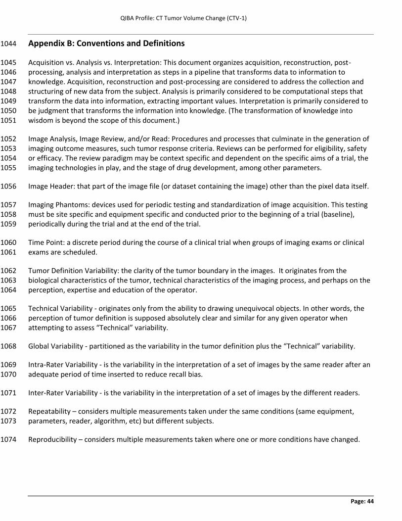
QIBA Profile: CT Tumor Volume Change (CTV-1)
Page: 44
Appendix B: Conventions and Definitions 1044
Acquisition vs. Analysis vs. Interpretation: This document organizes acquisition, reconstruction, post-1045 processing, analysis and interpretation as steps in a pipeline that transforms data to information to 1046 knowledge. Acquisition, reconstruction and post-processing are considered to address the collection and 1047 structuring of new data from the subject. Analysis is primarily considered to be computational steps that 1048 transform the data into information, extracting important values. Interpretation is primarily considered to 1049 be judgment that transforms the information into knowledge. (The transformation of knowledge into 1050 wisdom is beyond the scope of this document.) 1051
Image Analysis, Image Review, and/or Read: Procedures and processes that culminate in the generation of 1052 imaging outcome measures, such tumor response criteria. Reviews can be performed for eligibility, safety 1053 or efficacy. The review paradigm may be context specific and dependent on the specific aims of a trial, the 1054 imaging technologies in play, and the stage of drug development, among other parameters. 1055
Image Header: that part of the image file (or dataset containing the image) other than the pixel data itself. 1056
Imaging Phantoms: devices used for periodic testing and standardization of image acquisition. This testing 1057 must be site specific and equipment specific and conducted prior to the beginning of a trial (baseline), 1058 periodically during the trial and at the end of the trial. 1059
Time Point: a discrete period during the course of a clinical trial when groups of imaging exams or clinical 1060 exams are scheduled. 1061
Tumor Definition Variability: the clarity of the tumor boundary in the images. It originates from the 1062 biological characteristics of the tumor, technical characteristics of the imaging process, and perhaps on the 1063 perception, expertise and education of the operator. 1064
Technical Variability - originates only from the ability to drawing unequivocal objects. In other words, the 1065 perception of tumor definition is supposed absolutely clear and similar for any given operator when 1066 attempting to assess “Technical” variability. 1067
Global Variability - partitioned as the variability in the tumor definition plus the “Technical” variability. 1068
Intra-Rater Variability - is the variability in the interpretation of a set of images by the same reader after an 1069 adequate period of time inserted to reduce recall bias. 1070
Inter-Rater Variability - is the variability in the interpretation of a set of images by the different readers. 1071
Repeatability – considers multiple measurements taken under the same conditions (same equipment, 1072 parameters, reader, algorithm, etc) but different subjects. 1073
Reproducibility – considers multiple measurements taken where one or more conditions have changed. 1074


![WHY QIBA: PET-CT SPECIFICSqibawiki.rsna.org/images/0/08/Why_QIBA_PET-CT_specifics.pdf · Quantitative Imaging Test Discovery, Development, and Validation [Private & Academic Sectors]](https://static.fdocuments.in/doc/165x107/5f76183aa0c8a608ce5c1cb1/why-qiba-pet-ct-quantitative-imaging-test-discovery-development-and-validation.jpg)