Purinoceptor expression in regenerating skeletal muscle …s copies/CV1266.pdf · Purinoceptor...
34
The FASEB Journal express article 10.1096/fj.03-1175fje. Published online July 1, 2004. Purinoceptor expression in regenerating skeletal muscle in the mdx mouse model of muscular dystrophy and in satellite cell cultures Mina Ryten,* Shi Yu Yang, † Philip M. Dunn,* Geoffrey Goldspink, † and Geoffrey Burnstock † *Autonomic Neuroscience Institute, Royal Free & University College Medical School, Royal Free Campus; and † Molecular Tissue Repair Unit, Department of Surgery, Royal Free and University College Medical School, University College, London NW3 2PF, United Kingdom Corresponding author: Geoffrey Burnstock, Autonomic Neuroscience Institute, Royal Free & University College Medical School, Royal Free Campus, Rowland Hill Street, London NW3 2PF, UK. E-mail: [email protected] ABSTRACT ATP is an important extracellular signaling molecule mediating its effects by activation of P2X and P2Y receptors. P2 receptors are expressed during muscle development, and recent findings demonstrate that ATP can regulate myoblast proliferation and differentiation in vitro. However, the role of purinergic signaling during regeneration of injured skeletal muscle has not been investigated. To examine this process in a clinically relevant system, we used the mouse model of muscular dystrophy (mdx), in which muscle degeneration is rapidly followed by regeneration. The latter process, in vivo muscle regeneration, was the focus of this study, and to study the cellular mechanisms involved in it, a parallel study on normal rat skeletal myoblast cultures was conducted. Using immunohistochemistry, RT-PCR, and electrophysiology, we investigated the expression of the P2X 1-7 receptor subtypes and the P2Y 1,2,4,6 receptors. Experiments in vitro and in vivo demonstrated the sequential expression of the P2X 5 , P2Y 1 , and P2X 2 receptors during the process of muscle regeneration. The P2X 5 and P2Y 1 receptors were expressed first on activated satellite cells, and the P2Y 1 receptor was also expressed on infiltrating immune cells. Subsequent P2X 2 receptor expression on newly formed myotubes showed significant colocalization with AChRs, suggesting a role in regulation of muscle innervation. Thus, this study provides the first evidence for a role for purinergic signaling in muscle regeneration and raises the possibility of new therapeutic strategies in the treatment of muscle disease. Key words: ATP ● P2 receptor ● myotube dult skeletal muscle fibers are large, terminally differentiated cells containing multiple postmitotic nuclei. However, skeletal muscle shows a remarkable ability to recover from injury. The ability of mammalian skeletal muscle to respond to pathological damage is largely attributed to the presence of a small population of self-renewing muscle precursors, termed skeletal muscle satellite cells. Normally quiescent in response to muscle fiber injury satellite cells are activated, proliferate, and ultimately fuse to repair existing muscle fibers and form new myotubes (1–3). This process must be followed by myotube differentiation and growth A Page 1 of 34 (page number not for citation purposes)
Transcript of Purinoceptor expression in regenerating skeletal muscle …s copies/CV1266.pdf · Purinoceptor...
The FASEB Journal express article 10.1096/fj.03-1175fje. Published online July 1, 2004. Purinoceptor expression in regenerating skeletal muscle in the mdx mouse model of muscular dystrophy and in satellite cell cultures Mina Ryten,* Shi Yu Yang,† Philip M. Dunn,* Geoffrey Goldspink,† and Geoffrey Burnstock†
*Autonomic Neuroscience Institute, Royal Free & University College Medical School, Royal Free Campus; and †Molecular Tissue Repair Unit, Department of Surgery, Royal Free and University College Medical School, University College, London NW3 2PF, United Kingdom Corresponding author: Geoffrey Burnstock, Autonomic Neuroscience Institute, Royal Free & University College Medical School, Royal Free Campus, Rowland Hill Street, London NW3 2PF, UK. E-mail: [email protected] ABSTRACT ATP is an important extracellular signaling molecule mediating its effects by activation of P2X and P2Y receptors. P2 receptors are expressed during muscle development, and recent findings demonstrate that ATP can regulate myoblast proliferation and differentiation in vitro. However, the role of purinergic signaling during regeneration of injured skeletal muscle has not been investigated. To examine this process in a clinically relevant system, we used the mouse model of muscular dystrophy (mdx), in which muscle degeneration is rapidly followed by regeneration. The latter process, in vivo muscle regeneration, was the focus of this study, and to study the cellular mechanisms involved in it, a parallel study on normal rat skeletal myoblast cultures was conducted. Using immunohistochemistry, RT-PCR, and electrophysiology, we investigated the expression of the P2X1-7 receptor subtypes and the P2Y1,2,4,6 receptors. Experiments in vitro and in vivo demonstrated the sequential expression of the P2X5, P2Y1, and P2X2 receptors during the process of muscle regeneration. The P2X5 and P2Y1 receptors were expressed first on activated satellite cells, and the P2Y1 receptor was also expressed on infiltrating immune cells. Subsequent P2X2 receptor expression on newly formed myotubes showed significant colocalization with AChRs, suggesting a role in regulation of muscle innervation. Thus, this study provides the first evidence for a role for purinergic signaling in muscle regeneration and raises the possibility of new therapeutic strategies in the treatment of muscle disease. Key words: ATP P2 receptor myotube
dult skeletal muscle fibers are large, terminally differentiated cells containing multiple postmitotic nuclei. However, skeletal muscle shows a remarkable ability to recover from injury. The ability of mammalian skeletal muscle to respond to pathological damage is
largely attributed to the presence of a small population of self-renewing muscle precursors, termed skeletal muscle satellite cells. Normally quiescent in response to muscle fiber injury satellite cells are activated, proliferate, and ultimately fuse to repair existing muscle fibers and form new myotubes (1–3). This process must be followed by myotube differentiation and growth
A
Page 1 of 34(page number not for citation purposes)

before full muscle function can be regained. Many growth factors have been implicated in the regulation of muscle regeneration, including fibroblast growth factors, insulin-like growth factors, and nitric oxide (3). In this study, we explore the possibility that ATP might also be involved in regulating muscle regeneration. ATP and other extracellular nucleotides are now widely recognized as important extracellular messenger molecules with diverse biological effects ranging from smooth muscle contraction and synaptic transmission to cell proliferation and differentiation (4). The effects of ATP are mediated by activation of either P2X or P2Y receptors (5). P2Y receptors are G protein-coupled receptors, which act principally by activating phospholipase C, leading to formation of inositol 1,4,5-trisphophate and the mobilization of intracellular Ca2+ (5). In contrast, P2X receptors are ligand-gated ion channels and activation of these receptors by extracellular ATP elicits a flow of cations (Na+, K+, and Ca2+) across the plasma membrane. To date, seven mammalian P2X receptor subunits (P2X1-7) capable of assembling into homo- or heteromultimeric receptors have been cloned (6).
The expression of purinoceptors on developing skeletal muscle cells is well established. Responses characteristic of P2 receptor activation have been detected on myoblasts and myotubes cultured from embryonic chick muscle and C2C12 myoblasts (7–14). More recently the expression of specific P2X and P2Y receptor subtypes during skeletal muscle development has been demonstrated. P2Y1, P2X5, and P2X6 are expressed in chick skeletal muscle development (15–18), while expression of the P2X2, P2X5, P2X6, P2Y1, P2Y2, and P2Y4 receptors has been demonstrated in rat skeletal muscle development (19, 20).
In the case of the P2X5 and P2Y1 receptors, the functional significance of receptor expression has been explored. Whereas the P2Y1 receptor has been implicated in the regulation of acetylcholine receptor (AChR) and acetylcholinesterase (AChE) expression (17, 21), a role for the P2X5 receptor in the regulation of myoblast activity and differentiation has been demonstrated (22). In vitro experiments show that activation of the P2X5 receptor by ATP (but not adenosine, ADP, or UTP) results in a shift in the balance between myoblast proliferation and differentiation. Rat skeletal satellite cells (in primary culture) exposed to ATP have a reduced rate of proliferation but express markers of differentiation including myogenin and p21 mRNA (22). There is also evidence to suggest that adenosine, acting via purinergic (P1) receptors, may have trophic effects on muscle cells, causing apoptosis of C2C12 myoblasts (23).
There is also growing evidence for the release of ATP from multiple sites in skeletal muscle. As in all tissues, muscle fiber damage will result in the passive release of ATP and other nucleotides from cells. In addition, significant quantities of ATP are coreleased with ACh from motor nerve terminals (24, 25) on nerve activation and may be released from muscle fibers on contraction (26–28). Thus, skeletal muscle, particularly on injury, would be expected to contain high levels of extracellular ATP.
Together these findings suggest that purinergic signaling may play a significant role not only in skeletal muscle formation but also in muscle regeneration. To investigate this possibility, we studied purinoceptor expression and function in response to muscle injury both in vivo and in vitro. The mdx mouse model of Duchenne muscular dystrophy (DMD) was used as an example of a clinically relevant form of muscle damage and subsequent regeneration, the latter being the
Page 2 of 34(page number not for citation purposes)
focus of this study. DMD, the most common and severe of the muscular dystrophies, is caused by mutations in the dystrophin gene that result in the loss of dystrophin protein (29, 30). Dystrophin (as part of the dystrophin-glycoprotein complex) is involved in linking the cytoskeleton to the extracellular matrix, thereby stabilizing the periphery of the muscle fiber during contraction (31, 32). Loss of this protein renders the muscle plasma membrane susceptible to contraction-induced damage (33, 34), which (in the early stages of the disease) is followed rapidly by muscle regeneration. The mdx mouse has a genetic defect in a homologous region to that identified in humans and similarly lacks dystrophin protein (35, 30). This results in cycles of muscle fiber degeneration and subsequent regeneration. However, it must be noted that muscle regeneration in mdx mice does not result in such marked muscle weakness and premature death as occurs in human DMD patients.
Primary cultures of postnatal rat myoblasts exhibit a process of muscle regeneration in vivo that parallels that seen in vivo. Myoblasts in primary culture proliferate, align, and fuse to form contractile myotubes. This cellular morphogenesis is accompanied by muscle-specific gene expression, including the expression of the myogenic regulatory factors and proteins characteristic of terminal differentiation, such as myosin heavy chain (MHC) and nicotinic AChRs. By using this model of muscle regeneration, we have been able to apply a variety of techniques, including immunocytochemistry, RT-PCR, and patch clamping, to investigate the underlying signaling pathways. Thus, it was possible to investigate the timing, localization, and pharmacology of purinoceptors on satellite cells and myotubes during the process of muscle formation and maturation.
MATERIALS AND METHODS
Tissue preparation
Mdx and control, C57Bl/10 strain, mice were weaned at 3 wk and killed at 3.5 wk, 5 wk, and 2-3 months of age by exposing animals to a rising concentration of CO2, and death was confirmed by cervical dislocation. The tibialis anterior muscles were removed, placed onto cork blocks, covered with OCT compound, and frozen in liquid nitrogen-cooled isopentane. Cryostat sections were cut, producing transverse sections of the muscle at 12 µm or 7 µm and were collected on gelatinized slides.
Longitudinal sections of tibialis anterior muscle were prepared by pinning muscles on sylgard and then fixing the tissue in 4% paraformaldehyde for 2 h at room temperature. After being rinsed in PBS, muscles were placed longitudinally onto cork blocks, embedded in OCT and frozen in liquid nitrogen-cooled isopentane. Cryostat sections were cut at 50 µm and collected in PBS for staining as floating sections.
Immunohistochemistry (color reaction)
Immunostaining of 12 µm cryostat sections for the P2X1-7 and P2Y1,2,4 receptors was performed as described in Ryten et al. (20) and Cheung et al. (19); all sections were counterstained for nuclei with contrast green (Kirkegaard and Perry Laboratories) for 5 min at room temperature. Control experiments were carried out with the primary antibody omitted from the staining
Page 3 of 34(page number not for citation purposes)

procedure or with the primary antibody preabsorbed with the cognate peptides used to immunize the rabbits.
To make meaningful comparisons between the levels of immunoreactivity for a given receptor, sections were stained in sets containing tissues from control (C57Bl/10) and mdx animals at 3.5 wk, 5 wk, and 2-3 months of age and the color reaction was timed and equal for all samples.
Immunohistochemistry (fluorescence)
Air-dried cryostat sections and skeletal myoblasts plated on laminin-coated 8-well LabTek chamberslides (Nunc Life Technologies) were fixed in 4% formaldehyde in 0.1 M phosphate buffer (pH 7.4). After being washed in PBS, nonspecific binding sites were blocked using 10% NHS in PBS or 10% NGS. Due to the greater degree of permeabilization required for immunostaining for nuclear transcription factors, 7 µm thick cryostat sections were used when staining for MyoD and myogenin, and these sections were incubated in 10% NHS + 0.2% Triton X-100. Sections were incubated with primary antibodies, diluted in the respective blocking solutions, overnight at room temperature. The primary antibodies used were rabbit anti-P2X2 or anti-P2X5 (Roche Bioscience), rabbit anti-P2Y1 (Alomone Laboratories), rabbit anti-neurofilament 200 (Sigma), rabbit anti-MyoD1 (Autogen Bioclear), and rabbit anti-myogenin (Autogen Bioclear). These antibodies were used at the following concentrations: anti-P2X2 or -P2X5, 5 µg/ml; anti-P2Y1, 3 µg/ml; anti-neurofilament, 1:100; anti-MyoD1, 1:400; and anti-myogenin 1:400. After being washed in PBS, cells were incubated with fluorescence-labeled secondary antibodies for 1 h at 37°C. Donkey anti-rabbit Cy3 (1:500 in 1% NHS in PBS) or Oregon green-labeled goat anti-rabbit secondary antibodies (Stratech Scientific; 1:100 in 1% NGS in PBS) were applied as appropriate. In some cases, cell nuclei were also stained by incubation with DAPI (Sigma) at 0.6 µg/ ml in PBS for 1 h at room temperature.
Double labeling experiments for P2Y1 and CD11b were conducted as described above, except for an additional 1 h incubation with fluorescein-labeled rat anti-CD11b (Serotec). Immunostaining for P2X2 and AChRs was also performed using a similar protocol, except that sections were incubated for 1 h with Texas Red-labeled α-bungarotoxin (specific marker of the α-subunit of nicotine AChRs). Longitudinal, 50 µm thick sections were examined using confocal microscopy.
Control experiments were carried out with the primary antibody omitted from the staining procedure, and, in the case of the rabbit anti-P2X1-7, P2Y1, P2Y2, and P2Y4 antibodies, the primary antibody was preabsorbed with the peptides used to immunize the rabbits.
Quantitative analysis of P2X5 immunoreactivity
To measure the expression of P2X5 receptor staining, the number of nuclei present in P2X5-positive cells was counted in five randomly chosen fields (of 0.2 mm2) from a minimum of two muscle sections per muscle sample. Muscle samples from at least four animals were examined. Sections were taken from the mid-belly region of the TA muscle, photographed using a digital camera (Leica) attached to a Zeiss Axioplan microscope (Zeiss), and viewed on a screen with a grid applied to aid accuracy.
Page 4 of 34(page number not for citation purposes)

The total number of nuclei present in each field of view was also counted. This revealed that mdx muscle contained a significantly higher number of nuclei as compared with control muscle (204 ± 23 nuclei/ field of view in control, as compared with 353 ± 23 nuclei/ field of view in mdx tibialis anterior muscle). To correct for differences in nuclei number, the number of nuclei present in P2X5-positive cells was expressed as a percentage of the total number of nuclei.
Differences in the percentage of nuclei present in P2X5-positive cells in age-matched muscle samples were assessed for significance by one-way ANOVA followed by a post hoc Bonferroni’s multiple comparison test.
Tissue culture
Primary cultures of skeletal myoblasts were prepared as described in Ryten et al. (22). Cells were maintained in DMEM supplemented with 10% FCS, 10% normal horse serum (NHS; GIBCO), 0.5% gentamicin, and 0.5% ampicillin (Sigma) (termed growth medium, GM), or DMEM supplemented with 5% NHS (differentiation medium, DM), at 37°C and 5% CO2. Freshly isolated cells were used for each experiment.
Electrophysiology
Cells were plated at low density in 35 mm plastic culture dishes (Falcon) and maintained in culture for 1–7 days. Culture dishes were placed on the stage of an inverted microscope (Diaphot, Nikon), and cells were visualized under phase contrast at x600 magnification. Culture dishes were perfused with extracellular solution at room temperature, at a rate of 0.5 ml/min, while solution was applied locally to the cell of interest using a microperfusion device (36). Recordings were carried out using the conventional whole cell patch clamp technique (37) or the perforated patch technique. Patch electrodes were fabricated from thin wall borosilicate glass capillaries (Clark Electromedical, GC 150TF) and had a resistance of 2–4 MΩ when filled with “intracellular solution.” Membrane currents were recorded using an Axopatch 200B amplifier (Axon Instruments), displayed on a chart recorder (Gould TA240), and stored on digital audiotape using a DTR-1204 (Biologic, Claix) recorder for subsequent line analysis. Traces were acquired using Fetchex (pClamp software, Axon Instruments) and plotted using Origin 4 (Microcal).
Concentration effect data were fitted with the Hill equation: Y = A/[1+(K/X)n] where A is the maximum effect, K is the EC50, and n is the Hill coefficient, using Origin 4 (Microcal). The combined data from a number of cells were fitted, and the results are presented as values ± SE determined by the fitting routine.
Except where modified as indicated, the extracellular solution contained (mM): 154 NaCl, 4.7 KCl, 10 HEPES, 5.6 D-glucose, 1.2 MgCl2, 2.5 CaCl2 adjusted to pH 7.4 with NaOH. In experiments to investigate P2X mediated responses, the intracellular (pipette) solution contained (mM): 56 citric acid, 3 MgCl2, 20 CsCl, 40 HEPES, 0.1 EGTA, 10 TEA Cl, adjusted to pH 7.2 with CsOH (total Cs+ concentration 170 mM). To investigate P2Y-mediated responses, the pipette solution contained (mM): 56 tripotassium citrate, 25 KCl, 10 NaCl, 35 HEPES, 0.1 K EGTA, 0.26 amphotericin B.
Page 5 of 34(page number not for citation purposes)

Drugs used
ATP, ADP, UTP, DMPP, Reactive Blue 2, and suramin were purchased from Sigma. PPADS (tetrasodium salt) was supplied by Tocris Cookson Ltd (Bristol).
RT-PCR
Total RNA was extracted from skeletal myoblasts using the SV Total RNA Isolation System (Promega). RT-PCR was performed using Ready-to-Go RT-PCR beads (Amersham). Reverse transcription was performed using the Moloney murine leukemia virus reverse transcriptase. Primer sequences for P2X1-7 (38) and P2Y1,2,4,6 (39-41) were used for amplification reactions, as previously reported. The amplification reaction, performed in the same reaction tube, was conducted under the following conditions: 95°C for 30 s, the relevant annealing temperature for 30 s, and 72°C for 1 min, plus an additional cycle with an elongation time of 5 min. Amplification products were separated by electrophoresis and visualized by ethidium bromide staining. The presence of possible contaminants was investigated in all experiments, using control RT-PCR reactions in which either mRNA had been omitted or heating to 95°C had inactivated the reverse transcriptase.
RESULTS
Histology of mdx and control skeletal muscle
The tibialis anterior (TA) muscles of 3.5 wk, 5 wk, and 2-3 months old, mdx and age-matched control animals were studied. At all time points, muscle from control animals consisted of mature muscle fibers (Fig. 1A), characterized by peripheral my nuclei. Muscle fibers (in each age group) had similar diameters.
In contrast, degenerative and regenerative tissue changes were observed in skeletal muscle samples from mdx mice (Fig. 1A). As previously reported, muscle fiber degeneration and regeneration were initially episodic (42, 43). At birth, all the limb muscle fibers were relatively normal. However, by 3.5 wk of age histological evidence of muscle fiber degeneration in the TA muscle was obvious. Examination of sections by haematoxylin and eosin staining demonstrated a greatly reduced number of intact fibers and the heavy infiltration of phagocytic cells (Fig. 1A).
This acute phase of degeneration was followed by rapid regeneration, such that by 5 wk of age necrotic fibers had been replaced with small diameter myotubes (identified by their central nuclei) and the normal muscle architecture had been largely reestablished (Fig. 1A). In adult skeletal muscle samples, there was evidence of concurrent muscle fiber degeneration and regeneration. This resulted in a wide variation in muscle fiber diameter with newly formed, smaller diameter myotubes adjacent to larger, hypertrophied muscle fibers containing central nuclei (Fig. 1A).
Expression of P2 receptors in adult mdx and control skeletal muscle
Using specific antibodies against the P2X1-7 receptor subunits and the P2Y1,2,4 receptors we studied the expression of purinoceptor proteins in adult (2-3 month old) mdx and control skeletal
Page 6 of 34(page number not for citation purposes)
muscle. Control experiments, performed by preabsorbing the antibodies with the corresponding peptides, were used to confirm the specificity of the staining.
In normal, adult skeletal muscle, purinoceptor expression was restricted to vascular and nervous tissue but was absent from skeletal muscle cells. P2X1 and P2Y1 receptor proteins were detected in blood vessels, while the P2X5 receptor was detected in nerve fibers (Fig. 1B and C). These findings were confirmed by double labeling experiments (Fig. 1B and C). Colocalization of P2Y1 receptor protein (red) with smooth muscle actin (green) demonstrated expression of this receptor on vascular smooth muscle cells (Fig. 1B). Similarly, nerve bundles in both control and mdx muscle stained for P2X5, as demonstrated by colocalization of P2X5 receptor protein (red) and neurofilament (green) (Fig. 1C).
In contrast, adult mdx skeletal muscle was characterized by increased and widespread expression of three purinoceptor proteins, P2Y1, P2X5, and P2X2 (Fig. 2A). Whereas P2Y1 immunostaining was seen only in blood vessels in control samples, P2Y1 immunopositive cells were present throughout adult mdx muscle. These small, mononuclear cells appeared to be particularly concentrated in areas of muscle necrosis (Fig. 2A). Similarly, immunoreactivity for the P2X5 receptor was increased in mdx, as compared with adult skeletal muscle. P2X5-positive mononuclear cells and muscle fibers were scattered throughout adult mdx skeletal muscle (Fig. 2A). A pronounced increase in P2X2 expression was also observed in diseased as compared with control skeletal muscle. No immunoreactivity for P2X2 was detected on any cell type in control muscle samples, but there were many P2X2-immopositive myotubes and muscle fibers present in adult mdx skeletal muscle. Immunostaining was apparent as dark patches of immunoreactivity on the plasma membrane (Fig. 2A).
Specific purinoceptors are expressed in regeneration as opposed to degeneration of mdx muscle
The investigation of mdx and control skeletal muscle in 2- to 3-month-old mice demonstrated the increased and widespread expression of purinoceptors in mdx skeletal muscle, (as compared with control tissue). However, since degenerating and regenerating muscle fibers co-exist in adult mdx muscle, it was unclear whether P2 receptor expression was associated with the former and/or latter process. It is well recognized, both in human DMD and in the mdx mouse model, that muscle fiber degeneration and regeneration were initially episodic. We therefore investigated purinoceptor expression in the first cycle of damage and repair. Consistent with previous reports, the first degenerative phase in TA muscle was at 3.5 wk, while muscle regeneration was apparent at 5 wk of age (Fig. 1A) (42, 43).
With the use of immunohistochemistry, it was possible to demonstrate that expression of specific purinoceptors in mdx skeletal muscle was associated with specific time points, characterized by either degeneration or regeneration (Table 1). Whereas skeletal muscle from 3.5-wk-old mdx mice was strongly immunopositive for P2Y1, there was no immunoreactivity for P2X2 and little immunostaining for the P2X5 receptor in this tissue (Fig. 2B). As in adult mdx tissue, P2Y1 receptor immunostaining was expressed on small, mononuclear cells surrounding necrotic muscle fibers. This would suggest that while P2Y1 receptor expression was associated with muscle degeneration, the P2X5 and P2X2 receptors were not involved in this process (Table 1).
Page 7 of 34(page number not for citation purposes)
In contrast, at 5 wk of age when muscle regeneration was the predominant process occurring in mdx TA skeletal muscle (similar changes occur at different times in other muscles), both the P2X5 and P2X2 receptors were strongly expressed (Fig. 2C, Table 1). As noted in adult mdx muscle, immunoreactivity for P2X5 was present on mononuclear cells and myotubes, whereas immunoreactivity for P2X2 was restricted to distinct patches on muscle fiber and myotube membranes.
P2X5 receptor expression was quantified in control and mdx muscle from 3.5-wk, 5-wk, and 2- to 3-month-old mice. Receptor expression in each muscle sample was assessed by counting the number of nuclei present in P2X5-positive cells and the total number of nuclei in five randomly chosen fields from a minimum of two muscle sections. To correct for differences in nuclei number due predominantly to the age of the mice or the heavy infiltration of immune cells on muscle damage, the number of nuclei present in P2X5-positive cells was expressed as a percentage of the total number of nuclei. With the use of this method of quantifying P2X5 receptor expression, we demonstrated that in no sample did the number of nuclei present in P2X5-positive cells exceed 6% (Fig. 3A). However, there was a significant (P<0.1%) increase in P2X5 immunoreactivity in mdx, as compared with control skeletal muscle in 5-wk-old and adult mice. At 5 wk, 5.63% ± 0.34% of nuclei were present in P2X5-positive cells in mdx muscle, while only 0.47% ± 0.19% of nuclei were present in P2X5-positive cells in control tissue (Fig. 3A). There was no significant difference in immunoreactivity for P2X5 in mdx skeletal muscle from 5-wk-old, as compared with adult skeletal muscle (Fig. 3A).
A subpopulation of activated satellite cells in mdx muscle express P2 receptors
Sequential sections of 2- to 3-month-old mdx muscles were stained for myogenin, MyoD, P2X5, and P2Y1. These experiments revealed that many P2X5-positive mononuclear cells expressed the myogenic regulatory factors myogenin and MyoD, indicating that these cells were activated satellite cells (Fig. 3B, arrows). Furthermore, in some cases P2X5-positive myotubes contained nuclei positive for myogenin, suggesting that these fibers had been recently formed or repaired by satellite cell fusion. However, it was clear that there were also a number of cells that expressed myogenin, MyoD, or P2X5 alone. Thus, it appeared that only a subpopulation of activated satellite cells was positive for P2X5 and that some P2X5-positive cells were not activated satellite cells.
Immunostaining for P2Y1 was present in regions of regenerating muscle (as identified by MyoD or myogenin expression), and it was possible to identify a number of P2Y1-positive cells as activated satellite cells (Fig. 3B, arrowheads), though there were fewer of these than P2X5-positive satellite cells. This suggests that, as in the case of P2X5, only a subpopulation of activated satellite cells expresses P2Y1 and that the P2Y1 receptor is expressed by a variety of cell types.
P2Y1 receptor was expressed on inflammatory cells in mdx muscle
To determine the identity of P2Y1-positive cells, adult mdx muscle was double labeled for P2Y1 (red) and CD11b (green; Fig. 4A). CD11b is a marker for a large variety of inflammatory cells, including macrophages and B-lymphocytes. Significant colocalization of P2Y1 and CD11b (yellow) was detected (Fig. 4A). However, the fact that some CD11b cells were not
Page 8 of 34(page number not for citation purposes)
immunopositive for P2Y1 indicated that this receptor was not expressed by all infiltrating inflammatory cells.
Double labeling for P2X2 and AChRs
A number of studies demonstrate abnormalities in the localization and degradation of AChRs on mdx muscle from 5 wk of age onwards (43–45). Furthermore, there is growing evidence that P2X2 receptors and AChRs can functionally interact (46-50). Thus, the expression of AChRs and P2X2 receptors was investigated on adult skeletal muscle from control and mdx mice (Fig. 4B). Double staining experiments performed on muscle from control mice demonstrated largely continuous and well-organized clusters of AChRs at the neuromuscular junctions (NMJs) and the absence of any staining for P2X2 (Fig. 4B). In contrast, NMJs in adult mdx muscle consisted of numerous clusters of AChRs. Furthermore, P2X2 receptor expression was apparent on muscle fibers, and there were small areas of AChR and P2X2 receptor colocalization (yellow; Fig. 4B).
Myoblasts in primary culture express P2 receptors
Cells extracted from neonatal rat skeletal muscle were plated at low density and maintained in differentiation medium for 24 h. As demonstrated by staining for the myogenic transcription factor MyoD, 93.6 ± 2.5% of cells were myoblasts (Fig. 5A). These cells were also capable of fusing to form multinuclear myotubes that were positive for muscle-specific proteins, such as skeletal myosin heavy chain (Fig. 5A).
Using specific antibodies for P2X1-7 and P2Y1, P2Y2, and P2Y4, we investigated purinoceptor expression on myoblast cultures. Control experiments, performed by preabsorbing the antibodies with the corresponding peptides, were used to confirm the specificity of our findings. We found immunoreactivity for only two of the P2 receptors, P2X5 and P2Y1, on mononuclear cells in these cultures. Using RT-PCR, we were able to confirm the expression of both P2X5 and P2Y1 receptor mRNA in myoblasts (Fig. 5B, data for the P2X receptors not shown). RT-PCR experiments also demonstrated the expression of P2Y2, P2X4, P2X6, and P2X2 receptor mRNA.
Patch clamp recordings on myoblasts in primary culture
To investigate the functional properties of the P2 receptors present on myoblasts, we carried out whole cell patch clamp recording. Recordings were only made from small fusiform mononuclear cells. When the recording pipette contained a Cs+-based solution, rapid application of 10 µM ATP evoked a small, inward current of 9.7 ± 1.6 pA in 80% (17/21) of cells (Fig. 5C). In contrast, the nicotinic AChR agonist, 1,1-dimethyl-4-phenylpiperazinium iodide (DMPP), failed to produce an inward current in 80% (8/10) of cells tested. Responses to ATP reflected a current density of 0.87 ± 0.1 pA/pF and activated and inactivated quickly (Fig. 5C). The inward current activated by ATP was inwardly rectifying with a reversal potential close to 0 mV indicative of a nonselective cation conductance. UTP and ADP, which are potent agonists at some types of P2Y receptors failed to evoke any significant response under these recording conditions even at a concentration of 100 µM (Fig. 5D). Using perforated patch recording with a K+-based pipette solution, 4/4 cells responded to ATP with an outward current, which activated with a delay of several seconds (Fig. 5E). This would suggest the involvement of a metabotropic receptor and activation of Ca2+-activated potassium channels.
Page 9 of 34(page number not for citation purposes)

P2 receptor expression and regulation on myotubes
Myoblast cultures, maintained in DM to encourage myotube formation, were investigated 1, 3, 5, and 7 days after plating. Day 1 cultures contained mononucleated myoblasts, as confirmed by immunostaining for MyoD (Fig. 5A). However, by day 3 some myoblasts had aligned and fused to form multinuclear myotubes, as confirmed by the expression of skeletal myosin and the presence of three or more nuclei (Fig. 5A). This process continued such that by day 5 and 7, large, spontaneously contractile myotubes had formed. Staining procedures were carried out for all seven of the P2X receptors and the P2Y1, P2Y2, and P2Y4 receptors at all four time points. We found immunoreactivity only for the P2X2, P2X5, P2Y1, and P2Y4 receptors (Fig. 6A). These receptors were expressed sequentially, P2X5 and P2Y1 being expressed first, followed by P2Y4 and P2X2 (Fig. 6A; Table 2).
As discussed above, skeletal myoblasts (maintained in DM) stained strongly for the P2X5 and P2Y1 receptors (Fig. 6A). Similarly, early myotubes, present on day 3 cultures, expressed the P2X5 and P2Y1 receptors only (Fig. 6A; Table 2). However, with increasing time in culture, expression of these receptors on both mononucleated myoblasts and myotubes was greatly reduced. In the case of P2Y1, immunoreactivity on both unfused cells and myotubes had disappeared by day 5 (Fig. 6A; Table 2). Similarly in the case of P2X5, there was no immunostaining on unfused cells, and staining of myotubes was restricted to the ends of the cells (Fig. 6A). Since myotubes formed in vitro may branch to form multi- as opposed to bipolar cells, it was noted that the tips of all poles of the myotubes stained for P2X5. By day 7, no staining for P2X5 was detectable on any cell type (Fig. 6A).
Unlike P2X5 and P2Y1, immunoreactivity for the P2X2 and P2Y4 receptors could only be detected on myotubes (Fig. 6A; Table 2) but not on myoblasts. However, whereas immunoreactivity for P2Y4 could be detected on myotubes at all time points (from day 3 until day 7) (Fig. 6A; Table 2), P2X2 receptor expression could only be detected from day 5 (Fig. 6A; Table 2). Furthermore, unlike P2Y4 receptor expression, immunostaining for the P2X2 receptor was not homogenous (Fig. 6A). Although P2X2 was expressed along the whole length of the myotubes, there were clusters or areas of membrane with particularly high receptor staining. This pattern of staining was particularly apparent on the myotubes of day 7 cultures (Fig. 6A).
In summary, we were able to demonstrate the sequential expression of four purinoceptors during myotube formation, P2Y1, P2X5, P2Y4, and P2X2 (Table 2). Whereas the P2Y1 and P2X5 were expressed on both mononucleated myoblasts and myotubes, the P2Y4 and P2X2 receptors were only expressed on myotubes (Table 2).
Coexpression of P2X2 and AChRs on myotubes
To determine whether the pattern of P2X2 immunostaining was related to the well-documented clustering of AChRs on myotubes of aneural cultures we double-stained day 5 and 7 cultures for P2X2 receptors and AChRs (Fig. 6B). As previously reported, Texas Red-labeled α-bungarotoxin stained clusters of AChRs scattered across the myotube membrane (Fig. 6B). Using confocal microscopy to determine the precise location of receptor staining, we were able to demonstrate colocalization of P2X2 (green) and AChRs (red; Fig. 6B). Although there were clearly areas of
Page 10 of 34(page number not for citation purposes)

myotube membrane with significant colocalization (yellow), the same myotube also stained in other areas for P2X2 alone or AChRs alone (Fig. 6B).
Patch clamp recordings on myotubes
To avoid complications arising from poor space clamp, recordings were only made from small linear myotubes (<300 µm in length). When recordings were made with a Cs+-based pipette solution, these cells all responded to ATP with a small, rapidly activating inward current (Fig. 7A and B). This response was much smaller than that produced by the nicotinic AChR agonist DMPP (Fig. 7A). The effect of ATP was concentration-dependent, with an EC50 of 2.3 µM (Fig. 7C) and could not be replicated by application of either UTP or ADP (at concentrations of 30 µM; Fig. 7B). In agreement with the findings of Hume and Hönig (8, we were unable to observe any ATP-activated single channel currents in outside out patches excised from these myotubes. We investigated the pharmacology of the ATP-activated inward current in myotubes. Both suramin and Reactive Blue 2 produced a concentration-dependent, reversible antagonism of the ATP response with IC50 values of 3 and 0.97 µM, respectively (Fig. 7D and E). The most potent antagonist tested was pyridoxal 5-phosphate-6-azophenyl-2',4'-disulfonic acid (PPADS; Fig. 7F). This compound, at a concentration of 0.1µM, produced > 95% inhibition. However, the antagonism developed slowly over the course of 4 min and washed out very slowly, so that after 15 min washing, the response had recovered to <50% of the control value (Fig. 7F). As with the satellite cells, recording with a K+-based solution revealed a slowly activating outward current (Fig. 7G), which would suggest the involvement of a metabotropic receptor and activation of Ca2+-activated potassium channels.
DISCUSSSION
In this paper, we have shown for the first time that purinoceptor expression, in particular expression of the P2X5 and P2X2 receptors, is a key feature of muscle regeneration. In normal skeletal muscle, purinoceptor expression was absent from skeletal muscle cells, though present in vascular and nervous tissue. However, three purinoceptor proteins were strongly expressed in adult mdx muscle, namely P2X2, P2X5, and P2Y1. To determine more precisely the role of these receptors in the response to muscle injury, we investigated the expression and function of the purinoceptors at different time points in the mdx mouse lifetime and in primary postnatal rat myoblast cultures. With the use of RT-PCR, immunohistochemistry, and electrophysiology, these experiments revealed the sequential expression of the P2X5, P2Y1, and P2X2 receptors during the process of muscle regeneration. The P2X5 and P2Y1 receptors were expressed first on activated satellite cells and in the case of P2Y1, a range of immune cells. This was followed by the expression of the P2X2 receptor on newly formed myotubes both in vivo and in vitro. Thus these findings strongly suggest a role for purinergic signaling in the process of skeletal muscle regeneration.
The cause of muscle injury in mdx mice, as in DMD patients, appears to be the absence of the subsarcolemmal cytoskeletal protein dystrophin (35, 30). In the mdx mouse, the limb muscle fibers are histologically normal postnatally (51) but undergo degeneration soon after weaning (at 3 wk in this study) when motor activity increases. This first phase of muscle damage and the subsequent inflammatory response were characterized by a marked increase in immunoreactivity for the P2Y1 receptor. Double-staining experiments demonstrated that P2Y1 receptor protein was
Page 11 of 34(page number not for citation purposes)

strongly expressed on leukocytes (as identified by immunoreactivity for CD11b) and leukocyte infiltration could largely account for the increase in P2Y1 receptor expression in damaged muscle. This finding is consistent with past research demonstrating P2Y1 receptor expression on a range of white blood cells (including monocytes, dendritic cells, and thymocytes) and a role for this receptor in the regulation of cytokine release (52-54). Since some cytokines (including IL-6 and LIF) can act as growth factors in damaged skeletal muscle, it is possible that P2Y1 receptor expression may be involved in regulating leukocyte activity and indirectly muscle repair (55. 56).
The regeneration of muscle fibers in mdx muscle ensures that muscle function rapidly recovers (42). This process depends on the presence of skeletal muscle satellite cells. On muscle injury, satellite cells are activated, proliferate, and ultimately fuse to repair damaged fibers and form new myotubes. Past research on mdx muscle suggests that, while these regenerative processes continue in adult mice, they are most prominent at ~4-5 wk of age, when activated satellite cells are present in the greatest numbers (42, 43, 57). Thus, the increase in P2X5 receptor expression in mdx skeletal muscle from 5 wk of age onwards strongly suggests a role for this receptor in muscle regeneration. Immunostaining demonstrated a >10-fold increase in the expression of the P2X5 receptor in mdx muscle at 5 wk, as compared with age-matched control tissue and mdx muscle at 3.5 wk of age. Although a similar increase in P2X5 receptor expression was also demonstrated in adult mdx muscle when regeneration is less obvious, this may be due to a slight overestimate of receptor expression in this tissue. Although correcting for differences in the number of nuclei present in mdx and control skeletal muscle was an effective means of accounting for the infiltration of immune cells into mdx tissue, it was not an effective means of accounting for differences in muscle morphology when comparing mdx tissue from animals of different ages.
Immunolabeling of sequential muscle sections demonstrated that the marked increase in immunoreactivity was due to the expression of the P2X5 receptor on a subpopulation of activated satellite cells (as identified by the expression of the myogenic transcription factors MyoD or myogenin). A number of activated satellite cells were also positive for P2Y1 (though fewer than for P2X5). The absence of significant immunoreactivity for either of these receptors in mononucleated cells in control muscle samples suggested that purinoceptors were not expressed by quiescent satellite cells but only activated satellite cells.
In keeping with the observations in vivo, we found that postnatal mononucleated myoblasts in primary culture expressed both P2X5 and P2Y1 receptor mRNA and protein. Expression of these receptor proteins by satellite cells could account for the P2X- and P2Y-like responses detected by patch clamp recording on addition of ATP. Furthermore, since the cultures investigated were derived from normal rat skeletal muscle, this study strongly suggested that purinoceptor expression in mdx skeletal muscle was not directly due to a deficiency in the dystrophin protein, but occurred secondary to muscle regeneration (irrespective of the initial cause of injury). Thus, it seems likely that ATP is one of a number of extracellular signaling molecules, including mechano growth factor, fibroblast growth factors, and insulin-like growth factors, which regulate satellite cell activity (3, 58, 59).
However, there were some differences between myoblasts in culture and in vivo. It should be recognized that whereas all cells in culture expressed P2 receptors, P2X5 and P2Y1 receptor
Page 12 of 34(page number not for citation purposes)
proteins were expressed by only a subpopulation of myoblasts in mdx muscle. This discrepancy could be explained by the fact that high levels of receptor expression may be specific to particular cell states (for example proliferation or differentiation). Thus, in vivo, where satellite cells have not been activated simultaneously or maintained under identical conditions, differential expression of the P2X5 and P2Y1 receptors might be expected.
Recent research demonstrating the role of the P2X5 receptor in muscle formation (22) strongly suggests that the expression of this receptor on activated satellite cells in mdx muscle was of functional significance. Activation of the P2X5 receptor on rat skeletal satellite cells in primary culture has been shown to increase the rate of commitment to terminal differentiation and fusion to form multinuclear myotubes (22). Consistent with such a function for the P2X5 receptor, it was observed that newly formed myotubes in culture and small myotubes in vivo (presumably recently regenerated) express this receptor.
Although the functional significance of P2X2 receptor expression in skeletal muscle is not established, the timing of receptor expression in mdx muscle may indicate a possible role in the regulation of muscle reinnervation after damage. The absence of dystrophin in mdx muscle not only affects muscle architecture but also disrupts the relationship between motor nerve and muscle fiber (43). Abnormalities in the structure and function of neuromuscular junctions include NMJ fragmentation and changes in AChR function and the expression of embryonic-type AChRs. These appear after the first postnatal month and after the onset of muscle damage and repair and become increasingly obvious with age (44, 45). Similarly, expression of the P2X2 receptor in mdx muscle began only at 5 wk of age and immunoreactivity for this receptor was strongest in adult mdx muscle, when P2X2-immunopositive clusters were detected across the cell membranes of myotubes and muscle fibers. P2X2 receptor expression was also observed on aneural myotubes in vitro, confirming that receptor expression in mdx muscle was indeed a feature of muscle regeneration and not degeneration. Furthermore, patch clamp recordings conducted on myotubes in vitro demonstrated responses characteristic of P2X receptors and consistent with the expression of homomeric P2X2 receptors (ATP responses were sensitive to the antagonists, suramin, Reactive Blue 2, and PPADS). Although it is possible that heteromeric P2X2/P2X5 receptors were present (the P2X5 receptor protein being transiently expressed on myotubes and capable of coassembly with P2X2 subunits), the different distribution patterns and timing of P2X2 and P2X5 receptor subunits makes this unlikely.
Recent studies demonstrating that P2X2 and ACh receptors can interact may suggest a specific role for the P2X2 receptor in the regulation of AChR function and possibly distribution under conditions of abnormal innervation. Coactivation of P2X2 and nicotinic AChRs elicits currents significantly smaller than those predicted on the basis of activation of each channel separately (46–50). Thus, coexpression of P2X2 and AChRs could account for some of the abnormalities in AChR activity observed in mdx muscle. In fact, our double-staining experiments demonstrated colocalization of P2X2 and ACh receptors both in mdx muscle and in myotube cultures. As previously reported, AChRs were expressed in clusters on the myotube membrane in aneural cultures, and in many cases receptor clusters contained not only acetylcholine but also P2X2 receptors. Similarly, in adult mdx muscle, P2X2 receptor protein colocalized with AChRs at small receptor clusters at the NMJ. Since ATP is coreleased with ACh at the NMJ, coactivation of the nicotinic and P2X2 receptors could occur and cause previously observed reductions in the opening time and current amplitude of AChRs in dystrophic muscle (60, 37).
Page 13 of 34(page number not for citation purposes)

It is important to note that, contrary to the findings presented by Choi et al. (17), coexpression of P2Y1 and ACh receptors at the NMJs could not be demonstrated in either normal or mdx muscle at any time point. However, immunocytochemistry and patch clamp recording in vitro did demonstrate the expression of P2Y receptors on myotubes in vitro. Both P2Y1 and P2Y4 receptor protein were detected on myotubes in culture, though not on muscle fibers in mdx skeletal muscle. The presence of these receptors in culture is consistent with the findings of Henning et al. (12–14), showing that application of UTP and ADP to C2C12 myotubes can cause an increase in inositol-1,4,5-trisphophate levels and cAMP respectively. Thus, the differences in the expression of P2Y receptors in vivo and in vitro may be, at least in part, related to the absence of dystrophin in mdx mice.
In summary, the age-dependent pattern of P2 receptor expression in mdx muscle strongly suggests that purinergic signaling plays a key role in the response to muscle injury. The P2X2, P2X5, and P2Y1 receptors were strongly expressed in mdx skeletal muscle (as compared with controls), and these receptors were expressed on cells known to be important in muscle regeneration. In particular, the P2X5 and P2Y1 receptors were expressed on activated satellite cells. P2 receptors were also expressed on myotubes/ muscle fibers (P2X2) and on infiltrating immune cells (P2Y1). Since mdx muscle is likely to contain high levels of extracellular ATP due to muscle damage, and it is known that ATP and other extracellular nucleotides can affect satellite cell activity, it seems likely that P2 receptor expression in mdx muscle is of functional significance in muscle regeneration. Furthermore, strong similarities in the specific subtypes and pattern of P2 receptor expression in regenerating muscle in vivo and in vitro suggest that P2 receptors are part of the normal response to muscle injury. Thus, this study provides the first evidence for a role for purinergic signaling in muscle regeneration in vivo and indicates that purinergic receptors may be therapeutic targets for the treatment of muscle diseases.
ACKNOWLEDGMENTS
The MF20 monoclonal antibody developed by Donald A. Fischman was obtained from the Developmental Studies Hybridoma Bank developed under the auspices of the NICHD and maintained by the University of Iowa, Department of Biological Sciences, Iowa City, IA 52242. The authors thank Roche Bioscience (Palo Alto, CA), the Wellcome Trust, and the University College London Medical School (London, UK) for their financial support, and are grateful to Chrystalla Orphanides for editorial assistance.
REFERENCES
1. Bischoff, R. (1994) The satellite cell and muscle regeneration. In Myogenesis (Engel, A. G., and Franszini-Armstrong, C., eds.) p. 97-118, McGraw-Hill
2. Seale, P., and Rudnicki, M. A. (2000) A new look at the origin, function, and "stem-cell" status of muscle satellite cells. Dev. Biol. 218, 115–124
3. Hawke, T. J., and Garry, D. J. (2001) Myogenic satellite cells: physiology to molecular biology. J. Appl. Physiol. 91, 534–551
4. Burnstock, G. (1997) The past, present and future of purine nucleotides as signaling molecules. Neuropharmacology 36, 1127–1139
Page 14 of 34(page number not for citation purposes)

5. Ralevic, V., and Burnstock, G. (1998) Receptors for purines and pyrimidines. Pharmacol. Rev. 50, 413–492
6. Torres, G. E., Egan, T. M., and Voigt, M. M. (1999) Identification of a domain involved in ATP-gated ionotropic receptor subunit assembly. J. Biol. Chem. 274, 22359–22365
7. Kolb, H. A., and Wakelam, M. J. (1983) Transmitter-like action of ATP on patched membranes of cultured myoblasts and myotubes. Nature 303, 621–623
8. Hume, R. I., and Hönig, M. G. (1986) Excitatory action of ATP on embryonic chick muscle. J. Neurosci. 6, 681–690
9. Hume, R. I., and Thomas, S. A. (1988) Multiple actions of adenosine 5′-triphosphate on chick skeletal muscle. J. Physiol. 406, 503–524
10. Thomas, S. A., and Hume, R. I. (1990) Permeation of both cations and anions through a single class of ATP-activated ion channels in developing chick skeletal muscle. J. Gen. Physiol. 95, 569–590
11. Thomas, S. A., Zawisa, M. J., Lin, X., and Hume, R. I. (1991) A receptor that is highly specific for extracellular ATP in developing chick skeletal muscle in vitro. Br. J. Pharmacol. 1034, 1963–1969
12. Henning, R. H., Nelemans, A., van den Akker, J., and den Hertog, A. (1992) The nucleotide receptors on mouse C2C12 myotubes. Br. J. Pharmacol. 106, 853–858
13. Henning, R. H., Duin, M., den Hertog, A., and Nelemans, A. (1993a) Characterization of P2-purinoceptor mediated cyclic AMP formation in mouse C2C12 myotubes. Br. J. Pharmacol. 110, 133–138
14. Henning, R. H., Duin, M., den Hertog, A., and Nelemans, A. (1993b) Activation of the phospholipase C pathway by ATP is mediated exclusively through nucleotide type P2-purinoceptors in C2C12 myotubes. Br. J. Pharmacol. 110, 747–752
15. Meyer, M. P., Clarke, J. D., Patel, K., Townsend-Nicholson, A., and Burnstock, G. (1999a) Selective expression of purinoceptor cP2Y1 suggests a role for nucleotide signaling in development of the chick embryo. Dev. Dyn. 214, 152–158
16. Meyer, M. P., Groschel-Stewart, U., Robson, T., and Burnstock, G. (1999b) Expression of two ATP-gated ion channels, P2X5 and P2X6, in developing chick skeletal muscle. Dev. Dyn. 216, 442–449
17. Choi, R. C., Man, M. L., Ling, K. K., Ip, N. Y., Simon, J., Barnard, E. A., and Tsim, K. W. (2001) Expression of the P2Y1 nucleotide receptor in chick muscle: its functional role in the regulation of acetylcholinesterase and acetylcholine receptor. J. Neurosci. 21, 9224–9234
Page 15 of 34(page number not for citation purposes)

18. Ruppelt, A., Ma, W., Borchardt, K., Silberberg, S. D., and Soto, F. (2001) Genomic structure, developmental distribution and functional properties of the chicken P2X(5) receptor. J. Neurochem. 77, 1256–1265
19. Cheung, K. K., Ryten, M., and Burnstock, G. (2003) Abundant and dynamic expression of G protein-coupled P2Y receptors in mammalian development. Dev. Dyn. 228, 254–266
20. Ryten, M., Hoebertz, A., and Burnstock, G. (2001) Sequential expression of three receptor subtypes for extracellular ATP in developing rat skeletal muscle. Dev. Dyn. 221, 331–341
21. Choi, R. C., Siow, N. L., Cheng, A. W., Ling, K. K., Tung, E. K., Simon, J., Barnard, E. A., and Tsim, K. W. (2003) ATP acts via P2Y1 receptors to stimulate acetylcholinesterase and acetylcholine receptor expression: transduction and transcription control. J. Neurosci. 23, 4445–4456
22. Ryten, M., Dunn, P. M., Neary, J. T., and Burnstock, G. (2002) ATP regulates the differentiation of mammalian skeletal muscle by activation of a P2X5 receptor on satellite cells. J. Cell Biol. 158, 345–355
23. Ceruti, S., Giammarioli, A. M., Camurri, A., Falzono, L., Rufini, S., Frank, C., Fiorentini, C., Malorni, W., and Abbracchio, M. P. (2000) Adenosine- and 2-chloro-adenosine-induced cytopathic effects on myoblastic cells and myotubes: involvement of different intracellular mechanisms. Neuromuscul. Disord. 10, 436–446
24. Redman, R. S., and Silinsky, E. M. (1994) ATP released together with acetylcholine as the mediator of neuromuscular depression at frog motor nerve endings. J. Physiol. 477, 117–127
25. Silinsky, E. M., and Redman, R. S. (1996) Synchronous release of ATP and neurotransmitter within milliseconds of a motor nerve impulse in the frog. J. Physiol. 492, 815–822
26. Smith, D. O. (1991) Sources of adenosine released during neuromuscular transmission in the rat. J. Physiol. 432, 343–354
27. Cunha, R. A., and Sebastiao, A. M. (1993) Adenosine and adenine nucleotides are independently released from both the nerve terminals and the muscle fibres upon electrical stimulation of the innervated skeletal muscle of the frog. Pflugers Arch. 424, 503–510
28. Hellsten, Y., Maclean, D., Radegran, G., Saltin, B., and Bangsbo, J. (1998) Adenosine concentrations in the interstitium of resting and contracting human skeletal muscle. Circulation 98, 6–8
29. Hoffman, E. P., Brown, R. H., Jr., and Kunkel, L. M. (1987) Dystrophin: the protein product of the Duchenne muscular dystrophy locus. Cell 51, 919–928
30. Sicinski, P., Geng, Y., Ryder-Cook, A. S., Barnard, E. A., Darlison, M. G., and Barnard, P. J. (1989) The molecular basis of muscular dystrophy in the mdx mouse: a point mutation. Science 244, 1578–1580
Page 16 of 34(page number not for citation purposes)
31. Tinsley, J. M., Blake, D. J., Zuellig, R. A., and Davies, K. E. (1994) Increasing complexity of the dystrophin-associated protein complex. Proc. Natl. Acad. Sci. USA 91, 8307–8313
32. Straub, V., and Campbell, K. P. (1997) Muscular dystrophies and the dystrophin-glycoprotein complex. Curr. Opin. Neurol. 10, 168–175
33. Weller, B., Karpati, G., and Carpenter, S. (1990) Dystrophin-deficient mdx muscle fibers are preferentially vulnerable to necrosis induced by experimental lengthening contractions. J. Neurol. Sci. 100, 9–13
34. Petrof, B. J., Shrager, J. B., Stedman, H. H., Kelly, A. M., and Sweeney, H. L. (1993) Dystrophin protects the sarcolemma from stresses developed during muscle contraction. Proc. Natl. Acad. Sci. USA 90, 3710–3714
35. Bulfield, G., Siller, W. G., Wight, P. A., and Moore, K. J. (1984) X chromosome-linked muscular dystrophy (mdx) in the mouse. Proc. Natl. Acad. Sci. USA 81, 1189–1192
36. Dunn, P. M., Benton, D. C., Campos Rosa, J., Ganellin, C. R., and Jenkinson, D. H. (1996) Discrimination between subtypes of apamin-sensitive Ca(2+)-activated K+ channels by gallamine and a novel bis-quaternary quinolinium cyclophane, UCL 1530. Br. J. Pharmacol. 117, 35–42
37. Hamill, O. P., Marty, A., Neher, E., Sakmann, B., and Sigworth, F. J. (1981) Improved patch-clamp techniques for high-resolution current recording from cells and cell-free membrane patches. Pflugers Arch. 391, 85–100
38. Shibuya, I., Tanaka, K., Hattori, Y., Uezono, Y., Harayama, N., Noguchi, J., Ueta, Y., Izumi, F., and Yamashita, H. (1999) Evidence that multiple P2X purinoceptors are functionally expressed in rat supraoptic neurones. J. Physiol. 514, 351–367
39. Bailey, M. A., Imbert-Teboul, M., Turner, C., Marsy, S., Srai, K., Burnstock, G., and Unwin, R. J. (2000) Axial distribution and characterization of basolateral P2Y receptors along the rat renal tubule. Kidney Int. 58, 1893–1901
40. Bailey, M. A., Imbert-Teboul, M., Turner, C., Srai, S. K., Burnstock, G., and Unwin, R. J. (2001) Evidence for basolateral P2Y(6) receptors along the rat proximal tubule: functional and molecular characterization. J. Am. Soc. Nephrol. 12, 1640–1647
41. Hede, S. E., Amstrup, J., Christoffersen, B. C., and Novak, I. (1999) Purinoceptors evoke different electrophysiological responses in pancreatic ducts. P2Y inhibits K(+) conductance, and P2X stimulates cation conductance. J. Biol. Chem. 274, 31784–31791
42. Dangain, J., and Vrbová, G. (1984) Muscle development in mdx mutant mice. Muscle Nerve 7, 700–704
43. De la Porte, S., Morin, S., and Koenig, J. (1999) Characteristics of skeletal muscle in mdx mutant mice. Int. Rev. Cytol. 19,:99-148
Page 17 of 34(page number not for citation purposes)
44. Kong, J., and Anderson, J. E. (1999) Dystrophin is required for organizing large acetylcholine receptor aggregates. Brain Res. 839, 298–304
45. Grady, R. M., Zhou, H., Cunningham, J. M., Henry, M. D., Campbell, K. P., and Sanes, J. R. (2000) Maturation and maintenance of the neuromuscular synapse: genetic evidence for roles of the dystrophin–glycoprotein complex. Neuron 25, 279–293
46. Nakazawa, K. (1994) ATP activated current and its interaction with acetylcholine activated current in rat sympathetic neurons. J. Neurosci. 14, 740–750
47. Brajas-Lopez, C., Espinosa-Luna, R., and Zhu, Y. (1998) Functional interactions between nicotinic and P2X channels in short-term cultures of guinea pig submucosal neurons. J. Physiol. 513, 671–683
48. Searl, T. J., Redman, R. S., and Silinsky, E. M. (1998) Mutual occlusion of P2X ATP receptors and nicotinic receptors on sympathetic neurons of the guinea-pig. J. Physiol. 510, 783–791
49. Zhou, X., and Galligan, J. J. (1998) Non-additive interaction between nicotinic cholinergic and P2X purine receptors in guinea-pig enteric neurons in culture. J. Physiol. 513, 685–697
50. Khakh, B. S., Zhou, X., Sydes, J., Galligan, J. J., and Lester, H. A. (2000) State-dependent cross-inhibition between transmitter-gated cation channels. Nature 406, 405–410
51. Torres, L. F., and Duchen, L. W. (1987) The mutant mdx: inherited myopathy in the mouse. Morphological studies of nerves, muscles and end-plates. Brain 110, 269–299
52. Jin, J., Dasari, V. R., Sistare, F. D., and Kunapuli, S. P. (1998) Distribution of P2Y receptor subtypes on haematopoietic cells. Br. J. Pharmacol. 123, 789–794
53. Di Virgilio, F., Chiozzi, P., Ferrari, D., Falzoni, S., Sanz, J. M., Morelli, A., Torboli, M., Bolognesi, G., and Baricordi, O. R. (2001) Nucleotide receptors: an emerging family of regulatory molecules in blood cells. Blood 97, 587–600
54. Straub, R. H., Pongratz, G., Günzler, C., Michna, A., Baier, S., Kees, F., Falk, W., and Schölmerich, J. (2002) Immunoregulation of IL-6 secretion by endogenous and exogenous adenosine and by purinergic agonists in splenic tissue slices. J. Neuroimmunol. 125, 73–81
55. Austin, L., and Burgess, A. W. (1991) Stimulation of myoblast proliferation in culture by leukemia inhibitory factor and other cytokines. J. Neurol. Sci. 101, 193–197
56. Kurek, J. B., Nouri, S., Kannourakis, G., Murphy, M., and Austin, L. (1996) Leukemia inhibitory factor and interleukin-6 are produced by diseased and regenerating skeletal muscle. Muscle Nerve 19, 1291–1301
57. Jin, Y., Murakami, N., Saito, Y., Goto, Y., Koishi, K., and Nonaka, I. (2000) Expression of MyoD and myogenin in dystrophic mice, mdx and dy, during regeneration. Acta Neuropathol. (Berl.) 99, 619–627
Page 18 of 34(page number not for citation purposes)
58. Yang, S. Y., and Goldspink, G. (2002) Different roles of the IGF-I Ec peptide (MGF) and mature IGF-I in myoblast proliferation and differentiation. FEBS Lett. 522, 156–160
59. Hill, M., and Goldspink, G. (2003) Expression and splicing of the insulin-like growth factor gene in rodent muscle is associated with muscle satellite (stem) cell activation following local tissue damage. J. Physiol. 549, 409–418
60. Carlson, C. G. (1999) Spontaneous changes in acetylcholine receptor and calcium leakage activity in cell-attached patches from cultured dystrophic myotubes. Pflugers Arch. 437, 371–380
Received January 22, 2004; accepted May 10, 2004.
Page 19 of 34(page number not for citation purposes)
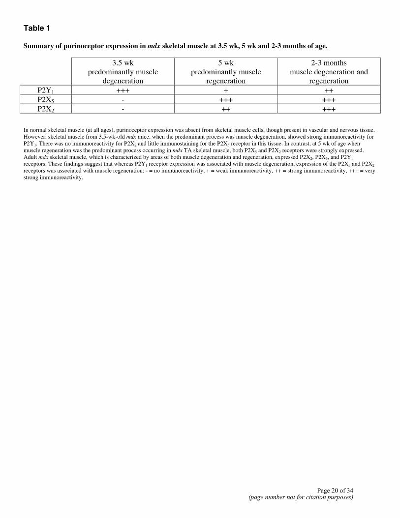
Table 1 Summary of purinoceptor expression in mdx skeletal muscle at 3.5 wk, 5 wk and 2-3 months of age.
3.5 wk predominantly muscle
degeneration
5 wk predominantly muscle
regeneration
2-3 months muscle degeneration and
regeneration
P2Y1 +++ + ++ P2X5 - +++ +++ P2X2 - ++ +++
In normal skeletal muscle (at all ages), purinoceptor expression was absent from skeletal muscle cells, though present in vascular and nervous tissue. However, skeletal muscle from 3.5-wk-old mdx mice, when the predominant process was muscle degeneration, showed strong immunoreactivity for P2Y1. There was no immunoreactivity for P2X2 and little immunostaining for the P2X5 receptor in this tissue. In contrast, at 5 wk of age when muscle regeneration was the predominant process occurring in mdx TA skeletal muscle, both P2X5 and P2X2 receptors were strongly expressed. Adult mdx skeletal muscle, which is characterized by areas of both muscle degeneration and regeneration, expressed P2X2, P2X5, and P2Y1 receptors. These findings suggest that whereas P2Y1 receptor expression was associated with muscle degeneration, expression of the P2X5 and P2X2 receptors was associated with muscle regeneration; - = no immunoreactivity, + = weak immunoreactivity, ++ = strong immunoreactivity, +++ = very strong immunoreactivity.
Page 20 of 34(page number not for citation purposes)
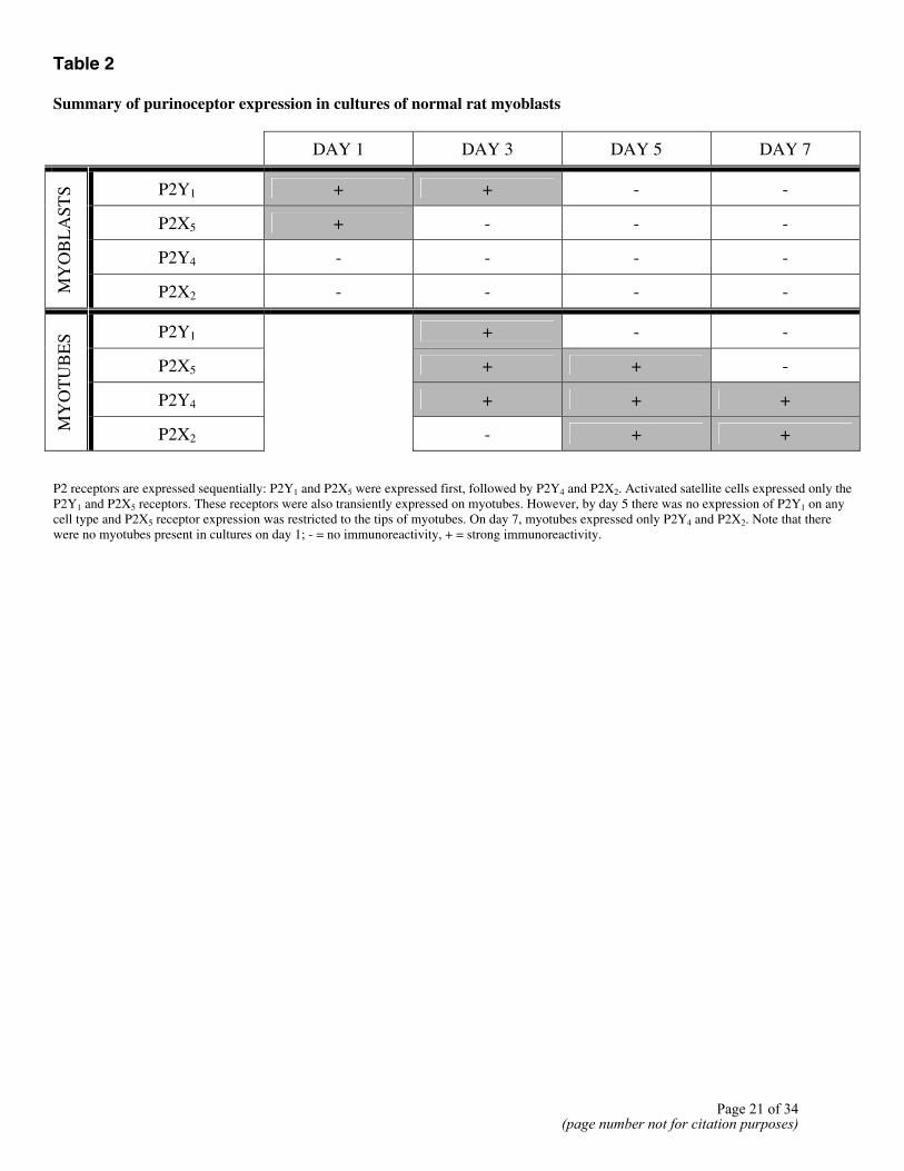
Table 2 Summary of purinoceptor expression in cultures of normal rat myoblasts
DAY 1 DAY 3 DAY 5 DAY 7
P2Y1 + + - -
P2X5 + - - -
P2Y4 - - - -
MY
OB
LA
STS
P2X2 - - - -
P2Y1 + - -
P2X5 + + -
P2Y4 + + +
MY
OT
UB
ES
P2X2
- + +
P2 receptors are expressed sequentially: P2Y1 and P2X5 were expressed first, followed by P2Y4 and P2X2. Activated satellite cells expressed only the P2Y1 and P2X5 receptors. These receptors were also transiently expressed on myotubes. However, by day 5 there was no expression of P2Y1 on any cell type and P2X5 receptor expression was restricted to the tips of myotubes. On day 7, myotubes expressed only P2Y4 and P2X2. Note that there were no myotubes present in cultures on day 1; - = no immunoreactivity, + = strong immunoreactivity.
Page 21 of 34(page number not for citation purposes)
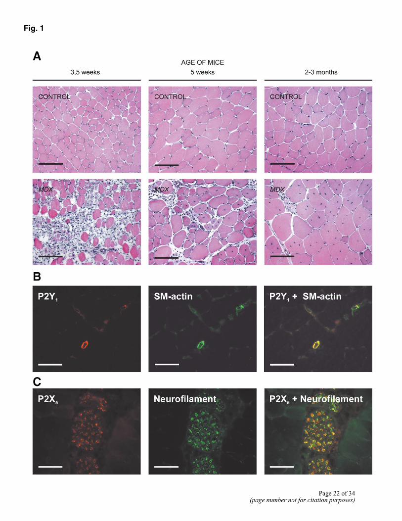
Fig. 1
Page 22 of 34(page number not for citation purposes)
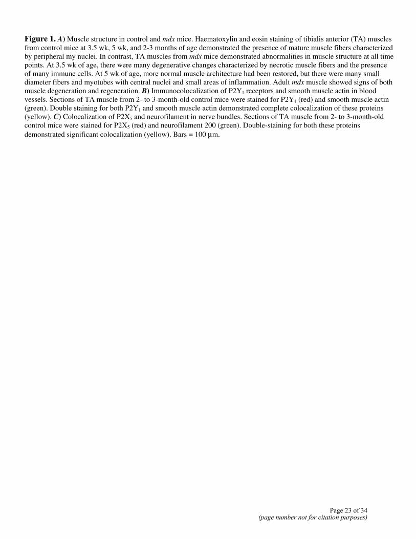
Figure 1. A) Muscle structure in control and mdx mice. Haematoxylin and eosin staining of tibialis anterior (TA) muscles from control mice at 3.5 wk, 5 wk, and 2-3 months of age demonstrated the presence of mature muscle fibers characterized by peripheral my nuclei. In contrast, TA muscles from mdx mice demonstrated abnormalities in muscle structure at all time points. At 3.5 wk of age, there were many degenerative changes characterized by necrotic muscle fibers and the presence of many immune cells. At 5 wk of age, more normal muscle architecture had been restored, but there were many small diameter fibers and myotubes with central nuclei and small areas of inflammation. Adult mdx muscle showed signs of both muscle degeneration and regeneration. B) Immunocolocalization of P2Y1 receptors and smooth muscle actin in blood vessels. Sections of TA muscle from 2- to 3-month-old control mice were stained for P2Y1 (red) and smooth muscle actin (green). Double staining for both P2Y1 and smooth muscle actin demonstrated complete colocalization of these proteins (yellow). C) Colocalization of P2X5 and neurofilament in nerve bundles. Sections of TA muscle from 2- to 3-month-old control mice were stained for P2X5 (red) and neurofilament 200 (green). Double-staining for both these proteins demonstrated significant colocalization (yellow). Bars = 100 µm.
Page 23 of 34(page number not for citation purposes)
Fig. 2
Page 24 of 34(page number not for citation purposes)
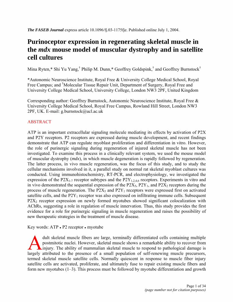
Figure 2. A) Changes in the expression of the P2Y1, P2X5, and P2X2 receptors in adult mdx muscle. Sections of TA muscle from 2- to 3-month-old C57Bl/10 (control) and mdx mice were stained for P2Y1, P2X5, and P2X2 (black) and counterstained for nuclei (green). Immunoreactivity for P2Y1 was only observed in blood vessels in control muscle samples; many immunopositive mononucleated cells were present in adult mdx muscle. Similarly, while immunoreactivity for P2X5 or P2X2 was absent in muscle samples from adult control mice, P2X5-immunopositive cells were scattered throughout mdx muscle, and strong immunoreactivity for P2X2 was observed on myotube and muscle fiber membranes (arrows). B) Sections of TA muscle from 3.5-wk-old mdx mice stained for P2Y1, P2X5, and P2X2 receptors (black) and counterstained for nuclei (green). P2Y1 receptor was strongly expressed during the first phase of mdx muscle degeneration. Neither the P2X2 nor P2X5 receptors were expressed at significant levels at this time. C) Sections of TA muscle from 5-wk-old mdx mice stained for P2Y1, P2X5 and P2X2 receptors (black) and counterstained for nuclei (green). Immunoreactivity for the P2X5 and P2X2 receptors was present at this age in mdx muscle, when the predominant process occurring is regeneration. Although P2Y1 receptor was also expressed at this time, immunoreactivity for this receptor appeared to be less widespread compared with that in adult mdx muscle. Bars = 100 µm.
Page 25 of 34(page number not for citation purposes)
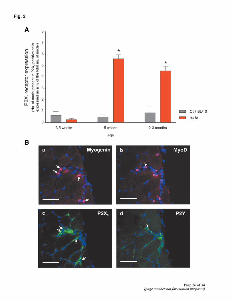
Fig. 3
Page 26 of 34
(page number not for citation purposes)
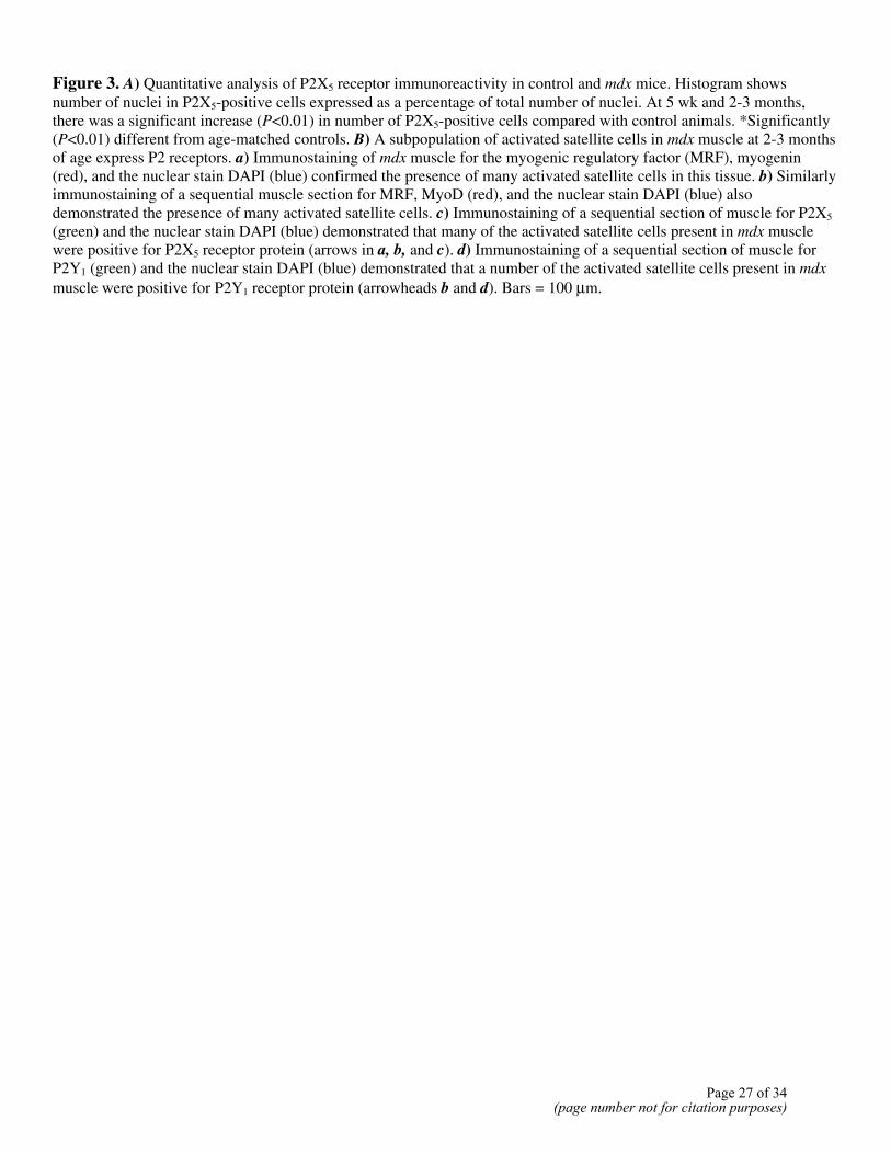
Figure 3. A) Quantitative analysis of P2X5 receptor immunoreactivity in control and mdx mice. Histogram shows number of nuclei in P2X5-positive cells expressed as a percentage of total number of nuclei. At 5 wk and 2-3 months, there was a significant increase (P<0.01) in number of P2X5-positive cells compared with control animals. *Significantly (P<0.01) different from age-matched controls. B) A subpopulation of activated satellite cells in mdx muscle at 2-3 months of age express P2 receptors. a) Immunostaining of mdx muscle for the myogenic regulatory factor (MRF), myogenin (red), and the nuclear stain DAPI (blue) confirmed the presence of many activated satellite cells in this tissue. b) Similarly immunostaining of a sequential muscle section for MRF, MyoD (red), and the nuclear stain DAPI (blue) also demonstrated the presence of many activated satellite cells. c) Immunostaining of a sequential section of muscle for P2X5 (green) and the nuclear stain DAPI (blue) demonstrated that many of the activated satellite cells present in mdx muscle were positive for P2X5 receptor protein (arrows in a, b, and c). d) Immunostaining of a sequential section of muscle for P2Y1 (green) and the nuclear stain DAPI (blue) demonstrated that a number of the activated satellite cells present in mdx muscle were positive for P2Y1 receptor protein (arrowheads b and d). Bars = 100 µm.
Page 27 of 34(page number not for citation purposes)

Fig. 4
Figure 4. A) Subpopulations of inflammatory cells in mdx muscle express the P2Y1 receptor. Immunostaining of adult mdx muscle for P2Y1 (red) and CD11b (green) (a marker for a variety of immune cells) revealed that there was a significant degree of colocalization of P2Y1 and CD11b (yellow) in adult mdx muscle. However, since some cells expressed only CD11b (green) or only P2Y1 (red), it is clear that the P2Y1 receptor was expressed by a subpopulation of inflammatory cells and that not all P2Y1-positive cells were immune cells. B) Representative neuromuscular junctions from control and mdx skeletal muscle. Longitudinal sections of adult skeletal muscle from control and mdx mice were double stained for ACh receptors (red) and P2X2 receptors (green). a) Immunostaining of muscle from control mice demonstrated largely continuous and well-organized clusters of AChRs at the NMJs and the absence of any staining for P2X2. b) In contrast, NMJs in adult mdx muscle consisted of multiple clusters of AChRs and were generally larger. Furthermore, P2X2 receptor expression was apparent on muscle fibers, and there were numerous small areas of AChR and P2X2 receptor colocalization (yellow). Bars = 100 µm.
Page 28 of 34(page number not for citation purposes)
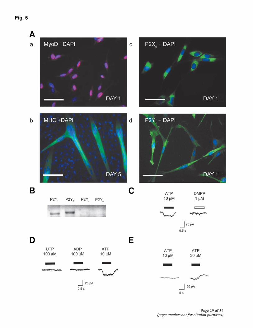
Fig. 5
Page 29 of 34(page number not for citation purposes)

Figure 5. A) Expression and function of purinoceptors in myoblasts in vitro. a) Immunocytochemistry for MyoD (red) and DAPI (blue) demonstrated that day 1 cultures contained 93.6 ± 2.5% myoblasts. Bar = 50 µm. b) After 5 days in culture, many myoblasts had fused to form multinuclear myotubes, which stain for myosin heavy chain (green) and DAPI (blue). Bar = 100 µm. c) Immunocytochemistry for P2X5 receptors (green) and DAPI (blue) demonstrated that cells maintained in differentiation medium (DM) for 24 h, stained strongly for P2X5 receptor protein. Bar = 50 µm. d) Immunocytochemistry for P2Y1 receptors (green) and DAPI (blue) demonstrated that the P2Y1 receptor was expressed by myoblasts in day 1 cultures. Bar = 50 µm. B) RT-PCR demonstrates the expression of P2Y receptor mRNA. Both P2Y1 and P2Y2 receptor mRNA was detected, but P2Y4 and P2Y6 were absent. C) Application of ATP to myoblasts voltage clamped at – 60 mV using a Cs+-based electrode solution, produced a rapidly activating inward current. In this and subsequent figures, the bar indicates duration of drug application. Most cells failed to respond to the nicotinic ACh receptor agonist DMPP. D) Effect of ATP was not mimicked by the P2Y1 and P2Y2/ 4 receptor agonists ADP and UTP. E) When recordings were carried out using the perforated patch technique, and a K+-based pipette solution, ATP evoked a concentration-dependent outward current, which was sometimes preceded by a small inward current.
Page 30 of 34(page number not for citation purposes)
Fig. 6
Page 31 of 34(page number not for citation purposes)
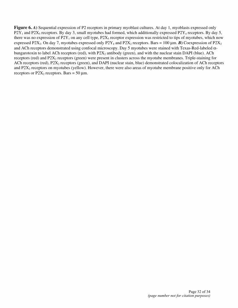
Figure 6. A) Sequential expression of P2 receptors in primary myoblast cultures. At day 1, myoblasts expressed only P2Y1 and P2X5 receptors. By day 3, small myotubes had formed, which additionally expressed P2Y4 receptors. By day 5, there was no expression of P2Y1 on any cell type, P2X5 receptor expression was restricted to tips of myotubes, which now expressed P2X2. On day 7, myotubes expressed only P2Y4 and P2X2 receptors. Bars = 100 µm. B) Coexpression of P2X2 and ACh receptors demonstrated using confocal microscopy. Day 5 myotubes were stained with Texas-Red-labeled α-bungarotoxin to label ACh receptors (red), with P2X2 antibody (green), and with the nuclear stain DAPI (blue). ACh receptors (red) and P2X2 receptors (green) were present in clusters across the myotube membranes. Triple-staining for ACh receptors (red), P2X2 receptors (green), and DAPI (nuclear stain, blue) demonstrated colocalization of ACh receptors and P2X2 receptors on myotubes (yellow). However, there were also areas of myotube membrane positive only for ACh receptors or P2X2 receptors. Bars = 50 µm.
Page 32 of 34(page number not for citation purposes)
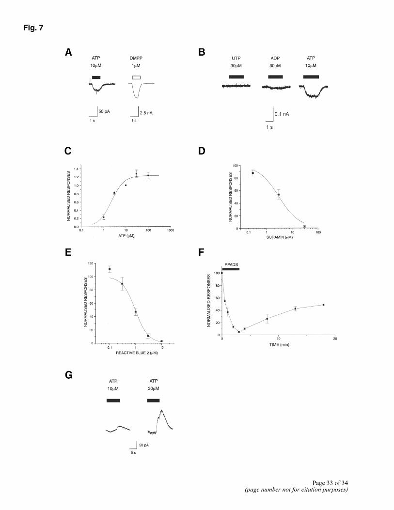
Fig. 7
Page 33 of 34
(page number not for citation purposes)
Figure 7. Patch clamp recording from myotubes. A) Application of ATP to small myotubes voltage clamped at –60 mV using a Cs+-based electrode solution, produced a small rapidly activating inward current. A much larger current was produced by the nicotinic AChR agonist, DMPP. B) Effect of ATP was not mimicked by the P2Y receptor agonists UTP and ADP, even when applied at higher concentrations. C) Concentration response curve for inward current activated by ATP. Fitting data with Hill equation gave an EC50 of 2.25 ± 0.05 µM. D) Concentration effect curve for inhibition of the inward current response to 10 µM ATP by suramin (using a Cs+-based electrode solution). Points are means ± SE from 3-5 cells. Fitting data to Hill equation gave an IC50 value of 3 ± 0.8 µM. E) Concentration effect curve for the inhibition of the response to 10 µM ATP by Reactive Blue 2 (using a Cs+-based electrode solution). Points are mean ± SE from 4 cells. Fitting the data to the Hill equation gave an IC50 of 0.97 ± 0.19 µM. F) Time course for inhibition of ATP response by PPADS (using a Cs+-based electrode solution). Responses to ATP (10 µM) were recorded at various time points during the application of 0.1 µM PPADS, and following washout of the antagonist. Points are means ± SE from 3 myotubes. G) When myotubes were voltage-clamped using the perforated patch technique and a K+-based pipette solution, ATP evoked a concentration-dependent outward current, which was often preceded by a much smaller inward current.
Page 34 of 34(page number not for citation purposes)