
Purification Characterization Aand B Clostridium difficileiai.asm.org/content/35/3/1032.full.pdf ·...
9
INFECTION AND IMMUNITY, Mar. 1982, p. 1032-1040 0019-9567/82/031032-09$02.00/0 Vol. 35, No. 3 Purification and Characterization of Toxins A and B of Clostridium difficile N. M. SULLIVAN, S. PELLETT, AND T. D. WILKINS* Department of Anaerobic Microbiology, Virginia Polytechnic Institute and State University, Blacksburg, Virginia 24061 Received 17 August 1981/Accepted 30 October 1981 Toxin preparations were obtained by growing Clostridium difficile VPI strain 10463 in 2-liter brain heart infusion dialysis flasks at 37°C for 3 days. The initial step of the purification scheme involved ultrafiltration through an XM-100 membrane filter. Two toxic activities, designated toxins A and B, were separated by ion-exchange chromatography on DEAE-NaCl gradients. Toxin A was purified to homogeneity by an acetic acid precipitation at pH 5.5. Other separation techniques, including CM Sepharose CL-6B, (NH4)2SO4 and acetic acid precipita- tions, and hydrophobic interaction chromatography, were examined in attempts to further purify toxin B. Although these methods failed to increase the specific activity of toxin B, they provided additional evidence that the two toxins are distinct molecules. The toxins are acid and heat labile and are inactivated by trypsin and chymotrypsin, but not by amylase. The molecular weight of toxin A, as estimated by gel filtration and gradient polyacrylamide electrophoresis, ranged from 440,000 to 500,000. The estimated molecular weight of toxin B was 360,000 to 470,000. Intestinal disturbances are common side ef- fects of antimicrobial therapy. Pseudomembran- ous colitis in humans and antibiotic-associated colitis in hamsters are diseases associated with the administration of clindamycin or other anti- biotics (6-8, 17, 20). The causative agent of most of these diseases appears to be Clostridium difficile and its toxin (7, 10, 17, 30). Fecal filtrates from over 90% of patients with docu- mented pseudomembranous colitis (6, 8) and cecal contents of hamsters injected with clinda- mycin are cytotoxic in tissue cultures and cause death when injected intracecally into normal hamsters (7, 8). The effects of the toxin are neutralized by antisera against partially purified C. difficile culture supernatants (16) and by a fortuitous cross-reaction with C. sordellii anti- sera obtained from the U.S. Bureau of Biologics (8, 10). Methods for the partial purification of C. difficile toxin (3, 20, 26, 28, 28a, 30) have been described by research groups, and there are discrepancies in the characterization of the tox- in. Recently, Bartlett et al. reported that two toxins are produced by C. difficile: one (toxin A) causes fluid accumulation in rabbit ileal loops and has been called an "enterotoxin," and the other (toxin B) has been described as primarily a cytotoxin (8, 28a; N. S. Taylor, G. M. Thorne, and J. G. Bartlett, Clin. Res., 28:285, 1980). We have found that both toxins are cytotoxic and animal lethal, although toxin B is at least 1,000- fold more cytotoxic than toxin A. Initial data indicate that each toxin is produced in sufficient concentrations in vitro and in vivo to cause cytopathic effects in tissue culture and to kill animals. At present, it is not known which toxin(s) is primarily responsible for the patholo- gy seen in pseudomembranous colitis and antibi- otic-associated colitis. In this paper, we describe the purification and characterization of toxin A and the partial purifi- cation and characterization of toxin B. MATERIALS AND METHODS Bacterial strain. C. difficile VPI strain 10463 was grown in 2-liter brain heart infusion (dialysis flasks for 72 h at 37°C (3). Chemicals and reagents. Column supports for ion- exchange chromatography (DEAE-Sepharose CL-6B and CM Sepharose CL-6B), hydrophobic interaction chromatography, concanavalin A 4B affinity chroma- tography, and gel filtration (Sepharose 6B) were ob- tained from Pharmacia Fine Chemicals, Piscataway, N.J. Reagents for the inactivation studies were obtained from Sigma Chemical Co., St. Louis, Mo., and includ- ed trypsin (bovine pancreatic, type III), chymotrypsin (bovine pancreatic), bacterial amylase (type Ila), and soybean inhibitor (type I-S). Polyacrylamide gels were prepared from the following chemicals: electrophoresis pure acrylamide (Bio-Rad Laboratories, Richmond, Calif.), electrophoresis pure sodium dodecyl sulfate (Bio-Rad), electrophoresis- pure N,N'-methylenebisacrylamide (Eastman Organic Chemicals, Rochester, N.Y.), N,N,N',N'-tetramethyl- 1032 on August 19, 2018 by guest http://iai.asm.org/ Downloaded from
Transcript of Purification Characterization Aand B Clostridium difficileiai.asm.org/content/35/3/1032.full.pdf ·...

INFECTION AND IMMUNITY, Mar. 1982, p. 1032-10400019-9567/82/031032-09$02.00/0
Vol. 35, No. 3
Purification and Characterization of Toxins A and B ofClostridium difficile
N. M. SULLIVAN, S. PELLETT, AND T. D. WILKINS*Department ofAnaerobic Microbiology, Virginia Polytechnic Institute and State University, Blacksburg,
Virginia 24061
Received 17 August 1981/Accepted 30 October 1981
Toxin preparations were obtained by growing Clostridium difficile VPI strain10463 in 2-liter brain heart infusion dialysis flasks at 37°C for 3 days. The initialstep of the purification scheme involved ultrafiltration through an XM-100membrane filter. Two toxic activities, designated toxins A and B, were separatedby ion-exchange chromatography on DEAE-NaCl gradients. Toxin A was purifiedto homogeneity by an acetic acid precipitation at pH 5.5. Other separationtechniques, including CM Sepharose CL-6B, (NH4)2SO4 and acetic acid precipita-tions, and hydrophobic interaction chromatography, were examined in attemptsto further purify toxin B. Although these methods failed to increase the specificactivity of toxin B, they provided additional evidence that the two toxins aredistinct molecules. The toxins are acid and heat labile and are inactivated bytrypsin and chymotrypsin, but not by amylase. The molecular weight of toxin A,as estimated by gel filtration and gradient polyacrylamide electrophoresis, rangedfrom 440,000 to 500,000. The estimated molecular weight of toxin B was 360,000to 470,000.
Intestinal disturbances are common side ef-fects of antimicrobial therapy. Pseudomembran-ous colitis in humans and antibiotic-associatedcolitis in hamsters are diseases associated withthe administration of clindamycin or other anti-biotics (6-8, 17, 20). The causative agent of mostof these diseases appears to be Clostridiumdifficile and its toxin (7, 10, 17, 30). Fecalfiltrates from over 90% of patients with docu-mented pseudomembranous colitis (6, 8) andcecal contents of hamsters injected with clinda-mycin are cytotoxic in tissue cultures and causedeath when injected intracecally into normalhamsters (7, 8). The effects of the toxin areneutralized by antisera against partially purifiedC. difficile culture supernatants (16) and by afortuitous cross-reaction with C. sordellii anti-sera obtained from the U.S. Bureau of Biologics(8, 10).Methods for the partial purification of C.
difficile toxin (3, 20, 26, 28, 28a, 30) have beendescribed by research groups, and there arediscrepancies in the characterization of the tox-in. Recently, Bartlett et al. reported that twotoxins are produced by C. difficile: one (toxin A)causes fluid accumulation in rabbit ileal loopsand has been called an "enterotoxin," and theother (toxin B) has been described as primarily acytotoxin (8, 28a; N. S. Taylor, G. M. Thorne,and J. G. Bartlett, Clin. Res., 28:285, 1980). Wehave found that both toxins are cytotoxic andanimal lethal, although toxin B is at least 1,000-
fold more cytotoxic than toxin A. Initial dataindicate that each toxin is produced in sufficientconcentrations in vitro and in vivo to causecytopathic effects in tissue culture and to killanimals. At present, it is not known whichtoxin(s) is primarily responsible for the patholo-gy seen in pseudomembranous colitis and antibi-otic-associated colitis.
In this paper, we describe the purification andcharacterization of toxin A and the partial purifi-cation and characterization of toxin B.
MATERIALS AND METHODSBacterial strain. C. difficile VPI strain 10463 was
grown in 2-liter brain heart infusion (dialysis flasks for72 h at 37°C (3).
Chemicals and reagents. Column supports for ion-exchange chromatography (DEAE-Sepharose CL-6Band CM Sepharose CL-6B), hydrophobic interactionchromatography, concanavalin A 4B affinity chroma-tography, and gel filtration (Sepharose 6B) were ob-tained from Pharmacia Fine Chemicals, Piscataway,N.J.
Reagents for the inactivation studies were obtainedfrom Sigma Chemical Co., St. Louis, Mo., and includ-ed trypsin (bovine pancreatic, type III), chymotrypsin(bovine pancreatic), bacterial amylase (type Ila), andsoybean inhibitor (type I-S).
Polyacrylamide gels were prepared from the followingchemicals: electrophoresis pure acrylamide (Bio-RadLaboratories, Richmond, Calif.), electrophoresis puresodium dodecyl sulfate (Bio-Rad), electrophoresis-pure N,N'-methylenebisacrylamide (Eastman OrganicChemicals, Rochester, N.Y.), N,N,N',N'-tetramethyl-
1032
on August 19, 2018 by guest
http://iai.asm.org/
Dow
nloaded from
PURIFICATION OF C. DIFFICILE TOXINS 1033
ethylenediamine (Bio-Rad), and electrophoresis-pureammonium persulfate (Isolab, Inc., Akron, Ohio).
Protein and chloride determination. Protein wasquantitated by the Bio-Rad method (Bio-Rad) (9).Chloride concentrations were determined by titration,using Sigma chloride kit no. 830 (Sigma).
Cytotoxicity and animal lethality assays. The cyto-toxicity assay was performed in a 92-well flat-bottommicrotiter plate (Costar, Data Packaging, Cambridge,Mass.). Chinese hamster ovary cells (CHO-Kl) (FlowLaboratories, Inc., McLean, Va.) were grown to con-fluency in F12 medium (Flow Laboratories) containing10%6 fetal calf serum. The cells were trypsinized(0.025% trypsin), incubated for 5 min in 5% CO2 at37°C, and washed with two 5-ml volumes of F12medium containing 2% fetal calf serum. Approximate-ly 1,000 cells were dispensed per well. Samples oftoxin preparations were titered in 2- or 10-fold dilu-tions within the wells after the cells had become fixedto the bottom of the well (3 h). The tissue culture dosewas defined as the reciprocal of the highest dilutionwhich rounded 100%o of the CHO-Kl cells.For the mouse lethality experiments, 6-week-old
ICR male mice were used (Flow Laboratories). Thetoxin preparations were diluted in twofold dilutions in50 mM Tris-hydrochloride buffer (pH 7.5), and 0.5 mlof each dilution was injected intraperitoneally. Deathswere recorded at 18 h. The LD10o was defined as thereciprocal of the highest dilution required to kill 100loof the mice injected.
Toxin purification. C. difficile VPI strain 10463 wasgrown in 12 2-liter dialysis culture flasks as previouslydescribed. After centrifugation at 8,000 x g for 10 minand filtration through a 0.45-,um membrane filter (Mil-lipore Corp., Bedford, Mass.), the culture supernatant(-750 ml) was concentrated to 50 ml by ultrafiltrationat 4°C, using an XM-100 membrane filter (AmiconCorp., Lexington, Mass.) with a thin-channel typeconcentrator. The retentate was washed with 1,500 mlof 50 mM Tris-hydrochloride buffer (pH 7.5) at 4°C andconcentrated to a final volume of 40 to 50 ml.The concentrated supernatant was loaded onto a
DEAE-Sepharose CL-6B column (2.5 by 10 cm) whichhad been equilibrated with 50 mM Tris-hydrochloride(pH 7.5). After the sample was loaded, the columnwas washed with 200 ml of 50 mM Tris-hydrochloride(pH 7.5) containing 0.05 M NaCl. The sample waseluted first with a 300-ml linear NaCl gradient in 50mM Tris-hydrochloride buffer (0.05 to 0.25 M NaCl),followed by 150 ml of 50 mM Tris-hydrochloride (pH7.5) containing 0.3 M NaCl. A second 300-ml lineargradient (0.3 to 0.6 M NaCI) in the same bufferfollowed the 0.3 M NaCl wash. The flow rate of thecolumns was 55 to 60 ml/h (gravity) at 4°C. Fractions(4.2 ml) were collected and assayed for cytotoxicity byusing CHO-Ki cells.The fractions containing the highest cytotoxic titers
were pooled, filter sterilized, and stored at 4°C. Thetoxins that eluted in the first and second NaCl gradi-ents were designated toxins A and B, respectively.From 5 to 10 ml of the toxic fractions from the first
DEAE gradient (toxin A) was dialyzed against 1 literof 0.01 M sodium acetate buffer (pH 5.5) at 4°C for 18to 24 h. The dialysate was centrifuged to recover theprecipitate at 169 x g for 10 min, washed with 5 ml ofthe same acetate buffer, and recentrifuged. The precip-itate was solubilized in S to 10 ml of 50 mM Tris-
hydrochloride, (pH 7.5) containing 0.05 M NaCl, andthe solution (toxin A) was filter sterilized and stored at40C.PAGE. Davis gels with a 5% lower gel and an upper
stacking gel (12) and gradient gels (2.5 to 27%) (Isolab)were used to demonstrate the removal of contaminat-ing proteins during the purification procedure. Slabgels (7.5 by 7.5 by 0.3 cm) were used for bothprocedures. Davis gel samples contained approximate-ly 100 p.g of protein, 10%o glycerol, and 1 to 2 p.l of0.05% bromphenol blue. The buffer system for theDavis gels was Tris-glycine (pH 8.4), whereas poly-acrylamide gel electrophoresis (PAGE) with gradientgels was performed with Tris-borate (pH 8.4). ForDavis gels, electrophoresis was performed at 60 Vuntil the marker dye was at the interface between thestacker and the lower gel, then increased to 125 V untilthe dye had migrated to 0.5 cm from the bottom of thegel. The gradient PAGE gels were first preelectropho-resed for 30 min at 75 V before sample loading, thenthe voltage was increased to 125 V for 18 to 24 hours.
After electrophoresis, both types of PAGE gelswere fixed with 10%o trichloroacetic acid for 1 h andstained with 0.025% Coomassie brilliant blue R-250 in10% acetic acid. The gels were destained with a 10%otrichloroacetic acid solution.
Crossed IEP. Crossed immunoelectrophoresis (IEP)was also used as a method to demonstrated the remov-al of contaminating proteins during the purificationprocedure. Crossed IEP was performed with the LKB2117 Multiphor apparatus and IEP kit (LKB Instru-ments, Inc., Rockville, Md.). The general methodolo-gy presented in the LKB instruction manuals andapplication notes 85 and 249 and in the quantitativeIEP manual of Axelsen et al. (4) was followed. Gelswere pressed, washed, stained, and destained. Theantisera used for the second dimension was goatantisera produced against crude culture supernatantsof C. difficile strain 10463 (16).
Elution of the toxins from PAGE gels. The two toxinswere eluted from PAGE gels to localize the toxicactivity for toxin B and to confirm that the purifiedtoxin A was the major protein band. Samples of toxinA (600 p.g) and toxin B (3.0 mg) were loaded onto anentire slab gel. When electrophoresis was completed,a longitudinal slice of the gel was cut, fixed, andstained with Coomassie blue as previously described.The remainder of the gels were cut horizontally in 3-mm slices, homogenized in 50 mM Tris-hydrochloride(pH 7.5) containing 0.05 M NaCl, and stored 18 to 24 hat 4°C. The fractions were then assayed for cytotoxicactivity.Gel filtration chromatography. Gel filtration chroma-
tography was used as a possible method of purifica-tion. Samples (5 ml) of crude culture supernatant wereput onto a column (2.5 by 7.0 cm) of Sepharose CL-6Band eluted at 4°C with 50 mM Tris-hydrochloridebuffer (pH 7.5) at a flow rate of 25 to 30 ml/h.
Determination of native molecular weight. Descend-ing chromatography, performed with a Sepharose CL-6B column (1.5 by 77 cm), was used for estimating thenative molecular weights of the toxins (1). The Kayvalues of high molecular weight standard proteins(Pharmacia Fine Chemicals) aldolase, catalase, ferm-tin, and thyroglobulin, were used to obtain a standardcurve. The cytotoxic titers of the fractions were usedto locate the toxins for the calculation of their Kav
VOL. 35, 1982
on August 19, 2018 by guest
http://iai.asm.org/
Dow
nloaded from
1034 SULLIVAN, PELLETT, AND WILKINS
values, and the molecular weights of the two toxinswere estimated from the standard curve.
Gradient polyacrylamide gels were also used toestimate the molecular weights of the toxins. Relativemobilities were calculated for each toxin. The toxinswere detected by eluting the activity from the gels.Ammonium sulfate precipitations. Ammonium sul-
fate precipitations were performed on crude culturesupernatants to determine the precipitation propertiesof the two toxins. Finely ground ammonium sulfatewas added slowly in 5% increments to crude culturesupernatant (4°C) with gentle mixing. After each addi-tion, the sample was mixed for 15 to 20 min, adjustedto pH 7.0 with 2 N NaOH, and then filtered through a0.45-p.m membrane filter. The samples were analyzedfor cytotoxicity and animal toxicity.
Acetic acid precipitations. Samples (1.0 ml) of toxinA or B were dialyzed for 18 h at 4°C against 100-mlvolumes of 0.01 M acetate buffer (pH 3.0 to 6.0) in 0.3-pH-unit increments. The dialysates were filtered witha 0.45-p.m membrane filter, and the filter, whichretained the precipitated material, was placed in 50mM Tris-hydrochloride (pH 7.5). The precipitates andsupernatant were assayed for cytotoxic activity andprotein concentrations.
Hydrophobic interaction chromatography. Toxin A(5 ml; 0.6 mg of protein per ml) and toxin B (5 ml; 0.7mg of protein per ml) were adsorbed separately ontophenyl-Sepharose 4B. In a batch procedure, the toxinswere diluted 1:10 (vol/vol) in 0.01 M phosphate buffer(pH 6.8) containing 0, 5, 10, 15, or 30% ammoniumsulfate and added to the gel. Each toxin was theneluted with increasing concentrations of ethylene gly-col (0 to 50%). In addition, toxin samples were diluted1:10 in 5% (NH4)2SO4-0.01 M P04 buffer (pH 6.8) andput onto phenyl-Sepharose 4B columns (2.5 by 5 cm).The columns were washed with 100 ml of the samebuffer, and a linear gradient was then applied consist-ing of 90 ml of 5% (NH4)2SO4 and 90 ml of 509oethylene glycol in 0.01 M P04 buffer (25). Fractions (2ml) were collected and assayed for cytotoxicity.
Carboxymethyl-Sepharose 6B chromatography. Ion-exchange chromatography on carboxymethyl-Sephar-ose 6B was examined as a possible method of purifica-tion for both toxins. Batch techniques were used withtwo different buffer systems: 0.003 M phosphate buffer
(pH 5.8) and 0.05 M acetate buffer (pH 3.8). Toxin A (2ml) and toxin B (5 ml) were concentrated to 0.5 ml in aB15 minicon concentrator (Amicon) and diluted 1:10with starting buffer. The columns (2.5 by 10.0 cm)were washed with 2 volumes of buffer, and sampleswere eluted by stepwise increases of sodium, andphosphate or acetate concentrations (18, 27).
Effects of storage at different temperatures. Aceticacid-precipitated toxin A (0.6 mg/ml), partially purifiedtoxin B (DEAE; 0.6 mg/ml), and crude culture super-natant (1.4 mg/ml) were stored at -20, 4, 20 to 25, and37°C. Samples were examined for loss of cytotoxicitydaily for 1 week and then once per week for 4 weeks.The toxins were also incubated at 56°C, and sampleswere taken at time intervals (0, 6, 10, 15, 30, 60, and120 min) and examined for loss of cytotoxic activity.
Effects of pH. Purified toxin A and partially purifiedtoxin B (both 0.6 mg/ml) were diluted 1:10 in thefollowing pH buffer systems and incubated for 2 h atroom temperature: 0.2M glycine-HCl (pH 2.0), 0.05 Macetate (pH 4.0), 0.05 M Tris-hydrochloride (pH 7.5),and 0.2 M glycine-NaOH (pH 10.0). The samples thenwere diluted 1:10 in 0.05 M Tris-hydrochloride buffer(pH 7.5) and examined for loss of activity by thecytotoxicity and animal lethality assays.Enzymatic inactivation of the toxins. Bacterial amy-
lase, trypsin, and chymotrypsin were examined fortheir effect on the toxins. To demonstrate that theenzymes were active before the toxin inactivationexperiments, standard enzyme assays were performedby the methods described in the Worthington EnzymesManual (Worthington Biochemicals Corp., Freehold,N.J.). Trypsin and chymotrypsin were mixed in ap-proximately equimolar amounts with the toxins andincubated at 370C for 24 h. Samples were taken forcytotoxicity assays at different time intervals duringthe incubation period. The final concentrations of thetoxins were 0.3 mg/ml. Soybean trypsin inhibitor wasadded to each sample before determination of cytotox-icity.The concentrations used for bacterial amylase were
10.0, 1.0, and 0.1 mg/ml. Equal volumes of the amy-lase concentrations and toxins (0.6 mg/ml) were mixedand incubated for 2 and 24 h at 25°C. A tube containingtoxin (0.6 mg/ml) and buffer was used as a control.Samples were examined for loss of cytotoxicity.
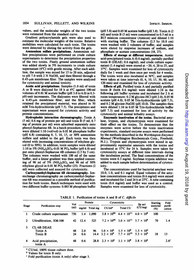
TABLE 1. Purification of toxins A and B of C. difficile
Vol Protein Cytotoxicity Sp act Starting FoldStage Purification step P material purifi-(ml) mllg/ml Total mg CU/mI0 Total CU (CU/mg) (%) cation
1 Crude culture supernatant 750 1.4 1,090 5.8 x 10⁸b 4.4 x 10¹¹ 4.0 x 10⁸ 100
2 Ultrafiltration, XM-100 42 12.4 525 7.2 x 10⁹b 3.0 x 10¹¹ 5.7 x 10⁸ 70 1.4
3 CL-6B DEAEToxin A 48 2.0 96 3.0 x 10⁶ 1.5 x 10⁸ 1.5 x 10⁶Toxin B 24 0.6 14.4 3.2 x 10⁹ 7.7 x 10"° 5.3 x 10⁹ 18 13
4 Acid precipitation, 48 0.6 28.8 2.3 x 10⁶ 1.1 X 10⁸ 3.8 x 10⁶ 2.6ctoxin A
a CU/ml, 100% tissue culture dose.b Values for toxin B only.c Fold purification (toxin A only) after stage 3.
INFECT. IMMUN.
on August 19, 2018 by guest
http://iai.asm.org/
Dow
nloaded from

PURIFICATION OF C. DIFFICILE TOXINS 1035
Compositional analyses of the toxins. Samples oftoxin A (0.5 ml) and toxin B (0.6 mg/ml) were exam-ined for the presence of hexose (15), pentose (14), andphosphorus (13).Because the cytotoxin (toxin B) was previously
reported to be a glycoprotein (26), a concanavalin Acolumn (0.5 by 3.0 cm) was used in an attempt toadsorb toxin B (2, 24). A 0.5-ml sample (0.2 mg ofprotein per ml) in 50 mM acetate buffer (pH 5.8)containing 0.9%o NaCl was loaded onto the column andwashed with 20 ml of the same buffer. Fractions (0.5ml) were collected and assayed for cytotoxicity.
Protease assays. Samples (0.1 ml) of crude cultureifitrate (1 mg/ml) and toxins A and B were examinedfor protease activity, with azocasein (Sigma) as thesubstrate (20).
Production of purified toxin A antiserum. Antiserumwas raised in a goat against a Formalin-treated prepa-ration of acetic acid-precipitated toxin A by the meth-ods previously described (16). The goat received asubcutaneous injection of 1.5 ml of the toxoid once perweek over a period of 8 weeks. The antiserum wasexamined by (i) neutralization tests in CHO-Kl cells(16) and (ii) by crossed IEP by using crude culturesupernatants in the first dimension.
RESULTS
Toxin purification. The cytotoxic activity ofthe cell-free culture supernatant (crude culturesupernatant) was 5.8 x 10⁸ cytotoxic units (CU)per ml. Toxin B was at least 1,000-fold morecytotoxic than toxin A; therefore, the cytotoxic-ity owing to toxin A in crude culture superna-tants could not be determined (Table 1).The specific activities of the toxins at each
step in the purification scheme are given inTables 1 and 2. There was an initial loss of about30% of the cytotoxicity (toxin B) when the crudeculture filtrate was concentrated with the thin-channel ultrafiltration device. Active cytotoxindid not go through the ultrafilter, so we assumethe loss was owing to either binding to the filteror inactivation.The DEAE-Sepharose CL-6B column sepa-
rated two toxin activities in C. diJfficile culturesupematant which we have designated toxin Aand toxin B (Fig. 1). Most of the protein elutedin the first gradient immediately after toxin A, sowe sacrificed some of the toxin A peak to
achieve greater purification. When this wasdone carefully, both PAGE and crossed IEPshowed that many of the contaminating proteinswere eliminated (Fig. 2 and 3). Approximatelysix protein bands were observed in the toxin Apreparation after ion-exchange chromatographywhen fractions were pooled carefully; at leastsix additional bands were observed when theentire cytotoxic peak was included. Toxin A wasfurther purified by precipitating the toxin inacetate buffer at pH 5.5. Under these conditions,the remaining contaminating proteins remainedin solution and the precipitated toxin A could beobtained by centrifugation. Analysis of the acid-precipitated toxin A by PAGE and crossed IEP(Fig. 2 and 3) revealed that the preparation washomogeneous and that the toxic activity wasassociated with the protein band (Fig. 4).Toxin B presented a greater problem. We had
a much higher specific activity in the toxin Bfraction after the DEAE-Sepharose CL-6B col-umn than had been reported previously for puri-fied cytotoxin, yet our preparation obviouslycontained several contaminants (Fig. 2). We arenot sure which, if any, of the protein bandsrepresents the toxin, since the toxicity waseluted from an area with at least three proteinbands (Fig. 4). Crossed IEP also showed severalcontaminants (Fig. 3). We have tried many dif-ferent methods to further purify toxin B, but inall cases the specific activity did not increaseand often decreased because of the instability ofthe toxin.
Biological activity. Both purified toxin A andthe toxin B preparations were cytotoxic andcaused the same morphological alterations inCHO-Kl cells. Toxin B was at least 1,000-foldmore cytotoxic than toxin A; the cytotoxicity oftoxin A was not because of slight contaminationwith toxin B, since specific antiserum to toxin B(21a) did not neutralize this cytotoxicity. Thespecific activities of toxins A and B were 3.8 x10⁶ CU/mg and 5.3 x 10⁹ CU/mg, respectively(Table 1). The specific activities of toxins in themouse lethality assay were 6.4 x 10³ LD1OJmgfor toxin A and 2.1 x 10⁴ LD10O/mg for toxin B(Table 2).
Molecular weight determinations. The Kay val-
TABLE 2. Mouse lethality of toxins A and B during purification
Stage Purification step Animal toxicity Sp act(LD1mIOOrf) (LD1OO}mg)
1 Crude culture supernatant 3.2 x 10³ 1.6 x 1032 Ultrafiltration, XM-10043 CL 6B DEAE
Toxin A 1.3 x 10⁴ 6.4 x 10³Toxin B 1.3 x 10⁴ 2.1 x 10⁴
4 Acid precipitation, toxin A 6.4 x 10³ 1.1 X 10⁴a Animal toxicities were not done on this stage.
VOL. 35, 1982
on August 19, 2018 by guest
http://iai.asm.org/
Dow
nloaded from

1036 SULLIVAN, PELLETT, AND WILKINS
0e0o: f
z0
-io
I)00x
o o
1Q J .
'~0C-wa
TOXIN A
TOXIN B
° o
,*, -o'I , -A,-A'-
x
&O
3.0 rkcasox
0,
-4
1.0 Z
I.- o.05-0.2SM NOIC I .3-U6 M NaC- I
FRACTION NUMBER
FIG. 1. Separation of toxins A and B by ion-exchange chromatography. A sample (42 ml) of concentratedculture supernatant was loaded onto a column (2.5 by 10.0 cm) of DEAE-Sepharose CL-6B, and toxins A and Bwere eluted with two linear NaCl gradients (0.05 to 0.25 M NaCI and 0.3 to 0.6 M NaCI). A 150-ml wash with 50mM Tris-hydrochloride (pH 7.5) containing 0.3 M NaCl followed the first gradient. Symbols: 0, cytotoxicity(100% tissue culture dose); O, protein concentration; and A, chloride molarity.
ues of both toxins, as determined by gel filtra-tion, corresponded to a molecular weight ofapproximately 440,000 to 500,000.The relative mobility, as determined by gradi-
ent PAGE, indicated that toxin A had a molecu-lar weight of 550,000, whereas the molecularweight of toxin B was 360,000.
Difference in the properties of toxins A and B.Various methods including (NH4)2SO4 precipita-tion, acetic acid precipitation, hydrophobic in-teraction chromatography, CM-Sepharose 6B
A B C I) E...; ~~~~~~~~~~~~~~~~~.....
..
FIG. 2. Analysis of toxin preparations by electro-phoresis in 5% Davis gels. Samples (100 p.g of protein)include: (A) crude culture supernatant, (B) toxin B(DEAE), (C) crude culture supemnatant, (D) toxin A(DEAE), and (E) toxin A (acetic acid precipitation).
chromatography, and agarose electrophoresiswere examined to determine whether they in-creased the specific activity of toxin B. Al-though these methods did not result in increasedpurification of toxin B, they did demonstratedistinct differences between toxins A and B(Table 3).
Stability of toxins A and B at different tempera-tures and pH. Both toxins were stable from -20to 37°C for up to 4 weeks. There was a 99% lossof activity within 6 min at 56°C for both toxins.Toxin A was stable at pH 4 and 10, whereas
toxin B was not stable at either pH. Both toxinswere inactivated at pH 2.0.
Trypsin and chymotrypsin inactivation. Tryp-sin and chymotrypsin inactivated both toxins.There was no activation nor interconversion ofone toxin to the other. C. difficile has beenreported to be weakly proteolytic (19); however,we could not detect proteolytic activity in theculture supernatant or in the toxin preparationswith the azocasein method.
Tests for carbohydrates and phosphorus. Rolfeand Finegold (26) has reported that the cytotoxin(toxin B) appeared to be a glycoprotein based onthe inactivation of the toxin by large amounts ofbacterial amylase (10 mg/ml). We were unable todemonstrate any inactivation of our toxin A or Bpreparations even at the unusually high-test con-centration of 5 mg of the enzyme per ml. A smallamount of hexose (24 ,ug/mg of protein) andpentose (60 pLg/mg of protein) was found in the
INFECT. IMMUN.
on August 19, 2018 by guest
http://iai.asm.org/
Dow
nloaded from

PURIFICATION OF C. DIFFICILE TOXINS 1037
A
C
B
D
A...
FIG. 3. Analysis of toxin preparations by crossed IEP. All of the wells in the first dimension initiallycontained 50 ,ug of protein. The upper part of each gel contained 125 ,ul of antiserum prepared against C. difficilestrains 10463 culture supernatant. (A) Crude culture supernatant, (B) toxin B (DEAE), (C) toxin A (DEAE), and(D) toxin A (acetic acid precipitation).
BBBBBBBBBBBBBBBtoxinB preparation (0.6 mg/ml), but toxin B was.... not adsorbed when passed through a concanava-
lin A column. Toxin A did not have detectableamounts of hexose or pentose. Neither toxinpreparation contained detectable phosphorus.
Specificity of toxin A antiserum. Neutralizationtests were done with the purified toxin A antise-rum. Toxin A activity was neutralized with theantiserum to a titer of 520 to 1,040. There was nodetectable neutralization of toxin B activity.
Analysis of the antiserum by crossed IEPshowed the presence of only one precipitin bandwhich corresponded to the toxin A band.
FIG. 4. Localization of cytotoxic activity of toxinsA and B on 5% Davis slab gels. Samples (600 p1d) oftoxin A (600 ,ug) and toxin B (3.0 mg) were layeredacross the surface of the gels. One vertical slice wasfixed and stained. The other gel was sliced horizontal-ly into 3-mm sections, and the sections were assayedfor cytotoxic activity. Cytotoxic activities are indicat-ed by arrows.
DISCUSSIONOur results confirm the observation of Bartlett
et al. that C. difficile produces at least two toxins(8). Other research groups had not separatedthese two toxins previously, both because thestrains of C. difficile that were used producedamounts of toxin A which were not detectableby cytotoxicity assay and because the methods
VOL. 35, 1982
r
on August 19, 2018 by guest
http://iai.asm.org/
Dow
nloaded from

1038 SULLIVAN, PELLETT, AND WILKINS
TABLE 3. Differences between toxins A and B by using different biochemical separation methodsMethod Toxin Result
Ammonium sulfate precipitation A Maximum precipitation at 45 to 50% saturationB Maximum precipitation at 60 to 65% saturation
Acetic acid precipitation A Maximum precipitation at pH 5.5(0.01 M acetate buffer) B Maximum precipitation at pH 3.8 (but 90% inactivated)
Hydrophobic interaction chromatography A Adsorbed at O%o ammonium sulfate and eluted with 20 to 25%(phenyl-Sepharose 4B) ethylene glycol
B Adsorbed at 5 to 10%o ammonium sulfate and eluted with 35 to40% ethylene glycol
Carboxymethyl-Sepharose CL-6B A Adsorbed at pH 5.8, not pH 3.8; eluted with 0.04 MP04-0.2 M NaCl
B Did not adsorb at pH 5.8 or 3.8
Agarose electrophoresis Toxin B migrated faster than toxin A
used did not clearly separate the two toxins. Wehave used a strain of C. difficile that produces1,000- to 10,000-fold more toxin than other re-ported strains (26, 28), and thus we were able todetect toxin A by cytotoxicity assay. We did notobserve the presence of the two toxins previous-ly (29) because of the purification methods for-merly used: (i) the cytotoxicity of toxin B wasnot easily eluted from polyacrylamide gels; thus,the observed cytotoxicity was eluted with thetoxin A band; (ii) both toxins have very similarmolecular weights and did not separate on mo-lecular sieve columns; and (iii) the brand ofDEAE ion-exchanger used (Biogel A) did notclearly separate the two toxins.The toxin that our research group had earlier
obtained in almost pure form (29), as we nowknow, was toxin A containing as a contaminantabout 1% toxin B. The contaminating toxin Baccounted for the greater cytotoxic specific ac-tivity of that preparation compared to the pres-ent toxin A preparation. The degree of purity ofa protein is based on the ability to detect con-taminants. Therefore, because we could notdetect contaminants in the pH 5.5 precipitatedtoxin A preparation by two types of acrylamideelectrophoresis and by the rigorous test ofcrossed IEP, we have referred to this prepara-tion as purified toxin A.We have attempted to reproduce results
which have been reported by Rolfe and Finegold(26) on the purification of the cytotoxin (toxin B)by using the same strain of C. difficile andseparation techniques that were described intheir paper. As reported, a major protein bandwas obtained on PAGE, however, analysis bycrossed IEP indicated that the major band wasnot the cytotoxin. Crossed IEP with monospe-cific toxin B antibodies showed that the cytotox-in was associated with a minor component of the
toxin preparation. Another discrepancy was ob-served with regard to the specific activitiesobtained with the final toxin preparation of Rolfeand Finegold. The specific activity of toxin B inour starting material (crude culture supematant)is 100-fold higher than the specific activity re-ported for the final purified toxin of Rolfe andFinegold. We considered that the sensitivity ofthe W138 tissue culture cells they used might bedifferent from our CHO-Kl cells; however,when we repeated the purification scheme withthe same bacterial strain, we obtained specificactivities similar to those which they reported.
After the completion of this work, we discov-ered that another group had also reported isola-tion of two toxins from C. difficile (5), and wenoted several discrepancies. One discrepancywas that their D-2 (toxin B) preparation did notkill mice; however, they did not state how muchtoxin B was injected, and therefore their resultsmay have been a concentration effect. The finalspecific activity of their toxin B preparation incell culture was similar to our results; however,they did not detect cytotoxicity in their toxin A(D-1) preparations. The cell line they used(HeLa) is not as sensitive as CHO-Ki cells fortoxin A (S. Donta and N. Sullivan, submitted forpublication), and their toxin A may not haveproduced cytopathic activity in their assay. Da-vis gels were used by Banno et al. as their onlycriterion for purity, and they analyzed lowamounts of protein (20 ,ug) on these gels. Ourtoxin B preparation also shows only one majorprotein band when this amount of protein isexamined. Thus, we feel that the cytotoxin of C.difficile (toxin B) has not been purified to date.Other fractionation methods for proteins, in-
cluding ion-exchange chromatography, aceticacid and (NH4)2SO4 precipitation, and hydro-phobic interaction chromatography were exam-
INFECT. IMMUN.
on August 19, 2018 by guest
http://iai.asm.org/
Dow
nloaded from

PURIFICATION OF C. DIFFICILE TOXINS 1039
ined for further purification of toxin B. None ofthese methods resulted in an increase in specificactivity, and in many cases, a decrease wasobserved. Currently we are attempting to purifytoxin B by affinity chromatography on a columnof Sepharose 4B with immobilized gamma glob-ulin specific for toxin B.Three standard sodium dodecyl sulfate-PAGE
procedures, using combinations of sodium dode-cyl sulfate, urea, and guanidine-hydrochloride(22, 23, 29), were used in an attempt to deter-mine the subunit structure of toxin A. Underthese conditions, toxin A was not dissociatedinto smaller units (J. Libby, unpublished data).These results were confirmed by another re-search group (J.-S. Chen and K. Blanchard,personal communication). Because of the largemass of toxin A, we believe it is composed ofseveral subunits, and we are currently investi-gating other procedures for the dissociation oftoxin A into subunits.The difficulty in separating the two toxins, and
their similarities, initially led us to believe thatthey were two interchangeable forms of thesame toxin. This appears much less likely be-cause Bartlett et al. obtained antiserum thatneutralized only toxin A (8) and we have nowproduced antisera to toxins A and B separatelywhich neutralized the respective toxin and donot cross-react with each other (21a). There areseveral procedures (Table 3) that separate thetoxins, and we have not observed the conver-sion of toxin A or B into the other during any ofthese manipulations. In addition, toxin A alsocauses fluid influx into the rabbit ileal loop,whereas toxin B does not (7, 21b), and toxin B isa more potent cytotoxin with at least a 1000-fold-greater specific activity than toxin A. This isfurther evidence which supports the idea that C.difficile produces two distinct toxins. It haspreviously been reported that toxin A is several-fold more lethal for mice than toxin B (8).However, we have found that toxin B is morelethal for mice than toxin A per milligram ofprotein. With the purified toxin A and the mono-specific gamma globulin we now have, we hopeto determine the mechanisms of action of thesetoxins both on tissue culture cells and the colon-ic mucosa.
ACKNOWLEDGMENTSWe thank David Lyerly for performing the IEP analyses and
J. Libby, J.-S. Chen, and K. Blanchard for performing thesodium dodecyl sulfate-PAGE studies.These studies were supported by Public Health Service
grant AI15749-03 from the National Institute of Allergy andInfectious Disease and in part by the State of Virginia.
LITERATURE CITED
1. Andrews, P. 1965. The gel filtration behaviour of proteinsrelated to their molecular weights over a wide range.
Biochem. J. 96:595-606.2. Aspberg, K., and J. Porath. 1970. Group-specific adsorp-
tion of glycoproteins. Acta Chem. Scand. 24:1839-1841.3. Aswell, J. E., M. Ehrich, R. Van Tassell, G. C. Tsai, L. V.
Holdeman, and T. D. Wilkins. 1979. Characterization andcomparison of C. difficile and other clostridial toxins, p.272-275. In D. Schlessinger (ed.), Microbiology-1979.American Society for Microbiology, Washington, D.C.
4. Axelsen, N. H., J. Kroll, and B. Weeke (ed.). 1973. Amanual of quantitative immunoelectrophoresis. Methodsand applications. Universitesforlaget, Oslo, Norway.
5. Banno, Y., T. Kobayashi, K. Watanabe, K. Ueno, and Y.Nozawa. 1981. Two toxins (D-1 and D-2) of Clostridiumdifficile causing antibiotic-associated colitis: purificationand some characterization. Biochem. Int. 2:629-635.
6. Bartlett, J. G., T.-W. Chang, M. Gurwith, S. L. Gorbach,and A. B. Onderdonk. 1978. Antibiotic-associated pseudo-membranous colitis due to toxin producing clostridia.New Engl. J. Med. 298:531-534.
7. Bartlett, J. G., A. B. Onderonk, R. L. Cisneros, and D. L.Kasper. 1977. Clindamycin-associated colitis due to atoxin-producing species of Clostridium in hamsters. J.Infect. Dis. 136:701-705.
8. Bartlett, J. G., N. S. Taylor, T.-W. Chang, and J. Dzink.1980. Clinical and laboratory observations in Clostridiumdifficile colitis. Am. J. Clin. Nutr. 33:2521-2526.
9. Bradford, M. M. 1976. A rapid and sensitive method forthe quantification of microgram quantities of protein uti-lizing the principle of protein-dye binding. Anal. Biochem.72:248-254.
10. Chang, T.-W., J. G. Bartlett, S. L. Gorbach, and A. B.Onderdonk. 1978. Clindamycin-induced enterocolitis inhamsters as a model of pseudomembranous colitis inpatients. Infect. Immun. 20:526-529.
11. Chang, T.-W., M. Lauerman, and J. G. Bartlett. 1979.Cytotoxicity assay in antibiotic-associated colitis. J. In-fect. Dis. 140:765-770.
12. Davis, B. J. 1965. Disc electrophoresis. II. Methods andapplication to human serum proteins. Ann. N.Y. Acad.Sci. 121:404-427.
13. DeSiervo, A. J. 1969. Alterations in the phospholipidcomposition of Escherichia coli B during growth at differ-ent temperatures. J. Bacteriol. 100:1342-1349.
14. Dische, Z. 1949. Spectophotometric method for the deter-mination of free pentose and pentose in nucleotides. J.Biol. Chem. 181:379-392.
15. Dubois, M., K. A. Giles, J. K. Hamilton, P. A. Rebers,and F. Smith. 1956. Colormetric method for determinationof sugars and related substances. Anal. Chem. 28:350-356.
16. Ehrich, M., R. L. Van Tassell, J. M. Libby, and T. D.Wilkins. 1980. Production of Clostridium difficile antitox-in. Infect. Immun. 28:1041-1043.
17. Fekety, R., J. Silva, R. A. Browne, G. D. Rifkin, and J. R.Ebright. 1979. Clindamycin-induced colitis. Am. J. Clin.Nutr. 32:244-250.
18. Hauschild, A. H. W., and R. Hilsheimer. 1971. Purificationand characteristics of the enterotoxin of Clostridiumperfringens type A. Can. J. Microbiol. 17:1425-1432.
19. Holdeman, L. V., E. P. Cato, and W. E. C. Moore (ed.).1977. Anaerobe laboratory manual, 4th ed. Virginia Poly-technic Institute and State University, Blacksburg, Va.
20. Humphrey, C. D., C. W. Condon, J. R. Cantey, and F. E.Pittman. 1979. Partial purification of a toxin found inhamsters with antibiotic-associated colitis. Gastroenterol-ogy 76:468-476.
21. Kreger, A. S., and L. D. Gray. 1978. Purification ofPseudomonas aeruginosa proteases and microscopiccharacterization of pseudomonal protease-induced rabbitcornial damage. Infect. Immun. 19:630-648.
21a.Libby, J. M., and T. D. WiLkins. 1982. Production ofantitoxins to two toxins of Clostridium diJficile and immu-nological comparison of the toxins by crossed-neutraliza-tion studies. Infect. Immun. 35:374-376.
21b.Lyerly, D. M., D. E. Lockwood, S. J. Richardson, and
VOL. 35, 1982
on August 19, 2018 by guest
http://iai.asm.org/
Dow
nloaded from
1040 SULLIVAN, PELLETT, AND WILKINS
T. D. Wilkins. 1982. Biological activities of toxins A and Bof Clostridium difficile. Immun. 35:1147-1150.
22. Matheka, H. D., P. J. Enzmann, H. L. Bachrach, and B.Migl. 1977. The influence of sodium dodecyl sulfate fromdifferent sources on the separation of virus proteins inpolyacrylamide gel electrophoresis. Anal. Biochem. 81:9-17.
23. Moldow, C., J. Robertson, and L. Rothfield. 1972. Purifi-cation of bacterial membrane proteins. J. Membr. Biol.10:137-152.
24. Murthy, R., and A. Herez. 1973. Purification of human a1-antitrypsin by affinity chromatography on Sepharosebound Concanavalin A. FEBS Lett. 32:243-246.
25. Ochoa, J. L. 1978. Hydrophobic (interaction) chromatog-raphy. Biochemistry 60:1-45.
26. Rolfe, R. D., and S. M. Finegold. 1979. Purification andcharacterization of Clostridium difficile toxin. Infect. Im-mun. 25:191-201.
INFECT. IMMUN.
27. Shannon, L. M., E. Kay, and J. Y. Lew. 1966. Peroxidaseisoenzymes from horseradish roots. I. Isolation and physi-cal properties. J. Biol. Chem. 241:2166-2172.
28. Taylor, N. S., and J. G. Bartlett. 1979. Partial purificationand characterization of a cytotoxin from Clostridiumdifficile. Rev. Infect. Dis. 1:379-385.
28a.Taylor, N. S., G. M. Thorne, and J. G. Barlett. 1981.Comparison of two toxins produced by Clostridium diffi-cile. Infect. Immun. 34:1036-1043.
29. Weber, K., and M. Osborn. 1969. The reliability ofmolecular weight determinations by dodecyl sulfate-poly-acrylamide gel electrophoresis. J. Biol. Chem. 244:4406-4412.
30. Wilkins, T. D., M. Ehrich, R. Van Tassell, and E. Balish.1980. Studies on the clostridial toxin associated withantibiotic-induced pseudomembranous colitis, p. 411-417.In D. Eaker and T. Wadstrom (ed.), Natural toxins.Pergamon Press, Inc., Elmsford, N.Y.
on August 19, 2018 by guest
http://iai.asm.org/
Dow
nloaded from