Purification and N-terminal amino acid sequence comparisons of ...
10
JOURNAL OF VIROLOGY, Sept. 1985, p. 778-787 0022-538X/85/090778-10$02.00/0 Copyright C) 1985, American Society for Microbiology Purification and N-Terminal Amino Acid Sequence Comparisons of Structural Proteins from Retrovirus-D/Washington and Mason-Pfizer Monkey Virus LOUIS E. HENDERSON,'* RAYMOND SOWDER,1 GARY SMYTHERS,' RAOUL E. BENVENISTE, 2 AND STEPHEN OROSZLAN' LBI-Basic Research Program, Laboratory of Molecular Virology and Carcinogenesis,' and Laboratory of Viral Carcinogenesis,2 National Cancer Institute-Cancer Research Facility, Frederick, Maryland 21701 Received 14 February 1985/Accepted 24 May 1985 A new D-type retrovirus originally designated SAIDS-D/Washington and here referred to as retrovirus- D/Washington (R-D/W) was recently isolated at the University of Washington Primate Center, Seattle, Wash., from a rhesus monkey with an acquired immunodeficiency syndrome and retroperitoneal fibromatosis. To better establish the relationship of this new D-type virus to the prototype D-type virus, Mason-Pfizer monkey virus (MPMV), we have purified and compared six structural proteins from each virus. The proteins purified from each D-type retrovirus include p4, p10, p12, p14, p27, and a phosphoprotein designated ppl8 for MPMV and pp2O for R-D/W. Amino acid analysis and N-terminal amino acid sequence analysis show that the p4, p12, p14, and p27 proteins of R-D/W are distinct from the homologous proteins of MPMV but that these proteins from the two different viruses share a high degree of amino acid sequence homology. The plO proteins from the two viruses have similar amino acid compositions, and both are blocked to N-terminal Edman degradation. The phosphoproteins from the two viruses each contain phosphoserine but are different from each other in amino acid composition, molecular weight, and N-terminal amino acid sequence. The data thus show that each of the R-D/W proteins examined is distinguishable from its MPMV homolog and that a major difference between these two D-type retroviruses is found in the viral phosphoproteins. The N-terminal amino acid sequences of D-type retroviral proteins were used to search for sequence homologies between D-type and other retroviral amino acid sequences. An unexpected amino acid sequence homology was found between R-D/W pp2O (a gag protein) and a 28-residue segment of the env precursor polyprotein of Rous sarcoma virus. The N-terminal amino acid sequences of the D-type major gag protein (p27) and the nucleic acid-binding protein (p14) show only limited amino acid sequence homology to functionally homologous proteins of C-type retroviruses. Mason-Pfizer monkey virus (MPMV) was first isolated from a rhesus monkey breast carcinoma (4). The virus buds from the cell membrane of infected cells by envelopment of preformed A particles, and the mature virions contain a cone-shaped, centrally located nucleoid. The virus-encoded reverse transcriptase requires magnesium rather than man- ganese as a divalent cation for full activity. These unique morphological and biochemical properties have led to the classification of MPMV as a D-type retrovirus. The virus is exogenous in macaques, since nucleic acid sequences ho- mologous to the virus are present only in infected animals (1, 8). Additional D-type retroviruses have also been isolated from the squirrel monkey, a New World primate (squirrel monkey retrovirus) (2, 13), and from the langur, an Old World monkey (45). Unlike MPMV, these viruses are en- dogenous, genetically transmitted retroviruses that are found in all cells of all animals of those species (1, 45). Nucleic acid hybridization studies have suggested that the langur virus is partially related to MPMV and that MPMV may have been derived from an endogenous virus of langurs or another closely related species (1). The inoculation of MPMV into macaques has not led to tumor formation, but after inoculation of newborn rhesus monkeys, a disease spectrum characterized by lympha- denopathy, weight loss, diarrhea, and opportunistic infec- tions has been observed (10). A similar syndrome has * Corresponding author. occurred spontaneously in various species of macaques housed at the New England, California, and Washington Regional Primate Research Centers (11, 20, 22). This dis- ease, called simian acquired immunodeficiency syndrome, has been thought to be associated with a recently discovered group of D-type retroviruses that are related to MPMV (5, 25, 43). These newly isolated viruses have been referred to as retrovirus-D or simian acquired immunodeficiency syn- drome virus. The simian acquired immunodeficiency syn- drome disease at the Washington Regional Primate Research Center is also associated with a fibromatous tumor termed retroperitoneal fibromatosis which is characterized by an aggressive proliferation of highly vascularized fibrous tissue (11). The immunological relatedness of retrovirus-D to the other D-type retroviruses (MPMV, langur virus, and squirrel monkey retrovirus) has been determined in specific radioim- munoassays for the major core protein (p27) and the enve- lope glycoprotein purified from MPMV. Retrovirus-D and langur virus share common antigenic determinants with MPMV p27 (25, 43). The retrovirus-D isolates from the three primate centers (Washington, New England, and California) have also been compared in radioimmunoassays to the MPMV gp7O and plO proteins. The results show that anti- gens of retrovirus-D/Washington (R-D/W) are immunologi- cally distinct from those present in retrovirus-D/New En- gland and retrovirus-D/California (25, 43; L. Arthur, unpub- lished data). Of this family of related D-type retroviruses, R-D/W is the only isolate that is associated with retroperi- 778 Vol. 55, No. 3
Transcript of Purification and N-terminal amino acid sequence comparisons of ...
JOURNAL OF VIROLOGY, Sept. 1985, p. 778-7870022-538X/85/090778-10$02.00/0Copyright C) 1985, American Society for Microbiology
Purification and N-Terminal Amino Acid Sequence Comparisons ofStructural Proteins from Retrovirus-D/Washington and Mason-Pfizer
Monkey VirusLOUIS E. HENDERSON,'* RAYMOND SOWDER,1 GARY SMYTHERS,' RAOUL E. BENVENISTE, 2
AND STEPHEN OROSZLAN'
LBI-Basic Research Program, Laboratory of Molecular Virology and Carcinogenesis,' and Laboratory of ViralCarcinogenesis,2 National Cancer Institute-Cancer Research Facility, Frederick, Maryland 21701
Received 14 February 1985/Accepted 24 May 1985
A new D-type retrovirus originally designated SAIDS-D/Washington and here referred to as retrovirus-D/Washington (R-D/W) was recently isolated at the University of Washington Primate Center, Seattle, Wash.,from a rhesus monkey with an acquired immunodeficiency syndrome and retroperitoneal fibromatosis. Tobetter establish the relationship of this new D-type virus to the prototype D-type virus, Mason-Pfizer monkeyvirus (MPMV), we have purified and compared six structural proteins from each virus. The proteins purifiedfrom each D-type retrovirus include p4, p10, p12, p14, p27, and a phosphoprotein designated ppl8 for MPMVand pp2O for R-D/W. Amino acid analysis and N-terminal amino acid sequence analysis show that the p4, p12,p14, and p27 proteins of R-D/W are distinct from the homologous proteins of MPMV but that these proteinsfrom the two different viruses share a high degree of amino acid sequence homology. The plO proteins from thetwo viruses have similar amino acid compositions, and both are blocked to N-terminal Edman degradation. Thephosphoproteins from the two viruses each contain phosphoserine but are different from each other in aminoacid composition, molecular weight, and N-terminal amino acid sequence. The data thus show that each of theR-D/W proteins examined is distinguishable from its MPMV homolog and that a major difference betweenthese two D-type retroviruses is found in the viral phosphoproteins. The N-terminal amino acid sequences ofD-type retroviral proteins were used to search for sequence homologies between D-type and other retroviralamino acid sequences. An unexpected amino acid sequence homology was found between R-D/W pp2O (a gagprotein) and a 28-residue segment of the env precursor polyprotein of Rous sarcoma virus. The N-terminalamino acid sequences of the D-type major gag protein (p27) and the nucleic acid-binding protein (p14) showonly limited amino acid sequence homology to functionally homologous proteins of C-type retroviruses.
Mason-Pfizer monkey virus (MPMV) was first isolatedfrom a rhesus monkey breast carcinoma (4). The virus budsfrom the cell membrane of infected cells by envelopment ofpreformed A particles, and the mature virions contain acone-shaped, centrally located nucleoid. The virus-encodedreverse transcriptase requires magnesium rather than man-ganese as a divalent cation for full activity. These uniquemorphological and biochemical properties have led to theclassification of MPMV as a D-type retrovirus. The virus isexogenous in macaques, since nucleic acid sequences ho-mologous to the virus are present only in infected animals (1,8).
Additional D-type retroviruses have also been isolatedfrom the squirrel monkey, a New World primate (squirrelmonkey retrovirus) (2, 13), and from the langur, an OldWorld monkey (45). Unlike MPMV, these viruses are en-dogenous, genetically transmitted retroviruses that are
found in all cells of all animals of those species (1, 45).Nucleic acid hybridization studies have suggested that thelangur virus is partially related to MPMV and that MPMVmay have been derived from an endogenous virus of langursor another closely related species (1).The inoculation of MPMV into macaques has not led to
tumor formation, but after inoculation of newborn rhesusmonkeys, a disease spectrum characterized by lympha-denopathy, weight loss, diarrhea, and opportunistic infec-tions has been observed (10). A similar syndrome has
* Corresponding author.
occurred spontaneously in various species of macaqueshoused at the New England, California, and WashingtonRegional Primate Research Centers (11, 20, 22). This dis-ease, called simian acquired immunodeficiency syndrome,has been thought to be associated with a recently discoveredgroup of D-type retroviruses that are related to MPMV (5,25, 43). These newly isolated viruses have been referred toas retrovirus-D or simian acquired immunodeficiency syn-drome virus. The simian acquired immunodeficiency syn-drome disease at the Washington Regional Primate ResearchCenter is also associated with a fibromatous tumor termedretroperitoneal fibromatosis which is characterized by an
aggressive proliferation of highly vascularized fibrous tissue(11).The immunological relatedness of retrovirus-D to the
other D-type retroviruses (MPMV, langur virus, and squirrelmonkey retrovirus) has been determined in specific radioim-munoassays for the major core protein (p27) and the enve-
lope glycoprotein purified from MPMV. Retrovirus-D andlangur virus share common antigenic determinants withMPMV p27 (25, 43). The retrovirus-D isolates from the threeprimate centers (Washington, New England, and California)have also been compared in radioimmunoassays to theMPMV gp7O and plO proteins. The results show that anti-gens of retrovirus-D/Washington (R-D/W) are immunologi-cally distinct from those present in retrovirus-D/New En-gland and retrovirus-D/California (25, 43; L. Arthur, unpub-lished data). Of this family of related D-type retroviruses,R-D/W is the only isolate that is associated with retroperi-
778
Vol. 55, No. 3

R-D/W AND MPMV STRUCTURAL PROTEIN AMINO ACID SEQUENCES
toneal fibromatosis. The tumor can be induced by inocula-tion of macaques with filtered cell culture media obtainedfrom mammalian cell lines producing R-D/W (R. Benveniste,unpublished data).
Little is known about the molecular structure of theD-type retroviruses; the nucleic acid sequence of the viralgenome is not known, and the N-terminal amino acid se-
quence of only one viral protein (MPMV p27) has beendetermined (29). As a first step toward elucidating thedetailed molecular structures of the D-type retroviruses andto provide a better understanding of the relationships be-tween various D-type isolates and the relationships betweenD-type retroviruses and other members of the retrovirusfamily, we have purified six structural proteins from MPMVand six structural proteins from R-D/W and determined theamino acid content and N-terminal amino acid sequences ofthese purified D-type viral proteins. Proteins isolated fromeach D-type virus were designated according to their molec-ular weights expressed in thousands, and wherever possible,the notation system used here is consistent with the proteindesighations used by others to denote the structural proteinsof MPMV (36, 37, 46). The proteins isolated from each virusinclude p4, plO, p12, pl4, and p27, as well as twophosphoproteins designated pp2O (R-D/W) and ppl8(MPMV). The data reveal that the p4, plO, p12, p14, and p27proteins of R-D/W are related to but not identical to thecomparable proteins of MPMV. The phosphoproteins ofR-D/W and MPMV each contain phosphoserine but are
unequal in size and have distinctly different N-terminalamino acid sequences and amino acid compositions. Thedata thus clearly indicate that R-D/W is related to MPMVbut that these two similar D-type retroviruses may havedifferent major phosphoproteins.
MATERIALS AND METHODS
Virus production. R-D/W virus was grown in dog thymuscells (FCf2Th) or a human lung adenocarcinoma cell line(A549) and harvested by centrifugation followed by sucrosedensity gradient centrifugation as previously described (1).MPMV was grown in A549 cells and purified in a similarfashion.
Chemicals. All reagents used in the liquid-phase spinningcup sequencer were purchased from Beckman Instruments,Palo Alto, Calif. Polybrene was purchased from AldrichChemical Co., Milwaukee, Wis. Guanidine-hydrochloride(enzyme grade) was purchased from Bethesda ResearchLaboratories, Inc., Gaithersburg, Md. Acetonitrile, 1-propanol, ethylacetate, benzene, and butyl chloride wereobtained from Burdick Jackson, Muskegon, Mo. Sequencergrade trifluoroacetic acid (TFA) was purchased from PierceChemical Co., Rockford, Ill.
Gel electrophoresis. Sodium dodecyl sulfate-polyacryl-amide gel electrophoresis (SDS-PAGE) was performed on10-to-20% gradient gels by the method of Laemmli (21).Proteins were visualized by staining with Coomassie brilliantblue R-250, Bio-Rad Laboratories, Richmond, Calif.Amino acid analysis. Samples for amino acid analysis were
hydrolyzed for 24 h in vacuo with 6 N HCl containing 0.1%liquid phenol and then dried by vacuum desiccation. Anal-ysis was performed with a Durrum D-500 amino acid ana-lyzer and ninhydrin detection of eluted amino acids.o-Phosphoamino acid analysis. Samples were hydrolyzed
in vacuo in 4 N HCl at 110°C for 1.5 h. After the 4 N HCl wasremoved by evaporation under reduced pressure, the sam-
ples were dissolved in 0.006 N HCl and injected onto an
anion exchange column (Synchropak AX300), and the o-phosphoamino acids were separated at 50°C by isocraticelution at a flow rate of 1.3 ml/min with 15 mM KH2PO4adjusted to pH 3.0 with concentrated H3PO4 and made12.5% (vol/vol) in methanol (48). Eluted phosphoamino acidswere detected by fluorescence after reaction with ortho-phthalaldehyde reagent (32).
Liquid-phase sequencing. Semiautomated microsequenc-ing was performed with an 890C Beckman sequencer up-dated and modified as previously described (15). Phen-ylthiohydantoin (PTH) derivatives of amino acid were quan-titatively identified as previously described (14).
Separation of viral proteins by RP-HPLC. For reversed-phase high-pressure liquid chromatography (RP-HPLC),concentrated suspensions of purified virus (1 mg/ml) in 0.1 Msodium phosphate (pH 7.0) were saturated with guanidinehydrochloride, made 10% (vol/vol) in mercaptoethanol andkept at 4°C overnight. The resulting slightly turbid solutionof disrupted virus was adjusted to pH 2.0 by addition ofTFAand injected into a high-pressure liquid chromatograph (Wa-ters Associates, Milford, Mass.), and viral proteins were
separated on a ,uBondapak C18 column (Waters Associates).Eluted proteins were detected by measuring UV A206 with amodel 450 variable-wavelength detector (Waters Associates)and collected manually to optimize separations and recover-ies. Solvents were removed by lyophilization. Flow rate,elution solvent, and gradient conditions are given in thefigure legends. After each separation of viral proteins, theRP-HPLC column was washed at 50°C with 120 ml of1-propanol-water-TFA (90:10:0.1) at a flow rate of 0.2ml/min. Columns were stored in 1-propanol or in acetonitrilecontaining 0.05% TFA. Columns were frequently monitoredfor their ability to separate a standard mixture of proteinsand peptides (50 pug each of bovine serum albumin,ovalbumin, lysozyme, ribonuclease, methionine-enkephalin,and leucine-enkephalin). When necessary, columns werecleaned and repacked as previously described (18).
RESULTSIn previous publications, we have described the separa-
tion of structural proteins from C-type (17) and B-type (19)retroviruses by RP-HPLC. In this communication, we willdescribe the separation of the structural proteins of D-typeretroviruses by RP-HPLC and show some of the structuraldifferences between two similar D-type viruses (R-D/W andMPMV).The proteins associated with purified R-D/W virus and
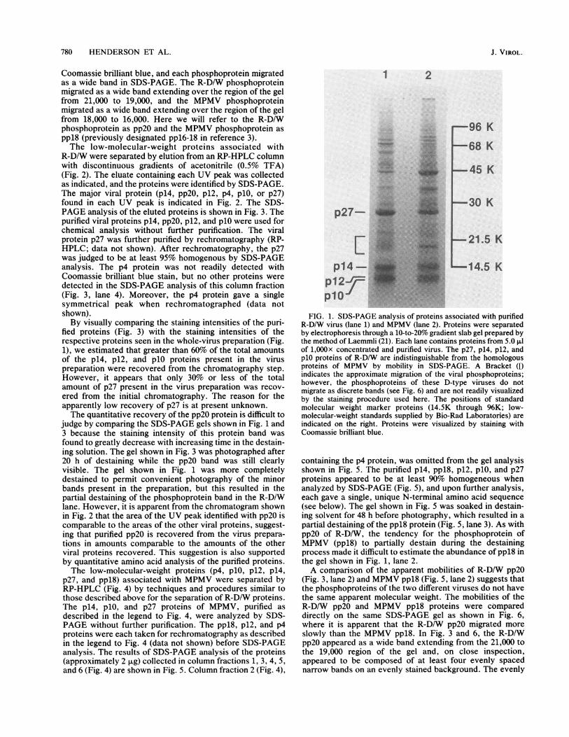
MPMV were first compared by SDS-PAGE (Fig. 1). In thelow-molecular-weight region of the gel (less than 30,000), thestructural proteins associated with each virus appeared quitesimilar. Each lane contained prominent bands labeled p27,p14, p12, and p1O. The mobilities of these bands in theR-D/W lane (Fig. 1, lane 1) appeared identical to the mobil-ities of the like bands in the MPMV lane (lane 2). As will beshown, these bands correspond to major structural proteinspresent in each virus preparation. In the region of the gelbracketed between 21,000 and 16,000 (Fig. 1, brackets) thetwo lanes appeared dissimilar. A band at 18,000 appearedmore prominent in the MPMV lane (Fig. 1, lane 2) than in theR-D/W lane (lane 1), and a region of staining between 21,000and 19,000 in the R-D/W lane (lane 1) was not apparent in theMPMV lane (lane 2). Each virus contained a phosphoproteinthat migrated in this region of the gel and was present in thevirus in amounts comparable to the amounts of p27, p14, p12and plO found in the virus. However, these phosphoproteinsdid not stain as intensely as did the other viral proteins with
VOL. 55, 1985 779
780 HENDERSON ET AL.
Coomassie brilliant blue, and each phosphoprotein migratedas a wide band in SDS-PAGE. The R-D/W phosphoproteinmigrated as a wide band extending over the region of the gelfrom 21,000 to 19,000, and the MPMV phosphoproteinmigrated as a wide band extending over the region of the gelfrom 18,000 to 16,000. Here we will refer to the R-D/Wphosphoprotein as pp20 and the MPMV phosphoprotein asppl8 (previously designated ppl6-18 in reference 3).The low-molecular-weight proteins associated with
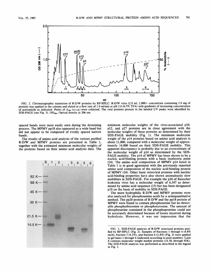
R-D/W were separated by elution from an RP-HPLC columnwith discontinuous gradients of acetonitrile (0.5% TFA)(Fig. 2). The eluate containing each UV peak was collectedas indicated, and the proteins were identified by SDS-PAGE.The major viral protein (p14, pp2O, p12, p4, plO, or p27)found in each UV peak is indicated in Fig. 2. The SDS-PAGE analysis of the eluted proteins is shown in Fig. 3. Thepurified viral proteins p14, pp2O, p12, and plO were used forchemical analysis without further purification. The viralprotein p27 was further purified by rechromatography (RP-HPLC; data not shown). After rechromatography, the p27was judged to be at least 95% homogenous by SDS-PAGEanalysis. The p4 protein was not readily detected withCoomassie brilliant blue stain, but no other proteins weredetected in the SDS-PAGE analysis of this column fraction(Fig. 3, lane 4). Moreover, the p4 protein gave a singlesymmetrical peak when rechromatographed (data notshown).By visually comparing the staining intensities of the puri-
fied proteins (Fig. 3) with the staining intensities of therespective proteins seen in the whole-virus preparation (Fig.1), we estimated that greater than 60% of the total amountsof the p14, p12, and p10 proteins present in the viruspreparation were recovered from the chromatography step.However, it appears that only 30% or less of the totalamount of p27 present in the virus preparation was recov-ered from the initial chromatography. The reason for theapparently low recovery of p27 is at present unknown.The quantitative recovery of the pp2O protein is difficult to
judge by comparing the SDS-PAGE gels shown in Fig. 1 and3 because the staining intensity of this protein band wasfound to greatly decrease with increasing time in the destain-ing solution. The gel shown in Fig. 3 was photographed after20 h of destaining while the pp2O band was still clearlyvisible. The gel shown in Fig. 1 was more completelydestained to permit convenient photography of the minorbands present in the preparation, but this resulted in thepartial destaining of the phosphoprotein band in the R-D/Wlane. However, it is apparent from the chromatogram shownin Fig. 2 that the area of the UV peak identified with pp2O iscomparable to the areas of the other viral proteins, suggest-ing that purified pp2O is recovered from the virus prepara-tions in amounts comparable to the amounts of the otherviral proteins recovered. This suggestion is also supportedby quantitative amino acid analysis of the purified proteins.The low-molecular-weight proteins (p4, plO, pl2, pl4,
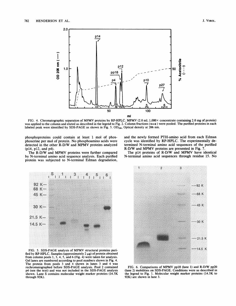
p27, and pp18) associated with MPMV were separated byRP-HPLC (Fig. 4) by techniques and procedures similar tothose described above for the separation of R-D/W proteins.The p14, plO, and p27 proteins of MPMV, purified asdescribed in the legend to Fig. 4, were analyzed by SDS-PAGE without further purification. The ppl8, p12, and p4proteins were each taken for rechromatography as describedin the legend to Fig. 4 (data not shown) before SDS-PAGEanalysis. The results of SDS-PAGE analysis of the proteins(approximately 2 ,ug) collected in column fractions 1, 3, 4, 5,and 6 (Fig. 4) are shown in Fig. 5. Column fraction 2 (Fig. 4),
1
p27-
Lp14-
pl21p10}
2
r---96 K-68 K
-45 K
-30 K
-21.5 K
- 14.5 K
FIG. 1. SDS-PAGE analysis of proteins associated with purifiedR-D/W virus (lane 1) and MPMV (lane 2). Proteins were separatedby electrophoresis through a 10-to-20% gradient slab gel prepared bythe method of Laemmli (21). Each lane contains proteins from 5.0 ,ulof 1,OOOx concentrated and purified virus. The p27, pl4, p12, andplO proteins of R-D/W are indistinguishable from the homologousproteins of MPMV by mobility in SDS-PAGE. A Bracket ([)indicates the approximate migration of the viral phosphoproteins;however, the phosphoproteins of these D-type viruses do notmigrate as discrete bands (see Fig. 6) and are not readily visualizedby the staining procedure used here. The positions of standardmolecular weight marker proteins (14.5K through 96K; low-molecular-weight standards supplied by Bio-Rad Laboratories) areindicated on the right. Proteins were visualized by staining withCoomassie brilliant blue.
containing the p4 protein, was omitted from the gel analysisshown in Fig. 5. The purified p14, ppl8, p12, p1O, and p27proteins appeared to be at least 90% homogeneous whenanalyzed by SDS-PAGE (Fig. 5), and upon further analysis,each gave a single, unique N-terminal amino acid sequence(see below). The gel shown in Fig. 5 was soaked in destain-ing solvent for 48 h before photography, which resulted in apartial destaining of the ppl8 protein (Fig. 5; lane 3). As withpp2O of R-D/W, the tendency for the phosphoprotein ofMPMV (ppl8) to partially destain during the destainingprocess made it difficult to estimate the abundance of ppl8 inthe gel shown in Fig. 1, lane 2.A comparison of the apparent mobilities of R-D/W pp2O
(Fig. 3, lane 2) and MPMV ppl8 (Fig. 5, lane 2) suggests thatthe phosphoproteins of the two different viruses do not havethe same apparent molecular weight. The mobilities of theR-D/W pp2O and MPMV ppl8 proteins were compareddirectly on the same SDS-PAGE gel as shown in Fig. 6,where it is apparent that the R-D/W pp2O migrated moreslowly than the MPMV ppl8. In Fig. 3 and 6, the R-D/Wpp2O appeared as a wide band extending from the 21,000 tothe 19,000 region of the gel and, on close inspection,appeared to be composed of at least four evenly spacednarrow bands on an evenly stained background. The evenly
J. VIROL.
R-D/W AND MPMV STRUCTURAL PROTEIN AMINO ACID SEQUENCES
I
Ec
ac00
'0e0.. .
0)
50 100 150ml
FIG. 2. Chromatographic separation of R-D/W proteins by RP-HPLC. R-D/W virus (2.0 ml; 1,000x concentrate containing 2.0 mg ofprotein) was applied to the column and eluted at a flow rate of 1.0 ml/min at pH 2.0 (0.5% TFA) with gradients of increasing concentrationof acetonitrile as indicated. Peaks of A206 (H-j) were collected. The viral proteins present in the labeled UV peaks were identified bySDS-PAGE (see Fig. 3). OD26, Optical density at 206 nm.
spaced bands were most easily seen during the destainingprocess. The MPMV ppl8 also appeared as a wide band butdid not appear to be composed of evenly spaced narrowbands.The results of amino acid analysis of the various purified
R-DIW and MPMV proteins are presented in Table 1,together with the estimated minimum molecular weights ofthe proteins based on their amino acid analysis data. The
Is 1 2 l 13141 5161
92 K--
68 K-
45 K-
30 K--
21.5 K- :*. I'll
minimum molecular weights of the virus-associated plO,p12, and p27 proteins are in close agreement with themolecular weights of these proteins as determined by theirSDS-PAGE mobility (Fig. 1). The minimum molecularweight of the p14 proteins based on amino acid analysis isabout 11,000, compared with a molecular weight of approx-imately 14,000 based on their SDS-PAGE mobility. Thisapparent discrepancy is probably due to an overestimate ofthe molecular weight of p14 as determined by the SDS-PAGE mobility. The p14 of MPMV has been shown to be anucleic acid-binding protein with a basic isoelectric point(24). The amino acid composition of MPMV p14 listed inTable 1 is in good agreement with the previously reportedamino acid composition of the nucleic acid-binding proteinof MPMV (24). Other basic retroviral proteins with nucleicacid-binding properties have also shown anomalously slowmobilities in SDS-PAGE. For example the plO of Rauscherleukemia virus has a molecular weight of 6,347 as deter-mined by amino acid sequence (15) but has been designatedplO on the basis of mobility in SDS-PAGE.The more hydrophilic R-D/W and MPMV proteins were
also analyzed for phosphoamino acids by a semiquantitativemethod. The pp2O protein of R-D/W and the ppl8 protein ofMPMV were found to contain phosphoserine but no detect-able phosphothreonine or phosphotyrosine. The amount ofphosphoserine contained in the phosphoproteins could notbe accurately determined because of losses incurred duringhydrolysis. However, it was our impression that the
FIG. 3. SDS-PAGE analysis of R-D/W structural proteins puri-fied by RP-HPLC (Fig. 2). Samples of fractions 1 through 4 (4.8%each), fraction 5 (4.2%), and fraction 6 (2.4%) (Fig. 2) were appliedto gel lanes 1 through 6 (indicated according to pool number). LaneS contains molecular weight marker proteins (14.5K through 92K).The SDS-PAGE analysis was performed as described in the legendto Fig. 1.
781VOL. 55, 1985
14.5 K-4-0
782 HENDERSON ET AL.
2.0
E
8 1,CM
0
50 100
0'-
U..4-
0_
:
mlFIG. 4. Chromatographic separation of MPMV proteins by RP-HPLC. MPMV (2.0 ml; 1,000X concentrate containing 2.0 mg of protein)
was applied to the column and eluted as described in the legend to Fig. 2. Column fractions (-i ) were pooled. The purified proteins in eachlabeled peak were identified by SDS-PAGE as shown in Fig. 5. OD26, Optical density at 206 nm.
phosphoproteins could contain at least 1 mol of phos-phoserine per mol of protein. No phosphoamino acids weredetected in the other R-D/W and MPMV proteins analyzed(p14, p12, and p4).The R-D/W and MPMV proteins were further compared
by N-terminal amino acid sequence analysis. Each purifiedprotein was subjected to N-terminal Edman degradation,
and the newly formed PTH-amino acid from each Edmancycle was identified by RP-HPLC. The experimentally de-termined N-terminal amino acid sequences of the purifiedR-D/W and MPMV proteins are presented in Fig. 7.The p14 proteins of R-D/W and MPMV have identical
N-terminal amino acid sequences through residue 15. No
I 2 3
ISI 11 1 31 141 51 1692 K-68 K45 K-
-92 K
-68 K
-45 K4.
4-, . - 30 K_.e
-21.5 K
FIG. 5. SDS-PAGE analysis of MPMV structural proteins puri-fied by RP-HPLC. Samples (approximately 2 jig) of protein obtainedfrom column pools 1, 3, 4, 5, and 6 (Fig. 4) were taken for analysis.Gel lanes are numbered according to pool numbers shown in Fig. 4.The protein from pools 3 and 4 shown in lanes 3 and 4 wasrechromatographed before SDS-PAGE analysis. Pool 2 containedp4 (see the text) and was not included in the SDS-PAGE analysisshown. Lane S contains molecular weight marker proteins (14.5Kthrough 92K).
-14.5 K
FIG. 6. Comparisons of MPMV ppl8 (lane 1) and R-D/W pp2O(lane 2) mobilities on SDS-PAGE. Conditions were as described inthe legend to Fig. 1. Molecular weight marker proteins (14.5K to92K) are shown in lane 3.
30 K-
21.5 K--14.5 K- 41.
J. VIROL.

R-D/W AND MPMV STRUCTURAL PROTEIN AMINO ACID SEQUENCES
TABLE 1. Amino acid analysis of proteins purified from R-D/W and MPMV
Mol of residue per mol of proteina:
Residue p14 pp20 pp18 p12 p4 plO p27
R-D/W MPMV R-D/W MPMV R-D/W MPMV R-D/W MPMV R-D/W MPMV R-D/W MPMV
Aspartic 13.0 (13) 12.2 (12) 28.0 (28) 24.7 (25) 7.3 (7) 6.4 (6) 4.1 (4) 4.0 (4) 11.1 (11) 9.3 (9) 23.9 (24) 24.7 (25)acid
Threonine 2.7 (3) 2.7 (3) 9.9 (10) 13.2 (13) 5.7 (6) 7.3 (7) 2.0 (2) 2.0 (2) 4.6 (5) 4.7 (5) 19.3 (19) 17.7 (18)Serine 5.0 (5) 3.1 (3) 16.0 (16) 8.0 (8) 10.1 (10) 5.2 (5) 3.9 (4) 2.8 (3) 2.6 (3) 3.6 (4) 11.3 (11) 15.1 (15)Glutamic 8.9 (9) 10.9 (11) 20.7 (21) 15.4 (15) 21.4 (21) 22.7 (23) 6.0 (6) 6.0 (6) 11.8 (12) 12.1 (12) 26.3 (26) 26.9 (27)
acidProline 8.0 (8) 8.2 (8) 18.0 (18) 30.6 (31) 6.7 (7) 8.5 (9) 6.5 (7) 7.0 (7) 5.0 (5) 4.8 (5) 13.4 (13) 12.3 (12)Glycine 10.8 (11) 11.3 (11) 4.8 (5) 4.2 (4) 5.6 (6) 6.9 (7) 1.1 (1) 1.0 (1) 6.0 (6) 6.0 (6) 22.7 (23) 21.2 (21)Alanine 6.6 (7) 8.8 (9) 14.6 (15) 14.1 (14) 1.7 (2) 7.3 (7) 2.0 (2) 2.6 (3) 3.8 (4) 5.0 (5) 28.4 (28) 26.0 (26)Valine 1.6 (2) 2.0 (2) 6.2 (6) 4.2 (4) 3.7 (4) 3.3 (3) 3.8 (4) 3.7 (4) 6.4 (6) 7.3 (7) 12.4 (12) 13.1 (3)Methio- 1.2 (1) 0 6.2 (6) 2.3 (2) 1.2 (1) 0 0 0 1.2 (1) 1.2 (1) 4.5 (5) 3.2 (3)
nineIsoleucine 0 1.3 (1) 4.2 (4) 0 8.0 (8) 7.4 (7) 1.0 (1) 0 4.6 (5) 3.3 (3) 10.7 (11) 7.3 (7)Leucine 4.3 (4) 2.9 (3) 10.6 (11) 6.5 (7) 11.6 (12) 11.0 (11) 1.1 (1) 1.2 (1) 8.2 (8) 7.8 (8) 17.2 (17) 19.9 (20)Tyrosine 1.1 (1) 1.0 (1) 4.3 (4) 5.3 (5) 2.5 (3) 0 1.0 (1) 1.0 (1) 3.6 (4) 3.8 (4) 9.8 (10) 8.7 (9)Phenyl- 3.8 (4) 4.0 (4) 2.0 (2) 3.0 (3) 1.3 (1) 1.4 (1) 1.9 (2) 1.9 (2) 6.1 (6) 5.5 (6) 10.0 (10) 10.4 (10)
alanineHistidine 4.0 (4) 5.7 (6) 1.9 (2) 3.0 (3) 4.0 (4) 5.1 (5) 0 0 1.4 (1) 1.7 (2) 4.8 (5) 5.0 (5)Lysine 12.8 (13) 13.6 (14) 14.7 (15) 8.8 (9) 12.9 (13) 14.1 (14) 0 1.1 (1) 10.4 (10) 8.7 (9) 12.9 (13) 13.2 (13)Arg' On 3b :3() 7: () 8 (5) 6 (4)5.3 (5) i 8 f@3 8 8 4.8 ffl 4A43 1.3! 2)1JP 9 tJD)
Trypto- NDC ND ND ND ND ND ND ND ND ND ND NDphan
Mol wt 10,600 10,720 18,540 15,900 12,580 12,210 3,745 3,745 10,680 10,490 26,210 26,010a Values are given as moles of residues per mole of protein and the value rounded to the nearest integer is given in parentheses. Values were calculated from
the mole percentage of amino acid residues by assuming a molecular weight consistent with SDS-PAGE mobility of the protein and adjusted to give the leastdeparture from the nearest integer values.
b Determined after performic acid oxidation of cystine residues.c ND, Not determined.
PTH-amino acid derivative could be identified for positions16 and 18 of either R-D/W or MPMV p14. The only observeddifference in the amino acid sequences of the pl4s occurredat position 19, where MPMV had a glutamic acid residue inplace of an asparagine in R-D/W. This substitution is com-patible with the differences observed by amino acid analysisof the two pi4s (Table 1).The N-terminal amino acid sequences of the phos-
phoproteins of R-D/W (pp2O) and MPMV (ppl8) were dis-tinct from each other (Fig. 7). This result is compatible withthe observation that these two viral proteins differed inapparent molecular weight (Fig. 6) and amino acid compo-sition (Table 1).The N-terminal amino acid sequences of the p12 protein
from R-D/W and the p12 protein from MPMV were nearlyidentical to each other except for substitutions in MPMV ofan alanine for serine at position 14, a glutamic acid forglycine at position 24, and an alanine for glutamine atposition 28 (Fig. 7). The data in Table 1 reveal that MPMVp12 contains more alanine by amino acid composition thandoes R-D/W p12.The p4 peptides from R-D/W and MPMV each contain
about 35 residues (Table 1). The N-terminal amino acidsequence of each peptide was determined for 26 of the 35residues (Fig. 7). The amino acid sequence of R-D/W p4differed from the amino acid sequence of MPMV p4 bysubstitutions at the following five positions in MPMV p4;position 9 (asparagine for serine), position 10 (lysine forserine), position 16 (serine for asparagine), position 18(proline for isoleucine), and position 26 (asparagine forproline). These substitutions account for all of the differ-
ences in amino acid compositions between R-D/W p4 andMPMV p4 except for one alanine in MPMV p4. Thisobservation suggests that there is at least one additionalposition in the unsequenced C-terminal nine residues of thep4 peptides in which R-D/W p4 differs from MPMV p4.Thus, the p4 peptides of R-D/W and MPMV differ by at leastsix amino acid substitutions out of 35 residues.The N-terminal amino acid sequences of the p27 proteins
from R-D/W and MPMV were determined and compared forthe first 24 residues of each protein (Fig. 7). Comparison ofthe N-terminal amino acid sequences of R-D/W and MPMVp27 proteins revealed only a single amino acid substitution atposition 6, a valine in MPMV for a methionine in R-D/W.The plO proteins of R-D/W and MPMV were each sub-
jected to 20 cycles of N-terminal Edman degradation, but noPTH-amino acid derivatives were obtained from either pro-tein. This strongly suggests that these proteins are cova-lently modified at their N-terminal amino group resulting inan amino terminus that is blocked to Edman degradation.To investigate possible relationships between the amino
acid sequence of D-type retroviral protein and amino acidsequences of other proteins, the N-terminal amino acidsequences of the R-D/W and MPMV proteins listed in Fig. 7were compared with the known amino acid sequences of gagprecursor polyproteins of other retroviruses, includingMoloney murine leukemia virus (42), Moloney murine sar-coma virus (47), feline leukemia virus (12), baboon endo-genous virus (M-7) (44), bovine leukemia virus (34), Roussarcoma virus (RSV) (40), human T-cell leukemia virus-I(41), and murine mammary tumor virus (9) and also to theenv precursor polyprotein of human T-cell leukemia virus-I
VOL. 55, 1985 783

784 HENDERSON ET AL.
Viral Protein N-Terminal Amino Acid Sequence
p14
R-D/W Al a-Al a-Al a-Phe-Ser-Gly-Gl n-Thr-Val-Lys-Asp-Phe-Leu-Asn-Asn- X -Asn- X -Asp-Arg-Gly-
to if SI to II IsS""S IS SI II"I go"I" " Is SI If Glu X"MPMV
phosphoprotei n
R-D/W Al a-Al a-Val-Thr-Gl n-Thr-Gl n-Lys-I 1 e-Leu-Lys-Val-Ser-Ser-Gl n-Thr-Asp-Leu-Arg-Asp-Lys-Ser-Gl n-Asn-Ser-
MPMV Leu-Thr-Al a-Gl n-Thr-Ser-Lys-Asp-Pro-Gl u-Asp-Pro-Asn-Pro-Ser-Gl u-Val -Asp-Trp-Asp-Gly-Leu-Gl u-
R-D/W Glu-Met-Asp- X -Ile-Ser-Leu-
p12
R-D/W Ala-Val-Val-Asn-Pro-Lys-Glu-Glu-Leu-Lys-Glu-Lys-Ile-Ser-Gln-Leu-Glu-Glu-Gln-Ile-Lys-Leu-Glu-Gly-Leu-II "I II of II II If" If If"I SI Al a " " s" IS II"I" Gl u "MPMV
R-D/W Hi s-Gl n-Gl n-Leu-I 1 e-I 1 e-Arg-Leu-Gl n
MPMV II of Al a " IIx" if
R-D/W Gly-Al a-Val -Ser-Phe-Val-Pro-Al a-Ser-Ser-Asn-Asn-Pro-Phe-Gl n-Asn-Leu-I 1 e-Gl u-Pro-Pro-rl n-(Gl u-Val-Gl n-Pro
S" II II "I If IS if Asn-Lys " " " " Ser " Pro "If IIS"" " AspMPMV
R-D/W Pro-Val-Thr-Gl u-Thr-Met-Asp-Gly-Gl n-Gly-Gl n-Al a-Trp-Arg-Hi s-Hi s-Asn-Gly-Phe-Asp-Phe-Thr-Val -Il e
MPMV " " " " " Val iSIIIf" " " If if" " " xII IsXFIG. 7. N-terminal amino acid sequences ofMPMV and R-D/W proteins purified as described in the legends to Fig. 2 and 4 (see the text).
At positions where the MPMV protein has the same residue as the R-D/W protein, the residue is listed in the R-D/W row and indicated byditto marks (") in the MPMV row. An X in a sequence indicates a position where we could not make a positive identification of a PTH-aminoacid derivative. The N-terminal amino acid sequence of each viral protein was determined by Edman degradation of 3 to 6 nmol of purifiedprotein. The PTH-amino acid derivative obtained at each Edman cycle was quantitatively and qualititatively identified by high-pressure liquidchromatography (14). The quantitative recovery of PTH-amino acids was compatible with an overall repetitive yield of 96% for each Edmancycle from the analysis of each of the proteins listed above except for the p14 proteins. In the analysis of both MPMV and R-D/W p14, therepetitive yield of PTH-amino acid was 96% for cycles 1 through 15, but the quantity of PTH-amino acids recovered from cycles 17 and onwas only about 10% of the expected value. The reason for the sharp decrease in the yield of PTH-amino acid after step 16 is unknown atpresent. The N-terminal amino acid sequence ofMPMV p27 reported here is in complete agreement with our previously reported N-terminalsequence for this protein (29) except that in our previous publication, the residues at positions 13 and 16 were not positively identified, andthe report contained typographical errors in positions 14 and 20.
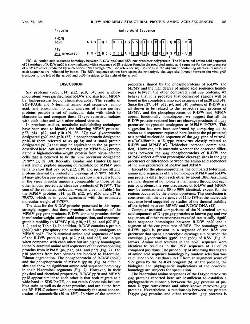
(41), Moloney murine leukemia virus (42), RSV (40), bovineleukemia virus (35), and murine mammary tumor virus (33).The D-type virus sequences were also compared with thoseof all the proteins in the Georgetown data base by the SearchProgram on the National Institutes of Health computer (23).These comparisons revealed statistically significant amino
acid sequence homology between the N-terminal amino acidsequence of R-D/W pp2O and a peptide segment found in theenv precursor polyprotein of RSV (Fig. 8). The N-terminalamino acid sequence of R-D/W pp2O did not show statisti-cally significant amino acid sequence homology (alignmentscores greater than 3.0) with any other known retrovirusamino acid sequences.The N-terminal amino acid sequences of all other D-type
gag proteins given in this report failed to show statisticallysignificant amino acid sequence homologies with any other
known amino acid sequences, including sequences of retro-viral origin. However, the N-terminal residues of theseD-type p27 proteins (Pro-Val-) are common with the N-terminal residues of HTLV-I p24 (31) and avian C-type p27(26) and are similar to the N-terminal residues of all knownmammalian C-type retrovirus p30 proteins (Pro-Leu) (29).The N-terminal alanine residue of these D-type nucleicacid-binding proteins (p14 proteins) is common to all otherknown retroviral nucleic acid-binding proteins from themurine C-type retroviruses (15) and RSV (K. S. Misono,F. S. Sharief, and J. Leis, Fed. Proc. 39:1611, 1980). Theseobservations suggest that the enzyme catalyzing theproteolytic cleavages of D-type gag precursor polyproteinsmay show a substrate specificity similar to that of othermammalian and avian C-type retrovirus proteases (7, 27, 28,30, 49).
J. VIROL.
R-D/W AND MPMV STRUCTURAL PROTEIN AMINO ACID SEQUENCES
Amino Acid Sequence
R-D/Wpp2O
RSVenv precursor
A A V T Q T Q K I L K V S SOT D L R D K S Q N S E MH
P N H T D ILK IL ANSS R T G I R LIR S V LH L nt
FIG. 8. Amino acid sequence homology between R-D/W pp20 and RSV env precursor polyprotein. The N-terminal amino acid sequenceof 28 residues of R-D/W pp2O is shown aligned with a sequence of 28 residues found in the predicted amino acid sequence for the env precursorof RSV (residues predicted by nucleotides 6202 through 6286; see reference 40). Positions in the alignment containing identical residues ineach sequence are indicated by boxes. The RSV sequence shown here spans the proteolytic cleavage site (arrow) between the viral gp85(residues to the left of the arrow) and gp36 (residues to the right of the arrow).
DISCUSSION
Six proteins (p27, p14, p12, plO, p4, and a phos-phoprotein) were purified from R-D/W and also from MPMVby high-pressure liquid chromatography. The results ofSDS-PAGE and N-terminal amino acid sequence, aminoacid, and phosphoamino acid analyses of these purifiedproteins provide a set of molecular data with which tocharacterize and compare these D-type retroviral isolateswith each other and with other related viruses.
In previous studies, metabolic radiolabeling techniqueshave been used to identify the following MPMV proteins:p27, pl4, p12, and plO (29, 36, 37); two glycoproteinsdesignated gp2O and gp7O (37); a phosphoprotein designatedppl6-18 (here referred to as ppl8); and a small proteindesignated p6 (3) that may be equivalent to the p4 proteindescribed here. Antiserum raised against MPMV-p27 precip-itated a high-molecular-weight protein from virus-infectedcells that is believed to be the gag precursor designatedPr78gag (3, 38, 39). Recently, Bradac and Hunter (3) haveused tryptic peptide mapping of radiolabeled MPMV pro-teins to show that p27, p14, p12, plO, and ppl8 are all gagproteins derived by proteolytic cleavage of Pr78gag. MPMVp4 may also be a gag protein since, as shown here, it is foundin the virus in molar amounts comparable to those of theother known proteolytic cleavage products of Pr78gag. Thesum of the estimated molecular weights given in Table 1 forthe MPMV proteins p14, ppl8, p12, p4, p10, and p27 is79,075, which is in good agreement with the estimatedmolecular weight of Pr78 ar.
The data for the R-D/W proteins presented in this reportstrongly suggest that these proteins are homologs of theMPMV gag gene products. R-D/W contains proteins similarin molecular weight, amino acid composition, and chromato-graphic mobility to MPMV p14, plO, p12, p4, and p27 (Fig.1, 2, and 4; Table 1). In addition, R-D/W contains a protein(pp2O) with phosphorylated serine residue(s) analogous toMPMV ppl8. The N-terminal amino acid sequences of fourof the R-D/W proteins (p4, p12, pl4, and p27) are uniquewhen compared with each other but are highly homologousto the N-terminal amino acid sequences of the correspondingproteins from MPMV (p4, p12, p14, and p27) (Fig. 7). TheplO proteins from both viruses are blocked to N-terminalEdman degradation. The phosphoprotein of R-D/W (pp2O)and the phosphoprotein of MPMV (ppl8) (Fig. 6) differ insize and show no significant amino acid sequence homologyin their N-terminal segments (Fig. 7). However, in theirphysical and chemical properties, R-D/W pp2O and MPMVppl8 appear similar to each other in that both migrate as awide band in SDS-PAGE, do not retain Coomassie brilliantblue stain as well as do other proteins, and are eluted fromthe RP-HPLC column with approximately the same concen-tration of acetonitrile (30 to 35%). In view of the common
properties shared by the phosphoproteins of R-D/W andMPMV and the high degree of amino acid sequence homol-ogies between the other compared viral gag proteins, webelieve that it is probable that conserved regions will befound in the complete amino acid sequences of pp2O and p18.Since the p27, p14, p12, p4, and plO proteins of R-D/W areall shown to be related to the respective gag proteins ofMPMV, and the phosphoproteins of R-D/W and MPMVappear functionally homologous, we suggest that all theR-D/W proteins reported here are cleavage products of a gagprecursor polyprotein analogous to MPMV Pr785ag. Thissuggestion has now been confirmed by comparing all theamino acid sequences reported here (except the p4 proteins)to a partial nucleotide sequence of the gag gene of retrovi-rus-D/California, a D-type retrovirus closely related toR-D/W and MPMV (G. Heidecker, personal communica-tion). However, it is uncertain whether the observed differ-ences between the gag phosphoproteins of R-D/W andMPMV reflect different proteolytic cleavage sites in the gagprecursors or differences between the amino acid sequencesof the gag precursors of R-D/W and MPMV.Except for the phosphoproteins, the compared N-terminal
amino acid sequences of the homologous MPMV and R-D/Wgag proteins differ from each other by about 10%. Assuminga similar degree of homology is maintained throughout eachpair of proteins, the gag precursors of R-D/W and MPMVmay be approximately 80 to 90% identical, except for theregions occupied by the phosphoproteins. This conclusion isconsistent with the divergency of 12 to 18% at the nucleotidesequence level suggested by studies of the thermal stabilityof the hybrid between MPMV and R-D/W DNA (43).
Computer-assisted comparisons of the N-terminal aminoacid sequences of D-type gag proteins to known gag and env
sequences of other retroviruses revealed statistically signif-icant sequence homologies between R-D/W pp2O and asegment of the env precursor of RSV. The homology withR-D/W pp2O is present in a segment of the RSV envprecursor that spans a proteolytic cleavage site between theenvelope glycoproteins (gp85 and gp36) of RSV (Fig. 8,arrow). Amino acid residues in the pp2O sequence wereidentical to residues in the RSV sequence at 11 of 28compared positions. The probability of observing this degreeof amino acid sequence homology by random selection wascalculated to be less than 1 in 107 from an alignment score of5.22 given by the ALIGN program (6). At the present, thebiological and phylogenetic implications of the observedhomology are subjects for speculation.The N-terminal amino sequences of the D-type retrovirus
gag proteins reported here are insufficient to establish aphylogenetic relationship between the gag proteins of pri-mate D-type retroviruses and other known retroviral gagproteins. Nevertheless, a relationship between the primateD-type gag proteins and other retroviral gag proteins is
Protein
VOL. 55, 1985 785
786 HENDERSON ET AL.
suggested. MPMV Pr789ag has been shown to incorporatemyristic acid, as do the gag precursors of many othermammalian retroviruses (39). In the murine retroviruses, thesite of myristylation has been identified as the N-terminalamino group of the gag precursor and its cleavage product,p15 (16). The D-type plO proteins are as hydrophobic as thep15 proteins of murine virus and are blocked to N-terminalEdman degradation. It is probable that the D-type plOproteins represent the N-terminal portion of the gag precur-sor and are N-terminally myristylated. The D-typephosphoproteins may be functionally homologous to thephosphoproteins of other retroviruses. The D-type p14 pro-teins isolated here appear similar if not identical to theprotein previously described by Long et al. (24) as a basicprotein with nucleic acid-binding properties present inMPMV. Typically, retroviral nucleic acid-binding proteinscontain at least three cysteine residues in a highly conservedsequence, and in some cases, the sequence is repeated (15,27). The D-type p14 proteins contain six cysteine residues(Table 1), suggesting that these proteins may contain therepeated sequence. The N-terminal proline residue of theD-type p27 proteins is common to the major gag protein ofall B- and C-type retroviruses, which shows that this com-mon feature of the proteolytic cleavage mechanism extendsto D-type viruses.The methods and data presented in this report provide a
molecular basis for the comparison of the gag proteins fromdifferent isolates of D-type virus. Similar studies with thestructural proteins of other D-type retroviruses, includingsquirrel monkey retrovirus, langur virus, and the isolates ofretrovirus-D from the California and New England PrimateCenters, will reveal any structural similarities between theseviruses. Since the various D-type isolates are associatedwith a variety of diseases in primates, detailed structuralstudies may help to elucidate the molecular nature of thispathogenic diversity.
ACKNOWLEDGMENTSWe thank W. B. Knott, R. W. Hill, and C. Hixson for technical
assistance.This research was sponsored in part by the National Cancer
Institute, Department of Health and Human Services, under con-tract no. NO1-CO-23909 with Litton Bionetics, Inc.
ADDENDUM IN PROOFThe nucleotide sequences of R-D/California (P. Marx and
P. Luciw, personal communication) and of MPMV (E.Hunter, J. Engler, L. Perez, and C. Barker, personalcommunication) have confirmed that the p4 protein isencoded in the gag gene. The nucleotide sequence of theMPMV gag gene also shows a translated sequencehomologous with the N-terminal amino acid sequence ofR-D/W pp2O upstream from the sequence coding for theN-terminal sequence of MPMV ppl8.
LITERATURE CITED1. Benveniste, R. E., and G. J. Todaro. 1977. Evolution of primate
oncornaviruses: an endogenous virus from langurs (Presbytisspp.) with related virogene sequences in other Old Worldmonkeys. Proc. Natl. Acad. Sci. U.S.A. 74:4557-4561.
2. Benveniste, R. E., and G. J. Todaro. 1978. Approaches to theisolation ofRNA tumor viruses from primates. Arthritis Rheum.21:2-16.
3. Bradac, J., and E. Hunter. 1984. Polypeptides of Mason-Pfizermonkey virus. I. Synthesis and processing of the gag-geneproducts. Virology 138:260-275.
4. Chopra, H. C., and M. M. Mason. 1970. A new virus in a
spontaneous mammary tumor of a rhesus monkey. Cancer Res.30:2081-2086.
5. Daniel, M. D., N. W. King, N. L. Letvin, R. D. Hunt, P. K.Sehgal, and R. C. Desrosiers. 1984. A new type D retrovirusisolated from macaques with an immunodeficiency syndrome.Science 223:602-605.
6. Dayhoff, M. 0. (ed.). 1976. Atlas of protein sequence andstructure, vol. 5, suppl. 2, p. 1-8. National Biomedical ResearchFoundation, Washington, D.C.
7. Dittmar, K. J., and K. Moelling. 1978. Biochemical properties ofp15-associated protease in an avian RNA tumor virus. J. Virol.28:106-118.
8. Drohan, W., D. Colcher, G. Schochetman, and J. Schlom. 1977.Distribution of Mason-Pfizer virus-specific sequences in theDNA of primates. J. Virol. 23:36-43.
9. Fasel, N., E. Buetti, J. Firzlaff, K. Pearson, and H. Diggelmann.1983. Nucleotide sequence of the 5' noncoding region and partof the gag gene of mouse mammary tumor virus: identificationof the 5' splicing site for subgenomic mRNAs. Nucleic AcidsRes. 11:6943-6955.
10. Fine, D. L., J. C. Landon, R. J. Pienta, M. T. Kubicek, M. J.Valerio, W. F. Loeb, and H. C. Chopra. 1975. Responses ofinfant rhesus monkeys to inoculation with Mason-Pfizer mon-key virus materials. J. Natl. Cancer Inst. 54:651-658.
11. Giddens, W. E., Jr., W. R. Morton, E. Hefti, S. Panem, and H.Ochs. 1983. Enzootic retroperitoneal fibromatosis in macacaspp., p. 249-253. In S. S. Kalter (ed.), Viral and immunologicaldiseases in nonhuman primates. Proceedings: symposium, viraland immunological disease. Alan R. Liss, Inc., New York.
12. Hampe, A., I. Laprevotte, and F. Galibert. 1982. Nucleotidesequences of feline retroviral oncogenes (v-fes) provide evi-dence for a family of tyrosine-specific protein kinase genes. Cell30:775-785.
13. Heberling, R. L., S. T. Barker, S. S. Kalter, G. C. Smith, and R.Helmke. 1977. Oncornavirus: isolation from a squirrel monkey(Saimiri sciureus) lung culture. Science 195:289-292.
14. Henderson, L. E., T. D. Copeland, and S. Oroszlan. 1980.Separation of all amino acid phenylthiohydantoins by high-performance liquid chromatography on phenylalkyl support.Anal. Biochem. 102:1-7.
15. Henderson, L. E., T. D. Copeland, R. C. Sowder, G. W. Smyth-ers, and S. Oroszlan. 1981. Primary structure of the low-molecular weight nucleic acid binding proteins of murine leuke-mia viruses. J. Biol. Chem. 256:8400-8406.
16. Henderson, L. E., H. C. Krutzsch, and S. Oroszlan. 1983.Myristyl amino-terminal acylation of murine retroviral proteins:a new posttranslational protein modification. Proc. Natl. Acad.Sci. U.S.A. 80:339-343.
17. Henderson, L. E., R. Sowder, T. D. Copeland, G. Smythers, andS. Oroszlan. 1984. Quantitative separation of murine leukemiavirus proteins by reversed-phase high-pressure liquid chroma-tography reveals newly described gag and env cleavage prod-ucts. J. Virol. 52:492-500.
18. Henderson, L. E., R. Sowder, and S. Oroszlan. 1981. Protein andpeptide purification by reversed-phase high-pressure chroma-tography using volatile solvents, p. 251-260. In D. T. Liu, A. N.Schechter, R. Heinriksson, and P. G. Condliffe (ed.), Chemicalsynthesis and sequencing of peptides and proteins. Else-vier/North-Holland Publishing Co., Amsterdam.
19. Henderson, L. E., R. Sowder, G. Smythers, and S. Oroszlan.1983. Terminal amino acid sequences and proteolytic cleavagesites of mouse mammary tumor virus env gene products. J.Virol. 48:314-319.
20. Henrickson, R. V., K. G. Osborn, D. L. Madden, J. H.Anderson, D. H. Maul, J. L. Sever, L. R. Ellingsworth, L. J.Lowenstine, and M. B. Gardner. 1983. Epidemic of acquiredimmunodeficiency in rhesus monkeys. Lancet i:388-390.
21. Laemmli, U. K. 1970. Cleavage of structural proteins during theassembly of the head of bacteriophage T4. Nature (London)227:680-685.
22. Letvin, J. L., K. A. Eaton, W. R. Aldrich, P. K. Sehgal, B. J.Blake, S. F. Schlossman, N. W. King, and R. D. Hunt. 1983.Acquired immunodeficiency syndrome in a colony of macaque
J. VIROL.

R-D/W AND MPMV STRUCTURAL PROTEIN AMINO ACID SEQUENCES
monkeys. Proc. Natl. Acad. Sci. U.S.A. 80:2718-2722.23. Lipman, D. J., and W. J. Wilber. 1983. Program for rapid
biosequence similarity analysis. Mathematical ResearchBranch, National Institute of Arthritis, Diabetes, and Digestiveand Kidney Diseases, National Institutes of Health, Bethesda,Md.
24. Long, C. W., L. E. Henderson, and S. Oroszlan. 1980. Isolationand characterization of low molecular weight DNA bindingproteins from retroviruses. Virology 104:491-496.
25. Marx, P. A., D. H. Maul, K. G. Osborn, N. W. Lerche, P.Moody, L. J. Lowenstine, R. V. Henrickson, L. 0. Arthur,R. V. Gilden, M. Gravell, W. T. London, J. L. Sever, J. A.Levy, R. J. Munn, and M. B. Gardner. 1984. Simian AIDS:isolation of a type D retrovirus and transmission of the disease.Science 223:1083-1086.
26. Niall, H. D., R. Sauer, and D. W. Allen. 1970. The N-terminalamino acid sequence of two avian leukosis group specificantigens. Proc. Natl. Acad. Sci. U.S.A. 67:1804-1809.
27. Oroszlan, S., and T. D. Copeland. 1985. Primary structure andprocessing of gag and env gene products of human T-cellleukemia viruses HTLV-ICR and HTLV-IATK. Curr. Top. Mi-crobiol. Immunol. 115:221-233.
28. Oroszlan, S., and R. V. Gilden. 1979. Amino acid sequences ofplant and animal viral proteins, p. 1-35. In H. Fraekel-Conratand R. W. Wagner (ed.), Comprehensive virology, vol. 13.Plenum Publishing Corp., New York.
29. Oroszlan, S., and R. V. Gilden. 1980. Primary structure analysisof retrovirus proteins, p. 299-344. In J. R. Stephenson (ed.),Molecular biology of RNA tumor viruses, Academic Press,Inc., New York.
30. Oroszlan, S., L. E. Henderson, J. R. Stephenson, T. D.Copeland, C. W. Long, J. N. Ihle, and R. V. Gilden. 1978.Amino- and carboxyl-terminal amino acid sequences of proteinscoded by gag gene of murine leukemia virus. Proc. Natl. Acad.Sci. U.S.A. 75:1404-1408.
31. Oroszlan, S., M. G. Sarngadharan, T. D. Copeland, V. S.Kalyanaraman, R. V. Gilden, and R. C. GaHo. 1982. Primarystructure analysis of the major internal protein p24 of humantype C T-cell leukemia virus. Proc. Natl. Acad. Sci. U.S.A.79:1291-1294.
32. Perini, F., J. Sadow, and C. Hixson. 1979. Fluorometric analysisof polyamines, histamine and 1-methylhistamine. Anal.Biochem. 94:431-439.
33. Redmond, S. M. S., and C. Dickson. 1983. Sequence andexpression of the mouse mammary tumor virus env gene.EMBO J. 2:125-131.
34. Rice, N. R., R. M. Stephens, A. Burny, and R. V. Gilden. 1985.The gag and pol genes of bovine leukemia virus: nucleotidesequence and analysis. Virology 142:357-377.
35. Rice, N., R. M. Stephens, D. Couez, D. Deschamps, R. Kett-
mann, A. Burny, and R. V. Gilden. 1984. The nucleotide se-quence of the env gene and post-env region of bovine leukemiavirus. Virology 138:82-93.
36. Schochetman, G., M. Boehm-Truitt, and J. Schlom. 1975. Anti-genic analysis of the major structural proteins of the Mason-Pfizer monkey virus. J. Immunol. 117:168-173.
37. Schochetman, G., K. Kortright, and J. Schlom. 1975. Mason-Pfizer monkey virus: analysis and localization of virion proteinsand glycoproteins. J. Virol. 16:1208-1219.
38. Schultz, A. M., L. E. Henderson, and S. Oroszlan. 1983.Myristylation of gag polyproteins of type B, C, and D retrovi-ruses and also of retroviral transforming proteins, p. 304. InM. A. Rich (ed.), Leukemia reviews international. MarcellDekker, Inc., New York.
39. Schultz, A. M., and S. Oroszlan. 1983. In vivo modification ofretroviral gag gene-encoded polyproteins by myristic acid. J.Virol. 46:355-361.
40. Schwartz, D. E., R. Tizard, and W. Gilbert. 1983. Nucleotidesequence of Rous sarcoma virus. Cell 32:853-869.
41. Seiki, M., S. Hattori, Y. Hirayama, and M. Yoshida. 1983.Human adult T-cell leukemia virus: complete nucleotide se-quence of the provirus genome integrated in leukemia cell DNA.Proc. Natl. Acad. Sci. U.S.A. 80:3618-3622.
42. Shinnick, T. M., R. A. Lerner, and J. G. Sutcliffe. 1981. Nucle-otide sequence of Moloney murine leukemia virus. Nature(London) 293:543-548.
43. Stromberg, K., R. E. Benveniste, L. 0. Arthur, H. Rabin, W. E.Giddens, H. E. Ochs, W. R. Morton, and C.-C. Tsai. 1984.Characterization of exogenous type D retrovirus from a fibromaof a macaque with simian AIDS and fibromatosis. Science224:289-292.
44. Tamura, T.-A. 1983. Provirus of M7 baboon endogenous virus:nucleotide sequence of the gag-pol region. J. Virol. 47:137-145.
45. Todaro, G. J., R. E. Benveniste, C. J. Sherr, J. Schlom, G.Schidlovsky, and J. R. Stephenson. 1978. Isolation and charac-terization of a new type D retrovirus from the asian primate,Presbytis obscurus (spectacled langur). Virology 84:189-194.
46. Tronick, S. R., J. R. Stephenson, and S. A. Aaronson. 1974.Immunological properties of two polypeptides of Mason-Pfizermonkey virus. J. Virol. 14:125-132.
47. Van Beveren, C., F. van Straaten, J. A. Galleshaw, and I. M.Verma. 1981. Nucleotide sequence of the genome of a murinesarcoma virus. Cell 27:97-108.
48. Yang, J. C., J. M. Fujitaki, and R. A. Smith. 1982. Separation ofphosphohydroxyamino acids by high performance liquid chro-matography. Anal. Biochem. 122:360-363.
49. Yoshinaka, Y., I. Katoh, T. D. Copeland, and S. Oroszlan. 1985.Murine leukemia virus protease in encoded by the gag-pol geneand is synthesized through suppression of an amber terminationcodon. Proc. Natl. Acad. Sci. U.S.A. 82:1618-1622.
VOL. 55, 1985 787