Pulseless algorithm
56
By: Bong, Tess & Albert
-
Upload
albert-abacan -
Category
Health & Medicine
-
view
63 -
download
7
Transcript of Pulseless algorithm

By: Bong, Tess & Albert

Pulseless Arrest (Cardiac Arrest)
Algorithm
The Pulseless Arrest Algorithm which is
now known as the Cardiac Arrest Algorithm
takes its place as the most important
algorithm in the ACLS Protocol. There are 4
rhythms that are seen with pulseless
cardiac arrest. These four rhythms are
pulseless ventricular tachycardia (VT),
ventricular fibrillation (VF), asystole, and
pulseless electrical activity (PEA).
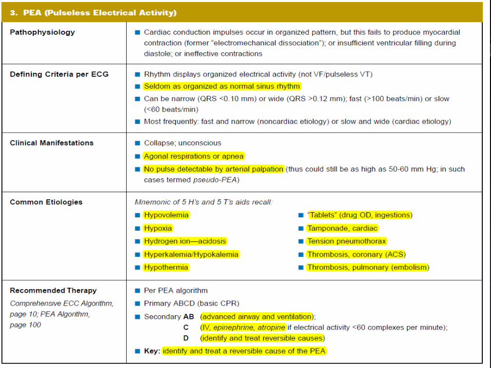
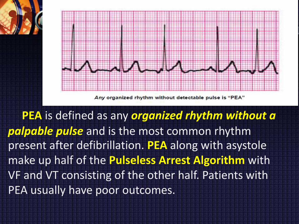
PEA is defined as any organized rhythm without a
palpable pulse and is the most common rhythm present after defibrillation. PEA along with asystole make up half of the Pulseless Arrest Algorithm with VF and VT consisting of the other half. Patients with PEA usually have poor outcomes.

Positive outcome of
an attempted
resuscitation
depends primarily
on two actions:
1. Providing
effective CPR; and


2. Identification and correction of
the cause of PEA.

Understanding the importance of diagnosing
and treating the underlying cause is
fundamental to management of all cardiac
arrest rhythms.

Treating Potentially Reversible
Causes of PEA/Asystole
PEA
reversible conditions
treated successfully if those conditions are identified
and corrected
Hypoxemia: advanced airway
severe volume loss or sepsis: administration of
empirical IV/IO crystalloid.
severe blood loss: blood transfusion
pulmonary embolism: empirical fibrinolytic
therapy (Class IIa, LOE B)
tension pneumothorax: needle decompression


Medications used in PEA
There are 2 medications used in the PEA algorithm,
epinephrine and vasopressin. These medications
should be given while maintaining high-quality CPR. 1
milligram of epinephrine is given IV or IO every 3-5
minutes. 40 Units of vasopressin can be given IV or IO
to replace the first or second dose of epinephrine.
Atropine is no
longer
recommended for
the treatment of
PEA per the 2010
ACLS guidelines.

ASYSTOLE


Asystole is defined as a cardiac arrest
rhythm in which there is no discernible
electrical activity on the ECG monitor.
Asystole is sometimes referred to as a “flat
line.”
Ensure that asystole is not another rhythm
that looks like a “flat line.” Fine VF can appear
to be asystole, and a “flat line” on a monitor
can be due to operator error or equipment
failure.
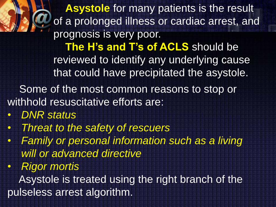
Asystole for many patients is the result
of a prolonged illness or cardiac arrest, and
prognosis is very poor.
The H’s and T’s of ACLS should be
reviewed to identify any underlying cause
that could have precipitated the asystole.
Some of the most common reasons to stop or
withhold resuscitative efforts are:
• DNR status
• Threat to the safety of rescuers
• Family or personal information such as a living
will or advanced directive
• Rigor mortis
Asystole is treated using the right branch of the
pulseless arrest algorithm.


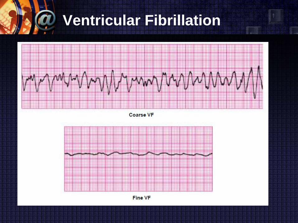
Ventricular Fibrillation
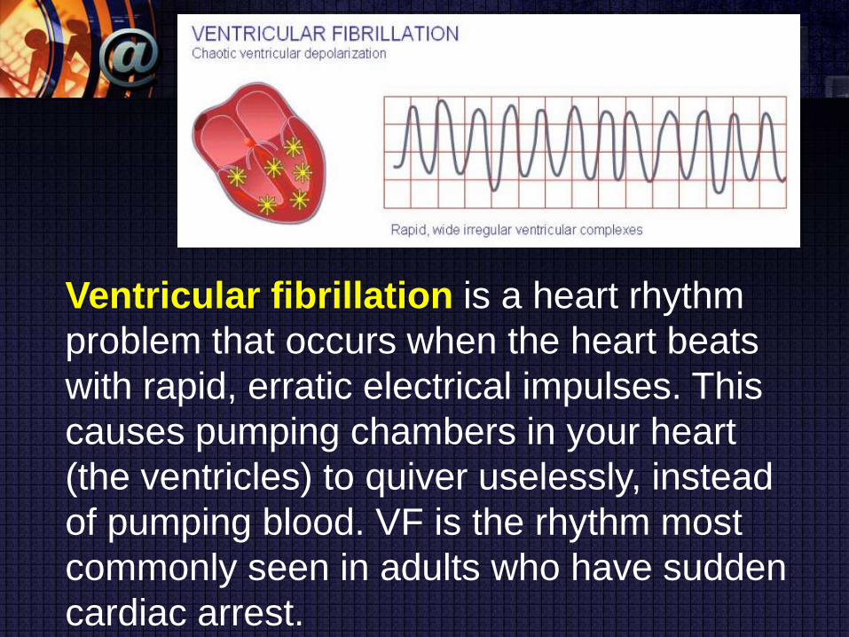
Ventricular fibrillation is a heart rhythm
problem that occurs when the heart beats
with rapid, erratic electrical impulses. This
causes pumping chambers in your heart
(the ventricles) to quiver uselessly, instead
of pumping blood. VF is the rhythm most
commonly seen in adults who have sudden
cardiac arrest.

Ventricular Tachycardia
Pulseless VT is associated with no
effective cardiac output, hence, no
effective pulse, and is a cause of
cardiac arrest.

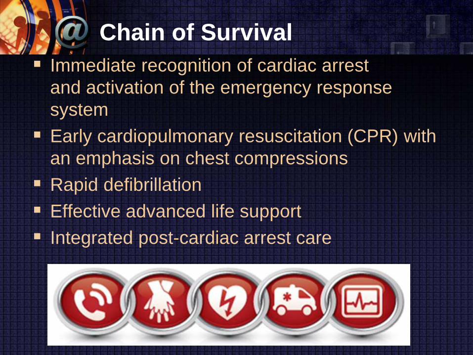
Chain of Survival
Immediate recognition of cardiac arrest
and activation of the emergency response
system
Early cardiopulmonary resuscitation (CPR) with
an emphasis on chest compressions
Rapid defibrillation
Effective advanced life support
Integrated post-cardiac arrest care

Survival from these cardiac arrest rhythms
requires
basic life support (BLS)
system of advanced cardiovascular life support
(ACLS)
integrated post– cardiac arrest care
Periodic pauses in CPR
brief as possible
assess rhythm, shock VF/VT, perform a pulse check
when an organized rhythm is detected, or place an
advanced airway

absence of an advanced airway
synchronized compression–ventilation ratio of 30:2
compression rate of at least 100 per minute.
After placement of a supraglottic airway or an
endotracheal tube
chest compressions should deliver at least 100
compressions per minute continuously without
pauses for ventilation
delivering ventilations should give 1 breath every 6-8
seconds (8-10 breaths per minute)
avoid delivering an excessive number of ventilations

Pulseless Arrest
(Cardiac Arrest)
Algorithm

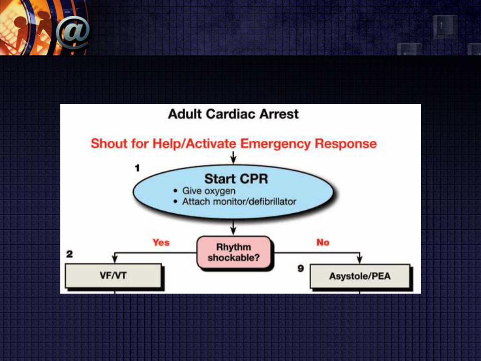

resume CPR while charges the defibrillator
chest compressions should switch at every 2-
minute cycle to minimize fatigue
CPR Before Defibrillation
Performing CPR while a defibrillator is readied
for use is strongly recommended for all patients
in cardiac arrest.
Defibrillation after minimally interrupted CPR
gives the best chance of success.
If ventricular fibrillation recurs, reinitiate
defibrillation at the energy level that previously
resulted in successful defibrillation.

Drug Therapy in VF/Pulseless
VT
Amiodarone: first-line antiarrhythmic agent
Magnesium sulfate
torsades de pointes associated with a long QT
interval
Class IIb, LOE B

Physiologic Parameters
Pulse
End-Tidal CO2
Coronary Perfusion Pressure and Arterial
Relaxation Pressure
Central Venous Oxygen Saturation
Pulse Oximetry
Arterial Blood Gases
Echocardiography
Pulse
No studies have shown the validity or clinical
utility of checking pulses during ongoing CPR.
Because there are no valves in the inferior vena
cava, retrograde blood flow into the venous
system may produce femoral vein pulsations.
Pulse
Carotid pulsations during CPR do not indicate
the efficacy of myocardial or cerebral perfusion
during CPR.
no more than 10 seconds to check for a pulse
If it is not felt within that time period chest
compressions should be started

End-Tidal CO2
During untreated cardiac arrest CO2 continues
to be produced in the body, but there is no CO2
delivery to the lungs.
Under these conditions PETCO2 will approach
zero with continued ventilation. With initiation of
CPR, cardiac output is the major determinant of
CO2 delivery to the lungs.
If ventilation is relatively constant, PETCO2
correlates well with cardiac output during CPR.
End-Tidal CO2
If PETCO2 is <10 mm Hg, it is reasonable to
consider trying to improve CPR quality by
optimizing chest compression parameters
(Class IIb, LOE C).
If PETCO2 abruptly increases to a normal value
(35-40 mm Hg), it is reasonable to consider that
this is an indicator of ROSC (Class IIa, LOE B).

Coronary Perfusion Pressure and Arterial
Relaxation Pressure
Increased CPP correlates with improved 24-
hour survival rates and is associated with
improved myocardial blood flow and ROSC
If the arterial relaxation “diastolic” pressure is
<20 mm Hg, it is reasonable to consider trying
to improve quality of CPR by optimizing chest
compression parameters or giving a
vasopressor or both (Class IIb, LOE C).

Pulse Oximetry
typically does not provide a reliable signal
because pulsatile blood flow is inadequate in
peripheral tissue beds

Arterial Blood Gases
Arterial blood gas monitoring during CPR is not
a reliable indicator of the severity of tissue
hypoxemia, hypercarbia, or tissue acidosis.
Routine measurement of arterial blood gases
during CPR has uncertain value (Class IIb, LOE
C).

ACCESS FOR PARENTERAL
MEDICATIONS DURING
CARDIAC ARREST
worsened survival for every minute that
antiarrhythmic drug delivery was delayed
Time to drug administration was also a predictor
of ROSC
insufficient evidence to specify exact time
parameters or the precise sequence with which
drugs

Peripheral IV Drug Delivery
bolus injection and followed with a 20-mL bolus
of IV fluid
IO Drug Delivery
all age groups

Central IV Drug Delivery
peak drug concentrations are higher and drug
circulation times shorter
interrupt CPR

Endotracheal Drug Delivery
lidocaine, epinephrine, atropine, naloxone, and
vasopressin
2-21⁄2 times the recommended IV dose
dilute the recommended dose in 5-10 mL of
sterile water or normal saline and inject the drug
directly into the endotracheal tube

ADVANCED AIRWAY

delayed endotracheal intubation combined with
passive oxygen delivery and minimally
interrupted chest compressions was associated
with improved neurologically intact survival after
out-of-hospital cardiac arrest in patients with
witnessed VF/VT
When an advanced airway (eg, endotracheal
tube or supraglottic airway) is placed, 2
providers no longer deliver cycles of
compressions interrupted with pauses for
ventilation.

1 breath every 6-8 seconds (8-10 breaths per
minute)

MEDICATIONS FOR ARREST
RHYTHMS

Epinephrine
It is reasonable to consider administering a 1
mg dose of IV/IO epinephrine every 3-5 minutes
during adult cardiac arrest (Class IIb, LOE A).
If IV/IO access is delayed or cannot be
established, epinephrine may be given
endotracheally at a dose of 2-2.5 mg.

Vasopressin
nonadrenergic peripheral vasoconstrictor
1 dose of vasopressin 40 units IV/IO may
replace either the first or second dose of
epinephrine in the treatment of cardiac arrest
(Class IIb, LOE A)

Amiodarone
An initial dose of 300 mg IV/IO can be followed
by 1 dose of 150 mg IV/IO.

Lidocaine
considered if amiodarone is not available (Class
IIb, LOE B)
The initial dose is 1-1.5 mg/kg IV.
If VF/pulseless VT persists, additional doses of
0.5-0.75 mg/kg IV push may be administered at
5- to 10-minute intervals to a maximum dose of
3 mg/kg.
Magnesium Sulfate
torsades de pointes
IV/IO bolus of magnesium sulfate at a dose of 1-
2 g diluted in 10 mL D5W (Class IIb, LOE C)

ANY QUESTIONS?