Pulsed Radiofrequency Electromagnetic Field Therapy: An
18
1 BioElectronics Chronic Wound Case Series Pulsed Radiofrequency Electromagnetic Field Therapy: An Adjunct Pain and Wound Healing Therapy
Transcript of Pulsed Radiofrequency Electromagnetic Field Therapy: An

1
BioElectronics Chronic Wound Case Series
Pulsed Radiofrequency Electromagnetic Field
Therapy:
An Adjunct Pain and Wound Healing Therapy

2
Introduction
Bioelectronics Corporation makes an innovative medical device, designed to replace larger
clinic-based devices that treat chronic wounds, pain and edema with pulsed radiofrequency
electromagnetic field energy. All therapeutic pulsed radiofrequency electromagnetic field
devices emit a safe form of non-ionizing electromagnetic radiation. BioElectronics medical
devices, as do many larger clinic based devices, use a frequency of 27.12 MHz, an assigned FCC
medical frequency. The unique device design by BioElectronics has refined the technology into
an 8gram wearable miniature form, which delivers the radiofrequency electromagnetic field at
a pulse rate of 1000 pulses per second and at 100 microsecond burst widths. Peak burst
output power of the 12 cm antenna is approximately 0.0098 watts covering a surface area of
approximate 100 cm2. The circuitry consists of low voltage (3 V) digital/analog electronics that
control all timing functions to produce
the therapeutic RF field, where the
antenna field is placed directly above
the therapeutic site.
Bioelectronics Medical Devices enable:
Continuous 24 hr. therapy Direct focus onto the wound Convenience – home-based Small, light and portable Easy to use Improved patient compliance Dramatically lower cost of therapy Safety and Elimination/Reduction of Drugs
Pulsed Radio frequency Electromagnetic field as a Chronic wound therapy
The use of pulsed radiofrequency electromagnetic field (PEMF), also termed (PRFE) therapy
has shown notable success in healing of chronic wounds. PEMF is a non-ionizing energy at the
shortwave radiofrequency band of the electromagnetic spectrum, commonly at a frequency of
Figure 1. A traditional Diapulse
delivering 30min x 2 daily required
treatments,
And the BioElectronics RecoveryRX
PEMF device incorporated into the
wound dressing giving continuous
24hr home based therapy

3
27.12MHz. Since the introduction of PEMF in the 1950s, clinical studies on healing of chronic
wounds(1-6) and surgical recovery(7, 8), as well orthopedic studies(9-12) have documented
PEMF as a successful clinical therapy. A series of case reports (13-17) and a retrospective study
on a wound registry(18) have re-introduced PEMF therapy as an adjunct wound healing
therapy, as newer more portable PEMF devices have been introduced.
Known Downstream Biological Effects of PEMF
While the exact mechanism by which PEMF interacts with cells to initiate the therapeutic
effects is not fully understood, cell studies have given valuable insight into the downstream
biological effects of PEMF therapy. Human fibroblasts exposed to PEMF signal show p42/44
MAP kinase activation(19), and increased cell proliferation. The MAP kinase family of
intracellular signaling proteins is activated by a range of stimuli and the activated MAP kinase
translocate to the nucleus and transactivate transcription factors, changing gene expression to
promote growth, differentiation or proliferation. Co-cultures of human epidermal
keratinocytes and human dermal fibroblasts which were studied by gene array demonstrated
an up-regulation of gene families associated with all phases of the wound healing cycle(20,
21). These included many genes involved in the inflammatory stage of wound repair and
expression of genes involved in angiogenesis and tissue remodeling. Cell studies on human
vascular endothelial cells confirm angiogenesis effects of PEMF fields(22), as well as up-
regulation of FGF-2(23), a growth factor that promotes angiogenesis. Nitric oxide is up-
regulated by PEMF, nitric oxide is a vasodilator and also promotes angiogenesis(24).
In a mouse models of diabetes, wound healing rates were increased when exposed to PEMF,
compared to animals that were sham PEMF treated(25). And notably increased proliferation of
dermal fibroblasts was determined, measured by the cell proliferation marker Ki67, a protein
that accumulates in the cell nucleus of cells progressing through the cell cycle.
PEMF Delivery
PEMF therapy is none invasive and is delivered through the wound dressing, and to
date has shown no unwanted side effects. With positive reports in the literature documenting
PEMF as an effective therapy, its wider adoption as an adjunct therapy seems warranted.
However, limitations exist, that have restricted its adoption as a widely employed wound
4
healing therapy. Current treatment regimens require 2 x 30 minute treatments per day and
are delivered by clinic-based, mains powered devices (figure1).
Considering that in many patients, chronic wounds can take many weeks to heal,
making clinic based therapies that are require twice daily treatments impractical for most
patients. The potential answer to this inherent limitation is portable lightweight wearable
PEMF technology, which ideally could become be an integral part of the wound dressing as
shown in Figure 1. Stiller et al 1992 (26), published a randomized control trial, in which a
portable, wearable device to deliver the PEMF therapy was used. The portable device allowed
for a home based treatment; in this case it was used for predominantly venous leg ulcers. The
PEMF delivery device that was used, weighed 508g with treatment protocol consisting of 3 hrs
per day. Significant decreases in wound area, wound depth, healthy granulation tissue and
decreased pain intensity favoring the active group were seen. This study suggests that
wearable, portable forms of PEMF could be an effective adjunct wounding healing therapy.
Nicole and Bentall 1982 (27)were the first to publish a study using wearable battery powered
extended use time PEMF device in which edema and bruising were reportedly decreased
following blepharoplasty. Bental, also published a paper documenting that extended use time
wearable PEMF, reduced the healing time of experimental human skin wounds(28). Healing
was shown to be 52 days in the untreated group compared to 39 days in the PEMF treated
group. Histological analysis also showed advanced wound architecture, including near normal
epidermal thickness in the treated wounds compared to a thin epidermis in the untreated
wounds. More recent studies, have demonstrated the effectiveness of small wearable
extended use time PEMF devices. A significant reduction in postoperative pain in randomized,
double blind placebo controlled studies has been reported (29-31). Plantar fasciitis, a
recalcitrant heel pain has also been shown to be treated with portable, wearable extended use
PEMF therapy(32). Given that postoperative pain is significantly controlled by wearable PEMF
it seems probable that chronic wounds can also be treated with these devices. Below is a
series of case studies that have utilized RecoveryRx to induce wound healing in chronic
wounds of various etiologies.

5
Bioelectronics RecoveryRx - Case Studies
Pain Therapy
Healing therapy
Introduction
The following case studies document patients who had long standing chronic
wounds of various etiologies that were treated with BioElectronics RecoveryRX
PEMF device. The patients were treated from three different clinics, in the USA,
Belgium and Holland. Each patient had previously been treated with a variety of
therapies which had failed to heal their chronic wounds. Persistent wound pain
was also present in a number of cases. Bioelectronics RecoveryRX PEMF patented
devices were introduced to the treatment protocol of each patient, at this time no
other treatment parameters were changed. Wound size and wound pain, were
evaluated for each patient, prior to introduction of RecoveryRX PEMF therapy, and
after subsequent RecoveryRx treatment.

6
Case Study 1 Exposed Vascular Prosthetic Vein
A left leg ulcer with exposed vascular prosthetic vein. Treatment consisted of
debridement and 2 weeks of negative pressure wound therapy. After which the
PEMF device RecoveryRx was introduced with Polymem dressing. The leg ulcer
reduced in size and was 50% reduced by 3 weeks of pulsed radiofrequency and
Polymem dressing treatment. Wound went onto complete closure.
A shows the patient’s venous stasis leg ulcer; B, the wound dressing incorporating
a RecoveryRx PEMF device; and C shows the wound after 3 weeks of RecoveryRx
PEMF therapy. Patient went to complete healing.
B. A
.
B.
C.

7
Case Study 2 Necrotic Toe Amputation
Patient 2 had an ischemic right foot with rapid evolution to a necrotic toe. An
urgent amputation was performed. The patient needed surgical debridement of
necrotic wound edges after which negative pressure vacuum therapy was started
and continued for 1 month. Split skin graft was used but an open wound was still
left. RecoveryRx PEMF therapy in combination with antiseptic Polymem silver was
introduced. The split skin graft was then successful and the remaining open wound
healed with the combination of RecoveryRx and Polymem silver dressing.
A. shows the split skin graft, and B. Polymem dressing with Recovery RX and C. 17
days after treatment.
B.
C.
A.

8
Case Study 3 – Venous Stasis Ulcer
A 72 yr patient with type II diabetes had a venous stasis ulcer that had undergone
multilayer compression therapy for 4 weeks without any appreciable healing and
was accompanied by significant pain. The venous stasis ulcer of patient 1 is shown
at A. week 0 , B. week 2, C. week, 4 and D. week 6 of RecoveryRx PEMF treatment.
Noteworthy pain relief was reported by the patient after 2 weeks of treatment.
The ulcer had decreased in size from 4.0 x 2.5 cm to 0.7 x 0.5 cm after 6 weeks
RecoveryRx treatment. The ulcer continued onto complete healing using the
RecoveryRx therapy.
B. A
.
B.
C. D.

9
Case Study 4 - Diabetic ulcer
A 62 year old patient with insulin controlled diabetes with a ulcer on his heel that
had not responded to debridement and application of triple antibiotic ointment
with offloading. Once the RecoveryRx device was added, triple antibiotic was
discontinued. The ulcer improved rapidly with RecoveryRx treatment; 50% of the
wound area was healed after 1 week of RecoveryRx PEMF treatment. The ulcer
progressed to complete healing at 3 weeks.
The ulcer is shown at A. week 0, B. week 1, C. week 2 and D. week 3.
B.
D.
A
.
B.
C.

10
Case Study 5 – Diabetic Ulcer
A 42 yr old truck driver with type II diabetes had a.5 x 0.5 cm diabetic ulcer.
Previous failed treatments included wound debridement, use of Promogran
matrix, and dry sterile dressing. Once the RecoveryRx device was added to the
regimen, Promogran was discontinued. The diabetic ulcer healed after 3 weeks
RecoveryRx PEMF therapy.
The ulcer at A. week 0, B. week 1 and C. week 3 is shown.
B. A. C.

11
Case Study 6- Pyoderma Ulcer
Pyoderma gangrenosum: Patient had a history of ulcerations and had two lesions:
one on the left dorsal midfoot and the other on medial heel/ankle that had been
present for 2 years. Therapy prior to RecoveryRx addition consisted of
compression, curettage, hydrofera blue, and silvadene treatments. The left dorsal
midfoot wound base was 80% red and 20% yellow with a moderate
serosanguinous exudate. His pain was 10 out of 10 on the Visual Analogue Scale.
Medial heel ankle wound ulcer base was 80% red and 20% yellow with moderate
serosanguinous drainage and a pain of 9 out of 10 on the Visual Analogue Scale.
Vicodin was used for pain medication.
Pain was rapidly resolved and the 2 yr old ulcers moved into the healing phase
after addition of RecoveryRx. The ulcers at A. week 0, B. week 2 and at C. week 11
are shown.
C. B. A.
C. A. B.
12
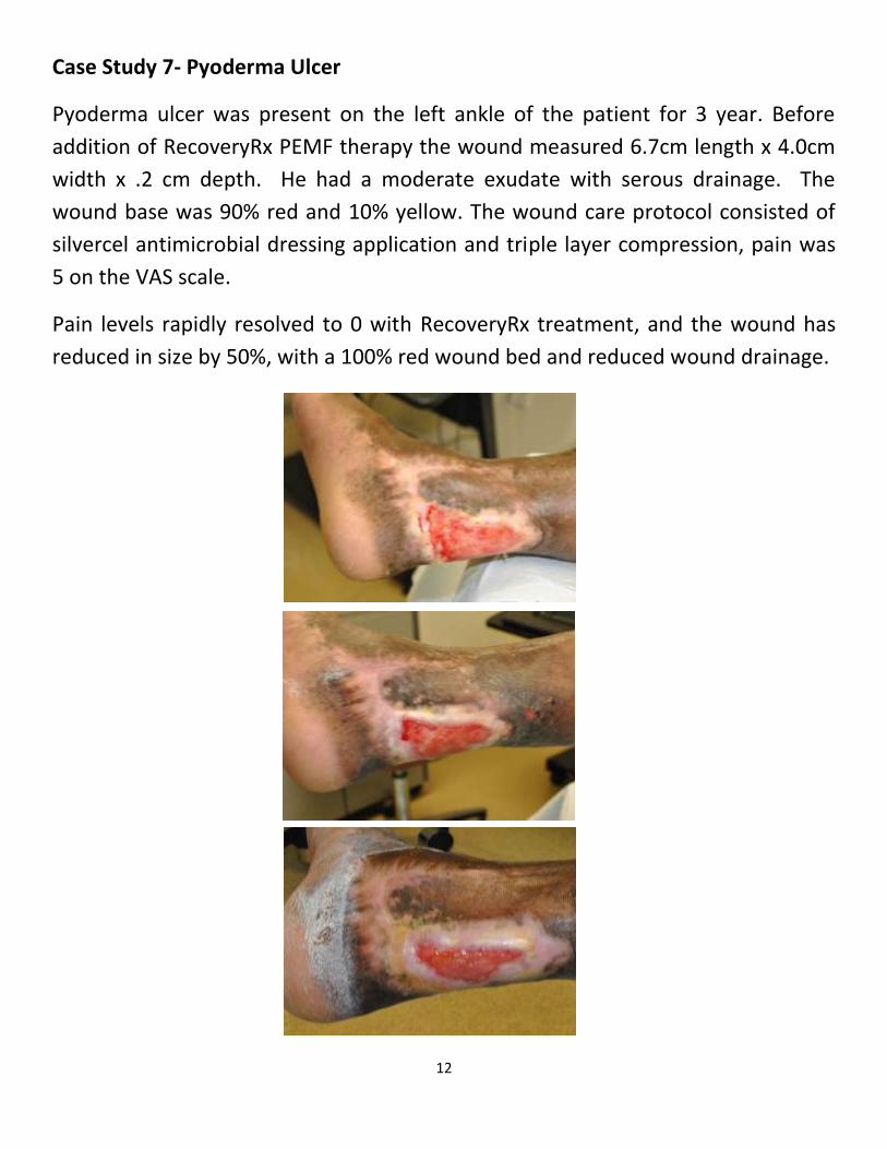
Case Study 7- Pyoderma Ulcer
Pyoderma ulcer was present on the left ankle of the patient for 3 year. Before
addition of RecoveryRx PEMF therapy the wound measured 6.7cm length x 4.0cm
width x .2 cm depth. He had a moderate exudate with serous drainage. The
wound base was 90% red and 10% yellow. The wound care protocol consisted of
silvercel antimicrobial dressing application and triple layer compression, pain was
5 on the VAS scale.
Pain levels rapidly resolved to 0 with RecoveryRx treatment, and the wound has
reduced in size by 50%, with a 100% red wound bed and reduced wound drainage.
13
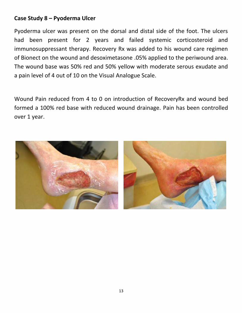
Case Study 8 – Pyoderma Ulcer
Pyoderma ulcer was present on the dorsal and distal side of the foot. The ulcers
had been present for 2 years and failed systemic corticosteroid and
immunosuppressant therapy. Recovery Rx was added to his wound care regimen
of Bionect on the wound and desoximetasone .05% applied to the periwound area.
The wound base was 50% red and 50% yellow with moderate serous exudate and
a pain level of 4 out of 10 on the Visual Analogue Scale.
Wound Pain reduced from 4 to 0 on introduction of RecoveryRx and wound bed
formed a 100% red base with reduced wound drainage. Pain has been controlled
over 1 year.

14
Case Study 9 – Venous Stasis Ulcer
Venous stasis ulcer received after fall in 2009, amputation was considered before
treatment began with Recovery Rx. The wound improved rapidly and the patient
was removed from the amputation list. The wound healed in 12 weeks.

15
Case Study 10 – diabetic Ulcer
105 yr old with a diabetic ulcer present for 2 yrs. Treated with RecoveryRx PEMF
therapy. The ulcer began to heal after 2 weeks treatment and closed after 12
weeks of PRFE therapy

16
Conclusion
The introduction of RecoveryRx PEMF therapy had a significant and positive effect
on all the chronic wounds under study. Chronic recalcitrant wounds that had been
present for up to 2 years, showed improvements, and in most cases complete
healing after the addition of RecoveryRx therapy. A second significant finding was
that persistent wound pain was also markedly reduced, usually within two weeks
of the beginning of RecoveryRx therapy. Wound healing is a series of complex
events that include inflammation, proliferation and maturation. Chronic wounds
are stalled in the inflammatory phase of wound healing. The RF energy from
RecoveryRx, unlike most wound therapies, works at a cellular level, rapidly
resolving edema and breaking the cycle of chronic inflammation. The cells re-
establish cell to cell contact and a healthy wound environment forms which allows
for cell proliferation and wound repair to progress.
Each of the wounds in the case studies had failed to heal with previous therapies
and addition of RecoveryRx PEMF therapy was the only change in the wound
treatment protocol. These findings are therefore notable in that the wounds
moved into a healing phase and strongly indicate that RecoveryRx is a significant
addition to the available wound healing therapies. The ease of use of RecoveryRx,
its low cost and compatibility with current conventional therapies and wound
dressings demonstrate that RecoveryRx can be widely applied as a first choice
wound healing therapy.
References 1. Comorosan, S., Vasilco, R., Arghiropol, M., Paslaru, L., Jieanu, V., and Stelea, S. (1993) The effect of diapulse
therapy on the healing of decubitus ulcer, Rom J Physiol 30, 41-45. 2. Goldin, J. H., Broadbent, N. R., Nancarrow, J. D., and Marshall, T. (1981) The effects of Diapulse on the healing of
wounds: a double-blind randomised controlled trial in man, Br J Plast Surg 34, 267-270. 3. Itoh, M., Montemayor, J. S., Jr., Matsumoto, E., Eason, A., Lee, M. H., and Folk, F. S. (1991) Accelerated wound
healing of pressure ulcers by pulsed high peak power electromagnetic energy (Diapulse), Decubitus 4, 24-25, 29-34.
17
4. Salzberg, C. A., Cooper-Vastola, S. A., Perez, F., Viehbeck, M. G., and Byrne, D. W. (1995) The effects of non-thermal pulsed electromagnetic energy on wound healing of pressure ulcers in spinal cord-injured patients: a randomized, double-blind study, Ostomy Wound Manage 41, 42-44, 46, 48 passim.
5. Tung, S., Khaski, A., Milano, E., and Kay, C. (1995) The application of Diapulse in the treatment of decubitus ulcers: case reports, Contemp Surg 47, 27-32.
6. Rawe, I. M., and Vlahovic, T. C. (2011) The use of a portable, wearable form of pulsed radio frequency electromagnetic energy device for the healing of recalcitrant ulcers: A case report, Int Wound J.
7. Kaplan, E. G., and Weinstock, R. E. (1968) Clinical evaluation of diapulse as adjunctive therapy following foot surgery, J Am Podiatry Assoc 58, 218-221.
8. Aronofsky, D. H. (1971) Reduction of dental postsurgical symptoms using nonthermal pulsed high-peak-power electromagnetic energy, Oral Surg Oral Med Oral Pathol 32, 688-696.
9. Pennington, G. M., Danley, D. L., Sumko, M. H., Bucknell, A., and Nelson, J. H. (1993) Pulsed, non-thermal, high-frequency electromagnetic energy (DIAPULSE) in the treatment of grade I and grade II ankle sprains, Mil Med 158, 101-104.
10. Foley-Nolan, D., Barry, C., Coughlan, R. J., O'Connor, P., and Roden, D. (1990) Pulsed high frequency (27MHz) electromagnetic therapy for persistent neck pain. A double blind, placebo-controlled study of 20 patients, Orthopedics 13, 445-451.
11. Foley-Nolan, D., Moore, K., Codd, M., Barry, C., O'Connor, P., and Coughlan, R. J. (1992) Low energy high frequency pulsed electromagnetic therapy for acute whiplash injuries. A double blind randomized controlled study, Scand J Rehabil Med 24, 51-59.
12. Callaghan, M. J., Whittaker, P. E., Grimes, S., and Smith, L. (2005) An evaluation of pulsed shortwave on knee osteoarthritis using radioleucoscintigraphy: a randomised, double blind, controlled trial, Joint Bone Spine 72, 150-155.
13. Fletcher, S. (2011) Successful treatment of venous stasis ulcers with combination compression therapy and pulsed radio frequency energy in a patient scheduled for amputation, J Wound Ostomy Continence Nurs 38, 91-94.
14. Frykberg, R., Martin, E., Tallis, A., and Tierney, E. (2011) A case history of multimodal therapy in healing a complicated diabetic foot wound: negative pressure, dermal replacement and pulsed radio frequency energy therapies, Int Wound J 8, 132-139.
15. Larsen, J. A., and Overstreet, J. (2008) Pulsed radio frequency energy in the treatment of complex diabetic foot wounds: two cases, J Wound Ostomy Continence Nurs 35, 523-527.
16. Porreca, E. G., and Giordano-Jablon, G. M. (2008) Treatment of severe (Stage III and IV) chronic pressure ulcers using pulsed radio frequency energy in a quadriplegic patient, Eplasty 8, e49.
17. Maier, M. (2011) Pulsed radio frequency energy in the treatment of painful chronic cutaneous wounds: a report of two cases, Pain Med 12, 829-832.
18. Frykberg, R. G., Driver, V. R., Lavery, L. A., Armstrong, D. G., and Isenberg, R. A. (2011) The Use of Pulsed Radio Frequency Energy Therapy in Treating Lower Extremity Wounds: Results of a Retrospective Study of a Wound Registry, Ostomy Wound Manage 57, 22-29.
19. Gilbert, T. L., Griffin, N., Moffett, J., Ritz, M. C., and George, F. R. (2002) The Provant® Wound Closure System Induces Activation of p44/42 MAP Kinase in Normal Cultured Human Fibroblasts, Ann. N.Y. Acad. Sci. 961, 168-171.
20. Moffett, J., Griffin, N. E., Ritz, M. C., and George, F. R. (2010) Pulsed radio frequency energy field treatment of cells in culture results in increased expression of genes involved in the inflammation phase of lower extremity diabetic wound healing., The Journal of Diabetic Foot Complications 2, 57-64.
21. Moffett, J., Kabut, N. J., Griffin, N. E., Ritz, M. C., and George, F. R. (2011) Pulsed radio frequency energy field treatment of cells in culture: Increased expression of genes involved in angiogenesis and tissue remodeling during wound healing., The Journal of Diabetic Foot Complications 3, 30-39.
22. Hopper, R. A., VerHalen, J. P., Tepper, O., Mehrara, B. J., Detch, R., Chang, E. I., Baharestani, S., Simon, B. J., and Gurtner, G. C. (2009) Osteoblasts stimulated with pulsed electromagnetic fields increase HUVEC proliferation via a VEGF-A independent mechanism, Bioelectromagnetics 30, 189-197.

18
23. Strauch, B., Patel, M. K., Navarro, J. A., Berdichevsky, M., Yu, H. L., and Pilla, A. A. (2007) Pulsed magnetic fields accelerate cutaneous wound healing in rats, Plastic and reconstructive surgery 120, 425-430.
24. Fitzsimmons, R. J., Gordon, S. L., Kronberg, J., Ganey, T., and Pilla, A. A. (2008) A pulsing electric field (PEF) increases human chondrocyte proliferation through a transduction pathway involving nitric oxide signaling, J Orthop Res 26, 854-859.
25. Li, Q., Kao, H., Matros, E., Peng, C., Murphy, G. F., and Guo, L. (2011) Pulsed Radio Frequency Energy (PRFE) Accelerates Wound Healing In Diabetic Mice, Plast Reconstr Surg.
26. Stiller, M. J., Pak, G. H., Shupack, J. L., Thaler, S., Kenny, C., and Jondreau, L. (1992) A portable pulsed electromagnetic field (PEMF) device to enhance healing of recalcitrant venous ulcers: a double-blind, placebo-controlled clinical trial, Br J Dermatol 127, 147-154.
27. Nicolle, F. V., and Bentall, R. M. (1982) Use of radio-frequency pulsed energy in the control of postoperative reaction in blepharoplasty, Aesthetic Plast Surg 6, 169-171.
28. Bentall, R. H. C. (1986) Low-level pulsed radiofrequency feilds and the treatment of soft-tissue injuries, Bioelectrochemistry and Bioenergetics 16, 531-548.
29. Heden, P., and Pilla, A. A. (2008) Effects of pulsed electromagnetic fields on postoperative pain: a double-blind randomized pilot study in breast augmentation patients, Aesthetic Plast Surg 32, 660-666.
30. Rawe, I. M., Lowenstein, A., Barcelo, C. R., and Genecov, D. G. (2011) Conrol of postoperative pain with a wearable continuously operating pulsed radio frequency energy device - A preliminary study, Aesthetic Plast Surg inpress.
31. Rohde, C., Chiang, A., Adipoju, O., Casper, D., and Pilla, A. A. (2010) Effects of pulsed electromagnetic fields on interleukin-1 beta and postoperative pain: a double-blind, placebo-controlled, pilot study in breast reduction patients, Plast Reconstr Surg 125, 1620-1629.
32. Brooke, J., Dauphinee, D. M., Korpinen, J., and Rawe, I. M. (2012) Pulsed Radio Frequency Electromagnetic Field Therapy: A Potential Novel Treatment for Plantar Fasciitis, J Foot Ankle Surg in press.