Pulmonary Hypertension and Congestive Heart Failure Stephen L. Rennyson MD August 11, 2011.

LUND UNIVERSITY
PO Box 117221 00 Lund+46 46-222 00 00
Pulmonary hypertension related to heart failure and hypoxia. Mechanisms, newtreatment strategies and impact on mortality following heart transplantation.Experiences from Skåne University Hospital in Lund
Lundgren, Jakob
2018
Document Version:Publisher's PDF, also known as Version of record
Link to publication
Citation for published version (APA):Lundgren, J. (2018). Pulmonary hypertension related to heart failure and hypoxia. Mechanisms, new treatmentstrategies and impact on mortality following heart transplantation. Experiences from Skåne University Hospital inLund. Lund University: Faculty of Medicine.
Total number of authors:1
General rightsUnless other specific re-use rights are stated the following general rights apply:Copyright and moral rights for the publications made accessible in the public portal are retained by the authorsand/or other copyright owners and it is a condition of accessing publications that users recognise and abide by thelegal requirements associated with these rights. • Users may download and print one copy of any publication from the public portal for the purpose of private studyor research. • You may not further distribute the material or use it for any profit-making activity or commercial gain • You may freely distribute the URL identifying the publication in the public portal
Read more about Creative commons licenses: https://creativecommons.org/licenses/Take down policyIf you believe that this document breaches copyright please contact us providing details, and we will removeaccess to the work immediately and investigate your claim.

JAK
OB
LUN
DG
REN
P
ulmonary hypertension related to heart failure and hypoxia
2018:100
Lund UniversityDepartment of Clinical Sciences Lund
Cardiology
Lund University, Faculty of Medicine Doctoral Dissertation Series 2018:100
ISBN 978-91-7619-668-7 ISSN 1652-8220
Pulmonary hypertension related to heart failure and hypoxia Mechanisms, new treatment strategies and impact on mortality following heart transplantation. Experiences from Skåne University Hospital in Lund
JAKOB LUNDGREN
FAcULty OF MEDiciNE | LUND UNivERsity
978
9176
1966
87Pr
inte
d by
Med
ia-T
ryck
, Lun
d 20
18
NO
RDIC
SW
AN
EC
OLA
BEL
304
1 09
03
Jakob Lundgren studied medicine at Lund University and graduated in 2015. He completed his research internship at Skåne University Hospital in Lund in 2017 where he is now training in car-diology. After completing his PhD thesis he naively consider himself a free man.

Pulmonary hypertension related to
heart failure and hypoxia
Mechanisms, new treatment strategies and impact on
mortality following heart transplantation.
Experiences from Skåne University Hospital in Lund
Jakob Lundgren, M.D.
DOCTORAL DISSERTATION
by due permission of the Faculty of Medicine, Lund University, Sweden.
To be defended at Segerfalksalen, BMC, Lund University, Lund.
September 28, 2018 09:00.
Faculty opponent
Prof. Jean-Luc Vachiéry, M.D., Ph.D., Dept. of Cardiology, CUB,
Erasme University Hospital, Brussels, Belgium
11111
2
Organization LUND UNIVERSITY
Document name DISSERTATION
Faculty of Medicine, Clinical Sciences Lund, Cardiology
Date of issue September 28, 2018
Author Jakob Lundgren
Sponsoring organizations: Actelion Pharmaceuticals Sweden AB, ALF, Anna-Lisa and Sven-Erik Lundgren’s Foundation, Crafoord Foundation, Copenhagen Muscle Research Centre, Maggie Stephens’ Foundation, Skåne University Hospital’s Foundation and Swedish Society of Pulmonary Hypertension.
Title Pulmonary hypertension related to heart failure and hypoxia – Mechanisms, new treatment strategies and impact on mortality following heart transplantation. Experiences from Skåne University Hospital in Lund. Abstract
Pulmonary hypertension due to left heart disease, caused by passive congestion of the pulmonary circulation is associated with poor prognosis. If long-standing, endothelial damage and subsequent vasoconstriction may occur, in some cases further complicated by vascular remodeling of the pulmonary vessels. Such remodeling may induce fixed elevated pulmonary vascular resistance, potentially impairing outcome after heart transplantation. Knowledge on physiological alterations after heart transplantation, both with regards to hemodynamics and the plasma concentrations of vasoactive substances, is scarce. There are furthermore few options for treating pulmonary hypertension due to left heart disease and hypoxic pulmonary vasoconstriction, the latter a condition which may aggravate pulmonary hypertension due to left heart disease. These issues were the focus of the present thesis.
First, in a porcine model of hypoxic pulmonary vasoconstriction, soluble guanylate cyclase stimulation alone or in combination with dual endothelin receptor blockade was found to completely reverse acute hypoxic pulmonary vasoconstriction without affecting oxygen consumption.
Second, in a retrospective review of the patients’ heart transplanted and post-operatively followed at Skåne University Hospital in Lund, Sweden between 1988 and 2010, the impact of pre-operative and post-operative pulmonary hypertension on long-term survival was investigated. Invasive hemodynamics during slight exercise were furthermore studied to identify the hemodynamic response to exercise after heart transplantation. The findings suggest that with careful patient selection and care, pre-operative pulmonary hypertension may not be a strict contraindication for heart transplantation. Pulmonary hypertension one year after heart transplantation was, however, associated with impaired survival. Also, whereas the post-operative response to slight exercise with regards to cardiac output was adequate after heart transplantation, the patients’ exhibited abnormally high ventricular filling pressures during exercise. The reason for the elevated filling pressures is probably multifactorial and possibly related to transplanted hearts being more dependent of the Frank Starling-mechanism to maintain adequate cardiac output, as well as decreased diastolic compliance.
Finally, in a prospective cohort of consecutive heart transplanted patients without severe cardiopulmonary complications, we analyzed plasma concentrations of substances in the nitric oxide pathway prior to and after heart transplantation. The L-Arginine/ADMA-ratio, a ratio previously associated with disease severity and outcome in heart failure, was found to be markedly improved after heart transplantation and inversely correlated to pulmonary vascular resistance, suggesting an improved nitric oxide mediated vasodilatation. Key words: Pulmonary hypertension, left heart disease, hypoxic pulmonary vasoconstriction, heart transplantation, hemodynamics, soluble guanylate cyclase, endothelin receptor antagonist, survival, L-Arginine, ADMA Classification system and/or index terms (if any)
Supplementary bibliographical information Language: English
ISSN and key title: 1652-8220 ISBN: 978-91-7619-668-7
Recipient’s notes Number of pages Price
Security classification
I, the undersigned, being the copyright owner of the abstract of the above-mentioned dissertation, hereby grant to all reference sources permission to publish and disseminate the abstract of the above-mentioned dissertation.
Signature Date 2018-08-23
94

Pulmonary hypertension related to
heart failure and hypoxia
Mechanisms, new treatment strategies and impact on
mortality following heart transplantation.
Experiences from Skåne University Hospital in Lund
Jakob Lundgren, M.D.
Supervisor:
Assoc. Prof. Göran Rådegran, M.D., M.Sc.Eng.Phys., D.M.Sc.
Assistant supervisor:
Lars Algotsson, M.D., Ph.D.
33333
4
Coverphoto – A pulmonary artery wedge pressure curve from a patient with
pulmonary hypertension. Designed with assistance from Christian Reitan.
Copyright Jakob Lundgren
Lund University, Faculty of Medicine
Department of Clinical Sciences Lund, Cardiology
ISBN 978-91-7619-668-7
ISSN 1652-8220
Lund University, Faculty of Medicine Doctoral Dissertation Series 2018:100
Printed in Sweden by Media-Tryck, Lund University
Lund 2018
Media-Tryck is an environmentally
certified and ISO 14001 certified
provider of printed material.
Read more about our environmental
work at www.mediatryck.lu.se
NO
RD
ICSWAN ECO
LAB
EL
1234 5678
4

To Sigrid
55555
6
Table of Contents
List of publications ...................................................................................................8
Abbreviations ...........................................................................................................9
Abstract ..................................................................................................................11
Swedish Summary (Populärvetenskaplig sammanfattning) ...................................13
Introduction ............................................................................................................16
Heart Transplantation ...................................................................................16 Hemodynamics in Heart Transplantation ............................................17
Mechanical Circulatory Support ..........................................................19 The Clinical and Research Programs for Heart Transplantation and
Mechanical Circulatory Support in Lund, Sweden..............................20
Pulmonary Hypertension ..............................................................................22 Pulmonary Arterial Hypertension ........................................................23
Pulmonary Hypertension due to Left Heart Disease ...........................26
Hypoxic Pulmonary Vasoconstriction .................................................33
Aims .......................................................................................................................34
Materials and Methods ...........................................................................................35
Paper I ..........................................................................................................35
In Vivo Pig Model of Acute Hypoxic Pulmonary Vasoconstriction ....35
Drugs ...................................................................................................37
Paper II-IV ...................................................................................................37
Study Population and Data Collection .................................................37
Right Heart Catheterization and Endomyocardial Biopsies ................41
Drugs and Devices ...............................................................................42
Paper V .........................................................................................................42
Study Population .................................................................................42
Plasma Samples and Biomarker Analysis ...........................................43
Ethics ............................................................................................................44
Statistical Analyses ......................................................................................44
Results ....................................................................................................................46
Paper I ..........................................................................................................46
6

Hypoxia ...............................................................................................46
Effects of Soluble Guanylate Cyclase Stimulation ..............................47
Effects of Soluble Guanylate Cyclase Stimulation in Combination
with Dual Endothelin Receptor Blockade ...........................................48
Paper II-IV ...................................................................................................50
Pre-operative Hemodynamics..............................................................50
Post-operative Hemodynamics ............................................................51
Acute Cellular Rejections and Post-operative Renal Function ............54
Survival ...............................................................................................55
Paper V .........................................................................................................58
Pre-operative and Post-operative Characteristics ................................58
Plasma Levels of Substances in the Nitric Oxide Pathway .................59
Correlations .........................................................................................59
Limitations ...................................................................................................60
Paper I ..................................................................................................60
Paper II-IV ...........................................................................................60
Paper V ................................................................................................61
Discussion...............................................................................................................62
Paper I ..........................................................................................................62
Response to Hypoxia ...........................................................................62
Soluble Guanylate Cyclase Stimulation and Dual Endothelin
Receptor Blockade Totally Reverses Acute HPV ...............................63
Paper II .........................................................................................................64
Pre-operative Hemodynamics..............................................................64
Post-operative Hemodynamics ............................................................65
Paper III ........................................................................................................66
Survival ...............................................................................................66
Diastolic Pressure Gradient .................................................................67
Bridge-to-Transplantation ...................................................................68
Paper IV .......................................................................................................68
Survival ...............................................................................................69
Paper V .........................................................................................................70
Pre-operative Concentrations ..............................................................70
Post-operative Concentrations .............................................................70
Conclusions ............................................................................................................72
Future Perspectives .................................................................................................74
Acknowledgements ................................................................................................76
References ..............................................................................................................77
77777
8
List of Publications
This thesis is based on the following papers, which will be referred to in the text by
their Roman numerals.
I. Lundgren J, Kylhammar D, Hedelin P, Rådegran G. sGC
stimulation totally reverses hypoxia-induced pulmonary vasoconstriction alone and
combined with dual endothelin-receptor blockade in a porcine model. Acta Physiol
(Oxf). 2012 Nov;206(3):178-94.
II. Lundgren J, Rådegran G. Hemodynamic Characteristics Including
Pulmonary Hypertension at Rest and During Exercise Before and After Heart
Transplantation. J Am Heart Assoc. 2015 Jul 21;4(7).
III. Lundgren J, Algotsson L, Kornhall B, Rådegran G. Preoperative
pulmonary hypertension and its impact on survival after heart transplantation. Scand
Cardiovasc J. 2014 Feb;48(1):47-58.
IV. Lundgren J, Söderlund C, Rådegran G. Impact of postoperative
pulmonary hypertension on outcome after heart transplantation. Scand Cardiovasc
J. 2017 Jun;51(3):172-181.
V. Lundgren J, Sandqvist A, Hedeland M, Bondesson U, Wikström G,
Rådegran G. Alterations in plasma L-arginine and methylarginines in heart failure
and after heart transplantation. Scand Cardiovasc J. 2018 Aug;52(4):196-204.
The following review forms the basis for parts of the introduction of the thesis.
Lundgren J, Rådegran G. Pathophysiology and potential treatments of pulmonary
hypertension due to systolic left heart failure. Acta Physiol (Oxf). 2014
Jun;211(2):314-33.
8

Abbreviations
ACR Acute cellular rejection
ADMA Asymmetric dimethylarginine
CO Cardiac output
Cpc-PH Combined pre-capillary and post-
capillary PH
CTEPH Chronic thromboembolic PH
DPG Diastolic pressure gradient
ECMO Extracorporeal membrane
oxygenation
EMB Endomyocardial biopsy
ESC European Society of Cardiology
FiO2 Inspiratory oxygen fraction
HPV Hypoxic pulmonary vasoconstriction
HR Heart rate
HT Heart transplantation
Ipc-PH Isolated post-capillary PH
ISHLT International Society for Heart and
Lung Transplantation
LVAD Left ventricular assist device
MAP Mean arterial pressure
MPAP Mean pulmonary artery pressure
MRAP Mean right atrial pressure
NO Nitric oxide
NYHA New York Heart Association
99999

10
PAH Pulmonary arterial hypertension
PAWP Pulmonary artery wedge pressure
PDE5i Phosphodiesterase type 5 inhibitor
PH Pulmonary hypertension
PH-LHD Pulmonary hypertension due to left
heart disease
PVR Pulmonary vascular resistance
RHC Right heart catheterization
SAOO2 Aortic O2 saturation
sGC Soluble guanylate cyclase
SPAO2 Pulmonary artery O2 saturation
SV Stroke volume
TPG Transpulmonary gradient
TPR Total pulmonary resistance
10

Abstract
Pulmonary hypertension due to left heart disease, caused by passive congestion of
the pulmonary circulation, is associated with poor prognosis. If long-standing,
endothelial damage and subsequent vasoconstriction may occur, in some cases
further complicated by vascular remodeling of the pulmonary vessels. Such
remodeling may induce fixed elevated pulmonary vascular resistance, potentially
impairing outcome after heart transplantation. Knowledge on physiological
alterations after heart transplantation, both with regards to hemodynamics and the
plasma concentrations of various vasoactive substances, is scarce. There are
furthermore few options for treating pulmonary hypertension due to left heart
disease and hypoxic pulmonary vasoconstriction, the latter a condition which may
aggravate pulmonary hypertension due to left heart disease. These issues were the
focus of the present thesis.
First, in a porcine model of hypoxic pulmonary vasoconstriction, soluble guanylate
cyclase stimulation alone or in combination with dual endothelin receptor blockade
was found to completely reverse acute hypoxic pulmonary vasoconstriction without
affecting oxygen consumption.
Second, in a retrospective review of the patients’ heart transplanted and post-
operatively followed at Skåne University Hospital in Lund, Sweden between 1988
and 2010, the impact of pre-operative and post-operative pulmonary hypertension
on long-term survival was investigated. Invasive hemodynamics during slight
exercise were furthermore studied to identify the hemodynamic response to exercise
after heart transplantation. The findings suggest that with careful patient selection
and care, pre-operative pulmonary hypertension may not be a strict contraindication
for heart transplantation. Pulmonary hypertension one year after heart
transplantation was, however, associated with impaired survival. Also, whereas the
post-operative response to slight exercise with regards to cardiac output was
adequate after heart transplantation, the patients’ exhibited abnormally high
ventricular filling pressures during exercise. The reason for the elevated filling
pressures is probably multifactorial and possibly related to transplanted hearts being
more dependent of the Frank Starling-mechanism to maintain adequate cardiac
output, as well as decreased diastolic compliance.
Finally, in a prospective cohort of consecutive heart transplanted patients without
severe cardiopulmonary complications, we analyzed plasma concentrations of
1111111111

12
substances in the nitric oxide pathway prior to and after heart transplantation. The
L-Arginine/ADMA-ratio, a ratio previously associated with disease severity and
outcome in heart failure, was found to be markedly improved after heart
transplantation and inversely correlated to pulmonary vascular resistance,
suggesting an improved nitric oxide mediated vasodilatation.
12

Swedish summary
Högt tryck i lungkretsloppet – pulmonell hypertension – kan uppstå till följd av ett
stort antal sjukdomstillstånd. Vänstersidig hjärtsjukdom, som i västvärlden främst
orsakas av olika former av hjärtsvikt och i mindre utsträckning av sjukdomar i
hjärtats klaffar, är den vanligaste orsaken till pulmonell hypertension och dess
förekomst vid vänstersidig hjärtsjukdom är förenat med dålig prognos. I ett första
skede är den pulmonella hypertensionen en direkt följd av den vänstersidiga
hjärtsjukdomen. Denna resulterar i en passiv stockning av blod bakåt i
lungkretsloppet, med förhöjda lungartärstryck som följd. Om förhöjda tryck
kvarstår under en längre tid kan skada på blodkärlens väggar uppstå, med ökad
kärlsammandragning som följd. Denna kan sedan ge upphov till en ombyggnad av
kärlväggen, vilken medför försämrad kärlvidgningsförmåga. Sådan ombyggnad är
särskilt allvarlig och kan i samband med hjärttransplantation leda till akut svikt av
höger hjärthalvas funktion. Detta då det nya hjärtat inte är anpassat till den ökade
belastning som förändringarna i lungkretsloppet medför. Om sådan svikt uppstår är
risken för tidig död efter hjärttransplantation förhöjd. Kunskapsläget kring vilka
patienter med pulmonell hypertension som har försämrad prognos är dock dåligt
med varierande resultat från tidigare studier. Dessutom är förståelsen för
fysiologiska förändringar efter hjärttransplantation bristfällig, både vad gäller
hjärtats pumparbete och normalnivåer av kärlaktiva substanser. Vidare saknas
specifik medicinsk behandling för pulmonell hypertension på basen av vänstersidig
hjärtsjukdom. Medicinsk behandling saknas också vid pulmonell hypertension
orsakad av ett antal andra tillstånd, däribland så kallad hypoxisk pulmonell
vasokonstriktion. Vid hypoxisk pulmonell vasokonstriktion drar blodådrorna i
lungan ihop sig till följd av låg syrehalt i den inandade luften eller till följd av nedsatt
lufttillförsel till delar av lungan. Det senare ses bland annat vid kroniskt obstruktiv
lungsjukdom (KOL), en sjukdom som inte sällan utgör ett samtillstånd vid
vänstersidig hjärtsjukdom. Kombinationen av vänstersidig hjärtsjukdom och KOL
kan därigenom förvärra den pulmonella hypertensionen ytterligare. Syftet med den
aktuella avhandlingen var att undersöka fysiologiska förändringar efter
hjärttransplantation, både vad gäller hjärtats pumparbete och kärlvidgande
substanser, samt utvärdera hur pulmonell hypertension påverkar överlevnaden efter
transplantation. Vi undersökte också om kärlvidgande behandling skulle kunna
användas för behandling av hypoxisk pulmonell vasokonstriktion.
1313131313

14
I delarbete I studerades, i en grismodell, effekten av två typer av kärlaktiva
substanser vid akut hypoxisk pulmonell vasokonstriktion. Substanserna, en sGC-
stimulerare som förmedlar kväveoxids kärlvidgande egenskaper och en dubbel
endotelinreceptorblockerare som hindrar kärlsammandragning, kunde häva den
hypoxi-orsakade pulmonella hypertensionen och akuta kärlsammandragningen utan
allvarliga sidoeffekter. Fynden kan dock ej appliceras på andra former av pulmonell
hypertension, inklusive pulmonell hypertension på basen av vänstersidig
hjärtsjukdom. Faktum är att ett antal kärlaktiva substanser i ovan undersökta
läkemedelsgrupper testats i kliniska studier på vänstersidig hjärtsjukdom. Den stora
majoriteten av dessa studier har varit negativa, varför denna typ av behandling ej
rekommenderas i nuläget.
I delarbete II-IV genomfördes en journalgenomgång av samtliga patienter som
hjärttransplanterats vid Skånes universitetssjukhus i Lund 1988-2010. I studierna
inkluderades de vuxna patienter som följts i Lund och som genomgått invasiva
vilomätningar av hjärtats pumparbete och tryckförhållanden – så kallad högersidig
hjärtkateterisering – före och efter hjärttransplantation. I delarbete II studerades
patienter som utöver vilomätningar även genomgått mätningar vid lätt arbete före
och efter hjärttransplantation. Vi fann att hjärtats pumparbete i vila ej nämnvärt
skiljer sig från vad som förväntas bland friska individer och att hjärtminutvolymen
vid lätt ansträngning efter hjärttransplantation även den är adekvat. Trots detta
kunde vi konstatera att hjärttransplanterade patienter, vid fysisk aktivitet, har
kraftigt förhöjda tryck i vänster förmak. Sannolikt kan detta förklaras av en
fysiologisk anpassning till följd av att det transplanterade hjärtat saknar
nervförsörjning. Huruvida denna avvikelse har betydelse för överlevnaden efter
hjärttransplantation är ej studerat och framtida arbeten krävs för att utröna om det
finns incitament för medicinsk behandling av de förhöjda trycken i vänster förmak.
I delarbete III-IV undersöktes hur pulmonell hypertension före och efter
hjärttransplantation påverkar långtidsöverlevnaden efter densamma. I vårt material
sågs ingen skillnad i postoperativ överlevnad baserat på om patienterna hade
preoperativ pulmonell hypertension, eller ej. Dessa fynd innebär inte att pulmonell
hypertension är ofarligt i samband med hjärttransplantation utan belyser snarare
svårigheterna att identifiera patienterna med ovan beskrivna ombyggnad av
lungkärlen. Med god patientselektion och vård är pulmonell hypertension i gruppen
som helhet alltså inget absolut hinder för hjärttransplantation. Detta utesluter inte
att en subgrupp med kraftig kärlombyggnad har försämrad överlevnad. Vi fann
däremot att pulmonell hypertension vid upprepade mätningar under, eller
kvarstående efter, första året efter hjärttransplantation är förenat med försämrad
långtidsöverlevnad.
Kväveoxid är centralt för kärlvidgning och produceras från aminosyran L-Arginin,
en process som hämmas av substansen ADMA. Det är välkänt att kväveoxid-
beroende kärlvidgning är minskad vid flera tillstånd, inklusive vänstersidig
14
hjärtsjukdom. Vid vänstersidig hjärtsvikt har man också funnit att kvoten mellan L-
Arginin och ADMA är ett bra mått på kväveoxid-orsakad kärlvidgning. Denna kvot
har dessutom visat sig vara korrelerad till såväl svårighetsgrad av, som överlevnad
vid, vänstersidig hjärtsvikt. För att studera förändring av kväveoxid-förmedlad
kärlvidgning efter hjärttransplantation genomfördes delarbete V i vilket
plasmanivåerna av dessa substanser studerades i 12 på varandra följande
hjärttransplantationspatienter utan hjärt-kärl komplikationer. Jämfört med
kontrollpersoners plasmanivåer konstaterades att L-Arginin var lågt och ADMA
högt hos patienter före hjärttransplantation, vilket resulterade i en låg L-
Arginin/ADMA-kvot. Efter transplantation normaliserades L-Arginin-nivåerna
medan ADMA var oförändrat. Följaktligen förbättrades även L-Arginin/ADMA-
kvoten, dock utan att nå normalnivåer. Denna kvot uppvisade också en omvänd
korrelation till resistansen i lungkärlen efter hjärttransplantation, vilket tyder på att
L-Arginin/ADMA-kvoten reflekterar lungkärlstonus efter hjärttransplantation samt
att den kväveoxid-beroende kärlvidgningen är förbättrad, men ej normaliserad.
Vilken betydelse dessa fynd har för behandling av patienter efter
hjärttransplantation är oklart och framtida arbeten krävs alltså för att utvärdera om
dessa patienter har nytta av kärlvidgande behandling.
Sammanfattningsvis bidrar denna avhandling med utökad insikt i den komplexa
situation, framförallt i relation till pulmonell hypertension, som en
hjärttransplantation utgör. I genomförda arbeten noteras en tydlig fysiologisk
skillnad i hjärtarbete vid lätt ansträngning efter hjärttransplantation, jämfört med
vad som förväntas hos friska individer. Överlevnad efter transplantation har vidare
utvärderats baserat på preoperativ pulmonell hypertension. I detta arbete hade
patienter med pulmonell hypertension i gruppen som helhet inte påverkad
överlevnad och pulmonell hypertension utgör således inget absolut hinder för
hjärttransplantation. Däremot har patienter med pulmonell hypertension efter
hjärttransplantation försämrad långtidsöverlevnad. Det är också möjligt att en
subgrupp av patienter med preoperativ PH och kärlombyggnad har ökad dödlighet.
På grund av detta, och utformning samt storlek av studierna i denna avhandling,
krävs framtida arbeten för att bekräfta resultaten och för att utarbeta definitioner
som tydligt identifierar patienter med ombyggnad av lungkärlen. Framtida arbeten
krävs också för att utvärdera om behandling med substanser som, i lungkretsloppet,
har kärlvidgande egenskaper medför gynnsamma effekter vid pulmonell
hypertension på basen av vänstersidig hjärtsjukdom eller efter hjärttransplantation.
1515151515

16
Introduction
Heart Transplantation
More than 50 years have passed since the first orthotropic human heart
transplantation (HT) was performed by Christian Barnard’s team at Groote Schuur
Hospital in Cape Town, South Africa (1). Despite successful surgery, the patient
died from pneumonia 18 days after HT. In the years that followed, a few hundred
HTs were performed worldwide (2). The initial results were discouraging (2), partly
due to poor understanding of issues related to rejection and immunosuppression.
The field was, however, revolutionized by the introduction of endomyocardial
biopsies (EMB), allowing the possibility of monitoring rejections (3). This was
followed by the discovery of cyclosporine, an immunosuppressive agent decreasing
t-cell activity by inhibiting calcineurin (4). The discovery of cyclosporine was, in
turn, followed by a number of newer immunosuppressive drugs (5). After a dramatic
increase in the number of HTs performed yearly after the introduction of
cyclosporine in the 1980’s, the number of HTs worldwide has remained relatively
constant, around 4000-5000 per year, during the last decades (Figure 1) (6).
Figure 1. Heart Transplantations 1982-2015 Worldwide heart transplantations reported to ISHLT. Printed with permission from ISHLT and UNOS, JHLT. 2017 Oct;36(10): 1037-1079. (6)
16

Today, the most common maintenance immunosuppression regimen includes a
combination of the calcineurin inhibitor tacrolimus, the antimetabolite
mycophenolate mofetil and steroids (6). As new immunosuppressants have been
introduced, the post-HT survival has steadily improved (Figure 2). However, in the
last decades the main improvement in survival has been during the first post-
operative year, whereas survival beyond then has remained relatively constant
(Figure 2). Despite improved therapies, long-term complications related to
immunosuppression, such as malignancies, infections and renal failure are among
the most common causes of cumulative death after HT (6).
Figure 2. Survival After Heart Transplantation 1982-2015 Worldwide survival after heart transplantation as reported by ISHLT. Printed with permission from ISHLT and UNOS, JHLT. 2017 Oct;36(10): 1037-1079. (6)
Hemodynamics in Heart Transplantation
Pulmonary Hypertension in Relation to Heart Transplantation
Severe pulmonary hypertension (PH) with pulmonary vascular remodeling and
“fixed” elevated pulmonary vascular resistance (PVR) increases the risk for acute
right heart failure after HT. In this setting, the right ventricle of the transplanted
heart is not adapted to the persistently increased resistance in the pulmonary circuit
after HT and may therefore fail to pump against it. Acute right heart failure due to
pre-operative PH has shown to be a common cause of early mortality after HT (7;8).
Based on early reports where pre-operative PH was associated with worse post-
operative outcome (9;10), PH with vascular remodeling is considered a relative
contraindication for HT according to the International Society for Heart and Lung
Transplantation (ISHLT) (7). However, the continuous improvement in survival
during the first year after HT suggests that pre-, intra- and post-operative care has
improved over the past decades. It is therefore likely that the thoracic intensive care
units of today are able to handle many of the potential issues related to pre-operative
1717171717

18
PH and that previous reports may be outdated. In support of this, in the most recent
report from ISHLT, patients who underwent HT 2004-2015 had similar post-HT
survival, irrespective of pre-operative PVR levels (Figure 3) (6). Yet, early mortality
remains high and with present hemodynamic definitions it is difficult to identify the
patients with an increased risk for acute right heart failure after HT (11-14).
Therefore, a good clinical evaluation is essential for adequate patient selection, to
further improve survival and decrease the risk of post-operative complications.
Figure 3. PVR and Survival After Heart Transplantation 2004-2015 Survival after heart transplantation based on pre-operative PVR. Printed with permission from ISHLT and UNOS, JHLT. 2017 Oct;36(10): 1037-1079. (6)
It is well established that pre-operative PH may persist early after HT (15). In
most cases it does, however, resolve with time (16). In contrast to multiple previous
studies on the impact of pre-operative PH on post-HT survival, the knowledge on
post-HT PH and its effect on long-term outcome is scarce. A few studies (17;18)
have found that persistently elevated PVR and mean pulmonary artery pressure
(MPAP) one year after HT is associated with worse prognosis. In these reports, the
common definition of PH (MPAP ≥ 25 mmHg) was not used and the impact of post-
operative PH therefore remains to be thoroughly investigated.
Exercise Hemodynamics After Heart Transplantation
Most reports on exercise after HT have used non-invasive methods and focused on
either exercise capacity or a potential reinnervation of the transplanted heart (19-
28). In contrast, complete invasive data on hemodynamic response to exercise after
HT is scarce (29-35). It has, however, been suggested that ventricular filling
pressures are elevated during exercise after HT (33;35). The precise reason is
unknown and has been attributed to factors such as increased dependence of the
Frank Starling Mechanism (31;36;37) and diastolic dysfunction secondary to
hypertension, myocardial ischemia or rejection (15;16;33;35). Fluid retention, lack
18

of heart rate reserve and lack of the Bainbridge effect have also been suggested to
contribute to the increased ventricular filling pressures (15;16;34;35).
Mechanical Circulatory Support
Over the past decades various mechanical circulatory supports, primarily
implantable left ventricular assist devices (LVADs), have been used as bridge-to-
transplant in severely ill patients. The ISHLT Mechanical Circulatory Support
Registry reports a 3-year survival with modern LVADs above 60% (38). With
improving devices and increasing organ shortage, the number of patients
transplanted from mechanical support has steadily increased, worldwide reaching
more than 50 % in recent years (Figure 4 a) (6). In the most recent report from the
ISHLT, no difference in post-operative survival was observed based on long-term
intracorporeal LVAD or not prior to HT, whereas the pre-operative use of
extracorporeal membrane oxygenation (ECMO) was associated with worse post-HT
survival (Figure 4 b) (6). In current ISHLT guidelines on mechanical circulatory
support, heart failure patients at high risk of one-year mortality should be referred
for HT or mechanical circulatory support, if there are no contraindications for such
therapy (39). The same guidelines support mechanical circulatory support both as
bridge-to-transplantation and as destination therapy. The latter is, however, still a
matter of debate, currently being investigated in Sweden in the SweVAD trial
(ClinicalTrials.gov Identifier: NCT02592499).
Figure 4 (a) and (b). Bridge-to-Transplantation and Post-Operative Survival (a) % of HT patients bridged to transplantation with LVAD, (b) Post-operative survival based on pre-operative inotropes and LVAD. Printed with permission from ISHLT and UNOS, JHLT. 2017 Oct;36(10): 1037-1079. (6)
LVAD is also used in patients with pharmacologically irreversible PH, as long
term LVAD may reverse fixed pulmonary pressures and resistances in these patients
(40-49). The positive hemodynamic effects with LVAD have been shown to remain
stable after HT (44;49). In patients with irreversible PH, mechanical circulatory
support as bridge to candidacy is therefore recommended by the ISHLT (50). In
patients where PH does not resolve with LVAD, concomitant phosphodiesterase
1919191919
20
type 5 inhibitor (PDE5i) may be considered (39), as this has been shown to reverse
PH during LVAD therapy in a single center study (51). The SOPRANO trial is
currently evaluating the dual endothelin receptor antagonist macitentan in the same
setting (ClinicalTrials.gov Identifier: NCT02554903).
An analysis of a large multi-national registry has shown that in over 20 % of
patients, LVAD-implantation is complicated by acute right heart failure within 30
days of surgery (52), as the failing right ventricle is not able to cope with the
increased flow generated by the device. Previously, several risk factors for right
heart failure has been presented (53-57), but adequate risk stratification has been
lacking. Recently, a new right heart failure score based on data from the
EUROMACS database was presented (52) and outperformed previously presented
risk scores (54;57).
Although LVAD therapy improve quality of life as well as survival (58-61) and
despite the fact that modern devices out-perform older generations (60;61), adverse
events, resulting in hospitalization, remain common (38;62). Therefore LVAD
speed optimization and close hemodynamic monitoring are of great importance (63-
65). When using the ramp test described by Uriel et al. (63) to optimize
hemodynamics, diastolic pressure gradient (DPG) > 5 mmHg is still common in
LVAD patients. In these patients, DPG > 5 mmHg is furthermore associated with
increased risk of heart failure readmission and death. In contrast, in a univariate
analyze of the same material, PVR did not predict death or heart failure readmission
(66). Previously, PVR has commonly been used as a therapeutic target in LVAD
patients, but recent data consequently suggests that DPG may be more appropriate
(66).
The Clinical and Research Programs for Heart Transplantation and
Mechanical Circulatory Support in Lund, Sweden
In February 1988, one month after the concept of brain death was formally enacted
by Swedish law, the first HT in Lund was carried out by the thoracic surgeon Jan
Otto Solem. Since 2011, Lund and Gothenburg serve as the two Swedish referral
centers for HT. At present, approximately 25-30 HTs are being performed yearly in
Lund.
After the first HT performed in Lund more than 30 years ago, a HT follow-up
program was initiated by cardiologist Stig Persson. During the years that have
passed, Dr. Björn Kornhall has further refined this extensive program. The follow-
up has included frequent visits to health care providers, blood tests and diagnostic
investigations. In the first year after HT, around 14 routine EMBs and 4-5
hemodynamic evaluations with right heart catheterizations (RHC) have been
performed (Figure 5 a).
20

Figure 5 (a-c). The Hemodynamic Lab in Lund and Left Ventricular Assist Devices as Bride-to-Transplantation (a) The Hemodynamic lab at Skåne University Hospital in Lund, (b) The patient with the first LVAD (Heartmate) implanted at Skåne University Hospital, Lund, in 1993 out walking with a large control unit and (c) A patient followed at Skåne University Hospital, Lund, currently being bridged to HT with a Heartmate III, visualizing todays small portable control unit. Printed with written permission of the patient.
With regard to LVADs, the patients have been followed in a similar matter to that
of the HT patients, including frequent blood tests and echocardiographic
evaluations. The first LVAD implantation in Lund was performed in 1993. Initially
the HeartMate IP and HeartMate VE were the most commonly used devices.
Thereafter, when the second generation of LVADs became available, the HeartMate
II and, less frequently, HeartWare were used for many years. In the most recent
years, the HeartMate III has been the device most commonly used (Figure 5 b and
c). Today approximately 40 % of the HT patients at Skåne University Hospital are
bridged to HT with a LVAD (Figure 6).
Figure 6. Heart Transplantations in Lund 1988-2017 Number of heart transplantations performed per year in Lund, including those bridged with LVAD. Designed by and printed with the permission of Dr. Björn Kornhall.
2121212121

22
The present thesis was performed within Lund Heart Transplantation and
Pulmonary Hypertension Research Network, utilizing data from the Lund Heart
Transplantation Research Register and blood samples collected at the
Hemodynamic Lab in Lund, stored in the Lund Cardio Pulmonary Register cohort
of Region Skåne´s Biobank. The network and registers have been established by
Assoc. Prof. Göran Rådegran to facilitate clinical research in HT and PH, based on
data assembled at the Hemodynamic Lab at Skåne University Hospital in Lund.
Pulmonary Hypertension
Pulmonary hypertension is divided into five groups depending on the disorder
causing the condition (14). PH group 1, pulmonary arterial hypertension (PAH),
results from remodeling and obstruction of the small pulmonary arteries. In contrast,
the two largest groups of PH namely group 2, PH due to left heart disease (PH-
LHD), and group 3, PH due to lung diseases and/or hypoxia, occur as a result of the
underlying condition and is often related to the severity of the corresponding
disease. PH group 4, chronic thromboembolic PH (CTEPH), is instead caused by
chronic obstruction of the pulmonary arteries due to fibrotic transformation of a
pulmonary arterial thrombus. Finally, PH group 5 is caused by unknown or
multifactorial mechanisms (Table 1).
Table 1. Clinical Classification of Pulmonary Hypertension
Adapted from Galiè et al. 2016 (14)
1. PULMONARY ARTERIAL HYPERTENSION (PAH)
1’. PULMONARY VENO-OCCLUSIVE DISEASE AND/OR PULM CAPILLARY HEMANGIOMATOSIS
1’’. PERSISTENT PULMONARY HYPERTENSION OF THE NEWBORN (PPHN)
2. PULMONARY HYPERTENSION DUE TO LEFT HEART DISEASE
2.1. Left ventricular systolic dysfunction
2.2. Left ventricular diastolic dysfunction
2.3. Valvular disease
2.4. Congenital/aquired left heart inflow/outflow tract obstruction and congenital cardiomyopathies
2.5 Congenital/aquired pulmonary veins stenosis
3. PULMONARY HYPERTENSION DUE TO LUNG DISEASE AND/OR HYPOXIA
3.1. Chronic obstructive pulmonary disease
3.2. Interstitial lung disease
3.3. Other pulmonary diseases with mixed restrictive and obstructive pattern
3.4. Sleep-disordered breathing
3.5. Alveolar hypoventilation disorders
3.6. Chronic exposure to high altitude
3.7. Developmental lung diseases
4. CHRONIC THROMBOEMBOLIC PULMONARY HYPERTENSION (CTEPH)
5. PULMONARY HYPERTENSION WITH UNCLEAR AND/OR MULTIFACTORIAL MECHANISMS
22

Pulmonary Arterial Hypertension
Over the years, research in PH has primarily been devoted to PAH where the
prognosis, if untreated, is very poor with an estimated survival of approximately 1-
2.8 years (67). PAH is a rare and complex pulmonary vasculopathy, primarily
affecting the small pulmonary arteries (68;69). Although it has been established that
several pathways including metabolic signaling, growth factors, cytokines etc. are
involved (70;71), the precise pathological processes that initiate PAH are largely
unknown. Initially, the disease is dominated by excessive vasoconstriction resulting
from endothelial dysfunction and imbalance between vasoactive substances
including, but not limited to, endothelin, nitric oxide (NO) and prostacyclin. As the
disease progresses, vascular remodeling characterized by medial hypertrophy,
intimal proliferation and fibrosis as well as adventitial thickening, become
increasingly prominent (68). Additional hallmarks of PAH include vascular
inflammation, the formation of plexiform lesions and in situ thrombosis, which
cause further obstruction of the vascular lumen (68;70;71). The reduced vascular
lumen and stiffening of the arteries result in increased PVR and subsequent right
ventricular overload, leading to right ventricle hypertrophy followed by dilatation.
The increased load causes right ventricular failure, syncope and ultimately death
(72).
Figure 7. Terapeutic Targets in PAH Printed with permission from ACTA Physiologica (73).
The scientific focus on PAH has resulted in improved pathophysiological
understanding and development of medical therapies targeting the endothelin, NO
and prostacyclin pathways (Figure 7) (14). These drugs primarily serve to
counteract the excessive pulmonary vasoconstriction and to slow disease
2323232323

24
progression which, however, cannot be entirely stopped even with modern therapy.
The disease consequently still carries a poor prognosis (74) and lung transplantation
remains the ultimate treatment available. Although strong evidence is lacking for
the use of PAH drugs in other forms of PH, off label use, particularly in CTEPH,
has been common (14;75).
The Endothelin Pathway
Endothelin-1 is a potent vasoconstrictor mediating its effects via endothelin A and
B receptors on vascular smooth muscle cells. Endothelin B receptors are also located
on endothelial cells where they mediate vasodilatory effects through the NO and
prostacyclin pathways (76;77). The constrictive effects does however seem to be the
most pronounced and clinically important (78).
In PAH, single and dual endothelin receptor blockade has been investigated in
multiple trials (79-82) and found to improve six minute walking distance (79;80)
and reduce hospitalizations as well as disease progression (81;82). Endothelin
receptor blockade has consequently become a cornerstone in the medical
management of PAH (14). In CTEPH, the dual endothelin receptor blocker bosentan
therapy was investigated in the BENEFIT trial in which PVR and six minute
walking distance were co-primary endpoints. In BENEFIT, treatment with bosentan
improved PVR but not six minute walking distance (83). In the recent MERIT trial
in inoperable CTEPH, the novel dual endothelin receptor blocker macitentan
resulted in significant reduction of the primary endpoint PVR (84).
Figure 8. The L-Arginine, NO and ADMA Pathway ERA – Endothelin receptor antagonist, sGC stim – sGC stimulator and PDE5i – Phosphodiesterase type 5 inhibitor. Printed with permission from Heart & Vessels (85).
24

The Nitric Oxide Pathway
Nitric oxide is produced from L-Arginine (86) and exerts pulmonary vasodilatory
effects (87) by activation of soluble guanylate cyclase (sGC) (Figure 8). sGC in turn
catalyzes the conversion of guanosine triphosphate to the second messenger cyclic
guanosine monophosphate which causes vasodilatation (88;89). The half-life of
cyclic guanosine monophosphate is short and the substance degraded by
phosphodiesterases. In the pulmonary circulation, PDE5 is the most abundant
isoform (90) and inhibition of PDE5 consequently results in vasodilatation. Apart
from the vasodilatory effects, NO influences vasoproliferation, angiogenesis,
cellular adhesion and platelet aggregation (91).
The conversion of L-Arginine to NO and Citrulline is catalyzed by NO synthase
(86) and inhibited by the competitive NO synthase inhibitor asymmetric
dimethylarginine (ADMA). ADMA, in turn, is primarily metabolized and to a lesser
extent excreted by the kidneys (91). ADMA has shown to be elevated in several
cardiovascular conditions including PAH and heart failure (85;92-96) and is
consequently thought to play a central part in the decreased NO production in these
conditions.
With regard to medical therapies in PAH, PDE5i’s have demonstrated
improvement in six minute walking distance in several trials (97;98). In
combination with single endothelin receptor blockade with ambrisentan, the PDE5i
tadalafil has also shown to reduce hospitalizations in PAH (82). The beneficial
effects of substances targeting the NO pathway in PAH have also been highlighted
in trials with the sGC stimulator riociguat, where therapy improved both primary
and secondary endpoints including six minute walking distance, functional class and
quality of life (99;100). Together with endothelin receptor blockade, substances
targeting the NO pathway are central in treatment of PAH (14).
Of note, in CTEPH riociguat improved six minute walking distance,
hemodynamics, biomarkers and functional class (101) and is currently the only
recommended medical therapy for this condition (14).
The Prostacyclin Pathway
Prostacyclin is a cyclooxygenase derived prostaglandin with vasodilatory, anti-
inflammatory, anti-proliferative, anti-platelet and anti-thrombotic effects. The
pulmonary vasodilatation is caused by the binding of prostacyclin to prostacyclin-
receptors on vascular smooth muscle cells, thereby increasing cyclic adenosine
monophosphate (102).
Prostacyclin analogues were the first drugs available for the treatment of PAH
(103;104) and can be administered as intravenous or subcutaneous infusions, as
inhalations or orally (71). These drugs are effective in improving outcomes in PAH
(103;104) but due to short half-life, complex routes of admission and frequent
adverse effects, they are rarely used as first line therapy in patients with WHO class
2525252525

26
II and III (14). Recently, the orally administered prostacyclin receptor agonist
selexipag, with a longer half-life than previous prostacyclin analogues, was proven
to reduce PAH related hospitalizations and disease progression in the GRIPHON
trial (105). Based on the beneficial effects and more convenient route of
administration, the use of substances targeting the prostacyclin pathway may
consequently increase (14;105). In the ongoing TRITON trial, it is investigated
whether initial triple combination with selexipag in combination with the endothelin
receptor antagonist macitentan and PDE5i tadalafil is superior to a dual combination
of the latter two drugs (ClinicalTrials.gov Identifier: NCT02558231).
Pulmonary Hypertension due to Left Heart Disease
Pulmonary hypertension due to left heart disease is by far the most common cause
of PH, accounting for up to 80 % of all cases (106). In developed countries, it is
primarily caused by systolic or diastolic heart failure and to a lesser extent by
valvular heart disease (14). As the prevalence of heart failure is increasing (107),
consequently so is PH-LHD. However, the definite prevalence of PH in left heart
disease is unknown and vary considerably depending on the method used but is
likely at least 50% (106;108). In a cohort of patients with systolic and diastolic heart
failure referred for RHC, PH was identified in 54-80 % depending on the underlying
diagnosis (109). PH-LHD is indeed a complex and heterogeneous condition
associated with poor prognosis in heart failure (110;111), yet PH is often overlooked
in the setting of left heart disease (112).
The precise pathophysiology and pathobiology of PH-LHD is multifactorial and
not completely understood. In acute left heart failure, increased left ventricular
pressures are transmitted to the pulmonary circuit, resulting in passive congestion
of the pulmonary veins, capillaries and arteries (113). In present guidelines this
passive PH is referred to as isolated post-capillary PH (Ipc-PH) (Figure 9) (14). The
acutely elevated capillary pressure causes increased stress on the capillary wall and
subsequent edema (106). These effects are initially reversible but may, if long-
standing, result in structural changes including thickened of the alveolar-capillary
membranes and thereby decreased diffusion capacity of the lungs (114).
Furthermore, long-standing elevation in pulmonary artery pressures may result in
endothelial damage and alterations in vasoactive substances, including increased
endothelin production (115-117) and decreased NO production (118). These
changes result in impaired smooth muscle relaxation and excessive pulmonary
vasoconstriction (117;118). Such “active” PH with a pre-capillary component
denotes as combined pre-capillary and post-capillary PH (Cpc-PH) (Figure 9) (14).
26
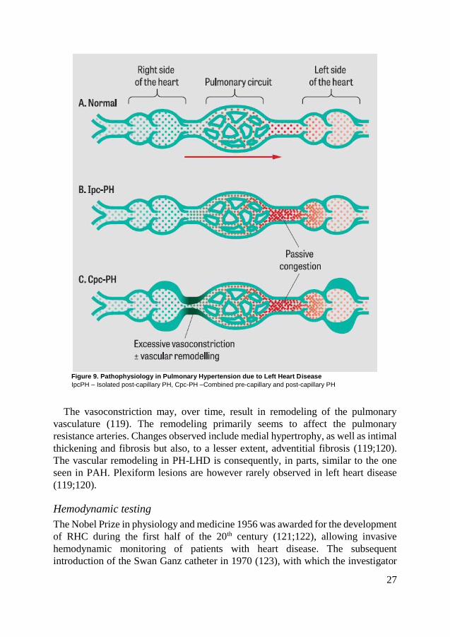
Figure 9. Pathophysiology in Pulmonary Hypertension due to Left Heart Disease
IpcPH – Isolated post-capillary PH, Cpc-PH –Combined pre-capillary and post-capillary PH
The vasoconstriction may, over time, result in remodeling of the pulmonary
vasculature (119). The remodeling primarily seems to affect the pulmonary
resistance arteries. Changes observed include medial hypertrophy, as well as intimal
thickening and fibrosis but also, to a lesser extent, adventitial fibrosis (119;120).
The vascular remodeling in PH-LHD is consequently, in parts, similar to the one
seen in PAH. Plexiform lesions are however rarely observed in left heart disease
(119;120).
Hemodynamic testing
The Nobel Prize in physiology and medicine 1956 was awarded for the development
of RHC during the first half of the 20th century (121;122), allowing invasive
hemodynamic monitoring of patients with heart disease. The subsequent
introduction of the Swan Ganz catheter in 1970 (123), with which the investigator
2727272727
28
can estimate left atrial pressure by inflating a balloon and temporarily obstruct the
pulmonary artery [pulmonary artery wedge pressure – PAWP], has further improved
the diagnostic value provided by RHC. Consequently, RHC has been central for the
increased understanding of the circulation and improved care for these diseases. To
date, RHC remains an important tool in the diagnosis of severe heart disease and
pulmonary hypertension.
In 1958 Paul Wood first defined PH as systolic pulmonary artery pressure above
30 mmHg and diastolic pulmonary artery pressure above 15 mmHg (124). In this
pioneer work, Wood also sub-classified post-capillary PH into a passive and a
reactive “out-of-proportion” group. The term “out-of-proportion” was used for
many years, referring to cases where MPAP was more pronouncedly increased than
what would be expected based on the levels of PAWP, thereby suggesting an active
pulmonary vascular component. The term, however, caused confusion, so it was
abandoned in the most recent European Society of Cardiology (ESC) guidelines
(14;108).
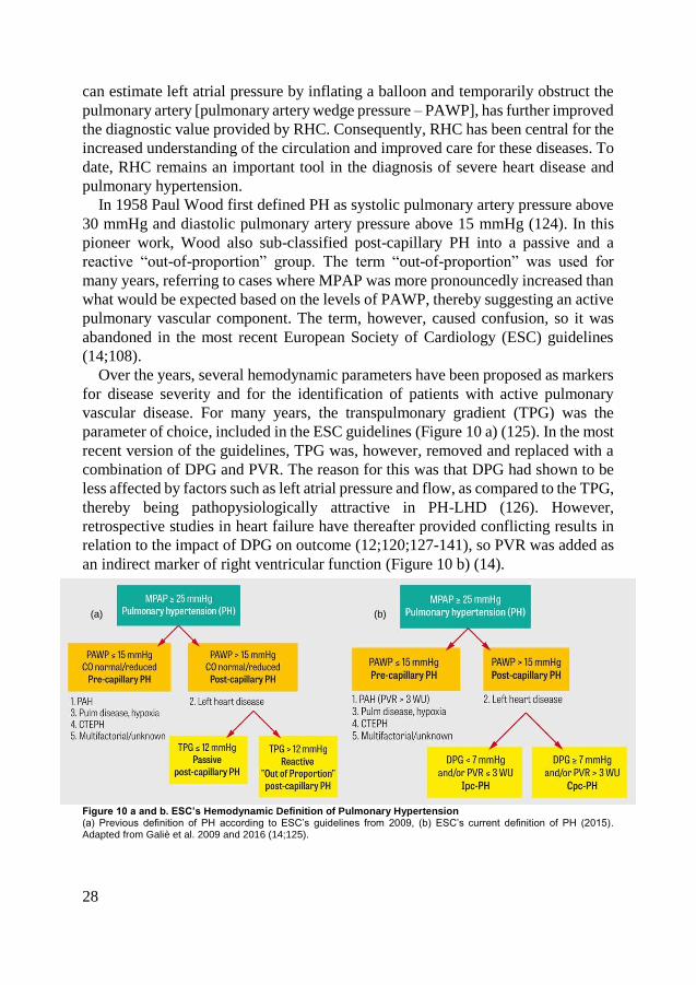
Over the years, several hemodynamic parameters have been proposed as markers
for disease severity and for the identification of patients with active pulmonary
vascular disease. For many years, the transpulmonary gradient (TPG) was the
parameter of choice, included in the ESC guidelines (Figure 10 a) (125). In the most
recent version of the guidelines, TPG was, however, removed and replaced with a
combination of DPG and PVR. The reason for this was that DPG had shown to be
less affected by factors such as left atrial pressure and flow, as compared to the TPG,
thereby being pathopysiologically attractive in PH-LHD (126). However,
retrospective studies in heart failure have thereafter provided conflicting results in
relation to the impact of DPG on outcome (12;120;127-141), so PVR was added as
an indirect marker of right ventricular function (Figure 10 b) (14).
Figure 10 a and b. ESC’s Hemodynamic Definition of Pulmonary Hypertension (a) Previous definition of PH according to ESC’s guidelines from 2009, (b) ESC’s current definition of PH (2015). Adapted from Galiè et al. 2009 and 2016 (14;125).
(b) (a)
28

Hemodynamic exercise testing
Exercise measurements during invasive hemodynamic assessments are commonly
performed. Exercise provides additional information on disease severity (142-147)
and improve early diagnosis of heart failure (148), as well as PH (149). It has been
established that, in health, an increase of cardiac output (CO) by 1 L·min-1 is
accompanied by, on average, an increase in MPAP by 1 mmHg (150;151) and that
these parameters rapidly return to resting values (152) after exercise. Large inter-
individual alterations in the flow-pressure relationship have, however, been
observed (153).
PH during exercise has previously been defined as MPAP > 30 mmHg (154). In
recent guidelines this definition was removed as sufficient evidence for the
hemodynamic thresholds which indicates exercise PH has been lacking (14;125)
and since MPAP has been found to increase beyond 30 mmHg in healthy individuals
(144). An adequate definition of exercise PH is also complicated by the fact that
hemodynamic alterations due to increasing age are more pronounced during
exercise than at rest (144;155;156) and that invasive data are particularly scarce in
the elderly. Patients with severe lung disease also represent a challenge, as marked
alterations in intrathoracic pressures during the respiratory cycle may complicate
the interpretation. Total pulmonary resistance (TPR = MPAP/CO) is affected by
alterations in respiratory pressure to a lesser extent than previously used parameters
and therefore seems to be a more reliable marker for diagnosis of exercise PH also
in this population (157). Moreover, the pressure-flow relationship assessed by TPR
does not seem to exceed 3 WU in healthy individuals (153;158)
With regards to PVR, an increase during exercise can be either due to pulmonary
vascular disease or congestion of the pulmonary circuit due to left heart disease
(153;158). PVR during exercise therefore has a poor diagnostic value in left heart
disease whereas the prognostic value in pulmonary vascular disease is very good
(149). Due to the limitations of MPAP and PVR, TPR has been suggested to be a
more adequate predictor of exercise PH. TPR > 3 WU has indeed been documented
to be an excellent discriminator for exercise PH alone or in combination with MPAP
> 30 mmHg (149;159), although findings may differ depending on how the
measurements are performed (159). Based on these findings, the European
Respiratory Society has released an official statement suggesting that exercise PH
should be defined as MPAP > 30 mmHg and TPR > 3 WU in the absence of MPAP
≥ 25 mmHg at rest (157). The natural history of exercise PH is not known and needs
further investigation (157).
Acute vasodilatation test
Resting hemodynamics is often insufficient to sub-define post-capillary PH and
identify patients with severe pulmonary vascular remodeling. As stated previously,
this is of major importance when evaluating patients for HT, as fixed elevated PVR
2929292929

30
may increase the risk for acute right heart failure after HT. Therefore, acute
vasodilatory testing is recommended in the work-up for HT when the systolic
pulmonary artery pressure is ≥ 50 mmHg and either TPG is ≥ 15 mmHg and/or PVR
is > 3 WU (7). A PVR which remains high without severe systemic hypotension is
considered a relative contraindication for HT (7). In an attempt to sub-define post-
capillary PH, an acute vasodilatation test is also included in the ISHLT definition of
PH (Figure 11) (11). Such a test is, however, lacking in the ESC definition of PH.
In fact, in recent studies using the novel ESC definition, acute vasodilatation tests
have failed to identify a sub group of patients with fixed PVR and worse prognosis
(134;141), suggesting that the current value of these tests, outside pre-HT
evaluations, is limited.
With regards to the drugs used for the vasodilatation test, the effects of several
vasoactive agents on pulmonary hemodynamics have been reported (160). Among
these substances, intravenous infusion of nitroprusside and inhaled NO are probably
best documented (13;160) and also commonly used.
Figure 11. ISHLT’s Hemodynamic Definition of Pulmonary Hypertension Adapted from Fang et al 2012 (11).
Therapies in Pulmonary Hypertension due to Left Heart Disease
There are no specific therapies for PH-LHD. Treatment instead focuses on
optimizing the underlying cause of the disease, as appropriate (14;161). This
includes standard heart failure medications as well as implantable devices and
surgery (161). Several studies, including observational single center reports and
large clinical trials, have investigated the use of pulmonary vasoactive drugs
targeting the endothelin, NO and prostacyclin pathways in left heart disease. Some
of these trials have exclusively included patients with post-capillary PH although
most have not.
30

Several trials with endothelin receptor antagonists have been carried out in
systolic left heart failure with and without PH. None of these have shown beneficial
effects of the treatment. Instead, safety concerns have arisen, with a tendency of
increased fluid retention using endothelin receptor antagonists (162-164). At
present, a trial with the endothelin receptor antagonist macitentan is carried out in
patients with diastolic heart failure and pulmonary vascular disease
(ClinicalTrials.gov Identifier: NCT03153111), a condition previously not
investigated. This is of great importance as the clinical manifestations of PAH and
PH due to diastolic heart failure may be similar. Consequently, if this trial is
negative even more effort has to be put into finding robust markers for
differentiation between PAH and PH due to diastolic heart failure.
With regards to the NO pathway several observational studies as well as small
randomized single-center trials have reported beneficial effects on hemodynamics,
quality of life and functional class with PDE5i’s, primarily sildenafil, in left heart
disease with or without PH. Despite these positive results and the potentially large
patient population available for these drugs, large clinical trials have, until recently,
been lacking. In recent years the trials RELAX and SIOVAC have been published,
both failing to meet their primary end-point (165;166). In fact, in SIOVAC where
study participants with persistent PH after valvular surgery were randomized to
PDE5i or placebo, treatment with PDE5i resulted in an increased risk for the primary
end-point, a composite of death, heart failure hospitalization, change in WHO
functional class and quality of life. Moreover, the sGC stimulator riociguat,
available for the treatment of PAH and CTEPH, has been investigated in systolic
and diastolic heart failure, also without a clear improvement in the primary end-
point change in MPAP. Riociguat did, however, improve several other
hemodynamic parameters in systolic heart failure (167;168), suggesting that sGC
stimulators may be beneficial in this condition. A novel sGC stimulator with longer
half-life, vericiguat, has therefore been evaluated in phase II trials on systolic and
diastolic heart failure (169;170). In SOCRATES-REDUCED, high doses of
vericiguat improved the primary end-point, change in NT-proBNP (169).
Finally, prostacyclin was evaluated for the treatment of severe heart failure in the
FIRST trial. Despite previous results suggesting beneficial effects of prostacyclin in
heart failure (171), FIRST was terminated early due to increased mortality in
patients receiving prostacyclin (172).
As randomized trials in general have failed to demonstrate beneficial effects of
PAH specific drugs in left heart disease, with or without PH, off label use outside
clinical trials should be avoided (Table 2) (14).
3131313131

32
Table 2. Completed Clinical Trials in Pulmonary Hypertension due to Left Heart Disease
Drug
Class
Study
acronym and
substance
Study
population
PH
inclusion
criteria
Primary
endpoint
Outcome
ERA
REACH-1
(162):
Bosentan
LVEF<35%,
NYHA IIIb-IV
(n = 370)
No Change in
clinical status.
Stopped prematurely due
to increased
hepatic transaminases.
Less adverse events after
3 months, in
patients who completed
the study.
ENABLE-1 & 2
(163):
Bosentan
LVEF<35%,
NYHA IIIb-IV
(n = 1613)
No All-cause
mortality or
heart failure
hospitalization.
No difference in primary
endpoint.
Increased risk of worsening
HF, likely due to fluid
retention.
MELODY-1
(164):
Macitentan
LVEF≥30%,
NYHA II-III (n
= 63)
Yes, Cpc-
PH
Significant fluid
retention or
worsening
NYHA class.
Trend towards higher
incidence of significant
fluid retention.
PDE5i
RELAX (165):
Sildenafil
LVEF≥50%,
NYHA II-IV (n
= 216)
No Change in peak
oxygen
consumption.
No improvement in
primary endpoint.
SIOVAC (166):
Sildenafil
Successful
valvular
replacement/r
epair > 1 year
before
inclusion.
Yes,
MPAP≥30
mmHg.
Composite of
death, heart
failure
hospitalization,
WHO class and
QoL.
Increased risk of primary
endpoint.
sGC
stim
LEPHT (167):
Riociguat
LVEF≤40%
(n = 201)
Yes,
MPAP≥25
mmHg
Change in
MPAP.
No improvement in
primary endpoint.
Improvement in CI and
PVR. Well tolerated.
DILATE (168):
Riociguat
HFpEF (n =
39)
Yes, post-
capillary
PH
Change in
MPAP.
No change in primary
endpoint. Well tolerated.
SOCRATES-
REDUCED
(169):
Vericiguat
LVEF<45%
Worsening
heart failure
(n = 456)
No Change in
NT-proBNP
No improvement in
primary endpoint in
pooled analysis.
Improvement in primary
endpont with high doses
of Vericiguat.
SOCRATES-
PRESERVED
(170):
Vericiguat
LVEF≥45%,
NYHA II-IV (n
= 477)
No Change in
NT-proBNP and
left atrial
volume
No improvement in
primary endpoint.
Improved QoL.
PGI2
FIRST (172):
Epoprostenol
HFrEF,
NYHA IIIb-IV
(n = 471)
No All-cause
mortality
Terminated early due to a
strong trend towards
increased mortality.
32

Hypoxic Pulmonary Vasoconstriction
Hypoxic pulmonary vasoconstriction (HPV) was first described in an in vivo cat
model in 1946 (173) and a year later in human (174). HPV mainly affects the smooth
muscle cells of the pulmonary resistance arteries (175;176) and is primarily caused
by a decreased partial pressure of oxygen in alveolar air. Lower mixed venous
saturation does also, to a lesser extent, contribute to HPV (177). HPV in focal
hypoxia is beneficial and contribute to optimized ventilation-perfusion matching
and gas exchange. In global hypoxia, e.g. during rapid ascent to high altitude it may,
however, be detrimental. HPV may then limit exercise capacity (178) as well as
contribute to the development of high altitude pulmonary edema and right heart
failure by increasing pulmonary pressures and resistances (179-181). Whereas acute
HPV is fully reversible (173), chronic hypoxia may result in pulmonary arterial and
venous remodeling, as well as PH (182-184). The remodeling of the pulmonary
arteries is characterized by medial hypertrophy as well as intimal and adventitial
thickening. In severe cases plexiform lesions may also occur (183). Interestingly,
the pulmonary veins furthermore undergo arterialization and intimal fibrosis (184).
As in PAH and PH-LHD, the endothelin, NO and prostacyclin pathways are also
central in HPV and in the development of hypoxic PH, including vascular
remodeling (185) (Figure 12). Figure 12. Chacaltaya Ski Lodge in Bolivia at Approximately 5300 m Above Sea Level. The partial pressure of oxygen at this altitude corresponds to a FiO2 of approximately 0.10 causing pronounced hypoxic pulmonary vasoconstriction, thereby impairing exercise capacity. Photographed by and printed with the permission of assoc. prof. Göran Rådegran.
Therapies in Hypoxic Pulmonary Vasoconstriction
Treatment of acute global hypoxia focuses, apart from removing the cause of
hypoxia, on increasing systemic oxygen supply via ventilation or ECMO. In acute
hypoxia, reports have shown beneficial effects with PDE5i and inhaled NO but not
with endothelin receptor antagonists or prostacyclin (185). In PH due to chronic
lung diseases, PAH drugs have shown to improve hemodynamics without
improving exercise capacity. In fact, several trials have resulted in further
impairment in arterial saturation and quality of life as they interfere with HPV,
thereby aggravating ventilation-perfusion mismatching (185-190). Guidelines
therefore advise against the use of drugs targeting the endothelin, NO and
prostacyclin pathways in PH due to lung diseases and/or hypoxia (14).
3333333333
34
Aims
The overall objectives of the present thesis were to characterize patients prior to and
after heart transplantation with regard to hemodynamics and blood borne
biomarkers, as well as to evaluate vasoactive substances in the treatment of acute
hypoxic pulmonary vasoconstriction, which may aggravate pulmonary hypertension
due to left heart disease.
The specific aims of the included papers were to investigate:
• The effects of two pulmonary vasodilators, namely the soluble guanylate
cyclase stimulator BAY 41-8543 and the dual endothelin receptor blocker
tezosentan, in acute hypoxic pulmonary vasoconstriction.
• The hemodynamic characteristics after heart transplantation, including
exercise response.
• The impact of pre-operative and post-operative pulmonary hypertension on
outcome after heart transplantation.
• Plasma concentrations of substances related the nitric oxide pathway and
nitric oxide dependent pulmonary vasodilatation prior to and after heart
transplantation.
34
Materials and Methods
Paper I
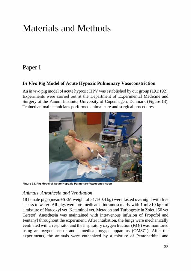
In Vivo Pig Model of Acute Hypoxic Pulmonary Vasoconstriction
An in vivo pig model of acute hypoxic HPV was established by our group (191;192).
Experiments were carried out at the Department of Experimental Medicine and
Surgery at the Panum Institute, University of Copenhagen, Denmark (Figure 13).
Trained animal technicians performed animal care and surgical procedures.
Figure 13. Pig Model of Acute Hypoxic Pulmonary Vasoconstriction
Animals, Anesthesia and Ventilation
18 female pigs (mean±SEM weight of 31.1±0.4 kg) were fasted overnight with free
access to water. All pigs were pre-medicated intramuscularly with 1 mL·10 kg-1 of
a mixture of Narcoxyl vet, Ketaminol vet, Metadon and Turbogesic in Zoletil 50 vet
Tørstof. Anesthesia was maintained with intravenous infusion of Propofol and
Fentanyl throughout the experiment. After intubation, the lungs were mechanically
ventilated with a respirator and the inspiratory oxygen fraction (FiO2) was monitored
using an oxygen sensor and a medical oxygen apparatus (OM871). After the
experiments, the animals were euthanized by a mixture of Pentobarbital and
3535353535

36
Lidocainhydrochloride. Further details can be found in the original publication
(193).
Invasive Procedures, Measurements and Hemodynamic Surveillance
Two skin incisions were made, one along the medial line of the throat and one in
the groin, to locate the internal jugular vein and the common carotid artery, as well
as the femoral artery, respectively. An infusion catheter was inserted through the
internal jugular vein and advanced into the right atrium. Catheters for arterial
pressure measurements were positioned in the aortic arch, through the common
carotid artery, as well as in the femoral artery. The femoral arterial catheter was
used as a back-up, if problems with the aortic catheter would occur (193).
A Swan Ganz catheter (Edwards Lifesciences, Irvine, CA, USA), introduced
through the right internal jugular vein, was used to measure mean right atrial
pressure (MRAP), pulmonary artery pressures and PAWP, as well as CO in
triplicates by thermodilution.
Electrocardiogram, heart rate (HR), mean arterial pressure (MAP), MRAP and
MPAP, were monitored and recorded throughout the experiment. PAWP was
measured intermittently. Non-invasive saturation was monitored by a pulse
oximetry probe placed on the tails of the pigs. PVR, systemic vascular resistance
and stroke volume (SV) were calculated using the following formulae: PVR =
(MPAP – PAWP) / CO, systemic vascular resistance = (MAP – MRAP) / CO, and
SV = CO / HR.
Blood samples from the pulmonary artery and the aortic arch were used for
immediate blood gas analysis, determining blood aorta (AO) and pulmonary artery
(PA) O2 saturation (SAOO2, SPAO2), hemoglobin concentration (HbAO, HbPA), O2-
pressure (pAOO2, pPAO2), pHAO and pHPA. This allowed calculation of aorta and
pulmonary artery O2-content (CAOO2, CPAO2): CAOO2 = (HbAO·1.34·SAOO2) +
(0.0031·pAOO2), and CPAO2 = (HbPA·1.34·SPAO2) + (0.0031·pPAO2), O2-extraction =
CAOO2 - CPAO2, and O2-consumption = CO·(CAOO2 - CPAO2). In the formulae, SO2
is expressed as a fraction of 1.0 and not %, Hb as g·dL-1, pO2 as mmHg, and the
correction factors 1.34 as mL·g-1 and 0.0031 as mL·dL-1·mmHg-1.
Induction of Hypoxia
Hypoxia was induced by slowly lowering the oxygen supply and airflow on the
respirator from FiO2~0.21, to a stable level at ~0.15 and ~0.10, respectively. The
respiratory rate and tidal volume were kept constant throughout the experiments.
Hemodynamic measurements and blood sampling were performed towards the end
of normoxia, 5 minutes into stable hypoxia at FiO2~0.15 and 15 minutes into stable
hypoxia at FiO2~0.10 (hypoxia baseline). During the induction of hypoxia, 1000 mL
of sodium chloride was administered to each animal to avoid sudden systemic
hypotension.
36

Six pigs were used for control measurements, where the stability of the HPV
response over time was evaluated. In this protocol, hemodynamic measurements
and blood sampling were performed after 5, 15, 22.5, 30, 37.5, 60, 67.5, 75, and
82.5 min of sustained hypoxia at FiO2~0.10, corresponding to the time-points when
measurements were performed in the intervention protocols described below.
Drugs
Twelve pigs were used to study the effects of the sGC stimulator BAY 41-8543
(Bayer Scheering Pharma AG, Wuppertal, Germany), alone (n=6) or in combination
with dual ET-receptor blockade with tezosentan (Actelion, Allschwil, Switzerland,
n=6).
In the first intervention protocol, the sGC stimulator BAY 41-8543 was infused
at increasing doses of 1, 3, 6, 9 and 12 μg·min-1·kg-1 for ~7.5 minutes each.
Measurements were performed at steady state, 5-7.5 min after starting infusion of
the sGC stimulator at each dose.
In the second protocol, 5 mg·kg-1 body weight of the dual endothelin A receptor
and B receptor antagonist tezosentan was delivered as a manual bolus injection,
followed by infusion of BAY 41-8543 at 1, 3 and 6 μg·min-1·kg-1 for ~7.5 minutes
each. Measurements were performed 5, 15, 30 and 60 min after injection of
tezosentan and at steady state between 5-7.5 min after starting infusion of the sGC
stimulator at each dose.
All drugs were administered in the right atrium during sustained hypoxia at
FiO2~0.10. An infusion pump (Model 11plus, Harvard apparatus, Maryland, USA)
was used to administer BAY 41-8543.
Paper II-IV
Study Population and Data Collection
Medical records from the 215 HT patients followed at Skåne University Hospital in
Lund, Sweden, 1988-2010 were retrospectively reviewed. A total of 219 HTs were
included, out of which 214 (98%) were first-time HTs. Five (2%) were re-HTs; three
within seven days, one 175 days and one 18 years after HT. 218 of the HTs had been
performed in Lund. One pediatric first time HT had been performed abroad. 72
(33%) of the patients received a mechanical assist prior to HT (Table 3). Children
under the age of 18 (n=21), re-HTs (n=5) and patients referred from, and
subsequently evaluated at, other hospitals (n=83) and/or with incomplete data prior
3737373737

38
to HT (n=12) were excluded. 94 adults evaluated at rest, at our hemodynamic lab,
prior to HT remained for analysis (Table 3).
Table 3. Baseline Characteristics of the Entire HT Population in Lund 1988-2010 and the 94 Included Patients
Risk factor category and group Entire population characteristics Study population characteristics
Mean SD +/- N % Mean SD +/- N %
Recipient characteristics
Gender of recipient 215 94
Male 147 68.4 70 74.4
Female 68 31.6 24 25.6
Age of recipient (years) 44.6 17.2 215 51.6 9.9 94
≤10 9 4.2 0 0
11-20 24 11.1 1 1.1
21-30 8 3.7 4 4.3
31-40 17 7.9 6 6.4
41-50 42 19.5 21 22.3
51-60 85 39.5 45 47.9
61≤ 30 14.0 17 18.1
Recipient length (cm) 167.3 25.3 197 173.4 7.9 93
Recipient weight (kg) 68.4 18.9 198 75.3 15.0 93
Recipient blood group 215 94
0 71 33.0 31 33.0
A 109 50.7 46 48.9
B 19 8.8 8 8.5
AB 16 7.4 9 9.6
Time on waiting list (days) 151.3 200.4 219 148.6 179.6 94
Indication for HT 215 94
ARVD 3 1.4 0 0
Cardiac Tumor 1 0.5 0 0
CHD 11 5.1 1 1.1
CVR 1 0.5 0 0
DCM 98 45.6 46 48.9
DCM (Adria) 3 1.4 1 1.1
DCM + AI/AS 9 4.2 5 5.3
HCM 10 4.7 8 8.5
Myocarditis 13 6.0 6 6.4
IHD 58 27.0 24 25.6
RCM 6 2.8 3 3.2
RHF + VT/VF 2 0.9 0 0
Donor characteristics
Gender of donor 219 94
Male 138 63.0 58 61.7
Female 81 37.0 36 38.3
Age of donor (years) 40.0 15.7 219 43.9 13.7 94
Donor length (cm) 172.4 18.7 210 176.3 9.5 92
Donor weight (kg) 74.1 18.5 211 79.4 13.5 93
Ischemic time (min) 184.7 63.2 201 182.6 61.5 86
Recipient-donor matching
Age difference (years) 4.6 15.9 219 7.7 14.7 94
Sex matching 219 94
Sex-matched 160 73.1 71 75.5
Sex-mismatched 59 26.9 23 24.5
AB0-matching 219 94
Identic 184 84.0 82 87.2
Compatible 32 14.6 12 12.8
Incompatible 3 1.4 0 0
38

Paper II
The 94 patients were hemodynamically characterized at rest prior to and after HT.
32 of the 94 patients exercised prior to and one year after HT. They were
subsequently characterized into two groups, one with patients≤50 years (n=12) and
one with patients>50 years (n=20), at the time of HT. This sub classification was
performed to compare our findings to those presented in healthy individuals
published in previous systematic reviews, as these have shown that the
hemodynamic response to exercise differ for healthy individuals aged ≤50 years vs.
>50 years (144;155).
Table 4. Previous Publications on Pre-Operative Pulmonary Hypertension and Their Reported Outcomes After Heart Transplantation
Author/Period Characterization Results
Costard-Jäckle
and Fowler (9)
1. “Low risk”: PVR<2.5 or PVR≤2.5
and sAP≥85 with vasodilatation
- Increased 3- month mortality in
high risk group
1980-1988 2. “High risk”: PVR≥2.5 or PVR≤2.5
but sAP≤85 with vasodilatation
Murali et al. (10)
1980-1991
1. TPG<15 and PVR<5
2. TPG<15 and PVR≥5
3. TPG≥15 and PVR<5
4. TPG≥15 and PVR≥5
- Increased 0-2 day mortality in
severe PH pre-HT (group 4)
- Increased 30-day mortality with
TPG>15
Lindelöw et al.
(32)
1988-1990
1. “Low PVR”: PVR≤3
2. “High PVR”: PVR≤3 and
MAP≥70 with vasodilatation
- No difference in mortality
between groups
Delgado et al.
(194)
1991-1996
1. No PH: TPG≤12 and PVR≤2.5
2. PH: TPG>12 and/or PVR>2.5
- Increased 30-day mortality with
PH
Klotz et al. (195)
1998-2001
1. No PH: TPG<12 and PVR<2.5
2. PH: TPG≥12 and/or PVR≥2.5
- No difference in 1 year
mortality between patients
without PH vs reversible PH
Chang et al.
(196)
1983-2000
1. No PH: PVR<2.5
2. Mild/moderate PH:
PVR 2.5-4.9
3. Severe PH: PVR≥5
- Increased 1, 3 and 6 month
survival with mild/moderate PH
- Increased 1 year mortality with
severe PH, otherwise
unchanged
Goland et al.
(17)
1989-2004
1. No PH: PVR<3, TPG<10
2. Mild-Moderate PH:
PVR 3-6, TPG 10-20
3. Severe PH: PVR>6, TPG>20
- No difference in mortality with
pre-HT PH
- Increased long term mortality
with sustained PH after HT
Klotz et al. (197)
1996-2005
1. No PH: TPG≤12 and PVR≤2.5
2. Rev PH: TPG≤12 and PVR≤2.5
with vasodilatation
- No difference in mortality
between groups
1. No PH: 2-3 of: sPAP≤50,
TPG≤10 and PVR≤2.5
- No difference in 1 year
mortality with pre-HT PH
2. PH: 2-3 of: sPAP≤50, TPG≤10
and PVR≤2.5 with vasodilatation
- Increased long term mortality
with sustained PH after HT
Gude et al. (18)
1983-2007
1. No PH: 2-3 of: SPAP≤50,
TPG≤10 and PVR≤2.5
2. PH: 2-3 of: SPAP≤50,
TPG≤10 and PVR≤2.5 with
vasodilatation
- No difference in long-term
mortality with pre-HT PH
- Increased long-term mortality
with sustained PH after HT
3939393939
40
Paper III
The 94 included patients were characterized according to: i. previously published
hemodynamic risk criteria for outcome after HT (Table 4) (9;10;17;18;18;32;194-
197); ii. ESC’s guidelines from 2009 (125); iii. ISHLT’s State of art summary
statement from 2012 (11); iv. ISHLT’s relative contraindications for HT from 2006
(7) and v. ISHLT’s criteria for increased risk of right heart failure and early death
after HT from 2006 (7) (Table 5). To separate the patients into groups according to
ISHLT’s State of art summary statement, the criteria for passive and reactive mixed
PH were modified, i.e. defined as TPG ≤ 15 mmHg and PVR ≤ 3 WU, instead of
TPG ≤ 15 mmHg or PVR ≤ 3 WU. Survival was compared between the subgroups
of each study.
Table 5. Previous Definitions of Pulmonary Hypertension and a High Risk Population According to ESC and ISHLT
Paper IV
Of the 94 patients, 89 were post-operatively evaluated with routine RHC at our
hemodynamic laboratory. The five patients who did not perform RHC after HT died
early after transplantation: three within one day, one after 14 days, and one after 25
days. All were men and three had pre-operative PH. No additional, clinically
indicated, hemodynamic evaluations were included in the study.
The patients were grouped depending on their hemodynamic data at the post-
operative evaluations. Survival was compared between the groups and hazard ratios
for death were estimated with a Cox regression analysis adjusted for gender, age at
the time of HT and kidney function one year after HT.
Definition/Author Characterization
PH- definitions according to ESC-
guidelines from 2009. Galiè et al.
(125)
1. No PH: mPAP<25
2. Pre-capillary PH: MPAP≥25 and PCWP≤15 and CO normal or reduced
3. Post-capillary PH: MPAP≥25 and PCWP>15 and CO normal or reduced
3a. Passive: TPG≤12
3b. Reactive: TPG>12
ISHLT’s State of art summary
statement. Fang et al. (11)
1. No PH: mPAP<25
2. Passive PH: MPAP≥25 and PCWP>15 and TPG≤15 or PVR≤3.0
3. Mixed PH: MPAP≥25 and PCWP>15 and TPG>15 or PVR>3.0
3a. Reactive PH (with vasodilatation) : TPG≤15 or PVR≤3.0
3b. Non-reactive PH (with vasodilatation): TPG>15 or PVR>3.0
ISHLT-guidelines for performing
vasodilation during RHC. Mehra et
al. (7)
1. sPAP≥50 and either TPG≥15 or PVR>3.0 while maintaining sAP>85
with vasodilatation
ISHLT’s defined relative
contraindications for HT. Mehra et
al.
1. PVR>5 or PVRI>6 or TPG>16-20
ISHLT’s defined increased risk of
right heart failure and early death
after HT. Mehra et al. (7)
1. sPAP>60 and either PVR>5 or PVRI>6 or TPG>16-20
2. PVR >2.5 with vasodilatation
3. PVR <2.5 but sAP falls to <85 with vasodilatation
40

Right Heart Catheterization and Endomyocardial Biopsies
Right heart catheterization (Figure 14 a) was performed at rest prior to HT, as well
as one and four weeks, three and six months and one year after HT. In all patients
capable to exercise, hemodynamic measurements were also performed during
supine bicycle exercise prior to HT, as well as four weeks, three and six months and
one year after HT. Men exercised at 50 W and women at 30 W, maintaining steady
revolutions at 60 per minute. Measurements were conducted at steady state,
approximately five minutes after initiation of exercise. There was no graded increase
in workload. If patients had more than one RHC prior to HT, the one closest to HT,
or assist implantation, was analyzed. Pre-operative RHC was on average performed
143.1 ± 113.3 days prior to HT.
Figure 14 (a) and (b). Right Heart Catheterization and Swan Ganz Catheter (a) The hemodynamic lab at Skåne University Hospital, Lund, (b) Schematic illustration of a Swan Ganz catheter
RHC was predominantly performed via the right internal jugular vein, using a
Swan Ganz catheter (Figure 14 b) (Baxter Health Care Corp, Santa Ana, CA).
During RHC MPAP, PAWP, MRAP and MAP, were measured. HR was recorded
from electrocardiography. CO was measured by thermodilution. TPG, DPG, SV,
cardiac index, stroke volume index, PVR, pulmonary vascular resistance index and
TPR were calculated by the following formulae: TPG=MPAP-PAWP,
DPG=diastolic pulmonary artery pressure/PAWP, SV=CO/HR, CI=CO/body
surface area, SVI=SV/body surface area, PVR=TPG/CO, PVRI=TPG/CI and
TPR=MPAP/CO. Left ventricular stroke work index and right ventricular stroke
work index were calculated using different formulae depending on
recommendations at the time of each study, i.e. left ventricular stroke work
index=(MAP-PAWP)·SV index and right ventricular stroke work index=(MPAP-
MRAP)·SV index (paper II) or left ventricular stroke work index=(MAP-
PAWP)·SV index·0.0136 and right ventricular stroke work index=(MPAP-
MRAP)·SV index·0.0136, where 0.0136 is a conversion factor (paper III).
4141414141

42
To monitor acute cellular rejection (ACR), EMBs were performed from the right
interventricular septum on a routine basis on 14 occasions during the first post-
operative year (week 1, 2, 3, 4, 6, 8, 10, 12, 16, 20, 24, 32, 40, and 52 after HT).
Some of the EMBs were performed in connection with the RHCs described above.
From 2005 onwards, EMBs were graded according to both the 1990 and the 2004
ISHLT grading systems (198;199). Prior to 2005, the EMBs were solely graded
according to the 1990 system (198).
With regard to renal function, glomerular filtration rate was measured at one year
after HT using the iohexol clearance method. In patients where iohexol clearance
measurements were missing, the creatinine-based CKD-EPI (Chronic Kidney
Disease Epidemiology Collaboration) equation recommended by KDIGO (200),
was used.
Drugs and Devices
A vasodilator test or mechanical assist was used when necessary to assess acute and
sustained vaso-reactivity, respectively. Of the 94 included patients, 23 patients were
exposed to a vasodilator, either intravenous nitroprusside (Nitropress, Hospira Inc,
Lake Forest, USA) or nitric oxide (NO, INOmax, INO Therapeutics AB, Lidingö,
Sweden) inhalation (INOvent, INO Therapeutics AB, Lidingö, Sweden) during
RHC. The doses for nitroprusside were ~0.5, 1, 2, 4 and 8 µg·kg-1·min-1 and for NO
20 or 40 parts per million.
21 of the 94 patients received a mechanical assist as a bridge to transplantation.
Of these, five were HeartMate IP (Thoratec, Pleasanton, CA, USA), five HeartMate
VE (Thoratec, Pleasanton, CA, USA), three Jarvik 2000 (Jarvik Heart Inc., New
York, NY, USA), one Abiomed (Abiomed Inc, Danvers, MA, USA) and seven
HeartMate II (Thoratec, Pleasanton, CA, USA). 12 patients with mechanical assist
were hemodynamically re-evaluated with the device prior to HT, three had a
HeartMate IP, four HeartMate VE, two Jarvik 2000 and three HeartMate II. Four
patients with mechanical assist exercised prior to HT and one year thereafter.
Paper V
Study Population
Clinical data was gathered from medical records of all 43 patients who underwent
HT at Skåne University Hospital in Lund June 2012 through February 2014. All
patients were included in a prospective cohort study and 12 patients were included
in the present investigation. The 12 patients were selected as they were
42

hemodynamically evaluated at our lab, at rest, prior to HT, as well as four weeks
and six months after HT, and as they at the four week and six month evaluations; i.
did not have rejections requiring specific therapy; ii. did not have post-operative PH
and; iii. had left ventricular ejection fraction≥50% (Table 6).
12 healthy, age-matched, non-smokers without drug treatment and no symptoms
or signs of common cold were used for comparison.
Table 6. Baseline Characteristics of the Included Patients and Controls
Parameter Patients Healthy subjects
n median (IQR) n median (IQR)
Clinical features
Sex, male/female 12 10/2 12 5/7
Age, years 12 50 (45-60) 12 51 (46-60)
Weight, kg 12 80 (71-92) N/A
Length, cm 12 179 (176-180) N/A
BSA, m2 12 2.0 (1.9-2.1) N/A
NYHA class, IIIb/IV 3/9 N/A
Etiology, DCM/HCM/IHD 12 8/1/3 N/A
Diabetes mellitus, yes/no 12 0/12 12 0/12
Hypertension, yes/no 12 0/12 12 0/12
AF, yes/no 12 2/10 12 0/12
History of smoking, yes/no 12 3/9 12 0/12
Medical history
Betablockers, % 12 12 (100) -
ACEi/ARB, % 12 8/4 (100) -
MRA, % 12 8 (67) -
Anticoagulants, % 12 10 (83) -
Statins, % 12 3 (25) -
Diuretics, % 12 12 (100) -
Biochemical indicators
L-Arginine, µM 12 56 (48-60) 12 86 (78-92) *
ADMA, µM 12 0.57 (0.47-0.71) 12 0.39 (0.32-0.42) *
SDMA, µM 12 0.85 (0.68-1.15) 12 0.41 (0.36-0.45) *
L-Arginine/ADMA 12 86 (68-128) 12 225 (203-273) *
P-Urea, mmol∙L-1 12 9 (8-13) N/A
P-Homocysteine, µM∙L-1 12 16 (13-27) N/A
P-NT-proBNP, ng∙L-1 12 4058 (3348-7408) N/A
P-creatinine, µM∙L-1 12 106 (95-131) N/A
eGFR, mL∙min-1∙1.73m-2 12 61 (44-78) N/A
Plasma Samples and Biomarker Analysis
Plasma samples of patients collected from the superior vena cava during RHC were
stored at -80˚C in the Lund Cardio Pulmonary Register cohort of Region Skåne’s
biobank. The samples were analyzed for plasma concentrations of ADMA,
symmetric dimethylarginine, L-Arginine, L-Ornithine and L-Citrulline with liquid
chromatography – tandem mass spectrometry (LC-MS/MS), at the Swedish
National Veterinary Institute in Uppsala, Sweden. The L-Arginine/ADMA-ratio, the
L-Arginine/L-Ornithine-ratio and the global arginine bioavailability ratio (L-
Arginine/(L-Ornithine+L-Citrulline)) were calculated.
4343434343

44
Control samples collected at Uppsala University Hospital and stored at -20˚C
were analyzed for L-Arginine, ADMA and symmetric dimethylarginine. The
analysis of control samples was performed prior to the analysis of patient samples,
at which point the L-Ornithine and L-Citrulline analyzes were not available.
Consequently, L-Ornithine and L-Citrulline levels are lacking in controls. The
methods used for the analysis of L-Arginine, ADMA and symmetric
dimethylarginine were identical in patients and controls.
Ethics
The experiments in paper I were ethically approved by Dyreforsøgstilsynet,
Copenhagen, Denmark (2009/561-1621) and performed in accordance with the
guidelines of the Department of Experimental Medicine and Surgery at the Panum
Institute, Copenhagen, Denmark.
Papers II-V were performed with approval from the local ethics boards in Lund
(Approval nos. 2014/92, 2011/777, 2011/368 and 2010/114 for papers II-IV and
2010/114, 2010/442, 2011/368, 2011/777, 2015/270 for paper V) and Uppsala
(Approval no. 2010/343, paper V). Papers II-V performed in accordance with the
Declarations of Helsinki and Istanbul.
Statistical Analyses
Parametric or non-parametric statistics were used, as appropriate, depending on the
distribution of data, i.e. Paired t-test or Wilcoxon Signed rank test, respectively,
when comparing one group prior to, and after, an intervention; One Way Repeated
Measures ANOVA (Tukey-test) or One Way Repeated Measures ANOVA on
Ranks (Tukey-test), respectively, when the same group was compared over time; t-
test or Mann-Whitney Rank Sum test, respectively, when two groups were
compared and Pearson- or Spearman Correlation, respectively, when measuring the
association between two variables. The χ2 test was used to compare the proportion
of EMBs with ACR between two groups. Survival curves were plotted using the
Kaplan-Meier method, differences between groups were investigated using the Log-
Rank test and hazard ratios of death were estimated with Cox proportional hazards
regression analysis. In papers III and IV, last day of follow-up was 30th of June
2012 (mean 8.2±5.8 years).
Continuous variables are presented as mean±SEM (paper I), mean±SD (paper II-
IV) or median and interquartile range (paper V), unless otherwise indicated.
Categorical variables are expressed as counts and percentages. With the exception
of the Cox proportional hazards regression analysis in paper IV, performed in SPSS
44

(IBM SPSS Statistics 20, IBM Corp., Armonk, NY, USA) all statistical analyses
were performed using SigmaStat/SigmaPlot 11 (Systat Software Inc., San José, CA,
USA. A p value<0.05 was considered statistically significant throughout.
4545454545

46
Results
Paper I
Hypoxia
Induction of Hypoxia
In a pooled analysis of all 18
pigs, hypoxia gradually, and
maximally, increased MPAP by
14.2±0.6 mmHg, PVR by
2.8±0.3 WU, MRAP by 2.4±0.6
mmHg, PAWP by 1.8±0.5
mmHg and CO by 0.7±0.2
L·min-1. Hypoxia also maximally
decreased SVR by 5.6±1.3 WU,
SAOO2 by 21.7±2.8 % and SPAO2
by 17.8±3.5 %. MAP, HR and
blood-O2-consumption were not
significantly altered by induction
of hypoxia (Figure 15).
Hypoxia protocol
During sustained hypoxia at
FiO2~0.10 for 82.5 min, all
hemodynamic variables as well
as blood-O2-consumption
remained stable, whereas blood
SAOO2 and SPAO2 decreased by
13.9±3.1 % and 14.0±2.4 %,
respectively. Figure 15. Hemodynamic and O2-consumption Response to Hypoxia * Indicates statistical significance as compared to normoxia, § Indicates statistical significance as compared to FiO2 0.15.
46
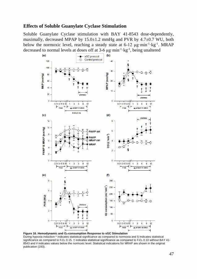
Effects of Soluble Guanylate Cyclase Stimulation
Soluble Guanylate Cyclase stimulation with BAY 41-8543 dose-dependently,
maximally, decreased MPAP by 15.0±1.2 mmHg and PVR by 4.7±0.7 WU, both
below the normoxic level, reaching a steady state at 6-12 μg·min-1·kg-1. MRAP
decreased to normal levels at doses off at 3-6 μg·min-1·kg-1, being unaltered
Figure 16. Hemodynamic and O2-consumption Response to sGC Stimulation During hypoxia induction * indicates statistical significance as compared to normoxia and § Indicates statistical significance as compared to FiO2 0.15. † indicates statistical significance as compared to FiO2 0.10 without BAY 41-8543 and # indicates values below the normoxic level. Statistical indications for MRAP are shown in the original publication (193).
4747474747

48
thereafter. Furthermore, BAY 41-8543 dose-dependently decreased MAP by
26.8±3.3 mmHg, levelling off at 6-12 μg·min-1·kg-1 and increased CO by maximally
1.4±0.3 L·min-1, levelling off at 3-12 μg·min-1·kg-1, as well as SPAO2 by 12.4±5.7 %
at doses of 12 μg·min-1·kg-1. PAWP, SAOO2 and blood-O2-consumption remained
unaltered (Figure 16).
Effects of Soluble Guanylate Cyclase Stimulation in Combination with
Dual Endothelin Receptor Blockade
Dual Endothelin Receptor Blockade
Dual ET-receptor blockade with tezosentan completely normalized PVR and MRAP
and maximally decreased MPAP by 11.8±1.2 mmHg, reaching a steady state 30-60
minutes after bolus injection of tezosentan for MPAP and PVR and after 15-60 min
for MRAP. Furthermore, tezosentan maximally decreased MAP by 27.2±2.8
mmHg, levelling off 30-60 minutes after injection and decreased SPAO2 by 6.3±2.3
% after 60 minutes. PAWP, CO, blood SAOO2, and blood O2-consumption were
unaltered by tezosentan injection (Figure 17).
Soluble Guanylate Cyclase Stimulation on Top of Dual Endothelin
Receptor Blockade
Among the variables decreased by tezosentan, BAY 41-8543 further, dose-
dependently decreased MPAP to normoxic levels as well as PVR and MAP by
1.9±0.4 WU and 20.5±2.9 mmHg, respectively, to levels below those in normoxia,
levelling off at infusion rates of 3-6 μg·min-1·kg-1 for all three variables. MRAP,
PAWP, CO, SAOO2, SPAO2 and blood O2-consumption remained unaltered with
BAY 41-8543 infusion in addition to tezosentan (Figure 17).
48

Figure 17. Hemodynamic and O2-consumption Response to sGC Stimulation in Combination with Endothelin Receptor Blockade During hypoxia induction * indicates statistical significance as compared to normoxia and § indicates statistical significance as compared to FiO2 0.15. During sustained FiO2 0.10, † indicates statistical significance as compared to FiO2 0.10 without tezosentan, ¤ indicates statistical significance with BAY 41-8543 as compared to hypoxia 60 min after tezosentan bolus infusion, ○ indicates return to normoxic values and # indicates values below the normoxic level. Statistical indications for MRAP are shown in the original publication (193).
4949494949

50
Paper II-IV
Pre-operative Hemodynamics
At rest, in all 94 patients, pre-operative MPAP was 30.1±8.8 mmHg, PAWP
20.5±8.2 mmHg, TPG 9.6±3.5 mmHg, DPG -1.2±4.6 mmHg, MRAP 7.9±6.1
mmHg, SV 46.7±17.1 mL·beat-1, CO 3.7±1.0 L·min-1 and PVR 2.8±1.3 WU.
32 patients exercised during RHC prior to HT and one year after HT. During the
pre-operative assessment, exercise, compared to rest, increased MPAP to 44.4±10.5
mmHg, PAWP to 30.5±9.5 mmHg, TPG to 14.0±5.3 mmHg, MRAP to 16.3±7.4
mmHg, HR to 111.9±19.6 beat·min-1, SV to 60.2±21.6 mL·beat-1 and CO to 6.4±1.7
L·min-1, whereas PVR and TPR remained unchanged (Figure 18).
Figure 18. Hemodynamic Response to Exercise Prior to and After Heart Transplantation * Indicates statistical significance as compared to rest.
50

With regard to age dependent differences, none of the hemodynamic parameters
above differed for patients ≤50 years and >50 years, neither at rest nor during
exercise. The percentage change did furthermore not differ between the groups.
Pre-operative Pulmonary Hypertension
Sixty-six of the 94 patients had PH prior to HT. Of these, 63 had post-capillary PH
and three had pre-capillary PH according to present definitions (14;125). 50 of the
patients with post-capillary PH had passive post-capillary PH and 13 had a reactive
post-capillary PH, according to ESC’s guidelines from 2009 (125). DPG≥7 mmHg
was present in three patients.
Acute Vasoreactivity Test
The 23 patients exposed to acute vasodilatation during RHC prior to HT showed a
decrease in MPAP by 4.6±10.4 mmHg and PVR by 1.6±1.3 WU. PVR did not
decrease to <2.5 WU in 12 patients. In four patients where PVR decreased to <2.5
WU, SAP decreased to <85 mmHg.
Hemodynamic Response to Left Ventricular Assist Device
Twelve patients were hemodynamically re-evaluated 77±47 days after assist-
implantation. Prior to implantation, MPAP in these patients was 29.3±7.8 mmHg,
PAWP was 21.8±8.3 mmHg, MRAP was 6.7±7.7 mmHg, DPG was -2.3±5.4
mmHg, CO was 3.4±0.7 L·min-1 and PVR was 2.2±0.7 WU. At re-evaluation,
MPAP, PAWP, and MRAP were normalized. DPG increased to 2.0±4.2 mmHg and
CO increased to 4.3±1.1 L·min-1 whereas PVR remained unaltered. Five of the 12
patients still had PVR>2.5 WU at the re-evaluation prior to HT.
Post-operative Hemodynamics
Hemodynamic Response to Heart Transplantation
Seventy-one patients performed RHC at rest one week after HT, 72 at four weeks
after HT, 63 at three months after HT, 72 at six months after HT and 78 at one year
after HT.
Compared to prior to HT, at rest, one week after HT, there was a decrease in
MPAP, PAWP, PVR and TPR, along with an increase in CO and SV. MRAP and
HR were unaltered (Figure 19).
HR and PVR remained constant throughout the first year after HT, whereas
MPAP, PAWP, MRAP, SV and TPR further decreased and CO further increased
(Figure 19).
5151515151
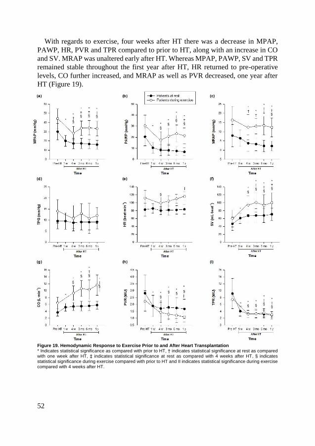
52
With regards to exercise, four weeks after HT there was a decrease in MPAP,
PAWP, HR, PVR and TPR compared to prior to HT, along with an increase in CO
and SV. MRAP was unaltered early after HT. Whereas MPAP, PAWP, SV and TPR
remained stable throughout the first year after HT, HR returned to pre-operative
levels, CO further increased, and MRAP as well as PVR decreased, one year after
HT (Figure 19).
Figure 19. Hemodynamic Response to Exercise Prior to and After Heart Transplantation * Indicates statistical significance as compared with prior to HT, † indicates statistical significance at rest as compared with one week after HT, ‡ indicates statistical significance at rest as compared with 4 weeks after HT, § indicates statistical significance during exercise compared with prior to HT and II indicates statistical significance during exercise compared with 4 weeks after HT.
52
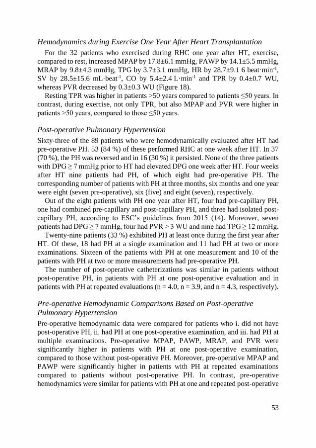
Hemodynamics during Exercise One Year After Heart Transplantation
For the 32 patients who exercised during RHC one year after HT, exercise,
compared to rest, increased MPAP by 17.8±6.1 mmHg, PAWP by 14.1±5.5 mmHg,
MRAP by 9.8±4.3 mmHg, TPG by 3.7±3.1 mmHg, HR by 28.7±9.1 6 beat·min-1,
SV by 28.5±15.6 mL·beat-1, CO by 5.4±2.4 L·min-1 and TPR by 0.4±0.7 WU,
whereas PVR decreased by 0.3±0.3 WU (Figure 18).
Resting TPR was higher in patients >50 years compared to patients ≤50 years. In
contrast, during exercise, not only TPR, but also MPAP and PVR were higher in
patients >50 years, compared to those ≤50 years.
Post-operative Pulmonary Hypertension
Sixty-three of the 89 patients who were hemodynamically evaluated after HT had
pre-operative PH. 53 (84 %) of these performed RHC at one week after HT. In 37
(70 %), the PH was reversed and in 16 (30 %) it persisted. None of the three patients
with DPG ≥ 7 mmHg prior to HT had elevated DPG one week after HT. Four weeks
after HT nine patients had PH, of which eight had pre-operative PH. The
corresponding number of patients with PH at three months, six months and one year
were eight (seven pre-operative), six (five) and eight (seven), respectively.
Out of the eight patients with PH one year after HT, four had pre-capillary PH,
one had combined pre-capillary and post-capillary PH, and three had isolated post-
capillary PH, according to ESC’s guidelines from 2015 (14). Moreover, seven
patients had DPG ≥ 7 mmHg, four had PVR > 3 WU and nine had TPG ≥ 12 mmHg.
Twenty-nine patients (33 %) exhibited PH at least once during the first year after
HT. Of these, 18 had PH at a single examination and 11 had PH at two or more
examinations. Sixteen of the patients with PH at one measurement and 10 of the
patients with PH at two or more measurements had pre-operative PH.
The number of post-operative catheterizations was similar in patients without
post-operative PH, in patients with PH at one post-operative evaluation and in
patients with PH at repeated evaluations (n = 4.0, n = 3.9, and n = 4.3, respectively).
Pre-operative Hemodynamic Comparisons Based on Post-operative
Pulmonary Hypertension
Pre-operative hemodynamic data were compared for patients who i. did not have
post-operative PH, ii. had PH at one post-operative examination, and iii. had PH at
multiple examinations. Pre-operative MPAP, PAWP, MRAP, and PVR were
significantly higher in patients with PH at one post-operative examination,
compared to those without post-operative PH. Moreover, pre-operative MPAP and
PAWP were significantly higher in patients with PH at repeated examinations
compared to patients without post-operative PH. In contrast, pre-operative
hemodynamics were similar for patients with PH at one and repeated post-operative
5353535353
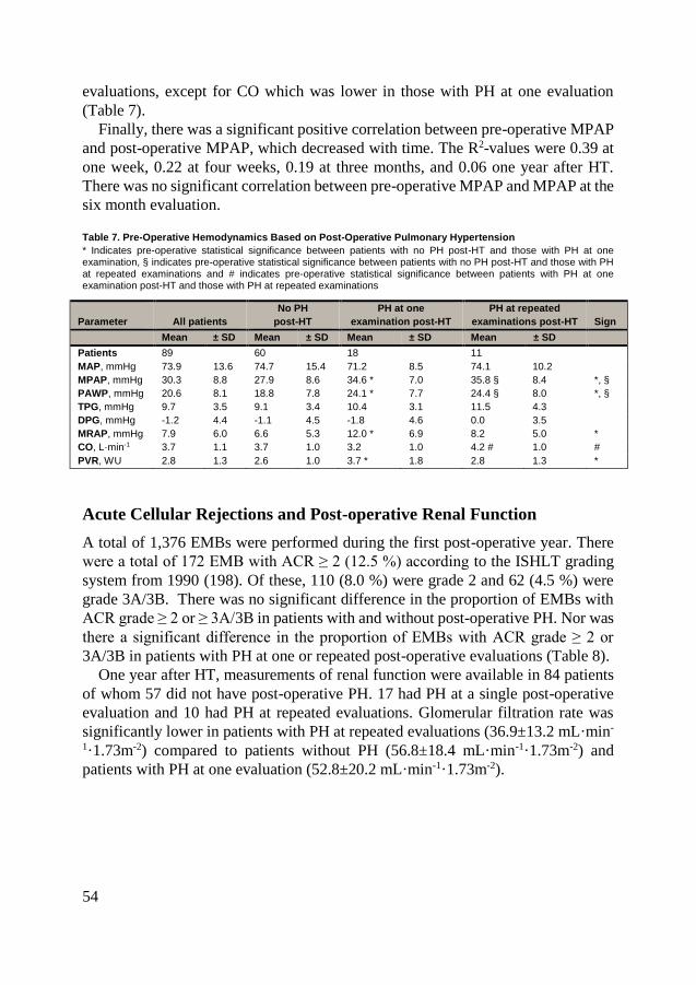
54
evaluations, except for CO which was lower in those with PH at one evaluation
(Table 7).
Finally, there was a significant positive correlation between pre-operative MPAP
and post-operative MPAP, which decreased with time. The R2-values were 0.39 at
one week, 0.22 at four weeks, 0.19 at three months, and 0.06 one year after HT.
There was no significant correlation between pre-operative MPAP and MPAP at the
six month evaluation.
Table 7. Pre-Operative Hemodynamics Based on Post-Operative Pulmonary Hypertension
* Indicates pre-operative statistical significance between patients with no PH post-HT and those with PH at one examination, § indicates pre-operative statistical significance between patients with no PH post-HT and those with PH at repeated examinations and # indicates pre-operative statistical significance between patients with PH at one examination post-HT and those with PH at repeated examinations
Parameter All patients
No PH
post-HT
PH at one
examination post-HT
PH at repeated
examinations post-HT Sign
Mean ± SD Mean ± SD Mean ± SD Mean ± SD
Patients 89 60 18 11
MAP, mmHg 73.9 13.6 74.7 15.4 71.2 8.5 74.1 10.2
MPAP, mmHg 30.3 8.8 27.9 8.6 34.6 * 7.0 35.8 § 8.4 *, §
PAWP, mmHg 20.6 8.1 18.8 7.8 24.1 * 7.7 24.4 § 8.0 *, §
TPG, mmHg 9.7 3.5 9.1 3.4 10.4 3.1 11.5 4.3
DPG, mmHg -1.2 4.4 -1.1 4.5 -1.8 4.6 0.0 3.5
MRAP, mmHg 7.9 6.0 6.6 5.3 12.0 * 6.9 8.2 5.0 *
CO, L·min-1 3.7 1.1 3.7 1.0 3.2 1.0 4.2 # 1.0 #
PVR, WU 2.8 1.3 2.6 1.0 3.7 * 1.8 2.8 1.3 *
Acute Cellular Rejections and Post-operative Renal Function
A total of 1,376 EMBs were performed during the first post-operative year. There
were a total of 172 EMB with ACR ≥ 2 (12.5 %) according to the ISHLT grading
system from 1990 (198). Of these, 110 (8.0 %) were grade 2 and 62 (4.5 %) were
grade 3A/3B. There was no significant difference in the proportion of EMBs with
ACR grade ≥ 2 or ≥ 3A/3B in patients with and without post-operative PH. Nor was
there a significant difference in the proportion of EMBs with ACR grade ≥ 2 or
3A/3B in patients with PH at one or repeated post-operative evaluations (Table 8).
One year after HT, measurements of renal function were available in 84 patients
of whom 57 did not have post-operative PH. 17 had PH at a single post-operative
evaluation and 10 had PH at repeated evaluations. Glomerular filtration rate was
significantly lower in patients with PH at repeated evaluations (36.9±13.2 mL·min-
1·1.73m-2) compared to patients without PH (56.8±18.4 mL·min-1·1.73m-2) and
patients with PH at one evaluation (52.8±20.2 mL·min-1·1.73m-2).
54
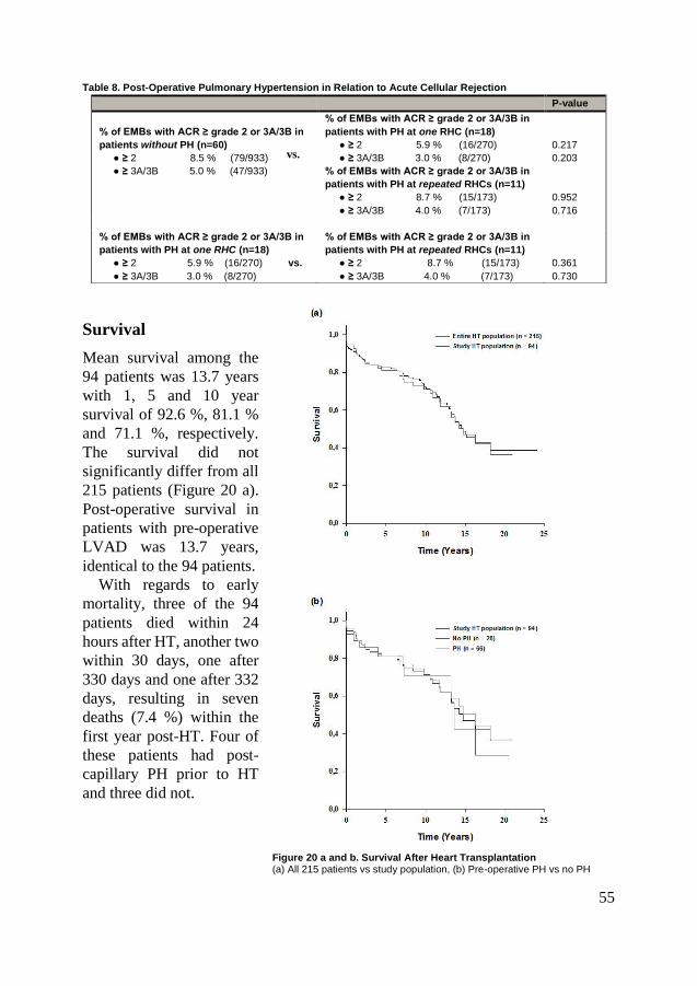
Table 8. Post-Operative Pulmonary Hypertension in Relation to Acute Cellular Rejection
Survival
Mean survival among the
94 patients was 13.7 years
with 1, 5 and 10 year
survival of 92.6 %, 81.1 %
and 71.1 %, respectively.
The survival did not
significantly differ from all
215 patients (Figure 20 a).
Post-operative survival in
patients with pre-operative
LVAD was 13.7 years,
identical to the 94 patients.
With regards to early
mortality, three of the 94
patients died within 24
hours after HT, another two
within 30 days, one after
330 days and one after 332
days, resulting in seven
deaths (7.4 %) within the
first year post-HT. Four of
these patients had post-
capillary PH prior to HT
and three did not.
Figure 20 a and b. Survival After Heart Transplantation (a) All 215 patients vs study population, (b) Pre-operative PH vs no PH
P-value
% of EMBs with ACR ≥ grade 2 or 3A/3B in
patients without PH (n=60)
● ≥ 2 8.5 % (79/933)
● ≥ 3A/3B 5.0 % (47/933)
% of EMBs with ACR ≥ grade 2 or 3A/3B in
patients with PH at one RHC (n=18)
● ≥ 2 5.9 % (16/270)
● ≥ 3A/3B 3.0 % (8/270)
% of EMBs with ACR ≥ grade 2 or 3A/3B in
patients with PH at repeated RHCs (n=11)
● ≥ 2 8.7 % (15/173)
● ≥ 3A/3B 4.0 % (7/173)
0.217
0.203
0.952
0.716
% of EMBs with ACR ≥ grade 2 or 3A/3B in
patients with PH at one RHC (n=18)
● ≥ 2 5.9 % (16/270) vs.
● ≥ 3A/3B 3.0 % (8/270)
% of EMBs with ACR ≥ grade 2 or 3A/3B in
patients with PH at repeated RHCs (n=11)
● ≥ 2 8.7 % (15/173)
● ≥ 3A/3B 4.0 % (7/173)
0.361
0.730
vs.
5555555555
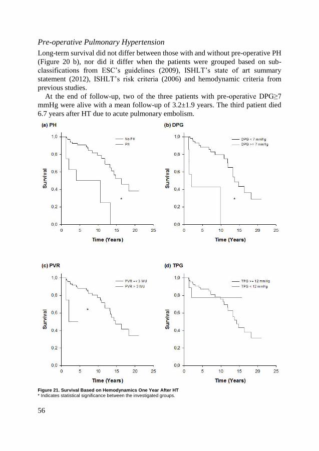
56
Pre-operative Pulmonary Hypertension
Long-term survival did not differ between those with and without pre-operative PH
(Figure 20 b), nor did it differ when the patients were grouped based on sub-
classifications from ESC’s guidelines (2009), ISHLT’s state of art summary
statement (2012), ISHLT’s risk criteria (2006) and hemodynamic criteria from
previous studies.
At the end of follow-up, two of the three patients with pre-operative DPG≥7
mmHg were alive with a mean follow-up of 3.2±1.9 years. The third patient died
6.7 years after HT due to acute pulmonary embolism.
Figure 21. Survival Based on Hemodynamics One Year After HT * Indicates statistical significance between the investigated groups.
56
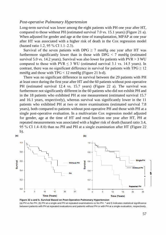
Post-operative Pulmonary Hypertension
Long-term survival was lower among the eight patients with PH one year after HT,
compared to those without PH (estimated survival 7.0 vs. 15.1 years) (Figure 21 a).
When adjusted for gender and age at the time of transplantation, MPAP at one year
after HT was associated with a higher risk of death in the Cox regression model
(hazard ratio 1.2, 95 % CI 1.1‒2.3).
Survival of the seven patients with DPG ≥ 7 mmHg one year after HT was
furthermore significantly lower than in those with DPG < 7 mmHg (estimated
survival 5.0 vs. 14.2 years). Survival was also lower for patients with PVR > 3 WU
compared to those with PVR ≤ 3 WU (estimated survival 3.1 vs. 14.5 years). In
contrast, there was no significant difference in survival for patients with TPG ≥ 12
mmHg and those with TPG < 12 mmHg (Figure 21 b-d).
There was no significant difference in survival between the 29 patients with PH
at least once during the first year after HT and the 60 patients without post-operative
PH (estimated survival 12.4 vs. 15.7 years) (Figure 22 a). The survival was
furthermore not significantly different in the 60 patients who did not exhibit PH and
in the 18 patients who exhibited PH at one measurement (estimated survival 15.7
and 16.1 years, respectively), whereas survival was significantly lower in the 11
patients who exhibited PH at two or more examinations (estimated survival 7.8
years), both compared to patients without post-operative PH and those with PH at a
single post-operative evaluation. In a multivariate Cox regression model adjusted
for gender, age at the time of HT and renal function one year after HT, PH at
repeated measurements was associated with a higher risk of death (hazard ratio 3.4,
95 % CI 1.4‒8.0) than no PH and PH at a single examination after HT (Figure 22
b).
Figure 22 a and b. Survival Based on Post-Operative Pulmonary Hypertension
(a) PH vs No PH, (b) PH at a single and PH at repeated examinations vs No PH. * and § Indicates statistical significance
between patients with PH at repeated evaluations and patients without PH or with PH at a single evaluation, respectively.
5757575757
58
Forty-six of the 60 patients (77 %) without post-operative PH, 13 of the 18
patients (72 %) with PH at one post-operative examination, and one of the 11
patients (9 %) with PH at repeated post-operative evaluations were alive at the end
of follow-up.
Cause of Death
At the end of follow-up, 34 patients had died. The most common cause of death was
rejection (n = 7), i.e. acute cellular and humoral rejections as well as coronary
allograft vasculopathy, followed by malignancy (n = 6), infection (n = 5) and
intracranial hemorrhage (n = 4). Other causes of death included, amongst others,
primary allograft dysfunction, acute coronary syndrome and pulmonary embolism.
Paper V
Pre-operative and Post-operative Characteristics
Two of the 12 patients were women and the median age was 50.0 years. The most
common diagnosis was dilated cardiomyopathy (n=8) followed by ischemic heart
disease (n=3). Three patients were ex-smokers and the remaining never smokers.
Prior to HT, MPAP was 35.0 (26.5-41.5) mmHg, PAWP 25.0 (19.5-29.8) mmHg,
MRAP 15.0 (11.5-18.5) mmHg, CO 3.4 (2.6-4.3) L∙min-1 and PVR 2.4 (1.9-3.0)
WU. Nine of the 12 patients had pre-operative PH. Of these, three had combined
pre-capillary and post-capillary PH and five had isolated post-capillary PH
according to ESC’s guidelines from 2015 (14).
Four weeks after HT there was an increase in CO to 6.0 (5.6-6.6) L∙min-1, as well
as a decrease in MPAP to 17.0 (11.5-20.5) mmHg, PAWP to 8.0 (4.5-10.5) mmHg
and PVR to 1.3 (1.1-1.5) WU, compared to prior to HT, remaining stable at the six
month evaluation. Six months after HT MRAP decreased to 1.5 (0.0-4.0) mmHg.
None of the patients had post-operative PH.
With regards to immunosuppression after HT, all patients were treated with a
triple combination of corticosteroids, a calcineurin inhibitor (10 Tacrolimus, 2
Cyclosporine) and mycophenolate mofetil/sodium. In one patient an mTOR
inhibitor was added prior to the six month evaluation due to renal dysfunction. Four
weeks and six months post-HT three and two patients, respectively, were diagnosed
with ACR grade 1R (according to 2005 system, corresponding to grade 1A/1B/2 in
the 1990 system). None needed specific rejection treatment.
58

Plasma Levels of Substances in the Nitric Oxide Pathway
Prior to HT, the plasma concentration of L-
Arginine was 56.0 (47.9-60.3) µM, ADMA
0.57 (0.47-0.71) µM and L-
Arginine/ADMA-ratio 85.7 (68.3-128.3).
L-Arginine was lower in patients than in
controls, whereas ADMA was higher,
resulting in a markedly lower L-
Arginine/ADMA-ratio in patients (Figure
23).
Four weeks after HT there was an
increase in the plasma concentration of; L-
Arginine to 82.5 (64.7-101.2) µM and L-
Arginine/ADMA-ratio to 164.0 (104.5-
188.7), compared to prior to HT. All
concentrations remained stable at the six
month evaluation. After HT, L-Arginine
was in line with healthy controls, whereas
L-Arginine/ADMA, although improved,
remained decreased. There was no change
in ADMA plasma levels after HT (Figure
23).
Correlations
In a univariate analysis six months after
HT, there was an inverse correlation
between PVR and the L-Arginine/ADMA-
ratio (R2: 0.39). There were no other
significant correlations, neither at the pre-
operative nor post-operative evaluations,
between the L-Arginine/ADMA-ratio and
the hemodynamic parameters, NT-
proBNP, creatinine or urea. Moreover,
neither the plasma levels of L-Arginine nor
ADMA correlated to PVR, nor to any of
the other investigated hemodynamic
parameters, after HT. Figure 23. Plasma Levels of L-Arginine and ADMA Prior to and After Heart Transplantation * Indicates statistical significance as compared to prior
to HT and § indicates statistical significance as
compared to controls.
5959595959
60
Limitations
The limitations of the papers in this thesis are addressed in detail below.
Paper I
Despite porcine and human hearts sharing a close hemodynamic resemblance (201),
there are interspecies variabilities in terms of the response to hypoxia and drugs,
including tezosentan, which have to be taken into account when interpreting the
results. Our findings can therefore not directly be applied in a human setting. Also,
in a hospital setting the most common cause of HPV is various chronic pulmonary
diseases including chronic obstructive pulmonary disease with emphysema, where
dilatation of the pulmonary vessels may be detrimental, causing deoxygenation of
the blood. Our findings in acute hypoxia therefore need confirmation in chronic
HPV.
Paper II-IV
The retrospective design of paper II-IV has disadvantages compared to prospective
studies. The long timespan and small number of patients in our cohort is furthermore
a major limitation which could potentially influence the results. When evaluating
the normal response to exercise in paper II it is also important to note that
hemodynamic evaluations in healthy controls were not performed. Instead the
results were compared to previously published systematic reviews on healthy
individuals. Although this may not be optimal, due to ethical issues of performing
heart catheterizations on healthy persons, we find our approach to be a valid
alternative. The studies did furthermore only include patients who underwent HT.
Pre-operative hemodynamic data may consequently not be applicable for patients
with severe heart failure who do not undergo HT. The response to exercise prior to
and after HT in paper II was, however, comparable to what has previously been
described in heart failure (147). With regard to hemodynamics we therefore believe
that the present observations are representative for patients with severe heart failure.
When investigating the impact of pre-operative PH on post-operative survival
there is also a risk of selection bias as patients with clinical signs of severe
pulmonary vascular disease may have been excluded from HT, irrespective of
hemodynamics. In paper III we did furthermore not adjust survival data for potential
confounders, which could influence the outcome. In paper IV we did not have the
possibility to histologically examine the pulmonary vessels of the patients who died,
so it cannot be determined whether repeated PH represented pulmonary vascular
disease.
60
However, demographics and survival were similar for the 94 included patients
and all 215 HT patients. Survival was furthermore constant during the entire period
of transplantations and hemodynamic measurements were all performed routinely
and in the same manner, mostly by the same physicians, minimizing the risk of
investigator bias despite the long study period. In summary we believe the findings
may be applicable on a larger HT population and are of interest, primarily to
generate hypotheses for future trials. However, they must be interpreted cautiously
and confirmation in larger prospective and controlled trials is needed.
Paper V
Although the small sample size was in line with previous pilot studies in HT, which
has stimulated the initiation of larger trials, the study size is a major limitation.
Furthermore, we did not measure NO and cGMP levels so we cannot be certain that
their production was decreased prior to HT. However, the decreased L-
Arginine/ADMA-ratio throughout our study, in combination with the fact that the
ratio reflects pulmonary vascular tone, points in the direction of NO-dependent
endothelial dysfunction prior to and after HT. Finally, medical therapies prior to and
after HT were individually adjusted. Betablockers (202) and RAAS-blockade (203)
have been shown to decrease circulating ADMA levels. These drugs are less
frequently used after HT which, in theory, could increase plasma levels of ADMA.
The use of these drugs does, however, represent a cornerstone in heart failure
therapy and a true clinical situation, so our findings are relevant to generate
hypotheses for future, larger, trials.
6161616161

62
Discussion
Paper I
sGC stimulation with BAY 41-8543 totally reversed HPV during acute hypoxia.
Dual endothelin receptor blockade with tezosentan furthermore markedly attenuated
acute HPV. When the sGC stimulator was administered on top of dual endothelin
receptor blockade, pulmonary pressures and resistances were even further
decreased. Blood arterial O2-saturation and O2-consumption remained unaltered
throughout the experiments, despite a marked systemic vasodilatation.
Response to Hypoxia
In our in vivo pig model there was a marked and stable increase in pulmonary artery
pressures and resistances during sustained hypoxia for 82.5 min. These findings are
similar to what has previously been described when various species have been
exposed to severe hypoxia (176) and highlights the profound HPV in our study.
Interestingly, during induction of acute hypoxia, left ventricular filling pressure
increased and was then unaltered throughout all experiments, suggesting that the
increase was not solely a result of ventricular interdependence due to increased right
heart load. Marked increases in left ventricular filling pressures upon exposure to
acute hypoxia have not been observed in humans, whereas the capillary pressures
increase and may contribute to high altitude pulmonary edema (204). On the other
hand, patients with previous high altitude pulmonary edema have been shown to
have high left ventricular filling pressures during exercise (205) and hypoxia has
indeed been shown to cause temporary diastolic dysfunction, even in young,
healthy, individuals (206). It consequently seems that the increase in left ventricular
filling pressure observed in our study in parts may be species specific and possibly
related to diastolic dysfunction during hypoxia.
62

Soluble Guanylate Cyclase Stimulation and Dual Endothelin Receptor
Blockade Totally Reverses Acute Hypoxic Pulmonary Vasoconstriction
Stimulation of sGC, by intravenous infusion of BAY 41-8543, was found to totally
reverse the pulmonary vasoconstrictor response to acute hypoxia and reduce the
load on the right side of the heart, thereby protecting against right ventricular failure.
If sustained, such benefits could potentially also prevent right ventricular failure in
the chronic setting. Whether sGC stimulation could prohibit hypoxia induced
pulmonary edema is unknown and needs further investigation. Considering the
systemic effects of sGC stimulators, also observed in several human studies
(99;101), it is however possible that the load of the left ventricle, and consequently
pulmonary capillaries, may decrease with such therapies.
Treatment with endothelin receptor antagonists have proven beneficial in PAH
(79-81). However, in PAH dual endothelin receptor blockade is seldom sufficient
to fully normalize the increased pulmonary vascular tone (14). In our study it was
therefore of importance to evaluate whether combination treatment, targeting
several pathways, could be tolerated and of additional value. Dual ET-receptor
blockade with tezosentan indeed effectively attenuated acute HPV. When adding
sGC stimulation, pulmonary pressures and resistances further decreased to values
as low as, or even lower than, those in normoxia. In recent years several studies on
PAH have shown beneficial effects of combination therapy, as compared to mono
therapy (81;82;105) and based on our findings such a combination treatment seems
feasible and effective also in reversing acute hypoxia-induced PH. The pronounced
reduction in systemic pressures observed in the present study may, however, be
harmful in critically ill patients. Therefore, if treating patients with the present
drugs, doses should be carefully titrated and systemic blood pressure closely
monitored. It should, however, be noted that the decrease in systemic blood pressure
with tezosentan in the present study has not been observed in humans (207) and is
likely species-dependent.
As endothelin receptor blockers as well as sGC stimulators may, besides their
vasodilating effects, also attenuate pulmonary vascular remodeling (208-210), our
findings are of importance in the development of new treatment strategies for PH of
various etiology, including PH-LHD and PH due to hypoxia. A few reports in
patients with chronic lung disease, including chronic obstructive pulmonary disease,
and fibrotic lung disease have, however, suggested otherwise (186-190). Although
therapies targeting the nitric oxide and endothelin pathways may improve
hemodynamics and decrease right ventricular load, they may also counter the
beneficial effects of HPV in focal hypoxia, thereby impairing ventilation-perfusion
matching and arterial oxygenation. Several negative studies have also been reported
with drugs targeting these pathways in left heart disease (162-168), perhaps due to
increased load on the left ventricle when dilating the pulmonary vessels. In contrast
to the findings in left heart disease and chronic pulmonary disease, sGC stimulation
6363636363
64
with the novel drug riociguat have shown beneficial effects in PAH and CTEPH
(99-101;211) and is now used for treatment of these conditions. Trials with riociguat
in systolic and diastolic left heart failure were also recently conducted. Riociguat
did not decrease pulmonary artery pressures in either of the trials (167;168) but
improved cardiac index and quality of life in systolic heart failure (167) and some
hemodynamic parameters in diastolic heart failure (168), indicating potential
beneficial effects in left heart disease. Therefore, the new sGC stimulator vericiguat
is currently being tested in a phase III trial on systolic left heart failure
(ClinicalTrials.gov Identifier: NCT02861534) after having shown to improve NT-
proBNP (169). The same drug has also shown to improve quality of life in diastolic
heart failure (170). At present, PAH targeted therapies are, however, not
recommended in PH-LHD or lung disease (14).
When the present PhD project was initiated we planned for further studies with
sGC stimulators in humans with PH-LHD. These plans were, however, abandoned
when the above trials were initiated.
Paper II
Hemodynamic parameters during rest and slight supine exercise were characterized
in patients undergoing HT. Hemodynamics markedly improved at rest and during
exercise one and four weeks after HT, respectively. All parameters remained
constant or improved even further throughout the first post-operative year. The post-
HT functional response to exercise, with regards to cardiac output, was also
adequate. Despite this, a profound increase in filling pressures was observed during
exercise after HT. So, although the hemodynamics at rest were normalized after HT,
the response to even slight exercise differed compared with the normal innervated
heart.
Pre-operative Hemodynamics
Prior to HT, exercise resulted in a marked increase in both left and right ventricular
filling pressures, indicating enhanced biventricular failure during exercise. Previous
reports in healthy volunteers found a close relationship between exercise-induced
increases in left and right ventricular filling pressures (150;151). Such a correlation
was not observed prior to HT, suggesting that the increased load on the right
ventricle is not solely due to increased left ventricular filling pressures, passive
congestion of the pulmonary circuit and subsequent post-capillary PH, but also due
to superimposed pulmonary artery pressures. The exaggerated increase in
pulmonary pressure could be due to several factors, including HPV (29) related to
64

low mixed venous PO2. Mixed venous PO2 was indeed markedly decreased during
exercise in the patients where these data were available.
With regards to exercise, the definition of exercise PH has been removed from
ESC’s guidelines on PH due to lack of robust data (14). However, the European
Respiratory Society have published a statement suggesting that exercise PH should,
at peak exercise, be defined as MPAP > 30 mmHg in combination with TPR > 3
WU, in absence of resting MPAP ≥ 25 mmHg (157). Recently it was also suggested
that reference values for exercise should be individually determined, as the response
to exercise was markedly altered by age (156). In contrast to previous findings in
healthy individuals, (144;155;156) the hemodynamics in severe heart failure did not
significantly differ between patients aged ≤ 50 and > 50 years during rest or
exercise. In end-stage heart failure, age does consequently not seem to play a
significant role in hemodynamic response to exercise. This is of importance as
exercise testing may be beneficial in early detection of heart failure (148) and PH
(149), as also suggested by our data. Exercise testing may furthermore aid in the
differentiation between PAH and PH-LHD, especially when the latter is caused by
diastolic dysfunction.
Post-operative Hemodynamics
Hemodynamics at rest after HT have been reported in several publications (15-
18;32-34;194;196;212-217). In line with these reports, our study shows a dramatic
hemodynamic improvement within the first week after HT, remaining stable or even
further improving throughout the first year following transplantation. In contrast to
the large number of studies on resting hemodynamics, publications on invasive
exercise hemodynamics after HT are scarce (29-35). In a few small reports it has
however been shown that MPAP and PAWP decrease early after HT (29) and that
during exercise in denervated hearts, CO is initially increased by augmented preload
and the Frank Starling mechanism and later by the effects of circulating
catecholamines (31). These reports have, however, lacked longitudinal follow-up of
patients. Our results therefore improve the understanding of post-operative
hemodynamic development over time.
During exercise after HT pulmonary artery pressure and, in particular, bilateral
ventricular filling pressures increased dramatically, despite normal values at rest.
The increase was more pronounced than what would be expected in healthy
individuals at the corresponding workload (144;155;156) and has been described
previously (33;35). Several reasons for these elevated filling pressures have been
suggested (15;16;31;33-37;218), including a prominent decrease in diastolic
compliance in denervated hearts (218). Denervated hearts may also be more
dependent on the Frank Starling mechanism than innervated hearts (31;36;37). The
ultimate cause remains unclear and is probably multifactorial (33).
6565656565
66
The HR in our study was, as expected due to denervation, slightly higher at rest
than what would be expected in healthy individuals (156). During slight exercise,
an increase to values as high as, or even slightly higher than, what has previously
been described in health, was furthermore observed (144;155;156). As the increase
in HR during slight exercise was similar to that of healthy persons, the chronotropic
insufficiency described after HT (35) primarily seem to play a part in high intensity
exercise.
Due to the increase in left ventricular filling pressures and subsequent increase in
pulmonary pressures induced by exercise after HT, the post-operative response in
PVR was comparable to what has been presented in healthy individuals (144;155).
On the other hand, the response in TPR to exercise was still deranged at one year
after HT. These findings suggest that TPR may give more adequate insight in the
pulmonary circulation during exercise than PVR, as it reflects the increase in MPAP
in relation to CO, independently of left ventricular filling pressures. Our findings
consequently support the recent statement from the European Respiratory Society,
suggesting that TPR should be used to define exercise PH (157).
Paper III
After HT no significant differences in survival was observed based on
hemodynamic criteria used in previous publications, including ESC’s guidelines
from 2009 (14) and ISHLT’s documents (7;11). Even though we consider severe
pre-operative PH to be a risk factor for right heart failure after HT, previous
hemodynamic criteria for PH seem insufficient to discriminate the impact of
pulmonary hemodynamics on survival after HT. Recent reports have furthermore
presented conflicting results on post-HT survival with regard to present ESC
guidelines (14) and DPG (136-138). Consequently, new risk criteria including
invasive hemodynamics as well as novel biomarkers and imaging techniques would
be of great value.
Survival
Although it is well established that severe pre-operative PH with vascular
remodeling increases the risk of acute right heart failure after HT, previous studies
have used different hemodynamic parameters and thresholds when evaluating
outcome after HT (7;9-11;17;18;32;125;194-197). Correct comparisons and
predictions on survival are therefore difficult to make. Moreover, as survival during
the first year after HT steadily rise (6), it is a reasonable assumption that today’s
post-operative care has greatly improved as compared to the care given in the early
HT era. Therefore, early hemodynamic reports on outcome after HT may be
66

outdated, making it even harder to define robust hemodynamic criteria for acute
right heart failure and early mortality after HT. The overall survival after HT was
excellent in our cohort, despite most of the patients having some degree of pre-
operative PH. The outcome of patients with PH was furthermore comparable to the
patients without pre-operative PH, which may be attributed to the pre-, intra- and
post-operative treatment at our thoracic intensive care unit, focusing on preventing
fluid accumulation by aggressive diuretic therapy, still maintaining adequate
perfusion pressure. Another possible explanation is that the patients in our cohort
was likely to have PH caused by passive congestion and excessive vasoconstriction,
rather than severe vascular remodeling. Our findings on post-operative outcome are
in line with some recent reports (6;17;18;137;195;197;219;220), whereas they are
in contrast to others (9;10;136;138;194). The diverging results indeed highlight the
difficulty in identifying a high risk population. Which hemodynamic parameters that
best predict outcome after HT and the precise magnitude at which they do so
consequently remains unclear.
Diastolic Pressure Gradient
DPG ≥ 7 mmHg has been suggested to be a marker of severe pulmonary vascular
disease and worse prognosis in PH-LHD (120). DPG was therefore introduced in
the most recent ESC guidelines (14). As indicated previously there are, however,
conflicting results on the clinical implications of DPG. Whereas some reports
indeed have shown that DPG is a prognostic factor (120;127-134), others have not
(12;135-141). In our material DPG ≥ 7 mmHg was only observed in three patients
prior to HT. None of these patients had elevated DPG or acute right heart failure
after HT and their survival was not significantly decreased. Diastolic pulmonary
artery pressure has shown to be less affected by changes in flow and left ventricular
filling pressures than MPAP (126), in theory making DPG an attractive marker in
left heart disease. However, DPG is to a greater extent affected by HR, which has
been presented as one of its major weaknesses (221;222). In patients with elevated
DPG prior to HT, the HR was indeed higher than in the overall cohort. It is
consequently likely that the high DPG in these patients was not due to pulmonary
vascular disease, which could explain why DPG was normalized and outcome was
not affected after HT. Finally, another complicating factor with DPG is the
measurement of PAWP. Whereas diastolic pulmonary artery pressure is measured
at end-diastole, measurements of PAWP vary potentially resulting in overestimation
of PAWP and misclassification of patients. This was recently illustrated in a report
where QRS-gated identified more patients with high DPG (12). In summary, there
is a sound physiological rationale for DPG in sub-defining PH due to left heart
failure, but there are several pitfalls which are not yet overcome. Consequently, the
role of DPG is still debated and needs further evaluation.
6767676767
68
With the controversies concerning DPG and PVR, another hemodynamic marker,
namely the pulmonary arterial compliance, has recently been highlighted in the
literature. In comparison to PVR, pulmonary arterial compliance accounts for the
pulsatility of blood flow and is a sensitive marker for right ventricular afterload.
Pulmonary arterial compliance was not investigated in our material but has indeed
been shown to be an independent predictor of outcome in left heart failure with or
without PH (135;141;223;224) and may consequently be implemented in future
clinical practice.
Bridge-to-Transplantation
Our study confirms that mechanical assist prior to HT improved hemodynamics and
normalized MPAP as well as PAWP. Moreover, none of the 21 patients who
received an assist died the first year after HT, and their mean survival did not differ
from the rest of the population. These findings suggest that LVAD therapy improve
hemodynamics as well as survival, to levels similar to the general HT-population.
These observations were confirmed in a recent publication where long-term LVAD
use resulted in lowering of PVR which had previously been irreversibly elevated.
The patients in this study subsequently underwent HT with outcome as good as other
HT patients (49). Consequently, patients should not be disqualified from HT based
purely on pre-LVAD PH. Of note, an ongoing trial is investigating the effects of the
dual endothelin receptor antagonist macitentan in patients with persistent PH and
PVR > 3 WU after LVAD implantation (ClinicalTrials.gov Identifier:
NCT02554903). A previous study has furthermore shown that PDE5i may reverse
PH that persists after LVAD implantation (51). It is therefore possible that PAH-
therapies may be of value to optimize pulmonary vascular tone when the left
ventricle is mechanically unloaded.
Paper IV
Patients with PH, DPG ≥ 7mmHg or PVR > 3 WU one year after HT had worse
long-term survival than patients without PH and those with low DPG or PVR,
respectively. TPG at the commonly used cut-off at 12 mmHg did, however, not
affect outcome. Of further interest, PH at repeated evaluations during the first post-
operative year was associated with impaired long term survival as compared to both
patients without PH and patients with PH at one post-operative examination,
whereas outcome did not significantly differ for the latter two groups.
68

Survival
As compared to the large number of studies investing the impact of pre-operative
PH on outcome after HT, there has previously been scarce knowledge on the role of
post-operative PH on survival. The few reports previously published have not used
the common definition of PH (MPAP ≥ 25 mmHg) (14). Therefore, although they
showed that high MPAP and PVR after HT was associated with poor prognosis, the
clinical implications of the findings have remained uncertain (17;18). Despite using
a different definition (14), our findings were in line with the previous reports (17;18)
emphasizing that abnormal pulmonary hemodynamics, including PH, one year after
HT is associated with reduced long-term survival. For the first time we also reported
post-operative survival based on results from repeated hemodynamic evaluations
during the first year after HT. The findings suggest that PH at repeated
examinations, but not at a single evaluation, is associated with impaired outcome,
which have several potential explanations. One may be that hemodynamics can be
temporarily affected by factors such as HPV, post-operative stress and therapies that
may influence pulmonary vascular tone. Multiple assessments may therefore
potentially decrease the risk of such transient confounders interfering with the
clinical evaluations, thereby identifying patients with “true” pulmonary vascular
disease. Performing repeated hemodynamic evaluations to sub-define post-capillary
PH has, in fact, recently been suggested after a report where DPG was found not to
affect survival (138;225).
As the pulmonary artery pressures were only moderately increased in patients
with PH at repeated evaluations and the causes of death were not primarily
cardiovascular, it is however unlikely that patients with repeated PH had severe
pulmonary vascular disease. Instead it is probable that repeated PH was attributed
to an overall worse condition. Nonetheless repeated PH was an independent marker
of death when adjusted for age and gender as well as renal function one year after
HT. ACR did furthermore not differ between the groups. Consequently, our findings
warrant further investigations to define the actual cause of repeated PH.
Interestingly, when investigating pre-operative hemodynamics based on whether
patients had PH at one or repeated post-operative evaluations, no marked differences
were identified. The lack of pre-operative differences indeed highlight the
importance of close hemodynamic monitoring before and after HT to, if possible,
identify a high-risk population at an earlier stage. To determine whether this
population may benefit from vasoactive therapies, randomized controlled trials are
urged for (73;106).
Finally, patients with DPG ≥ 7mmHg or PVR > 3 WU one year after HT had
worse long-term survival than patients with low DPG and PVR, respectively.
However, the groups were small and not all patients with high DPG or PVR had PH.
Consequently, definite conclusions on the role of these parameters on outcome after
HT cannot be made based on our findings. As described previously, conflicting
6969696969

70
results on the impact of pre-operative DPG and PVR on outcome after HT have been
presented in recent years. Due to the conflicting results, both DPG and PVR need
further investigation, not only prior to HT but also thereafter.
Paper V
Plasma concentrations of substances related to the NO pathway were investigated
prior to and after HT. Compared to controls, L-Arginine was low and ADMA high
prior to HT, resulting in a low L-Arginine/ADMA-ratio. Although improved, the L-
Arginine/ADMA-ratio was not normalized after HT. The inverse correlation to PVR
furthermore suggest that the L-Arginine/ADMA-ratio reflects pulmonary vascular
tone after HT and that NO-dependent endothelial function is still partly impaired.
As all patients were free of post-operative PH, as well as ACR equal to or higher
than grade 1R and had normal left heart function at all post-operative evaluations
included, the findings may represent the normal plasma concentrations of these
substances after HT.
Pre-operative Concentrations
In severe heart failure, L-Arginine was considerably lower than in controls. The
levels also seemed to be lower than in patients with mild to moderate heart failure,
recently reported by our group (85), possibly representing the progressive
endothelial dysfunction and congestion of the pulmonary circuit with worsening
heart failure. Similar results have been observed in other studies, where ADMA has
been shown to correlate to NYHA functional class (92;95;226) and pulmonary
pressures (227). Prior to HT, the plasma levels of the investigated substances were
in fact in line with those of patients with PAH, in our previous report (85). There
was also a non-significant trend towards worse states in our patients with, compared
to without, PH. Further investigation is therefore encouraged to define whether or
not there is a true difference between heart failure patients with and without PH and
whether the concentrations represent pulmonary endothelial dysfunction.
Post-operative Concentrations
The L-Arginine/ADMA-ratio has been suggested to better reflect NO production
and endothelial function, than L-Arginine and ADMA separately (228). The L-
Arginine/ADMA-ratio has furthermore shown to be associated with disease severity
(226), as well as mortality (229), in heart failure. Our results with improved yet not
normalized L-Arginine/ADMA-ratio after HT where the ratio may also reflect
70
pulmonary vascular tone, as illustrated by its inverse correlation to PVR, suggest
that NO-dependent endothelial function is still partly impaired early after HT. If
using therapies targeting the NO-pathway prior to or after HT it consequently seems
reasonable to use drugs which act independent of NO, such as sGC stimulators,
rather than NO-dependent substances.
Arginase catalyzes the reaction where L-Arginine is converted to urea and L-
Ornithine. Arginase has shown to be elevated in heart failure (230) and increases
during oxidative stress and infections (231). Although L-Arginine increased after
HT, L-Ornithine remained unaltered. Our findings do consequently not support that
major alterations in arginase activity is the reason for increased L-Arginine
concentrations following HT. L-Citrulline, produced together with NO, was also
unaltered by HT. As NO and cyclic guanosine monophosphate-levels were not
measured, we cannot be certain that NO production increased after transplantation.
Most of the circulating L-Citrulline is, however, released from the intestines rather
than from the endothelium (232). Therefore, an increased release of L-Citrulline
from the endothelium may not be sufficient to significantly alter plasma
concentrations of L-Citrulline. Unaltered plasma L-Citrulline levels does therefore
not preclude increased NO production. Moreover, after HT CO increases, possibly
reducing inflammation (233). With reduced inflammation it is reasonable to assume
that the vascular sensitivity for NO is increased, leading to partly restored NO-
mediated vasodilatation, irrespective of whether or not NO production is increased.
7171717171

72
Conclusions
The present thesis includes a pig model of acute HPV, a retrospective review of
heart transplanted patients and an analysis of plasma concentrations of substances
related to the nitric oxide pathway in a prospective heart transplantation cohort. The
following conclusions were drawn:
• sGC stimulation alone or in combination with dual endothelin receptor
blockade, in an in vivo porcine model, dose-dependently attenuated acute
HPV illustrated by normalization of MPAP and PVR, thereby reducing
MRAP and consequently right ventricular afterload. There was a
simultaneous systemic vasodilatation and reduction in MAP which,
however, did not alter blood arterial O2-saturation or O2-consumption.
Together, the findings suggest that sGC stimulation alone, or in
combination with endothelin receptor blockade, may be used to prevent
acute right heart failure in acute hypoxia.
• Hemodynamics during rest and slight exercise rapidly recovered after HT.
The improvement in relevant hemodynamic parameters was maintained, or
even further improved, throughout the first year after transplantation.
Despite this improvement and despite an adequate functional response to
exercise after HT, as illustrated by an increase in CO, there was a marked
increase in ventricular filling pressures after HT, highlighted by elevated
PAWP and MRAP as well as high MPAP. Such a pronounced increase has
not previously been observed in healthy individuals, nor in patients with
severe heart failure prior to HT. The alterations in exercise hemodynamics
may partly be attributed to the fact that transplanted hearts lack innervation,
being more dependent on the Frank Starling mechanism to increase CO, as
compared to innervated hearts. However, the complete reason is probably
multifactorial and a decreased diastolic compliance is also likely to play a
part. If this is the case, the increased ventricular filling pressures may be the
first sign of a post-operative diastolic dysfunction. Finally, age-dependent
differences in the hemodynamic response to exercise have previously been
described in healthy individuals. In end-stage heart failure, age did not
significantly affect this response, an important finding as exercise criteria
for PH may improve early diagnosis and outcome. Yet, such classifications
72
are currently lacking, partly due to previously described age-dependent
alterations.
• Post-operative survival was not significantly influenced by pre-operative
hemodynamic status, probably related to careful patient selection, as well
as modern pre-, intra- and post-operative care. Excessive pulmonary
vascular remodeling prior to HT may, however, still affect long-term
outcome. Although patients with clinical signs of severe pulmonary
vascular disease may have been excluded from HT, influencing the results,
the findings highlight that previous and present hemodynamic definitions
are insufficient to identify patients with vascular remodeling. The findings
also show that LVAD is a feasible option for improving hemodynamics and
bridging patients to HT. Consequently, PH per se, prior to mechanical
unloading, is not an absolute contraindication for HT. Instead, with careful
treatment of these patient, the potential complications caused by PH can, to
a certain extent, be overcome and these patients transplanted without
increased risk of early mortality.
• Patients with PH at one year after HT had impaired long-term survival, as
compared to those without PH. Also, PH at repeated evaluations the first
year after HT was associated with worse outcome than PH at a single
examination. The PH in patients with PH at repeated measurements was
moderate and is likely to be a result of an overall worse condition, rather
than pulmonary vascular disease. Early and repeated hemodynamic
measurements after HT may nonetheless be useful to identify patients with
potentially impaired survival.
• Plasma concentrations of L-Arginine were low and ADMA concentrations
high in severe heart failure, as compared to in healthy individuals. Whereas
ADMA was unaltered by HT, L-Arginine rapidly returned to normal levels
thereafter. Consequently, the L-Arginine/ADMA-ratio was improved, but
not normalized and furthermore inversely correlated to PVR after HT. The
findings thereby suggest that the L-Arginine/ADMA-ratio reflects post-
operative pulmonary vascular tone and that NO-dependent endothelial
vasodilation was improved although not normalized after HT. Whether this
has implications on long-term outcomes remains to be investigated. Based
on these findings, if targeting the NO-pathway after HT, NO independent
substances such as sGC stimulators may be more appealing than NO-
dependent vasodilators.
7373737373

74
Future Perspectives
The incidence of heart failure is increasing and consequently, so is PH-LHD. Some
patients with PH-LHD develop pulmonary vascular remodeling associated with
impaired survival (120). In an attempt to improve diagnosis, ESC’s guidelines were
in 2015 updated with a new hemodynamic sub-classification of PH-LHD, where
DPG was introduced (14). Subsequent retrospective studies on the prognostic value
of DPG have, however, been conflicting and based on discussions at the Sixth World
Symposium on PH it is likely that the definition again will be revised. The frequent
revisions of the guidelines illustrate the complexity of the disease and difficulties in
sub-defining post-capillary PH with present methods. Studies on invasive and non-
invasive methods are needed to increase the understanding of disease progression
in PH-LHD. A key aspect in this matter is the role of acute vasodilatation tests and
exercise during invasive hemodynamic measurements. Inclusion of such variables
in the guidelines may improve diagnosis and sub-classification of post-capillary PH,
but studies on these aspects are lacking. Structured trials are therefore needed, in
healthy individuals as well as in patients with heart failure, to increase the
understanding of hemodynamics. A project on the influence of age on hemodynamic
parameters at rest and during supine exercise was recently carried out in healthy
individuals (156), stimulating further research. Hemodynamic monitoring is,
however, unlikely to be sufficient to identify all patients with vascular remodeling.
A structured work-up, also including non-invasive imaging and biomarkers, may
improve adequate diagnosis. This needs to be investigated and relevant biomarkers
identified.
Despite more than half a century has passed since the first HT, understanding of
physiologic alterations after HT remain poor. Our results show a dramatic increase
in filling pressures during exercise the first year after HT, but little is known of the
long-term implications of these findings, as well as of the hemodynamic response
to exercise late after HT. Whereas survival have steadily improved early after HT,
survival beyond the first year has remained relatively constant (6). This highlights
the importance of further research to increase the understanding of endothelial
function as well as of long-term complications such as coronary allograft
vasculopathy and diastolic dysfunction after HT. A key aspect is circulating
biomarkers of endothelial function. We are currently investigating blood borne
markers of inflammation and angiogenesis after HT but prospective multi-center
74

studies are needed to identify potential targets for medical therapies, which may
slow the development of complications such as diastolic dysfunction, diabetes
mellitus and renal impairment, thereby improving long-term survival.
For the time being, HT remains the ultimate treatment for end stage heart failure
and specific medical therapies are lacking for the pulmonary component of PH-
LHD. Several trials with medical therapies used to treat PAH, targeting the NO,
endothelin and prostacyclin pathways, have been negative in left heart disease with
or without PH (162-168;172), as well as in PH due to lung diseases (187-190),
including chronic HPV. However, acute HPV may aggravate PH-LHD and in this
setting knowledge on novel medical therapies is scarce. Interestingly, in contrast to
previously negative findings the novel sGC stimulator vericiguat was, in a recent
phase II trial on systolic heart failure, shown to reduce NT-proBNP (169) and a
phase III trial is currently ongoing (ClinicalTrials.gov Identifier: NCT02861534).
However, as hypotension often is a limiting factor in the treatment of heart failure,
it remains to be seen whether this therapeutical approach has beneficial effects as
compared to standard heart failure medications. It is instead plausible that the future
in treatment of severe left heart disease, including PH-LHD, may lay in medical
devices. In the present thesis the hemodynamic improvement with long-term use of
LVADs as bridge to HT has been highlighted and it is clear that their long-term use
may reverse pulmonary vascular changes. The first generations of these devices
have been associated with a high rate of complications. There is, however, a steady
technical improvement and in the two-year follow-up of the MOMENTUM 3 trial,
Heartmate III was associated with a 19% absolute risk reduction in the composite
primary end-point of survival free of disabling stroke or reoperation, as compared
to Heartmate II (61). With further improvement and fully implantable devices,
without percutaneous drive-lines, future generations of LVADs may be a long-term
treatment option for patients with PH-LHD.
7575757575
76
Acknowledgement
Associate professor Göran Rådegran, my very enthusiastic supervisor, for
providing the opportunity to do my PhD, for constantly putting me forward and for
always being available. Thank you!
Dr Lars Algotsson, my brilliant assistant supervisor, for invaluable clinical input
and thoughtful scientific comments throughout my PhD project.
A special thanks to my fellow PhD students Anna Werther-Evaldsson, Annika
Ingvarsson, Habib Bouzina, Eveline Löfdahl and, in particular, David
Kylhammar and Carl Söderlund for collaboration, assistance and support, as well
as for great friendship outside the numerous scientific sessions we have attended.
Senior colleagues Drs. Björn Kornhall, Öyvind Reitan, Johan Holm, Carl-
Johan Höijer, Carl Meurling, Oscar Braun, Grunde Gjesdal and Gustav Smith
at the Clinic for Heart Failure and Valvular Disease. Thanks for always taking your
time to share your expertise in severe heart failure and pulmonary hypertension as
well as for providing a stimulating scientific environment. Your inclusive and
genuine response has allowed my interest in the field to increase even further.
Anneli Ahlqvist for your very important work with LCPR.
Drs. Pontus Andell, Christian Reitan, Josef Dankiewicz, Moman A.
Mohammad, Kristina Torngren and Sofia Karlsson for being great friends as
well as colleagues. I am very lucky to have started my career among so many
talented young clinicians and researchers. To many more laughs, late nights and
early mornings.
Lena Lindén and Monica Magnusson for making the impossible possible and for
all the happy moments around the breakfast table.
Dr. Patrik Gilje, my clinical supervisor, for interesting discussions and careful,
pragmatic and clever guidance through the world of clinical cardiology.
My parents and siblings Anneli Olsson Lundgren, Anders Lundgren, Paula
Lundgren, Simon Lundgren and Nils Lundgren for always being supportive,
showing interest in my work and providing me with an intellectually stimulating
environment. Thanks also to my younger sisters Clara and Nora Lundgren for
bringing laughs and playfulness.
Finally, to my beloved wife Sigrid for everlasting support and love.
76
References
(1) Barnard CN. The operation. A human cardiac transplant: an interim report of a
successful operation performed at Groote Schuur Hospital, Cape Town. S Afr
Med J 1967;41(48):1271-4.
(2) DiBardino DJ. The history and development of cardiac transplantation. Tex Heart
Inst J 1999;26(3):198-205.
(3) Caves PK, Stinson EB, Billingham M, Shumway NE. Percutaneous transvenous
endomyocardial biopsy in human heart recipients. Experience with a new
technique. Ann Thorac Surg 1973;16(4):325-36.
(4) Borel JF. Comparative study of in vitro and in vivo drug effects on cell-mediated
cytotoxicity. Immunology 1976;31(4):631-41.
(5) Soderlund C, Radegran G. Immunosuppressive therapies after heart
transplantation--The balance between under- and over-immunosuppression.
Transplant Rev (Orlando) 2015;29(3):181-9.
(6) Lund LH, Khush KK, Cherikh WS, Goldfarb S, Kucheryavaya AY, Levvey BJ, et
al. The Registry of the International Society for Heart and Lung Transplantation:
Thirty-fourth Adult Heart Transplantation Report-2017; Focus Theme: Allograft
ischemic time. J Heart Lung Transplant 2017;36(10):1037-46.
(7) Mehra MR, Kobashigawa J, Starling R, Russell S, Uber PA, Parameshwar J, et al.
Listing criteria for heart transplantation: International Society for Heart and Lung
Transplantation guidelines for the care of cardiac transplant candidates--2006. J
Heart Lung Transplant 2006;25(9):1024-42.
(8) Stobierska-Dzierzek B, Awad H, Michler RE. The evolving management of acute
right-sided heart failure in cardiac transplant recipients. J Am Coll Cardiol
2001;38(4):923-31.
(9) Costard-Jackle A, Fowler MB. Influence of preoperative pulmonary artery
pressure on mortality after heart transplantation: testing of potential reversibility
of pulmonary hypertension with nitroprusside is useful in defining a high risk
group. J Am Coll Cardiol 1992;19(1):48-54.
(10) Murali S, Kormos RL, Uretsky BF, Schechter D, Reddy PS, Denys BG, et al.
Preoperative pulmonary hemodynamics and early mortality after orthotopic
cardiac transplantation: the Pittsburgh experience. Am Heart J 1993;126(4):896-
904.
(11) Fang JC, DeMarco T, Givertz MM, Borlaug BA, Lewis GD, Rame JE, et al.
World Health Organization Pulmonary Hypertension group 2: pulmonary
hypertension due to left heart disease in the adult--a summary statement from the
7777777777
78
Pulmonary Hypertension Council of the International Society for Heart and Lung
Transplantation. J Heart Lung Transplant 2012;31(9):913-33.
(12) Wright SP, Moayedi Y, Foroutan F, Agarwal S, Paradero G, Alba AC, et al.
Diastolic Pressure Difference to Classify Pulmonary Hypertension in the
Assessment of Heart Transplant Candidates. Circ Heart Fail 2017;10(9).
(13) Ramu B, Houston BA, Tedford RJ. Pulmonary Vascular Disease: Hemodynamic
Assessment and Treatment Selection-Focus on Group II Pulmonary Hypertension.
Curr Heart Fail Rep 2018;15(2):81-93.
(14) Galie N, Humbert M, Vachiery JL, Gibbs S, Lang I, Torbicki A, et al. 2015
ESC/ERS Guidelines for the diagnosis and treatment of pulmonary hypertension:
The Joint Task Force for the Diagnosis and Treatment of Pulmonary
Hypertension of the European Society of Cardiology (ESC) and the European
Respiratory Society (ERS): Endorsed by: Association for European Paediatric and
Congenital Cardiology (AEPC), International Society for Heart and Lung
Transplantation (ISHLT). Eur Heart J 2016;37(1):67-119.
(15) Tamburino C, Corcos T, Feraco E, Leger P, Desruennes M, Vaissier E, et al.
Hemodynamic parameters one and four weeks after cardiac transplantation. Am J
Cardiol 1989;63(9):635-7.
(16) Corcos T, Tamburino C, Leger P, Vaissier E, Rossant P, Mattei MF, et al. Early
and late hemodynamic evaluation after cardiac transplantation: a study of 28
cases. J Am Coll Cardiol 1988;11(2):264-9.
(17) Goland S, Czer LS, Kass RM, De Robertis MA, Mirocha J, Coleman B, et al. Pre-
existing pulmonary hypertension in patients with end-stage heart failure: impact
on clinical outcome and hemodynamic follow-up after orthotopic heart
transplantation. J Heart Lung Transplant 2007;26(4):312-8.
(18) Gude E, Simonsen S, Geiran OR, Fiane AE, Gullestad L, Arora S, et al.
Pulmonary hypertension in heart transplantation: discrepant prognostic impact of
pre-operative compared with 1-year post-operative right heart hemodynamics. J
Heart Lung Transplant 2010;29(2):216-23.
(19) Renlund DG, Taylor DO, Ensley RD, O'Connell JB, Gilbert EM, Bristow MR, et
al. Exercise capacity after heart transplantation: influence of donor and recipient
characteristics. J Heart Lung Transplant 1996;15(1 Pt 1):16-24.
(20) Osada N, Chaitman BR, Donohue TJ, Wolford TL, Stelken AM, Miller LW.
Long-term cardiopulmonary exercise performance after heart transplantation. Am
J Cardiol 1997;79(4):451-6.
(21) Givertz MM, Hartley LH, Colucci WS. Long-term sequential changes in exercise
capacity and chronotropic responsiveness after cardiac transplantation.
Circulation 1997;96(1):232-7.
(22) Savin WM, Haskell WL, Schroeder JS, Stinson EB. Cardiorespiratory responses
of cardiac transplant patients to graded, symptom-limited exercise. Circulation
1980;62(1):55-60.
(23) Kavanagh T, Yacoub MH, Mertens DJ, Kennedy J, Campbell RB, Sawyer P.
Cardiorespiratory responses to exercise training after orthotopic cardiac
transplantation. Circulation 1988;77(1):162-71.
78

(24) Kavanagh T, Mertens DJ, Shephard RJ, Beyene J, Kennedy J, Campbell R, et al.
Long-term cardiorespiratory results of exercise training following cardiac
transplantation. Am J Cardiol 2003;91(2):190-4.
(25) Lord SW, Brady S, Holt ND, Mitchell L, Dark JH, McComb JM. Exercise
response after cardiac transplantation: correlation with sympathetic reinnervation.
Heart 1996;75(1):40-3.
(26) Squires RW, Leung TC, Cyr NS, Allison TG, Johnson BD, Ballman KV, et al.
Partial normalization of the heart rate response to exercise after cardiac
transplantation: frequency and relationship to exercise capacity. Mayo Clin Proc
2002;77(12):1295-300.
(27) Richard R, Verdier JC, Duvallet A, Rosier SP, Leger P, Nignan A, et al.
Chronotropic competence in endurance trained heart transplant recipients: heart
rate is not a limiting factor for exercise capacity. J Am Coll Cardiol
1999;33(1):192-7.
(28) Labovitz AJ, Drimmer AM, McBride LR, Pennington DG, Willman VL, Miller
LW. Exercise capacity during the first year after cardiac transplantation. Am J
Cardiol 1989;64(10):642-5.
(29) Naeije R, Lipski A, Abramowicz M, Lejeune P, Melot C, Antoine M, et al. Nature
of pulmonary hypertension in congestive heart failure. Effects of cardiac
transplantation. Am J Respir Crit Care Med 1994;149(4 Pt 1):881-7.
(30) Campeau L, Pospisil L, Grondin P, Dyrda I, Lepage G. Cardiac catheterization
findings at rest and after exercise in patients following cardiac transplantation.
Am J Cardiol 1970;25(5):523-8.
(31) Pope SE, Stinson EB, Daughters GT, Schroeder JS, Ingels NB, Jr., Alderman EL.
Exercise response of the denervated heart in long-term cardiac transplant
recipients. Am J Cardiol 1980;46(2):213-8.
(32) Lindelow B, Andersson B, Waagstein F, Bergh CH. High and low pulmonary
vascular resistance in heart transplant candidates. A 5-year follow-up after heart
transplantation shows continuous reduction in resistance and no difference in
complication rate. Eur Heart J 1999;20(2):148-56.
(33) Pflugfelder PW, McKenzie FN, Kostuk WJ. Hemodynamic profiles at rest and
during supine exercise after orthotopic cardiac transplantation. Am J Cardiol
1988;61(15):1328-33.
(34) Hosenpud JD, Pantely GA, Morton MJ, Wilson RA, Norman DJ, Cobanoglu AM,
et al. Lack of progressive "restrictive" physiology after heart transplantation
despite intervening episodes of allograft rejection: comparison of serial rest and
exercise hemodynamics one and two years after transplantation. J Heart
Transplant 1990;9(2):119-23.
(35) Kao AC, Van TP, III, Shaeffer-McCall GS, Shaw JP, Kuzil BB, Page RD, et al.
Central and peripheral limitations to upright exercise in untrained cardiac
transplant recipients. Circulation 1994;89(6):2605-15.
(36) Donald DE. Capacity for exercise after denervation of the heart. Circulation
1968;38(2):225-6.
(37) Carleton RA, Heller SJ, Najafi H, Clark JG. Hemodynamic performance of a
transplanted human heart. Circulation 1969;40(4):447-52.
7979797979

80
(38) Kirklin JK, Xie R, Cowger J, de By TMMH, Nakatani T, Schueler S, et al.
Second annual report from the ISHLT Mechanically Assisted Circulatory Support
Registry. J Heart Lung Transplant 2018;37(6):685-91.
(39) Feldman D, Pamboukian SV, Teuteberg JJ, Birks E, Lietz K, Moore SA, et al.
The 2013 International Society for Heart and Lung Transplantation Guidelines for
mechanical circulatory support: executive summary. J Heart Lung Transplant
2013;32(2):157-87.
(40) Gallagher RC, Kormos RL, Gasior T, Murali S, Griffith BP, Hardesty RL.
Univentricular support results in reduction of pulmonary resistance and improved
right ventricular function. ASAIO Trans 1991;37(3):M287-M288.
(41) John R, Liao K, Kamdar F, Eckman P, Boyle A, Colvin-Adams M. Effects on
pre- and posttransplant pulmonary hemodynamics in patients with continuous-
flow left ventricular assist devices. J Thorac Cardiovasc Surg 2010;140(2):447-
52.
(42) Smedira NG, Massad MG, Navia J, Vargo RL, Patel AN, Cook DJ, et al.
Pulmonary hypertension is not a risk factor for RVAD use and death after left
ventricular assist system support. ASAIO J 1996;42(5):M733-M735.
(43) Zimpfer D, Zrunek P, Roethy W, Czerny M, Schima H, Huber L, et al. Left
ventricular assist devices decrease fixed pulmonary hypertension in cardiac
transplant candidates. J Thorac Cardiovasc Surg 2007;133(3):689-95.
(44) Nair PK, Kormos RL, Teuteberg JJ, Mathier MA, Bermudez CA, Toyoda Y, et al.
Pulsatile left ventricular assist device support as a bridge to decision in patients
with end-stage heart failure complicated by pulmonary hypertension. J Heart
Lung Transplant 2010;29(2):201-8.
(45) Salzberg SP, Lachat ML, von HK, Zund G, Turina MI. Normalization of high
pulmonary vascular resistance with LVAD support in heart transplantation
candidates. Eur J Cardiothorac Surg 2005;27(2):222-5.
(46) Mikus E, Stepanenko A, Krabatsch T, Loforte A, Dandel M, Lehmkuhl HB, et al.
Reversibility of fixed pulmonary hypertension in left ventricular assist device
support recipients. Eur J Cardiothorac Surg 2011;40(4):971-7.
(47) Kutty RS, Parameshwar J, Lewis C, Catarino PA, Sudarshan CD, Jenkins DP, et
al. Use of centrifugal left ventricular assist device as a bridge to candidacy in
severe heart failure with secondary pulmonary hypertension. Eur J Cardiothorac
Surg 2013;43(6):1237-42.
(48) Martin J, Siegenthaler MP, Friesewinkel O, Fader T, van de Loo A, Trummer G,
et al. Implantable left ventricular assist device for treatment of pulmonary
hypertension in candidates for orthotopic heart transplantation-a preliminary
study. Eur J Cardiothorac Surg 2004;25(6):971-7.
(49) Moayedifar R, Zuckermann A, Aliabadi-Zuckermann A, Riebandt J, Angleitner
P, Dimitrov K, et al. Long-term heart transplant outcomes after lowering fixed
pulmonary hypertension using left ventricular assist devices. Eur J Cardiothorac
Surg 2018.
(50) Mehra MR, Canter CE, Hannan MM, Semigran MJ, Uber PA, Baran DA, et al.
The 2016 International Society for Heart Lung Transplantation listing criteria for
heart transplantation: A 10-year update. J Heart Lung Transplant 2016;35(1):1-23.
80

(51) Tedford RJ, Hemnes AR, Russell SD, Wittstein IS, Mahmud M, Zaiman AL, et
al. PDE5A inhibitor treatment of persistent pulmonary hypertension after
mechanical circulatory support. Circ Heart Fail 2008;1(4):213-9.
(52) Soliman OII, Akin S, Muslem R, Boersma E, Manintveld OC, Krabatsch T, et al.
Derivation and Validation of a Novel Right-Sided Heart Failure Model After
Implantation of Continuous Flow Left Ventricular Assist Devices: The
EUROMACS (European Registry for Patients with Mechanical Circulatory
Support) Right-Sided Heart Failure Risk Score. Circulation 2018;137(9):891-906.
(53) Matthews JC, Koelling TM, Pagani FD, Aaronson KD. The right ventricular
failure risk score a pre-operative tool for assessing the risk of right ventricular
failure in left ventricular assist device candidates. J Am Coll Cardiol
2008;51(22):2163-72.
(54) Kormos RL, Teuteberg JJ, Pagani FD, Russell SD, John R, Miller LW, et al.
Right ventricular failure in patients with the HeartMate II continuous-flow left
ventricular assist device: incidence, risk factors, and effect on outcomes. J Thorac
Cardiovasc Surg 2010;139(5):1316-24.
(55) Fitzpatrick JR, III, Frederick JR, Hiesinger W, Hsu VM, McCormick RC, Kozin
ED, et al. Early planned institution of biventricular mechanical circulatory
support results in improved outcomes compared with delayed conversion of a left
ventricular assist device to a biventricular assist device. J Thorac Cardiovasc Surg
2009;137(4):971-7.
(56) Drakos SG, Janicki L, Horne BD, Kfoury AG, Reid BB, Clayson S, et al. Risk
factors predictive of right ventricular failure after left ventricular assist device
implantation. Am J Cardiol 2010;105(7):1030-5.
(57) Atluri P, Goldstone AB, Fairman AS, MacArthur JW, Shudo Y, Cohen JE, et al.
Predicting right ventricular failure in the modern, continuous flow left ventricular
assist device era. Ann Thorac Surg 2013;96(3):857-63.
(58) Ikegami H, Kurlansky P, Takeda K, Naka Y. Challenges faced in long term
ventricular assist device support. Expert Rev Med Devices 2016;13(8):727-40.
(59) Estep JD, Starling RC, Horstmanshof DA, Milano CA, Selzman CH, Shah KB, et
al. Risk Assessment and Comparative Effectiveness of Left Ventricular Assist
Device and Medical Management in Ambulatory Heart Failure Patients: Results
From the ROADMAP Study. J Am Coll Cardiol 2015;66(16):1747-61.
(60) Mehra MR, Naka Y, Uriel N, Goldstein DJ, Cleveland JC, Jr., Colombo PC, et al.
A Fully Magnetically Levitated Circulatory Pump for Advanced Heart Failure. N
Engl J Med 2017;376(5):440-50.
(61) Mehra MR, Goldstein DJ, Uriel N, Cleveland JC, Jr., Yuzefpolskaya M, Salerno
C, et al. Two-Year Outcomes with a Magnetically Levitated Cardiac Pump in
Heart Failure. N Engl J Med 2018;378(15):1386-95.
(62) de By TMMH, Mohacsi P, Gahl B, Zittermann A, Krabatsch T, Gustafsson F, et
al. The European Registry for Patients with Mechanical Circulatory Support
(EUROMACS) of the European Association for Cardio-Thoracic Surgery
(EACTS): second report. Eur J Cardiothorac Surg 2017.
8181818181

82
(63) Uriel N, Sayer G, Addetia K, Fedson S, Kim GH, Rodgers D, et al. Hemodynamic
Ramp Tests in Patients With Left Ventricular Assist Devices. JACC Heart Fail
2016;4(3):208-17.
(64) Abraham WT, Adamson PB, Bourge RC, Aaron MF, Costanzo MR, Stevenson
LW, et al. Wireless pulmonary artery haemodynamic monitoring in chronic heart
failure: a randomised controlled trial. Lancet 2011;377(9766):658-66.
(65) Benza RL, Raina A, Abraham WT, Adamson PB, Lindenfeld J, Miller AB, et al.
Pulmonary hypertension related to left heart disease: insight from a wireless
implantable hemodynamic monitor. J Heart Lung Transplant 2015;34(3):329-37.
(66) Imamura T, Chung B, Nguyen A, Rodgers D, Sayer G, Adatya S, et al.
Decoupling Between Diastolic Pulmonary Artery Pressure and Pulmonary
Capillary Wedge Pressure as a Prognostic Factor After Continuous Flow
Ventricular Assist Device Implantation. Circ Heart Fail 2017;10(9).
(67) D'Alonzo GE, Barst RJ, Ayres SM, Bergofsky EH, Brundage BH, Detre KM, et
al. Survival in patients with primary pulmonary hypertension. Results from a
national prospective registry. Ann Intern Med 1991;115(5):343-9.
(68) Pietra GG, Capron F, Stewart S, Leone O, Humbert M, Robbins IM, et al.
Pathologic assessment of vasculopathies in pulmonary hypertension. J Am Coll
Cardiol 2004;43(12 Suppl S):25S-32S.
(69) Tuder RM, Archer SL, Dorfmuller P, Erzurum SC, Guignabert C, Michelakis E,
et al. Relevant issues in the pathology and pathobiology of pulmonary
hypertension. J Am Coll Cardiol 2013;62(25 Suppl):D4-12.
(70) Schermuly RT, Ghofrani HA, Wilkins MR, Grimminger F. Mechanisms of
disease: pulmonary arterial hypertension. Nat Rev Cardiol 2011;8(8):443-55.
(71) Thenappan T, Ormiston ML, Ryan JJ, Archer SL. Pulmonary arterial
hypertension: pathogenesis and clinical management. BMJ 2018;360:j5492.
(72) Rubin LJ. Primary pulmonary hypertension. N Engl J Med 1997;336(2):111-7.
(73) Lundgren J, Radegran G. Pathophysiology and potential treatments of pulmonary
hypertension due to systolic left heart failure. Acta Physiol (Oxf)
2014;211(2):314-33.
(74) Radegran G, Kjellstrom B, Ekmehag B, Larsen F, Rundqvist B, Blomquist SB, et
al. Characteristics and survival of adult Swedish PAH and CTEPH patients 2000-
2014. Scand Cardiovasc J 2016;50(4):243-50.
(75) Pepke-Zaba J, Delcroix M, Lang I, Mayer E, Jansa P, Ambroz D, et al. Chronic
thromboembolic pulmonary hypertension (CTEPH): results from an international
prospective registry. Circulation 2011;124(18):1973-81.
(76) Berti F, Rossoni G, Della BD, Villa LM, Buschi A, Trento F, et al. Nitric oxide
and prostacyclin influence coronary vasomotor tone in perfused rabbit heart and
modulate endothelin-1 activity. J Cardiovasc Pharmacol 1993;22(2):321-6.
(77) Hirata Y, Emori T, Eguchi S, Kanno K, Imai T, Ohta K, et al. Endothelin receptor
subtype B mediates synthesis of nitric oxide by cultured bovine endothelial cells.
J Clin Invest 1993;91(4):1367-73.
82

(78) Chen SJ, Chen YF, Meng QC, Durand J, Dicarlo VS, Oparil S. Endothelin-
receptor antagonist bosentan prevents and reverses hypoxic pulmonary
hypertension in rats. J Appl Physiol (1985) 1995;79(6):2122-31.
(79) Rubin LJ, Badesch DB, Barst RJ, Galie N, Black CM, Keogh A, et al. Bosentan
therapy for pulmonary arterial hypertension. N Engl J Med 2002;346(12):896-
903.
(80) Galie N, Olschewski H, Oudiz RJ, Torres F, Frost A, Ghofrani HA, et al.
Ambrisentan for the treatment of pulmonary arterial hypertension: results of the
ambrisentan in pulmonary arterial hypertension, randomized, double-blind,
placebo-controlled, multicenter, efficacy (ARIES) study 1 and 2. Circulation
2008;117(23):3010-9.
(81) Pulido T, Adzerikho I, Channick RN, Delcroix M, Galie N, Ghofrani HA, et al.
Macitentan and morbidity and mortality in pulmonary arterial hypertension. N
Engl J Med 2013;369(9):809-18.
(82) Galie N, Barbera JA, Frost AE, Ghofrani HA, Hoeper MM, McLaughlin VV, et
al. Initial Use of Ambrisentan plus Tadalafil in Pulmonary Arterial Hypertension.
N Engl J Med 2015;373(9):834-44.
(83) Jais X, D'Armini AM, Jansa P, Torbicki A, Delcroix M, Ghofrani HA, et al.
Bosentan for treatment of inoperable chronic thromboembolic pulmonary
hypertension: BENEFiT (Bosentan Effects in iNopErable Forms of chronIc
Thromboembolic pulmonary hypertension), a randomized, placebo-controlled
trial. J Am Coll Cardiol 2008;52(25):2127-34.
(84) Ghofrani HA, Simonneau G, D'Armini AM, Fedullo P, Howard LS, Jais X, et al.
Macitentan for the treatment of inoperable chronic thromboembolic pulmonary
hypertension (MERIT-1): results from the multicentre, phase 2, randomised,
double-blind, placebo-controlled study. Lancet Respir Med 2017;5(10):785-94.
(85) Sandqvist A, Schneede J, Kylhammar D, Henrohn D, Lundgren J, Hedeland M, et
al. Plasma L-arginine levels distinguish pulmonary arterial hypertension from left
ventricular systolic dysfunction. Heart Vessels 2018;33(3):255-63.
(86) Palmer RM, Ashton DS, Moncada S. Vascular endothelial cells synthesize nitric
oxide from L-arginine. Nature 1988;333(6174):664-6.
(87) Pepke-Zaba J, Higenbottam TW, Dinh-Xuan AT, Stone D, Wallwork J. Inhaled
nitric oxide as a cause of selective pulmonary vasodilatation in pulmonary
hypertension. Lancet 1991;338(8776):1173-4.
(88) Moncada S, Higgs A. The L-arginine-nitric oxide pathway. N Engl J Med
1993;329(27):2002-12.
(89) Evgenov OV, Pacher P, Schmidt PM, Hasko G, Schmidt HH, Stasch JP. NO-
independent stimulators and activators of soluble guanylate cyclase: discovery
and therapeutic potential. Nat Rev Drug Discov 2006;5(9):755-68.
(90) Thomas MK, Francis SH, Corbin JD. Characterization of a purified bovine lung
cGMP-binding cGMP phosphodiesterase. J Biol Chem 1990;265(25):14964-70.
(91) Visser M, Paulus WJ, Vermeulen MA, Richir MC, Davids M, Wisselink W, et al.
The role of asymmetric dimethylarginine and arginine in the failing heart and its
vasculature. Eur J Heart Fail 2010;12(12):1274-81.
8383838383

84
(92) Usui M, Matsuoka H, Miyazaki H, Ueda S, Okuda S, Imaizumi T. Increased
endogenous nitric oxide synthase inhibitor in patients with congestive heart
failure. Life Sci 1998;62(26):2425-30.
(93) Dimitroulas T, Giannakoulas G, Sfetsios T, Karvounis H, Dimitroula H, Koliakos
G, et al. Asymmetrical dimethylarginine in systemic sclerosis-related pulmonary
arterial hypertension. Rheumatology (Oxford) 2008;47(11):1682-5.
(94) Kielstein JT, Bode-Boger SM, Hesse G, Martens-Lobenhoffer J, Takacs A, Fliser
D, et al. Asymmetrical dimethylarginine in idiopathic pulmonary arterial
hypertension. Arterioscler Thromb Vasc Biol 2005;25(7):1414-8.
(95) Hsu CP, Lin SJ, Chung MY, Lu TM. Asymmetric dimethylarginine predicts
clinical outcomes in ischemic chronic heart failure. Atherosclerosis
2012;225(2):504-10.
(96) Zairis MN, Patsourakos NG, Tsiaousis GZ, Theodossis GA, Melidonis A,
Makrygiannis SS, et al. Plasma asymmetric dimethylarginine and mortality in
patients with acute decompensation of chronic heart failure. Heart
2012;98(11):860-4.
(97) Galie N, Ghofrani HA, Torbicki A, Barst RJ, Rubin LJ, Badesch D, et al.
Sildenafil citrate therapy for pulmonary arterial hypertension. N Engl J Med
2005;353(20):2148-57.
(98) Galie N, Brundage BH, Ghofrani HA, Oudiz RJ, Simonneau G, Safdar Z, et al.
Tadalafil therapy for pulmonary arterial hypertension. Circulation
2009;119(22):2894-903.
(99) Ghofrani HA, Galie N, Grimminger F, Grunig E, Humbert M, Jing ZC, et al.
Riociguat for the treatment of pulmonary arterial hypertension. N Engl J Med
2013;369(4):330-40.
(100) Rubin LJ, Galie N, Grimminger F, Grunig E, Humbert M, Jing ZC, et al.
Riociguat for the treatment of pulmonary arterial hypertension: a long-term
extension study (PATENT-2). Eur Respir J 2015;45(5):1303-13.
(101) Ghofrani HA, D'Armini AM, Grimminger F, Hoeper MM, Jansa P, Kim NH, et
al. Riociguat for the treatment of chronic thromboembolic pulmonary
hypertension. N Engl J Med 2013;369(4):319-29.
(102) Moncada S. Prostacyclin, from discovery to clinical application. J Pharmacol
1985;16 Suppl 1:71-88.
(103) Barst RJ, Rubin LJ, Long WA, McGoon MD, Rich S, Badesch DB, et al. A
comparison of continuous intravenous epoprostenol (prostacyclin) with
conventional therapy for primary pulmonary hypertension. N Engl J Med
1996;334(5):296-301.
(104) Badesch DB, Tapson VF, McGoon MD, Brundage BH, Rubin LJ, Wigley FM, et
al. Continuous intravenous epoprostenol for pulmonary hypertension due to the
scleroderma spectrum of disease. A randomized, controlled trial. Ann Intern Med
2000;132(6):425-34.
(105) Sitbon O, Channick R, Chin KM, Frey A, Gaine S, Galie N, et al. Selexipag for
the Treatment of Pulmonary Arterial Hypertension. N Engl J Med
2015;373(26):2522-33.
84
(106) Rosenkranz S, Gibbs JS, Wachter R, De MT, Vonk-Noordegraaf A, Vachiery JL.
Left ventricular heart failure and pulmonary hypertensiondagger. Eur Heart J
2016;37(12):942-54.
(107) Zarrinkoub R, Wettermark B, Wandell P, Mejhert M, Szulkin R, Ljunggren G, et
al. The epidemiology of heart failure, based on data for 2.1 million inhabitants in
Sweden. Eur J Heart Fail 2013;15(9):995-1002.
(108) Vachiery JL, Adir Y, Barbera JA, Champion H, Coghlan JG, Cottin V, et al.
Pulmonary hypertension due to left heart diseases. J Am Coll Cardiol 2013;62(25
Suppl):D100-D108.
(109) Gerges M, Gerges C, Pistritto AM, Lang MB, Trip P, Jakowitsch J, et al.
Pulmonary Hypertension in Heart Failure. Epidemiology, Right Ventricular
Function, and Survival. Am J Respir Crit Care Med 2015;192(10):1234-46.
(110) Kjaergaard J, Akkan D, Iversen KK, Kjoller E, Kober L, Torp-Pedersen C, et al.
Prognostic importance of pulmonary hypertension in patients with heart failure.
Am J Cardiol 2007;99(8):1146-50.
(111) Bursi F, McNallan SM, Redfield MM, Nkomo VT, Lam CS, Weston SA, et al.
Pulmonary pressures and death in heart failure: a community study. J Am Coll
Cardiol 2012;59(3):222-31.
(112) Luscher TF. Pulmonary embolism and pulmonary hypertension: two issues often
neglected in cardiology. Eur Heart J 2015;36(10):581-3.
(113) Drazner MH, Hamilton MA, Fonarow G, Creaser J, Flavell C, Stevenson LW.
Relationship between right and left-sided filling pressures in 1000 patients with
advanced heart failure. J Heart Lung Transplant 1999;18(11):1126-32.
(114) Guazzi M. Alveolar gas diffusion abnormalities in heart failure. J Card Fail
2008;14(8):695-702.
(115) Cody RJ, Haas GJ, Binkley PF, Capers Q, Kelley R. Plasma endothelin correlates
with the extent of pulmonary hypertension in patients with chronic congestive
heart failure. Circulation 1992;85(2):504-9.
(116) Tsutamoto T, Wada A, Maeda Y, Adachi T, Kinoshita M. Relation between
endothelin-1 spillover in the lungs and pulmonary vascular resistance in patients
with chronic heart failure. J Am Coll Cardiol 1994;23(6):1427-33.
(117) Ooi H, Colucci WS, Givertz MM. Endothelin mediates increased pulmonary
vascular tone in patients with heart failure: demonstration by direct
intrapulmonary infusion of sitaxsentan. Circulation 2002;106(13):1618-21.
(118) Cooper CJ, Jevnikar FW, Walsh T, Dickinson J, Mouhaffel A, Selwyn AP. The
influence of basal nitric oxide activity on pulmonary vascular resistance in
patients with congestive heart failure. Am J Cardiol 1998;82(5):609-14.
(119) Rich S, Rabinovitch M. Diagnosis and treatment of secondary (non-category 1)
pulmonary hypertension. Circulation 2008;118(21):2190-9.
(120) Gerges C, Gerges M, Lang MB, Zhang Y, Jakowitsch J, Probst P, et al. Diastolic
pulmonary vascular pressure gradient: a predictor of prognosis in "out-of-
proportion" pulmonary hypertension. Chest 2013;143(3):758-66.
(121) COURNAND A, Riley RL, Breed ES, Baldwin ED, Richards DW, Lester MS, et
al. MEASUREMENT OF CARDIAC OUTPUT IN MAN USING THE
8585858585

86
TECHNIQUE OF CATHETERIZATION OF THE RIGHT AURICLE OR
VENTRICLE. J Clin Invest 1945;24(1):106-16.
(122) BLOOMFIELD RA, LAUSON HD, COURNAND A, Breed ES, Richards DW.
RECORDING OF RIGHT HEART PRESSURES IN NORMAL SUBJECTS
AND IN PATIENTS WITH CHRONIC PULMONARY DISEASE AND
VARIOUS TYPES OF CARDIO-CIRCULATORY DISEASE. J Clin Invest
1946;25(4):639-64.
(123) Swan HJ, Ganz W, Forrester J, Marcus H, Diamond G, Chonette D.
Catheterization of the heart in man with use of a flow-directed balloon-tipped
catheter. N Engl J Med 1970;283(9):447-51.
(124) WOOD P. Pulmonary hypertension with special reference to the vasoconstrictive
factor. Br Heart J 1958;20(4):557-70.
(125) Galie N, Hoeper MM, Humbert M, Torbicki A, Vachiery JL, Barbera JA, et al.
Guidelines for the diagnosis and treatment of pulmonary hypertension: the Task
Force for the Diagnosis and Treatment of Pulmonary Hypertension of the
European Society of Cardiology (ESC) and the European Respiratory Society
(ERS), endorsed by the International Society of Heart and Lung Transplantation
(ISHLT). Eur Heart J 2009;30(20):2493-537.
(126) Naeije R, Vachiery JL, Yerly P, Vanderpool R. The transpulmonary pressure
gradient for the diagnosis of pulmonary vascular disease. Eur Respir J
2013;41(1):217-23.
(127) Ibe T, Wada H, Sakakura K, Ikeda N, Yamada Y, Sugawara Y, et al. Pulmonary
hypertension due to left heart disease: The prognostic implications of diastolic
pulmonary vascular pressure gradient. J Cardiol 2016;67(6):555-9.
(128) Nagy AI, Venkateshvaran A, Merkely B, Lund LH, Manouras A. Determinants
and prognostic implications of the negative diastolic pulmonary pressure gradient
in patients with pulmonary hypertension due to left heart disease. Eur J Heart Fail
2017;19(1):88-97.
(129) Dragu R, Rispler S, Habib M, Sholy H, Hammerman H, Galie N, et al. Pulmonary
arterial capacitance in patients with heart failure and reactive pulmonary
hypertension. Eur J Heart Fail 2015;17(1):74-80.
(130) O'Sullivan CJ, Wenaweser P, Ceylan O, Rat-Wirtzler J, Stortecky S, Heg D, et al.
Effect of Pulmonary Hypertension Hemodynamic Presentation on Clinical
Outcomes in Patients With Severe Symptomatic Aortic Valve Stenosis
Undergoing Transcatheter Aortic Valve Implantation: Insights From the New
Proposed Pulmonary Hypertension Classification. Circ Cardiovasc Interv
2015;8(7):e002358.
(131) Naeije R, Gerges M, Vachiery JL, Caravita S, Gerges C, Lang IM. Hemodynamic
Phenotyping of Pulmonary Hypertension in Left Heart Failure. Circ Heart Fail
2017;10(9).
(132) Rezaee ME, Nichols EL, Sidhu M, Brown JR. Combined Post- and Precapillary
Pulmonary Hypertension in Patients With Heart Failure. Clin Cardiol
2016;39(11):658-64.
86

(133) Yamabe S, Dohi Y, Fujisaki S, Higashi A, Kinoshita H, Sada Y, et al. Prognostic
Factors for Survival in Pulmonary Hypertension Due to Left Heart Disease. Circ J
2016;80(1):243-9.
(134) Ghio S, Crimi G, Temporelli PL, Traversi E, La Rovere MT, Cannito A, et al.
Haemodynamic effects of an acute vasodilator challenge in heart failure patients
with reduced ejection fraction and different forms of post-capillary pulmonary
hypertension. Eur J Heart Fail 2018;20(4):725-34.
(135) Palazzini M, Dardi F, Manes A, Bacchi Reggiani ML, Gotti E, Rinaldi A, et al.
Pulmonary hypertension due to left heart disease: analysis of survival according
to the haemodynamic classification of the 2015 ESC/ERS guidelines and insights
for future changes. Eur J Heart Fail 2018;20(2):248-55.
(136) Vakil K, Duval S, Sharma A, Adabag S, Abidi KS, Taimeh Z, et al. Impact of
pre-transplant pulmonary hypertension on survival after heart transplantation: a
UNOS registry analysis. Int J Cardiol 2014;176(3):595-9.
(137) Tedford RJ, Beaty CA, Mathai SC, Kolb TM, Damico R, Hassoun PM, et al.
Prognostic value of the pre-transplant diastolic pulmonary artery pressure-to-
pulmonary capillary wedge pressure gradient in cardiac transplant recipients with
pulmonary hypertension. J Heart Lung Transplant 2014;33(3):289-97.
(138) Tampakakis E, Leary PJ, Selby VN, De MT, Cappola TP, Felker GM, et al. The
diastolic pulmonary gradient does not predict survival in patients with pulmonary
hypertension due to left heart disease. JACC Heart Fail 2015;3(1):9-16.
(139) Caravita S, Dewachter C, Soranna D, D'Araujo SC, Khaldi A, Zambon A, et al.
Haemodynamics to predict outcome in pulmonary hypertension due to left heart
disease: a meta-analysis. Eur Respir J 2018;51(4).
(140) Adir Y, Guazzi M, Offer A, Temporelli PL, Cannito A, Ghio S. Pulmonary
hemodynamics in heart failure patients with reduced or preserved ejection
fraction and pulmonary hypertension: Similarities and disparities. Am Heart J
2017;192:120-7.
(141) Al-Naamani N, Preston IR, Paulus JK, Hill NS, Roberts KE. Pulmonary Arterial
Capacitance Is an Important Predictor of Mortality in Heart Failure With a
Preserved Ejection Fraction. JACC Heart Fail 2015;3(6):467-74.
(142) Tolle JJ, Waxman AB, Van Horn TL, Pappagianopoulos PP, Systrom DM.
Exercise-induced pulmonary arterial hypertension. Circulation
2008;118(21):2183-9.
(143) Oudiz RJ, Rubin LJ. Exercise-induced pulmonary arterial hypertension: a new
addition to the spectrum of pulmonary vascular diseases. Circulation
2008;118(21):2120-1.
(144) Kovacs G, Berghold A, Scheidl S, Olschewski H. Pulmonary arterial pressure
during rest and exercise in healthy subjects: a systematic review. Eur Respir J
2009;34(4):888-94.
(145) Naeije R. In defence of exercise stress tests for the diagnosis of pulmonary
hypertension. Heart 2011;97(2):94-5.
(146) Whyte K, Hoette S, Herve P, Montani D, Jais X, Parent F, et al. The association
between resting and mild-to-moderate exercise pulmonary artery pressure. Eur
Respir J 2012;39(2):313-8.
8787878787

88
(147) Lewis GD, Murphy RM, Shah RV, Pappagianopoulos PP, Malhotra R, Bloch KD,
et al. Pulmonary vascular response patterns during exercise in left ventricular
systolic dysfunction predict exercise capacity and outcomes. Circ Heart Fail
2011;4(3):276-85.
(148) Borlaug BA, Nishimura RA, Sorajja P, Lam CS, Redfield MM. Exercise
hemodynamics enhance diagnosis of early heart failure with preserved ejection
fraction. Circ Heart Fail 2010;3(5):588-95.
(149) Herve P, Lau EM, Sitbon O, Savale L, Montani D, Godinas L, et al. Criteria for
diagnosis of exercise pulmonary hypertension. Eur Respir J 2015;46(3):728-37.
(150) Reeves JT, Groves BM, Sutton JR, Wagner PD, Cymerman A, Malconian MK, et
al. Operation Everest II: preservation of cardiac function at extreme altitude. J
Appl Physiol (1985) 1987;63(2):531-9.
(151) Reeves JT, Groves BM, Cymerman A, Sutton JR, Wagner PD, Turkevich D, et al.
Operation Everest II: cardiac filling pressures during cycle exercise at sea level.
Respir Physiol 1990;80(2-3):147-54.
(152) Argiento P, Chesler N, Mule M, D'Alto M, Bossone E, Unger P, et al. Exercise
stress echocardiography for the study of the pulmonary circulation. Eur Respir J
2010;35(6):1273-8.
(153) Naeije R, Vanderpool R, Dhakal BP, Saggar R, Saggar R, Vachiery JL, et al.
Exercise-induced pulmonary hypertension: physiological basis and
methodological concerns. Am J Respir Crit Care Med 2013;187(6):576-83.
(154) Galie N, Torbicki A, Barst R, Dartevelle P, Haworth S, Higenbottam T, et al.
Guidelines on diagnosis and treatment of pulmonary arterial hypertension. The
Task Force on Diagnosis and Treatment of Pulmonary Arterial Hypertension of
the European Society of Cardiology. Eur Heart J 2004;25(24):2243-78.
(155) Kovacs G, Olschewski A, Berghold A, Olschewski H. Pulmonary vascular
resistances during exercise in normal subjects: a systematic review. Eur Respir J
2012;39(2):319-28.
(156) Wolsk E, Bakkestrom R, Thomsen JH, Balling L, Andersen MJ, Dahl JS, et al.
The Influence of Age on Hemodynamic Parameters During Rest and Exercise in
Healthy Individuals. JACC Heart Fail 2017;5(5):337-46.
(157) Kovacs G, Herve P, Barbera JA, Chaouat A, Chemla D, Condliffe R, et al. An
official European Respiratory Society statement: pulmonary haemodynamics
during exercise. Eur Respir J 2017;50(5).
(158) Lewis GD, Bossone E, Naeije R, Grunig E, Saggar R, Lancellotti P, et al.
Pulmonary vascular hemodynamic response to exercise in cardiopulmonary
diseases. Circulation 2013;128(13):1470-9.
(159) Godinas L, Lau EM, Chemla D, Lador F, Savale L, Montani D, et al. Diagnostic
concordance of different criteria for exercise pulmonary hypertension in subjects
with normal resting pulmonary artery pressure. Eur Respir J 2016;48(1):254-7.
(160) Guglin M, Mehra S, Mason TJ. Comparison of drugs for pulmonary hypertension
reversibility testing: A meta-analysis. Pulm Circ 2013;3(2):406-13.
(161) Ponikowski P, Voors AA, Anker SD, Bueno H, Cleland JGF, Coats AJS, et al.
2016 ESC Guidelines for the diagnosis and treatment of acute and chronic heart
88
failure: The Task Force for the diagnosis and treatment of acute and chronic heart
failure of the European Society of Cardiology (ESC)Developed with the special
contribution of the Heart Failure Association (HFA) of the ESC. Eur Heart J
2016;37(27):2129-200.
(162) Packer M, McMurray J, Massie BM, Caspi A, Charlon V, Cohen-Solal A, et al.
Clinical effects of endothelin receptor antagonism with bosentan in patients with
severe chronic heart failure: results of a pilot study. J Card Fail 2005;11(1):12-20.
(163) Packer M, McMurray JJV, Krum H, Kiowski W, Massie BM, Caspi A, et al.
Long-Term Effect of Endothelin Receptor Antagonism With Bosentan on the
Morbidity and Mortality of Patients With Severe Chronic Heart Failure: Primary
Results of the ENABLE Trials. JACC Heart Fail 2017;5(5):317-26.
(164) Vachiery JL, Delcroix M, Al-Hiti H, Efficace M, Hutyra M, Lack G, et al.
Macitentan in pulmonary hypertension due to left ventricular dysfunction. Eur
Respir J 2018;51(2).
(165) Redfield MM, Chen HH, Borlaug BA, Semigran MJ, Lee KL, Lewis G, et al.
Effect of phosphodiesterase-5 inhibition on exercise capacity and clinical status in
heart failure with preserved ejection fraction: a randomized clinical trial. JAMA
2013;309(12):1268-77.
(166) Bermejo J, Yotti R, Garcia-Orta R, Sanchez-Fernandez PL, Castano M, Segovia-
Cubero J, et al. Sildenafil for improving outcomes in patients with corrected
valvular heart disease and persistent pulmonary hypertension: a multicenter,
double-blind, randomized clinical trial. Eur Heart J 2018;39(15):1255-64.
(167) Bonderman D, Ghio S, Felix SB, Ghofrani HA, Michelakis E, Mitrovic V, et al.
Riociguat for patients with pulmonary hypertension caused by systolic left
ventricular dysfunction: a phase IIb double-blind, randomized, placebo-
controlled, dose-ranging hemodynamic study. Circulation 2013;128(5):502-11.
(168) Bonderman D, Pretsch I, Steringer-Mascherbauer R, Jansa P, Rosenkranz S,
Tufaro C, et al. Acute hemodynamic effects of riociguat in patients with
pulmonary hypertension associated with diastolic heart failure (DILATE-1): a
randomized, double-blind, placebo-controlled, single-dose study. Chest
2014;146(5):1274-85.
(169) Gheorghiade M, Greene S, Butler J, Filippatos G, Lam C, Maggioni A, et al.
Effect of Vericiguat, a Soluble Guanylate Cyclase Stimulator, on Natriuretic
Peptide Levels in Patients With Worsening Chronic Heart Failure and Reduced
Ejection Fraction: The SOCRATES-REDUCED Randomized Trial. JAMA
2015;314(21):2251-62.
(170) Pieske B, Maggioni AP, Lam CSP, Pieske-Kraigher E, Filippatos G, Butler J, et
al. Vericiguat in patients with worsening chronic heart failure and preserved
ejection fraction: results of the SOluble guanylate Cyclase stimulatoR in heArT
failurE patientS with PRESERVED EF (SOCRATES-PRESERVED) study. Eur
Heart J 2017;38(15):1119-27.
(171) Sueta CA, Gheorghiade M, Adams KF, Jr., Bourge RC, Murali S, Uretsky BF, et
al. Safety and efficacy of epoprostenol in patients with severe congestive heart
failure. Epoprostenol Multicenter Research Group. Am J Cardiol
1995;75(3):34A-43A.
8989898989
90
(172) Califf RM, Adams KF, McKenna WJ, Gheorghiade M, Uretsky BF, McNulty SE,
et al. A randomized controlled trial of epoprostenol therapy for severe congestive
heart failure: The Flolan International Randomized Survival Trial (FIRST). Am
Heart J 1997;134(1):44-54.
(173) von Euler US, Liljestrand G. Observations on the pulmonary arterial blood
pressure in the cat. Acta Physiol Scand 1946;12:301-20.
(174) MOTLEY HL, COURNAND A, . The influence of short periods of induced acute
anoxia upon pulmonary artery pressures in man. Am J Physiol 1947;150(2):315-
20.
(175) Kato M, Staub NC. Response of small pulmonary arteries to unilobar hypoxia and
hypercapnia. Circ Res 1966;19(2):426-40.
(176) Sylvester JT, Shimoda LA, Aaronson PI, Ward JP. Hypoxic pulmonary
vasoconstriction. Physiol Rev 2012 ;92(1):367-520.
(177) Marshall C, Marshall B. Site and sensitivity for stimulation of hypoxic pulmonary
vasoconstriction. J Appl Physiol Respir Environ Exerc Physiol 1983;55(3):711-6.
(178) Naeije R. Physiological adaptation of the cardiovascular system to high altitude.
Prog Cardiovasc Dis 2010;52(6):456-66.
(179) Bartsch P, Mairbaurl H, Maggiorini M, Swenson ER. Physiological aspects of
high-altitude pulmonary edema. J Appl Physiol (1985 ) 2005;98(3):1101-10.
(180) Maggiorini M, Brunner-La Rocca HP, Peth S, Fischler M, Bohm T, Bernheim A,
et al. Both tadalafil and dexamethasone may reduce the incidence of high-altitude
pulmonary edema: a randomized trial. Ann Intern Med 2006;145(7):497-506.
(181) Preston IR. Clinical perspective of hypoxia-mediated pulmonary hypertension.
Antioxid Redox Signal 2007;9(6):711-21.
(182) Naeije R, Dedobbeleer C. Pulmonary hypertension and the right ventricle in
hypoxia. Exp Physiol 2013;98(8):1247-56.
(183) Carlsen J, Hasseriis AK, Boesgaard S, Iversen M, Steinbruchel D, Bogelund AC.
Pulmonary arterial lesions in explanted lungs after transplantation correlate with
severity of pulmonary hypertension in chronic obstructive pulmonary disease. J
Heart Lung Transplant 2013;32(3):347-54.
(184) Andersen KH, Andersen CB, Gustafsson F, Carlsen J. Pulmonary venous
remodeling in COPD-pulmonary hypertension and idiopathic pulmonary arterial
hypertension. Pulm Circ 2017;7(2):514-21.
(185) Kylhammar D, Radegran G. The principal pathways involved in the in vivo
modulation of hypoxic pulmonary vasoconstriction, pulmonary arterial
remodelling and pulmonary hypertension. Acta Physiol (Oxf) 2017;219(4):728-
56.
(186) Blanco I, Gimeno E, Munoz PA, Pizarro S, Gistau C, Rodriguez-Roisin R, et al.
Hemodynamic and gas exchange effects of sildenafil in patients with chronic
obstructive pulmonary disease and pulmonary hypertension. Am J Respir Crit
Care Med 2010;181(3):270-8.
(187) Blanco I, Santos S, Gea J, Guell R, Torres F, Gimeno-Santos E, et al. Sildenafil to
improve respiratory rehabilitation outcomes in COPD: a controlled trial. Eur
Respir J 2013;42(4):982-92.
90
(188) Goudie AR, Lipworth BJ, Hopkinson PJ, Wei L, Struthers AD. Tadalafil in
patients with chronic obstructive pulmonary disease: a randomised, double-blind,
parallel-group, placebo-controlled trial. Lancet Respir Med 2014;2(4):293-300.
(189) Stolz D, Rasch H, Linka A, Di VM, Meyer A, Brutsche M, et al. A randomised,
controlled trial of bosentan in severe COPD. Eur Respir J 2008;32(3):619-28.
(190) Corte TJ, Keir GJ, Dimopoulos K, Howard L, Corris PA, Parfitt L, et al. Bosentan
in pulmonary hypertension associated with fibrotic idiopathic interstitial
pneumonia. Am J Respir Crit Care Med 2014;190(2):208-17.
(191) Hedelin P, Kylhammar D, Radegran G. Dual endothelin receptor blockade with
tezosentan markedly attenuates hypoxia-induced pulmonary vasoconstriction in a
porcine model. Acta Physiol (Oxf) 2012;204(3):419-34.
(192) Kylhammar D. Modulators of hypoxic pulmonary vasoconstriction and
pulmonary hypertension - Implications for new treatment strategies 2016.
(193) Lundgren J, Kylhammar D, Hedelin P, Radegran G. sGC stimulation totally
reverses hypoxia-induced pulmonary vasoconstriction alone and combined with
dual endothelin-receptor blockade in a porcine model. Acta Physiol (Oxf)
2012;206(3):178-94.
(194) Delgado JF, Gomez-Sanchez MA, Saenz dlC, Sanchez V, Escribano P,
Hernandez-Afonso J, et al. Impact of mild pulmonary hypertension on mortality
and pulmonary artery pressure profile after heart transplantation. J Heart Lung
Transplant 2001;20(9):942-8.
(195) Klotz S, Deng MC, Hanafy D, Schmid C, Stypmann J, Schmidt C, et al.
Reversible pulmonary hypertension in heart transplant candidates--pretransplant
evaluation and outcome after orthotopic heart transplantation. Eur J Heart Fail
2003;5(5):645-53.
(196) Chang PP, Longenecker JC, Wang NY, Baughman KL, Conte JV, Hare JM, et al.
Mild vs severe pulmonary hypertension before heart transplantation: different
effects on posttransplantation pulmonary hypertension and mortality. J Heart
Lung Transplant 2005;24(8):998-1007.
(197) Klotz S, Wenzelburger F, Stypmann J, Welp H, Drees G, Schmid C, et al.
Reversible pulmonary hypertension in heart transplant candidates: to transplant or
not to transplant. Ann Thorac Surg 2006;82(5):1770-3.
(198) Billingham ME, Cary NR, Hammond ME, Kemnitz J, Marboe C, McCallister
HA, et al. A working formulation for the standardization of nomenclature in the
diagnosis of heart and lung rejection: Heart Rejection Study Group. The
International Society for Heart Transplantation. J Heart Transplant 1990;9(6):587-
93.
(199) Stewart S, Winters GL, Fishbein MC, Tazelaar HD, Kobashigawa J, Abrams J, et
al. Revision of the 1990 working formulation for the standardization of
nomenclature in the diagnosis of heart rejection. J Heart Lung Transplant
2005;24(11):1710-20.
(200) KDIGO 2012 Clinical Practice Guideline for the Evaluation and
Management of Chronic Kidney Disease. Kidney International Supplements
2013.
9191919191

92
(201) Lelovas PP, Kostomitsopoulos NG, Xanthos TT. A comparative anatomic and
physiologic overview of the porcine heart. J Am Assoc Lab Anim Sci
2014;53(5):432-8.
(202) Tang WH, Shrestha K, Wang Z, Troughton RW, Klein AL, Hazen SL.
Diminished global arginine bioavailability as a metabolic defect in chronic
systolic heart failure. J Card Fail 2013;19(2):87-93.
(203) Chen JW, Hsu NW, Wu TC, Lin SJ, Chang MS. Long-term angiotensin-
converting enzyme inhibition reduces plasma asymmetric dimethylarginine and
improves endothelial nitric oxide bioavailability and coronary microvascular
function in patients with syndrome X. Am J Cardiol 2002;90(9):974-82.
(204) Maggiorini M, Melot C, Pierre S, Pfeiffer F, Greve I, Sartori C, et al. High-
altitude pulmonary edema is initially caused by an increase in capillary pressure.
Circulation 2001;103(16):2078-83.
(205) Eldridge MW, Podolsky A, Richardson RS, Johnson DH, Knight DR, Johnson
EC, et al. Pulmonary hemodynamic response to exercise in subjects with prior
high-altitude pulmonary edema. J Appl Physiol (1985 ) 1996;81(2):911-21.
(206) Kjaergaard J, Snyder EM, Hassager C, Olson TP, Oh JK, Johnson BD. The effect
of 18 h of simulated high altitude on left ventricular function. Eur J Appl Physiol
2006;98(4):411-8.
(207) Dingemanse J, Clozel M, van Giersbergen PL. Pharmacokinetics and
pharmacodynamics of tezosentan, an intravenous dual endothelin receptor
antagonist, following chronic infusion in healthy subjects. Br J Clin Pharmacol
2002;53(4):355-62.
(208) Dumitrascu R, Weissmann N, Ghofrani HA, Dony E, Beuerlein K, Schmidt H, et
al. Activation of soluble guanylate cyclase reverses experimental pulmonary
hypertension and vascular remodeling. Circulation 2006;113(2):286-95.
(209) Weissmann N, Hackemack S, Dahal BK, Pullamsetti SS, Savai R, Mittal M, et al.
The soluble guanylate cyclase activator HMR1766 reverses hypoxia-induced
experimental pulmonary hypertension in mice. Am J Physiol Lung Cell Mol
Physiol 2009;297(4):L658-L665.
(210) Vermeersch P, Buys E, Pokreisz P, Marsboom G, Ichinose F, Sips P, et al.
Soluble guanylate cyclase-alpha1 deficiency selectively inhibits the pulmonary
vasodilator response to nitric oxide and increases the pulmonary vascular
remodeling response to chronic hypoxia. Circulation 2007;116(8):936-43.
(211) Simonneau G, D'Armini AM, Ghofrani HA, Grimminger F, Hoeper MM, Jansa P,
et al. Riociguat for the treatment of chronic thromboembolic pulmonary
hypertension: a long-term extension study (CHEST-2). Eur Respir J
2015;45(5):1293-302.
(212) Young JB, Leon CA, Short HD, III, Noon GP, Lawrence EC, Whisennand HH, et
al. Evolution of hemodynamics after orthotopic heart and heart-lung
transplantation: early restrictive patterns persisting in occult fashion. J Heart
Transplant 1987;6(1):34-43.
(213) Bhatia SJ, Kirshenbaum JM, Shemin RJ, Cohn LH, Collins JJ, Di Sesa VJ, et al.
Time course of resolution of pulmonary hypertension and right ventricular
92
remodeling after orthotopic cardiac transplantation. Circulation 1987;76(4):819-
26.
(214) Bourge RC, Kirklin JK, Naftel DC, White C, Mason DA, Epstein AE. Analysis
and predictors of pulmonary vascular resistance after cardiac transplantation. J
Thorac Cardiovasc Surg 1991;101(3):432-44.
(215) von SW, Ziegler U, Kemkes BM, Erdmann E. Heart transplantation:
hemodynamics over a five-year period. J Heart Lung Transplant 1991;10(3):342-
50.
(216) Tenderich G, Koerner MM, Stuettgen B, Mirow N, Arusoglu L, Morshuis M, et
al. Pre-existing elevated pulmonary vascular resistance: long-term hemodynamic
follow-up and outcome of recipients after orthotopic heart transplantation. J
Cardiovasc Surg (Torino) 2000;41(2):215-9.
(217) Wahl A, Feller M, Wigger E, Tanner H, Stoupis C, Carrel T, et al. Pretransplant
pulmonary hypertension and long-term allograft right ventricular function. Eur J
Cardiothorac Surg 2010;37(1):61-7.
(218) Sullivan MJ, Cobb FR, Higginbotham MB. Stroke volume increases by similar
mechanisms during upright exercise in normal men and women. Am J Cardiol
1991;67(16):1405-12.
(219) Liden H, Haraldsson A, Ricksten SE, Kjellman U, Wiklund L. Does pretransplant
left ventricular assist device therapy improve results after heart transplantation in
patients with elevated pulmonary vascular resistance? Eur J Cardiothorac Surg
2009;35(6):1029-34.
(220) Kettner J, Dorazilova Z, Netuka I, Maly J, Al-Hiti H, Melenovsky V, et al. Is
severe pulmonary hypertension a contraindication for orthotopic heart
transplantation? Not any more. Physiol Res 2011;60(5):769-75.
(221) Tampakakis E, Tedford RJ. Reply: characterization of pulmonary hypertension in
heart failure using the diastolic pressure gradient: the conundrum of high and low
diastolic pulmonary gradient. JACC Heart Fail 2015;3(5):426-7.
(222) Enson Y, Wood JA, Mantaras NB, Harvey RM. The influence of heart rate on
pulmonary arterial-left ventricular pressure relationships at end-diastole.
Circulation 1977;56(4 Pt 1):533-9.
(223) Dupont M, Mullens W, Skouri HN, Abrahams Z, Wu Y, Taylor DO, et al.
Prognostic role of pulmonary arterial capacitance in advanced heart failure. Circ
Heart Fail 2012;5(6):778-85.
(224) Miller WL, Grill DE, Borlaug BA. Clinical features, hemodynamics, and
outcomes of pulmonary hypertension due to chronic heart failure with reduced
ejection fraction: pulmonary hypertension and heart failure. JACC Heart Fail
2013;1(4):290-9.
(225) Chatterjee NA, Lewis GD. Characterization of Pulmonary Hypertension in Heart
Failure Using the Diastolic Pressure Gradient: Limitations of a Solitary
Measurement. JACC Heart Fail 2015;3(1):17-21.
(226) Seljeflot I, Nilsson BB, Westheim AS, Bratseth V, Arnesen H. The L-arginine-
asymmetric dimethylarginine ratio is strongly related to the severity of chronic
heart failure. No effects of exercise training. J Card Fail 2011;17(2):135-42.
9393939393

94
(227) Shao Z, Wang Z, Shrestha K, Thakur A, Borowski AG, Sweet W, et al.
Pulmonary hypertension associated with advanced systolic heart failure:
dysregulated arginine metabolism and importance of compensatory
dimethylarginine dimethylaminohydrolase-1. J Am Coll Cardiol
2012;59(13):1150-8.
(228) Boger RH, Sullivan LM, Schwedhelm E, Wang TJ, Maas R, Benjamin EJ, et al.
Plasma asymmetric dimethylarginine and incidence of cardiovascular disease and
death in the community. Circulation 2009;119(12):1592-600.
(229) Anderssohn M, Rosenberg M, Schwedhelm E, Zugck C, Lutz M, Luneburg N, et
al. The L-Arginine-asymmetric dimethylarginine ratio is an independent predictor
of mortality in dilated cardiomyopathy. J Card Fail 2012;18(12):904-11.
(230) Quitter F, Figulla HR, Ferrari M, Pernow J, Jung C. Increased arginase levels in
heart failure represent a therapeutic target to rescue microvascular perfusion. Clin
Hemorheol Microcirc 2013;54(1):75-85.
(231) Pernow J, Jung C. Arginase as a potential target in the treatment of cardiovascular
disease: reversal of arginine steal? Cardiovasc Res 2013;98(3):334-43.
(232) Windmueller HG, Spaeth AE. Source and fate of circulating citrulline. Am J
Physiol 198;241(6):E473-E480.
(233) Cugno M, Mari D, Meroni PL, Gronda E, Vicari F, Frigerio M, et al. Haemostatic
and inflammatory biomarkers in advanced chronic heart failure: role of oral
anticoagulants and successful heart transplantation. Br J Haematol
2004;126(1):85-92.
94

Paper I


sGC stimulation totally reverses hypoxia-induced
pulmonary vasoconstriction alone and combined with dual
endothelin-receptor blockade in a porcine model
J. Lundgren,1,2 D. Kylhammar,1,2,3 P. Hedelin1,2,3 and G. Radegran1,2,3
1 The Oresund Cardiovascular Research Collaboration, The Clinic for Heart Failure and Valvular Disease, Skane University Hospital,
Lund, Sweden
2 The Department of Cardiology, Clinical Sciences, Lund University, Lund, Sweden
3 The Copenhagen Muscle Research Centre, Rigshospitalet, and the Panum Institute, University of Copenhagen, Copenhagen,
Denmark
Received 5 December 2011,
revision requested 21 December
2011,
final revision received 29 March
2012,
accepted 12 April 2012
Correspondence: G. Radegran,
The Clinic for Heart Failure and
Valvular Disease, EA15, Skane
University Hospital, Lund,
Sweden.
E-mail: [email protected]
Abstract
Aim: Stimulation of soluble guanylate cyclase (sGC) with BAY 41-8543
was hypothesized to attenuate acute hypoxic pulmonary vasoconstriction
alone and combined with dual endothelin (ET)-receptor antagonist tezos-
entan.Methods: Measurements were taken in 18 anaesthetized pigs with a
mean ± SEM weight of 31.1 ± 0.4 kg, in normoxia (FiO2~0.21) and
hypoxia (FiO2~0.10) without (control protocol, n = 6), and with right
atrial infusion of BAY 41-8543 at 1, 3, 6, 9 and 12 lg min�1per kg (pro-
tocol 2, n = 6) or tezosentan at 5 mg kg�1 followed by BAY 41-8543 at
1, 3 and 6 lg min�1per kg (protocol 3, n = 6).Results: Hypoxia (n = 18) increased (P < 0.001) mean pulmonary artery
pressure (MPAP) and pulmonary vascular resistance (PVR) by
14.2 ± 0.6 mmHg and 2.8 ± 0.3 WU respectively. During sustained
hypoxia without treatment, MPAP and PVR remained stable. BAY 41-
8543 (n = 6) dose-dependently decreased (P < 0.001) MPAP and PVR by
15.0 ± 1.2 mmHg and 4.7 ± 0.7 WU respectively. Tezosentan (n = 6)
decreased (P < 0.001) MPAP and PVR by 11.8 ± 1.2 mmHg and
2.0 ± 0.2 WU, respectively, whereafter BAY 41-8543 (n = 6) further
decreased (P < 0.001) MPAP and PVR by 6.6 ± 0.9 mmHg and 1.9 ± 0.4
WU respectively. Both BAY 41-8543 and tezosentan decreased (P < 0.001)
systemic arterial pressure and systemic vascular resistance. Blood-O2 con-
sumption remained unaltered (P = ns) during all interventions.Conclusion: BAY 41-8543 totally reverses the effects of acute hypoxia-
induced pulmonary vasoconstriction, and enhances the attenuating effects
of tezosentan, without affecting oxygenation. Thus, sGC stimulation,
alone or combined with dual ET-receptor blockade, could offer a means
to treat pulmonary hypertension related to hypoxia and potentially other
causes.
Keywords BAY 41-8543, hypoxia, pulmonary vasoconstriction, soluble
guanylate cyclase, tezosentan.
© 2012 The AuthorsActa Physiologica © 2012 Scandinavian Physiological Society, doi: 10.1111/j.1748-1716.2012.02445.x178
Acta Physiol 2012, 206, 178–194

Pulmonary hypertension (PH), defined by a mean pul-
monary artery pressure (MPAP) � 25 mmHg at rest
(Galie et al. 2009), is a heterogeneous disease with
severe consequences. According to the DANA POINT
classification, PH may be related to (1) pulmonary
arterial hypertension (PAH), (2) left heart disease, (3)
lung diseases and/or hypoxia, (4) chronic thromboem-
bolism, or be of (5) unclear and/or multifactorial
mechanisms (Simmoneau et al. 2009). Even though
groups 1–5 differ with regard to their aetiology, they
may share similar pathological features.
Hypoxia, as exhibited in chronic obstructive and
restrictive pulmonary diseases, as well as upon exposure
to high altitude, is thus one factor involved in the patho-
genesis of PH (Simmoneau et al. 2009). Whereas chronic
hypoxia may induce structural and functional changes of
the pulmonary arterial vascular bed, including remodel-
ling with proliferation and migration of vascular smooth
muscle cells and increased extracellular matrix formation
(Humbert et al. 2004), acute hypoxia causes immediate
pulmonary arteriolar vasoconstriction, known as hyp-
oxic pulmonary vasoconstriction (HPV) (von Euler &
Liljestrand 1946). Furthermore, as similar mediators
may be involved in HPV and hypoxia-induced vascular
remodelling, as in the pathogenesis of PAH, HPV has
evolved as a means to evaluate factors that control pul-
monary vascular tone.
Soluble guanylate cyclase (sGC) is a heterodimeric
haeme protein, consisting of a large a-subunit and a
small haeme-binding b-subunit (Boerrigter & Burnett
2010). It is the main enzyme activated by nitric oxide
(NO) in the NO signalling pathway and catalyses the
conversion of guanosine-5′-triphosphate into the sec-
ond messenger cyclic guanosine-3′-5′-monophosphate
(cGMP). cGMP in turn activates downstream effectors
leading to several different cellular processes (Evgenov
et al. 2006). There are both stimulators and activators
of sGC, which can be either dependent or independent
of NO (Stasch et al. 2011). The stimulators sensitize
sGC to low levels of bioavailable NO, maintaining
the enzyme in its active form, or increase sGC activity
in the absence of NO (Stasch et al. 2011). sGC activa-
tors activate sGC when it is in its oxidized, or haeme-
free, state (Stasch et al. 2011). Stimulation of sGC
has, in this context, been suggested to, independently,
as well as in synergy with NO, produce anti-aggrega-
tory, anti-proliferative and vasodilator effects. sGC
stimulation could thus offer a new means to treat PH
of various causes (Evgenov et al. 2006). A prerequisite
for the NO-induced activation of sGC is the presence
of the reduced Fe2+ haeme moiety. Its removal or oxi-
dation to Fe3+, which may occur under oxidative
stress, as for instance in hypoxia and atherosclerosis,
leads to the formation of an NO-insensitive form (Ign-
arro et al. 1986). Such impairment of the NO-sGC-
cGMP pathway has, besides endothelin-1 (ET-1), been
suggested to influence pulmonary vascular remodelling
(Vermeersch et al. 2007). This implies that sGC also
may be a fundamental mechanism that influences vas-
cular structure and tone. Stimulation and/or activation
of sGC could thus consequently offer a means to
decrease pulmonary vascular tone, not only by vasodi-
latation, but also by inhibiting pulmonary vascular
remodelling.
With respect to the effects of ET-receptor blockade
on acute HPV, our laboratory recently showed, in a
porcine model, that dual ET-receptor blockade with
tezosentan, infused in the right atrium, markedly
attenuated the acute hypoxia-induced increase in pul-
monary arterial pressure and completely abolished the
pulmonary vascular resistance (PVR) increase to
hypoxia (Hedelin et al. 2011). In the present study,
we utilized the same model of acute normobaric
hypoxia in the pig, to study whether sGC stimulation
alone, or on top of dual ET-receptor blockade, attenu-
ates HPV. Such findings could be of importance to
develop new single or combination treatments for PH
related to hypoxia and potentially other causes,
including PAH, as well as to clarify the potency of
various endogenous factors that control pulmonary
vascular tone. We hypothesized that right atrial infu-
sion of the, NO-independent, sGC stimulator BAY
41-8543 (Stasch et al. 2002) alone, or on top of dual
ET-receptor blockade with tezosentan (Clozel et al.
1999), would attenuate acute HPV.
Materials and methods
Animals and ethical approval
Eighteen female pigs of mixed Danish landrace and
Yorkshire were fasted overnight with free access to
water. They were subsequently studied, in accordance
with the guidelines of the Department of Experimental
Medicine and Surgery at the Panum Institute, Copen-
hagen, Denmark. The experiments were ethically
approved by Dyreforsøgstilsynet, Copenhagen, Den-
mark (2009/561-1621).
Anaesthesia and ventilation
All pigs were pre-medicated intramuscularly with
1 mL 10 kg�1 of a mixture of 6.25 mL Narcoxyl vet
(Xylazin 20 mg mL�1; Intervet, Roskilde, Denmark),
1.25 mL Ketaminol vet (Ketamin 100 mg mL; Inter-
vet), 2 mL Metadon (10 mg mL�1; Nycomed,
Roskilde, Denmark) and 2 mL Turbogesic (Butorpha-
noltartrat 10 mg mL�1; Scanvet, Fredensborg,
Denmark) in Zoletil 50 vet Tørstof (Virbac, Kolding,
Denmark). Anaesthesia was maintained with 1.5 mL
© 2012 The AuthorsActa Physiologica © 2012 Scandinavian Physiological Society, doi: 10.1111/j.1748-1716.2012.02445.x 179
Acta Physiol 2012, 206, 178–194 J Lundgren et al. · sGC stim & ET-rec block reverse HPV

kg�1 per h of Propofol (10 mg mL�1; B Braun, Fred-
eriksberg, Denmark) and 1 mL 10 kg�1 per h of Fen-
tanyl (50 µg mL�1; Janssen-Cilag, Birkerød,
Denmark) administered intravenously throughout the
experiment with a B Braun Infusomat®Space and B
Braun Perfusor®Compact infusion pump respectively.
After intubation, the lungs were mechanically venti-
lated with a respirator (Dameca, Rødovre, Denmark)
and the inspiratory oxygen fraction (FiO2) was moni-
tored using an oxygen sensor and a medical oxygen
apparatus OM871 (Dameca). After the experiments,
the animals were killed by a 10–15 mL mixture of
Pentobarbital (200 mg mL�1) and Lidocainhydrochlo-
ride (20 mg mL�1) (Veterinærapoteket, Copenhagen
University, Copenhagen, Denmark).
Invasive procedures, measurements and haemodynamic
surveillance
Two small skin incisions were made, one along the
medial line of the throat and one in the groin, to
locate the internal jugular vein and the common caro-
tid artery, as well as the femoral artery respectively.
An infusion catheter (Baby feeding tube No. 8; Uno-
medical, Birkerød, Denmark) was inserted through the
internal jugular vein and advanced into the right
atrium. Catheters for arterial pressure measurements
(Baby feeding tube No. 8; Unomedical) were posi-
tioned in the aortic arch, through the common carotid
artery, as well as in the femoral artery. The femoral
arterial catheter was only used as a backup, if prob-
lems with the aortic catheter would occur.
A standard Swan Ganz catheter (93IHF75 1.5 mL
Cap; Edwards Lifesciences, Irvine, CA, USA), designed
for use in adult humans, introduced through the right
internal jugular vein, with its tip placed in the pulmonary
artery, was used to measure right atrial-, pulmonary
artery- and pulmonary capillary wedge pressures (PCWP),
as well as CO in triplicates by thermodilution, infusing
10 mL bolus doses of cold saline (Pavek et al. 1964). CO
was calculated using a Baxter-Vigilance® monitor
(Edwards Critical-Care, Saint-Prex, Switzerland).
ECG, heart rate (HR), mean aortic pressure (MAP),
mean femoral artery pressure and mean right atrial
pressure (MRAP) as well as MPAP were continuously
monitored on a surveillance screen (Hewlett Packard,
Berkshire, UK) and recorded using a PowerLab 8/30
system (AD Instruments, Oxfordshire, UK). PCWP
was measured intermittently. By evaluating the pres-
sure curves, it was ensured that the Swan Ganz cathe-
ter was correctly placed throughout the experiment.
The data were processed using the PowerLab software
Chart, version 5.5.6 (AD Instruments, Oxfordshire,
UK). Non-invasive saturation was monitored by a
pulsoximetry probe (Philips M1193A; Philips Medico
A/S, Copenhagen, Denmark) placed on the tails of the
pigs, as a visual continuous feedback during the
experiments. PVR, systemic vascular resistance (SVR)
and stroke volume (SV) were calculated according to
the formulas: PVR = (MPAP�PCWP)/CO, SVR =(MAP�MRAP)/CO, and SV = CO/HR.
Blood samples from the pulmonary artery and the
aortic arch were used for immediate blood gas analysis
with an ABL 700 Series apparatus (Radiometer, Copen-
hagen, Denmark), determining blood aorta (AO) and
pulmonary artery (PA) O2 saturation (SAOO2 and
SPAO2), haemoglobin concentration (HbAO and HbPA),
O2 pressure (pAOO2 and pPAO2), pHAO and pHPA. This
allowed calculation of aorta and pulmonary artery O2
content (CAOO2 and CPAO2), where: CAOO2 =(HbAO � 1.34 � SAOO2) + (0.0031 � pAOO2), and CPAO2 =(HbPA � 1.34 � SPAO2) + (0.0031 � pPAO2), O2 extrac-
tion = CAOO2�CPAO2 and O2 consumption =CO � (CAOO2�CPAO2). In the formulas, SO2 is
expressed as a fraction of 1.0 and not%, Hb as g dL�1,
pO2 as mmHg, and the correction factors 1.34 as
mL g�1 and 0.0031 as mL dL�1 per mmHg.
Drugs
In protocol 2, 9000 lg of the sGC stimulator BAY
41-8543 (Bayer Schering Pharma AG, Wuppertal,
Germany) was dissolved in a 50 mL solution consist-
ing of glycerol/sterile water/polyethylene glycol 400
(60/100/949 = g g�1 per g). The solutions were glyc-
erol 99% (Sigma-Aldrich, Bornem, Belgium), sterile
water and polyethylene glycol (Sigma-Aldrich). An
infusion pump (Model 11plus; Harvard apparatus,
Holliston, MA, USA) was utilized for right atrial infu-
sion of BAY 41-8543. In protocol 3, 5 mg kg�1 body
weight of the dual ET A- and B-receptor antagonist
tezosentan (Actelion Pharmaceuticals, Allschwil, Swit-
zerland) was dissolved in 10 mL of sterile water. The
tezosentan solution was delivered as a manual bolus
injection in the right atrium during hypoxia. Tezosen-
tan was chosen as a dual ET-receptor antagonist, as it
has been developed for iv use and rapidly reaches a
plateau in its effect. The half-life of tezosentan has
been reported to be 0.5–2 h in various species (Clozel
et al. 1999). Subsequently, an infusion pump (Model
11plus; Harvard apparatus) was utilized for right
atrial infusion of BAY 41-8543, dissolved as described
in protocol 2.
Induction of hypoxia
In 18 pigs, with a mean ± SEM weight of
31.1 ± 0.4 kg, hypoxia was induced by slowly lower-
ing the oxygen supply and airflow on the respirator
from FiO2~0.21, to a stable level at approx. 0.15 and
© 2012 The AuthorsActa Physiologica © 2012 Scandinavian Physiological Society, doi: 10.1111/j.1748-1716.2012.02445.x180
sGC stim & ET-rec block reverse HPV · J Lundgren et al. Acta Physiol 2012, 206, 178–194

approx. 0.10 (hypoxia baseline) respectively. The
respiratory rate and tidal volume were kept constant
throughout the experiments. Haemodynamic measure-
ments and blood sampling were performed at the end
of normoxia, 5 min into stable hypoxia at FiO2~0.15and 15 min into stable hypoxia at FiO2~0.10 (hypoxia
baseline), corresponding to the altitudes of approx.
2800 and approx. 5500 m respectively. In protocol
1–3, approx. 1000 mL of sodium chloride was admin-
istered to each animal during the induction of hypoxia
to avoid a too large drop in systemic blood pressure.
Subsequently, six pigs were used for control mea-
surements, for this and related studies (protocol 1),
where the stability of the HPV response over time was
evaluated. Twelve pigs were used to study the effects
of the sGC stimulator BAY 41-8543, alone (protocol
2, n = 6) or in combination with dual ET-receptor
blockade with tezosentan (protocol 3, n = 6).
Protocol 1 – hypoxia control experiments
After having taken measurements in normoxia and
hypoxia at FiO2~0.15 and approx. 0.10 (hypoxia
baseline) at the above-mentioned time-points, in six
pigs (30.2 ± 1.6 kg), haemodynamic measurements
were taken and blood sampling were performed after
5, 15, 22.5, 30, 37.5, 60, 67.5, 75 and 82.5 min of
sustained hypoxia at FiO2~0.10. These measurements
correspond to the time-points when measurements
were taken in protocol 2–3.
Protocol 2 – the effects of infusion of the sGC
stimulator BAY 41-8543
After having taken measurements in normoxia and
hypoxia at FiO2~0.15 and approx. 0.10 (hypoxia
baseline) at the above-mentioned time-points, in six
pigs (28.7 ± 0.3 kg), haemodynamic measurements
were taken and blood sampling were performed dur-
ing right atrial infusion of BAY 41-8543 at increasing
doses of 1, 3, 6, 9 and 12 lg min�1 per kg for
approx. 7.5 min each during sustained hypoxia at
FiO2~0.10. Measurements were taken at steady state
between 5 and 7.5 min after starting the sGC infusion
at each dose. A stepwise increment was used to mimic
a clinical situation of administrating a drug, to reach
a stable satisfactory response, eliminating ‘on’ and
‘off’ effects when infusing the drug.
Protocol 3 – the effects of dual ET-receptor blockade
with tezosentan and subsequent infusion of the sGC
stimulator BAY 41-8543
After having taken measurements in normoxia and
hypoxia at FiO2~0.15 and approx. 0.10 (hypoxia
baseline) at the above-mentioned time-points, in six
pigs (31.5 ± 0.6 kg), haemodynamic measurements
were taken and blood sampling were performed dur-
ing sustained hypoxia at FiO2~0.10 after 5, 15, 30
and 60 min from the time of right atrial bolus infu-
sion of tezosentan at 5 mg kg�1, and subsequently
after infusion of BAY 41-8543 at 1, 3 and 6 lg min�1
per kg for approx. 7.5 min each. During infusion of
BAY 41-8543, measurements were taken at steady
state between 5 and 7.5 min after starting the sGC
infusion at each dose.
Statistics and data analysis
A SigmaStat System (SigmaPlot 11.0) was used for sta-
tistical analysis. Parametric or non-parametric statistics
were used depending on the distribution of data, that is,
one-way repeated measures ANOVA (Tukey’s test) or
one-way repeated measures ANOVA on Ranks (Tukey’s
test). In specific, for the parameters in protocol 1, 2 and
3, an analysis was first performed between normoxia
(FiO2~0.21) and hypoxia at FiO2~0.15 and FiO2~0.10,respectively, for each protocol, as well as for the pooled
response of all pigs. In protocol 1, hypoxia baseline
(FiO2~0.10) was then compared to the repeated mea-
surements taken during sustained hypoxia at FiO2~0.10to verify stability in the hypoxic response. In protocol
2, hypoxia baseline (FiO2~0.10) was compared with
infusion of BAY 41-8543 at increasing infusion rates
during sustained hypoxia at FiO2~0.10, and whether
the response normalized to normoxic level. In protocol
3, hypoxia baseline (FiO2~0.10) was compared to the
temporal response to infusion of tezosentan during sus-
tained hypoxia at FiO2~0.10, and subsequently whether
infusion of BAY 41-8543 could further alter the
response during sustained hypoxia at FiO2~0.10, as
compared to the effect of tezosentan, 60 min after bolus
infusion, and whether normalization to a level as in
normoxia occured. A P value < 0.05 was considered
statistically significant. P = ns indicates not statistically
significant. The values are mean ± SEM, unless other-
wise indicated.
Results
Induction of hypoxia
Haemodynamic measurements. As compared to norm-
oxia, hypoxia at FiO2~0.10 (n = 18) increased MPAP
by 14.2 ± 0.6 mmHg (P < 0.001), PVR by 2.8 ± 0.3
WU (P < 0.001), MRAP by 2.4 ± 0.6 mmHg
(P < 0.001), PCWP by 1.8 ± 0.5 mmHg (P < 0.001),
CO by 0.7 ± 0.2 L min�1 (P < 0.002) and SV by
4.9 ± 1.8 mL (P < 0.012), and decreased (P < 0.05)
SVR by 5.6 ± 1.3 WU. MAP and HR were not altered
© 2012 The AuthorsActa Physiologica © 2012 Scandinavian Physiological Society, doi: 10.1111/j.1748-1716.2012.02445.x 181
Acta Physiol 2012, 206, 178–194 J Lundgren et al. · sGC stim & ET-rec block reverse HPV
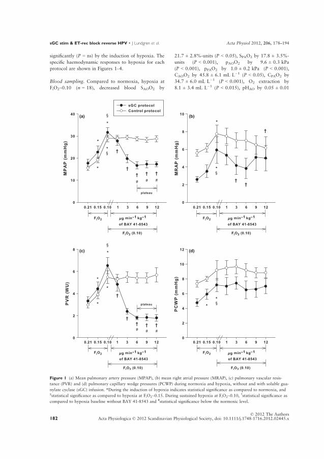
significantly (P = ns) by the induction of hypoxia. The
specific haemodynamic responses to hypoxia for each
protocol are shown in Figures 1–4.
Blood sampling. Compared to normoxia, hypoxia at
FiO2~0.10 (n = 18), decreased blood SAOO2 by
21.7 ± 2.8%-units (P < 0.05), SPAO2 by 17.8 ± 3.5%-
units (P < 0.001), pAOO2 by 9.6 ± 0.3 kPa
(P < 0.001), pPAO2 by 1.0 ± 0.2 kPa (P < 0.001),
CAOO2 by 45.8 ± 6.1 mL L�1 (P < 0.05), CPAO2 by
34.7 ± 6.0 mL L�1 (P < 0.001), O2 extraction by
8.1 ± 3.4 mL L�1 (P < 0.015), pHAO by 0.05 ± 0.01
(a)
(d)(c)
(b)
Figure 1 (a) Mean pulmonary artery pressure (MPAP), (b) mean right atrial pressure (MRAP), (c) pulmonary vascular resis-
tance (PVR) and (d) pulmonary capillary wedge pressures (PCWP) during normoxia and hypoxia, without and with soluble gua-
nylate cyclase (sGC) infusion. *During the induction of hypoxia indicates statistical significance as compared to normoxia, and§statistical significance as compared to hypoxia at FiO2~0.15. During sustained hypoxia at FiO2~0.10,
†statistical significance as
compared to hypoxia baseline without BAY 41-8543 and #statistical significance below the normoxic level.
© 2012 The AuthorsActa Physiologica © 2012 Scandinavian Physiological Society, doi: 10.1111/j.1748-1716.2012.02445.x182
sGC stim & ET-rec block reverse HPV · J Lundgren et al. Acta Physiol 2012, 206, 178–194

(a)
(d)
(e)
(c)
(b)
Figure 2 (a) Mean aortic pressure (MAP), (b) heart rate (HR), (c) CO, (d) stroke volume (SV) and (e) systemic vascular resis-
tance (SVR) during normoxia and hypoxia, without and with soluble guanylate cyclase (sGC) infusion. *During the induction of
hypoxia indicates statistical significance as compared to normoxia, and §statistical significance as compared to hypoxia
at FiO2~0.15. During sustained hypoxia at FiO2~0.10,†statistical significance as compared to hypoxia baseline without BAY
41-8543.
© 2012 The AuthorsActa Physiologica © 2012 Scandinavian Physiological Society, doi: 10.1111/j.1748-1716.2012.02445.x 183
Acta Physiol 2012, 206, 178–194 J Lundgren et al. · sGC stim & ET-rec block reverse HPV

(P < 0.05), pHPA by 0.04 ± 0.01 (P < 0.001), HbAOby 1.2 ± 0.3 g dL�1 (P < 0.05) and HbPA by
1.3 ± 0.2 g dL�1 (P < 0.001), whereas blood-O2
consumption was unchanged (P = ns) by the induction
of hypoxia. The specific responses in the blood param-
eters for each protocol are shown in Tables 1–3.
(a)
(d)(c)
(b)
Figure 3 (a) Mean pulmonary artery pressure (MPAP), (b) mean right atrial pressure (MRAP), (c) pulmonary vascular resis-
tance (PVR) and (d) pulmonary capillary wedge pressures (PCWP) during normoxia and hypoxia, without and with dual endo-
thelin (ET)-receptor blockade, followed by soluble guanylate cyclase (sGC) infusion. *During the induction of hypoxia indicates
statistical significance as compared to normoxia, and §statistical significance as compared to hypoxia at FiO2~0.15. During sus-
tained hypoxia at FiO2~0.10,†statistical significance as compared to hypoxia baseline without tezosentan, ¤statistical
significance with BAY 41-8543 as compared to hypoxia 60 min after tezosentan bolus infusion without BAY 41-8543, and ○notsignificantly different (P = ns) as compared to normoxia, that is, return to normoxia baseline. #Statistical significance below the
normoxic level.
© 2012 The AuthorsActa Physiologica © 2012 Scandinavian Physiological Society, doi: 10.1111/j.1748-1716.2012.02445.x184
sGC stim & ET-rec block reverse HPV · J Lundgren et al. Acta Physiol 2012, 206, 178–194
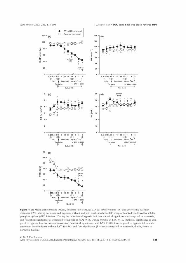
(a)
(d)
(e)
(c)
(b)
Figure 4 (a) Mean aortic pressure (MAP), (b) heart rate (HR), (c) CO, (d) stroke volume (SV) and (e) systemic vascular
resistance (SVR) during normoxia and hypoxia, without and with dual endothelin (ET)-receptor blockade, followed by soluble
guanylate cyclase (sGC) infusion. *During the induction of hypoxia indicates statistical significance as compared to normoxia,
and §statistical significance as compared to hypoxia at FiO2~0.15. During hypoxia at FiO2~0.10,†statistical significance as com-
pared to hypoxia baseline without tezosentan, ¤statistical significance with BAY 41-8543 as compared to hypoxia 60 min after
tezosentan bolus infusion without BAY 41-8543, and ○not significance (P = ns) as compared to normoxia, that is, return to
normoxia baseline.
© 2012 The AuthorsActa Physiologica © 2012 Scandinavian Physiological Society, doi: 10.1111/j.1748-1716.2012.02445.x 185
Acta Physiol 2012, 206, 178–194 J Lundgren et al. · sGC stim & ET-rec block reverse HPV

Table
1Bloodaortaandpulm
onary
arteryO
2saturation,pO
2,O
2content,pH,Hb,O
2extractionandO
2consumptionduringnorm
oxia
andhypoxia
FiO
2~0
.21
FiO
2~0
.15
FiO
2~0
.10
(baseline)
5min
(FiO
2~0
.10)
15min
(FiO
2~0
.10)
22.5
min
(FiO
2~0
.10)
30min
(FiO
2~0
.10)
37.5
min
(FiO
2~0
.10)
60min
(FiO
2~0
.10)
67.5
min
(FiO
2~0
.10)
75min
(FiO
2~0
.10)
82.5
min
(FiO
2~0
.10)
SAOO
2(%
)97.7
±0.2
95.2
±0.4
77.1
±2.7*§
76.6
±3.5
73.5
±3.8
74.0
±3.4
74.0
±4.0
70.3
±4.6
69.8
±5.4
66.7
±5.2†
68.5
±6.6†
63.2
±5.6†
SPAO
2(%
)56.6
±2.0
54.0
±2.2
40.7
±4.1*§
39.9
±4.5
37.2
±4.9
37.2
±5.2
36.7
±5.2
34.1
±5.8
33.5
±6.3†
30.7
±5.7†#
30.7
±5.4†#
26.7
±4.8†#
‡¤■●
pAOO
2(kPa)
16.7
±0.4
11.5
±0.2*
6.9
±0.4*§
6.8
±0.4
6.6
±0.5
6.6
±0.4
6.5
±0.4
6.3
±0.5
6.4
±0.6
6.0
±0.5†#
‡¤6.3
±0.6
5.8
±0.5†#
‡¤■
pPAO
2(kPa)
4.9
±0.1
4.8
±0.2
4.2
±0.2*§
4.1
±0.2
4.0
±0.3
4.0
±0.3
4.0
±0.3
3.8
±0.3
3.8
±0.4
3.7
±0.3†#
3.7
±0.3†#
3.5
±0.3†#
‡¤■●
CAOO
2
(mLL�1)
135.8
±3.2
119.1
±2.7*
93.2
±4.9*§
90.4
±3.9
85.7
±2.9
87.0
±2.5
86.5
±3.1
84.0
±3.5
84.4
±5.5
82.6
±5.2
81.8
±6.7†
85.9
±5.3†#
¤
CPAO
2
(mLL�1)
78.8
±2.8
72.0
±3.5
50.8
±5.4*§
49.9
±6.3
44.6
±5.1
45.4
±5.4
45.1
±5.8
42.2
±6.9
42.3
±7.7
38.4
±6.7†#
38.3
±6.2†#
34.0
±5.8†#
‡¤■
O2extraction
(mLL�1)
57.0
±3.4
47.1
±3.7*
42.4
±3.4*
40.5
±3.9
41.2
±2.8
41.6
±3.3
41.4
±3.6
41.8
±3.8
42.1
±3.0
44.2
±1.7
43.5
±2.5
41.9
±1.9
O2consumption
(mLmin
�1)
156.7
±9.1
133.8
±6.9*
154.2
±7.5§
148.7
±7.0
155.6
±10.0
148.6
±5.2
149.7
±11.6
146.8
±6.8
157.6
±13.4
169.0
±13.5
163.2
±13.1
158.9
±15.0
pH
AO
7.49±0.02
7.48±0.02
7.43±0.02*§
7.43±0.03
7.42±0.03
7.42±0.03
7.42±0.03
7.42±0.03
7.41±0.03
7.41±0.03†
7.41±0.031†
7.41±0.03†#
pH
PA
7.44±0.02
7.43±0.02
7.40±0.02*§
7.39±0.02
7.39±0.03
7.38±0.03
7.38±0.03
7.38±0.03
7.38±0.03
7.37±0.03
7.37±0.03
7.4
±0.03†#
‡
HbAO(g
dL�1)
10.1
±0.2
9.1
±0.2*
8.9
±0.4*
8.7
±0.3
8.6
±0.3
8.7
±0.3
8.6
±0.3
8.8
±0.3
9.0
±0.4
9.2
±0.4
8.9
±0.4
8.9
±0.4
HbPA(g
dL�1)
10.2
±0.2
9.8
±0.3
9.2
±0.4*
9.2
±0.4
9.0
±0.4
9.1
±0.3
9.1
±0.3
9.2
±0.3
9.4
±0.4
9.3
±0.4
9.3
±0.3
9.4
±0.4
Duringtheinductionofhypoxia;
*Statistical
sign
ificance
ascomparedto
norm
oxia,
§Statisticalsignificance
comparedto
hypoxia
atFiO
2~0
.15.
Duringsustained
hypoxia
atFiO
2~0.10;
†Statisticalsignificance
comparedto
hypoxia
baseline,
#Statistical
sign
ificance
comparedto
the5min
measurement,
‡Statisticalsignificance
comparedto
the15min
measurement,
¤Statisticalsignificance
comparedto
the22.5
min
measurement,
■Statisticalsign
ificance
comparedto
the30min
measurement,and
●Statisticalsign
ificance
comparedto
the37.5
min
measurement.
© 2012 The AuthorsActa Physiologica © 2012 Scandinavian Physiological Society, doi: 10.1111/j.1748-1716.2012.02445.x186
sGC stim & ET-rec block reverse HPV · J Lundgren et al. Acta Physiol 2012, 206, 178–194

Table
2BloodS A
OO
2,S P
AO
2,pAOO
2,pPAO
2,CAOO
2,CPAO
2,O
2extraction,O
2consumption,pH
AOandpH
PA,duringnorm
oxia
andhypoxia,withoutandwithsoluble
guanylate
cyclase
infusion
FiO
2~0
.21
FiO
2~0
.15
FiO
2~0
.10
(baseline)
1lg
min
�1per
kg
(FiO
2~0
.10)
3lgmin
�1per
kg
(FiO
2~0
.10)
6lgmin
�1per
kg
(FiO
2~0
.10)
9lg
min
�1per
kg
(FiO
2~0
.10)
12lg
min
�1per
kg
(FiO
2~0
.10)
S AOO
2(%
)99.3
±0.47
95.1
±1.4*
68.1
±6.5*§
68.7
±7.9
67.8
±8.8
67.9
±8.8
69.6
±8.2
77.2
±8.2
S PAO
2(%
)66.7
±2.5
60.0
±3.0*
33.3
±7.6*§
35.2
±8.1
38.0
±8.3
39.4
±7.5
42.0
±7.7
45.7
±7.3†
pAOO
2(kPa)
15.1
±0.6
10.3
±0.3*
6.0
±0.4*§
6.0
±0.5
6.0
±0.5
5.9
±0.5
5.9
±0.3
6.2
±0.3
pPAO
2(kPa)
5.5
±0.3
5.1
±0.2*
3.6
±0.3*§
3.6
±0.4
3.8
±0.4
3.9
±0.4
4.0
±0.4
4.1
±0.3
CAOO
2
(mLL�1)
162.9
±11.1
156.1
±7.3
101.3
±12.2*§
98.9
±12.7
97.3
±13.8
88.6
±9.5
97.3
±10.4
102.1
±9.0
CPAO
2(m
LL�1)
114.3
±5.9
98.9
±5.8*
49.8
±11.8*§
53.3
±12.3
57.3
±12.6
55.6
±11.2
57.5
±10.2
61.2
±9.5
O2extraction
(mL·L
�1)
46.6
±10.2
57.2
±6.5
51.4
±4.6
45.6
±5.6
40.0
±7.8
33.0
±2.2
39.7
±2.5
41.0
±5.3
O2consumption
(mLmin
�1)
180.0
±37.5
214.0
±16.7
194.7
±9.7
196.2
±15.3
196.5
±18.3
188.5
±21.6
242.2
±22.6
245.0
±30.9
pH
AO
7.47±0.04
7.44±0.04
7.40±0.05*§
7.39±0.05
7.39±0.05
7.38±0.05
7.37±0.05†
7.37±0.05†
pH
PA
7.4
±0.04
7.4
±0.03*
7.4
±0.04*§
7.4
±0.04
7.4
±0.05
7.4
±0.05
7.3
±0.05
7.3
±0.05†
HbAO(g
dL�1)
12.0
±0.8
12.0
±0.5
10.8
±0.4
10.6
±0.5
10.5
±0.3
9.8
±0.6
10.4
±0.3
9.8
±0.3
HbPA(g
dL�1)
12.6
±0.4
12.1
±0.4
10.8
±0.4*§
11.0
±0.3
10.9
±0.3
10.2
±0.4
10.2
±0.3
10.0
±0.3
Duringtheinductionofhypoxia;
*Statisticalsignificance
ascomparedto
norm
oxia,
§Statisticalsign
ificance
ascomparedto
hypoxia
atFiO
2~0
.15.
Duringsustained
hypoxia
atFiO
2~0
.10;
†Statisticalsign
ificance
comparedto
hypoxia
baselinewithoutBAY
41-8543.
© 2012 The AuthorsActa Physiologica © 2012 Scandinavian Physiological Society, doi: 10.1111/j.1748-1716.2012.02445.x 187
Acta Physiol 2012, 206, 178–194 J Lundgren et al. · sGC stim & ET-rec block reverse HPV
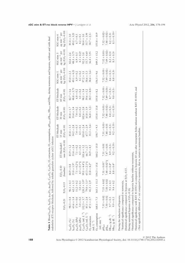
Table
3BloodS A
OO
2,S P
AO
2,pAOO
2,pPAO
2,CAOO
2,CPAO
2,O
2extraction,O
2consumption,pH
AO,pH
PA,HbAOandHbPAduringnorm
oxia
andhypoxia,withoutandwithdual
endothelin
(ET)-receptorblockade,
followed
bysoluble
guanylate
cyclase(sGC)infusion
FiO
2~0
.21
FiO
2~0
.15
FiO
2~0
.10
(baseline)
ET-blockade5
min
(FiO
2~0
.10)
ET-blockade
15min
(FiO
2~0
.10)
ET-blockade
30min
(FiO
2~0
.10)
ET-blockade
60min
(FiO
2~0
.10)
SGC-stim
1
lgmin
�1per
kg(F
iO2~0
.10)
SGC-stim
3
lgmin
�1per
kg(F
iO2~0
.10)
SGC-stim
6
lgmin
�1per
kg(F
iO2~0
.10)
S AOO
2(%
)98.3
±0.4
96.1
±0.3
84.9
±0.8
83.9
±1.2
85.8
±1.5
85.8
±1.7
82.7
±1.6
83.0
±2.3
82.6
±2.6
81.4
±3.0
S PAO
2(%
)63.6
±1.4
62.1
±1.5
55.0
±1.9*
53.4
±2.4
54.2
±2.8
53.4
±3.4
48.8
±3.7†
49.1
±4.2†
48.1
±3.7†
49.2
±3.2†
pAOO
2(kPa)
17.6
±0.6
11.2
±0.2
7.1
±0.2*§
7.1
±0.2
7.4
±0.3
7.4
±0.2
6.8
±0.2
6.9
±0.2
6.9
±0.3
6.7
±0.3
pPAO
2(kPa)
5.0
±0.2
5.0
±0.1
4.5
±0.1*§
4.5
±0.1
4.5
±0.1
4.4
±0.2
4.1
±0.2
4.1
±0.2
4.1
±0.2†
4.2
±0.1
CAOO
2(m
LL�1)
132.3
±3.7
120.3
±3.9*
99.1
±4.4*§
101.6
±3.2
103.0
±2.4
102.6
±2.8
98.1
±3.4
97.2
±3.1
92.1
±3.0
90.4
±3.0
CPAO
2(m
LL�1)
85.1
±2.4
79.0
±3.1*
65.3
±2.1*§
66.7
±2.7
67.7
±2.9
65.8
±3.9
59.6
±5.2
58.6
±5.3
55.7
±4.6†
55.7
±3.9†
O2extraction
(mLL�1)
47.1
±2.7
41.3
±2.9
33.8
±4.2*
34.9
±4.5
35.3
±3.6○
36.8
±3.2○
38.5
±4.0○
38.6
±3.6○
36.4
±3.9○
34.7
±2.1○
O2consumption
(mLmin
�1)
168.1
±7.3
165.1
±11.5
154.3
±15.6
160.2
±15.0
158.7
±9.3
153.8
±13.8
155.8
±8.2
156.5
±9.6
149.5
±13.2
155.8
±10.9
pH
AO
7.55±0.02
7.53±0.02
7.51±0.01*
7.51±0.01
7.51±0.01○
7.52±0.01○
7.52±0.01○
7.52±0.01○
7.52±0.01○
7.52±0.02○
pH
PA
7.50±0.02
7.50±0.02
7.48±0.02*§
7.47±0.01
7.48±0.01
7.48±0.02○
7.49±0.01○
7.49±0.02○
7.49±0.02○
7.48±0.02○
HbAO(g
dL�1)
9.7
±0.3
9.1
±0.3
8.6
±0.4*
8.9
±0.3○
8.8
±0.3○
8.8
±0.3○
8.7
±0.3○
8.6
±0.3○
8.2
±0.3¤
8.2
±0.3¤
HbPA(g
dL�1)
9.9
±0.3
9.4
±0.2
8.8
±0.4*
9.2
±0.3†○
9.2
±0.3†○
9.1
±0.3†○
9.0
±0.3○
8.8
±0.3¤
8.5
±0.3¤
8.3
±0.3†¤
Duringtheinductionofhypoxia;
*Statistical
sign
ificance
comparedto
norm
oxia,
§Statisticalsignificance
comparedto
hypoxia
atFiO
2~0
.15.
Duringsustained
hypoxia
atFiO
2~0
.10;
†Statisticalsignificance
comparedto
hypoxia
baselinewithouttezosentan,
¤Statisticalsignificance
withBAY
41-8543ascomparedto
hypoxia
60min
after
tezosentanbolusinfusionwithoutBAY
41-8543,and
○Notsign
ificantlydifferent(P
=ns)
comparedto
norm
oxia,thatis,return
tonorm
oxia
baseline.
© 2012 The AuthorsActa Physiologica © 2012 Scandinavian Physiological Society, doi: 10.1111/j.1748-1716.2012.02445.x188
sGC stim & ET-rec block reverse HPV · J Lundgren et al. Acta Physiol 2012, 206, 178–194
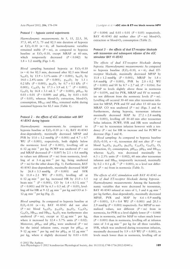
Protocol 1 – hypoxia control experiments
Haemodynamic measurements. At 5, 15, 22.5, 30,
37.5, 60, 67.5, 75 and 82.5 min duration of hypoxia
at FiO2~0.10 (n = 6), all haemodynamic variables
remained stable (P = ns), as compared to hypoxia
baseline at FiO2~0.10, except MRAP at 60 min,
which temporarily decreased (P < 0.042) by
1.8 ± 1.2 mmHg (Figs 1–4).
Blood sampling. Sustained hypoxia at FiO2~0.10(n = 6) for 82.5 min, decreased after 82.5 min blood
SAOO2 by 13.9 ± 3.1%-units (P < 0.001), SPAO2 by
14.0 ± 2.4%-units (P < 0.001), pAOO2 by 1.1 ±0.2 kPa (P < 0.001), pPAO2 by 0.7 ± 0.1 kPa (P <0.001), CAOO2 by 17.3 ± 3.9 mL L�1 (P < 0.001),
CPAO2 by 16.8 ± 3.1 mL L�1 (P < 0.001), pHAO by
0.03 ± 0.01 (P < 0.009) and pHPA by 0.03 ± 0.01
(P < 0.05), whereas blood-O2 extraction, blood-O2
consumption, HbAO and HbPA remained stable during
sustained hypoxia for 82.5 min (Table 1).
Protocol 2 – the effects of sGC stimulation with BAY
41-8543 during hypoxia
Haemodynamic measurements. As compared to
hypoxia baseline at FiO2~0.10 (n = 6), BAY 41-8543
dose-dependently, maximally decreased MPAP and
PVR by 15.0 ± 1.2 mmHg (P < 0.001) and 4.7 ± 0.7
WU (P < 0.001), respectively, both slightly below
the normoxic level (P < 0.001), levelling off at
6–12 lg min�1 per kg. PCWP was unaltered (P = ns)
and MRAP decreased (P < 0.007) by 2.1 ± 0.6 mmHg
to values not different (P = ns) from normoxia, level-
ling of at 3–6 lg min�1 per kg, being unaltered
(P = ns) for the other doses (Fig. 1). Furthermore, BAY
41-8543 dose-dependently, maximally decreased MAP
by 26.8 ± 3.3 mmHg (P < 0.001) and SVR
by 11.0 ± 2.1 WU (P < 0.05), levelling off at
6–12 lg min�1 per kg, increased HR by 21.0 ± 5.5
beats min�1 (P < 0.001), CO by 1.4 ± 0.3 L min�1
(P < 0.001) and SV by 6.5 ± 0.5 mL (P < 0.05), level-
ling off for HR at 9–12 lg min�1 per kg and CO at 3–
12 lg min�1 per kg (Fig. 2).
Blood sampling. As compared to hypoxia baseline at
FiO2~0.10 (n = 6), BAY 41-8543 did not alter
(P = ns) blood SAOO2, pAOO2, pPAO2, CAOO2,
CPAO2, HbAO and HbPA. SPAO2 was furthermore also
unaltered (P = ns), except at 12 lg min�1 per kg,
where it increased by 12.4 ± 5.7% units (P < 0.02).
Moreover, pHAO and pHPA were unaltered (P = ns)
for the initial infusion rates, except for pHAO at
9–12 lg min�1 per kg and for pHPA at 12 lg min�1
per kg, where it slightly decreased by 0.03 ± 0.01
(P < 0.004) and 0.03 ± 0.01 (P < 0.05) respectively.
BAY 41-8543 did neither alter (P = ns) blood-O2
extraction or blood-O2 consumption (Table 2).
Protocol 3 – the effects of dual ET-receptor blockade
with tezosentan and subsequent infusion of the sGC
stimulator BAY 41-8543
The effects of dual ET-receptor blockade during
hypoxia. Haemodynamic measurements: As compared
to hypoxia baseline (FiO2~0.10, n = 6), dual ET-
receptor blockade, maximally decreased MPAP by
11.8 ± 1.2 mmHg (P < 0.001), MRAP by 1.8 ±0.4 mmHg (P < 0.001), PVR by 2.0 ± 0.2 WU
(P < 0.001) and SV by 8.7 ± 2.7 mL (P < 0.016). For
MPAP to levels slightly above those in normoxia
(P < 0.039), and for PVR, MRAP and SV to normal-
ize not different from the normoxic levels (P = ns).
Levelling off occured 30–60 min after tezosentan infu-
sion for MPAP, PVR and SV and after 15–60 min for
MRAP. CO was unaltered (P = ns) (Figs. 3 and 4).
Furthermore, during hypoxia, tezosentan infusion
maximally decreased MAP by 27.2 ± 2.8 mmHg
(P < 0.001), levelling off 30–60 min after tezosentan
bolus infusion. PCWP, SVR and HR were unaltered
(P = ns) by tezosentan infusion however with a ten-
dency (P = ns) for HR to increase and for PCWP to
decrease (Figs 3 and 4).
Blood sampling: As compared to hypoxia baseline
(FiO2~0.10, n = 6), tezosentan did not alter (P = ns)
blood SAOO2, pAOO2, pPAO2, CAOO2, CPAO2, O2
extraction, O2 consumption, pHAO, pHPA and HbAO,
whereas SPAO2 was decreased maximally by
6.3 ± 2.3% units (P < 0.002), 60 min after tezosentan
infusion and HbPA temporarily increased, maximally
by 0.2 ± 0.1 g dL�1 (P < 0.001), to a level not differ-
ent (P = ns) from in normoxia (Table 3).
The effects of sGC stimulation with BAY 41-8543 on
top of dual ET-receptor blockade during hypoxia.
Haemodynamic measurements: Among the haemody-
namic variables that were decreased by tezosentan,
BAY 41-8543 infused at rates of 1, 3 and 6 lg min�1
per kg further, dose-dependently, maximally decreased
MPAP, PVR and MAP by 6.6 ± 0.9 mmHg
(P < 0.001), 1.9 ± 0.4 WU (P < 0.001) and 20.5 ±2.9 mmHg (P < 0.001) respectively. For MPAP to nor-
malized values, not different (P = ns) from in
normoxia, for PVR to a level slightly lower (P < 0.048)
than in normoxia, and for MAP to values much lower
(P < 0.001) than in normoxia, levelling off at infusion
rates of 3–6 lg min�1 per kg for all three variables.
SVR, which was unaltered during tezosentan infusion,
maximally decreased by 5.9 ± 0.9 WU (P < 0.001), to
levels much lower than in normoxia, levelling off at
© 2012 The AuthorsActa Physiologica © 2012 Scandinavian Physiological Society, doi: 10.1111/j.1748-1716.2012.02445.x 189
Acta Physiol 2012, 206, 178–194 J Lundgren et al. · sGC stim & ET-rec block reverse HPV

infusion rates of 3–6 lg min�1 per kg. MRAP, SV,
PCWP, HR and CO remained unaltered (P = ns) with
BAY 41-8543 infusion (Figs 3 and 4).
Blood sampling: Blood HbPA, which temporarily
increased by dual ET-receptor blockade during
hypoxia (FiO2~0.10), dose-dependently decreased,
maximally by 0.7 ± 0.07 g dL�1 (P < 0.001), after
subsequent infusion of the sGC stimulator BAY 41-
8543. Among the variables that were unaltered by
tezosentan bolus infusion, BAY 41-8543 infusion
dose-dependently decreased blood HbAO, maximally
by 0.6 ± 0.01 g dL�1 (P < 0.002), whereas blood
SAOO2, pAOO2, pPAO2, CAOO2, CPAO2, O2 extrac-
tion, O2 consumption, pHAO and pHPA remained
unaltered (P = ns) with BAY 41-8543 infusion. Blood
SPAO2, which was decreased by tezosentan bolus infu-
sion, remained unaltered (P = ns) with BAY 41-8543
infusion (Table 3).
Discussion
The present study shows that sGC stimulation with
BAY 41-8543, during acute hypoxia at FiO2~0.10,dose-dependently attenuates and totally reverses the
acute hypoxia-induced pulmonary vasoconstrictor
response. This is manifested by the reduction of the
hypoxia-induced increases in MPAP and PVR to val-
ues even slightly below those in normoxia. Our study
furthermore confirms that dual ET-receptor blockade
with tezosentan markedly attenuates the acute HPV
response, reducing the hypoxia-induced increases in
MPAP and PVR, for MPAP to levels slightly above
those in normoxia and for PVR to normoxic levels.
Subsequent sGC stimulation on top of dual ET-recep-
tor blockade further dose-dependently decreases
MPAP to ‘low’ normoxic values and PVR to values
slightly below those in normoxia. Neither sGC stimu-
lation nor dual ET-receptor blockade, or a combina-
tion of the two, altered blood arterial-O2 saturation
or blood-O2 consumption. Both sGC stimulation and
dual ET-receptor blockade, however, caused a marked
systemic vasodilatation, as illustrated by the decreases
in MAP and SVR, although during stable circulatory
conditions. These results suggest that sGC stimulation
alone, or in combination with dual ET-receptor block-
ade, could provide a rapid and effective means to
reduce generalized acute hypoxia-induced pulmonary
vasoconstriction, without affecting blood arterial oxy-
genation and blood-O2 consumption.
Response to hypoxia
Hypoxic pulmonary vasoconstriction, first identified
by von Euler and Liljestrand (1946), is primarily effec-
tive in the pre-capillary resistance arteries of the pul-
monary circulation (Kato & Staub 1966), although
approx. 20% of the response may be related to the
venous circuit. HPV may be beneficial in focal
hypoxia, such as in atelectasis (Glasser et al. 1983),
diverting blood from poorly to better ventilated areas
of the lung. In such situations, impairment of HPV
may consequently lead to insufficient oxygenation.
However, HPV may be detrimental in global hypoxia,
such as at rapid ascent to high altitude. HPV may
then increase pulmonary artery pressure and PVR,
and contribute to the development of pulmonary
oedema, as well as induce right heart failure (Bartsch
et al. 2005, Maggiorini et al. 2006, Preston 2007).
When wanting to treat global acute hypoxia, the pri-
mary choice of treatment is the removal of the cause
of hypoxia and to increase systemic oxygen supply via
ventilation or extra-corporal membrane oxygenation.
In the present study, though, we evaluated whether
sGC stimulation alone, or in combination with dual
ET-receptor blockade, may be a means to alleviate
acute hypoxia-induced pulmonary vasoconstriction.
This is of importance for developing new treatment
strategies for PH of various aetiology, and potentially
for developing alternative clinical strategies to infusion
of organic nitrates, such as nitroglycerin, as the latter
may be associated with the development of tolerance,
protein nitrosation and oxidative stress (Warnholtz
et al. 2002).
The present study indeed showed, in our porcine
model, a marked acute HPV response to hypoxia at
FiO2~0.10. This was illustrated by the marked
increase in MPAP and PVR. The concomitant increase
in MRAP with hypoxia may thus reflect an acute
increase in load on the right heart owing to pulmo-
nary vasoconstriction. Our hypoxia control protocol
(protocol 1), where no treatment was given, further-
more showed that the HPV response remained stable
during sustained hypoxia for 82.5 min, because MPAP
and PVR remained at a stable elevated level for this
period of time (Fig. 1).
Stimulation of sGC with BAY 41-8543 totally reverses
HPV
Stimulation of sGC, by right atrial infusion of BAY
41-8543, was in the present study found to potently
and dose-dependently attenuate, and in fact totally
reverse, the pulmonary vasoconstrictor response to
acute hypoxia, by reducing MPAP and PVR to values
even slightly below those in normoxia. MRAP was
also decreased to normoxic levels and if a similar low-
ering of MRAP occurred in sustained chronic hypoxia,
right heart failure could potentially be prohibited.
PCWP remained unaltered, suggesting that the
increase in PCWP upon the induction of hypoxia was
© 2012 The AuthorsActa Physiologica © 2012 Scandinavian Physiological Society, doi: 10.1111/j.1748-1716.2012.02445.x190
sGC stim & ET-rec block reverse HPV · J Lundgren et al. Acta Physiol 2012, 206, 178–194

not a direct result of a ventricular interdependence
owing to the PH-induced increased load on the right
side of the heart. Whether sGC stimulation addition-
ally could be a means of prohibiting hypoxia-induced
pulmonary oedema remains, however, unresolved.
Our in vivo findings add important information to
previous in vitro studies, where the sGC activator
HMR1766 has been found to dose-dependently inhibit
the pressor response to acute hypoxia in the isolated
perfused mouse lung (Weissmann et al. 2009).
HMR1766 has additionally been found to partially
reverse the haemodynamic changes and structural
remodelling of the lung vasculature and right ventricle
occurring in chronic hypoxia (Weissmann et al.
2009). Both BAY 41-2272, which stimulates sGC
directly and enhances the sensitivity of sGC to NO,
and BAY 58-2667, which activates sGC even in its
oxidized or haeme-free form, independently of NO,
have been found to reverse the haemodynamic and
structural changes associated with chronic hypoxia, as
well as monocrotaline-induced PH in rats (Dumitrascu
et al. 2006). sGCa1 seems furthermore essential for
the NO-mediated pulmonary, but not systemic, vaso-
dilatation and limits chronic hypoxia-induced vascular
remodelling by inhibiting smooth muscle cell prolifer-
ation (Vermeersch et al. 2007). sGCa1 has, however,
not been found to regulate baseline pulmonary vascu-
lar tone or the acute response to hypoxia (Vermeersch
et al. 2007). Even though sGCa1 did not seem to par-
ticipate in mediating HPV (Vermeersch et al. 2007),
we show that treatment with sGC stimulation may
totally reverse HPV. Thus, early sGC stimulator treat-
ment for patients with hypoxic PH may both reverse
HPV and positively affect pulmonary vascular remod-
elling, as has been shown in rats (Dumitrascu et al.
2006). However, the extent of effect in chronic
hypoxia, compared with in acute hypoxia cannot be
determined from the present study.
Moreover, therapies that act in synergy with endog-
enous NO, such as NO- and haeme-independent sGC
activators and NO-independent haeme-dependent sGC
stimulators, may be of future importance in PH to
optimize ventilation/perfusion matching and modulate
pulmonary vascular tone (Mittendorf et al. 2009).
Stimulators and activators of sGC may potentially
also be used as a complement to for instance PDE5
inhibitors and ET-receptor blockers, in the treatment
for PAH (Evgenov et al. 2006). Additionally, it is of
great interest that sGC stimulators may be adminis-
tered via inhalation to directly act in the target organ,
as described by Evgenov et al. (2006). With such a
route of administration, the systemic effects of sGC
stimulation could potentially be minimized and, fur-
thermore, with respect to hypoxia-induced PH, the
drug would primarily act in well-ventilated areas of
the lung. On the other hand, inhalation often requires
administration 4–8 times a day, which may be cum-
bersome for the patients.
Stimulation of sGC with BAY 41-8543 potentiates the
effects of dual ET-receptor blockade in attenuating HPV
Endothelin-1 is a potent vasoconstrictor, synthesized
by the endothelium, mediating its effects via ETA-
and/or ETB-receptors on vascular smooth muscle- and
endothelial cells (Sato et al. 1995, McCulloch et al.
1998). Hypoxia per se has been found to increase the
amount of ET-1 mRNA in the lung and right atrium
(Kourembanas et al. 1991, Elton et al. 1992), the ET-
1 immunoreactivity in plasma, as well as stimulate
preproendothelin-1 promotor activity (Aversa et al.
1997) and up-regulate the amount of ET-receptors
(Soma et al. 1999). Although it is widely recognized
that ET-1 plays an important role in the effects of
chronic hypoxia, its role in acute HPV has been con-
troversial. Studies on ET-receptor blockade, both
in vivo and in vitro, in different species, have shown
both attenuation (Bonvallet et al. 1994, Chen et al.
1995, 1997, DiCarlo et al. 1995, Oparil et al. 1995,
Holm et al. 1996, 1998, Franco-Cereceda & Holm
1998, Sato et al. 2000, Goirand et al. 2003, Modesti
et al. 2006, Petersen et al. 2008) and no attenuation
(Douglas et al. 1993, Takeoka et al. 1995, Lazor
et al. 1996, Hubloue et al. 2003, Biarent et al. 2006)
of the HPV response.
We recently showed, in our porcine model, that
dual ET-receptor blockade with tezosentan markedly
attenuates the acute hypoxia-induced increase in pul-
monary arterial pressure and completely attenuates
the PVR increase to hypoxia (Hedelin et al. 2011). As
dual ET-receptor blockade, for instance in the setting
of PAH, is not always sufficient to fully normalize the
increased pulmonary vascular tone, it was important
in the present study to evaluate whether combination
treatments, for instance with sGC stimulation and
dual ET-receptor blockade, could be additionally
effective. In the present study, we confirmed that dual
ET-receptor blockade with tezosentan, during acute
hypoxia, effectively attenuates the hypoxia-induced
increase in MPAP, PVR and MRAP, by decreasing
MPAP to values slightly higher than in normoxia and
by decreasing PVR and MRAP to normoxic levels,
whereas PCWP was unaltered. sGC stimulation on
top of the dual ET-receptor blockade further
decreased MPAP to ‘low’ normoxic values and PVR
to values slightly below those in normoxia, whereas
MRAP and PCWP remained unaltered. Thus, such a
combination treatment seems feasible and effective in
reversing acute hypoxia-induced PH. Furthermore,
such a strategy may also be of importance in the
© 2012 The AuthorsActa Physiologica © 2012 Scandinavian Physiological Society, doi: 10.1111/j.1748-1716.2012.02445.x 191
Acta Physiol 2012, 206, 178–194 J Lundgren et al. · sGC stim & ET-rec block reverse HPV

treatment for PH related to other causes, such as
PAH, because both ET-receptor blockers and sGC
stimulation may, besides their vasodilating effects,
also attenuate pulmonary vascular remodelling.
Systemic responses to hypoxia and stimulation of sGC
with BAY 41-8543 alone and on top of dual ET-
receptor blockade
The present study showed that sGC stimulation, as
well as dual ET-receptor blockade also exert potent
systemic effects, however, without causing a circula-
tory ‘collapse’. Hypoxia decreased SVR, increased CO
and SV, but left MAP and HR unaltered. In protocol
2, sGC stimulation during hypoxia decreased MAP
and further decreased SVR to levels below those in
normoxia, whereas HR, CO, SV and blood-O2 con-
sumption remained unaltered. In protocol 3, dual ET-
receptor blockade also decreased MAP and SVR to
values below the normoxic levels, whereas HR, CO,
SV and blood-O2 consumption were unaltered. A
major part of the drop in SVR and MAP induced by
tezosentan seems, however, to be species-related, as
the same pronounced effect has not been observed
with tezosentan in rats (Tabrizchi & Ford 2003) and
with bosentan in humans (Clozel et al. 1994). Subse-
quent sGC stimulation, on top of dual ET-receptor
blockade, further decreased MAP and SVR. This con-
firms a potent systemic effect of sGC stimulation. A
pronounced drop in MAP, as observed with BAY 41-
8543 in the present study, may be harmful in critically
ill patients and furthermore induce the release of vaso-
constrictor substances. Therefore, if wanting to treat
PH with BAY 41-8543, doses should be carefully
titrated and systemic blood pressure be monitored.
However, the systemic blood pressure decreases
observed with BAY 41-8543 indicate that sGC stimu-
lation could have potential implications in the treat-
ment for systemic hypertension and hypertensive
crisis.
Conclusion
The present study uniquely shows, in a porcine model,
that sGC stimulation alone, during acute hypoxia,
dose-dependently attenuates the hypoxia-induced pul-
monary vasoconstrictor response by decreasing MPAP,
PVR and MRAP to normoxic values. Furthermore,
sGC stimulation on top of dual ET-receptor blockade
during hypoxia decreases MPAP and MRAP to ‘low’
normoxic values and PVR to values slightly lower
than in normoxia. sGC stimulation alone, or in com-
bination with tezosentan, accomplished its attenuation
of the HPV response simultaneously with a marked
dose-dependent systemic vasodilator response, without
affecting blood arterial-O2 saturation and blood-O2
consumption. These findings suggest that sGC stimula-
tion alone, or in combination with dual ET-receptor
blockade, may provide a valuable means to attenuate
the vasoconstrictor response to acute hypoxia, which,
in turn, may ease the tension and excessive load on
the right heart.
Conflict of interest
The authors have declared no conflict of interest.
We would like to acknowledge the support of the animal
technicians at the Department of Experimental Surgery and
Medicine, at the Panum Institute, University of Copenhagen,
Copenhagen, Denmark, as well as the staff at the Copenha-
gen Muscle Research Centre, Rigshospitalet, Copenhagen,
Denmark, and at the Department of Cardiology, Lund Uni-
versity and the Clinic for Heart Failure and Valvular disease,
Skane University Hospital, Lund, Sweden. We furthermore
would like to acknowledge the support of Bayer Schering
Pharma AG (Wuppertal, Germany) for providing BAY 41-
8543 and Actelion Pharmaceuticals (Allschwil, Switzerland)
for providing tezosentan for our experiments. We would fur-
thermore like to acknowledge the financial support from the
Copenhagen Muscle Research Centre, Copenhagen, Den-
mark, as well as the Maggie Stephens-, Crafoord-, ‘ALF’-
and Skane University Hospital Foundations, Lund, Sweden.
References
Aversa, C.R., Oparil, S., Caro, J., Li, H., Sun, S.D., Chen, Y.
F., Swerdel, M.R., Monticello, T.M., Durham, S.K., Minc-
henko, A., Lira, S.A. & Webb, M.L. 1997. Hypoxia stimu-
lates human preproendothelin-1 promoter activity in
transgenic mice. Am J Physiol Lung Cell Mol Physiol 273,
L848–L855.
Bartsch, P., Mairbaurl, H., Maggiorini, M. & Swenson, E.R.
2005. Physiological aspects of high-altitude pulmonary
edema. J Appl Physiol 98, 1101–1110.
Biarent, D., Hubloue, I., Bejjani, G., Melot, C., Jespers, P.,
Naeije, R. & Leeman, M. 2006. Role of endothelins and
nitric oxide in the pulmonary circulation of perinatal
lambs during hyperoxia and hypoxia. Pediatr Res 59, 131–
136.
Boerrigter, G. & Burnett, J.C. Jr 2010. Soluble guanylate
cyclase: not a dull enzyme. Circulation 119, 2752–2754.
Bonvallet, S.T., Zamora, M.R., Hasunuma, K., Sato, K.,
Hanasato, N., Anderson, D., Sato, K. & Stelzner, T.J.
1994. BQ123, an ETA-receptor antagonist, attenuates hyp-
oxic pulmonary hypertension in rats. Am J Physiol 266,
H1327–H1331.
Chen, S.J., Chen, Y.-F., Meng, Q.C., Durand, J., DiCarlo, V.
S. & Oparil, S. 1995. Endothelin-receptor antagonist bos-
entan prevents and reverses hypoxic pulmonary hyperten-
sion in rats. J Appl Physiol 79, 2122–2131.
Chen, S.J., Chen, Y.-F., Opgenorth, T.J., Wessale, J.L., Meng,
Q.C., Durand, J., DiCarlo, V.S. & Oparil, S. 1997. The
orally active nonpeptide endothelin A-receptor antagonist
© 2012 The AuthorsActa Physiologica © 2012 Scandinavian Physiological Society, doi: 10.1111/j.1748-1716.2012.02445.x192
sGC stim & ET-rec block reverse HPV · J Lundgren et al. Acta Physiol 2012, 206, 178–194

A-127722 prevents and reverses hypoxia induced pulmonary
hypertension and pulmonary vascular remodeling in Spra-
gue-Dawley rats. J Cardiovasc Pharmacol 29, 713–725.
Clozel, M., Volker, B., Gray, G.A., Kalina, B., Loffler,
B.-M., Burri, K., Cassal, J.-M., Hirth, G., Muller, M.,
Neidhart, W. & Ramuz, H. 1994. Pharmacological charac-
terization of Bosentan, a new potent orally active nonpep-
tide endothelin receptor antagonist. J Pharmacol Exp Ther
270, 228–235.
Clozel, M., Ramuz, H., Clozel, J.P., Breu, V., Hess, P., Lof-
fler, B.M., Coassolo, P. & Roux, S. 1999. Pharmacology
of tezosentan, new endothelin receptor antagonist designed
for parenteral use. J Pharmacol Exp Ther 290, 840–846.
DiCarlo, V.S., Chen, S.J., Meng, Q.C., Durand, J., Yano,
M., Chen, Y.-F. & Oparil, S. 1995. ETA-receptor antago-
nist prevents and reverses chronic hypoxia-induced pulmo-
nary hypertension in rat. Am J Physiol Lung Cell Mol
Physiol 269, L690–L697.
Douglas, S.A., Vickery-Clark, L.M. & Ohlstein, E.H. 1993.
Endothelin-1 does not mediate hypoxic vasoconstriction in
canine isolated blood vessels: effect of BQ-123. Br J Phar-
macol 108, 418–421.
Dumitrascu, R., Weissmann, N., Ghofrani, H.A., Dony, E.,
Beuerlein, K., Schmidt, H., Stasch, J.P., Gnoth, M.J., See-
ger, W., Grimminger, F. & Schermuly, R.T. 2006. Activa-
tion of soluble guanylate cyclase reverses experimental
pulmonary hypertension and vascular remodeling. Circula-
tion 113, 286–295.
Elton, T.S., Oparil, S., Taylor, G.R., Hicks, P.H., Yang,
R.-H., Jin, H. & Chen, Y.F. 1992. Normobaric hypoxia
stimulates endothelin-1 gene expression in the rat. Am J
Physiol Regul Integr Comp Physiol 263, R1260–R1264.
von Euler, U.S. & Liljestrand, G. 1946. Observations on the
pulmonary arterial blood pressure in the cat. Acta Physiol
Scand 12, 301–320.
Evgenov, O.V., Pacher, P., Schmidt, P.M., Hasko, G.,
Schmidt, H.H. & Stasch, J.P. 2006. NO-independent stim-
ulators and activators of soluble guanylate cyclase: discov-
ery and therapeutic potential. Nat Rev Drug Discov 5, 755
–768.
Franco-Cereceda, A. & Holm, P. 1998. Selective or nonselec-
tive endothelin antagonists in porcine hypoxic pulmonary
hypertension? J Cardiovasc Pharmacol 31, S447–S452.
Galie, N., Hoeper, M.M., Humbert, M., Torbicki, A., Vachi-
ery, J.L., Barbera, J.A., Beghetti, M., Corris, P., Gaine, S.,
Gibbs, J.S. et al.2009. Guidelines for the diagnosis and
treatment of pulmonary hypertension: the Task Force for
the Diagnosis and Treatment of Pulmonary Hypertension
of the European Society of Cardiology (ESC) and the Euro-
pean Respiratory Society (ERS), endorsed by the Interna-
tional Society of Heart and Lung Transplantation (ISHLT).
Eur Heart J 30, 2493–2537.
Glasser, S.A., Domino, K.B., Lindgren, L., Parcella, P., Mar-
shall, C. & Marshall, B.E. 1983. Pulmonary blood pres-
sure and flow during atelectasis in the dog. Anesthesiology
58, 225–231.
Goirand, F., Bardou, M., Guerard, P., Dumas, J.-P., Roch-
ette, L. & Dumas, M. 2003. ETA, mixed ETA/ETB recep-
tor antagonists, and protein kinase C inhibitor prevent
acute hypoxic pulmonary vasoconstriction: influence of
potassium channels. J Cardiovasc Pharmacol 41, 117–
125.
Hedelin, P., Kylhammar, D. & Radegran, G. 2011. Dual
endothelin receptor blockade with tezosentan markedly
attenuates hypoxia-induced pulmonary vasoconstriction in
a porcine model. Acta Physiol 204, 419–434.
Holm, P., Liska, J., Clozel, M. & Franco-Cereceda, A. 1996.
The endothelin antagonist bosentan: hemodynamic effects
during normoxia and hypoxic pulmonary hypertension in
pigs. J Thorac Cardiovasc Surg 112, 890–897.
Holm, P., Liska, J. & Franco-Cereceda, A. 1998. The ETA
receptor antagonist, BMS-182874, reduces acute hypoxic
pulmonary hypertension in pigs in vivo. Cardiovasc Res
37, 765–771.
Hubloue, I., Biarent, D., Abdel Kafi, S., Bejjani, G., Kerbaul,
F., Naeije, R. & Leeman, M. 2003. Endogenous endothe-
lins and nitric oxide in hypoxic pulmonary vasoconstric-
tion. Eur Respir J 21, 19–24.
Humbert, M., Morrell, N.W., Archer, S.L., Stenmark, K.R.,
MacLean, M.R., Lang, I.M., Christman, B.W., Weir, E.K.,
Eickelberg, O., Voelkel, N.F. & Rabinovitch, M. 2004.
Cellular and molecular pathobiology of pulmonary arterial
hypertension. J Am Coll Cardiol 43, 13S–24S.
Ignarro, L.J., Adams, J.B., Horwitz, P.M. & Wood, K.S.
1986. Activation of soluble guanylate cyclase by NO-he-
moproteins involves NO-heme exchange. Comparison of
heme-containing and heme-deficient enzyme forms. J Biol
Chem 261, 4997–5002.
Kato, M. & Staub, N.C. 1966. Response of small pulmonary
arteries to unilobar hypoxia and hypercapnia. Circ Res 19,
426–440.
Kourembanas, S., Marsden, P.A., McQuillan, L.P. & Faller,
D.V. 1991. Hypoxia induces endothelin gene expression
and secretion in cultured human endothelium. J Clin Invest
88, 1054–1057.
Lazor, R., Feihl, F., Waeber, B., Kucera, P. & Perret, C.
1996. Endothelin-1 does not mediate the endothelium-
dependent hypoxic contractions of small pulmonary arter-
ies in rats. Chest 110, 189–197.
Maggiorini, M., Brunner-La Rocca, H.-P., Peth, S., Fischler,
M., Bohm, T., Bernheim, A., Kiencke, S., Bloch, K.E., De-
hnert, C., Naeije, R., Lehmann, T., Bartsch, P. & Mair-
baurl, H. 2006. Both tadalafil and dexamethasone may
reduce the incidence of high-altitude pulmonary edema: a
randomized trial. Ann Intern Med 145, 497–506.
McCulloch, K.M., Docherty, C. & MacLean, M.R. 1998.
Endothelin receptors mediating contraction of rat and
human pulmonary resistance arteries: effect of chronic
hypoxia in the rat. Br J Pharmacol 123, 1621–1630.
Mittendorf, J., Weigand, S., Alonso-Alija, C., Bischoff, E.,
Feurer, A., Gerisch, M., Kern, A., Knorr, A., Lang, D.,
Muenter, K. et al.2009. Discovery of riociguat (BAY 63-
2521): a potent, oral stimulator of soluble guanylate
cyclase for the treatment of pulmonary hypertension.
ChemMedChem 4, 853–865.
Modesti, P.A., Vanni, S., Morabito, M., Modesti, A., March-
etta, M., Gamberi, T., Sofi, F., Savia, G., Mancia, G., Gen-
sini, G.F. & Parati, G. 2006. Role of endothelin-1 in
© 2012 The AuthorsActa Physiologica © 2012 Scandinavian Physiological Society, doi: 10.1111/j.1748-1716.2012.02445.x 193
Acta Physiol 2012, 206, 178–194 J Lundgren et al. · sGC stim & ET-rec block reverse HPV

exposure to high altitude: Acute Mountain Sickness and
Endothelin-1 (ACME-1) study. Circulation 114, 1410–1416.
Oparil, S., Chen, S.J., Meng, Q.C., Elton, T.S., Yano, M. &
Chen, Y.F. 1995. Endothelin-A receptor antagonist pre-
vents acute hypoxia-induced pulmonary hypertension in
the rat. Am J Physiol 268, L95–L100.
Pavek, K., Boska, D. & Selecky, F.V. 1964. Measurement of
cardiac output by thermodilution with constant rate injec-
tion of indicator. Circ Res 15, 311–319.
Petersen, B., Deja, M., Bartholdy, R., Donaubauer, B., Laudi,
S., Francis, R.C.E., Boemke, W., Kaisers, U. & Busch, T.
2008. Inhalation of the ETA receptor antagonist LU-
135252 selectiveley attenuates hypoxic pulmonary vaso-
constriction. Am J Physiol Regul Integr Comp Physiol
294, R601–R605.
Preston, I.R. 2007. Clinical perspective of hypoxia-mediated
pulmonary hypertension. Antioxid Redox Signal 9, 711–
721.
Sato, K., Oka, M., Hasunuma, K., Ohnishi, M., Sato, K. &
Kira, S. 1995. Effects of separate and combined ETA and
ETB blockade on ET-1-induced constriction in perfused rat
lungs. Am J Physiol 269, L668–L672.
Sato, K., Morio, Y., Morris, K.G. Jr, Rodman, D.M. &
McMurtry, I.F. 2000. Mechanism of hypoxic pulmonary
vasoconstriction involves ETA receptor inhibition of KATP
channel. Am J Physiol Lung Cell Mol Physiol 278, L434–
L442.
Simmoneau, G., Robbins, I.M., Beghetti, M., Channick, R.
N., Delcroix, M., Denton, C.P., Elliott, C.G., Gaine, S.P.,
Gladwin, M.T., Jing, Z.C., Krowka, M.J., Langleben, D.,
Nakanishi, N. & Souza, R. 2009. Updated clinical classifi-
cation of pulmonary hypertension. J Am Coll Cardiol 54,
43–54.
Soma, S., Takahashi, H., Muramatsu, M., Oka, M. & Fuku-
chi, Y. 1999. Localization and distribution of endothelin
receptor subtypes in pulmonary vasculature of normal and
hypoxia-exposed rats. Am J Respir Cell Mol Biol 20, 620–
630.
Stasch, J.P., Alonso-Alija, C., Apeler, H., Dembowsky, K.,
Feurer, A., Minuth, T., Perzborn, E., Schramm, M. &
Straub, A. 2002. Pharmacological actions of a novel NO-
independent guanylyl cyclase stimulator, BAY 41-8543: in
vitro studies. Br J Pharmacol 135, 333–343.
Stasch, J.P., Pacher, P. & Evgenov, O.V. 2011. Soluble gua-
nylate cyclase as an emerging therapeutic target in cardio-
pulmonary disease. Circulation 123, 2263–2273.
Tabrizchi, R. & Ford, C.A. 2003. Haemodynamic effects of
endothelin receptor antagonist, tezosentan, in tumour
necrosis factor-a treated anaesthetized rats. Naunyn-Schmi-
edeberg’s Arch Pharmacol 367, 156–167.
Takeoka, M., Ishizaki, T., Sakai, A., Chang, S.W., Shigemori,
K., Higashi, T. & Ueda, G. 1995. Effect of BQ123 on vaso-
constriction as a result of either hypoxia or endothelin-1 in
perfused rat lungs. Acta Physiol Scand 155, 53–60.
Vermeersch, P., Buys, E., Pokreisz, P., Marsboom, G., Ichi-
nose, F., Sips, P., Pellens, M., Gillijns, H., Swinnen, M.,
Graveline, A., Collen, D., Dewerchin, M., Brouckaert, P.,
Bloch, K.D. & Janssens, S. 2007. Soluble guanylate
cyclase-alpha1 deficiency selectively inhibits the pulmonary
vasodilator response to nitric oxide and increases the pul-
monary vascular remodeling response to chronic hypoxia.
Circulation 116, 936–943.
Warnholtz, A., Mollnau, H., Heitzer, T., Kontush, A., Mol-
ler-Bertram, T., Lavall, D., Giaid, A., Beisiegel, U., Markl-
und, S.L., Walter, U., Meinertz, T. & Munzel, T. 2002.
Adverse effects of nitroglycerin treatment on endothelial
function, vascular nitrotyrosine levels and cGMP-depen-
dent protein kinase activity in hyperlipidemic Watanabe
rabbits. J Am Coll Cardiol 40, 1356–1363.
Weissmann, N., Hackemack, S., Dahal, B.K., Pullamsetti, S.
S., Savai, R., Mittal, M., Fuchs, B., Medebach, T., Dumi-
trascu, R., van Eickels, M., Ghofrani, H.A., Seeger, W.,
Grimminger, F. & Schermuly, R.T. 2009. The soluble gua-
nylate cyclase activator HMR1766 reverses hypoxia-
induced experimental pulmonary hypertension in mice. Am
J Physiol Lung Cell Mol Physiol 297, L658–L665.
© 2012 The AuthorsActa Physiologica © 2012 Scandinavian Physiological Society, doi: 10.1111/j.1748-1716.2012.02445.x194
sGC stim & ET-rec block reverse HPV · J Lundgren et al. Acta Physiol 2012, 206, 178–194

Copyright of Acta Physiologica is the property of Wiley-Blackwell and its content may not be copied or
emailed to multiple sites or posted to a listserv without the copyright holder's express written permission.
However, users may print, download, or email articles for individual use.

Paper II


Hemodynamic Characteristics Including Pulmonary Hypertension atRest and During Exercise Before and After Heart TransplantationJakob Lundgren, MD; G€oran R�adegran, MD, MS Eng Phys, DMSc
Background-—Little is known about the hemodynamic response to exercise in heart failure patients at various ages before andafter heart transplantation (HT). This information is important because postoperative hemodynamics may be a predictor of survival.To investigate the hemodynamic response to HT and exercise, we grouped our patients based on preoperative age and examinedtheir hemodynamics at rest and during exercise before and after HT.
Methods and Results-—Ninety-four patients were evaluated at rest prior to HT with right heart catheterization at our laboratory. Ofthese patients, 32 were evaluated during slight supine exercise before and 1 year after HT. Postoperative evaluations wereperformed at rest 1 week after HT and at rest and during exercise at 4 weeks, 3 months, 6 months, and 1 year after HT. Theexercise patients were divided into 2 groups based on preoperative age of ≤50 or >50 years. There were no age-dependentdifferences in the preoperative hemodynamic exercise responses. Hemodynamics markedly improved at rest and during exerciseat 1 and 4 weeks, respectively, after HT; however, pulmonary and, in particular, ventricular filling pressures remained high duringexercise at 1 year after HT, resulting in normalized pulmonary vascular resistance response but deranged total pulmonary vascularresistance response.
Conclusions-—Our findings suggest that, (1) in patients with heart failure age ≤50 or >50 years may not affect the hemodynamicresponse to exercise to the same extent as in healthy persons, and (2) total pulmonary vascular resistance may be more adequatethan pulmonary vascular resistance for evaluating the exercise response after HT. ( J Am Heart Assoc. 2015;4:e001787 doi:10.1161/JAHA.115.001787)
Key Words: catheterization • exercise • heart failure • heart transplantation
P hysical activity imposes prominent stress on the cardio-vascular system because adequate delivery of oxygen
and substrates is a necessity to sustain muscular activity.1
The provision of sufficient oxygen delivery requires bothventilatory and cardiovascular adaptations, includingincreases in ventilation, heart rate, and cardiac output (CO),in parallel with increased vascular conductance in activemuscles and increased vascular resistance in inactive tissue.2
In severe heart failure (HF), both cardiac output andendothelial function are impaired.3 Consequently, in HF, both
central and peripheral circulation may compromise sufficientoxygen delivery to active muscles, limiting exercise capacityand daily life activities.
Knowledge of systemic and pulmonary hemodynamics isimportant for the treatment of HF. The normal hemodynamicmagnitudes have previously been clearly defined at rest4,5;however, there is a lack of understanding of the normalhemodynamic response to exercise, especially with regard topulmonary circulation but also related to sex, body position,and different exercise intensities.6–8 This is further compli-cated by that the hemodynamic alterations due to age seemto be more prominent during exercise than at rest.7,8 Eventhough several invasive studies have investigated hemody-namics during exercise,9–13 the normal response has not beenclearly defined. This lack of consensus resulted in abandoningof the “exercise criteria” in the definition of pulmonaryhypertension at the World Symposium on Pulmonary Hyper-tension in 2008.4
Moreover, the mechanisms behind exaggerated exercise-induced increases in pulmonary artery pressures are poorlyunderstood. For these reasons, it remains of value to furthercharacterize hemodynamics during exercise before and afterHT. Such characteristics could assist in defining an abnormal
From the Haemodynamic Lab, The Section for Heart Failure and ValvularDisease, The Heart and Lung Clinic, Sk�ane University Hospital, Lund, Sweden(J.L., G.R.) and Department of Clinical Sciences Lund, Cardiology, LundUniversity, Lund, Sweden (J.L., G.R.).
Correspondence to: Jakob Lundgren, MD, The Haemodynamic Lab, TheSection for Heart Failure and Valvular Disease, The Heart and Lung Clinic, Sk�aneUniversity Hospital, Lund, Sweden. E-mail: [email protected]
Received January 21, 2015; accepted May 20, 2015.
ª 2015 The Authors. Published on behalf of the American Heart Association,Inc., by Wiley Blackwell. This is an open access article under the terms of theCreative Commons Attribution-NonCommercial License, which permits use,distribution and reproduction in any medium, provided the original work isproperly cited and is not used for commercial purposes.
DOI: 10.1161/JAHA.115.001787 Journal of the American Heart Association 1
ORIGINAL RESEARCH
at UN
IVE
RSIT
ET
SSJUK
HU
SET
on September 13, 2016
http://jaha.ahajournals.org/D
ownloaded from

response to exercise and potentially aid in a new definition ofexercise-induced pulmonary hypertension. This is importantbecause pulmonary hypertension during exercise might be abetter prognostic marker for disease severity than measure-ments at rest.14–19 Such a definition is also relevant becausethere is conflicting evidence regarding the preoperativemagnitudes of different hemodynamic parameters at restthat may affect outcome after HT. This aspect illustrates thecomplexity in differentiating between patients with excessivevasoconstriction and those with vascular remodeling20 andhighlights the need for refined criteria for defining pulmonaryhypertension. Improved insight into exercise hemodynamicsmay also provide guidance for medical treatment, for listingfor HT, and for detection of pulmonary vascular abnormalitiesearlier in HF and after HT.
The aim of the present study was therefore to characterizethe hemodynamics of patients with severe end-stage HF atrest and during exercise in relation to age before and afterHT and to relate that information to data from healthypersons in previously published systematic reviews.7,8 Ourpurpose was to clarify how patients with severe HF respondto slight exercise and how hemodynamics recover at rest andduring exercise after HT, as well as to highlight potentialabnormalities in the post-transplant response to exercise. Wehypothesized that hemodynamics at rest and during exerciseimprove early after HT but with significant abnormalitiesremaining during exercise at 1 year after HT. We alsohypothesized that any age-dependent differences in thehemodynamic response to exercise would be attenuated inpatients with severe HF.
Methods
Study Design and PopulationThis retrospective single-center study reviewed the 215 HTpatients followed at Sk�ane University Hospital in Lund,Sweden, during 1988–2010. The study was performed withinformed consent and approval by the ethics board in Lund(DNR 2014/92, Dnr 2011/777, Dnr 2011/368, Dnr 2010/114) and in accordance with the Declarations of Helsinki andIstanbul. A total of 219 HTs were included, of which 214 (98%)were first-time HTs, and 72 patients (33%) received amechanical assist prior to HT.
The study focused on adult patients evaluated at ourhemodynamic laboratory prior to HT. Patients referred fromand investigated at other university hospitals (n=87), childrenaged <18 years (n=21), repeated HTs (n=5), and other patientswith incomplete hemodynamic data prior to HT (n=12) wereexcluded. After exclusion, 94 adults (study population)remained for analysis, of which 24 were women (25.6%). Themean age was 51.6 years, and age ranged from 19 to
69 years. The most common indications for HT were dilated(52 patients) and ischemic (24 patients) cardiomyopathy.21
Data Collection and AnalysisRight heart catheterization (RHC) was performed at rest priorto HT and at 1 and 4 weeks, 3 and 6 months, and 1 year afterHT. In all patients who were capable of exercise, RHCmeasurements were also performed during supine bicycleexercise prior to HT and at 4 weeks, 3 and 6 months, and1 year after HT. Men exercised at 50 W, and womenexercised at 30 W. Measurements were conducted at steadystate �5 minutes after initiation of exercise. There was nograded increase in workload.
Thirty-two of the 94 patients exercised prior to and 1 yearafter HT. These patients were subsequently characterized in 2groups, 1 with patients aged ≤50 years (n=12) and 1 withpatients aged >50 years (n=20) at the time of HT (Table 1). Thissubclassification was performed to compare our findings withthose presented for healthy persons published in recentsystematic reviews.7,8Overall, 21 of the 94 includedHTpatientswere bridged to transplantation using a mechanical assist. Ofthese patients, 4 exercised prior to and 1 year after HT.
Right Heart CatheterizationRHC was predominantly performed via the right internaljugular vein, using a Swan Ganz catheter (Baxter Health CareCorp). If patients had >1 RHC prior to HT, the one closest toHT or assist implantation was analyzed. Mean pulmonaryartery pressure (MPAP), pulmonary artery wedge pressure(PAWP), mean right atrial pressure (MRAP), and mean arterialpressure were recorded during RHC. Heart rate was recordedfrom ECG. CO was measured by thermodilution. Cardiacindex, stroke volume (SV), SV index, left ventricular strokework index (LVSWI), right ventricular stroke work index(RVSWI), transpulmonary gradient (TPG), pulmonary vascularresistance (PVR), PVR index, and total PVR (TPVR) werecalculated using the following formulas: cardiac index=CO/body surface area; SV=CO/heart rate; SV index=SV/bodysurface area; LVSWI=(mean arterial pressure�PAWP)9SVindex; RVSWI=(MPAP�MRAP)9SV index; TPG=MPAP�PAWP;PVR=TPG/CO; PVR index=TPG/cardiac index; and TPVR=MPAP/CO.
StatisticsA SigmaStat system (SigmaPlot 11.0) was used for statisticalanalysis. Parametric or nonparametric statistics were useddepending on the distribution of data. One-way repeated-measures ANOVA (Tukey test) or One-way repeated-measuresANOVA on ranks (Tukey test) were used when multiple groups
DOI: 10.1161/JAHA.115.001787 Journal of the American Heart Association 2
Hemodynamics Before and After Heart Transplant Lundgren and R�adegran
ORIG
INALRESEARCH
at UN
IVE
RSIT
ET
SSJUK
HU
SET
on September 13, 2016
http://jaha.ahajournals.org/D
ownloaded from

Table 1. Characteristics of the Patients Who Exercised Prior to and 1 Year After HT, Grouped According to Age at the Time of HT
Patients Aged ≤50 Years at the Time of HT Patients Aged >50 Years at the Time of HT
Mean�SD n % Mean�SD n %
Recipient characteristics
Sex of recipient 12 20
Female 6 50.0 4 20.0
Age of recipient, y 42.8�7.3 12 56.6�3.7 20
≤10 0 0 0 0
11 to 20 0 0 0 0
21 to 30 1 8.3 0 0
31 to 40 2 16.7 0 0
41 to 50 9 75.0 0 0
51 to 60 0 0 16 80.0
≥61 0 0 4 20.0
Recipient length, cm 171.0�8.6 12 175.8�8.2 20
Recipient weight, kg 78.1�20.3 12 81.0�15.6 20
Recipient blood group 12 20
0 3 25.0 7 35.0
A 8 66.7 9 45.0
B 0 0 4 20
AB 1 8.3 0 0
Time on waiting list, days 121.5�146.6 12 138.9�130.1 20
Indication for HT 12 20
DCM 8 66.7 12 60.0
HCM 0 0 1 5
Myocarditis 2 16.7 1 5
IHD 0 0 6 30.0
RCM 2 16.7 0 0
Donor characteristics
Sex of donor 12 20
Female 5 41.7 9 45.0
Age of donor, y 35.5�13.9 12 45.2�15.4 20
Donor length, cm 175.7�9.1 12 176.3�10.3 18
Donor weight, kg 76.2�18.8 12 82.5�13.4 20
Ischemic time, min 163.3�63.5 12 182.7�56.5 20
Recipient–donor matching
Age difference, y 13.7�9.4 12 13.3�12.6 20
Sex matching 12 20
Sex matched 9 75.0 15 75.0
AB0 matching 12 20
Identic 9 75.0 19 95.0
Compatible 3 25.0 1 5.0
Incompatible 0 0 0 0
DCM indicates dilated cardiomyopathy; HCM, hypertrophic cardiomyopathy; HT, heart transplantation; IHD, ischemic heart disease; RCM, restrictive cardiomyopathy.
DOI: 10.1161/JAHA.115.001787 Journal of the American Heart Association 3
Hemodynamics Before and After Heart Transplant Lundgren and R�adegran
ORIG
INALRESEARCH
at UN
IVE
RSIT
ET
SSJUK
HU
SET
on September 13, 2016
http://jaha.ahajournals.org/D
ownloaded from

were compared. The Student t test and Mann-Whitney rank-sum test were used, respectively, when 2 groups werecompared. Paired Student t test and Wilcoxon signed ranktest were used, respectively, when comparing 1 group beforeand after an intervention. Pearson and Spearman correlationswere used, respectively, when measuring the associationbetween 2 variables. A P value <0.05 was consideredstatistically significant. All values are mean�SD.
Results
Hemodynamics at Rest and During Exercise Priorto Heart TransplantationBaseline hemodynamics for all 94 included patients and forthe subgroup of patients who exercised (n=32) are shown inTable 2.
During the preoperative assessment of the 32 patients whoexercised during RHC, exercise increased MPAP (P<0.001),PAWP (P<0.001), TPG (P<0.001), MRAP (P<0.001), CO(P<0.001), and SV (P<0.01) compared with performance atrest, whereas PVR and TPVR remained unchanged (P value not
significant) (Table 2). There was no correlation between changein PAWP andMRAP (R2=0.02, P value not significant) (Figure 1).
Exercise in patients aged ≤50 years increased MPAPby 15.7�10.0 mm Hg (58.1%, P<0.001), PAWP by10.3�8.2 mm Hg (53.8%, P<0.001), TPG by 5.4�5.9 mm Hg(68.6%, P<0.001), MRAP by 10.2�4.0 mm Hg (125.5%,P<0.001), CO by 2.2�0.7 L9min�1 (54.2%, P<0.001), andSV by 6.3�7.8 mL9beat�1 (11.3%, P<0.04), whereas PVRand TPVR remained unaltered (P value not significant)(Figures 2 and 3).
Similarly, in patients aged >50 years, exercise increasedMPAP by 18.8�11.5 mm Hg (69.4%, P<0.001), PAWP by13.5�11.3 mm Hg (77.2%, P<0.001), TPG by 5.4�4.2 mm Hg(55.3%, P<0.001), MRAP by 10.8�3.8 mm Hg (259.0%,P<0.001), and CO by 2.0�1.7 L9min�1 (48.1%, P<0.001),whereas SV, PVR, and TPVR remained unaltered (P value notsignificant) (Figures 2 and 3).
With regard to pre-HT age-dependent differences, none ofthese parameters differed for patients aged ≤50 or >50 years(P value not significant) at rest and during exercise. Thepercentage change did neither differ between the groups (Pvalue not significant) (Figures 2 and 3).
Table 2. Preoperative Haemodynamic Characteristics of All 94 Patients and the 32 Patients Who Exercised Prior to, and 1 YearAfter, HT
Parameter
Entire Study Population(at Rest)
Patients Who Exercised(at Rest)
Patients Who Exercised(During Exercise)
Patients ≤50 Years(at Rest)
Patients >50 Years(at Rest)
SignificanceMean�SD Mean�SD Mean�SD Mean�SD Mean�SD
Patients, n 94 32 32 12 20
MAP, mm Hg 74.1�13.6 75.4�15.8 87.9�19.7* 73.5�10.9 77.1�18.1 *
MPAP, mm Hg 30.1�8.8 27.2�9.8 44.4�10.5* 29.1�9.7 27.9�10.1 *
PAWP, mm Hg 20.5�8.2 18.3�8.8 30.5�9.5* 18.2�9.3 18.4�8.7 *
TPG, mm Hg 9.6�3.5 8.9�3.1 14.0�5.3* 7.9�2.3 9.5�3.3 *
MRAP, mm Hg 7.9�6.1 6.1�5.0 16.3�7.4* 7.6�5.5 5.3�4.7† *,†
HR, beat 9 min�1 82.6�16.8 82.2�15.0 111.9�19.6* 77.5�16.1 84.8�14.1 *
CO, L 9 min�1 3.7�1.0 4.3�0.9† 6.4�1.7* 4.3�1.1 4.3�0.9† *,†
SV, mL 9 beat�1 46.7�17.1 53.1�15.3† 60.2�21.6* 57.4�19.5 50.7�12.4 *,†
PVR (WU) 2.8�1.3 2.2�0.7† 2.2�0.8 2.0�0.6† 2.3�0.8 †
TPVR (WU) 9.1�4.3 6.7�2.8† 7.4�2.6 6.5�3.1† 6.8�2.7† †
LVSWI,mm Hg 9 mL 9 m�2
1395.0�730.1 1615.9�717.8 1894.7�1174.1 1680.5�689.9 1580.4�747.8
RVSWI,mm Hg 9 mL 9 m�2
546.2�271.1 594.6�333.2 814.8�404.4* 563.2�333.8 611.9�340.2 *
CO indicates cardiac output; HR, heart rate; HT, heart transplantation; LVSWI, left ventricular stroke work index; MAP, mean atrial pressure; MPAP, mean pulmonary artery pressure; MRAP,mean atrial pressure; PAWP, pulmonary artery wedge pressure; PVR, pulmonary vascular resistance; RVSWI, right ventricular stroke work index; SV, stroke volume; TPG, transpulmonarygradient; TPVR, total pulmonary vascular resistance; WU, Wood units.*Indicates significance (P<0.05) between rest and exercise in patients who exercised.†Indicates a significance (P<0.05) compared to the entire study population.
DOI: 10.1161/JAHA.115.001787 Journal of the American Heart Association 4
Hemodynamics Before and After Heart Transplant Lundgren and R�adegran
ORIG
INALRESEARCH
at UN
IVE
RSIT
ET
SSJUK
HU
SET
on September 13, 2016
http://jaha.ahajournals.org/D
ownloaded from

Hemodynamic Improvement After HeartTransplantationThe hemodynamic response and improvement in relation to HTare shown in Figures 4 and 5 for all 94 patients at rest and for32 patients during slight supine exercise. For the 94 patients atrest 1 week after HT, there was a decrease in MPAP (P<0.001),PAWP (P<0.001), PVR (P<0.001), and TPVR (P<0.001) alongwith an increase in mean arterial pressure (P<0.001), CO(P<0.001), and SV (P<0.001) compared with performance atrest prior to HT (Figures 4 and 5). TPG and MRAP wereunaltered (P value not significant) (Figures 4 and 5).
Of these parameters, PVR remained constant (P value notsignificant) throughout the first year after HT, whereas MPAPfurther decreased (P<0.009) andmean arterial pressure furtherincreased (P<0.02) at 4 weeks versus 1 week after HT andremained constant (P value not significant) thereafter. MRAPalso decreased (P<0.001) at 4 weeks as compared to bothprior to HT and one week after HT, and then temporarilydecreased (P<0.04) at 6 months compared with 4 weeks afterHT. SV further increased (P<0.01) at 3 months compared with1 week after HT and remained constant (P value not signifi-cant) thereafter. Finally, PAWP and TPVR further decreased(P<0.001 and P<0.02, respectively) and CO further increased(P<0.03) at 6 months compared with 1 week after HT andremained constant (P value not significant) thereafter.
Compared with performance during exercise prior to HT, at4 weeks after HT, there was a decrease in MPAP (P<0.001),PAWP (P<0.001), PVR (P<0.003), and TPVR (P<0.001) alongwith an increase in mean arterial pressure (P<0.001), CO(P<0.001), and SV (P<0.001). TPG and MRAP were unaltered(P value not significant) (Figures 4 and 5). All of theseparameters remained stable (P value not significant) through-
out the first year after HT except for CO, which furtherincreased (P<0.004), and for MRAP and PVR, which decreased(P<0.005 and P<0.03, respectively), at 1 year after HT(Figures 4 and 5).
Hemodynamic Response to Exercise at 1 YearAfter Heart TransplantationFor the 32 patients who performed RHC at rest and duringexercise 1 year after HT, exercise increased (P<0.001 for allparameters) MPAP by 17.8�6.1 mm Hg (114.6%), PAWP by14.1�5.5 mm Hg (193.1%), TPG by 3.7�3.1 mm Hg (44.4%),MRAP by 9.8�4.3 mm Hg (371.4%), CO by 5.4�2.4 L9min�1
(88.1%), SV by 28.5�15.6 mL9beat�1 (39.8%), and TPVR by0.4�0.7 Wood units (WU; 15.4%) compared with performanceat rest, whereas PVR decreased by 0.3�0.3 WU (22.1%,P<0.001) (Figures 2 and 3). Change in MPAP from rest toexercise correlated to change in PAWP (R2=0.75, P<0.001)(Figure 6).
Exercise in patients aged ≤50 years increased MPAP by14.8�4.6 mm Hg (104.1%, P<0.001), PAWP by 12.1�4.3mm Hg (190.8%, P<0.001), TPG by 2.8�3.3 mm Hg (34.7%,P<0.016), MRAP by 8.6�5.3 mm Hg (412.0%, P<0.001), CO by5.8�3.4 L9min�1 (88.7%, P<0.001), SV by 30.9�19.3mL9beat�1 (41.7%, P<0.001), and TPVR by 0.2�0.4 WU(11.1%, P<0.05), whereas PVR decreased by 0.3�0.3 WU(26.7%, P<0.004) (Figures 2 and 3).
Similarly, exercise in patients aged >50 years increasedMPAP by 19.6�6.4 mm Hg (118.5%, P<0.001), PAWP by15.3�6.1 mm Hg (194.3%, P<0.001), TPG by 4.3�2.8mm Hg (49.7%, P<0.001), MRAP by 10.5�4.0 mm Hg(354.2%, P<0.001), CO by 5.2�1.6 L9min�1 (87.7%,P<0.001), SV by 27.1�16.0 mL9beat�1 (38.6%, P<0.001),and TPVR by 0.5�0.8 WU (17.5%, P<0.008), whereasPVR decreased by 0.3�0.3 WU (19.7%, P<0.001) (Figures 2and 3).
Resting TPVR was generally higher in patients aged>50 years compared with patients aged ≤50 years(P<0.03). In contrast, during exercise, not only TPVR but alsoMPAP and PVR were higher (P<0.05) in patients aged>50 years compared with patients aged ≤50 years; however,the percentage change in these parameters did not differbetween groups (Figures 2 and 3).
DiscussionThe present study evaluated the hemodynamics at rest andduring slight supine exercise in patients with severe HF beforeand after HT. Our results showed that already at 1 week afterHT, hemodynamics markedly improved at rest and remainedconstant or further improved during the first year of follow-up
Δ PA
WP
(m
m H
g)
Figure 1. Correlation between preoperative changes to exercisein PAWP and MRAP. MRAP indicates mean right atrial pressure;PAWP, pulmonary artery wedge pressure.
DOI: 10.1161/JAHA.115.001787 Journal of the American Heart Association 5
Hemodynamics Before and After Heart Transplant Lundgren and R�adegran
ORIG
INALRESEARCH
at UN
IVE
RSIT
ET
SSJUK
HU
SET
on September 13, 2016
http://jaha.ahajournals.org/D
ownloaded from

A B
DC
Figure 2. Hemodynamic response to exercise with regard to pulmonary and intracardiac pressures prior to and 1 year after HT.● Patients aged ≤50 years prior to HT. ○ Patients aged ≤50 years 1 year after HT. ▼ Patients aged >50 years prior to HT;D Patients aged >50 years 1 year after HT. ■ All 32 patients prior to HT. □ All 32 patients 1 year after HT. A, MPAP. B, PAWP. C,MRAP. D, TPG. †A statistically significant difference for MPAP during exercise between patients aged ≤50 and >50 years. ‡Astatistically significant difference for MPAP, PAWP, MRAP, and TPG between rest and exercise prior to HT for patients aged≤50 years. §A statistically significant difference for MPAP, PAWP, MRAP, and TPG between rest and exercise prior to HT for patientsaged >50 years. ‖A statistically significant difference for MPAP, PAWP, MRAP, and TPG between rest and exercise at 1 year after HTfor patients aged ≤50 years. #A statistically significant difference for MPAP, PAWP, MRAP, and TPG between rest and exercise at1 year after HT for patients aged >50 years. HT indicates heart transplantation; MPAP, mean pulmonary artery pressure; MRAP,mean right atrial pressure; PAWP, pulmonary artery wedge pressure; TPG, transpulmonary gradient.
DOI: 10.1161/JAHA.115.001787 Journal of the American Heart Association 6
Hemodynamics Before and After Heart Transplant Lundgren and R�adegran
ORIG
INALRESEARCH
at UN
IVE
RSIT
ET
SSJUK
HU
SET
on September 13, 2016
http://jaha.ahajournals.org/D
ownloaded from

A B
C D
Figure 3. Hemodynamic response to exercise with regard to cardiac output, stroke volume, and pulmonary vascular resistances priorto and 1 year after HT. ● Patients aged ≤50 years prior to HT. ○ Patients aged ≤50 years 1 year after HT. ▼ Patients aged >50 yearsprior to HT. D Patients aged >50 years 1 year after HT. ■ All 32 patients prior to HT. □ All 32 patients 1 year after HT. A, CO. B, SV. C,PVR. D, TPVR. *A statistically significant difference for TPVR at rest between patients aged ≤50 and >50 years 1 year after HT. †Astatistically significant difference for PVR and TPVR during exercise between patients aged ≤50 and >50 years. ‡A statisticallysignificant difference for CO and SV between rest and exercise prior to HT for patients aged ≤50 years. §A statistically significantdifference for CO between rest and exercise prior to HT for patients aged >50 years. ‖A statistically significant difference for CO, SV,PVR, and TPVR between rest and exercise at 1 year after HT for patients aged ≤50 years. #A statistically significant difference for CO,SV, PVR, and TPVR between rest and exercise at 1 year after HT for patients aged >50 years. CO indicates cardiac output; HT, hearttransplantation; PVR, pulmonary vascular resistance; SV, stroke volume; TPVR, total pulmonary vascular resistance; WU, Wood units.
DOI: 10.1161/JAHA.115.001787 Journal of the American Heart Association 7
Hemodynamics Before and After Heart Transplant Lundgren and R�adegran
ORIG
INALRESEARCH
at UN
IVE
RSIT
ET
SSJUK
HU
SET
on September 13, 2016
http://jaha.ahajournals.org/D
ownloaded from

A B
C D
E F
Figure 4. Hemodynamic characteristics with regard to pulmonary and intracardiac pressuresin the 94 patients at rest prior to and after HT. ● Patients at rest. ○ Patients during exercise.Graphs show (A) MPAP, (B) PAWP, (C) MRAP (D) TPG, (E) PVR, and (F) TPVR at rest and duringexercise before and after HT. *A statistically significant difference at rest compared with prior toHT. †A statistically significant difference at rest compared with 4 weeks after HT. ‡A statisticallysignificant difference at rest compared with 4 weeks after HT. §A statistically significantdifference during exercise compared with prior to HT. ‖A statistically significant differenceduring exercise compared with 4 weeks after HT. Of the 32 patients who exercised prior to HT,16 exercised at 4 weeks, 22 at 3 months, 22 at 6 months, and 32 at 1 year after HT. HTindicates heart transplantation; MPAP, mean pulmonary artery pressure; MRAP, mean rightatrial pressure; PAWP, pulmonary artery wedge pressure; PVR, pulmonary vascular resistance;TPG, transpulmonary gradient; TPVR, total pulmonary vascular resistance; WU, Wood units.
DOI: 10.1161/JAHA.115.001787 Journal of the American Heart Association 8
Hemodynamics Before and After Heart Transplant Lundgren and R�adegran
ORIG
INALRESEARCH
at UN
IVE
RSIT
ET
SSJUK
HU
SET
on September 13, 2016
http://jaha.ahajournals.org/D
ownloaded from
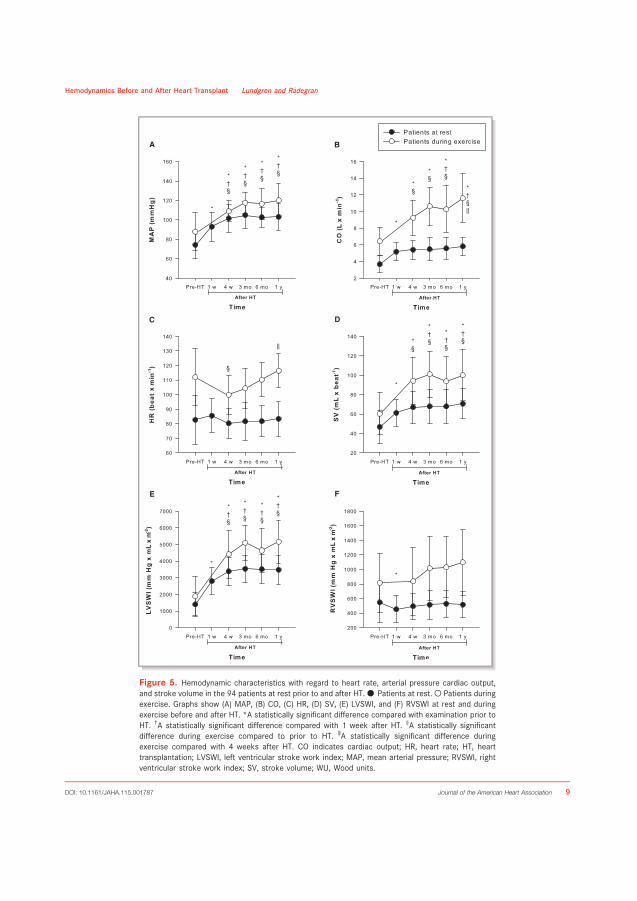
A B
C D
E F
Figure 5. Hemodynamic characteristics with regard to heart rate, arterial pressure cardiac output,and stroke volume in the 94 patients at rest prior to and after HT.● Patients at rest.○ Patients duringexercise. Graphs show (A) MAP, (B) CO, (C) HR, (D) SV, (E) LVSWI, and (F) RVSWI at rest and duringexercise before and after HT. *A statistically significant difference compared with examination prior toHT. †A statistically significant difference compared with 1 week after HT. §A statistically significantdifference during exercise compared to prior to HT. ‖A statistically significant difference duringexercise compared with 4 weeks after HT. CO indicates cardiac output; HR, heart rate; HT, hearttransplantation; LVSWI, left ventricular stroke work index; MAP, mean arterial pressure; RVSWI, rightventricular stroke work index; SV, stroke volume; WU, Wood units.
DOI: 10.1161/JAHA.115.001787 Journal of the American Heart Association 9
Hemodynamics Before and After Heart Transplant Lundgren and R�adegran
ORIG
INALRESEARCH
at UN
IVE
RSIT
ET
SSJUK
HU
SET
on September 13, 2016
http://jaha.ahajournals.org/D
ownloaded from

(Figures 4 and 5). The functional response to exercise wasalso substantially improved after HT, represented by anadequate increase in CO and SV (Figure 3); however, MPAPand particularly PAWP and MRAP remained high (Figures 2and 5). The absolute response to exercise of these pressureswas similar before and after HT, resulting in a higher relativeresponse to exercise after HT (Figure 2). Moreover, the age-dependent differences in the hemodynamic response toexercise previously reported in healthy persons7,8 were notobserved in our patients with severe end-stage HF prior to HT.Together, these findings suggest that in patients with severeHF, age may not be as dominant in affecting hemodynamicresponse to exercise as in healthy persons. The findings alsoindicate that despite normalized hemodynamics at rest, theresponse to even slight exercise after HT differed comparedwith the normal innervated heart.
Exercise Hemodynamics Prior to HeartTransplantationThe patients in our study who exercised prior to HT generallyresponded with a marked increase in PAWP and MRAP,suggesting enhanced biventricular failure on exertion(Table 2). Previous reports in healthy volunteers found a closerelationship between exercise-induced increases in PAWP andMRAP.22,23 Such a correlation was not observed in our patients(Figure 1), suggesting that the increased load on the rightventricle is not solely due to increased left ventricular fillingpressures and subsequent pulmonary venous hypertension orventricular interdependence but also is due to an exaggeratedincrease in MPAP. Indeed, the TPG in our patients, whichincreased by 5.4 mm Hg, is in line with the findings by Lewis
and colleagues, who investigated 60 patients with stable NewYork Heart Association class II to IV during slight uprightexercise.19 In contrast to the findings by Lewis et al19 andothers24,25 investigating slight upright exercise in HF patients,in our study, PVR did not significantly decrease during slightsupine exercise. This finding is also in contrast to the findings inhealthy persons reported previously8 and further supports anexaggerated increase in MPAP compared with the increase inPAWP and CO (Table 2). The increase in MPAP could be due toseveral factors. One such factor that may explain a part of theincrease is hypoxic pulmonary vasoconstriction26 related todecreased mixed venous PO2. In support of this hypothesis, themixed venous saturation decreased from 67% at rest to 26%during exercise in the 9 patients for whom these data wereavailable (data not shown).
It is well known that during exercise in healthy persons,there is great interindividual variation in TPVR.6 Despite thisknowledge, exercise-induced pulmonary hypertension wasrecently suggested to be defined as TPVR >3 WU.27,28 With amean TPVR of 7.4 WU in our patients, the preoperativeexercise TPVR was higher than in healthy persons27,28 but inline with previous results in patients with HF.19 This finding,together with the increase in TPG, suggests that although ourstudy exclusively included patients referred for HT, the resultswith regard to hemodynamics are representative for patientswith severe HF.
Hemodynamic Response to Heart TransplantationPrevious investigations of exercise after HT have used mainlynoninvasive methods and focused primarily on exercisecapacity and/or reinnervation of the transplanted heart29–38;however, a few studies have evaluated hemodynamics obtainedfrom RHC during exercise after HT.26,39–44 In 1980, Pope andcolleagues presented a report on the hemodynamic andcatecholamine response to exercise in 9 long-term HT survi-vors. They demonstrated that in denervated hearts, CO isincreased by augmented preload and the Frank Starlingmechanism early in exercise and later by the effects ofincreased levels of catecholamines.40 This report40 did notexamine patients early after HT; however, this work wasperformed in a later study of 20 patients.26 The authors thenfound that MPAP and PAWP decreased early after HT. Incontrast to these reports, we illustrated the complete hemo-dynamic picture, at rest and during exercise prior to HT, inwhich the same patients were followed repeatedly during thefirst year after HT, enabling the study of hemodynamic changesover time.
In contrast to exercise hemodynamics after HT, severalstudies have evaluated hemodynamics at rest early and lateafter HT.41–43,45–56 In line with those reports, we showed thateven at 1 week after HT, resting hemodynamics improved
Figure 6. Correlation between changes to exercise in PAWPand MPAP at 1 year after HT. HT indicates heart transplantation;PAWP, pulmonary artery wedge pressure; MPAP, mean pulmonaryartery pressure.
DOI: 10.1161/JAHA.115.001787 Journal of the American Heart Association 10
Hemodynamics Before and After Heart Transplant Lundgren and R�adegran
ORIG
INALRESEARCH
at UN
IVE
RSIT
ET
SSJUK
HU
SET
on September 13, 2016
http://jaha.ahajournals.org/D
ownloaded from

dramatically and were maintained throughout the first yearafter HT. Pulmonary artery pressures as well as CO, SV, TPVR,and LVSWI continued to improve during the first year after HT(Figures 4 and 5). In contrast to what is expected in healthypersons based on previous systematic reviews,7,8 the pulmo-nary artery pressure, PAWP, and MRAP responses to exercisewere exaggerated (Figures 2 and 4). The absolute increasewas, in fact, similar to that seen prior to HT (Figure 2),resulting in a greater percentage increase after transplanta-tion. Moreover, there was a correlation between the increasein MPAP and PAWP (Figure 6), suggesting that the MPAPresponse during exercise is caused, at least in part, by theelevated PAWP. This exaggerated increase in ventricular fillingpressures has been described previously.42,44 The reason forsimilar pressure increases during exercise after versus beforeHT, despite lower resting values, needs further investigation.It could be hypothesized that, in our population, these findingswere due to a prominent decrease in diastolic compliance indenervated hearts that reaches its maximum during slight tomoderate exercise.57 This is supported in that the denervatedheart, due to lack of sympathetic stimulation, is moredependent on the Frank Starling mechanism than is theinnervated heart.40,58,59 Factors such as lack of heartrate reserve and Bainbridge reflex, rejections, fluid retention,hypertension, myocardial ischemia, and diastolic dysfunctionhave also been suggested to account for post-transplantabnormalities.42–44,47,48 These studies, however, show diverg-ing results. The ultimate cause remains unclear and possiblyis multifactorial.42 In 1994, Kao and colleagues performedinvasive hemodynamic measurements during upright gradedexercise in 30 HT patients with normal left ventricular ejectionfraction at 3 to 16 months after HT and compared theirfindings with healthy controls.44 They showed that in theirpopulation, heart rate reserve and SV index were impairedafter HT, suggesting that during maximal exercise, chrono-tropic insufficiency and diastolic dysfunction explained theincreased filling pressures.44 These findings, however, differfrom ours. Based on what has been described previously inhealthy persons,8 the patients in our study at 1 year after HTresponded during slight supine exercise with adequateincrease in heart rate and SV. In fact, the SV in our patientswas in line with that of the healthy persons in the study byKao et al during submaximal upright exercise.44 Moreover,heart rate increased to 116.4�11.6 beats9min�1, which is inthe upper limit of what is expected during slight exercise inhealthy persons8 and in line with other previous reports in HTpatients.40 The difference between our findings and thosepresented by Kao et al could potentially be explained by thedifference in body positions in the studies; however, they mayalso, at least in part, be due to less healthy patients in thestudy by Kao et al, with low maximum oxygen consumption(12.3�4.0 mL 9 Kg�1 9 min�1) despite normal ejection
fraction.44 Moreover, in contrast to all previous reports, inthe present study, we evaluated the age-dependent differ-ences in exercise response after HT and investigated theimpact of the elevated filling pressures on pulmonaryresistance.
Age-Dependent Exercise ResponseIt has been shown previously that the hemodynamic responseto exercise differs for healthy persons aged ≤50 versus>50 years.7,8 In the most recent of these reports, Kovacs andcolleagues analyzed the hemodynamic response to exercise in222 healthy persons from 24 different studies in a systematicreview and stratified the participants according to age.8 Theresults confirmed earlier reports and showed that slight supineexercise results in an increase in CO by 85% in participants aged≤50 years and is accompanied by an increase in MPAP by 41%and a decrease in PVR and TPVR by 12% and 25%, respectively.In contrast, in participants aged >50 years, an increase in COby 71% is accompanied by an increase in MPAP by 66% and adecrease in PVR by 19%, whereas TPVR remained virtuallyunchanged.8 These findings differ from those in our HFpopulation both before and 1 year after HT. The MPAPresponse prior to HT was exaggerated in both groups whenrelated to the corresponding increase in CO, whereas the PVRand TPVR responses were attenuated (Figures 2 and 3). AfterHT, the CO response to exercise was adequate, but the MPAPresponse was still increased when related to the increase in CO(Figures 2 and 3). Because the exercise increase in PAWP waseven further exaggerated, PVR decreased in patients aged≤50 and >50 years, comparable to expectations for healthypersons.7,8 TPVR, which is not directly affected by PAWP, wasstill deranged at 1 year after HT and increased during exercisein patients aged ≤50 and >50 years (Figures 2 and 3).
In contrast to previous findings in healthy persons,7,8
MPAP, PVR, and TPVR of our patients prior to HT did not differsignificantly between patients aged ≤50 and >50 years at rest(Figures 2 and 3). Furthermore, no difference was notedbetween patients aged ≤50 and >50 years during exerciseprior to HT (Figures 2 and 3). Considering the low number ofpatients in our trial, these findings have to be interpreted withcaution. Indeed, MRAP tended to be higher in patients aged≤50 years at rest prior to HT and could indicate that thesepatients had more pronounced biventricular heart failure,which may affect the results. PAWP, CO, and SV werehowever similar in both groups, and TPG and PVR tended tobe even lower in the younger patients. Consequently,differences in disease severity between patients aged ≤50and >50 years are unlikely to play a major part in hemody-namic response. In contrast, at 1 year after transplantation,TPVR at rest and MPAP, PVR, and TPVR during exercise werehigher among patients aged >50 years (Figures 2 and 3).
DOI: 10.1161/JAHA.115.001787 Journal of the American Heart Association 11
Hemodynamics Before and After Heart Transplant Lundgren and R�adegran
ORIG
INALRESEARCH
at UN
IVE
RSIT
ET
SSJUK
HU
SET
on September 13, 2016
http://jaha.ahajournals.org/D
ownloaded from
Although the absolute and percentage responses did notdiffer significantly for any of the parameters, there was atendency toward greater changes in pressures in the patientsaged >50 years. Together, these observations suggest that inpatients with end-stage HF, the age-dependent response toexercise may be attenuated due to the severity of the disease,and that parts of this response may be restored after HT.Because MPAP and particularly PAWP remained deranged at1 year after HT, the findings also suggest that although thePVR response to exercise is normalized, PVR may notadequately reflect pulmonary circulation after HT. In thissetting, TPVR may be a better marker because it reflects theincrease in MPAP in relation to CO, independent of PAWP.
LimitationsAlthough a retrospective study such as the present hasdisadvantages compared with a prospective ditto, and thatthe long time span of our study could influence the results,the findings are of interest. This is supported by the fact thatour study population was representative with regard to similardemographic parameters and survival compared with ourentire HT population.21 However, the small sample size couldmask age-dependent differences in the response to exercise;therefore, these results must be interpreted with caution. Thisis particularly true after HT, where patients aged >50 yearsseem to have a greater increase in pulmonary and ventricularfilling pressures, whereas the response to exercise prior to HTseems to be similar between the groups, despite higherresting MRAP in the younger patients. When discussing thenormal response to exercise, one must also keep in mind thatwe did not study healthy controls in the present report.Instead, we evaluated our results based on previouslypublished systematic reviews of healthy persons. Althoughthis approach may not be optimal, given the ethical issues ofperforming heart catheterizations on healthy persons, we findour approach to be a valid alternative. Furthermore, our studyincluded only patients evaluated for HT; therefore, the findingsprior to HT may not be applicable to all patients with severeHF. Nonetheless, considering that the TPG and TPVRresponses to exercise in our patients were comparable toprevious results in patients with HF,19 we believe that ourobservations are representative with regard to hemodynamicsfor patients with severe HF. Finally, previous studies haveshown that rejections,43,44,48,60 ischemic time of the donorheart,43,44,48,60 and systemic hypertension44,60 do notaccount for the hemodynamic abnormalities seen after HT.It is possible, however, that other donor characteristics anddonor–recipient mismatches as well as patients’ medicaltherapies may influence the findings. This possibility was notinvestigated in the present study, apart from what is shown inTable 1, and further research is encouraged.
ConclusionThe present study illustrates the hemodynamic response toexercise in patients with severe end-stage HF prior to andafter HT. Our findings reveal that resting hemodynamicsrecover within the first week after HT and are maintained oreven improved throughout the first year after transplanta-tion. Moreover, the age-dependent differences in response toexercise in healthy persons were not observed in ourpatients with severe end-stage HF prior to HT. This suggeststhat the hemodynamic response to exercise may be less agedependent in HF patients than in the normal population.Finally, although the hemodynamics at rest and duringexercise were greatly improved after HT, and the functionalresponse to exercise with regard to CO and SV is adequateat 1 year after HT, there is an exaggerated increase inMPAP, PAWP, and MRAP in response to exercise. This isevident for patients aged ≤50 and >50 years and, at least inpart, is likely due to increased PAWP. As a result of theexaggerated increase in PAWP, the PVR response to exerciseis normalized after HT, masking the increased MPAP.Consequently, TPVR, which is independent of PAWP, maybe a better marker than PVR for hemodynamic evaluationsafter HT. Larger studies are encouraged to confirm thesefindings and to define the normal hemodynamic magnitudesduring exercise prior to and after HT to explain theexaggerated increase in PAWP to exercise.
AcknowledgmentsThis work is dedicated to Professor Bengt Saltin. We acknowledgethe support of the staff at the Hemodynamics Laboratory, Clinic forHeart Failure and Valvular Disease, Sk�ane University Hospital, andthe Department of Cardiology, Clinical Sciences, Lund University,Lund, Sweden.
Sources of FundingThis work was supported by unrestricted research grants fromAnna-Lisa and Sven-Erik Lundgren’s and ALF’s Foundations,the Swedish Society of Pulmonary Hypertension and ActelionPharmaceuticals Sweden AB. The funding organizations playedno role in the collection, analysis or interpretation of the dataand have no right to restrict the publishing of the article.
DisclosuresMr Lundgren reports an unrestricted research grant from TheSwedish Society of Pulmonary Hypertension, and Dr R�adegranreports unrestricted research grants from Anna-Lisa andSven-Erik Lundgren’s, Maggie Stephen’s, ALF’s and ActelionPharmaceuticals Sweden AB, during the conduct of the study.Mr Lundgren reports personal lecture fees from Actelion
DOI: 10.1161/JAHA.115.001787 Journal of the American Heart Association 12
Hemodynamics Before and After Heart Transplant Lundgren and R�adegran
ORIG
INALRESEARCH
at UN
IVE
RSIT
ET
SSJUK
HU
SET
on September 13, 2016
http://jaha.ahajournals.org/D
ownloaded from
Pharmaceuticals Sweden AB and GlaxoSmithKline outside thesubmitted work. Dr R�adegran reports personal lecture feesfrom Actelion Pharmaceuticals Sweden AB, GlaxoSmithKline,Bayer and Sandoz/Novartis outside the submitted work. DrR�adegran is, and has been primary-, or co-, investigator in;clinical PAH trials for GlaxoSmithKline, Actelion Pharmaceu-ticals Sweden AB, Pfizer, Bayer and United Therapeutics, andin clinical heart transplantation immuno-suppression trials forNovartis. The companies had no role in the data collection,analysis, or interpretation; or in the preparation or approval ofthe manuscript.
References1. Saltin B. Hemodynamic adaptations to exercise. Am J Cardiol. 1985;55:42D–
47D.
2. Saltin B, Radegran G, Koskolou MD, Roach RC. Skeletal muscle blood flow inhumans and its regulation during exercise. Acta Physiol Scand. 1998;162:421–436.
3. Shantsila E, Wrigley BJ, Blann AD, Gill PS, Lip GY. A contemporary view onendothelial function in heart failure. Eur J Heart Fail. 2012;14:873–881.
4. Galie N, Hoeper MM, Humbert M, Torbicki A, Vachiery JL, Barbera JA,Beghetti M, Corris P, Gaine S, Gibbs JS, Gomez-Sanchez MA, Jondeau G,Klepetko W, Opitz C, Peacock A, Rubin L, Zellweger M, Simonneau G; ESCCommittee for Practice Guidelines (CPG). Guidelines for the diagnosis andtreatment of pulmonary hypertension: the Task Force for the Diagnosis andTreatment of Pulmonary Hypertension of the European Society of Cardiology(ESC) and the European Respiratory Society (ERS), endorsed by theInternational Society of Heart and Lung Transplantation (ISHLT). Eur HeartJ. 2009;30:2493–2537.
5. Vachiery JL, Adir Y, Barbera JA, Champion H, Coghlan JG, Cottin V, De Marco T,Galie N, Ghio S, Gibbs JS, Martinez F, Semigran M, Simonneau G, Wells A,Seeger W. Pulmonary hypertension due to left heart diseases. J Am CollCardiol. 2013;62:D100–D108.
6. Naeije R, Chesler N. Pulmonary circulation at exercise. Compr Physiol.2012;2:711–741.
7. Kovacs G, Berghold A, Scheidl S, Olschewski H. Pulmonary arterial pressureduring rest and exercise in healthy subjects: a systematic review. Eur Respir J.2009;34:888–894.
8. Kovacs G, Olschewski A, Berghold A, Olschewski H. Pulmonary vascularresistances during exercise in normal subjects: a systematic review. Eur RespirJ. 2012;39:319–328.
9. Riley RL, Himmelstein A. Studies of the pulmonary circulation at rest andduring exercise in normal individuals and in patients with chronic pulmonarydisease. Am J Physiol. 1948;152:372–382.
10. Holmgren A, Jonsson B, Sjostrand T. Circulatory data in normal subjects atrest and during exercise in recumbent position, with special reference to thestroke volume at different work intensities. Acta Physiol Scand.1960;49:343–363.
11. Granath A, Jonsson B, Strandell T. Circulation, in healthy, old men, studied byright heart catheterization at rest and during exercise in supine and sittingposition. Acta Med Scand. 1964;176:425–446.
12. Damato AN, Galante JG, Smith WM. Hemodynamic response to treadmillexercise in normal subjects. J Appl Physiol. 1966;21:959–966.
13. Ehrsam RE, Perruchoud A, Oberholzer M, Burkart F, Herzog H. Influence of ageon pulmonary haemodynamics at rest and during supine exercise. Clin Sci(Lond). 1983;65:653–660.
14. Tolle JJ, Waxman AB, Van Horn TL, Pappagianopoulos PP, Systrom DM.Exercise-induced pulmonary arterial hypertension. Circulation.2008;118:2183–2189.
15. Oudiz RJ, Rubin LJ. Exercise-induced pulmonary arterial hypertension: a newaddition to the spectrum of pulmonary vascular diseases. Circulation.2008;118:2120–2121.
16. Kovacs G, Maier R, Aberer E, Brodmann M, Scheidl S, Tr€oster N, Hesse C,Salmhofer W, Graninger W, Gruenig E, Rubin LJ, Olschewski H. Borderlinepulmonary arterial pressure is associated with decreased exercise capacity inscleroderma. Am J Respir Crit Care Med. 2009;180:881–886.
17. Naeije R. In defence of exercise stress tests for the diagnosis of pulmonaryhypertension. Heart. 2011;97:94–95.
18. Whyte K, Hoette S, Herve P, Montani D, Ja€ıs X, Parent F, Savale L, Natali D,O’Callaghan DS, Garcia G, Sitbon O, Simonneau G, Humbert M, Chemla D. Theassociation between resting and mild-to-moderate exercise pulmonary arterypressure. Eur Respir J. 2012;39:313–318.
19. Lewis GD, Murphy RM, Shah RV, Pappagianopoulos PP, Malhotra R, Bloch KD,Systrom DM, Semigran MJ. Pulmonary vascular response patterns duringexercise in left ventricular systolic dysfunction predict exercise capacity andoutcomes. Circ Heart Fail. 2011;4:276–285.
20. Lundgren J, Radegran G. Pathophysiology and potential treatments ofpulmonary hypertension due to systolic left heart failure. Acta Physiol (Oxf).2014;211:314–333.
21. Lundgren J, Algotsson L, Kornhall B, Radegran G. Preoperative pulmonaryhypertension and its impact on survival after heart transplantation. ScandCardiovasc J. 2014;48:47–58.
22. Reeves JT, Groves BM, Cymerman A, Sutton JR, Wagner PD, Turkevich D,Houston CS. Operation Everest II: cardiac filling pressures during cycleexercise at sea level. Respir Physiol. 1990;80:147–154.
23. Reeves JT, Groves BM, Sutton JR, Wagner PD, Cymerman A, Malconian MK,Rock PB, Young PM, Houston CS. Operation Everest II: preservation of cardiacfunction at extreme altitude. J Appl Physiol. 1985;1987:531–539.
24. Sullivan MJ, Knight JD, Higginbotham MB, Cobb FR. Relation between centraland peripheral hemodynamics during exercise in patients with chronic heartfailure. Muscle blood flow is reduced with maintenance of arterial perfusionpressure. Circulation. 1989;80:769–781.
25. Janicki JS, Weber KT, Likoff MJ, Fishman AP. The pressure-flow response of thepulmonary circulation in patients with heart failure and pulmonary vasculardisease. Circulation. 1985;72:1270–1278.
26. Naeije R, Lipski A, Abramowicz M, Lejeune P, Melot C, Antoine M, De SMET JM,Leclerc JL, Primo G. Nature of pulmonary hypertension in congestive heartfailure. Effects of cardiac transplantation. Am J Respir Crit Care Med.1994;149:881–887.
27. Naeije R, Vanderpool R, Dhakal BP, Saggar R, Saggar R, Vachiery JL, Lewis GD.Exercise-induced pulmonary hypertension: physiological basis and methodo-logical concerns. Am J Respir Crit Care Med. 2013;187:576–583.
28. Lewis GD, Bossone E, Naeije R, Grunig E, Saggar R, Lancellotti P, Ghio S, VargaJ, Rajagopalan S, Oudiz R, Rubenfire M. Pulmonary vascular hemodynamicresponse to exercise in cardiopulmonary diseases. Circulation.2013;128:1470–1479.
29. Renlund DG, Taylor DO, Ensley RD, O’Connell JB, Gilbert EM, Bristow MR, MaH, Yanowitz FG. Exercise capacity after heart transplantation: influence ofdonor and recipient characteristics. J Heart Lung Transplant. 1996;15:16–24.
30. Osada N, Chaitman BR, Donohue TJ, Wolford TL, Stelken AM, Miller LW. Long-term cardiopulmonary exercise performance after heart transplantation. Am JCardiol. 1997;79:451–456.
31. Givertz MM, Hartley LH, Colucci WS. Long-term sequential changes in exercisecapacity and chronotropic responsiveness after cardiac transplantation.Circulation. 1997;96:232–237.
32. Savin WM, Haskell WL, Schroeder JS, Stinson EB. Cardiorespiratory responsesof cardiac transplant patients to graded, symptom-limited exercise. Circula-tion. 1980;62:55–60.
33. Kavanagh T, Yacoub MH, Mertens DJ, Kennedy J, Campbell RB, Sawyer P.Cardiorespiratory responses to exercise training after orthotopic cardiactransplantation. Circulation. 1988;77:162–171.
34. Kavanagh T, Mertens DJ, Shephard RJ, Beyene J, Kennedy J, Campbell R,Sawyer P, Yacoub M. Long-term cardiorespiratory results of exercise trainingfollowing cardiac transplantation. Am J Cardiol. 2003;91:190–194.
35. Lord SW, Brady S, Holt ND, Mitchell L, Dark JH, McComb JM. Exerciseresponse after cardiac transplantation: correlation with sympathetic reinner-vation. Heart. 1996;75:40–43.
36. Squires RW, Leung TC, Cyr NS, Allison TG, Johnson BD, Ballman KV, Wagner JA,Olson LJ, Frantz RP, Edwards BS, Kushwaha SS, Dearani JA, Daly RC, McGregorCG, Rodeheffer RJ. Partial normalization of the heart rate response to exerciseafter cardiac transplantation: frequency and relationship to exercise capacity.Mayo Clin Proc. 2002;77:1295–1300.
37. Richard R, Verdier JC, Duvallet A, Rosier SP, Leger P, Nignan A, Rieu M.Chronotropic competence in endurance trained heart transplant recipients:heart rate is not a limiting factor for exercise capacity. J Am Coll Cardiol.1999;33:192–197.
38. Labovitz AJ, Drimmer AM, McBride LR, Pennington DG, Willman VL, Miller LW.Exercise capacity during the first year after cardiac transplantation. Am JCardiol. 1989;64:642–645.
39. Campeau L, Pospisil L, Grondin P, Dyrda I, Lepage G. Cardiac catheterizationfindings at rest and after exercise in patients following cardiac transplantation.Am J Cardiol. 1970;25:523–528.
DOI: 10.1161/JAHA.115.001787 Journal of the American Heart Association 13
Hemodynamics Before and After Heart Transplant Lundgren and R�adegran
ORIG
INALRESEARCH
at UN
IVE
RSIT
ET
SSJUK
HU
SET
on September 13, 2016
http://jaha.ahajournals.org/D
ownloaded from

40. Pope SE, Stinson EB, Daughters GT, Schroeder JS, Ingels NB Jr, Alderman EL.Exercise response of the denervated heart in long-term cardiac transplantrecipients. Am J Cardiol. 1980;46:213–218.
41. Lindelow B, Andersson B, Waagstein F, Bergh CH. High and low pulmonaryvascular resistance in heart transplant candidates. A 5-year follow-up afterheart transplantation shows continuous reduction in resistance and nodifference in complication rate. Eur Heart J. 1999;20:148–156.
42. Pflugfelder PW, McKenzie FN, Kostuk WJ. Hemodynamic profiles at rest andduring supine exercise after orthotopic cardiac transplantation. Am J Cardiol.1988;61:1328–1333.
43. Hosenpud JD, Pantely GA, Morton MJ, Wilson RA, Norman DJ, Cobanoglu AM,Starr A. Lack of progressive “restrictive” physiology after heart transplantationdespite intervening episodes of allograft rejection: comparison of serial restand exercise hemodynamics one and 2 years after transplantation. J HeartTransplant. 1990;9:119–123.
44. Kao AC, Van TP III, Shaeffer-McCall GS, Shaw JP, Kuzil BB, Page RD,Higginbotham MB. Central and peripheral limitations to upright exercise inuntrained cardiac transplant recipients. Circulation. 1994;89:2605–2615.
45. Young JB, Leon CA, Short HD III, Noon GP, Lawrence EC, Whisennand HH, PrattCM, Goodman DA, Weilbaecher D, Quinones MA. Evolution of hemodynamicsafter orthotopic heart and heart-lung transplantation: early restrictive patternspersisting in occult fashion. J Heart Transplant. 1987;6:34–43.
46. Bhatia SJ, Kirshenbaum JM, Shemin RJ, Cohn LH, Collins JJ, Di Sesa VJ, YoungPJ, Mudge GH Jr, Sutton MG. Time course of resolution of pulmonaryhypertension and right ventricular remodeling after orthotopic cardiactransplantation. Circulation. 1987;76:819–826.
47. Corcos T, Tamburino C, Leger P, Vaissier E, Rossant P, Mattei MF, Daudon P,Gandjbakhch I, Pavie A, Cabrol A, Cabrol C. Early and late hemodynamicevaluation after cardiac transplantation: a study of 28 cases. J Am Coll Cardiol.1988;11:264–269.
48. Tamburino C, Corcos T, Feraco E, Leger P, Desruennes M, Vaissier E,Gandjbakhch I, Pavie A, Cabrol A, Cabrol C. Hemodynamic parameters one and4 weeks after cardiac transplantation. Am J Cardiol. 1989;63:635–637.
49. Bourge RC, Kirklin JK, Naftel DC, White C, Mason DA, Epstein AE. Analysis andpredictors of pulmonary vascular resistance after cardiac transplantation.J Thorac Cardiovasc Surg. 1991;101:432–444.
50. von Scheidt W, Ziegler U, Kemkes BM, Erdmann E. Heart transplantation:hemodynamics over a 5-year period. J Heart Lung Transplant. 1991;10:342–350.
51. Tenderich G, Koerner MM, Stuettgen B, Mirow N, Arusoglu L, Morshuis M,Bairaktaris A, Minami K, Koerfer R. Pre-existing elevated pulmonary vascularresistance: long-term hemodynamic follow-up and outcome of recipients afterorthotopic heart transplantation. J Cardiovasc Surg (Torino). 2000;41:215–219.
52. Delgado JF, Gomez-Sanchez MA, Saenz de la Calzada C, Sanchez V, EscribanoP, Hernandez-Afonso J, Tello R, Gomez de la Camara A, Rodriguez E,Rufilanchas JJ. Impact of mild pulmonary hypertension on mortality andpulmonary artery pressure profile after heart transplantation. J Heart LungTransplant. 2001;20:942–948.
53. Chang PP, Longenecker JC, Wang NY, Baughman KL, Conte JV, Hare JM,Kasper EK. Mild vs severe pulmonary hypertension before heart transplanta-tion: different effects on posttransplantation pulmonary hypertension andmortality. J Heart Lung Transplant. 2005;24:998–1007.
54. Goland S, Czer LS, Kass RM, De Robertis MA, Mirocha J, Coleman B, Capelli C,Raissi S, Cheng W, Fontana G, Trento A. Pre-existing pulmonary hypertensionin patients with end-stage heart failure: impact on clinical outcome andhemodynamic follow-up after orthotopic heart transplantation. J Heart LungTransplant. 2007;26:312–318.
55. Wahl A, Feller M, Wigger E, Tanner H, Stoupis C, Carrel T, Mohacsi P, Hullin R.Pretransplant pulmonary hypertension and long-term allograft right ventricularfunction. Eur J Cardiothorac Surg. 2010;37:61–67.
56. Gude E, Simonsen S, Geiran OR, Fiane AE, Gullestad L, Arora S, Relbo A,Andreassen AK. Pulmonary hypertension in heart transplantation: discrepantprognostic impact of pre-operative compared with 1-year post-operative rightheart hemodynamics. J Heart Lung Transplant. 2010;29:216–223.
57. Sullivan MJ, Cobb FR, Higginbotham MB. Stroke volume increases by similarmechanisms during upright exercise in normal men and women. Am J Cardiol.1991;67:1405–1412.
58. Donald DE. Capacity for exercise after denervation of the heart. Circulation.1968;38:225–226.
59. Carleton RA, Heller SJ, Najafi H, Clark JG. Hemodynamic performance of atransplanted human heart. Circulation. 1969;40:447–452.
60. Greenberg ML, Uretsky BF, Reddy PS, Bernstein RL, Griffith BP, Hardesty RL,Thompson ME, Bahnson HT. Long-term hemodynamic follow-up of cardiactransplant patients treated with cyclosporine and prednisone. Circulation.1985;71:487–494.
DOI: 10.1161/JAHA.115.001787 Journal of the American Heart Association 14
Hemodynamics Before and After Heart Transplant Lundgren and R�adegran
ORIG
INALRESEARCH
at UN
IVE
RSIT
ET
SSJUK
HU
SET
on September 13, 2016
http://jaha.ahajournals.org/D
ownloaded from

Paper III

Correspondence: G ö ran R å degran, The Haemodynamic Lab, The Clinic for Heart Failure and Valvular Disease, Sk å ne University Hospital, Lund, Sweden. Business Tel: � 46-(0)46-172633. Home Tel: � 46737066006. Fax: 0046-(0)46-307984. E-mail: [email protected]
(Received 10 June 2013 ; revised 16 December 2013 ; accepted 16 December 2013 )
ORIGINAL ARTICLE
Preoperative pulmonary hypertension and its impact on survival after heart transplantation
JAKOB LUNDGREN 1,2 , LARS ALGOTSSON 3 , BJ Ö RN KORNHALL 1 & G Ö RAN R Å DEGRAN 1,2
1 The Haemodynamic Lab, The Clinic for Heart Failure and Valvular Disease, Sk å ne University Hospital, Lund, Sweden, 2 Department of Cardiology, Clinical Sciences, Lund University, Lund, Sweden, and 3 Department of Cardiothoracic Surgery, Anaesthesia and Intensive Care, Sk å ne University Hospital, Lund, Sweden
Abstract Objectives. Pulmonary hypertension (PH) due to left heart disease may impair outcome after heart transplantation (HT). To evaluate to what extent previous, and present, haemodynamic criteria discriminate the impact of pre-operative-PH on sur-vival, we characterized the PH in our HT-patients according to ESC ’ s guidelines, ISHLT ’ s summary statement and ISHLT ’ s relative contraindications and criteria for early risk of death after HT. Design. Records from the 215 HT-patients in Lund during 1988 – 2010 were reviewed. Subsequent analysis included adults ( n � 94) evaluated with right-heart-catheterization at our lab, at rest before HT. End of follow-up was 30th of June 2012. Results . Survival (mean, n ) did not differ (p � ns) for the 94 HT-patients; without (13.0 years, n � 28) or with (13.9 years, n � 66) PH, passive (13.8 years, n � 50) or reactive (12.2 years, n � 13) post-capillary-PH, “ modifi ed ” passive (13.1 years, n � 40), mixed (16.6 years, n � 23), “ modifi ed ” reac-tive (12.6 years, n � 7) or non-reactive (12.2 years, n � 8) post-capillary-PH; or for ISHLT ’ s relative contraindications (12.0 years, n � 22) or increased risk of right-heart-failure and early death (16.5 years, n � 23) after HT. Conclusions. As previous and present haemodynamic criteria did not suffi ciently discriminate the impact of pre-operative-PH for survival after HT at our centre, larger multi-centre studies are encouraged to redefi ne criteria that may infl uence outcome.
Key words: heart failure , heart transplantation , haemodynamic , pulmonary hypertension , survival
Introduction
Pulmonary hypertension (PH) is defi ned by a mean pulmonary artery pressure (MPAP) � 25 mmHg, and sub-categorized by the magnitude of pulmo-nary capillary wedge pressure (PCWP), cardiac out-put (CO) and trans-pulmonary gradient (TPG) (Table Ia) (1). Major efforts have focused on clari-fying the cause of, and treatments for, pulmonary arterial hypertension (PAH), leading to improved survival in PAH, which from diagnosis, if untreated, was as low as ~ 1 – 2.8 years (2). In contrast, the pre-cise pathophysiology behind PH due to left heart disease remains, however, unclear. Furthermore, there is no specifi c therapy for the pulmonary com-ponent of PH due to left heart disease, other than optimizing left heart function. This is important as left heart disease is the most common cause of PH, where PH may be a marker of disease severity (3)
and a predictor of early mortality after heart transplantation (HT) (4).
The threshold magnitude for haemodynamic parameters, including pulmonary vascular resistance (PVR) and TPG, that increase the risk of right heart failure and early mortality after HT, furthermore remains unclear. Several characterizations of PH severity and its impact on outcome have therefore been suggested (Table Ia, b) (1,3 – 13). Systolic pulmonary artery pressure (SPAP) � 60 mmHg and either PVR � 5 WU or PVRI � 6 WU ⋅ m � 2 or TPG � 16 – 20 mmHg have been proposed as risk criteria for developing right heart failure and early death after HT (Table Ia) (4). A PVR � 5 WU or PVRI � 6 WU ⋅ m � 2 or TPG � 16 – 20 mmHg have consequently been defi ned as relative contraindica-tions for HT (4). In addition, irreversible PVR � 2.5 WU has been suggested to increase the risk for early
Scandinavian Cardiovascular Journal, 2014; Early Online: 1–12
ISSN 1401-7431 print/ISSN 1651-2006 online © 2014 Informa HealthcareDOI: 10.3109/14017431.2013.877153
Scan
d C
ardi
ovas
c J
Dow
nloa
ded
from
info
rmah
ealth
care
.com
by
UM
AS
on 0
1/27
/14
For
pers
onal
use
onl
y.
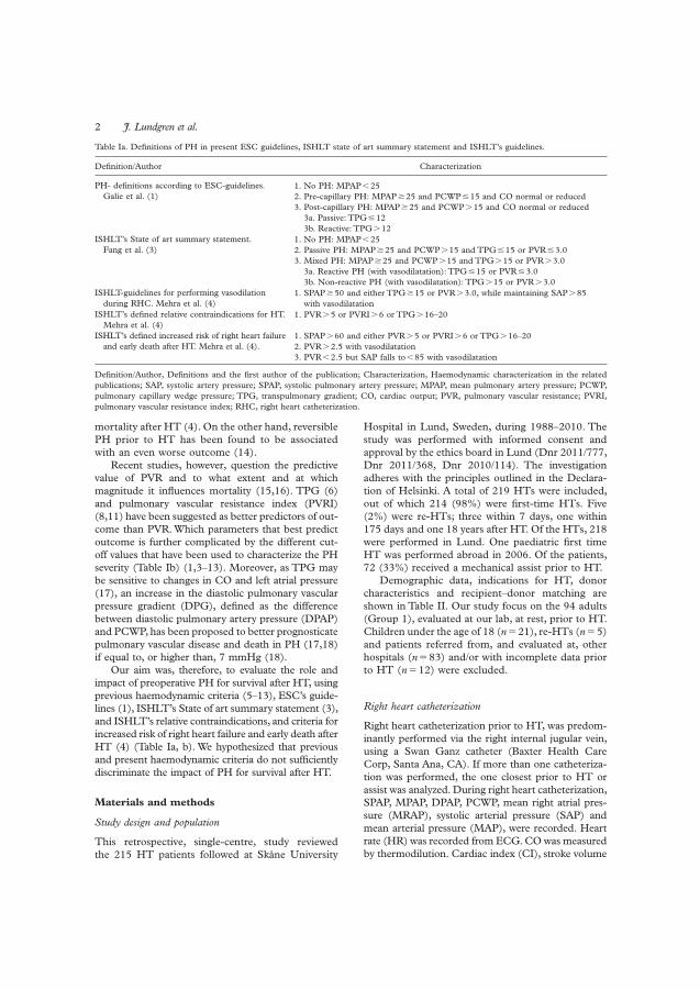
2 J. Lundgren et al.
mortality after HT (4). On the other hand, reversible PH prior to HT has been found to be associated with an even worse outcome (14).
Recent studies, however, question the predictive value of PVR and to what extent and at which magnitude it infl uences mortality (15,16). TPG (6) and pulmonary vascular resistance index (PVRI) (8,11) have been suggested as better predictors of out-come than PVR. Which parameters that best predict outcome is further complicated by the different cut-off values that have been used to characterize the PH severity (Table Ib) (1,3 – 13). Moreover, as TPG may be sensitive to changes in CO and left atrial pressure (17), an increase in the diastolic pulmonary vascular pressure gradient (DPG), defi ned as the difference between diastolic pulmonary artery pressure (DPAP) and PCWP, has been proposed to better prognosticate pulmonary vascular disease and death in PH (17,18) if equal to, or higher than, 7 mmHg (18).
Our aim was, therefore, to evaluate the role and impact of preoperative PH for survival after HT, using previous haemodynamic criteria (5 – 13), ESC ’ s guide-lines (1), ISHLT ’ s State of art summary statement (3), and ISHLT ’ s relative contraindications, and criteria for increased risk of right heart failure and early death after HT (4) (Table Ia, b). We hypothesized that previous and present haemodynamic criteria do not suffi ciently discriminate the impact of PH for survival after HT.
Materials and methods
Study design and population
This retrospective, single-centre, study reviewed the 215 HT patients followed at Sk å ne University
Hospital in Lund, Sweden, during 1988 – 2010. The study was performed with informed consent and approval by the ethics board in Lund (Dnr 2011/777, Dnr 2011/368, Dnr 2010/114). The investigation adheres with the principles outlined in the Declara-tion of Helsinki. A total of 219 HTs were included, out of which 214 (98%) were fi rst-time HTs. Five (2%) were re-HTs; three within 7 days, one within 175 days and one 18 years after HT. Of the HTs, 218 were performed in Lund. One paediatric fi rst time HT was performed abroad in 2006. Of the patients, 72 (33%) received a mechanical assist prior to HT.
Demographic data, indications for HT, donor characteristics and recipient – donor matching are shown in Table II. Our study focus on the 94 adults (Group 1), evaluated at our lab, at rest, prior to HT. Children under the age of 18 ( n � 21), re-HTs ( n � 5) and patients referred from, and evaluated at, other hospitals ( n � 83) and/or with incomplete data prior to HT ( n � 12) were excluded.
Right heart catheterization
Right heart catheterization prior to HT, was predom-inantly performed via the right internal jugular vein, using a Swan Ganz catheter (Baxter Health Care Corp, Santa Ana, CA). If more than one catheteriza-tion was performed, the one closest prior to HT or assist was analyzed. During right heart catheterization, SPAP, MPAP, DPAP, PCWP, mean right atrial pres-sure (MRAP), systolic arterial pressure (SAP) and mean arterial pressure (MAP), were recorded. Heart rate (HR) was recorded from ECG. CO was measured by thermodilution. Cardiac index (CI), stroke volume
Table Ia. Defi nitions of PH in present ESC guidelines, ISHLT state of art summary statement and ISHLT ’ s guidelines.
Defi nition/Author Characterization
PH- defi nitions according to ESC-guidelines. Galie et al. (1)
1. No PH: MPAP � 25 2. Pre-capillary PH: MPAP � 25 and PCWP � 15 and CO normal or reduced3. Post-capillary PH: MPAP � 25 and PCWP � 15 and CO normal or reduced 3a. Passive: TPG � 12 3b. Reactive: TPG � 12
ISHLT ’ s State of art summary statement. Fang et al. (3)
1. No PH: MPAP � 252. Passive PH: MPAP � 25 and PCWP � 15 and TPG � 15 or PVR � 3.03. Mixed PH: MPAP � 25 and PCWP � 15 and TPG � 15 or PVR � 3.0 3a. Reactive PH (with vasodilatation): TPG � 15 or PVR � 3.0 3b. Non-reactive PH (with vasodilatation): TPG � 15 or PVR � 3.0
ISHLT-guidelines for performing vasodilation during RHC. Mehra et al. (4)
1. SPAP � 50 and either TPG � 15 or PVR � 3.0, while maintaining SAP � 85 with vasodilatation
ISHLT ’ s defi ned relative contraindications for HT. Mehra et al. (4)
1. PVR � 5 or PVRI � 6 or TPG � 16 – 20
ISHLT ’ s defi ned increased risk of right heart failure and early death after HT. Mehra et al. (4).
1. SPAP � 60 and either PVR � 5 or PVRI � 6 or TPG � 16 – 202. PVR � 2.5 with vasodilatation3. PVR � 2.5 but SAP falls to � 85 with vasodilatation
Defi nition/Author, Defi nitions and the fi rst author of the publication; Characterization, Haemodynamic characterization in the related publications; SAP, systolic artery pressure; SPAP, systolic pulmonary artery pressure; MPAP, mean pulmonary artery pressure; PCWP, pulmonary capillary wedge pressure; TPG, transpulmonary gradient; CO, cardiac output; PVR, pulmonary vascular resistance; PVRI, pulmonary vascular resistance index; RHC, right heart catheterization.
Scan
d C
ardi
ovas
c J
Dow
nloa
ded
from
info
rmah
ealth
care
.com
by
UM
AS
on 0
1/27
/14
For
pers
onal
use
onl
y.
Pulmonary hypertension and heart transplantation 3
Table Ib. Characterization of HT-patients according to PH in previous publications.
Author/Period Characterization Patients Investigation aim Results
Costard-Jackle and Fowler (5) 1980 – 1988
1. “ Low risk ” : PVR � 2.5 or PVR � 2.5 and SAP � 85 with vasodilatation
218 3-month mortality
– Increased 3-month mortality in high-risk group
2. “ High risk ” : PVR � 2.5 or
PVR � 2.5 but SAP � 85 with vasodilatation
72
Murali et al. (6) 1980 – 1991
1. TPG � 15 and PVR � 5 2. TPG � 15 and PVR � 5 3. TPG � 15 and PVR � 5 4. TPG � 15 and PVR � 5
332 15 46 32
0 – 2, 3 – 7 and 8 – 30 days mortality
– Increased 0 – 2-day mortality in severe PH pre-HT (group 4)
– Increased 30-day mortality with TPG � 15
Lindelow et al. (7) 1988 – 1990
1. “ Low PVR ” : PVR � 3 2. “ High PVR ” : PVR � 3 and
MAP � 70 with vasodilatation
44 36
Short-, intermediate- and long-term outcome
– No difference in mortality between groups
Delgado et al. (8) 1991 – 1996
1. No PH: TPG � 12 and PVR � 2.5
2. PH: TPG � 12 and/or PVR � 2.5
71
41
30 – day mortality – Increased 30-day mortality with PH
Klotz et al. (9) 1998 – 2001
1. No PH: TPG � 12 and PVR � 2.5
2. PH: TPG � 12 and/or PVR � 2.5
90
61
1-year mortality – No difference in 1-year mortality between patients without PH vs. reversible PH
Chang et al. (10) 1983 – 2000
1. No PH: PVR � 2.5 2. Mild/moderate PH: PVR 2.5 – 4.9
3. Severe PH: PVR � 5
101 55
16
Short- and long-term outcome
– Increased 1-, 3- and 6-month survival with mild/moderate PH
– Increased 1-year mortality with severe PH, otherwise unchanged
Goland et al. (11) 1989 – 2004
1. No PH: PVR � 3, TPG � 10 2. Mild – Moderate PH:
PVR 3 – 6, TPG 10 – 20 3. Severe PH: PVR � 6, TPG � 20
266 112
32
Short- and long-term outcome
– No difference in mortality with pre-HT PH
– Increased long-term mortality with sustained PH after HT
Klotz et al. (12) 1996 – 2005
1. No PH: TPG � 12 and PVR � 2.5
2. Rev PH: TPG � 12 and PVR � 2.5 with vasodilatation
168
49
Short- and long-term outcome
– No difference in mortality between groups
Gude et al. (13) 1983 – 2007
1. No PH: 2 – 3 of: SPAP � 50, TPG � 10 and PVR � 2.5
365 Long-term outcome
– No difference in long-term mortality with pre-HT PH
2. PH: 2 – 3 of: SPAP � 50, TPG � 10 and PVR � 2.5 with vasodilatation
135 – Increased long-term mortality with sustained PH after HT
Author/Period, First author and years of investigation in the related publication; Characterization, PH-characterization in the related publications; Patients, Number of patients investigated in each group in the related publications; Investigation aim, The outcome investigated in the related publications; Results, Summary of fi ndings in the related publications; SAP, systolic artery pressure; SPAP, systolic pulmonary artery pressure; MPAP, mean pulmonary artery pressure; PCWP, pulmonary capillary wedge pressure; TPG, transpulmonary gradient; CO, cardiac output; PVR, pulmonary vascular resistance.
(SV), stroke volume index (SVI), left ventricular stroke work index, (LVSWI), right ventricular stroke work index (RVSWI), PVR, PVRI, TPG, DPG and systemic vascular resistance (SVR) were calculated by the following formulas: CI � CO/body surface area (BSA), SV � CO/HR, SVI � SV/BSA, LVSWI � (MAP-PCWP) ⋅ SVI ⋅ 0.0136, RVSWI � (MPAP-MRAP) ⋅ SVI ⋅ 0.0136, where 0.0136 is a conversion factor, PVR � TPG/CO, PVRI � TPG/CI, TPG � MPAP-PCWP, DPG � DPAP-PCWP and SVR � (MAP-MRAP)/CO.
Data analysis and characterization
Our HT patients were characterized according to i) previous haemodynamic risk characterizations
(5 – 13), ii) ESC ’ s guidelines (1), iii) ISHLT ’ s State of art summary statement (3), iv) ISHLT ’ s relative con-traindications and v) ISHLT ’ s criteria for increased risk of right heart failure and early death after HT (4) (Table Ia, b). To separate the patients into groups according to ISHLT ’ s State of art summary statement (Table Ia) (3), the criteria for passive and reactive mixed PH were modifi ed. “ Modifi ed ” passive PH was redefi ned as TPG � 15 and PVR � 3 instead of TPG � 15 or PVR � 3. “ Modifi ed ” reactive mixed PH was redefi ned as TPG � 15 and PVR � 3 with vasodi-latation instead of TPG � 15 or PVR � 3 with vasodi-latation.
The 94 patients in Group 1 were furthermore characterized according to whether or not they were re-evaluated prior to HT at rest with vasodilatation
Scan
d C
ardi
ovas
c J
Dow
nloa
ded
from
info
rmah
ealth
care
.com
by
UM
AS
on 0
1/27
/14
For
pers
onal
use
onl
y.

4 J. Lundgren et al.
Table II. Characteristics for the entire HT- and study population at Sk å ne University Hospital in Lund during 1988 – 2010.
Risk factor category and group *
Entire population chatacteristics Study population characteristics
Mean SD N % Mean SD N %
Recipient characteristics Gender of recipient 215 94 Female 68 31.6 24 25.6 Age of recipient (years) 44.6 17.2 215 51.6 9.9 94 � 10 9 4.2 0 011 – 20 24 11.1 1 1.121 – 30 8 3.7 4 4.331 – 40 17 7.9 6 6.441 – 50 42 19.5 21 22.351 – 60 85 39.5 45 47.961 � 30 14.0 17 18.1 Recipient ’ s height (cm) 167.3 25.3 197 173.4 7.9 93 Recipient ’ s weight (kg) 68.4 18.9 198 75.3 15.0 93 Recipient ’ s bloodgroup 215 94 0 71 33.0 31 33.0A 109 50.7 46 48.9B 19 8.8 8 8.5AB 16 7.4 9 9.6 Time on waiting list (days) 151.3 200.4 219 148.6 179.6 94 Indication for HT 215 94 ARVC 5 2.3 0 0Cardiac Tumour 1 0.5 0 0CHD 11 5.1 1 1.1CVR 1 0.5 0 0DCM 110 51.2 52 55.3HCM 10 4.7 8 8.5Myocarditis 13 6.0 6 6.4IHD 58 27.0 24 25.6RCM 6 2.8 3 3.2 Donor characteristics Gender of donor 219 94 Female 81 37.0 36 38.3 Age of donor (years) 40.0 15.7 219 43.9 13.7 94 Donor ’ s height (cm) 172.4 18.7 210 176.3 9.5 92 Donor ’ s weight (kg) 74.1 18.5 211 79.4 13.5 93 Ischaemic time (min) 184.7 63.2 201 182.6 61.5 86 Recipient – donor matching Age difference (years) 12.5 10.8 219 12.7 10.6 94 Sex matching 219 94 Sex-matched 160 73.1 71 75.5 ABO-matching 219 94 Identic 184 84.0 82 87.2Compatible 32 14.6 12 12.8Incompatible 3 1.4 0 0
ARVC, Arrythmogenic right ventricular cardiomyopathy; CHD, congenital heart disease; CVR, Chronic vascular rejection; DCM, Dilated cardiomyopathy; HCM, Hypertrophic cardiomyopathy; IHD, Ischaemic heart disease; RCM, Restrictive cardiomyopathy. * Risk factor category is marked in bold text and corresponding group is marked in normal text.
(Group 2, n � 23) or mechanical assist (Group 3b, n � 12), the latter performed 77 47 days after implantation (Table III). Survival was compared for the different PH groups and characterizations.
Drugs and devices
Acute and sustained vaso-reactivity was assessed, according to ISHLT ’ s guidelines (4), by a vasodilator
test or mechanical assist, respectively. Twenty-three patients were exposed to a vasodilator, either intravenous nitroprusside (Nitropress, Hospira Inc, Lake Forest, USA) or nitric oxide (NO, INOmax, INO Therapeutics AB, Liding ö , Sweden) inhalation (INOvent, INO Therapeutics AB, Liding ö , Sweden). The doses for nitroprusside were ~ 0.5, 1, 2, 4 and 8 μ g ⋅ kg � 1 ⋅ min � 1 and for NO were 20 or 40 parts per million (ppm). Of the 21 patients in Group 1 that
Scan
d C
ardi
ovas
c J
Dow
nloa
ded
from
info
rmah
ealth
care
.com
by
UM
AS
on 0
1/27
/14
For
pers
onal
use
onl
y.
Pulmonary hypertension and heart transplantation 5
received a mechanical assist (Group 3a), fi ve were HeartMate IP (Thoratec, Pleasanton, CA, USA), fi ve HeartMate VE (Thoratec, Pleasanton, CA, USA), three Jarvik 2000 (Jarvik Heart Inc., New York, NY, USA), one Abiomed (Abiomed Inc, Danvers, MA, USA) and seven HeartMate II (Thoratec, Pleasanton, CA, USA). Of the 12 patients with assist (Group 3b) who were haemodynamically re-evaluated prior to HT, three had a HeartMate IP, four HeartMate VE, two Jarvik 2000 and three HeartMate II.
Statistics
A SigmaStat System (SigmaPlot 11.0) was used for statistical analysis. Parametric or non-parametric statistics were used depending on the distribution of data; that is paired t-test or Wilcoxon signed rank test, respectively, when comparing one group prior to- and after an intervention, and a t-test or a Mann – Whitney rank sum test when two groups were compared. Survival curves were plotted using the Kaplan – Meier method. The Log-Rank test was performed when
comparing two or more groups. Last day of follow-up was 30th of June 2012. A p � 0.05 was considered signifi cant. All values are mean SD.
Results
Haemodynamic characterization at baseline with vasodilatation or mechanical assist
The haemodynamic measurements in Group 1 ( n � 94), prior to HT, are shown in Table III. The 23 patients exposed to vasodilatation showed a decrease in MPAP ( p � 0.048), PVR ( p � 0.001) and PVRI ( p � 0.001), by 4.6 10.4 mmHg, 1.6 1.3 WU and 2.9 2.3 WU · m � 2 , respectively, at the cost of SAP ( p � 0.015) and MAP ( p � 0.013), which decreased by 10.5 16.8 and 8.0 14.0 mmHg, respectively (Table III). PVR did not decrease to � 2.5 WU in 12 patients. In four patients where PVR decreased to � 2.5 WU, SAP decreased to � 85 mmHg, identify-ing 16 patients with an increased risk of right heart failure and early death after HT.
Table III. Haemodynamic characteristics of our HT study population without or with vasodilation or mechanical assist.
Parameter
Group 1 Group 2Group 2 after vasodilatation Group 3a Group 3b
Group 3b with mechanical assist
SignMean SD Mean SD Mean SD Mean SD Mean SD Mean SD
Patients 94 23 23 21 12 12SAP (mmHg) 99.3 16.9 102.8 15.8 92.3 § 17.8 92.4 19.7 92.5 24.9 – – § MAP (mmHg) 74.1 13.6 75.7 12.3 67.7 § 15.6 71.4 16.5 70.5 20.7 – – § SPAP (mmHg) 45.4 13.3 52.4 * 15.0 47.9 14.3 46.2 11.4 43.3 11.3 24.3 # 7.7 * , #DPAP (mmHg) 19.3 8.0 22.4 8.9 18.9 § 8.5 20.8 8.2 19.5 9.5 7.1 # 6.7 § , #MPAP (mmHg) 30.1 8.8 34.6 * 9.6 30.0 § 10.8 31.3 7.6 29.3 7.8 14.7 # 6.3 * , § , #PCWP (mmHg) 20.5 8.2 22.6 9.1 20.7 12.7 23.2 7.1 21.8 8.3 5.1 # 6.4 #TPG (mmHg) 9.6 3.5 12.0 * 3.6 9.4 § 4.5 8.1 * 3.9 7.5 * 2.8 9.6 3.1 * , § DPG (mmHg) � 1.2 4.6 � 0.1 4.2 � 2.6 6.9 � 2.3 4.7 � 2.3 5.4 2.0 # 4.2 #MRAP (mmHg) 7.9 6.1 9.5 6.6 7.7 7.9 7.6 6.3 6.7 7.7 2.8 # 4.3 #HR (beat · min � 1 ) 82.6 16.8 79.7 15.7 81.3 17.3 93.1 * 17.4 88.3 14.8 82.5 10.7 * CO (L · min � 1 ) 3.7 1.0 3.0 * 0.7 3.8 1.5 3.5 0.8 3.4 0.7 4.3 # 1.1 * , #CI (L · min � 1 ⋅ m � 2 ) 1.9 0.5 1.7 * 0.4 2.1 0.8 1.8 0.4 1.8 0.4 2.3 # 0.6 * , #SV (mL · beat � 1 ) 46.7 17.1 39.4 11.1 47.4 18.8 38.7 11.3 39.5 9.5 55.7 # 13.5 #SVI (mL · beat � 1 · m � 2 ) 24.7 8.7 21.6 6.0 26.3 10.1 20.5 6.1 21.1 5.8 29.7 # 8.0 #SVR (WU) 19.0 6.5 22.8 * 7.2 19.6 11.4 17.8 5.0 18.0 5.9 – – * PVR (WU) 2.8 1.3 4.2 * 1.6 2.6 § 0.9 2.4 1.1 2.2 0.7 2.2 0.9 * , § PVRI (WU · m � 2 ) 5.2 2.3 7.6 * 2.7 4.7 § 1.6 4.5 * 2.1 4.2 * 1.3 4.4 1.6 * LVSWI (g ⋅ beat � 1 · m � 2 ) 19.0 9.9 15.7 6.1 16.3 5.9 14.7 7.6 15.1 8.5 33.6 # 16.4 #RVSWI (g ⋅ beat � 1 · m � 2 ) 7.4 3.7 7.5 3.7 7.7 3.3 6.7 2.8 6.6 2.3 4.5 # 1.6 #Survival (years) 13.7 13.8 13.7 13.2
SAP, systolic arterial pressure; MAP, mean atrial pressure; SPAP, systolic pulmonary artery pressure; DPAP, diastolic pulmonary artery pressure; MPAP, mean pulmonary artery pressure; PCWP, pulmonary capillary wedge pressure; TPG, transpulmonary gradient; DPG, diastolic pulmonary vascular pressure gradient; MRAP, mean atrial pressure; HR, heart rate; CO, cardiac output; CI, cardiac index; SV, stroke volume; SVI, stroke volume index; SVR, systemic vascular resistance; PVR, pulmonary vascular resistance; PVRI, pulmonary vascular resistance index; LVSWI, left ventricular stroke work index; RVSWI, right ventricular stroke work index. SAP, MAP and SVR are not reported in patients with mechanical assist due to use of both pulsatile- and continuous-fl ow devices; Group 1, HT study population; Group 2, patients who performed vasodilatation test during RHC; Group 3a, patients bridged to HT with mechanical assist; Group 3b, patients bridged to HT with mechanical assist who were re-evaluated whilst on mechanical assist. * Indicates a statistical signifi cance ( p � 0.05) compared with Group 1 (study population). § Indicates statistical signifi cance ( p � 0.05) in Group 2 after vasodilation as compared with Group 2 prior to vasodilatation. # Indicates statistical signifi cance ( p � 0.05) in Group 3b with mechanical assist as compared with Group 3b without mechanical assist.
Scan
d C
ardi
ovas
c J
Dow
nloa
ded
from
info
rmah
ealth
care
.com
by
UM
AS
on 0
1/27
/14
For
pers
onal
use
onl
y.
6 J. Lundgren et al.
In the patients re-evaluated 77 47 days after assist-implantation (Group 3b), MPAP, PCWP and MRAP decreased by 14.7 5.4 ( p � 0.001), 16.8 6.5 ( p � 0.001) and 3.8 5.1 ( p � 0.027) mmHg, respec-tively, to normal magnitudes (Table III). CI, SVI and DPG increased by 0.5 0.4 L ⋅ min � 1 ⋅ m � 2 ( p � 0.003), 8.1 4.7 mL ⋅ m � 2 ( p � 0.001) and 4.3 3.8 mmHg ( p � 0.002) after implantation, respectively. PVR, PVRI and TPG remained unchanged (p � ns). Five of the 12 patients with assist, that were re-evaluated, still exhibited an irreversible PVR � 2.5 WU prior to HT; resulting together with the 16 patients remaining at increased risk of right heart failure and early death after acute vasodilatation (as mentioned above) in 21 patients with an increased risk of right heart failure and early death. If including the two patients with SPAP � 60 mmHg and either, PVR � 5 WU or PVRI � 6 WU ⋅ m � 2 or TPG � 16 – 20 mmHg, a total of 23 patients were considered at increased risk of right heart failure and early death after HT (Table IV, Supplementary Table I to be found online at http://informahealthcare.com/doi/abs/10.3109/14017431.2013.877153).
Furthermore, 66 of the 94 patients in Group 1 showed PH prior to HT. Of these, 63 had a post-capillary PH due to left heart disease (Table IV, Supplementary Table II to be found online at http://informahealthcare.com/doi/abs/10.3109/14017431.2013.877153) and three had a pre-capillary PH according to present defi nitions. Forty patients with post-capillary PH exhibited a “ modifi ed ” passive PH and 23 exhibited a mixed PH (Table IV, Supplemen-tary Table III to be found online at http://informa-healthcare.com/doi/abs/10.3109/14017431.2013.877153).
Fifteen of the 23 patients with mixed PH were assessed for acute vaso-reactivity. Of these, eight patients had a non-reactive PH prior to HT (Table IV, Supplementary Table III to be found online at http://informahealthcare.com/doi/abs/10.3109/14017431.2013.877153). DPG equal to or above the cut-off value of 7 mmHg was only present in three patients.
Survival after HT
Mean survival of the 94 versus all 215 HT patients was 13.7 versus 15.1 years, respectively; 1-, 5- and 10-year survival was 92.6%, 81.1% and 71.1%, versus 91.3%, 82.6% and 71.3%, respectively, not signifi cantly different from each other (Figure 1a). Mean follow-up of the 94 versus all 215 HT patients was 8.2 5.8 and 8.2 5.6 years, respectively (p � ns). Survival did neither differ between groups, with suffi cient number of patients, characterized depending on the magnitude of SPAP, MPAP, PCWP,
TPG and PVR (Table IV). At the end of follow-up two of the three patients with DPG � 7 mmHg were still alive with a mean follow-up of 3.2 1.9 years. The third patient died 6.7 years after HT due to acute pulmonary embolism.
Moreover, survival (mean, n) of the 94 HT patients (Group 1) did not differ (p � ns); for those without (13.0 years, n � 28) or with (13.9 years, n � 66) PH (Figure 1b), passive (13.8 years, n � 50) or reactive (12.2 years, n � 13) post-capillary PH (Table IV, Supplementary Table II to be found online at http://informahealthcare.com/doi/abs/10.3109/14017431.2013.877153, Figure 1c); “ mod-ifi ed ” passive (13.1 years, n � 40) or mixed (16.6 years, n � 23) post-capillary PH (Table IV, Supple-mentary Table II to be found online at http://informahealthcare.com/doi/abs/10.3109/14017431.2013.877153, Figure 1d); “ modifi ed ” reactive (12.6 years, n � 7) or non-reactive (12.2 years, n � 8) PH (Table IV, Supplementary Table III to be found online at http://informahealthcare.com/doi/abs/10.3109/14017431.2013.877153); or for those with ISHLT ’ s relative contraindications (12.0 years, n � 22) and criteria for increased risk of right heart failure and early death (9.1 years, n � 7) after HT, or for those assessed with vasodilator or assist that remained in the high-risk group (16.2 years, n � 21) (Table IV, Supplementary Table I to be found online at http://informahealthcare.com/doi/abs/10.3109/14017431.2013.877153, Figure 1e). Combining the latter two groups, 23 patients were considered at increased risk of right-heart-failure and early death after HT. Their survival (16.5 years, n � 23) did, however, not differ (p � ns) from the rest.
In addition, the survival of the patients with base-line PH who received LVAD ( n � 18) versus those patients with baseline PH who did not receive LVAD ( n � 48) did not differ (p � ns). Their mean survival was similar (p � ns); for example 14.0 years for patients with LVAD ( n � 18) versus 13.6 years for patients without LVAD ( n � 48).
With regards to early mortality, three of the 94 patients in the study population died within 24 h after HT, another two within 30 days, one after 330 days and one after 332 days; resulting in seven deaths within the fi rst year after HT. Four of these patients had post-capillary PH prior to HT (three passive and one “ modifi ed ” reactive) and three did not have PH. None of these patients had a DPG � 7 mmHg. The causes of death were vasoplegia in two cases, cere-brovascular insult in two and primary organ failure, cardiac fi brosis and liver cirrhosis in one case each.
Scan
d C
ardi
ovas
c J
Dow
nloa
ded
from
info
rmah
ealth
care
.com
by
UM
AS
on 0
1/27
/14
For
pers
onal
use
onl
y.

Pulmonary hypertension and heart transplantation 7 T
able
IV.
Cha
ract
eriz
atio
n of
our
HT
-stu
dy p
opul
atio
n ac
cord
ing
to P
H-c
hara
cter
izat
ion
in p
revi
ous
publ
icat
ions
and
cur
rent
gui
delin
es.
Aut
hor
Cha
ract
eriz
atio
nP
atie
nts
(n)
SPA
P(m
mH
g)M
PAP
(mm
Hg)
PC
WP
(mm
Hg)
TP
G(m
mH
g)P
VR
(WU
)
Mea
n su
rviv
al(Y
ears
)
Cos
tard
-J ä c
kle
and
Fow
ler
(5)
1. “
Low
ris
k ” :
PV
R �
2.5
or
PV
R �
2.5
and
SA
P �
85
wit
h va
sodi
lata
tion
5542
.3
13.
028
.0
9.2
20.3
9
.57.
8
2.3
2.0
0
.412
.42.
“ H
igh
risk
” : P
VR
� 2
.5 o
r P
VR
� 2
.5 b
ut S
AP
� 8
5 w
ith
vaso
dila
tati
on39
48.2
1
3.0
31.9
7
.921
.5
9.1
10.4
4
.13.
1
0.7
14.6
Mur
ali
et a
l. (6
)1.
TP
G �
15
and
PV
R �
579
42.6
1
2.1
28.5
8
.620
.0
8.6
8.5
2
.42.
4
0.7
14.0
2. T
PG
� 1
5 an
d P
VR
� 5
556
.4
11.
836
.2
3.3
23.4
4
.712
.8
1.8
6.3
0
.810
.33.
TP
G �
15
and
PV
R �
57
57.3
1
0.9
37.1
5
.821
.1
6.4
16.0
1
.24.
0
0.7
8.4
4. T
PG
� 1
5 an
d P
VR
� 5
367
.3
7.2
43.3
3
.825
.7
3.8
17.7
2
.95.
5
0.5
10.9
Lin
delo
w e
t al
. (7
)1.
“ L
ow P
VR
” : P
VR
� 3
6442
.8
12.
628
.3
8.8
20.2
8
.78.
1
2.5
2.1
0
.512
.7 *
2. “
Hig
h P
VR
” : P
VR
� 3
and
MA
P �
70
wit
h va
sodi
lata
tion
346
.0
17.
132
.7
9.7
25.3
1
1.0
7.3
1
.52.
6
0.3
1.4
3. E
xclu
ded:
2751
.8
13.
234
.3
7.7
21.8
7
.212
.5
3.3
4.2
1
.216
.4 *
Del
gado
et
al.
(8)
1. N
o P
H: T
PG
� 1
2 an
d P
VR
� 2
.551
41.0
1
2.5
27.1
8
.919
.4
8.8
7.8
2
.11.
9
0.4
12.8
2. P
H: T
PG
� 1
2 an
d/or
PV
R �
2.5
4350
.4
12.
533
.5
7.5
21.8
7
.311
.7
3.7
3.8
1
.314
.6K
lotz
et
al.
(9)
1. N
o P
H: T
PG
� 1
2 an
d P
VR
� 2
.546
40.3
1
1.6
26.6
8
.319
.2
8.4
7.4
2
.21.
9
0.4
13.3
2. P
H: T
PG
� 1
2 an
d/or
PV
R �
2.5
4850
.0
13.
233
.3
8.2
21.7
7
.911
.6
3.3
3.7
1
.314
.1C
hang
et
al.
(10)
1. N
o P
H:
PV
R �
2.5
4740
.6
11.
627
.0
8.6
19.5
8
.57.
5
2.3
1.9
0
.412
.52.
Mild
/mod
erat
e P
H:
PV
R 2
.5 – 4
.939
47.7
1
2.7
31.9
8
.220
.9
8.3
11.0
3
.13.
2
0.6
14.1
3. S
ever
e P
H:
PV
R �
58
60.5
1
1.3
38.9
4
.924
.3
4.3
14.6
3
.26.
0
0.8
12.2
Gol
and
et a
l. (1
1)1.
No
PH
: P
VR
� 3
, TP
G �
10
4440
.6
11.
727
.6
8.5
20.7
8
.46.
9
1.8
2.0
0
.512
.52.
Mild
– Mod
erat
e P
H:
PV
R 3
– 6, T
PG
10 –
2046
48.3
1
2.9
31.7
8
.720
.1
8.3
11.6
2
.63.
3
1.0
14.3
3. S
ever
e P
H:
PV
R �
6, T
PG
� 2
04
61.3
1
3.6
38.5
5
.523
.3
4.3
15.3
3
.96.
6
0.7
7.8
Klo
tz e
t al
. (1
2)1.
No
PH
: TP
G �
12
and
PV
R �
2.5
5240
.9
12.
427
.1
8.8
19.4
8
.87.
7
2.2
1.9
0
.412
.42.
Rev
PH
: TP
G �
12
and
PV
R �
2.5
wit
h va
sodi
lata
tion
653
.5
12.
137
.0
7.9
30.7
9
.26.
3
1.8
1.8
0
.67.
03.
Exc
lude
d.36
49.8
1
2.4
32.9
7
.321
.0
7.0
11.9
3
.43.
8
1.3
15.6
Gud
e et
al.
(13)
1. N
o P
H:
2 – 3
of:
SPA
P �
50,
TP
G �
10
and
PV
R �
2.5
5239
.1
10.
826
.4
8.1
19.0
8
.37.
4
2.0
2.0
0
.512
.62.
PH
: 2 –
3 of
: S
PAP
� 5
0, T
PG
� 1
0 an
d P
VR
� 2
.5 w
ith
vaso
dila
tati
on42
52.5
1
2.3
34.4
7
.722
.3
7.8
12.2
3
.13.
8
1.3
14.5
PH
-defi
nit
ion
acco
rdin
g to
E
SC
-gui
delin
es b
y G
alie
et
al.
(1)
1. N
o P
H:
2. P
re-c
apill
ary
PH
:3.
Pos
t-ca
pilla
ry P
H:
3
a P
assi
ve:
3
b R
eact
ive:
28 3 63 50 13
31.3
6
.342
.3
6.1
51.5
1
1.1
49.7
1
0.6
58.1
9
.9
19.2
3
.527
.3
1.5
34.7
6
.233
.7
6.2
38.4
4
.7
11.0
3
.912
.7
2.1
24.8
5
.825
.3
6.0
22.7
4
.1
8.2
2
.414
.7
1.2
9.9
3
.78.
4
2.2
15.7
2
.1
2.2
0
.73.
2
0.8
3.1
1
.42.
6
1.0
4.9
1
.2
13.0 XX
14.0
13.8
12.2
(Con
tinue
d)
Scan
d C
ardi
ovas
c J
Dow
nloa
ded
from
info
rmah
ealth
care
.com
by
UM
AS
on 0
1/27
/14
For
pers
onal
use
onl
y.

8 J. Lundgren et al.
Discussion
The present study shows that short- and long-term survival was similar after HT, no matter whether or not the patients had preoperative post-capillary PH, irrespective of type and PH characterization used (Table IV, Supplementary Tables I – III to be found online at http://informahealthcare.com/doi/abs/10.3109/14017431.2013.877153), Figure 1b – e). Thus, preoperative PH was, as defi ned previously and at present (1,3 – 13), at our centre not as decisive for outcome as previously postulated (Figure 1b – e). Consequently, even though we still consider preop-erative, post-capillary, PH to be a risk factor for right heart failure and early death after HT, previous and present, haemodynamic criteria (Tables I and IV, Supplementary Tables I – III to be found online at http://informahealthcare.com/doi/abs/10.3109/14017431.2013.877153) (1,3 – 13), do not suffi ciently dis-criminate the impact of PH for survival after HT. Consequently, new haemodynamic risk criteria are urged for.
Previous studies have proposed different thresh-olds, of various haemodynamic parameters that may predict outcome after HT (Table I) (1,3 – 13). Of our 94 patients evaluated before HT, 28 did not have and 66 had PH (1). However, survival at our centre was not impaired in the HT patients with preoperative post-capillary PH, irrespective of severity and hae-modynamic criterion analyzed (Table IV, Supple-mentary Table I – III to be found online at http://informahealthcare.com/doi/abs/10.3109/14017431.2013.877153), Figure 1b – e). Thus, in support of recent fi ndings (11,13,15,16), the precise magnitude of preoperative elevation of PVR and TPG, or other haemodynamic parameters that may infl uence out-come after HT remains unclear. DPG � 7 mmHg, which recently was suggested to indicate severe pul-monary vascular disease and increased mortality in patients with PH due to left heart disease (18), was only observed in three of our 94 patients prior to HT. If DPG � 7 is assumed as a new risk criterion also for outcome after HT, the low DPG in our study could be a reason for the good outcome in our PH patients. The low DPG could furthermore also sug-gest that the PH in our population was mainly due to passive congestion, rather than pulmonary vascu-lar remodelling.
Our study furthermore confi rms that mechanical assist prior to HT improves haemodynamics, making more patients eligible for HT (15,16,19 – 21). Of interest, a RVSWI � 5 g ⋅ beat � 1 ⋅ m � 2 in HT candi-dates, or a RVSWI � 399 mmHg · mL · m � 2 prior to mechanical assist implantation, has been described as risk factor for complications (22,23). In our material, four of twelve patients re-evaluated on mechanical assist had a RVSWI below the levels thought to A
utho
rC
hara
cter
izat
ion
Pat
ient
s(n
)S
PAP
(mm
Hg)
MPA
P(m
mH
g)P
CW
P(m
mH
g)T
PG
(mm
Hg)
PV
R(W
U)
Mea
n su
rviv
al(Y
ears
)
Sug
gest
ed P
H-d
efi n
itio
n1.
No
PH
2831
.3
6.3
19.2
3
.511
.0
3.9
8.2
2
.42.
2
0.7
13.0
acco
rdin
g to
Fan
g et
al.
(3)
2. P
re-c
apill
ary
PH
:3
42.3
6
.127
.3
1.5
12.7
2
.114
.7
1.2
3.2
0
.8X
X3.
“ M
odifi
ed ”
Pas
sive
PH
:39
49.2
1
0.5
33.4
6
.425
.3
6.0
8.1
2
.52.
2
0.5
13.8
4. M
ixed
PH
:23
55.2
1
0.9
36.8
5
.323
.9
5.2
13.0
3
.54.
6
1.2
16.6
4
a “ M
odifi
ed ”
Rea
ctiv
e:7
49.1
1
2.1
33.3
8
.325
.6
10.
27.
7
2.4
2.2
0
.412
.6
4b
Non
-rea
ctiv
e:8
58.0
1
0.7
37.3
4
.322
.8
4.2
14.5
3
.35.
7
1.3
12.2
4
c U
ntes
ted:
852
.3
10.
335
.0
5.2
23.1
3
.411
.9
3.4
3.9
0
.5Y
Y
Cha
ract
eriz
atio
n, P
H-c
hara
cter
izat
ion
in t
he r
elat
ed p
ublic
atio
ns;
Pat
ient
s, N
umbe
r of
our
HT
stu
dy p
atie
nts
inve
stig
ated
in
each
gro
up w
hen
char
acte
rize
d in
acc
orda
nce
to t
he r
elat
ed
publ
icat
ion;
Exc
lude
d, P
atie
nts
in w
hom
the
hae
mod
ynam
ics
wer
e to
o de
rang
ed t
o in
clud
e th
em in
the
cor
resp
ondi
ng s
tudy
. SPA
P, s
ysto
lic p
ulm
onar
y ar
tery
pre
ssur
e; M
PAP,
mea
n pu
lmon
ary
arte
ry p
ress
ure;
PC
WP,
pul
mon
ary
capi
llary
wed
ge p
ress
ure;
TP
G,
tran
spul
mon
ary
grad
ient
; P
VR
, pu
lmon
ary
vasc
ular
gra
dien
t; M
ean
surv
ival
, m
ean
surv
ival
in
year
s in
eac
h gr
oup.
*
Indi
cate
s si
gnifi
canc
e ( p
� 0
.05)
in
surv
ival
com
pare
d w
ith
the
“ hig
h-ri
sk ”
grou
p, h
owev
er o
nly
wit
h th
ree
pati
ents
. X
X i
ndic
ates
tha
t no
mea
n su
rviv
al c
ould
be
calc
ulat
ed.
YY
ind
icat
es t
hat
no m
ean
surv
ival
cou
ld b
e ca
lcul
ated
as
all
pati
ents
liv
ed a
t th
e en
d of
fol
low
up
(mea
n fo
llow
up
in t
his
grou
p w
as 6
.7
4.1
yea
rs).
“ M
odifi
ed ”
Pas
sive
� T
PG
� 1
5 an
d P
VR
� 3
ins
tead
of T
PG
� 1
5 or
PV
R �
3,
in o
rder
to
enab
le g
roup
ing
of t
he p
atie
nts
to e
ithe
r a
pass
ive
or m
ixed
gro
up.
“ Mod
ifi ed
” R
eact
ive
� T
PG
� 1
5 an
d P
VR
� 3
aft
er v
asod
ilata
tion
ins
tead
of
TP
G �
15
or P
VR
� 3
aft
er v
asod
ilata
tion
, in
ord
er t
o en
able
gro
upin
g of
the
pat
ient
s to
eit
her
a re
acti
ve o
r no
n-re
acti
ve g
roup
.
Tab
le I
V (
Con
tinue
d)
Scan
d C
ardi
ovas
c J
Dow
nloa
ded
from
info
rmah
ealth
care
.com
by
UM
AS
on 0
1/27
/14
For
pers
onal
use
onl
y.

Pulmonary hypertension and heart transplantation 9
Figure 1. Survival curves for the entire HT population, HT study population and patients characterized with different types of PH at Sk å ne University Hospital in Lund during 1988 – 2010. Survival was not different (p � ns) for our HT study population (Group 1, n � 94) as compared with; (a) the entire population of HT patients (n � 215), (b) patients without ( n � 28) or with ( n � 66) PH (1,3), (c) with passive ( n � 50) or reactive ( n � 13) post-capillary PH, defi ned according to ESC guidelines (1), or for (d) patients with “ modifi ed ” passive ( n � 40) or mixed ( n � 23) post-capillary PH, defi ned according to ISHLT ’ s state of art summary statement (3), (e) Survival was neither different (p � ns) for the patients with ISHLT ’ s defi ned relative contraindications for HT ( n � 22) and criteria for increased risk of right heart failure and early death after HT ( n � 23) (4) as compared with our HT study population (Group 1, n � 94).
Scan
d C
ardi
ovas
c J
Dow
nloa
ded
from
info
rmah
ealth
care
.com
by
UM
AS
on 0
1/27
/14
For
pers
onal
use
onl
y.

10 J. Lundgren et al.
increase the risk for complications, as defi ned above. RVSWI further decreased after assist implantation, however, without infl uencing survival after HT. Moreover, none of the 21 patients who received an assist died the fi rst year after HT, and their mean survival did not differ from the rest of the population. These fi ndings suggest that LVAD therapy improve haemo dynamics as well as survival, to levels similar to the general HT-population. Therefore, patients should not be disqualifi ed for HT based purely on PH prior to LVAD implantation.
Another explanation for the good outcome in our PH patients may be the pre-, intra- and postopera-tive treatment at our thoracic intensive care unit, focusing on keeping fi lling pressures low, maintain-ing adequate perfusion pressure and, by aggressive diuretic therapy, preventing patients from accumu-lating fl uids. The treatment has included drugs such as dobutamin, milrinone, inhaled NO and prostacy-clines, as well as levosimendan since year 2000. Veno – arterial extracorporeal membrane oxygenation (ECMO) has furthermore been used, when neces-sary. Transthoracic- and/or transesophageal echocar-diography has routinely been utilized intra- and post-operatively since 1989 till 1990. Future, com-plimentary clinical trials are encouraged to evaluate whether novel PAH-targeted therapies may exert additional benefi cial effects in specifi c cases of PH due to left heart disease, as previous studies have shown diverging results (24 – 30).
Finally, even though a retrospective study like the present may have disadvantages compared with a prospective study, and that the relative small sample size and long timespan of the present study could infl uence the results, our fi ndings are of interest. This is supported by that our study popu-lation was representative with regards to similar demographic parameters (Table II) and survival, as compared with our entire HT-population (Fig-ure 1a). Survival was furthermore similar during the entire period of transplantations. However, due to the small number of patients in some groups, these results have to be interpreted with caution, illustrating the value of larger, future, multi-centre, studies.
In conclusion, the present study showed that at our centre survival did not differ between patients with or without preoperative, post-capillary, PH (Figure 1b – e), defi ned according to present ESC ’ s guidelines (1) and ISHLT ’ s state of art summary statement (3). Survival did neither differ for previous haemodynamic criteria (5 – 13), nor for ISHLT ’ s relative contraindications and criteria for increased risk of right heart failure and early death after HT (4) (Table IV, Supplementary Table I – III to be found online at http://informahealthcare.com/doi/abs/10.
3109/14017431.2013.877153). Our fi ndings there-fore suggest that preoperative PH prior to HT, as defi ned according to the previous and present hae-modynamic criteria, does not have to be associated with worse outcome. Thus, larger multi-centre stud-ies are encouraged to fi nd the best haemodynamic predictors, and their magnitudes, for outcome after HT, thereby providing better guidance for whom to list for HT. In this context, DPG could be a new interesting marker, but its impact needs further evaluation.
Acknowledgements
We acknowledge the support of the staff at Haemo-dynamic Lab, The Clinic for Heart Failure and Valvular Disease, Sk å ne University Hospital, and the Department of Cardiology, Clinical Sciences, Lund University, Lund, Sweden.
Funding
This work was supported by unrestricted research grants from Anna-Lisa and Sven-Erik Lundgren ’ s, Maggie Stephen ’ s, ALF ’ s, Sk å ne University Hospital ’ s Foundations, as well as from the Swedish Society of Pulmonary Hypertension and Actelion Pharmaceuticals Sweden AB. The foundations have no role in the data collection, analysis and interpretation, or publication of this manuscript.
Declaration of interest: The authors report no declarations of interest. The authors alone are respon-sible for the content and writing of the paper.
Mr. Lundgren reports an unrestricted research grant from The Swedish Society of Pulmonary Hypertension, and Dr. R å degran reports unrestricted research grants from Anna-Lisa and Sven-Erik Lundgren ’ s, Maggie Stephen ’ s, ALF ’ s, Sk å ne University Hospital ’ s Foundations and Actelion Pharmaceuticals Sweden AB, during the conduct of the study.
Mr. Lundgren reports personal lecture fees from Actelion Pharmaceuticals Sweden AB outside the submitted work. Dr. R å degran reports personal lec-ture fees from Actelion Pharmaceuticals Sweden AB and Sandoz/Novartis outside the submitted work. Dr. Kornhall reports personal lecture fees from Actelion Pharmaceuticals Sweden AB, Bayer, Roche and Orion Pharma, outside the submitted work. Dr. Algotsson reports personal lecture fees from Orion Pharma and Abbvie, outside the submitted work.
Dr. R å degran is, and has been, primary- or co-investigator in; clinical PAH trials for Glaxo-
Scan
d C
ardi
ovas
c J
Dow
nloa
ded
from
info
rmah
ealth
care
.com
by
UM
AS
on 0
1/27
/14
For
pers
onal
use
onl
y.

Pulmonary hypertension and heart transplantation 11
SmithKline, Actelion Pharmaceuticals Sweden AB, Pfi zer, Bayer and United Therapeutics, and in clini-cal HT imuno-suppression trials for Novartis.
The companies have no role in the data collec-tion, analysis, interpretation, or publication.
References
Galie N , Hoeper MM , Humbert M , Torbicki A , Vachiery JL , 1. Barbera JA , et al . Guidelines for the diagnosis and treatment of pulmonary hypertension: the Task Force for the Diagnosis and Treatment of Pulmonary Hypertension of the European Society of Cardiology (ESC) and the European Respiratory Society (ERS), endorsed by the International Society of Heart and Lung Transplantation (ISHLT) . Eur Heart J. 2009 ; 30 : 2493 – 537 . D ’ Alonzo GE , Barst RJ , Ayres SM , Bergofsky EH , Brundage 2. BH , Detre KM , et al . Survival in patients with primary pul-monary hypertension. Results from a national prospective registry . Ann Intern Med. 1991 ; 115 : 343 – 9 . Fang JC , DeMarco T , Givertz MM , Borlaug BA , Lewis GD , 3. Rame JE , et al . World Health Organization Pulmonary Hypertension group 2: pulmonary hypertension due to left heart disease in the adult – a summary statement from the Pulmonary Hypertension Council of the International Soci-ety for Heart and Lung Transplantation . J Heart Lung Transplant. 2012 ; 31 : 913 – 33 . Mehra MR , Kobashigawa J , Starling R , Russell S , Uber PA , 4. Parameshwar J , et al . Listing criteria for heart transplantation: International Society for Heart and Lung Transplantation guidelines for the care of cardiac transplant candidates – 2006 . J Heart Lung Transplant. 2006; 25 : 1024 – 42 . Costard-Jackle A , Fowler MB . Infl uence of preoperative 5. pulmonary artery pressure on mortality after heart trans-plantation: testing of potential reversibility of pulmonary hypertension with nitroprusside is useful in defi ning a high risk group . J Am Coll Cardiol. 1992 ; 19 : 48 – 54 . Murali S , Kormos RL , Uretsky BF , Schechter D , Reddy PS , 6. Denys BG , et al . Preoperative pulmonary hemodynamics and early mortality after orthotopic cardiac transplantation: the Pittsburgh experience . Am Heart J. 1993 ; 126 : 896 – 904 . Lindelow B , Andersson B , Waagstein F , Bergh CH . High and 7. low pulmonary vascular resistance in heart transplant candi-dates. A 5-year follow-up after heart transplantation shows continuous reduction in resistance and no difference in com-plication rate . Eur Heart J . 1999 ; 20 : 148 – 56 . Delgado JF , Gomez-Sanchez MA , Saenzdl C , Sanchez V , 8. Escribano P , Hernandez-Afonso J , et al . Impact of mild pul-monary hypertension on mortality and pulmonary artery pressure profi le after heart transplantation . J Heart Lung Transplant. 2001 ; 20 : 942 – 8 . Klotz S , Deng MC , Hanafy D , Schmid C , Stypmann J , 9. Schmidt C , et al . Reversible pulmonary hypertension in heart transplant candidates – pretransplant evaluation and outcome after orthotopic heart transplantation . Eur J Heart Fail. 2003 ; 5 : 645 – 53 . Chang PP , Longenecker JC , Wang NY , Baughman KL , 10. Conte JV , Hare JM , Kasper EK . Mild vs severe pulmonary hypertension before heart transplantation: different effects on posttransplantation pulmonary hypertension and mortal-ity . J Heart Lung Transplant. 2005 ; 24 : 998 – 1007 . Goland S , Czer LS , Kass RM , De Robertis MA , Mirocha J , 11. Coleman B , et al . Pre-existing pulmonary hypertension in patients with end-stage heart failure: impact on clinical out-come and hemodynamic follow-up after orthotopic heart transplantation . J Heart Lung Transplant. 2007 ; 26 : 312 – 8 .
Klotz S , Wenzelburger F , Stypmann J , Welp H , Drees G , 12. Schmid C , Scheld HH . Reversible pulmonary hypertension in heart transplant candidates: to transplant or not to trans-plant . Ann Thorac Surg. 2006 ; 82 : 1770 – 3 . Gude E , Simonsen S , Geiran OR , Fiane AE , Gullestad L , 13. Arora S , et al . Pulmonary hypertension in heart transplanta-tion: discrepant prognostic impact of pre-operative compared with 1-year post-operative right heart hemodynamics . J Heart Lung Transplant. 2010 ; 29 : 216 – 23 . Butler J , Stankewicz MA , Wu J , Chomsky DB , Howser RL , 14. Khadim G , et al . Pre-transplant reversible pulmonary hyper-tension predicts higher risk for mortality after cardiac trans-plantation . J Heart Lung Transplant. 2005 ; 24 : 170 – 7 . Liden H , Haraldsson A , Ricksten SE , Kjellman U , 15. Wiklund L . Does pretransplant left ventricular assist device therapy improve results after heart transplantation in patients with elevated pulmonary vascular resistance? Eur J Cardiot-horac Surg. 2009 ; 35 : 1029 – 34 . Kettner J , Dorazilova Z , Netuka I , Maly J , Al-Hiti H , 16. Melenovsky V , et al . Is severe pulmonary hypertension a contraindication for orthotopic heart transplantation? Not any more. Physiol Res. 2011 ; 60 : 769 – 75 . Naeije R , Vachiery J , Yerly P , Vanderpool R . The transpulmo-17. nary pressure gradient for the diagnosis of pulmonary vas-cular disease . Eur Respir J. 2012 . Gerges C , Gerges M , Lang MB , Zhang Y , Jakowitsch J , 18. Probst P , et al . Diastolic pulmonary vascular pressure gradi-ent: a predictor of prognosis in “ out-of-proportion ” pulmo-nary hypertension . Chest. 2013 ; 143 : 758 – 66 . John R , Liao K , Kamdar F , Eckman P , Boyle A , 19. Colvin-Adams M . Effects on pre- and posttransplant pulmonary hemodynamics in patients with continuous-fl ow left ventricular assist devices . J Thorac Cardiovasc Surg. 2010 ; 140 : 447 – 52 . Nair PK , Kormos RL , Teuteberg JJ , Mathier MA , Bermudez 20. CA , Toyoda Y , et al . Pulsatile left ventricular assist device support as a bridge to decision in patients with end-stage heart failure complicated by pulmonary hypertension . J Heart Lung Transplant. 2010 ; 29 : 201 – 8 . John R , Kamdar F , Eckman P , Colvin-Adams M , Boyle A , 21. Shumway S , et al . Lessons learned from experience with over 100 consecutive HeartMate II left ventricular assist devices . Ann Thorac Surg. 2011 ; 92 : 1593 – 9 . Kato TS , Stevens GR , Jiang J , Christian SP , Gukasyan N , 22. Lippel M , et al . Risk stratifi cation of ambulatory patients with advanced heart failure undergoing evaluation for heart transplantation . J Heart Lung Transplant. 2013 ; 32 : 333 – 40 . Kato TS , Chokshi A , Singh P , Khawaja T , Iwata S , Homma 23. S , et al . Markers of extracellular matrix turnover and the development of right ventricular failure after ventricular assist device implantation in patients with advanced heart failure . J Heart Lung Transplant. 2012 ; 31 : 37 – 45 . Mogollon MV , Lage E , Cabezon S , Hinojosa R , 24. Ballesteros S , Aranda A , et al . Combination therapy with sildenafi l and bosentan reverts severe pulmonary hyperten-sion and allows heart transplantation: case report . Transplant Proc. 2006 ; 38 : 2522 – 3 . Haddad F , Kudelko K , Mercier O , Vrtovec B , Zamanian RT , 25. de Jesus Perez V . Pulmonary hypertension associated with left heart disease: characteristics, emerging concepts, and treat-ment strategies . Prog Cardiovasc Dis. 2011 ; 54 : 154 – 67 . De Santo LS , Romano G , Maiello C , Buonocore M , Cefarelli 26. M , Galdieri N , et al . Pulmonary artery hypertension in heart transplant recipients: how much is too much? Eur J Cardi-othorac Surg. 2012 ; 42 : 864 – 9 .
Scan
d C
ardi
ovas
c J
Dow
nloa
ded
from
info
rmah
ealth
care
.com
by
UM
AS
on 0
1/27
/14
For
pers
onal
use
onl
y.

12 J. Lundgren et al.
Pons J , Leblanc MH , Bernier M , Cantin B , Bourgault C , 27. Bergeron S , et al . Effects of chronic sildenafi l use on pulmonary hemodynamics and clinical outcomes in heart transplantation . J Heart Lung Transplant. 2012 ; 31 : 1281 – 7 . Califf RM , Adams KF , McKenna WJ , Gheorghiade M , 28. Uretsky BF , McNulty SE , et al . A randomized controlled trial of epoprostenol therapy for severe congestive heart fail-ure: The Flolan International Randomized Survival Trial (FIRST) . Am Heart J. 1997 ; 134 : 44 – 54 .
Kalra PR , Moon JC , Coats AJ . Do results of the ENABLE 29. (Endothelin Antagonist Bosentan for Lowering Cardiac Events in Heart Failure) study spell the end for non-selective endothe-lin antagonism in heart failure? Int J Cardiol. 2002 ; 85 : 195 – 7 . Anand I , McMurray J , Cohn JN , Konstam MA , Notter T , 30. Quitzau K , et al . Long-term effects of darusentan on left-ventricular remodelling and clinical outcomes in the Endo-thelinA Receptor Antagonist Trial in Heart Failure (EARTH): randomised, double-blind, placebo-controlled trial . Lancet. 2004 ; 364 : 347 – 54 .
Supplementary material available online
Supplementary Supplementary Table I – III.
Scan
d C
ardi
ovas
c J
Dow
nloa
ded
from
info
rmah
ealth
care
.com
by
UM
AS
on 0
1/27
/14
For
pers
onal
use
onl
y.

Paper IV


ORIGINAL ARTICLE
Impact of postoperative pulmonary hypertension on outcome afterheart transplantation
Jakob Lundgrena,b, Carl S€oderlunda,b and G€oran Rådegrana,b
aDepartment of Clinical Sciences Lund, Cardiology, Lund University, Lund, Sweden; bThe Hemodynamics Laboratory, Section for HeartFailure and Valvular Disease, VO Heart and Lung Medicine, Skåne University Hospital, Lund, Sweden
ABSTRACTObjectives: We wanted to investigate the effects of postoperative pulmonary hypertension (PHpostop:mean pulmonary artery pressure [MPAP]� 25mmHg), diastolic pressure gradient (DPG), pulmonary vas-cular resistance (PVR), and repeated hemodynamic measurements on long-term survival after hearttransplantation (HT).Design: Eighty-nine patients who underwent HT at Skåne University Hospital in Lund in the period1988–2010 and who were evaluated with right-heart-catheterization at rest, prior to HT and repeatedlyduring the first postoperative year, were grouped based on their MPAP, DPG, and PVR.Results: One year after HT, survival was lower in patients with PHpostop than in those without, inpatients with DPG�7mmHg than in those with DPG<7mmHg, and in patients with PVR>3 WU thanin those with PVR�3 WU. Moreover, compared to patients with no PHpostop or with PHpostop at oneevaluation during the first year after HT, PHpostop at repeated evaluations was associated with highermortality (hazard ratio 3.4, 95% CI 1.4–8.0). There was no significant difference in acute cellular rejec-tion between patients with and without PHpostop, but postoperative kidney function was worse inpatients with repeated PHpostop.Conclusions: When defined according to present guidelines, PH one year after HT may emerge as aprognostic marker for long-term outcome after HT. Moreover, PHpostop at repeated evaluations duringthe first year after HT had stronger prognostic value than PHpostop at a single examination, illustratinga means of identifying a high-risk population. However, confirmation in larger multi-center studies iswarranted.
ARTICLE HISTORYReceived 4 January 2017Revised 15 February 2017Accepted 1 March 2017
KEYWORDSDiastolic pressure gradient;pulmonary vascularresistance; hearttransplantation; pulmonaryhypertension; right heartcatheterization; survival
Introduction
Pulmonary hypertension (PH) due to left heart disease isa negative prognostic marker in left heart failure (LHF)[1]. Early in its course, the PH is caused by passive post-capillary congestion due to elevated left ventricular fillingpressures [2]. If the congestion is sustained, the elevatedpressures may cause endothelial damage and dysfunction,leading to an imbalance between vasoactive substancesand resulting in excessive pulmonary vasoconstriction. Insome patients, this vasoconstriction may be further com-plicated by vascular remodeling of the pulmonary vessels[3]. With previous definitions of PH, it has, however,been difficult to differentiate between patients with exces-sive vasoconstriction who do or do not have remodelingof the pulmonary arteries [3]. In this context, it has beensuggested that the diastolic pressure gradient (DPG) maydifferentiate between vasoconstriction and remodeling ofthe pulmonary arteries. DPG has therefore been assumedto be a better marker for subdefining post-capillary PHthan previously used parameters, such as the transpulmo-nary gradient (TPG), as DPG is less dependent of volume
and flow [4]. DPG �7mmHg was also recently shown tobe associated with pulmonary vascular abnormalities andimpaired outcome in patients with left heart disease [5].Thus, despite some conflicting results [6,7], DPG in com-bination with pulmonary vascular resistance (PVR)� 3WU has been included in the new PH guidelines to sub-define PH due to left heart disease [2].
Despite PH being associated with poor prognosis in LHF,there is conflicting evidence regarding its effect on survivalafter heart transplantation (HT). Whereas some studies haveshown that preoperative PH and elevated TPG or PVR mayinfluence postoperative outcome [8,9], other studies havefound no difference in postoperative survival based on pre-operative hemodynamic data [6,10–12]. A few studies haveinstead suggested that elevated mean pulmonary artery pres-sure (MPAP) and PVR one year after HT affect outcome[10,11]. These studies have, however, not used the recom-mended definition of PH (MPAP �25mmHg) [2] wheninvestigating survival, so their clinical implications remainuncertain. Moreover, very little is known about theimpact of postoperative DPG on outcome after HT.
CONTACT Jakob Lundgren [email protected] The Hemodynamics Lab, Section for Heart Failure and Valvular Disease, VO Heart and Lung,Medicine, Skåne University Hospital, SE-222 41 Lund, Sweden
Supplemental data for this article can be accessed here.
� 2017 Informa UK Limited, trading as Taylor & Francis Group
SCANDINAVIAN CARDIOVASCULAR JOURNAL, 2017http://dx.doi.org/10.1080/14017431.2017.1304569
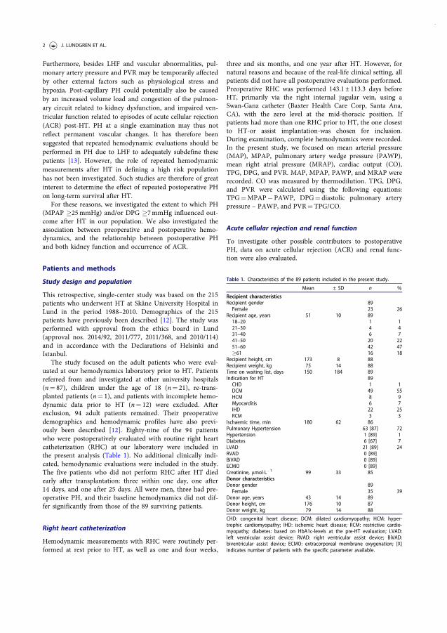
Furthermore, besides LHF and vascular abnormalities, pul-monary artery pressure and PVR may be temporarily affectedby other external factors such as physiological stress andhypoxia. Post-capillary PH could potentially also be causedby an increased volume load and congestion of the pulmon-ary circuit related to kidney dysfunction, and impaired ven-tricular function related to episodes of acute cellular rejection(ACR) post-HT. PH at a single examination may thus notreflect permanent vascular changes. It has therefore beensuggested that repeated hemodynamic evaluations should beperformed in PH due to LHF to adequately subdefine thesepatients [13]. However, the role of repeated hemodynamicmeasurements after HT in defining a high risk populationhas not been investigated. Such studies are therefore of greatinterest to determine the effect of repeated postoperative PHon long-term survival after HT.
For these reasons, we investigated the extent to which PH(MPAP �25mmHg) and/or DPG �7mmHg influenced out-come after HT in our population. We also investigated theassociation between preoperative and postoperative hemo-dynamics, and the relationship between postoperative PHand both kidney function and occurrence of ACR.
Patients and methods
Study design and population
This retrospective, single-center study was based on the 215patients who underwent HT at Skåne University Hospital inLund in the period 1988–2010. Demographics of the 215patients have previously been described [12]. The study wasperformed with approval from the ethics board in Lund(approval nos. 2014/92, 2011/777, 2011/368, and 2010/114)and in accordance with the Declarations of Helsinki andIstanbul.
The study focused on the adult patients who were eval-uated at our hemodynamics laboratory prior to HT. Patientsreferred from and investigated at other university hospitals(n¼ 87), children under the age of 18 (n¼ 21), re-trans-planted patients (n¼ 1), and patients with incomplete hemo-dynamic data prior to HT (n¼ 12) were excluded. Afterexclusion, 94 adult patients remained. Their preoperativedemographics and hemodynamic profiles have also previ-ously been described [12]. Eighty-nine of the 94 patientswho were postoperatively evaluated with routine right heartcatheterization (RHC) at our laboratory were included inthe present analysis (Table 1). No additional clinically indi-cated, hemodynamic evaluations were included in the study.The five patients who did not perform RHC after HT diedearly after transplantation: three within one day, one after14 days, and one after 25 days. All were men, three had pre-operative PH, and their baseline hemodynamics did not dif-fer significantly from those of the 89 surviving patients.
Right heart catheterization
Hemodynamic measurements with RHC were routinely per-formed at rest prior to HT, as well as one and four weeks,
three and six months, and one year after HT. However, fornatural reasons and because of the real-life clinical setting, allpatients did not have all postoperative evaluations performed.Preoperative RHC was performed 143.1 ± 113.3 days beforeHT, primarily via the right internal jugular vein, using aSwan-Ganz catheter (Baxter Health Care Corp, Santa Ana,CA), with the zero level at the mid-thoracic position. Ifpatients had more than one RHC prior to HT, the one closestto HT-or assist implantation-was chosen for inclusion.During examination, complete hemodynamics were recorded.In the present study, we focused on mean arterial pressure(MAP), MPAP, pulmonary artery wedge pressure (PAWP),mean right atrial pressure (MRAP), cardiac output (CO),TPG, DPG, and PVR. MAP, MPAP, PAWP, and MRAP wererecorded. CO was measured by thermodilution. TPG, DPG,and PVR were calculated using the following equations:TPG¼MPAP�PAWP, DPG¼ diastolic pulmonary arterypressure – PAWP, and PVR¼TPG/CO.
Acute cellular rejection and renal function
To investigate other possible contributors to postoperativePH, data on acute cellular rejection (ACR) and renal func-tion were also evaluated.
Table 1. Characteristics of the 89 patients included in the present study.
Mean ± SD n %
Recipient characteristicsRecipient gender 89
Female 23 26Recipient age, years 51 10 89
18–20 1 121–30 4 431–40 6 741–50 20 2251–60 42 47�61 16 18
Recipient height, cm 173 8 88Recipient weight, kg 75 14 88Time on waiting list, days 150 184 89Indication for HT 89
CHD 1 1DCM 49 55HCM 8 9Myocarditis 6 7IHD 22 25RCM 3 3
Ischaemic time, min 180 62 86Pulmonary Hypertension 63 [87] 72Hypertension 1 [89] 1Diabetes 6 [67] 7LVAD 21 [89] 24RVAD 0 [89]BiVAD 0 [89]ECMO 0 [89]Creatinine, lmol�L�1 99 33 85Donor characteristicsDonor gender 89
Female 35 39Donor age, years 43 14 89Donor height, cm 176 10 87Donor weight, kg 79 14 88
CHD: congenital heart disease; DCM: dilated cardiomyopathy; HCM: hyper-trophic cardiomyopathy; IHD: ischemic heart disease; RCM: restrictive cardio-myopathy; diabetes: based on HbA1c-levels at the pre-HT evaluation; LVAD:left ventricular assist device; RVAD: right ventricular assist device; BiVAD:biventricular assist device; ECMO: extracorporeal membrane oxygenation; [X]indicates number of patients with the specific parameter available.
2 J. LUNDGREN ET AL.

The occurrence of ACR was monitored with endomyo-cardial biopsies (EMBs), which were performed on a rou-tine basis on 14 occasions during the first postoperativeyear (week 1, 2, 3, 4, 6, 8, 10, 12, 16, 20, 24, 32, 40, and52). Thus, some of the EMBs were performed in connec-tion with the RHCs previously described. Additional EMBswere also performed where there were symptoms, or previ-ous episodes, of ACR. From 2005 onwards, EMBs weregraded according to both the 1990 and the 2004International Society for Heart and Lung Transplantation(ISHLT) grading systems [14,15]. Before then, EBMs weresolely graded according to the 1990 system. Therefore, allEMBs in the present study were histopathologically ana-lyzed and graded according to the 1990 ISHLT workingformulation [14].
With regard to renal function, glomerular filtration rate(GFR) was measured at one year post-HT using the iohexolclearance method. During the measurements, patients weregiven a dose-adjusted solution of iohexol (Omnipaque; GEHealthcare), after which plasma sampling was performed inrelation to patient morphometrics. High-performance liquidchromotography was used to determine the iohexol concen-trations. In a few cases where iohexol clearance measure-ments were missing, the creatinine-based CKD-EPI (ChronicKidney Disease Epidemiology Collaboration) equation, e.g.the currently recommended GFR estimating equation byKDIGO [16], was used to increase the amount of data avail-able for analysis [16,17].
Statistics
SigmaStat/SigmaPlot version 11.0 (Systat Software Inc,San Jose, CA) was used for statistical analysis. Parametric ornon-parametric statistics were used, depending on the distri-bution of data: i.e. t-test or Mann-Whitney rank sum test,respectively, when differences between two groups werebeing compared and Pearson- or Spearman correlation,respectively, when measuring the association between twovariables. Survival curves were plotted using the Kaplan-Meier method and hazard ratios (HRs) for death were esti-mated with Cox proportional hazards regression analysisand adjusted for gender, age at the time of HT, and kidneyfunction one year after HT. The last day of follow-up wasJune 30, 2012. Mean follow-up was 8.2 ± 5.8 years. Anyp-value of <.05 was considered to be statistically significant.All values are presented as mean ± SD. Estimated survival,when presented, is based on unadjusted data.
Results
Preoperative pulmonary hypertension and elevateddiastolic pressure gradient
Sixty-three patients had preoperative PH. Of these63 patients, 53 (84%) performed RHC at one week after HT.In 37 (70%), the PH was reversed and in 16 (30%) it per-sisted one week after HT. With regard to DPG, only threeof the patients had DPG �7mmHg prior to HT. In allthree, DPG was normalized one week after HT.
Postoperative pulmonary hypertension and its effect onlong-term survival
Survival, based on hemodynamics at the different evalua-tions, are shown in Figure 1(a–e). Long-term survival waslower in patients with PH at three months than in thosewithout (estimated survival 10.1 vs. 15.7 years, p< .006),and also at one year (estimated survival 7.0 vs. 15.1 years,p< .001). There was, however, no significant difference insurvival whether or not the patients had PH at one or fourweeks after HT-or at six months (Figure 1(a–e)). Of theeight patients with PH at one year after HT, four had preca-pillary PH, one had combined precapillary and postcapillaryPH, and three had isolated postcapillary PH. When adjustedfor gender and age at the time of transplantation, MPAP atone year after HT was associated with a higher risk of deathin the Cox regression model (HR 1.2, 95% CI 1.1–2.3). Thehemodynamic characteristics of the patients with postopera-tive PH are (for each examination) shown in SupplementalTable 1.
Diastolic pulmonary gradient, pulmonary vascularresistance, and transpulmonary gradient one year afterheart transplantation
One year after HT, DPG, PVR and TPG data were availablefrom 67, 78, and 78 patients (75%, 88%, and 88%), respect-ively. Seven patients had DPG �7mmHg. Their survival wassignificantly lower than in those with DPG <7mmHg (esti-mated survival 5.0 vs. 14.2 years, p< .001). Moreover, sur-vival was significantly lower in patients with PVR >3 WU(n¼ 4) than in those with PVR �3 WU (n¼ 74; estimatedsurvival 3.1 vs. 14.5 years, p< .006). In contrast, there wasno significant difference in survival between the patientswith TPG �12mmHg (n¼ 9) and in those with TPG<12mmHg (n¼ 69).
Effect of postoperative pulmonary hypertension duringrepeated measurements after heart transplantation
Survival in patients without postoperative PH and thosewith PH at one or repeated evaluations is shown in Figure2(a,b). Twenty-nine patients (33%) exhibited PH at leastonce during the first year after HT, and 60 (67%) did nothave PH at any of the postoperative evaluations. There wasno significant difference in survival between these twogroups (estimated survival 12.4 vs. 15.7 years) (Figure 2(a)).Of the 29 patients with postoperative PH, 18 had PH at asingle examination and 11 had PH at two or more examina-tions. Sixteen of the patients with PH at one measurement,and 10 of the patients with PH at two or more measure-ments, also had PH preoperatively. Of the 18 patients whoexhibited PH at a single examination, nine had PH at oneweek, three at four weeks, two at three months, one at sixmonths, and three at one year after HT. The survival wasnot significantly different in the 60 patients who did notexhibit PH and in the 18 patients who exhibited PH at onemeasurement (estimated survival 15.7 and 16.1 years,respectively), whereas survival was significantly lower in the
SCANDINAVIAN CARDIOVASCULAR JOURNAL 3

11 patients who exhibited PH at two or more examinations(estimated survival 7.8 years, p< .001) (Figure 2(b)). In amultivariate Cox regression model adjusted for gender andage at the time of HT, PH at repeated measurements wasassociated with a higher risk of death (HR 4.4, 95% CI
2.0–9.8) than no PH and PH at a single examination afterHT.
There was no significant difference in the number ofpostoperative catheterizations performed in patients withoutpostoperative PH, in patients with PH at one postoperative
(a) (b)
(c)
(e)
(d)
Figure 1. Survival in patients with and without PH after HT. (a) One week, (b) four weeks, (c) three months, (d) six months, and (e) one year after HT. �indicatesstatistical significance between patients with PH and patients without PH.
4 J. LUNDGREN ET AL.

evaluation, and in patients with PH at repeated evaluations(n¼ 4.0, n¼ 3.9, and n¼ 4.3, respectively).
Preoperative hemodynamic comparisons based onpostoperative pulmonary hypertension
Preoperative hemodynamics were compared for patientswho, after HT: (1) did not exhibit PH, (2) exhibited PH at
one examination, or (3) exhibited PH at several examina-tions (Table 2). Compared to patients without postoperativePH, preoperative MPAP, PAWP, MRAP, and PVR were sig-nificantly higher (p< .005, p< .02, p< .004, and p< .02,respectively) in patients with PH at one examination, andpreoperative MPAP and PAWP were significantly higher(p< .008 and p< .04, respectively) in patients with PH at
(a)
(b)
Figure 2. Survival in patients with no PH, with PH at one examination, and with PH at several examinations after HT. Comparison between (a) patients with no PHand with PH after HT, and: (b) patients without PH, with PH at a single examination, and with PH at repeated examinations after HT. �indicates statistical signifi-cance between patients without PH and patients with PH at repeated examinations. § indicates statistical significance between patients with PH at one examinationand patients with PH at repeated examinations.
SCANDINAVIAN CARDIOVASCULAR JOURNAL 5

repeated examinations (Table 2). Moreover, CO was signifi-cantly lower (p< .02) in patients with PH at one examin-ation than in patients with PH at repeated examinations.Finally, there was a weak positive correlation between pre-operative MPAP and MPAP at all postoperative examina-tions, except at the six-month evaluation. This associationdecreased with time. The R2-values were 0.39 (p< .001) atone week, 0.22 (p< .001) at four weeks, 0.19 (p< .001) atthree months, and 0.06 (p< .03) one year after HT.
Acute cellular rejection and renal function
The proportions of EMBs with ACR grade �2 or �3A/3Bin patients with and without postoperative PH are shown inTable 3. A total of 1,376 EMBs were performed during thefirst postoperative year. There was no significant differencein the proportion of EMBs with ACR grade �2 or �3A/3Bin patients with and without PH at one or repeated postop-erative evaluations. Nor was there any significant differencein the proportion of EMBs with ACR grade �2 or 3A/3B inpatients with PH at one or repeated postoperative evalua-tions (Table 3).
Kidney function one year after HT in patients with andwithout postoperative PH is shown in Figure 3. One yearafter HT, measurements of renal function were available
from 57 of the 60 patients (95%) without postoperative PH,in 17 of the 18 patients (94%) with PH at a single evalu-ation, and in 10 of the 11 patients (91%) with PH atrepeated evaluations. There was no significant difference inrenal function between patients without postoperative PHand those with PH at a single postoperative evaluation. Incontrast, renal function was lower in patients with repeatedPH, both when compared to patients without postoperativePH (p< .003) and when compared to those with PH at onepostoperative evaluation (p< .04) (Figure 3). However, whenkidney function was included in the multivariate regression,PH at repeated evaluations remained an independent pre-dictor of mortality (HR 3.4, 95% CI 1.4–8.0), compared tono PH and to PH at a single evaluation.
Kidney function and corresponding MPAP were availablefrom 77 patients at one year after HT, and a negative correl-ation was observed between the parameters (R2¼ .11,p< .004).
Cause of death
The causes of death in the different patient groups are givenin Table 4. Forty-six of the 60 patients (77%) without post-operative PH, 13 of the 18 patients (72%) with PH at onepostoperative examination, and one of the 11 patients (9%)
Table 2. Preoperative hemodynamic characteristics of patients with PH and without PH after HT.
All patients No PH post-HTPH at one exam-ination post-HT
PH at repeatedexaminations
post-HT
Parameter Mean ± SD Mean ± SD Mean ± SD Mean ± SD Sign
Patients 89 60 18 11MAP, mmHg 73.9 13.6 74.7 15.4 71.2 8.5 74.1 10.2MPAP, mmHg 30.3 8.8 27.9 8.6 34.6� 7.0 35.8§ 8.4 �,§PAWP, mmHg 20.6 8.1 18.8 7.8 24.1� 7.7 24.4§ 8.0 �,§TPG, mmHg 9.7 3.5 9.1 3.4 10.4 3.1 11.5 4.3DPG, mmHg �1.2 4.4 �1.1 4.5 �1.8 4.6 0.0 3.5MRAP, mmHg 7.9 6.0 6.6 5.3 12.0� 6.9 8.2 5.0 �CO, L�min�1 3.7 1.1 3.7 1.0 3.2 1.0 4.2# 1.0 #
PVR, WU 2.8 1.3 2.6 1.0 3.7� 1.8 2.8 1.3 �Mean: mean values from the preoperative hemodynamic evaluation; �indicates preoperative statistical significance betweenpatients with no PH post-HT and those with PH at one examination; §indicates preoperative statistical significance betweenpatients with no PH post-HT and those with PH at repeated examinations; #indicates preoperative statistical significance betweenpatients with PH at one examination post-HT and those with PH at repeated examinations; MAP: mean atrial pressure; MPAP:mean pulmonary artery pressure; PAWP: pulmonary artery wedge pressure; TPG: transpulmonary gradient; DPG: diastolic pressuregradient; MRAP: mean right atrial pressure; CO: cardiac output; PVR: pulmonary vascular resistance.
Table 3. Pulmonary hypertension in relation to acute cellular rejection.
P
% of EMBs with ACR� grade 2 or 3A/3B in patients withoutPH (n5 60)
% of EMBs with ACR� grade 2 or 3A/3B in patients with PH atone RHC (n5 18)
� �2� �3A/3B
8.5%5.0%
(79/933)(47/933)
� �2� �3A/3B
5.9%3.0%
(16/270)(8/270)
0.2170.203
vs. % of EMBs with ACR� grade 2 or 3A/3B in patients with PH atrepeated RHCs (n5 11)� �2� �3A/3B
8.7%4.0%
(15/173)(7/173)
0.9520.716
% of EMBs with ACR� grade 2 or 3A/3B in patients with PH atone RHC (n5 18)
vs. % of EMBs with ACR� grade 2 or 3A/3B in patients with PH atrepeated RHCs (n5 11)
� �2� �3A/3B
5.9%3.0%
(16/270)(8/270)
� �2� �3A/3B
8.7%4.0%
(15/173)(7/173)
0.3610.730
PH: pulmonary hypertension; ACR: acute cellular rejection; EMB: endomyocardial biopsy; RHC: right heart catheterization.
6 J. LUNDGREN ET AL.

with PH at repeated postoperative evaluations were alive atthe end of follow-up. Of the 29 patients who died, the mostcommon cause of death was rejection (n¼ 7, 24%) – includ-ing cellular and humoral rejections, as well as coronary allo-graft vasculopathy – followed by malignancy (n¼ 6, 21%)and infection (n¼ 5, 17%).
Discussion
The present study defined the characteristics of hemo-dynamics in HT patients during repeated measurements inthe first year after HT, and investigated the effects of post-operative PH and elevated DPG on survival. Our resultsindicate that persistent PH after HT may influence outcome.Moreover, DPG of �7mmHg and PVR > 3 WU one yearafter HT was also associated with a lower long-term survival,whereas TPG >12mmHg was not. Furthermore, survivalwas similar for patients with PH at a single postoperativeevaluation and in patients without any postoperative PH. In
contrast, survival was markedly lower in those who had PHat two or more examinations during the first postoperativeyear, with only one of 11 patients being alive at the end offollow-up. This illustrates the importance of early andrepeated catheterizations after HT, to identify patients withpersistent PH who may have impaired survival.
With the same cohort of patients, we recently showed thatpre-HT PH, defined according to previous and present defi-nitions, does not have to be associated with impaired survivalafter HT [12]. As shown in our previous study, patientsselected for HT showed reversibility of PVR in acute vaso-dilatation tests. Consequently, the patients included in boththe previous study and the present study probably did notshow extensive vascular remodeling, but instead passive con-gestion and excessive vasoconstriction. In agreement with thelack of effect of preoperative PH in the previous report, Gudeand colleagues did not observe any difference in survival in acohort of 500 HT patients whether or not they had preopera-tive PH [11]. Similarly, Goland and colleagues did notobserve any difference in survival in a cohort of 427 HTpatients, based on their preoperative hemodynamics. Instead,they found that MPAP >20mmHg six months and one yearafter HT [11], and PVR �3 WU one year after HT [10], wereassociated with lower long-term outcome. These reports didnot, however, use the current definitions of PH when defin-ing a high-risk population, so the impact of the current ESCguidelines in this regard remains uncertain. Consequently,the present study is the first to use the current definition ofPH [2] when evaluating the influence of post-HT PH on sur-vival. Despite using a different definition [2], our findings arein line with a few previous reports [10,11] emphasizing thatpersistent PH one year after HT is associated with reducedlong-term survival. This is further supported by the findingthat MPAP at one year after HT in our Cox regression modelwas associated with a higher risk of death, indicating that PHmay emerge as a prognostic marker for outcome followingHT.
Moreover, to the best of our knowledge, this is the firsttime that findings from repeated measurements during the
Table 4. Causes of death in the different patient groups.
All patientsNo PH post-
HT
PH at oneexaminationpost-HT
PH atrepeated
examinationspost-HT
Cause of death N % N % N % N %
Patients 89 60 18 11Deaths 29 14 5 10Infection 5 17 2 14 0 – 3 30Kidney Failure 1 3 1 7 0 – 0 –Malignancy 6 21 3 21 1 20 2 20Rejection 7 24 4 29 3 60 0 –Intracranial bleeding 3 10 2 14 0 – 1 10Ruptured aortic aneurysm 1 3 0 – 0 – 1 10Circulatory failure 1 3 0 – 0 – 1 10Acute coronary syndrome 1 3 1 7 0 – 0 –Amyloidosis 1 3 0 – 1 20 0 –Multi organ failure 1 3 0 – 0 – 1 10Pulmonary embolism 1 3 1 7 0 – 0 –Unknown 1 3 0 – 0 – 1 10
%: percent of the total number of deaths in the different groups; Unknown: one patient died under unclear circumstanceswhen abroad, therefore the cause of death is unclear.
Figure 3. GFR one year after HT in relation to postoperative PH. GFR, glomeru-lar filtration rate; PH, pulmonary hypertension; RHC, right heart catheterization.�indicates statistical significance between patients without PH and patientswith PH at repeated examinations. §indicates statistical significance betweenpatients with PH at one examination and patients with PH at repeatedexaminations.
SCANDINAVIAN CARDIOVASCULAR JOURNAL 7
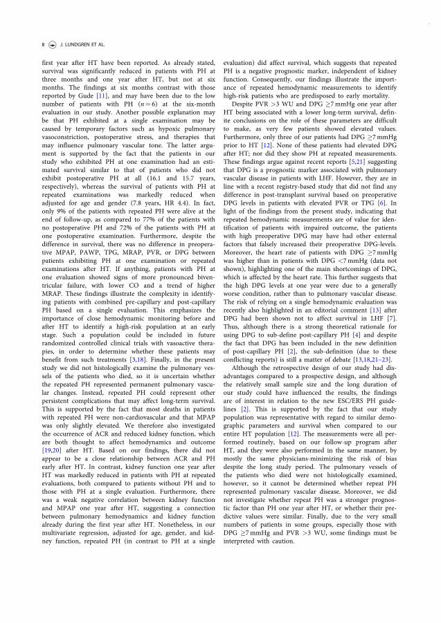
first year after HT have been reported. As already stated,survival was significantly reduced in patients with PH atthree months and one year after HT, but not at sixmonths. The findings at six months contrast with thosereported by Gude [11], and may have been due to the lownumber of patients with PH (n¼ 6) at the six-monthevaluation in our study. Another possible explanation maybe that PH exhibited at a single examination may becaused by temporary factors such as hypoxic pulmonaryvasoconstriction, postoperative stress, and therapies thatmay influence pulmonary vascular tone. The latter argu-ment is supported by the fact that the patients in ourstudy who exhibited PH at one examination had an esti-mated survival similar to that of patients who did notexhibit postoperative PH at all (16.1 and 15.7 years,respectively), whereas the survival of patients with PH atrepeated examinations was markedly reduced whenadjusted for age and gender (7.8 years, HR 4.4). In fact,only 9% of the patients with repeated PH were alive at theend of follow-up, as compared to 77% of the patients withno postoperative PH and 72% of the patients with PH atone postoperative examination. Furthermore, despite thedifference in survival, there was no difference in preopera-tive MPAP, PAWP, TPG, MRAP, PVR, or DPG betweenpatients exhibiting PH at one examination or repeatedexaminations after HT. If anything, patients with PH atone evaluation showed signs of more pronounced biven-tricular failure, with lower CO and a trend of higherMRAP. These findings illustrate the complexity in identify-ing patients with combined pre-capillary and post-capillaryPH based on a single evaluation. This emphasizes theimportance of close hemodynamic monitoring before andafter HT to identify a high-risk population at an earlystage. Such a population could be included in futurerandomized controlled clinical trials with vasoactive thera-pies, in order to determine whether these patients maybenefit from such treatments [3,18]. Finally, in the presentstudy we did not histologically examine the pulmonary ves-sels of the patients who died, so it is uncertain whetherthe repeated PH represented permanent pulmonary vascu-lar changes. Instead, repeated PH could represent otherpersistent complications that may affect long-term survival.This is supported by the fact that most deaths in patientswith repeated PH were non-cardiovascular and that MPAPwas only slightly elevated. We therefore also investigatedthe occurrence of ACR and reduced kidney function, whichare both thought to affect hemodynamics and outcome[19,20] after HT. Based on our findings, there did notappear to be a close relationship between ACR and PHearly after HT. In contrast, kidney function one year afterHT was markedly reduced in patients with PH at repeatedevaluations, both compared to patients without PH and tothose with PH at a single evaluation. Furthermore, therewas a weak negative correlation between kidney functionand MPAP one year after HT, suggesting a connectionbetween pulmonary hemodynamics and kidney functionalready during the first year after HT. Nonetheless, in ourmultivariate regression, adjusted for age, gender, and kid-ney function, repeated PH (in contrast to PH at a single
evaluation) did affect survival, which suggests that repeatedPH is a negative prognostic marker, independent of kidneyfunction. Consequently, our findings illustrate the import-ance of repeated hemodynamic measurements to identifyhigh-risk patients who are predisposed to early mortality.
Despite PVR >3 WU and DPG �7mmHg one year afterHT being associated with a lower long-term survival, defin-ite conclusions on the role of these parameters are difficultto make, as very few patients showed elevated values.Furthermore, only three of our patients had DPG �7mmHgprior to HT [12]. None of these patients had elevated DPGafter HT; nor did they show PH at repeated measurements.These findings argue against recent reports [5,21] suggestingthat DPG is a prognostic marker associated with pulmonaryvascular disease in patients with LHF. However, they are inline with a recent registry-based study that did not find anydifference in post-transplant survival based on preoperativeDPG levels in patients with elevated PVR or TPG [6]. Inlight of the findings from the present study, indicating thatrepeated hemodynamic measurements are of value for iden-tification of patients with impaired outcome, the patientswith high preoperative DPG may have had other externalfactors that falsely increased their preoperative DPG-levels.Moreover, the heart rate of patients with DPG �7mmHgwas higher than in patients with DPG <7mmHg (data notshown), highlighting one of the main shortcomings of DPG,which is affected by the heart rate. This further suggests thatthe high DPG levels at one year were due to a generallyworse condition, rather than to pulmonary vascular disease.The risk of relying on a single hemodynamic evaluation wasrecently also highlighted in an editorial comment [13] afterDPG had been shown not to affect survival in LHF [7].Thus, although there is a strong theoretical rationale forusing DPG to sub-define post-capillary PH [4] and despitethe fact that DPG has been included in the new definitionof post-capillary PH [2], the sub-definition (due to theseconflicting reports) is still a matter of debate [13,18,21–23].
Although the retrospective design of our study had dis-advantages compared to a prospective design, and althoughthe relatively small sample size and the long duration ofour study could have influenced the results, the findingsare of interest in relation to the new ESC/ERS PH guide-lines [2]. This is supported by the fact that our studypopulation was representative with regard to similar demo-graphic parameters and survival when compared to ourentire HT population [12]. The measurements were all per-formed routinely, based on our follow-up program afterHT, and they were also performed in the same manner, bymostly the same physicians-minimizing the risk of biasdespite the long study period. The pulmonary vessels ofthe patients who died were not histologically examined,however, so it cannot be determined whether repeat PHrepresented pulmonary vascular disease. Moreover, we didnot investigate whether repeat PH was a stronger prognos-tic factor than PH one year after HT, or whether their pre-dictive values were similar. Finally, due to the very smallnumbers of patients in some groups, especially those withDPG �7mmHg and PVR >3 WU, some findings must beinterpreted with caution.
8 J. LUNDGREN ET AL.

Conclusions
In conclusion, our study suggests that persistent PH, definedaccording to present guidelines, at one year after HT isrelated to impaired long-term survival. Moreover, the find-ings indicate that PH at repeated measurements during thefirst postoperative year is a negative prognostic marker,associated with worse outcome than PH at a single examin-ation. This highlights that repeated and early hemodynamicmeasurements after HT could be used to identify patientswith persistent PH who may exhibit impaired survival.Whether repeated PH is a stronger prognosticator of deaththan PH at one year after HT needs investigation in futuretrials. Further studies in larger patient cohorts are alsoencouraged to confirm our findings and to clarify whetherpatients who exhibit PH at repeated examinations after HThave signs of pulmonary vascular changes or whether thefindings represent a generally worse condition.
Acknowledgements
We acknowledge the support of the staff at (1) the Hemodynamics Labat the Section for Heart Failure and Valvular Disease, VO Heart andLung Medicine, Skåne University Hospital, and (2) the Department ofCardiology, Clinical Sciences, Lund University, Lund, Sweden.
Disclosure statement
Dr Lundgren reports receiving personal lecture fees from ActelionPharmaceuticals Sweden AB and GlaxoSmithKline for services outwiththe work submitted. Dr S€oderlund reports receiving personal lecturefees from Sandoz/Novartis for services outwith the work submitted. DrRådegran reports receiving personal lecture fees from ActelionPharmaceuticals Sweden AB, Bayer Healthcare, GlaxoSmithKline,NordicInfu Care, and Sandoz/Novartis for services outwith the worksubmitted.
Dr. Rådegran is, and has been, primary investigator or co-investiga-tor in clinical PAH trials for GlaxoSmithKline, ActelionPharmaceuticals Sweden AB, Pfizer, Bayer, and United Therapeutics,and in clinical heart transplantation immunosuppression trials forNovartis.
The companies had no role in the collection, analysis, and inter-pretation of data relating to this article and had no right in disapprov-ing of the manuscript.
Funding
We acknowledge financial support (unrestricted research grants) fromthe Anna-Lisa and Sven-Erik Lundgren Foundation, ALF, and theSkåne University Hospital Foundation, Lund, Sweden. We alsoreceived unrestricted research grants from the Swedish Society forPulmonary Hypertension (SveFPH) and from Actelion PharmaceuticalsSweden AB. The foundations had no role in the collection, analysis,and interpretation of data and had no right in disapproving of themanuscript.
References
[1] Bursi F, McNallan SM, Redfield MM, et al. Pulmonary pres-sures and death in heart failure: a community study. J Am CollCardiol. 2012;59:222–231.
[2] Galie N, Humbert M, Vachiery JL, et al. 2015 ESC/ERSGuidelines for the diagnosis and treatment of pulmonary hyper-tension: the Joint Task Force for the diagnosis and treatment of
pulmonary hypertension of the European Society of Cardiology(ESC) and the European Respiratory Society (ERS): endorsedby: association for European Paediatric and CongenitalCardiology (AEPC), International Society for Heart and LungTransplantation (ISHLT). Eur Heart J. 2016;37:67–119.
[3] Lundgren J, Radegran G. Pathophysiology and potential treat-ments of pulmonary hypertension due to systolic left heart fail-ure. Acta Physiol (Oxf). 2014;211:314–333.
[4] Naeije R, Vachiery JL, Yerly P, et al. The transpulmonary pres-sure gradient for the diagnosis of pulmonary vascular disease.Eur Respir J. 2013;41:217–223.
[5] Gerges C, Gerges M, Lang MB, et al. Diastolic pulmonary vas-cular pressure gradient: a predictor of prognosis in 'out-of-pro-portion' pulmonary hypertension. Chest. 2013;143:758–766.
[6] Tedford RJ, Beaty CA, Mathai SC, et al. Prognostic value of thepre-transplant diastolic pulmonary artery pressure-to-pulmon-ary capillary wedge pressure gradient in cardiac transplantrecipients with pulmonary hypertension. J Heart LungTransplant. 2014;33:289–297.
[7] Tampakakis E, Leary PJ, Selby VN, et al. The diastolic pulmon-ary gradient does not predict survival in patients with pulmon-ary hypertension due to left heart disease. JACC Heart Fail.2015;3:9–16.
[8] Costard-Jackle A, Fowler MB. Influence of preoperative pul-monary artery pressure on mortality after heart transplantation:testing of potential reversibility of pulmonary hypertension withnitroprusside is useful in defining a high risk group. J Am CollCardiol. 1992;19:48–54.
[9] Vakil K, Duval S, Sharma A, et al. Impact of pre-transplant pul-monary hypertension on survival after heart transplantation: AUNOS registry analysis. Int J Cardiol. 2014;176:595–599.
[10] Goland S, Czer LS, Kass RM, et al. Pre-existing pulmonaryhypertension in patients with end-stage heart failure: impact onclinical outcome and hemodynamic follow-up after orthotopicheart transplantation. J Heart Lung Transplant. 2007;26:312–318.
[11] Gude E, Simonsen S, Geiran OR, et al. Pulmonary hypertensionin heart transplantation: discrepant prognostic impact of pre-operative compared with 1-year post-operative right hearthemodynamics. J Heart Lung Transplant. 2010;29:216–223.
[12] Lundgren J, Algotsson L, Kornhall B, et al. Preoperative pul-monary hypertension and its impact on survival after hearttransplantation. Scand Cardiovasc J. 2014;48:47–58.
[13] Chatterjee NA, Lewis GD. Characterization of pulmonaryhypertension in heart failure using the diastolic pressure gradi-ent: limitations of a Solitary Measurement. JACC Heart Fail.2015;3:17–21.
[14] Billingham ME, Cary NR, Hammond ME, et al. A working for-mulation for the standardization of nomenclature in the diag-nosis of heart and lung rejection: heart Rejection Study Group.The International Society for Heart Transplantation. J HeartTransplant. 1990;9:587–593.
[15] Stewart S, Winters GL, Fishbein MC, et al. Revision of the 1990working formulation for the standardization of nomenclature inthe diagnosis of heart rejection. J Heart Lung Transplant.2005;24:1710–1720.
[16] Kidney Disease: Improving Global Outcomes (KDIGO) CKDWork Group. KDIGO 2012 clinical practice guideline for theevaluation and management of chronic kidney disease. KidneyInt Suppl. 2013;3:1–150.
[17] Levey AS, Stevens LA, Schmid CH, et al. A new equation toestimate glomerular filtration rate. Ann Intern Med.2009;150:604–612.
[18] Rosenkranz S, Gibbs JS, Wachter R, et al. Left ventricular heartfailure and pulmonary hypertensiondagger. Eur Heart J.2016;37:942–954.
[19] Soderlund C, Ohman J, Nilsson J, et al. Acute cellular rejectionthe first year after heart transplantation and its impact on sur-vival: a single-centre retrospective study at Skane UniversityHospital in Lund 1988-2010. Transpl Int. 2014;27:482–492.
SCANDINAVIAN CARDIOVASCULAR JOURNAL 9

[20] Soderlund C, Lofdahl E, Nilsson J, et al. Chronic kidney diseaseafter heart transplantation: a single-centre retrospective study atSkane University Hospital in Lund 1988-2010. Transpl Int.2016;29:529–539.
[21] Gerges C, Gerges M, Lang IM. Characterization of pulmonaryhypertension in heart failure using the diastolic pressure gradi-ent: the conundrum of high and low diastolic pulmonary gradi-ent. JACC Heart Fail. 2015;3:424–425.
[22] Naeije R. Measurement to predict survival: the caseof diastolic pulmonary gradient. JACC Heart Fail. 2015;3:425.
[23] Tampakakis E, Tedford RJ. Reply: characterization of pul-monary hypertension in heart failure using the diastolicpressure gradient: the conundrum of high and low dia-stolic pulmonary gradient. JACC Heart Fail. 2015;3:426–427.
10 J. LUNDGREN ET AL.

Paper V


ORIGINAL ARTICLE
Alterations in plasma L-arginine and methylarginines in heart failure and afterheart transplantation
Jakob Lundgrena,b, Anna Sandqvistc, Mikael Hedelandd,e, Ulf Bondessond,e, Gerhard Wikstr€omf andG€oran Rådegrana,b
aDepartment of Clinical Sciences Lund, Cardiology, Lund University, Lund, Sweden; bThe Hemodynamic Lab, The Section for Heart Failureand Valvular Disease, The Heart and Lung Clinic, Skåne University Hospital, Lund, Sweden; cDepartment of Pharmacology and ClinicalNeuroscience, Clinical Pharmacology, Umeå University, Umeå, Sweden; dDepartment of Chemistry, Environment and Feed Hygiene, NationalVeterinary Institute (SVA), Uppsala, Sweden; eDepartment of Medicinal Chemistry, Analytical Pharmaceutical Chemistry, Uppsala University,Uppsala, Sweden; fDepartment of Medical Sciences, Cardiology, Uppsala University, Uppsala University Hospital, Uppsala, Sweden
ABSTRACTObjective. Endothelial function, including the nitric oxide (NO)-pathway, has previously been extensivelyinvestigated in heart failure (HF). In contrast, studies are lacking on the NO pathway after hearttransplantation (HT). We therefore investigated substances in the NO pathway prior to and after HT inrelation to hemodynamic parameters. Design. 12 patients (median age 50.0 yrs, 2 females), heart trans-planted between June 2012 and February 2014, evaluated at our hemodynamic lab, at rest, prior toHT, as well as four weeks and six months after HT were included. All patients had normal left ventricu-lar function post-operatively and none had post-operative pulmonary hypertension or acute cellularrejection requiring therapy at the evaluations. Plasma concentrations of ADMA, SDMA, L-Arginine,L-Ornithine and L-Citrulline were analyzed at each evaluation. Results. In comparison to controls, theplasma L-Arginine concentration was low and ADMA high in HF patients, resulting in low L-Arginine/ADMA-ratio pre-HT. Already four weeks after HT L-Arginine was normalized whereas ADMA remainedhigh. Consequently the L-Arginine/ADMA-ratio improved, but did not normalize. The biomarkersremained unchanged at the six-month evaluation and the L-Arginine/ADMA-ratio correlated inverselyto pulmonary vascular resistance (PVR) six months post-HT. Conclusions. Plasma L-Arginine concentra-tions normalize after HT. However, as ADMA is unchanged, the L-Arginine/ADMA-ratio remained lowand correlated inversely to PVR. Together these findings suggest that (i) the L-Arginine/ADMA-ratiomay be an indicator of pulmonary vascular tone after HT, and that (ii) NO-dependent endothelial func-tion is partly restored after HT. Considering the good postoperative outcome, the biomarker levels maybe considered “normal” after HT.
ARTICLE HISTORYReceived 21 November 2017Revised 19 March 2018Accepted 20 March 2018
KEYWORDSNitric Oxide; ADMA;L-Arginine; hearttransplantation; heartfailure; right heartcatheterization
Introduction
Heart failure (HF) is common and despite continuousimprovement in HF therapy, the condition carries a poorprognosis, where heart transplantation (HT) resides as theultimate treatment. Pulmonary hypertension (PH) is a com-plicating factor in HF, associated with worse prognosis [1].With the present methods and definitions [2,3], it is difficultto differentiate between isolated post-capillary PH (Ipc-PH),caused by passive congestion of the pulmonary vasculatureand combined pre-capillary and post-capillary PH (Cpc-PH), complicated by endothelial dysfunction, excessive vaso-constriction and, in some cases, vascular remodelling [2].Adequately sub-defining post-capillary PH is, however, ofmajor importance in HT where Cpc-PH with vascularremodelling may increase the risk for acute right heart fail-ure and early mortality after HT.
Endothelial dysfunction is central in PH, but is also pre-sent in HF. This dysfunction is, at least in part, caused by
impaired nitric oxide (NO) production secondary to endo-thelial damage [4]. The L-Arginine/NO pathway is a com-plex system with effects on vasomotor tone as well as oncellular adhesion, platelet aggregation, vascular proliferationand angiogenesis [5]. NO is produced from L-Arginine,which can form either NO and citrulline or urea and orni-thine, respectively. The former reaction is catalysed by thenitric oxide synthase (NOS) family and the latter by argi-nase [6,7].
Moreover, the competitive NOS inhibitor asymmetricdimethylarginine (ADMA) has been shown to be elevated inplasma and serum in various cardiovascular conditions [8,9]as well as to be an independent predictor of outcome inacutely decompensated chronic HF [10]. ADMA is primarilymetabolized by dimethylarginine dimethylaminohydrolase-1(DDAH-1) and to a lesser extent excreted by the kidneys[5]. DDAH-1 is primarily expressed in the kidneys and liver[11] and its activity is reduced by oxidative stress andimpaired in the above-mentioned conditions. In contrast,
CONTACT Jakob Lundgren [email protected] The Hemodynamic Lab, The Section for Heart Failure and Valvular Disease, Skåne UniversityHospital, Lund, Sweden
Supplemental data for this article can be accessed here.
� 2018 Informa UK Limited, trading as Taylor & Francis Group
SCANDINAVIAN CARDIOVASCULAR JOURNAL, 2018https://doi.org/10.1080/14017431.2018.1459823
Arginase activity has been shown to be elevated in HF [12].This, together, leads to decreased NO synthesis andincreased production of urea, which in turn results in lowerL-Arginine available for NO production [12].
In contrast to in HF, there is scarce knowledge on theNO pathway in relation to HT. One study has shown thatNO production is impaired after HT, primarily due toincreased inflammation [13]. Another study has shown thatthe immunosuppressive compound sirolimus, but not myco-phenolate mofetil, decrease ADMA in HT patients [14].However, little is known about the levels of various substan-ces of the nitric oxide pathway, their development over timeand their correlation to hemodynamics after HT. We there-fore evaluated substances related to the NO pathway insevere HF patients prior to and after HT and related thefindings to commonly used hemodynamic parameters. Theaim was to define the “normal” post-HT plasma levels ofsubstances in the NO pathway, as well as to identify poten-tial biomarkers and targets for future medical therapies.
Materials and methods
Study design and population
Between June 2012 and February 2014, 43 patients wereheart transplanted in Lund and included in a prospectivecohort study. To identify a “normal” post-HT population,the 12 patients (median age 50.0 yrs, 2 females) who werehemodynamically evaluated at our lab, at rest, prior to HT,as well as four weeks and six months after HT, prior toaugust 2014, and who, at the four week and six month eval-uations, (i) did not have post-operative rejections requiringspecific therapy; (ii) did not have postoperative PH and; (iii)had left ventricular ejection fraction �50%, were included inthe present study. For comparison, we included 12 healthy,age-matched, non-smokers without drug treatment and nosymptoms or signs of common cold.
The study was performed with informed consent andapproval by the ethics board in Lund and Uppsala(Approval nos. 2010/114, 2010/343, 2010/442, 2011/368,2011/777, 2015/270) and in accordance with theDeclarations of Helsinki and Istanbul.
Right heart catheterization
Right heart catheterization (RHC) were performed at restprior to HT, as well as four weeks and six months after HT.RHC was performed via the right internal jugular vein,using a Swan Ganz catheter (Baxter Health Care Corp, SantaAna, CA). Complete pulmonary hemodynamics wererecorded. In the present study we focused on mean pulmon-ary artery pressure (MPAP), pulmonary artery wedge pres-sure (PAWP), mean right atrial pressure (MRAP), cardiacoutput (CO), transpulmonary gradient (TPG), diastolic pres-sure gradient (DPG) and pulmonary vascular resistance(PVR). MPAP, PAWP and MRAP were recorded. COwas measured by thermodilution. TPG, DPG and PVRwere calculated using the formulas: TPG¼MPAP-PAWP,
DPG¼ diastolic pulmonary artery pressure-PAWP andPVR¼TPG/CO.
Biomarker analysis
Plasma samples from patients, collected from the pulmonaryarteries during RHC, were stored at �80 �C in the LundCardio Pulmonary Register (LCPR) cohort of RegionSkåne’s biobank. The samples were analyzed for plasma con-centrations of ADMA, symmetric dimethylarginine (SDMA),L-Arginine, L-Ornithine and L-Citrulline with liquid chro-matography – tandem mass spectrometry (LC-MS/MS), atthe Swedish National Veterinary Institute in Uppsala,Sweden, as described previously [15]. The L-Arginine/ADMA-ratio as well as the L-Arginine/L-Ornithine-ratiowere evaluated and the global arginine bioavailability ratio(GABR) calculated using the formula: GABR¼ (L-Arginine/(L-Ornithineþ L-Citrulline)). Control samples were collectedat Uppsala University Hospital and analyzed for L-Arginine,ADMA and SDMA as described previously [16]. Thepatients and controls were analyzed at different time points.The methods used were the same. However, at the time ofanalysis of the samples from controls, the L-Ornithine andL-Citrulline analyzes were not available so these markers arelacking in controls.
To monitor acute cellular rejections (ACR), endomyocar-dial biopsies were routinely performed from the right inter-ventricular septum, and graded according to the 2005International Society for Heart and Lung Transplantationworking formulation [17].
Statistics
A SigmaStat/SigmaPlot version 11.0 (Systat Software Inc,San Jose, CA) was used for statistical analysis. Parametric ornon-parametric statistics were used depending on the distri-bution of data: i.e. One Way Repeated Measures ANOVA(Tukey-test) or One Way Repeated Measures ANOVA onRanks (Tukey-test), when the same group was comparedover time; t-test or Mann-Whitney Rank Sum test, respect-ively, when two groups were compared and Pearson- orSpearman Correlation, respectively, when measuring theassociation between two variables. A p value <.05 was con-sidered statistically significant. All values are presented asmedian and interquartile range.
Results
Study population
Baseline demographical data are shown in Table 1. Nine ofthe 12 patients had pre-operative PH (MPAP �25mmHg).Of these patients, three had combined pre-capillary andpost-capillary PH (Cpc-PH, DPG �7mmHg and/or PVR>3 WU) and five had isolated post-capillary PH (Ipc-PH,DPG <7mmHg and/or PVR �3 WU). One patient hadvery severe orthopnea at the RHC when blood samples werecollected and adequate PAWP measurements could not be
2 J. LUNDGREN ET AL.

performed, wherefore PH-subgrouping is lacking in thispatient. However, the extensive pre-HT evaluation revealedno signs of pulmonary vascular disease and in a secondRHC, MPAP and PAWP were 30mmHg and 24mmHg,respectively, with a PVR of 1.5 WU and a DPG of 0mmHg.Prior to the second RHC the patient had been medicallyoptimized with intravenous furosemide and levosimendanand was subsequently accepted for HT.
Follow-up data, with regards to clinical features and med-ications, are shown in Table 2. Four weeks after HT threepatients exhibited myocardial biopsies with rejection grade1R and six months after HT two patients had rejectiongrade 1R. None needed specific rejection treatment.
Substances in the nitric oxide pathway and theircharacteristics in relation to heart transplantation
Prior to HT, the plasma concentration of L-Arginine was56 (48–60) mM, L-Ornithine 93 (71–119) mM, L-Citrulline26 (19–33) mM, ADMA 0.57 (0.47–0.71) mM, SDMA 0.85(0.68–1.15) mM, L-Arginine/ADMA-ratio 86 (68–128),L-Arginine/L-Ornithine-ratio 0.57 (0.46–0.74) and GABR0.43 (0.37–0.55) (Figures 1 and 2). L-Arginine was lower(p< .001) in patients than in controls, whereas ADMA andSDMA were higher (p< .003 and p< .001, respectively),resulting in a markedly lower L-Arginine/ADMA-ratio inpatients (p< .001).
There was a trend (p¼ ns) towards higher plasmaconcentration of SDMA, as well as lower L-Arginine/ADMA-ratio, GABR and L-Arginine/Ornithine-ratio inpatients with, compared to those without, pre-operative PH(Table 3).
Four weeks after HT there was an increase in the plasmaconcentration of; L-Arginine to 83 (65–101) mM (p< .008),L-Arginine/ADMA-ratio to 164 (105–189) (p< .006),L-Arginine/L-Ornithine-ratio to 0.89 (0.82–1.08) (p< .05)and GABR to 0.75 (0.65–0.85) (p< .05), compared to priorto HT. All concentrations remained stable (p¼ ns) at the sixmonth evaluation. After HT, L-Arginine was in line withhealthy controls (p¼ ns), whereas L-Arginine/ADMA,although improved, remained decreased (p< .001). Therewas no change in ADMA, SDMA, L-Ornithine andL-Citrulline levels after HT (p¼ ns, Figures 1 and 2).
Hemodynamic characteristics in relation to hearttransplantation
Hemodynamics prior to HT, as well as four weeks and sixmonths thereafter, are shown in Table 4. Prior to HT,MPAP was 35 (27–42) mmHg, PAWP 25 (20–30) mmHg,TPG 9 (6–12) mmHg, DPG 1.0 (–0.8–4.8) mmHg, MRAP15 (12–19) mmHg, CO 3.4 (2.6–4.3) L�min�1 and PVR 2.4(1.9–3.0) WU.
Table 1. Baseline characteristics of patients and healthy controls.
Patients Healthy subjects
Parameter n median (IQR) n median (IQR)
Clinical featuresSex, male/female 12 10/2 12 5/7Age, years 12 50 (45–60) 12 51 (46–60)Weight, kg 12 80 (71–92) N/ALength, cm 12 179 (176–180) N/ABSA, m2 12 2.0 (1.9–2.1) N/ANYHA class, IIIb/IV 3/9 N/AEtiology, DCM/HCM/IHD 12 8/1/3 N/ADiabetes mellitus, yes/no 12 0/12 12 0/12Hypertension, yes/no 12 0/12 12 0/12AF, yes/no 12 2/10 12 0/12History of smoking, yes/no 12 3/9 12 0/12
Medical historyBetablockers, % 12 12 (100) –ACEi/ARB, % 12 8/4 (100) –MRA, % 12 8 (67) –Anticoagulants, % 12 10 (83) –Statins, % 12 3 (25) –Diuretics, % 12 12 (100) –
Biochemical indicatorsL-Arginine, mM 12 56 (48–60) 12 86 (78–92)�ADMA, mM 12 0.57 (0.47–0.71) 12 0.39 (0.32–0.42)�SDMA, mM 12 0.85 (0.68–1.15) 12 0.41 (0.36–0.45)�L-Arginine/ADMA 12 86 (68–128) 12 225 (203–273)�P-Urea, mmol�L�1 12 9 (8–13) N/AP-Homocysteine, mM�L�1 12 16 (13–27) N/AP-NT-proBNP, ng�L�1 12 4058 (3348–7408) N/AP-Creatinine, mM�L�1 12 106 (95–131) N/AeGFR, mL�min�1�1.73m�2 12 61 (44–78) N/A
IQR: interquartile range; n: individuals where the specific parameter was available; BSA: body surface area;NYHA: New York Heart Association; DCM: dilated cardiomyopathy; HCM: hypertrophic cardiomyopathy; IHD:ischemic heart disease; AF: atrial fibrillation; ACEi: angiotensin converting enzyme inhibitor; ARB: angiotensin IIreceptor blocker; MRA: mineralocorticoid receptor antagonist; ADMA: asymmetric dimethylarginine; SDMA:symmetric dimethylarginine; N/A: not assessed.�Indicates statistical significant difference (p< .05) between patients and controls.
SCANDINAVIAN CARDIOVASCULAR JOURNAL 3

Four weeks after HT there was an increase in CO to 6.0(5.6–6.6) L�min�1 (p< .001), as well as a decrease in MPAPto 17 (12–21) mmHg (p< .05), PAWP to 8 (5–11) mmHg(p< .001) and PVR to 1.3 (1.1–1.5) WU (p< .05), comparedto prior to HT, remaining stable (p¼ ns) at the six monthevaluation. Six months after HT MRAP decreased to 1.5(0.0–4.0) mmHg (p< .05), as compared to prior to HT.There was no change (p¼ ns) in DPG and TPG after,compared to prior to, HT. None of the patients had post-operative PH (Tables 2 and 4).
In a univariate analysis prior to HT there was a modestcorrelation between GABR and creatinine (R2: 0.37, p< .04).Six months after HT, there was an inverse correlationbetween PVR and the L-Arginine/ADMA-ratio (R2: 0.39,p< .03) as well as GABR (R2: 0.40, p< .03) (Figure 3(a,b)).There were no other significant correlations, neither at thepre-operative nor post-operative evaluations, between theL-Arginine/ADMA-ratio or GABR and the hemodynamicparameters, NT-proBNP, creatinine or urea (Supplementaltable 1). Moreover, neither the plasma levels of L-Argininenor ADMA correlated to PVR, or any of the other investi-gated hemodynamic parameters, after HT (p¼ ns). In con-trast, there was a strong correlation between the L-Arginine/ADMA ratio and aspartate aminotransferase (ASAT) andglutamyl transferase (GT), as well as between GABR andASAT, GT and alanine aminotransferase (ALAT)(Supplemental table 1). However, these correlations wereprimarily due to the correlation between L-Arginine and thetransferases, whereas no correlation was observed betweenADMA and these markers.
Discussion
In the present study, we investigated the plasma concentra-tions of substances related to the NO pathway in HF-patients prior to and after HT, without post-operative PHand rejections above 1R, as well as with normal left heartfunction after HT. In support of previous findings, we foundimpaired L-Arginine, ADMA and L-Arginine/ADMA-ratioprior to HT. Whereas L-Arginine greatly improved, to levelscomparable to healthy individuals, already four weeks afterHT, ADMA, L-Citrulline and L-Ornithine remainedunchanged. Consequently, the L-Arginine/ADMA-ratio wasimproved, but not fully normalized. The L-Arginine/ADMA-ratio was furthermore inversely correlated to PVR,suggesting a partly restored NO-dependent endothelial func-tion. Based on the good post-operative outcome, withoutsevere complications, our findings indicate the “normal”plasma concentrations of these biomarkers after HT.
The 12 patients included in the present study had severeHF with low CO and high filling pressures as well asseverely deranged biomarkers prior to HT. They presentedwith significantly lower plasma concentrations of L-Argininebut higher ADMA and SDMA compared to healthy controls,as well as compared to patients with mild to moderate HF,recently reported by our group [15]. In fact, the plasma lev-els of the investigated biomarkers related to the NO pathwaywere, prior to HT, in line with those of patients with PAH,in our previous report by Sandqvist et al. [15]. These find-ings may represent the progressive nature of HF, withworsening endothelial function and congestion of the
Table 2. Characteristics of the patients four weeks and six months after HT.
Four weeks Six months
Parameter n median (IQR) n median (IQR)
Clinical featuresWeight, kg 12 80 (71–92) 12 79 (67–93)BSA, m2 12 2.0 (1.9–2.1) 12 2.0 (1.8–2.1)LVEF, low/normal 12 0/12 11 0/11Pulmonary hypertension, yes/no 12 0/12 12 0/12Acute cellular rejection, yes/no 12 3/9 12 2/10ACR¼ grade 1A/1B 3 2ACR� grade 2 0 0
Biochemical indicatorsS-Urea, mmol�L�1 12 13 (9–16) 12 8 (7–11)P-Creatinine, mM�L�1 12 115 (97–148) 12 102 (94–160)eGFR, mL�min�1�1.73m�2 12 39 (27–58) 12 N/A
Medications n (%) n (%)Betablockers 12 4 (33) 12 7 (58)ACEi/ARB 12 2 (17) 12 0/4 (33)MRA 12 2 (17) 12 –Anticoagulants 12 11 (92) 12 8 (67)Statins 12 5 (42) 12 5 (42)Diuretics 12 10 (83) 12 6 (50)Insulin 12 4 (33) 12 5 (42)Steroids 12 12 (100) 12 12 (100)Tacrolimus 12 10 (83) 12 10 (83)Cyclosporine 12 2 (17) 12 2 (17)Mycophenolate mofetil 12 12 (100) 12 10 (83)Mycophenolate sodium 12 – 12 2 (17)Everolimus 12 – 12 1 (8)
IQR: interquartile range; n: individuals where the specific parameter was available; BSA: body surface area; LVEF: left ventricularejection fraction; ACR: acute cellular rejection; ACEi: angiotensin converting enzyme inhibitor; ARB: angiotensin II receptor blocker;MRA: mineralocorticoid receptor antagonist; N/A: not assessed.
4 J. LUNDGREN ET AL.
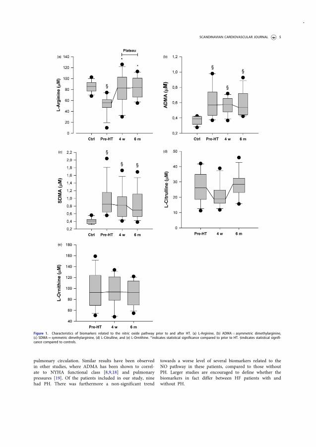
pulmonary circulation. Similar results have been observedin other studies, where ADMA has been shown to correl-ate to NYHA functional class [8,9,18] and pulmonarypressures [19]. Of the patients included in our study, ninehad PH. There was furthermore a non-significant trend
towards a worse level of several biomarkers related to theNO pathway in these patients, compared to those withoutPH. Larger studies are encouraged to define whether thebiomarkers in fact differ between HF patients with andwithout PH.
Figure 1. Characteristics of biomarkers related to the nitric oxide pathway prior to and after HT. (a) L-Arginine, (b) ADMA¼ asymmetric dimethylarginine,(c) SDMA¼ symmetric dimethylarginine, (d) L-Citrulline, and (e) L-Ornithine. �indicates statistical significance compared to prior to HT. §indicates statistical signifi-cance compared to controls.
SCANDINAVIAN CARDIOVASCULAR JOURNAL 5
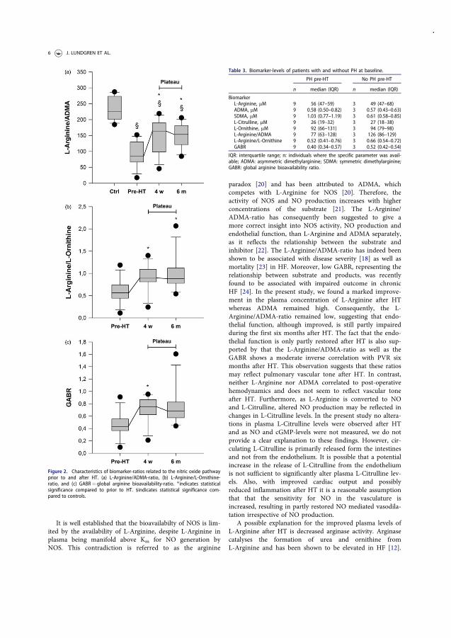
It is well established that the bioavailabilty of NOS is lim-ited by the availability of L-Arginine, despite L-Arginine inplasma being manifold above Km for NO generation byNOS. This contradiction is referred to as the arginine
paradox [20] and has been attributed to ADMA, whichcompetes with L-Arginine for NOS [20]. Therefore, theactivity of NOS and NO production increases with higherconcentrations of the substrate [21]. The L-Arginine/ADMA-ratio has consequently been suggested to give amore correct insight into NOS activity, NO production andendothelial function, than L-Arginine and ADMA separately,as it reflects the relationship between the substrate andinhibitor [22]. The L-Arginine/ADMA-ratio has indeed beenshown to be associated with disease severity [18] as well asmortality [23] in HF. Moreover, low GABR, representing therelationship between substrate and products, was recentlyfound to be associated with impaired outcome in chronicHF [24]. In the present study, we found a marked improve-ment in the plasma concentration of L-Arginine after HTwhereas ADMA remained high. Consequently, the L-Arginine/ADMA-ratio remained low, suggesting that endo-thelial function, although improved, is still partly impairedduring the first six months after HT. The fact that the endo-thelial function is only partly restored after HT is also sup-ported by that the L-Arginine/ADMA-ratio as well as theGABR shows a moderate inverse correlation with PVR sixmonths after HT. This observation suggests that these ratiosmay reflect pulmonary vascular tone after HT. In contrast,neither L-Arginine nor ADMA correlated to post-operativehemodynamics and does not seem to reflect vascular toneafter HT. Furthermore, as L-Arginine is converted to NOand L-Citrulline, altered NO production may be reflected inchanges in L-Citrulline levels. In the present study no altera-tions in plasma L-Citrulline levels were observed after HTand as NO and cGMP-levels were not measured, we do notprovide a clear explanation to these findings. However, cir-culating L-Citrulline is primarily released form the intestinesand not from the endothelium. It is possible that a potentialincrease in the release of L-Citrulline from the endotheliumis not sufficient to significantly alter plasma L-Citrulline lev-els. Also, with improved cardiac output and possiblyreduced inflammation after HT it is a reasonable assumptionthat that the sensitivity for NO in the vasculature isincreased, resulting in partly restored NO mediated vasodila-tation irrespective of NO production.
A possible explanation for the improved plasma levels ofL-Arginine after HT is decreased arginase activity. Arginasecatalyses the formation of urea and ornithine fromL-Arginine and has been shown to be elevated in HF [12].
Figure 2. Characteristics of biomarker-ratios related to the nitric oxide pathwayprior to and after HT. (a) L-Arginine/ADMA-ratio, (b) L-Arginine/L-Ornithine-ratio, and (c) GABR¼ global arginine bioavailability-ratio. �indicates statisticalsignificance compared to prior to HT. §indicates statistical significance com-pared to controls.
Table 3. Biomarker-levels of patients with and without PH at baseline.
PH pre-HT No PH pre-HT
n median (IQR) n median (IQR)
BiomarkerL-Arginine, mM 9 56 (47–59) 3 49 (47–68)ADMA, mM 9 0.58 (0.50–0.82) 3 0.57 (0.43–0.63)SDMA, mM 9 1.03 (0.77–1.19) 3 0.61 (0.58–0.85)L-Citrulline, mM 9 26 (19–32) 3 27 (18–38)L-Ornithine, mM 9 92 (66–131) 3 94 (79–98)L-Arginine/ADMA 9 77 (63–128) 3 126 (86–129)L-Arginine/L-Ornithine 9 0.52 (0.41–0.76) 3 0.66 (0.54–0.72)GABR 9 0.40 (0.34–0.57) 3 0.52 (0.42–0.54)
IQR: interquartile range; n: individuals where the specific parameter was avail-able; ADMA: asymmetric dimethylarginine; SDMA: symmetric dimethylarginine;GABR: global arginine bioavailability ratio.
6 J. LUNDGREN ET AL.

Arginase increases during oxidative stress and infections[25] and as it competes with NOS for L-Arginine it cancause “uncoupling” of NOS, leading, in sequence, toincreased superoxide production, diminished NO production
and endothelial dysfunction [25]. In our material, theL-Arginine/Ornithine-ratio, which can be used to assessarginase activity, increased after HT. However, the increasewas solely due to increase in plasma concentration ofL-Arginine and it is unlikely that arginase, to a greaterextent, is affected by HT. The present study does moreovernot provide a solid explanation to the persistently elevatedADMA levels after HT. Considering the low plasma concen-trations of L-Arginine prior to HT, L-Arginine is an appeal-ing therapy in HF. However, despite the strong rationale forsuch treatment, long-term investigations have been incon-clusive [26,27]. It is possible that the lack of effect in thesetrials is due to that L-Arginine levels do not represent endo-thelial function. This is supported by the present study, inwhich correlations between post-operative hemodynamicsand L-Arginine were lacking. Based on our findings, withlow pre-operative plasma levels of L-Arginine, in combin-ation with the high plasma levels of ADMA and decreasedL-Arginine/ADMA-ratio throughout the study, where theL-Arginine/ADMA-ratio indeed reflected post-HT pulmon-ary vascular tone, it is likely that NO production is reducedboth prior to and after HT. Consequently, drugs targetingthe NO pathway independent of NO, such as soluble guany-late cyclase (sGC) stimulators, may be more beneficial thanL-Arginine as well as NO dependent substances. Indeed, inpatients with systolic HF, a phase II trial with the novelsGC-stimulator vericiguat recently demonstrated positiveeffects on NT-proBNP levels in a dose-dependent manner[28], supporting this hypothesis. Future studies may eluci-date the potential of such a therapeutic approach.
Limitations
A major limitation of the present study is the small samplesize. However, the size is in line with previous pilot studiesin HT which has stimulated the initiation of larger trials.Moreover, in previous studies [29] we have, in larger HT-populations from our hospital, shown similar pre-operativeand post-operative hemodynamics as in the present group,suggesting that the current findings can be applied in largerpopulations. Furthermore, we did not measure NO andcGMP levels so we cannot be certain that the production isdecreased. However, the findings of decreased L-Arginine/ADMA-ratio throughout our study, in combination with the
Table 4. Selected hemodynamic parameters at baseline and during follow-up.
Pre-HT Four weeks post-HT Six months post-HT
n median (IQR) n median (IQR) n median (IQR)
Hemodynamic parameterMPAP, mmHg 12 35 (27–42) 12 17 (12–21)� 12 15 (12–18)�DPG, mmHg 11 1.0 (–0.8–4.8) 11 2.0 (0.3–5.0) 12 3.0 (0.0–5.0)TPG, mmHg 11 9 (6–12) 12 7.5 (6.0–9.0) 12 8.0 (6.0–9.5)PAWP, mmHg 11 25 (20–30) 12 8 (5–11)� 12 5.5 (4.0–8.5)�MRAP, mmHg 12 15 (12–19) 12 4.0 (3.0–6.0) 12 1.5 (0.0–4.0)�CO, L�min�1 12 3.4 (2.6–4.3) 12 6.0 (5.6–6.6)� 12 5.6 (5.1–5.8)�PVR, WU 11 2.4 (1.9–3.0) 12 1.3 (1.1–1.5)� 12 1.4 (1.1–1.7)
IQR: interquartile range; n: individuals where the specific parameter was available; MPAP: mean pulmonary artery pres-sure; DPG: diastolic pressure gradient; TPG: transpulmonary gradient; PAWP: pulmonary artery wedge pressure; MRAP:mean right atrial pressure; CO: cardiac output; PVR: pulmonary vascular resistance.�indicates statistical significant difference (p< .05) compared to prior to HT.
Figure 3. (a) Correlation between the L-Arginine/ADMA-ratio and PVR sixmonths after HT. (b) Correlation between GABR and PVR six months after HT.GABR: global arginine bioavailability ratio; PVR: pulmonary vascular resistance.
SCANDINAVIAN CARDIOVASCULAR JOURNAL 7
fact that the ratio reflect pulmonary vascular tone, points inthe direction of NO dependent endothelial dysfunction priorto and after HT. Finally, medical therapies prior to and afterHT were individually adjusted. As some of these medica-tions, such as betablockers [24], ACEi and ARB [30] havebeen shown to decrease circulating ADMA levels, dimin-ished use of these medications after HT could, in theory,increase plasma levels of ADMA and the results in the pre-sent study must consequently be interpreted with this as aprecaution. However, the use of these drugs represent acornerstone in heart failure therapy and a true clinical situ-ation so our findings are relevant to generate hypotheses forfuture, larger, trials.
Conclusions
The present study shows that plasma concentrations ofL-Arginine, by an unknown mechanism, normalizes afterHT. However, as ADMA plasma levels are unaltered, theL-Arginine/ADMA-ratio remains low and correlatesinversely to PVR. This suggests that the L-Arginine/ADMA-ratio reflects pulmonary vascular tone after HT and thatNO-dependent endothelial function is post-operativelyimproved, yet not normalized. Moreover, considering thegood post-operative outcome without complications, ourresults could represent normal plasma magnitudes of theinvestigated substances after HT and the plasma derivedL-Arginine/ADMA-ratio may therefore be used to assesspulmonary vascular tone after HT. Finally, based on the lowL-Arginine/ADMA-ratio prior to and after HT, it is reason-able to conclude that, if targeting the NO pathway in HF,treatment with NO independent vasoactive substances maybe more beneficial than NO dependent drugs. Larger trialson this subject are therefore encouraged.
Acknowledgements
We acknowledge the support of the staff at the Hemodynamic Lab atThe Section for Heart Failure and Valvular Disease, Skåne UniversityHospital, and the Department of Cardiology, Clinical Sciences, LundUniversity, Lund, Sweden. In specific, we acknowledge the work of Ms.Karin Paulsson, Ms. Anneli Ahlqvist and Ms. Marie Wildt for theirsupport in the assembly of the plasma samples within the Lund CardioPulmonary Register as well as Mrs. Sofia Berg Blomqvist and Mrs.Karin Kjellstr€om at Uppsala University Hospital, Uppsala, Sweden forsampling of blood plasma from healthy subjects. We furthermoreacknowledge the biobank services and retrieval of blood samples per-formed at Labmedicin Skåne, University and Regional Laboratories,Region Skåne, Sweden. Finally, we are grateful to Mrs. ElisabethFredriksson at the National Veterinary Institute (SVA) for carrying outthe LC-MC/MS analyses.
Disclosure statement
Dr. Lundgren and PhD Sandqvist report unrestricted research grantsfrom The Swedish Society of Pulmonary Hypertension, and Dr.Rådegran reports unrestricted research grants from Anna-Lisa andSven-Erik Lundgren’s, Maggie Stephen’s, ALF’s, Skåne UniversityHospital’s Foundations and Actelion Pharmaceuticals Sweden AB,during the conduct of the study.
Dr. Lundgren reports personal lecture fees from ActelionPharmaceuticals Sweden AB and GlaxoSmithKline outside the
submitted work. Dr. Wikstr€om and Dr. Rådegran report personal lec-ture fees from Actelion Pharmaceuticals Sweden AB, Bayer Healthcare,GlaxoSmithKline and Sandoz/Novartis outside the submitted work.
Dr. Wikstr€om is, and has been primary-, or co-, investigator in;clinical PAH trials for GlaxoSmithKline, Actelion PharmaceuticalsSweden AB, Pfizer, Bayer Healthcare and United Therapeutics, and inclinical heart failure trials for Novartis. Dr. Rådegran is, and has beenprimary-, or co-, investigator in; clinical PAH trials forGlaxoSmithKline, Actelion Pharmaceuticals Sweden AB, Pfizer, BayerHealthcare and United Therapeutics, and in clinical heart transplant-ation immuno-suppression trials for Novartis.
Adjunct prof. Hedeland and prof. Bondesson report no conflictsof interest.
Since the completion of the study and initial submission of themanuscript, PhD Sandqvist has started working at ActelionPharmaceuticals Sweden AB. The company has no role in data collec-tion, analysis, interpretation of data or publishing of the manuscript.
Funding
The work was supported by unrestricted research grants from Anna-Lisa and Sven-Erik Lundgren’s and ALF’s Foundations, the SwedishSociety of Pulmonary Hypertension and Actelion PharmaceuticalsSweden AB. The funding organizations played no role in the collection,analysis or interpretation of the data and have no right to restrict thepublishing of the manuscript.
References
[1] Bursi F, McNallan SM, Redfield MM, et al. Pulmonary pres-sures and death in heart failure: a community study. J Am CollCardiol. 2012;59:222–231.
[2] Rosenkranz S, Gibbs JS, Wachter R, et al. Left ventricular heartfailure and pulmonary hypertensiondagger. Eur Heart J.2016;37:942–954.
[3] Lundgren J, Soderlund C, Radegran G. Impact of postoperativepulmonary hypertension on outcome after heart transplant-ation. Scand Cardiovasc J. 2017;51:172–181.
[4] Cooper CJ, Jevnikar FW, Walsh T, et al. The influence of basalnitric oxide activity on pulmonary vascular resistance inpatients with congestive heart failure. Am J Cardiol. 1998;82:609–614.
[5] Visser M, Paulus WJ, Vermeulen MA, et al. The role of asym-metric dimethylarginine and arginine in the failing heart and itsvasculature. Eur J Heart Fail. 2010;12:1274–1281.
[6] Palmer RM, Ashton DS, Moncada S. Vascular endothelial cellssynthesize nitric oxide from L-arginine. Nature. 1988;333:664–666.
[7] Durante W, Johnson FK, Johnson RA. Arginase: a critical regu-lator of nitric oxide synthesis and vascular function. Clin ExpPharmacol Physiol. 2007;34:906–911.
[8] Usui M, Matsuoka H, Miyazaki H, et al. Increased endogenousnitric oxide synthase inhibitor in patients with congestive heartfailure. Life Sci. 1998;62:2425–2430.
[9] Hsu CP, Lin SJ, Chung MY, et al. Asymmetric dimethylargininepredicts clinical outcomes in ischemic chronic heart failure.Atherosclerosis. 2012;225:504–510.
[10] Zairis MN, Patsourakos NG, Tsiaousis GZ, et al. Plasma asym-metric dimethylarginine and mortality in patients with acutedecompensation of chronic heart failure. Heart. 2012;98:860–864.
[11] Palm F, Onozato ML, Luo Z, et al. Dimethylarginine dimethyla-minohydrolase (DDAH): expression, regulation, and function inthe cardiovascular and renal systems. Am J Physiol Heart CircPhysiol. 2007;293:H3227–H3245.
[12] Quitter F, Figulla HR, Ferrari M, et al. Increased arginase levelsin heart failure represent a therapeutic target to rescue micro-vascular perfusion. Clin Hemorheol Microcirc. 2013;54:75–85.
8 J. LUNDGREN ET AL.

[13] Holm T, Aukrust P, Aagaard E, et al. Hypertension in relationto nitric oxide, asymmetric dimethylarginine, and inflammation:different patterns in heart transplant recipients and individualswith essential hypertension. Transplantation. 2002;74:1395–1400.
[14] Potena L, Fearon WF, Sydow K, et al. Asymmetric dimethylar-ginine and cardiac allograft vasculopathy progression: modula-tion by sirolimus. Transplantation. 2008;85:827–833.
[15] Sandqvist A, Schneede J, Kylhammar D, et al. PlasmaL-arginine levels distinguish pulmonary arterial hypertensionfrom left ventricular systolic dysfunction. Heart Vessels.2018;33:255–263.
[16] Henrohn D, Sandqvist A, Egerod H, et al. Changes in plasmalevels of asymmetric dimethylarginine, symmetric dimethylargi-nine, and arginine after a single dose of vardenafil in patientswith pulmonary hypertension. Vascul Pharmacol. 2015;73:71–77.
[17] Stewart S, Winters GL, Fishbein MC, et al. Revision of the 1990working formulation for the standardization of nomenclature inthe diagnosis of heart rejection. J Heart Lung Transplant.2005;24:1710–1720.
[18] Seljeflot I, Nilsson BB, Westheim AS, et al. The L-arginine-asymmetric dimethylarginine ratio is strongly related to theseverity of chronic heart failure. No effects of exercise training.J Card Fail. 2011;17:135–142.
[19] Shao Z, Wang Z, Shrestha K, et al. Pulmonary hypertensionassociated with advanced systolic heart failure: dysregulatedarginine metabolism and importance of compensatory dimethy-larginine dimethylaminohydrolase-1. J Am Coll Cardiol.2012;59:1150–1158.
[20] Boger RH. Asymmetric dimethylarginine, an endogenousinhibitor of nitric oxide synthase, explains the “L-arginine para-dox” and acts as a novel cardiovascular risk factor. J Nutr.2004;134(10 Suppl):2842S–2847S.
[21] Creager MA, Gallagher SJ, Girerd XJ, et al. L-arginine improvesendothelium-dependent vasodilation in hypercholesterolemichumans. J Clin Invest. 1992;90:1248–1253.
[22] Boger RH, Sullivan LM, Schwedhelm E, et al. Plasmaasymmetric dimethylarginine and incidence of cardiovasculardisease and death in the community. Circulation. 2009;119:1592–1600.
[23] Anderssohn M, Rosenberg M, Schwedhelm E, et al. TheL-Arginine-asymmetric dimethylarginine ratio is an independ-ent predictor of mortality in dilated cardiomyopathy. J CardFail. 2012;18:904–911.
[24] Tang WH, Shrestha K, Wang Z, et al. Diminished global argin-ine bioavailability as a metabolic defect in chronic systolic heartfailure. J Card Fail. 2013;19:87–93.
[25] Pernow J, Jung C. Arginase as a potential target in the treat-ment of cardiovascular disease: reversal of arginine steal?Cardiovasc Res. 2013;98:334–343.
[26] Rector TS, Bank AJ, Mullen KA, et al. Randomized, double-blind, placebo-controlled study of supplemental oral L-arginine in patients with heart failure. Circulation. 1996;93:2135–2141.
[27] Schulman SP, Becker LC, Kass DA, et al. L-arginine therapy inacute myocardial infarction: the Vascular Interaction With Agein Myocardial Infarction (VINTAGE MI) randomized clinicaltrial. JAMA. 2006;295:58–64.
[28] Gheorghiade M, Greene S, Butler J, et al. Effect of vericiguat, asoluble guanylate cyclase stimulator, on natriuretic peptide lev-els in patients with worsening chronic heart failure and reducedejection fraction: the SOCRATES-REDUCED randomized trial.JAMA. 2015;314:2251–2262.
[29] Lundgren J, Radegran G. Hemodynamic characteristics includ-ing pulmonary hypertension at rest and during exercise beforeand after heart transplantation. J Am Heart Assoc.2015;4:e001787. doi:10.1161/JAHA.115.001787
[30] Chen JW, Hsu NW, Wu TC, et al. Long-term angiotensin-con-verting enzyme inhibition reduces plasma asymmetric dimethy-larginine and improves endothelial nitric oxide bioavailability andcoronary microvascular function in patients with syndrome X.Am J Cardiol. 2002;90:974–982.
SCANDINAVIAN CARDIOVASCULAR JOURNAL 9


REVIEW
Pathophysiology and potential treatments of pulmonary
hypertension due to systolic left heart failure
J. Lundgren1,2 and G. R�adegran1,2
1 The Haemodynamic Laboratory, The Clinic for Heart Failure and Valvular Disease, Sk�ane University Hospital, Lund, Sweden
2 Department of Cardiology, Clinical Sciences, Lund University, Lund, Sweden
Received 9 December 2013,
revision requested 3 February
2014,
revision received 11 March 2014,
accepted 28 March 2014
Correspondence: J. Lundgren, The
Haemodynamic Laboratory, The
Clinic for Heart Failure and
Valvular Disease, Sk�ane University
Hospital, Lund, Sweden.
E-mail: jakob_lundgren@hotmail.
com
Abstract
Pulmonary hypertension (PH) due to left heart failure is becoming increas-
ingly prevalent and is associated with poor outcome. The precise patho-
physiological mechanisms behind PH due to left heart failure are,
however, still unclear. In its early course, PH is caused by increased left
ventricular filling pressures, without pulmonary vessel abnormalities. Con-
ventional treatment for heart failure may partly reverse such passive PH
by optimizing left ventricular function. However, if increased pulmonary
pressures persist, endothelial damage, excessive vasoconstriction and struc-
tural changes in the pulmonary vasculature may occur. There is, at pres-
ent, no recommended medical treatment for this active component of PH
due to left heart failure. However, as the vascular changes in PH due to
left heart failure may be similar to those in pulmonary arterial hyperten-
sion (PAH), a selected group of these patients may benefit from PAH treat-
ment targeting the endothelin, nitric oxide or prostacyclin pathways. Such
potent pulmonary vasodilators could, however, be detrimental in patients
with left heart failure without pulmonary vascular pathology, as selective
pulmonary vasodilatation may lead to further congestion in the pulmonary
circuit, resulting in pulmonary oedema. The use of PAH therapies is there-
fore currently not recommended and would require the selection of suit-
able patients based on the underlying causes of the disease and careful
monitoring of their progress. The present review focuses on the following:
(i) the pathophysiology behind PH resulting from systolic left heart failure,
and (ii) the current evidence for medical treatment of this condition, espe-
cially the role of PAH-targeted therapies in systolic left heart failure.
Keywords left heart failure, pulmonary arterial hypertension, pulmonary
hypertension, therapy.
According to the classification presented at the Fifth
World Symposium on PH (WSPH) in Nice 2013,
pulmonary hypertension (PH) is divided into five
groups: (i) pulmonary arterial hypertension (PAH), (ii)
PH due to left heart disease, (iii) PH due to lung dis-
ease and/or hypoxaemia, (iv) chronic thromboembolic
pulmonary hypertension and (v) PH with unclear and/
or multifactorial mechanisms. Group 1 also includes
the subgroups 10. pulmonary veno-occlusive disease
and/or pulmonary capillary haemangiomatosis and 10 0.persistent PH of the newborn (Simonneau et al. 2013).
Group 2 is defined as post-capillary PH, while the oth-
ers are defined as pre-capillary PH, depending on the
location of the pathological changes (Galie et al. 2009).
© 2014 Scandinavian Physiological Society. Published by John Wiley & Sons Ltd, doi: 10.1111/apha.12295314
Acta Physiol 2014, 211, 314–333

Left heart disease is the most common cause of PH
and is mainly caused by left heart systolic or diastolic
dysfunction (Ghio et al. 2001, Lam et al. 2009) but
also, to a lesser extent, by valvular heart disease
(Simonneau et al. 2013). The pathophysiology behind
PH due to left heart disease is not completely under-
stood. Furthermore, there are no specific therapies for
the pulmonary component of PH due to left heart dis-
ease, other than optimizing left ventricular and valvu-
lar function (Galie et al. 2009). This is of importance
as PH is a negative prognostic marker in left heart
disease (Lucas et al. 2000, Gheorghiade et al. 2006).
Pulmonary hypertension is diagnosed by right heart
catheterization and defined by a mean pulmonary
artery pressure (MPAP) ≥ 25 mmHg. It is subcatego-
rized into pre- or post-capillary PH depending on
whether the pulmonary artery wedge pressure
(PAWP), previously also called pulmonary capillary
wedge pressure or pulmonary artery occlusion pres-
sure, is ≤ 15 or > 15 mmHg, respectively, at normal
or reduced cardiac output (Galie et al. 2009) (Fig. 1a).
Depending on the definition used, post-capillary PH is
further subcategorized according to the levels of the
transpulmonary gradient (TPG) (Galie et al. 2009)
(Fig. 1a) and/or pulmonary vascular resistance (PVR)
(Fang et al. 2012) (Fig. 1b). According to the ESC
guidelines, post-capillary PH may then be divided into
passive or reactive post-capillary PH (Fig. 1a) (Galie
et al. 2009) or, according to the International Society
for Heart and Lung Transplantation (ISHLT) state-of-
the-art summary statement, into passive or mixed
post-capillary PH (Fig. 1b) (Fang et al. 2012). The
mixed group may furthermore be subclassified into
reactive or non-reactive post-capillary PH, based on
the response to a vasodilatation test.
Recently, the diastolic pressure difference (DPD),
calculated by subtracting PAWP from the diastolic
pulmonary artery pressure, was introduced as a new
means of subclassifying post-capillary PH. DPD has
been suggested to identify pulmonary vascular disease
in left heart failure (LHF) more correctly than TPG,
as diastolic pulmonary artery pressure is less flow
dependent than MPAP (Naeije et al. 2013). In support
of this, Gerges et al. found that a DPD ≥ 7 mmHg
indicated severe pulmonary vascular disease and
increased mortality in patients with PH due to LHF
(Fig. 1c) (Gerges et al. 2013). DPD has consequently
been suggested as a new important parameter in the
definition of PH, based on the discussions at the Fifth
WSPH (Fig. 1d) (Vachiery et al. 2013).
Concerning the lack of therapies for PH due to LHF,
it has been debated whether PAH-targeted therapies
could be used to treat this condition. However, selec-
tive dilation of the pulmonary vessels in patients with
impaired left ventricular function, without simultaneous
(a)
(b)
(c)
(d)
Figure 1 (a) Haemodynamic definition of pulmonary
hypertension (PH) according to the ESC guidelines, (b)
Haemodynamic definition of pulmonary hypertension (PH)
according to the ISHLT state-of-the-art summary statement,
(c) Haemodynamic definition of PH according to Gerges
et al., (d) Haemodynamic definition suggested after the Fifth
World Symposium on PH. MPAP, mean pulmonary artery
pressure; PAWP, pulmonary artery wedge pressure; CO,
cardiac output; TPG, transpulmonary gradient; PAH, pulmo-
nary arterial hypertension; CTEPH, chronic thromboembolic
pulmonary hypertension; DPD, diastolic pressure difference;
PVR, pulmonary vascular resistance; LV, left ventricular.
© 2014 Scandinavian Physiological Society. Published by John Wiley & Sons Ltd, doi: 10.1111/apha.12295 315
Acta Physiol 2014, 211, 314–333 J Lundgren and G R�adegran · Pulmonary hypertension due to left heart failure

unloading of the left ventricle, could be detrimental,
causing further congestion of the pulmonary veins
(Haywood et al. 1992, Loh et al. 1994, Michelakis
et al. 2002). This congestion may, in turn, lead to pul-
monary oedema and rapid worsening of the patient’s
condition (Packer et al. 2005). As a consequence,
PAH-targeted therapies have not been recommended
for the treatment of patients with LHF, with or with-
out PH. However, as it has been suggested that the
changes observed in the pulmonary vessels, in persisting
PH due to LHF, may be similar to those in PAH,
selected patients with excessive vasoconstriction with
or without secondary vascular remodelling could,
potentially, benefit from PAH-targeted therapies.
The present review focuses on the pathophysiology
and pathogenesis of PH due to LHF related to systolic
dysfunction, as well as the existing knowledge on and
potential role of PAH-targeted therapies for this
patient population.
Pathophysiology of pulmonary hypertension
Pulmonary arterial hypertension
During the past thirty years, considerable effort has
been devoted to clarifying the cause of PAH. PAH is a
rare, but complex and serious disease, and if not trea-
ted, the survival has been reported to be as low as
approx. 1–2.8 years, depending on its underlying cause
(D’Alonzo et al. 1991). It has been suggested that
PAH is triggered by an imbalance between vasodilative
and vasoconstrictive factors, causing excessive pulmo-
nary arteriolar vasoconstriction (Schermuly et al.
2011). This initial vasoconstriction, in combination
with other, unknown, factors, is thought to lead to
vascular remodelling of the vessel layers of the pulmo-
nary arteries (Schermuly et al. 2011). Smooth muscle
cell proliferation, inflammation and the development
of plexiform lesions are additional hallmarks of PAH
(Schermuly et al. 2011). Together with in situ throm-
bosis, this remodelling results in a further reduction in
the size of the arterial lumen (Schermuly et al. 2011).
As PAH progresses, the influence of vascular remodel-
ling seems to become the most prominent feature of
the disease, leading to a further increase in PVR and
increased load on the right ventricle. This, in turn,
leads to right ventricular hypertrophy. However, at a
certain threshold, the right ventricle fails to cope with
the increasing pressure and begins to dilate, leading to
syncope and, ultimately, death (Rubin 1997).
Pulmonary hypertension due to left heart disease
The precise pathophysiology of PH due to LHF is
unclear and is probably multifactorial. In the acute
setting, LHF leads to increased pressures in the
pulmonary veins and capillaries. This venous PH is
due to passive congestion in the pulmonary vessels,
caused by the increased left ventricular filling pres-
sures (Drazner et al. 1999). The increased pressures
in the pulmonary capillaries result in increased stress
on the alveolar capillary wall, potentially causing
acute vascular wall damage and subsequent oedema
(Kurdak et al. 1995, West & Mathieu-Costello 1995).
Initially, the damage is reversible, but if it persists, it
may lead to irreversible, structural changes and
remodelling, causing the walls of the alveolar capillary
membrane to thicken, resulting in reduced diffusion
capacity of the lungs (Guazzi 2008).
In addition to affecting the pulmonary capillaries,
LHF may also lead to secondary increased pressure in
the pulmonary arteries. Such passive PH is caused by
persisting, increased left ventricular filling pressures
and is seen as increased pulmonary artery, and wedge
pressures. At this stage, however, TPG and PVR
remain low (Galie et al. 2009, Fang et al. 2012)
(Fig. 2). Moreover, if the increased pulmonary artery
pressures persist over a long period, this may lead to
endothelial damage, suggested to be the result of
impaired Ca2+ signalling and cytoskeletal reorganiza-
tion (Kerem et al. 2010). The endothelial damage in
turn causes an imbalance between vasoactive sub-
stances, resulting in impaired smooth muscle relaxa-
tion and, consequently, vasoconstriction (Cooper
et al. 1998, Ooi et al. 2002). In LHF, as in PAH,
nitric oxide (NO)-dependent vasodilation is impaired,
and endothelin-1 levels may be increased (Cody et al.
1992, Tsutamoto et al. 1994, Cooper et al. 1998, Ooi
et al. 2002). Such active PH is illustrated by increased
TPG and/or PVR, in addition to the increase in pul-
monary artery and wedge pressures (Fig. 2). The ESC
guidelines refer to this as reactive PH (Galie et al.
2009) (Fig. 1a), whereas ISHLT state-of-the-art sum-
mary statement (Fang et al. 2012) refers to it as mixed
PH (Fig. 1b). Such excessive vasoconstriction may
initially be reversible with an acute vasodilatation
test, but long-standing vasoconstriction may become
so-called fixed, that is, not acutely reversible, due
to changes in the pulmonary vasculature (Rich &
Rabinovitch 2008) (Fig. 2). The vascular changes that
occur include intimal thickening and fibrosis as well
as medial hypertrophy and may be similar to, but not
precisely the same as, those seen in PAH (Rich &
Rabinovitch 2008) (Galie et al. 2009). For instance,
plexiform lesions are generally not seen in PH due
to LHF.
The reasons for the lack of knowledge on the
pathophysiology of, and the lack of therapy for, PH
due to systolic LHF are many. One may be the lack
of good animal models for chronic heart failure with
© 2014 Scandinavian Physiological Society. Published by John Wiley & Sons Ltd, doi: 10.1111/apha.12295316
Pulmonary hypertension due to left heart failure · J Lundgren and G R�adegran Acta Physiol 2014, 211, 314–333
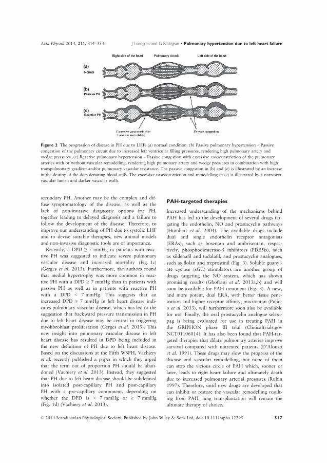
secondary PH. Another may be the complex and dif-
fuse symptomatology of the disease, as well as the
lack of non-invasive diagnostic options for PH,
together leading to delayed diagnosis and a failure to
follow the development of the disease. Therefore, to
improve our understanding of PH due to systolic LHF
and to devise suitable therapies, new animal models
and non-invasive diagnostic tools are of importance.
Recently, a DPD ≥ 7 mmHg in patients with reac-
tive PH was suggested to indicate severe pulmonary
vascular disease and increased mortality (Fig. 1c)
(Gerges et al. 2013). Furthermore, the authors found
that medial hypertrophy was more common in reac-
tive PH with a DPD ≥ 7 mmHg than in patients with
passive PH as well as in patients with reactive PH
with a DPD < 7 mmHg. This suggests that an
increased DPD ≥ 7 mmHg in left heart disease indi-
cates pulmonary vascular disease, which has led to the
suggestion that backward pressure transmission in PH
due to left heart disease may be central in triggering
myofibroblast proliferation (Gerges et al. 2013). This
new insight into pulmonary vascular disease in left
heart disease has resulted in DPD being included in
the new definition of PH due to left heart disease.
Based on the discussions at the Fifth WSPH, Vachiery
et al. recently published a paper in which they urged
that the term out of proportion PH should be aban-
doned (Vachiery et al. 2013). Instead, they suggested
that PH due to left heart disease should be subdefined
into isolated post-capillary PH and post-capillary
PH with a pre-capillary component, depending on
whether the DPD is < 7 mmHg or ≥ 7 mmHg
(Fig. 1d) (Vachiery et al. 2013).
PAH-targeted therapies
Increased understanding of the mechanisms behind
PAH has led to the development of several drugs tar-
geting the endothelin, NO and prostacyclin pathways
(Humbert et al. 2004). The available drugs include
dual and single endothelin receptor antagonists
(ERAs), such as bosentan and ambrisentan, respec-
tively, phosphodiesterase-5 inhibitors (PDE5is), such
as sildenafil and tadalafil, and prostacyclin analogues,
such as flolan and treprostinil (Fig. 3). Soluble guanyl-
ate cyclase (sGC) stimulators are another group of
drugs targeting the NO system, which has shown
promising results (Ghofrani et al. 2013a,b) and will
soon be available for PAH treatment (Fig. 3). A new,
and more potent, dual ERA, with better tissue pene-
tration and higher receptor affinity, macitentan (Pulid-
o et al. 2013), will furthermore soon also be available
for use. Finally, the oral prostacyclin analogue selexi-
pag is being evaluated for use in treating PAH in
the GRIPHON phase III trial (Clinicaltrials.gov
NCT01106014). It has also been found that PAH-tar-
geted therapies that dilate pulmonary arteries improve
survival compared with untreated patients (D’Alonzo
et al. 1991). These drugs may slow the progress of the
disease and vascular remodelling, but none of them
can stop the vicious circle of PAH which, sooner or
later, leads to right heart failure and ultimately death
due to increased pulmonary arterial pressures (Rubin
1997). Therefore, until new drugs are developed that
can inhibit or restore the vascular remodelling result-
ing from PAH, lung transplantation will remain the
ultimate therapy of choice.
(a)
(b)
(c)
Figure 2 The progression of disease in PH due to LHF: (a) normal condition. (b) Passive pulmonary hypertension – Passive
congestion of the pulmonary circuit due to increased left ventricular filling pressures, rendering high pulmonary artery and
wedge pressures. (c) Reactive pulmonary hypertension – Passive congestion with excessive vasoconstriction of the pulmonary
arteries with or without vascular remodelling, rendering high pulmonary artery and wedge pressures in combination with high
transpulmonary gradient and/or pulmonary vascular resistance. The passive congestion in (b) and (c) is illustrated by an increase
in the destiny of the dots denoting blood cells. The excessive vasoconstriction and remodelling in (c) is illustrated by a narrower
vascular lumen and darker vascular walls.
© 2014 Scandinavian Physiological Society. Published by John Wiley & Sons Ltd, doi: 10.1111/apha.12295 317
Acta Physiol 2014, 211, 314–333 J Lundgren and G R�adegran · Pulmonary hypertension due to left heart failure

The treatment of PH due to LHF
Although PH is common in LHF, and some vascular
changes seen in this condition may be similar to those
in PAH, there is no specific therapy for the pulmonary
component of PH due to LHF, other than optimizing
the function of the left heart (Galie et al. 2009). Con-
ventional medical therapy for systolic LHF, including
beta-blockers, angiotensin-converting enzyme inhibi-
tors and/or angiotensin receptor blockers, mineralo-
corticoid receptor antagonists, If (funny current)
inhibitor (ivabradine), digoxin and diuretics (McMur-
ray et al. 2012), may reduce the PH caused by passive
congestion, by optimizing the ventricular function of
the heart. However, they are not thought to affect the
excessive vasoconstriction or remodelling of the pul-
monary vasculature seen in reactive PH. Therefore, it
is of great importance to develop and evaluate new
therapies for PH due to LHF, directly related to the
pulmonary vascular component. This is further
strengthened by the fact that PH in LHF is a marker
of disease severity and a suggested risk factor for early
mortality after heart transplantation (Mehra et al.
2006). However, in a recent study, our group found
that with improved medical therapies and mechanical
assist devices, the pre-operative magnitudes of the hae-
modynamic parameters that best predict outcome after
heart transplantation remained unclear (Lundgren
et al. 2014). Although pre-operative PH may be asso-
ciated with poorer prognosis after heart transplanta-
tion, it appears that, with the diagnostic methods
currently used, and when PH is defined at current
magnitudes, it is difficult to differentiate between
patients with excessive vasoconstriction without vas-
cular remodelling and those with remodelling. There-
fore, it is difficult to decide which patients are eligible
for heart transplantation. If the assumptions regarding
DPD are correct, and that DPD ≥ 7 mmHg indeed
indicates severe pulmonary vascular disease in patients
with PH due to LHF, this parameter could possibly
help in the future selection of patients suitable for
heart transplantation.
It has been suggested that PAH-targeted therapies
could be of value in selected cases of PH due to LHF.
However, as previously mentioned, selective pulmo-
nary vasodilation, in patients with LHF, may lead to
increased left ventricular filling pressures (Haywood
et al. 1992, Loh et al. 1994, Michelakis et al. 2002)
and the worsening of heart failure. For this reason,
and the lack of appropriate studies, the use of such
therapies has not been recommended. It is, however,
possible that PAH-targeted therapies could prove valu-
able in PH patients with excessive vasoconstriction of
the pulmonary arteries, with or without secondary
vascular remodelling.
Left ventricular assist device
Patients with heart failure can remain stable for a rel-
atively long time due to improvements in medical
therapy (McMurray et al. 2012). Consequently, the
number of patients suffering from end-stage heart fail-
ure and secondary PH has increased, illustrating the
need for therapies that also target the pulmonary com-
ponent of the disease. Left ventricular assist devices
(LVADs) may be useful in this context. In addition to
improving LHF, unloading of the left ventricle with a
LVAD for a few months has also been shown to
reverse passive and reactive PH (John et al. 2010,
Nair et al. 2010, Kutty et al. 2013). These findings
were recently confirmed by our group, in patients who
received a LVAD as a bridge to heart transplantation
(Lundgren et al. 2014). We found that the use of a
LVAD (mean 77 days) decreased MPAP, PAWP and
mean right atrial pressure and increased cardiac out-
put, resulting in normal values (Fig. 4) (Lundgren
et al. 2014). Long-term use of LVADs may, further-
more, reverse PH in patients who are not responsive
to vasodilatation in the acute phase (Gallagher et al.
1991, Smedira et al. 1996). However, LVAD implan-
Vessel lumen
Endothelium
Subendothelium
Smooth muscle cells
Prostacyclin pathway
Endothelin pathway
Prostacyclin(Prostaglandin I2)
Prostacyclin synthase
Arachidonic acidEndothelin 1
Endothelin converting enzyme
Pro-Endothelin-1
Endothelin 1
Prostacyclinreceptor
Nitric oxidepathway
NOS
L-arg L-cit + Nitric oxide
Exogenous NO
+
Soluble guanylate cyclase
GTP cGMP
sGC stimulators
+
Vasodilatation and antiproliferation
Phosphodiesterasetype 5 inhibitors
Phosphodiesterase type 5
GMP
Prostacyclinanalogues
ETAR ETBR
ETBRETBR
Vasoconstriction and proliferation
Endothelinreceptor
antagonists
Vasodilatation and antiproliferation
++
Figure 3 Therapeutic pathways in PAH.
NOS, nitric oxide synthase; L-arg,
L-arginin; L-cit, L-citrulline; NO, nitric
oxide; sGC, soluble guanylate cyclase;
ETAR, endothelin receptor A; ETBR,
endothelin receptor B; GTP, guanosine
triphosphate; cGMP, cyclic guanosine
monophosphate.
© 2014 Scandinavian Physiological Society. Published by John Wiley & Sons Ltd, doi: 10.1111/apha.12295318
Pulmonary hypertension due to left heart failure · J Lundgren and G R�adegran Acta Physiol 2014, 211, 314–333

tation is an invasive procedure associated with poten-
tial complications related to surgery, driveline infec-
tions, gastrointestinal bleeding and thrombosis (Felix
et al. 2012). Therefore, it is also important to develop
new drugs directly targeting the PH component of
LHF. Furthermore, in the 12 patients investigated in
our study, we did not observe a decrease in PVR as a
result of LVAD therapy (Fig. 4). In contrast, a reduc-
tion in PVR was observed in 23 patients following
acute vasodilatation (Fig. 5) (Lundgren et al. 2014),
suggesting that medical vasodilatation therapy, alone
or in combination with a LVAD, could prove useful
in treating PH due to LHF. Several studies have, in
fact, investigated the effects of PDE5i and ERAs in left
heart disease without (Sutsch et al. 1998, Katz et al.
2000, Cotter et al. 2001, Louis et al. 2001, Schalcher
et al. 2001, Torre-Amione et al. 2001a,b, Coletta
et al. 2002, Luscher et al. 2002, Schofield et al. 2003,
Anand et al. 2004, Guazzi et al. 2004, 2007, 2009,
2010, 2011, Hirata et al. 2005, Packer et al. 2005,
Lewis et al. 2007a, McMurray et al. 2007, Behling
et al. 2008, De Santo et al. 2008, Boffini et al. 2009,
Amin et al. 2013) and with (Angel Gomez-Sanchez
et al. 2004, Gomez-Moreno et al. 2005, Jabbour et al.
2007, Lewis et al. 2007b, Maruszewski et al. 2007,
Zakliczynski et al. 2007, Kaluski et al. 2008, Tedford
et al. 2008, De Santo et al. 2012, Guazzi et al. 2012,
Hefke et al. 2012, Pons et al. 2012, Potter et al.
2012, Imamura et al. 2013b, Perez-Villa et al. 2013,
Reichenbach et al. 2013) secondary PH. These studies
are discussed and illustrated in the text and tables
below (Tables 1–3).
Phosphodiesterase-5 inhibition
Studies with the PDE5i sildenafil have generally shown
positive results in patients with LHF, with or without
PH. The effects seem to be short term (Schofield et al.
2003, Angel Gomez-Sanchez et al. 2004, Guazzi et al.
2004, Hirata et al. 2005, Lepore et al. 2005, Lewis
(a) (b)
(c) (d)
(e) (f)Figure 4 Haemodynamic response to
LVAD therapy with regards to: (a)
MPAP, mean pulmonary artery pressure;
(b) PAWP, pulmonary artery wedge
pressure; (c) TPG, transpulmonary gradi-
ent; (d) MRAP, mean right atrial pres-
sure; (e) CO, cardiac output; (f) PVR,
pulmonary vascular resistance; LVAD,
left ventricular assist device. *indicates a
statistically significant difference com-
pared with baseline measurements.
© 2014 Scandinavian Physiological Society. Published by John Wiley & Sons Ltd, doi: 10.1111/apha.12295 319
Acta Physiol 2014, 211, 314–333 J Lundgren and G R�adegran · Pulmonary hypertension due to left heart failure
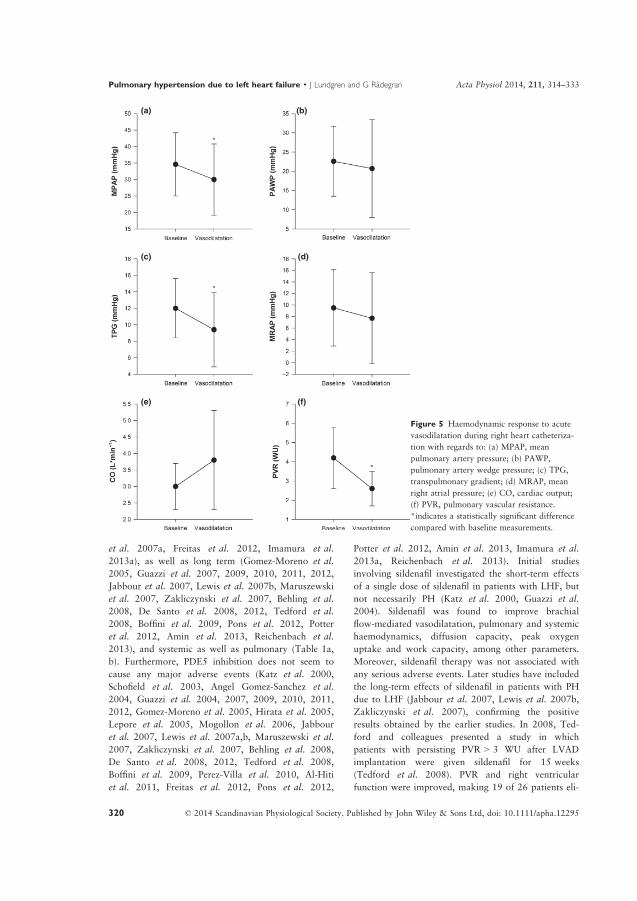
et al. 2007a, Freitas et al. 2012, Imamura et al.
2013a), as well as long term (Gomez-Moreno et al.
2005, Guazzi et al. 2007, 2009, 2010, 2011, 2012,
Jabbour et al. 2007, Lewis et al. 2007b, Maruszewski
et al. 2007, Zakliczynski et al. 2007, Behling et al.
2008, De Santo et al. 2008, 2012, Tedford et al.
2008, Boffini et al. 2009, Pons et al. 2012, Potter
et al. 2012, Amin et al. 2013, Reichenbach et al.
2013), and systemic as well as pulmonary (Table 1a,
b). Furthermore, PDE5 inhibition does not seem to
cause any major adverse events (Katz et al. 2000,
Schofield et al. 2003, Angel Gomez-Sanchez et al.
2004, Guazzi et al. 2004, 2007, 2009, 2010, 2011,
2012, Gomez-Moreno et al. 2005, Hirata et al. 2005,
Lepore et al. 2005, Mogollon et al. 2006, Jabbour
et al. 2007, Lewis et al. 2007a,b, Maruszewski et al.
2007, Zakliczynski et al. 2007, Behling et al. 2008,
De Santo et al. 2008, 2012, Tedford et al. 2008,
Boffini et al. 2009, Perez-Villa et al. 2010, Al-Hiti
et al. 2011, Freitas et al. 2012, Pons et al. 2012,
Potter et al. 2012, Amin et al. 2013, Imamura et al.
2013a, Reichenbach et al. 2013). Initial studies
involving sildenafil investigated the short-term effects
of a single dose of sildenafil in patients with LHF, but
not necessarily PH (Katz et al. 2000, Guazzi et al.
2004). Sildenafil was found to improve brachial
flow-mediated vasodilatation, pulmonary and systemic
haemodynamics, diffusion capacity, peak oxygen
uptake and work capacity, among other parameters.
Moreover, sildenafil therapy was not associated with
any serious adverse events. Later studies have included
the long-term effects of sildenafil in patients with PH
due to LHF (Jabbour et al. 2007, Lewis et al. 2007b,
Zakliczynski et al. 2007), confirming the positive
results obtained by the earlier studies. In 2008, Ted-
ford and colleagues presented a study in which
patients with persisting PVR > 3 WU after LVAD
implantation were given sildenafil for 15 weeks
(Tedford et al. 2008). PVR and right ventricular
function were improved, making 19 of 26 patients eli-
(a) (b)
(c) (d)
(e) (f)
Figure 5 Haemodynamic response to acute
vasodilatation during right heart catheteriza-
tion with regards to: (a) MPAP, mean
pulmonary artery pressure; (b) PAWP,
pulmonary artery wedge pressure; (c) TPG,
transpulmonary gradient; (d) MRAP, mean
right atrial pressure; (e) CO, cardiac output;
(f) PVR, pulmonary vascular resistance.
*indicates a statistically significant difference
compared with baseline measurements.
© 2014 Scandinavian Physiological Society. Published by John Wiley & Sons Ltd, doi: 10.1111/apha.12295320
Pulmonary hypertension due to left heart failure · J Lundgren and G R�adegran Acta Physiol 2014, 211, 314–333
Table 1 (a) Previous studies (2000–2007) with sildenafil in patients with LHF with or without PH; (b) Previous studies (2008–
2013) with sildenafil in patients with LHF with or without PH
Design, author and year Patient population
PH inclusion
criteria Outcome
(a)
Randomized 12.5–50 mg
single dose: Katz et al.
2000
Stable NYHA II-III for
3 months (n = 48)
LVEF<40%
Optimized HF therapy
for 4 weeks
No 25 and 50 mg of sildenafil increased brachial
flow-mediated vasodilatation and arterial
occlusion
No cases of hypotension.
Observational 50 mg single
dose: Schofield et al. 2003
HT (n = 15 male, mean
38 months post-HT)
Hypertension therapy
No Improved BP, aortic augmentation index
and LV stress. No case of hypotension.
Randomized 50 mg single
dose: Guazzi et al. 2004
NYHA II-III (n = 16
male)
LVEF≤40%FEV1/FVC>70%
No Improved MPAP, PVR, DLCO, DM, peak VO2,
recovery tau, brachial reactive hyperaemia,
DVO2/DWR, exercise capacity and ventilation
efficiency. The effects did not remain after 24 h.
Observational 100 mg
single sublingual dose:
G�omez-S�anchez et al.
2004
NYHA III-IV (n = 7)
Optimal HF therapy
Yes;
TPG>12 mmHg
and PVR>2.5 WU
Decreased MPAP, TPG and PVR.
Case report 25 mg t.i.d.
(4 months): Gomez-
Moreno et al. 2005
NYHA III-IV (n = 1)
LVEF<15%
Yes; MPAP
32 mmHg, PVR
5 WU
PH was partly reversed and the patient accepted
for HT.
Randomized 50 mg single
dose: Hirata et al. 2005
NYHA II-III (n = 20)
LVEF<35%
No Acutely improved cardiac performance.
Randomized 50 mg b.i.d.
(6 months): Guazzi et al.
2007
NYHA class II-III
(n = 46
male<65 years)
LVEF≤45%FEV1/FVC>70%
sAP 110-140
No Improved sPAP, ergoreflex effect on ventilation,
brachial artery flow-mediated dilatation,
exercise performance, ventilation efficiency and
QoL after 3 months. No further improvement
after 6 months.
Randomized 25–75 mg
t.i.d. (12 week): Lewis
et al. 2007
NYHA II-IV (n = 34)
LVEF<40%
Standard HF therapy.
Yes;
MPAP>25 mmHg
Improved peak VO2, 6MWD, QoL, PVR,
PVR/SVR, RVEF, CO and SV.
Fewer hospitalizations.
Observational 25–50 mg
t.i.d. (68 � 58 days):
Jabbour et al. 2007
Severe DCM (n = 6). Yes; Irreversible
TPG>15 mmHg
Improved TPG, PAWP, PVR and CO. 4 patients
listed for HT. Three patients had undergone
successful HT at the end of follow-up.
Observational 50 mg b.i.d.
(1 month): Zakliczynski
et al. 2007
NYHA III-IV (n = 6
male)
LVEF<25%
Yes; Irreversible
TPG>12 mmHg
and/or PVR>2.5
WU
Normal, or reversible, TPG and PVR in
5 patients, subsequently listed for HT.
Observational 50 mg single
dose: Lewis et al. 2007
Stable NYHA III
(n = 13;
3 months)
LVEF<35%
No Improved exercise capacity, ventilation
efficiency, MPAP, CI, PVR, RVEF and
PVR/SVR in PH patients (n = 7). No
improvement in patients without PH (n = 6).
Observational 25–50 mg
t.i.d.: Maruszewski et al.
2007
HT (n = 6) Yes; TPG>12 and/
or PVR>2.5
4 of 6 patients survived the early post-
operative period.
(b)
Observational 3 mg∙kg�1 –
250 mg∙day�1 (1 month):
De Santo et al. 2008
Acute RVD after HT
(n = 13)
No RVD resolved within 72 h in all patients.
12 patients survived the post-operative period.
Observational 25–75 mg
t.i.d. (15 weeks): Tedford
et al. 2008
Persistent PH 7–
14 days after LVAD
(n = 26)
Normalized PAWP
Yes; PVR>3 WU Improved PVR and RV function.
Nineteen patients considered eligible for HT.
Ten underwent HT at the end of follow-up.
1 developed acute RVF.
© 2014 Scandinavian Physiological Society. Published by John Wiley & Sons Ltd, doi: 10.1111/apha.12295 321
Acta Physiol 2014, 211, 314–333 J Lundgren and G R�adegran · Pulmonary hypertension due to left heart failure

Table 1 Continued
Design, author and year Patient population PH inclusion criteria Outcome
Randomized 50 mg t.i.d.
(4 weeks): Behling et al. 2008
LVEF≤40% (n = 19)
Standard HF therapy
No Improved functional capacity, ventilator
efficiency, oxygen uptake and sPAP.
Observational 25 mg t.i.d.
(6 months): Guazzi et al.
2009
HF (n = 40 male)
Optimal medical
therapy
No Improved heart rate recovery.
Observational 12.5 mg
b.i.d. – 50 mg t.i.d.
(approx. 1 months):
Boffini et al. 2009
RVD after HT (n = 10) No Improved sPAP 1 month after HT.
Other haemodynamic parameters did not
differ with sildenafil therapy.
Observational 50 mg t.i.d.
(3 months): Guazzi et al.
2010
HF (n = 40 male)
Optimal medical
therapy
No Improved sPAP, PVR, exercise performance,
ventilation efficiency and QoL.
Randomized 50 mg t.i.d.
(1 year): Guazzi et al. 2011
Stable NYHA II-III
(n = 45 male;
6 months)
LVEF<40%
FEV1/FVC>70%
LV diastolic
dysfunction
No Improved LAVI, LVEDV, LVMI, LVEF,
E’, exercise performance, ventilation
efficiency, NT-proBNP and QoL after
6 months. No further improvement after
1 year of therapy.
Randomized 50 mg t.i.d.
(1 year): Guazzi et al. 2012
NYHA III-IV (n = 32
male)
Exercise oscillatory
breathing
Yes; MPAP 25-
35 mmHg
Reversed EOB in 15 of 16 patients.
Improved functional performance,
exercise ventilation efficiency, QoL, MPAP,
PAWP and PVR.
Observational 20–60 mg
t.i.d. (prior to, and after,
HT): Pons et al. 2012
High risk prior to HT
(n = 15)
Yes; irreversible
PVR>2.5 WU and/
or TPG>12 mmHg
All patients underwent successful HT.
Survival was similar to patients without
PH (n = 104).
Observational
102.5 � 54 mg∙day�1
(mean 260 days): Potter
et al. 2012
On HT waiting list
(n = 16)
Yes; significant PH
reversibility
Improved sPAP and CI.
Eight patients underwent successful HT.
Two patients were removed from HT
waiting list due to improvement.
Observational 20–60 mg
t.i.d.: Reichenbach et al.
2012
Advanced HF (n = 47)
LVEF<40%
Yes; irreversible
TPG>15 mmHg
Improved PVR, TPG, CO and NYHA class.
Maintained bodyweight and improved
30-day survival after HT.
Observational 25–75 mg
t.i.d. (12 weeks): De Santo
et al. 2012
Stable NYHA III
(n = 32; 3 months)
LVEF<35%
Maximal medical
therapy
Yes; irreversible
PVR>6 WU
Improved MPAP, PAWP, TPG and PVR.
31 patients underwent HT (some still at
high risk according to ISHLT guidelines).
3 developed acute RHF. 1-year mortality
was 6.5%.
Randomized 25 mg b.i.d. –
50 mg t.i.d. (12 weeks):
Amin et al. 2013
Stable NYHA II- III
(n = 106; 3 months)
LVEF<35% on
optimal HF therapy
Primary endpoint:
Change in MAP.
No MAP was maintained.
No improvement in 6MWD or
hospitalization.
b.i.d., twice a day; BP, blood pressure; CI, cardiac index; CO, cardiac output; DCM, dilated cardiomyopathy; DLCO, carbon
monoxide diffusion capacity, DM, alveolar capillary membrane conductance; E’, mitral annulus early velocity; EOB, exercise
oscillatory breathing; FEV1, forced expiratory volume in 1 s; FVC, forced vital capacity; HF, heart failure; HT, heart transplan-
tation; ISHLT, International Society of Heart and Lung Transplantation; LAVI, left atrial volume index; LVAD, left ventricular
assist device; LVEDV, left ventricular end-diastolic volume; LVEF, left ventricular ejection fraction; LVMI, left ventricular mass
index; MAP, mean artery pressure; MPAP, mean pulmonary artery pressure; NYHA, New York Heart Association; PAWP, pul-
monary artery wedge pressure; PH, pulmonary hypertension; PVR, pulmonary vascular resistance; QoL, quality of life; RVD,
right ventricular dysfunction; RVF, right ventricular failure; recovery tau, recovery time constant for VO2; RVEF, right ventricu-
lar ejection fraction; sAP, systolic artery pressure; sPAP, systolic pulmonary artery pressure; SV, stroke volume; SVR, systemic
vascular resistance; t.i.d., three times a day; TPG, transpulmonary gradient; VO2, oxygen uptake; WR, work rate; 6MWD, six-
minute walking distance.
© 2014 Scandinavian Physiological Society. Published by John Wiley & Sons Ltd, doi: 10.1111/apha.12295322
Pulmonary hypertension due to left heart failure · J Lundgren and G R�adegran Acta Physiol 2014, 211, 314–333

Table 2 (a) Previous studies on patients with LHF with or without PH, using bosentan; (b) Previous studies on patients with
LHF with or without PH, using tezosentan or darusentan
Design, author and year Patient population PH inclusion criteria Outcome
(a)
Randomized 1 g b.i.d.
(2 weeks): Sutsch et al.
1998
Stable NYHA III (n = 36 male)
LVEF<30% Optimal
conventional therapy
PAWP≥15 mmHg and/or
CI≤2.5 L∙min�1∙m�2
No Improved CO, MPAP, PAWP, MRAP,
PVR. Reduced SVR.
One case of symptomatic hypotension
resulting in discontinuation of therapy.
REACH-1 500 mg b.i.d.
target (6 months): 1998
NYHA IIIb-IV (n = 370)
LVEF<35%Diuretic/ACEi therapy and
Hospitalization for HF within
the previous 12 months/
6MWD<375 m.
Primary endpoint: clinical
status of the patients
after 6 months.
No Stopped prematurely due to increased
hepatic transaminases.
Less adverse events after 3 months, in
patients who completed the study.
ENABLE 125 mg b.i.d.
(18 months): 2002
NYHA IIIb-IV (n = 1613)
LVEF<35%Optimal conventional therapy
Primary endpoint: All-cause
mortality or hospitalization.
No No improvement in primary endpoint.
Increased risk of worsening HF, likely
due to fluid retention.
Randomized 8–125 mg
b.i.d. (20 weeks): Kaluski
et al. 2008
NYHA IIIB-IV (n = 90)
LVEF<35%Hospitalized for worsening HF
within 6 months prior to study.
Primary endpoint: change in
ECHO-measured sPAP.
Yes;
sPAP≥40 mmHg
No difference in primary
endpoint.
Increased risk of adverse
events.
Case report 125 once daily
(3 months): Imamura
et al. 2012
Progressing HF (n = 1 male)
LVAD
Yes; persistent
PH 1 month
after LVAD
implantation
PH was reversed.
Observational 125 mg
b.i.d. target: Hefke
et al. 2012
On HT waiting list (n = 84) Yes; MPAP>35,TPG>15 and/or
PVR>240dyn � s � cm�5
Improved HD and 1-year survival on
HT waiting list.
Independent predictor of reduced
mortality on HT waiting list.
Decreased haematocrit and in 3
patients ASAT and/or ALAT increased.
Observational 125 mg
b.i.d. (4 months–1 year):
Perez-Villa et al. 2013
HT candidate (n = 24) Yes; irreversible
PVR>2.5 WU
PH reversed in 13 patients after
4 months and in 19 patients after
12 months.
14 patients underwent HT. 1-year
survival was 93% compared with 83%
in 56 patients with PVR≤2.5 WU
during the same period.
(b)
Randomized tezosentan
5–100 mg h�1: Cotter
et al. 2001
NYHA III (n = 38)
LVEF≤35%PAWP≥15 mmHg
CI≤2.7 L�min�1�m�2
No Dose-dependent improvement in PAWP,
PVR, cardiac power index and SVR
index. No decrease in MAP.
Randomized 5, 20, 50 or
100 mg h�1 (6 h):
Torre-Amione et al. 2001
NYHA III-IV (n = 61, 19–
70 years)
LVEF<35%CI<2.5 L�min�1�m�2
PAWP>18 mmHg
No Dose-dependent improvement in MPAP,
PAWP, CI, PVR and SVR. No case of
symptomatic hypotension.
Randomized tezosentan
20 mg h�1 or 50 mg h�1
(48 h): Torre-Amione
et al. 2001
NYHA III-IV (n = 14)
LVEF<35%.
No Improved ECHO sPAP, RAP, PAWP, CI
and SV. No serious adverse events.
© 2014 Scandinavian Physiological Society. Published by John Wiley & Sons Ltd, doi: 10.1111/apha.12295 323
Acta Physiol 2014, 211, 314–333 J Lundgren and G R�adegran · Pulmonary hypertension due to left heart failure

gible for heart transplantation (Tedford et al. 2008).
However, these results must be interpreted with cau-
tion, as sildenafil therapy was initiated only 7–14 days
after LVAD implantation. In the light of the current
knowledge on LVAD therapy, it is likely that a sub-
stantial part of the PH was reversed by left ventricle
unloading by the LVAD (Gallagher et al. 1991, Smed-
ira et al. 1996). Finally, in 2012, three studies on
patients with irreversible PH were published (De
Santo et al. 2012, Pons et al. 2012, Reichenbach et al.
2013). Two of these showed that sildenafil was effec-
tive in reversing PH previously defined as irreversible
(De Santo et al. 2012, Reichenbach et al. 2013), and
all three showed that heart transplantation could be
performed without an increased risk of acute right
heart failure or early mortality.
It could be hypothesized that the positive results
observed with sildenafil in patients with LHF, without
and with PH, are due to the dilatation of pulmonary
as well as systemic vessels. Such vasodilation would
result in unloading of both the left and right ventri-
cle, decreasing the risk of further pulmonary conges-
tion. PDE5is could thus provide a new, promising
therapy in patients with LHF, without or with, PH. It
is notable that no further haemodynamic improvement
was observed in any of the published studies beyond
3 months of therapy, resulting in a plateau phase
thereafter. Finally, as no large, multicentre, placebo-
controlled trials involving the treatment of LHF using
sildenafil have been published, further research is
necessary.
Dual endothelin receptor blockade
The dual ERA bosentan has been found to improve
pulmonary as well as systemic haemodynamics in
patients with LHF (Sutsch et al. 1998). However, the
two randomized, multicentre, placebo-controlled trials
Table 2 Continued
Design, author and year Patient population
PH inclusion
criteria Outcome
RITZ-2 tezosentan 50 or
100 mg�h�1: 2001
NYHA class III-IV (n = 184)
CI<2.5 L�min�1�m�2
PAWP>15 mmHg
No Improved CI, PAWP and dysponea
score. Less side effects with 50 mg�h�1
than 100 mg�h�1.
Randomized tezosentan
5–100 mg�h�1: Schalcher
et al. 2001
NYHA III (n = 38)
LVEF≤35%PAWP≥15 mmHg
CI≤2.7 L�min�1�m�2
Dose-dependent increase in CI and
stroke index.
Decrease in PVR and SVR. No decrease
in MAP. No serious adverse events.
VERITAS tezosentan
5 mg�h�1 for 30 min,
followed by 1 mg�h�1 for
24–72 h: 2007
Hospitalized due to acute HF within
the previous 24 h (n = 1448)
Persistent dysponea (RR>24/min)
Primary endpoints: change in
dysponea over 24 h and incidence
of death or worsening of HF at
7 days.
No Improved MPAP, MRAP, PAWP, PVR
and SVR.
No difference in primary endpoints.
More adverse events, mainly
hypotension.
HEAT darusentan 30,
100 or 300 mg�day�1
(3 weeks): 2002
NYHA class III (n = 157)
for at least 3 months
LVEF<35%
PAWP≥12 mmHg
CI≤2.6 L�min�1�m�2
Primary endpoint: 3-week change in
CI and PAWP.
No Increased CI, most pronounced
in the 100 mg�day�1 group, and
decreased SVR. More adverse events.
EARTH darusentan 10,
25, 50, 100 or
300 mg�day�1
(24 weeks): 2004
NYHA II-IV (n = 642)
LVEF<35%.
No No improvement in primary endpoint.
No beneficial effects on clinical
outcomes.
b.i.d., twice a day; CI, cardiac index; CO, cardiac output; HD, haemodynamic; HF, heart failure; HT, heart transplantation;
LVAD, left ventricular assist device; LVEF, left ventricular ejection fraction; MAP, mean arterial pressure; MPAP, mean pulmo-
nary artery pressure; MRAP, mean right atrial pressure; NYHA, New York Heart Association; PAWP, pulmonary artery wedge
pressure; PH, pulmonary hypertension; PVR, pulmonary vascular resistance; RAP, right atrial pressure; RR, respiratory rate;
sPAP, systolic pulmonary artery pressure; SV, stroke volume; SVR, systemic vascular resistance; TPG, transpulmonary gradient;
6MWD, six-minute walking distance.
© 2014 Scandinavian Physiological Society. Published by John Wiley & Sons Ltd, doi: 10.1111/apha.12295324
Pulmonary hypertension due to left heart failure · J Lundgren and G R�adegran Acta Physiol 2014, 211, 314–333
REACH-1 (Packer et al. 2005) and ENABLE (Coletta
et al. 2002) failed to meet their primary endpoints:
clinical status after 6 months and all-cause mortality
or hospitalization respectively. Bosentan was instead
found to increase the risk of adverse events such as
hepatic transaminase elevation and fluid retention
(Coletta et al. 2002, Packer et al. 2005). In fact, the
REACH-1 trial was terminated early due to increased
hepatic transaminases in the patients given bosentan
(Packer et al. 2005). However, both these trials were
performed in patients with LHF, but not necessarily
with secondary PH (Table 2a). Similar findings have
Table 3 Previous studies on patients with LHF with or without PH, using multiple drugs
Design, author and year Patient population PH inclusion criteria Outcome
Case report sildenafil
75 mg∙day�1 (12 months)
and bosentan 62.5 mg∙day�1
(6 months): Mogoll�on et al.
2006
Dilated, alcohol-
induced
cardiomyopathy
(n = 1 male)
LVEF<20%
Yes; irreversible
MPAP 59 mmHg and
PVR 6.4 WU
No HD improvement after 6 months of
sildenafil therapy. Normalized MPAP,
PAWP, TPG, CO and PVR after 6 months
of combination therapy. Underwent HT
without RHF.
Observational sildenafil
20–80 mg t.i.d. or bosentan
62.5–125 mg b.i.d. (16 weeks):
Perez-Villa et al. 2010
Ineligible for HT
(n = 18)
Yes; irreversible
PVR>2.5 WU
Greater reduction in PVR with bosentan.
Improved sPAP, dPAP and TPG with
bosentan.
12 patients (4 sildenafil, 8 bosentan)
underwent successful HT.
Observational sildenafil 100 mg
single dose or nitroprusside 1–
2 lg∙kg.1∙min�1: Freitas et al.
2012
NYHA III-IV
(n = 29)
LVEF<45%
Formal indication
of HT
No Reduced PH, maintained MAP and
improved left and right cardiac functions,
pO2 and SvO2 with sildenafil.
Improved left and right cardiac functions
and decreased MAP, pO2 and SvO2 with
nitroprusside. No improvement in PH.
Observational NO 5–30 ppm or
iloprost 50 lg: Sablotzki et al.2002
Dilated or
ischaemic DCM
(n = 14 male)
Yes; PVR≥180dyn�s�1�cm�5
Decreased MPAP and PVR with both
drugs. Increased CI and SVI with iloprost.
Greater decrease in MPAP with iloprost.
Observational NO 5–40 ppm or
epoprostenol 1-
12 ng∙kg.1∙min�1: Mogollon
Jimenez et al. 2008,
Pre-HT evaluation
(n = 54)
Yes; MPAP>35 mmHg
or PVR>4 WU
Improved MPAP, PVR and CO with
epoprostenol. NO did not improve HD.
Randomized NO 40-80 ppm and
PGE1 0.05–0.5 lg∙kg.1∙min�1
(crossover): Radovancevic
et al. 2005
Potential HT
candidates
(n = 19)
Optimal medical
therapy
Yes; sPAP>60 mmHg,
TPG>12 mmHg or
PVR>4 WU
TPG and PVR decreased with both drugs.
MAP decreased with PGE1. PH was
reversed in 6 patients with both drugs, in
4 with PGE1 only and in 4 with NO only.
Observational sildenafil 40 mg
or PGE1 200 ng�kg�1�min�1:
Al-Hiti
et al. 2011
Potential HT
candidates
(n = 13)
Yes; TPG>15 mmHg
or PVR>3 WU
Greater decrease in TPG and PVR with
sildenafil. Similar decrease in MAP, MPAP
and PAWP and similar increase in CI with
both drugs. Decreased PVR/SVR ratio
with sildenafil. More patients with PVR
acceptable for HT with sildenafil.
Observational sildenafil 50 mg
single dose and NO 80 ppm:
Lepore et al. 2005
NYHA III-IV
(n = 11)
LVEF<40%
Yes; MPAP>25 mmHg Improved CI, PVR, SVR and PVR/SVR
ratio with combination therapy
compared with single therapy with
either agent. No case of hypotension.
Case report sildenafil 20 mg single
dose or NO 20 ppm for 10 min:
Imamura et al. 2013
DCM (n = 1)
LVEF<28%
Yes; MPAP 44 mmHg
and PVR 5.96 WU
Decreased PVR with both drugs. Decreased
PAWP with sildenafil, increased PAWP
with NO.
b.i.d., twice a day; CI, cardiac index; CO, cardiac output; DCM, dilated cardiomyopathy; dPAP, diastolic pulmonary artery
pressure; HD, haemodynamic; HT, heart transplantation; LVEF, left ventricular ejection fraction; MAP, mean arterial pressure;
MPAP, mean pulmonary artery pressure; NO, nitric oxide; NYHA, New York Heart Association; PAWP, pulmonary artery
wedge pressure; PH, pulmonary hypertension; pO2, oxygen partial pressure; PVR, pulmonary vascular resistance; RHF, right
heart failure; sPAP, systolic pulmonary arterial pressure, SVI, stroke volume index; SvO2, venous oxygen saturation; SVR, sys-
temic vascular resistance; t.i.d., three times a day; TPG, transpulmonary gradient.
© 2014 Scandinavian Physiological Society. Published by John Wiley & Sons Ltd, doi: 10.1111/apha.12295 325
Acta Physiol 2014, 211, 314–333 J Lundgren and G R�adegran · Pulmonary hypertension due to left heart failure

been reported on the effects of tezosentan and darus-
entan in patients with LHF (Cotter et al. 2001, Louis
et al. 2001, Schalcher et al. 2001, Torre-Amione et al.
2001a,b, Luscher et al. 2002, Anand et al. 2004,
McMurray et al. 2007) (Table 2b).
A few studies on the use of bosentan in patients
with PH due to LHF have on the other hand shown
promising long-term effects (Hefke et al. 2012, Imam-
ura et al. 2013b, Perez-Villa et al. 2013). Hefke and
colleagues reported that bosentan was an independent
predictor of reduced mortality in a group of 84
patients on the waiting list for heart transplantation
(Hefke et al. 2012). Perez-Villa et al. reported that
12 months of bosentan therapy reversed previously
defined irreversible PVR in 19 of 26 patients, making
them eligible for heart transplantation (Perez-Villa
et al. 2013). They also observed that one-year survival
among the 14 patients who underwent heart trans-
plantation was similar to that in the 56 patients who
did not have increased PVR prior to heart transplanta-
tion (Perez-Villa et al. 2013) (Table 2a). One study in
which PH was based on echocardiographic measure-
ments and defined as sPAP≥40 mmHg is the only
study in PH due to left heart disease not reporting
positive results following bosentan therapy (Kaluski
et al. 2008) (Table 2a). However, echocardiographic
measurements of the tricuspid regurgitation and the
gradient between the right ventricle and right atrium
may be influenced by the echo-signal detected as well
as changes in cardiac output. Moreover, the positive
effects seen with bosentan in both small and large
studies have been shown to increase with the length of
therapy (Packer et al. 2005, Perez-Villa et al. 2013).
Consequently, a ‘plateau’ similar to the one seen with
sildenafil does not seem to occur with bosentan. Inter-
estingly, sildenafil and bosentan have been compared
in one case report (Mogollon et al. 2006) and a small
study with 18 subjects (Perez-Villa et al. 2010). The
results of both studies suggested that bosentan was
more effective than sildenafil in reversing PH and in
making patients eligible for heart transplantation
(Mogollon et al. 2006, Perez-Villa et al. 2010)
(Table 3).
The reason for the diverging results in the studies
on bosentan therapy could be the pathophysiology of
the patients’ disease in combination with the pulmo-
nary selectivity of bosentan. In the studies showing
negative results, either PH was not used as an inclu-
sion criterion, or the patients were included based on
their pulmonary artery pressures, that is, passive PH.
In contrast, the studies in which positive results were
found all included patients with reactive PH, with
increased PVR and/or TPG (Mogollon et al. 2006,
Perez-Villa et al. 2010, 2013, Hefke et al. 2012,
Imamura et al. 2013b). Based on these findings, it can
be hypothesized that if the PH is worsened by an
active component, it may be partly reversed by selec-
tive pulmonary vasodilation, leading to unloading of
the right ventricle. There is, however, also a risk of
congestion in these patients, and they should thus be
closely monitored.
Prostaglandins
Early studies were performed in the 1980s and 1990s
on patients with LHF using prostacyclin and the pros-
tacyclin analogue epoprostenol in patients with LHF.
Yui et al. found improved short-term haemodynamics
with prostacyclin (Yui et al. 1982), while Sueta et al.
reported improved 6-min walking distance and left
ventricular ejection fraction (LVEF) in a 12-week trial
using epoprostenol (Sueta et al. 1995). The FIRST
study using epoprostenol, in patients with LHF but
not necessarily with PH, was, however, terminated
early due to a strong tendency towards increased mor-
tality in the group given the drug (Califf et al. 1997).
Since then, only a few small studies have been per-
formed on the effects of prostacyclin analogues during
right heart catheterization (Weston et al. 2001, Sab-
lotzki et al. 2002a,b, Mogollon Jimenez et al. 2008)
(Tables 3 and 4). All of these studies demonstrated
improvement in pulmonary haemodynamics together
with an increase in cardiac output or cardiac index,
depending on the parameter observed.
A few studies have also been carried out on the
effects of the prostaglandin PGE1 in PH due to LHF.
In three of these studies, the short-term effects of
PGE1 infusion were investigated and found to improve
the haemodynamics (Radovancevic et al. 2005, von
Scheidt et al. 2006, Al-Hiti et al. 2011). However, in
one study in which PGE1 was compared with a single
dose of sildenafil, it was found that sildenafil was
more potent in reversing the PH, as well as being
more pulmonary specific than PGE1 (Al-Hiti et al.
2011). In 2011, Serra and colleagues presented a study
on PGE1 therapy in patients with severe LHF and
increased sPAP based on echocardiographic measure-
ments (Serra et al. 2011). PGE1 was administered for
three consecutive days each month for 36 months and
was found to improve the New York Heart Associa-
tion (NYHA) classification of heart failure and LVEF.
Furthermore, the 36-month mortality was 27% in the
PGE1 group, compared with 44% in the controls
(Serra et al. 2011). However, in this study, the PH
was diagnosed by echocardiography and 25% of the
patients were reported to have pre-capillary PH, and
these results must thus be interpreted with caution.
Considering the positive findings from these early
studies, it would be interesting to perform similar
© 2014 Scandinavian Physiological Society. Published by John Wiley & Sons Ltd, doi: 10.1111/apha.12295326
Pulmonary hypertension due to left heart failure · J Lundgren and G R�adegran Acta Physiol 2014, 211, 314–333

investigations in patients with post-capillary PH diag-
nosed by right heart catheterization.
Soluble guanylate cyclase stimulators
The novel sGC stimulator riociguat was recently eval-
uated in a phase IIb study in patients with PH and
LHF, but not necessarily with increased filling pres-
sures (Bonderman et al. 2013) (Table 5). The primary
endpoint, that is, change in MPAP, was not met.
However, several other parameters such as cardiac
index, stroke volume index, PVR and the Minnesota
Living with Heart Failure score improved as a result
of sGC stimulation. Further studies on the effects of
sGC stimulators in patients with PH due to LHF and
increased left ventricular filling pressures, with more
precise endpoints than MPAP, should be performed to
further investigate the clinical implications of these
findings. This is of interest as sGC stimulators, in con-
trast to PDE5 inhibitors, work in both the presence
and absence of NO. Therefore, therapy with sGC
stimulators may prove more valuable in patients with
impaired NO synthesis and signalling.
Future therapies
Recently, two randomized, placebo-controlled, multi-
centre, trials were initiated in patients with PH due to
LHF. The drugs planned for investigation were the
PDE5is sildenafil (Cooper et al. 2013) and tadalafil
(Clinicaltrials.gov NCT01910389), both acting on the
NO pathway. The tadalafil-trial was recently termi-
nated by the funding agency. However, the sildenafil-
trial is still ongoing and if it is successful it could
herald a new era in treating PH due to LHF (Table 6).
In addition, a trial with a novel sGC stimulator
Table 4 Previous studies on patients with LHF with or without PH using prostaglandins
Design, Author and Year
Patient
population PH inclusion criteria Outcome
Observational prostacyclin
22 � 11 ng∙kg�1∙min�1:
Yui et al. 1982
NYHA IV due to
CAD (n = 9)
Conventional
medical therapy
No Decreased MAP, PAWP, SVR and PVR.
Increased HR, CI, SVI and plasma
epinephrine.
No serious adverse events.
Randomized epoprostenol
8.8 � 3.5 ng∙kg�1∙min�1
(12 weeks): Sueta et al. 1995
NYHA III-IV
(n = 33)
LVEF≤30%Conventional
medical therapy
No Improved 6MWD with epoprostenol
compared with control. Improved LVEF.
3 patients withdrawn due to serious
adverse events. 2 deaths in epoprostenol
group and 8 in controls.
FIRST Epoprostenol
4 ng∙kg�1∙min�1: 1997
NYHA class IIIb-IV
(n = 471) Low
LVEF
No Terminated early due to a strong trend
towards increased mortality.
Observational iloprost
580 lg (mean) single dose:
Weston et al. 2001
Pre-HT evaluation
(n = 6)
Yes; irreversible
sPAP>50 mmHg,
TPG>15 mmHg or
PVR>3 WU
Improved sPAP, MPAP, PAWP, TPG, CO
and PVR/SVR ratio. 4 patients underwent
successful HT.
Observational iloprost inhal
50 lg: Sablotzki et al. 2002HT candidates
(n = 30)
Yes; increased PVR
(206 � 94 dyn�s�1�cm�5)
Improved MPAP, PAWP, CI and PVRI.
PROPHET PGE1
173 � 115 ng∙kg�1∙min�1:
2006
Potential HT
candidates
(n = 92)
Yes; PVR>2.5 WU or
TPG > 12 mmHg
Decreased MAP, MPAP, PAWP, SVR and
PVR. Increased CO.
No case of symptomatic hypotension.
90 patients were accepted for HT
(PVR<4 WU).
Observational PGE1
10 ng∙kg�1∙min�1 (mean)
(3 consecutive days per
month for 36 months):
Serra et al. 2011
NYHA III-IV
(n = 22)
LVEF<35%
Yes; ECHO-
sPAP>3 m∙s�1
Improved sPAP, NYHA class and LVEF.
One case of severe hypotension.
36-month mortality was 27% in PGE1
group vs. 44% in controls.
CAD, coronary artery disease; CI, cardiac index; CO, cardiac output; HR, heart rate; HT, heart transplantation; LVEF, left ven-
tricular ejection fraction; MAP, mean arterial pressure; MPAP, mean pulmonary artery pressure; NYHA, New York Heart Asso-
ciation; PAWP, pulmonary artery wedge pressure; PH, pulmonary hypertension; PVR, pulmonary vascular resistance; sPAP,
systolic pulmonary arterial pressure; SVI, stroke volume index; SVR, systemic vascular resistance; TPG, transpulmonary gradi-
ent; 6MWD, six-minute walking distance.
© 2014 Scandinavian Physiological Society. Published by John Wiley & Sons Ltd, doi: 10.1111/apha.12295 327
Acta Physiol 2014, 211, 314–333 J Lundgren and G R�adegran · Pulmonary hypertension due to left heart failure

(BAY1021189) is planned in patients with systolic
LHF (Clinicaltrials.gov NCT01951625) (Table 6). It
is also of interest to perform a similar study with the
new, more potent ERA, macitentan, as smaller studies
with bosentan have shown promising results in
patients with reactive PH. In fact, a phase II trial with
Macitentan, aiming to evaluate the safety and tolera-
bility of the drug in patients with combined pre- and
post-capillary PH (Vachiery et al. 2013), is currently
being initiated (Clinicaltrials.gov NCT02070991)
(Table 6).
Conclusions
Pulmonary hypertension is common in LHF, and is a
marker of disease severity. Early in its course, PH is
caused by passive congestion in the pulmonary vessels
due to elevated left ventricular filling pressures. If
these elevated pulmonary pressures persist, endothelial
damage may occur, leading to an active PH compo-
nent. In its early stages, active PH is characterized by
excessive vasoconstriction and may be reversible.
However, long-standing PH, may not be acutely
reversible due to remodelling of the pulmonary vascu-
lature. To date, there is no specific recommended ther-
apy for the pulmonary component of PH due to LHF.
However, studies suggest that some PAH-specific
drugs could be beneficial. PDE5is in particular have
shown encouraging results on studies in LHF patients
with and without PH. Ongoing trials with PDE5i in
PH due to LHF may clarify the potential of drugs tar-
geting the NO pathway, and could pave the way for
novel forms of treating PH due to LHF. In this light,
it would also be of interest to evaluate the impli-
cations of findings from previous studies on sGC
stimulation in a PH population with excessive vaso-
constriction with or without vascular remodelling.
Recent studies also suggest that ERAs may be useful
in patients with reactive PH, and complimentary clini-
cal trials with ERAs would, therefore, be of interest in
this specific patient population. Finally, although some
PAH specific drugs may prove valuable in selected
cases of PH due to LHF, it should be borne in mind
that selective pulmonary vasodilation without simulta-
neous unloading of the left ventricle can lead to
pulmonary oedema and worsening of heart failure.
Table 6 Ongoing, or planned, trials with PAH targeted therapies in patients with LHF without or with PH
Drug Study acronym Patient population
PH as inclusion
criteria
Sildenafil 40 mg t.i.d
(6 mo)
SilHF NYHA II-III (n = 210)
LVEF ≤ 40%
6MWD<400 m
Optimal medical therapy
Primary endpoint: Patient global
assessment and 6MWD
Yes; sPAP ≥ 40 mmHg
BAY1021189 (12 wk) SOCRATES-
REDUCED
LVEF ≤ 45% (n = 410)
Worsening chronic HF requiring
hospitalization (or iv. diuretics)
Primary endpoint: NT-proBNP
No
Macitentan 10 mg once daily
(12 wk)
MELODY-1 Left ventricular dysfunction (n�60)
Optimized diuretic therapy
Primary endpoint: Safety and tolerability
Yes; MPAP ≥ 25 mmHg,
PAWP > 15 mmHg and
DPD > 7 mmHg
HF, heart failure; LVEF, left ventricular ejection fraction; mo, month; NYHA, New York heart association; PH, pulmonary
hypertension; sPAP, systolic pulmonary artery pressure; wk, week; 6MWD, six minute walking distance.
Table 5 Previous study on patients with PH due to LHF using sGC stimulation
Design, author
and year Patient population PH inclusion criteria Outcome
LEPHT riociguat
0.5–2 mg t.i.d.
(16 weeks): 2013
LVEF≤40% (n = 201) Optimal
medical therapy
Primary endpoint: change in MPAP.
Yes; MPAP≥25mmHg
No change in primary endpoint. Improved CI,
SVI, PVR and Minnesota Living With Heart
Failure score. No serious adverse events.
CI, cardiac index; LVEF, left ventricular ejection fraction; MPAP, mean pulmonary artery pressure; PH, pulmonary hyperten-
sion; PVR, pulmonary vascular resistance; SVI, stroke volume index; t.i.d., three times daily.
Pulmonary hypertension due to left heart failure · J Lundgren and G R�adegran Acta Physiol 2014, 211, 314–333
© 2014 Scandinavian Physiological Society. Published by John Wiley & Sons Ltd, doi: 10.1111/apha.12295328

Due to these potentially detrimental effects, careful
patient selection and monitoring are required if
selected LHF patients are to be treated with PAH-tar-
geted therapies. At present, PAH-targeted therapies
are therefore generally not recommended in PH due to
LHF. However, further investigations are warranted
in well controlled clinical settings and trials.
Conflict of interest
Mr. Lundgren reports an unrestricted research grant from
The Swedish Society of Pulmonary Hypertension, and Dr.
R�adegran reports unrestricted research grants from Anna-Lisa
and Sven-Erik Lundgren’s, Maggie Stephen’s, ALF’s, Sk�ane
University Hospital’s Foundations and Actelion Pharmaceuti-
cals Sweden AB, during the conduct of the study.
Mr. Lundgren reports personal lecture fees from Actelion
Pharmaceuticals Sweden AB and GlaxoSmithKline outside
the submitted work. Dr. R�adegran reports personal lecture
fees from Actelion Pharmaceuticals Sweden AB, Glaxo-
SmithKline and Sandoz/Novartis outside the submitted work.
Dr R�adegran is and has been primary-, or co-, investigator
in clinical PAH trials for GlaxoSmithKline, Actelion Pharma-
ceuticals Sweden AB, Pfizer, Bayer and United Therapeutics,
and in clinical heart transplantation imunosuppression trials
for Novartis.
The companies have no role in the data collection, analysis
and interpretation and have no right in disapproving of the
manuscript.
We acknowledge the support of the staff at the Haemody-
namics Lab, Clinic for Heart Failure and Valvular Disease,
Sk�ane University Hospital, and the Department of Cardiol-
ogy, Clinical Sciences, Lund University, Lund, Sweden.
This work was supported by unrestricted research grants
from ALF’s, the Sk�ane University Hospital Foundations, the
Swedish Society of Pulmonary Hypertension and Actelion
Pharmaceuticals Sweden AB. The funding organizations
played no role in the collection, analysis or interpretation of
the data and have no right to restrict the publishing of the
manuscript.
References
Al-Hiti, H., Melenovsky, V., Syrovatka, P., Kettner, J.,
Malek, I. & Kautzner, J. 2011. Sildenafil is more selective
pulmonary vasodilator than prostaglandin E1 in patients
with pulmonary hypertension due to heart failure. Physiol
Res 60, 303–308.
Amin, A., Mahmoudi, E., Navid, H. & Chitsazan, M. 2013.
Is chronic sildenafil therapy safe and clinically beneficial in
patients with systolic heart failure? Congest Heart Fail 19,
99–103.
Anand, I., McMurray, J., Cohn, J.N., Konstam, M.A., Not-
ter, T., Quitzau, K., Ruschitzka, F. & Luscher, T.F. 2004.
Long-term effects of darusentan on left-ventricular remod-
elling and clinical outcomes in the EndothelinA Receptor
Antagonist Trial in Heart Failure (EARTH): randomised,
double-blind, placebo-controlled trial. Lancet 364, 347–
354.
Angel Gomez-Sanchez, M., Saenz, D.L.C., Escribano, S.P.,
Francisco Delgado, J.J., Lazaro, S.M., Albarran, G.A. &
Cea, C.L. 2004. Pilot assessment of the response of several
pulmonary hemodynamic variables to sublingual sildenafil
in candidates for heart transplantation. Eur J Heart Fail 6,
615–617.
Behling, A., Rohde, L.E., Colombo, F.C., Goldraich, L.A.,
Stein, R. & Clausell, N. 2008. Effects of 50-phosphodies-terase four-week long inhibition with sildenafil in patients
with chronic heart failure: a double-blind, placebo-con-
trolled clinical trial. J Card Fail 14, 189–197.
Boffini, M., Sansone, F., Ceresa, F., Ribezzo, M., Patane, F.,
Comoglio, C. & Rinaldi, M. 2009. Role of oral sildenafil
in the treatment of right ventricular dysfunction after heart
transplantation. Transplant Proc 41, 1353–1356.
Bonderman, D., Ghio, S., Felix, S.B., Ghofrani, H.A.,
Michelakis, E., Mitrovic, V., Oudiz, R.J., Boateng, F., Sca-
lise, A.V., Roessig, L. & Semigran, M.J. 2013. Riociguat
for patients with pulmonary hypertension caused by sys-
tolic left ventricular dysfunction: a phase IIb double-blind,
randomized, placebo-controlled, dose-ranging hemodynam-
ic study. Circulation 128, 502–511.
Califf, R.M., Adams, K.F., McKenna, W.J., Gheorghiade,
M., Uretsky, B.F., McNulty, S.E., Darius, H., Schulman,
K., Zannad, F., Handberg-Thurmond, E., Harrell, F.E. Jr,
Wheeler, W., Soler-Soler, J. & Swedberg, K. 1997. A ran-
domized controlled trial of epoprostenol therapy for severe
congestive heart failure: The Flolan International Random-
ized Survival Trial (FIRST). Am Heart J 134, 44–54.
Cody, R.J., Haas, G.J., Binkley, P.F., Capers, Q. & Kelley,
R. 1992. Plasma endothelin correlates with the extent of
pulmonary hypertension in patients with chronic conges-
tive heart failure. Circulation 85, 504–509.
Coletta, A., Thackray, S., Nikitin, N. & Cleland, J.G. 2002.
Clinical trials update: highlights of the scientific sessions of
The American College of Cardiology 2002: LIFE, DA-
NAMI 2, MADIT-2, MIRACLE-ICD, OVERTURE,
OCTAVE, ENABLE 1 & 2, CHRISTMAS, AFFIRM,
RACE, WIZARD, AZACS, REMATCH, BNP trial and
HARDBALL. Eur J Heart Fail 4, 381–388.
Cooper, C.J., Jevnikar, F.W., Walsh, T., Dickinson, J., Mou-
haffel, A. & Selwyn, A.P. 1998. The influence of basal
nitric oxide activity on pulmonary vascular resistance in
patients with congestive heart failure. Am J Cardiol 82,
609–614.
Cooper, T.J., Guazzi, M., Al-Mohammad, A., Amir, O., Ben-
gal, T., Cleland, J.G. & Dickstein, K. 2013. Sildenafil in
Heart failure (SilHF). An investigator-initiated multina-
tional randomized controlled clinical trial: rationale and
design. Eur J Heart Fail 15, 119–122.
Cotter, G., Kiowski, W., Kaluski, E., Kobrin, I., Milova-
nov, O., Marmor, A., Jafari, J., Reisin, L., Krakover, R.,
Vered, Z. & Caspi, A. 2001. Tezosentan (an intravenous
endothelin receptor A/B antagonist) reduces peripheral
resistance and increases cardiac power therefore prevent-
© 2014 Scandinavian Physiological Society. Published by John Wiley & Sons Ltd, doi: 10.1111/apha.12295 329
Acta Physiol 2014, 211, 314–333 J Lundgren and G R�adegran · Pulmonary hypertension due to left heart failure

ing a steep decrease in blood pressure in patients
with congestive heart failure. Eur J Heart Fail 3, 457–
461.
D’Alonzo, G.E., Barst, R.J., Ayres, S.M., Bergofsky, E.H.,
Brundage, B.H., Detre, K.M., Fishman, A.P., Goldring,
R.M., Groves, B.M. & Kernis, J.T. 1991. Survival in
patients with primary pulmonary hypertension. Results
from a national prospective registry. Ann Intern Med 115,
343–349.
De Santo, L.S., Mastroianni, C., Romano, G., Amarelli, C.,
Marra, C., Maiello, C., Galdieri, N., Della, C.A., Cotrufo, M.
& Caianiello, G. 2008. Role of sildenafil in acute posttrans-
plant right ventricular dysfunction: successful experience in
13 consecutive patients. Transplant Proc 40, 2015–2018.
De Santo, L.S., Romano, G., Maiello, C., Buonocore, M.,
Cefarelli, M., Galdieri, N., Nappi, G. & Amarelli, C.
2012. Pulmonary artery hypertension in heart transplant
recipients: how much is too much? Eur J Cardiothorac
Surg 42, 864–869.
Drazner, M.H., Hamilton, M.A., Fonarow, G., Creaser, J.,
Flavell, C. & Stevenson, L.W. 1999. Relationship between
right and left-sided filling pressures in 1000 patients with
advanced heart failure. J Heart Lung Transplant 18, 1126–
1132.
Fang, J.C., DeMarco, T., Givertz, M.M., Borlaug, B.A.,
Lewis, G.D., Rame, J.E., Gomberg-Maitland, M., Murali,
S., Frantz, R.P., McGlothlin, D., Horn, E.M. & Benza,
R.L. 2012. World Health Organization Pulmonary Hyper-
tension group 2: pulmonary hypertension due to left heart
disease in the adult–a summary statement from the Pulmo-
nary Hypertension Council of the International Society for
Heart and Lung Transplantation. J Heart Lung Transplant
31, 913–933.
Felix, S.E., Martina, J.R., Kirkels, J.H., Klopping, C., Nat-
hoe, H., Sukkel, E., Hulstein, N., Ramjankhan, F.Z., Do-
evendans, P.A., Lahpor, J.R. & de Jonge, N. 2012.
Continuous-flow left ventricular assist device support in
patients with advanced heart failure: points of interest for
the daily management. Eur J Heart Fail 14, 351–356.
Freitas, A.F. Jr, Bacal, F., Oliveira Junior, J.L., Fiorelli, A.I.,
Santos, R.H., Moreira, L.F., Silva, C.P., Mangini, S.,
Tsutsui, J.M. & Bocchi, E.A. 2012. Sildenafil vs. sodium
before nitroprusside for the pulmonary hypertension
reversibility test before cardiac transplantation. Arq Bras
Cardiol 99, 848–856.
Galie, N., Hoeper, M.M., Humbert, M., Torbicki, A., Vachi-
ery, J.L., Barbera, J.A., Beghetti, M., Corris, P., Gaine, S.,
Gibbs, J.S. et al. 2009. Guidelines for the diagnosis and
treatment of pulmonary hypertension: the Task Force for
the Diagnosis and Treatment of Pulmonary Hypertension
of the European Society of Cardiology (ESC) and the Euro-
pean Respiratory Society (ERS), endorsed by the Interna-
tional Society of Heart and Lung Transplantation (ISHLT).
Eur Heart J 30, 2493–2537.
Gallagher, R.C., Kormos, R.L., Gasior, T., Murali, S.,
Griffith, B.P. & Hardesty, R.L. 1991. Univentricular sup-
port results in reduction of pulmonary resistance and
improved right ventricular function. ASAIO Trans 37,
M287–M288.
Gerges, C., Gerges, M., Lang, M.B., Zhang, Y., Jakowitsch,
J., Probst, P., Maurer, G. & Lang, I.M. 2013. Diastolic
pulmonary vascular pressure gradient: a predictor of prog-
nosis in ‘out-of-proportion’ pulmonary hypertension. Chest
143, 758–766.
Gheorghiade, M., Filippatos, G., De, L.L. & Burnett, J.
2006. Congestion in acute heart failure syndromes: an
essential target of evaluation and treatment. Am J Med
119, S3–S10.
Ghio, S., Gavazzi, A., Campana, C., Inserra, C., Klersy, C.,
Sebastiani, R., Arbustini, E., Recusani, F. & Tavazzi, L.
2001. Independent and additive prognostic value of right
ventricular systolic function and pulmonary artery pressure
in patients with chronic heart failure. J Am Coll Cardiol
37, 183–188.
Ghofrani, H.A., D’Armini, A.M., Grimminger, F., Hoeper,
M.M., Jansa, P., Kim, N.H., Mayer, E., Simonneau, G.,
Wilkins, M.R., Fritsch, A., Neuser, D., Weimann, G. &
Wang, C. 2013a. Riociguat for the treatment of chronic
thromboembolic pulmonary hypertension. N Engl J Med
369, 319–329.
Ghofrani, H.A., Galie, N., Grimminger, F., Grunig, E., Hum-
bert, M., Jing, Z.C., Keogh, A.M., Langleben, D., Kilama,
M.O., Fritsch, A., Neuser, D. & Rubin, L.J. 2013b. Rio-
ciguat for the treatment of pulmonary arterial hyperten-
sion. N Engl J Med 369, 330–340.
Gomez-Moreno, S., Lage, E., Hernandez, A., Campos, A.,
Cabezon, S., Ordonez, A. & Hinojosa, R. 2005. Use of
oral sildenafil in patients with irreversible pulmonary
hypertension not eligible for heart transplantation. Trans-
plant Proc 37, 1550–1551.
Guazzi, M. 2008. Alveolar gas diffusion abnormalities in
heart failure. J Card Fail 14, 695–702.
Guazzi, M., Tumminello, G., Di, M.F., Fiorentini, C. &
Guazzi, M.D. 2004. The effects of phosphodiesterase-5
inhibition with sildenafil on pulmonary hemodynamics and
diffusion capacity, exercise ventilatory efficiency, and oxy-
gen uptake kinetics in chronic heart failure. J Am Coll
Cardiol 44, 2339–2348.
Guazzi, M., Samaja, M., Arena, R., Vicenzi, M. & Guazzi,
M.D. 2007. Long-term use of sildenafil in the therapeutic
management of heart failure. J Am Coll Cardiol 50, 2136–
2144.
Guazzi, M., Arena, R., Pinkstaff, S. & Guazzi, M.D. 2009. Six
months of Sildenafil therapy improves heart rate recovery in
patients with heart failure. Int J Cardiol 136, 341–343.
Guazzi, M., Myers, J., Peberdy, M.A., Bensimhon, D., Chase,
P. & Arena, R. 2010. Ventilatory efficiency and dyspnea
on exertion improvements are related to reduced pulmo-
nary pressure in heart failure patients receiving Sildenafil.
Int J Cardiol 144, 410–412.
Guazzi, M., Vicenzi, M., Arena, R. & Guazzi, M.D. 2011.
PDE5 inhibition with sildenafil improves left ventricular dia-
stolic function, cardiac geometry, and clinical status in
patients with stable systolic heart failure: results of a 1-year,
prospective, randomized, placebo-controlled study. Circ
Heart Fail 4, 8–17.
Guazzi, M., Vicenzi, M. & Arena, R. 2012. Phosphodiester-
ase 5 inhibition with sildenafil reverses exercise oscillatory
© 2014 Scandinavian Physiological Society. Published by John Wiley & Sons Ltd, doi: 10.1111/apha.12295330
Pulmonary hypertension due to left heart failure · J Lundgren and G R�adegran Acta Physiol 2014, 211, 314–333

breathing in chronic heart failure: a long-term cardiopul-
monary exercise testing placebo-controlled study. Eur J
Heart Fail 14, 82–90.
Haywood, G.A., Sneddon, J.F., Bashir, Y., Jennison, S.H.,
Gray, H.H. & McKenna, W.J. 1992. Adenosine infusion
for the reversal of pulmonary vasoconstriction in biventric-
ular failure. A good test but a poor therapy. Circulation
86, 896–902.
Hefke, T., Zittermann, A., Fuchs, U., Schulte-Eistrup, S.,
Gummert, J.F. & Schulz, U. 2012. Bosentan effects on he-
modynamics and clinical outcome in heart failure patients
with pulmonary hypertension awaiting cardiac transplanta-
tion. Thorac Cardiovasc Surg 60, 26–34.
Hirata, K., Adji, A., Vlachopoulos, C. & O’Rourke, M.F.
2005. Effect of sildenafil on cardiac performance in
patients with heart failure. Am J Cardiol 96, 1436–1440.
Humbert, M., Sitbon, O. & Simonneau, G. 2004. Treatment
of pulmonary arterial hypertension. N Engl J Med 351,
1425–1436.
Imamura, T., Kinugawa, K., Hatano, M., Kato, N., Minatsu-
ki, S., Muraoka, H., Inaba, T., Maki, H., Kimura, M., Ki-
noshita, O., Shiga, T., Yao, A., Kyo, S., Ono, M. &
Komuro, I. 2013a. Acute pulmonary vasoreactivity test
with sildenafil or nitric monoxide before left ventricular
assist device implantation. J Artif Organs 16, 389–392.
Imamura, T., Kinugawa, K., Hatano, M., Kato, N., Minatsu-
ki, S., Muraoka, H., Inaba, T., Maki, H., Shiga, T., Yao,
A., Kyo, S., Ono, M. & Nagai, R. 2013b. Bosentan
improved persistent pulmonary hypertension in a case after
implantation of a left ventricular assist device. J Artif
Organs 16, 101–104.
Jabbour, A., Keogh, A., Hayward, C. & Macdonald, P.
2007. Chronic sildenafil lowers transpulmonary gradient
and improves cardiac output allowing successful heart
transplantation. Eur J Heart Fail 9, 674–677.
John, R., Liao, K., Kamdar, F., Eckman, P., Boyle, A. &
Colvin-Adams, M. 2010. Effects on pre- and posttrans-
plant pulmonary hemodynamics in patients with continu-
ous-flow left ventricular assist devices. J Thorac
Cardiovasc Surg 140, 447–452.
Kaluski, E., Cotter, G., Leitman, M., Milo-Cotter, O., Krak-
over, R., Kobrin, I., Moriconi, T., Rainisio, M., Caspi, A.,
Reizin, L., Zimlichman, R. & Vered, Z. 2008. Clinical and
hemodynamic effects of bosentan dose optimization in symp-
tomatic heart failure patients with severe systolic dysfunction,
associated with secondary pulmonary hypertension–a multi-
center randomized study. Cardiology 109, 273–280.
Katz, S.D., Balidemaj, K., Homma, S., Wu, H., Wang, J. &
Maybaum, S. 2000. Acute type 5 phosphodiesterase inhibi-
tion with sildenafil enhances flow-mediated vasodilation in
patients with chronic heart failure. J Am Coll Cardiol 36,
845–851.
Kerem, A., Yin, J., Kaestle, S.M., Hoffmann, J., Schoene,
A.M., Singh, B., Kuppe, H., Borst, M.M. & Kuebler,
W.M. 2010. Lung endothelial dysfunction in congestive
heart failure: role of impaired Ca2+ signaling and cytoskel-
etal reorganization. Circ Res 106, 1103–1116.
Kurdak, S.S., Namba, Y., Fu, Z., Kennedy, B., Mathieu-Cos-
tello, O. & West, J.B. 1995. Effect of increased duration
of high perfusion pressure on stress failure of pulmonary
capillaries. Microvasc Res 50, 235–248.
Kutty, R.S., Parameshwar, J., Lewis, C., Catarino, P.A.,
Sudarshan, C.D., Jenkins, D.P., Dunning, J.J. & Tsui, S.S.
2013. Use of centrifugal left ventricular assist device as a bridge
to candidacy in severe heart failure with secondary pulmonary
hypertension. Eur J Cardiothorac Surg 43, 1237–1242.
Lam, C.S., Roger, V.L., Rodeheffer, R.J., Borlaug, B.A., End-
ers, F.T. & Redfield, M.M. 2009. Pulmonary hypertension
in heart failure with preserved ejection fraction: a commu-
nity-based study. J Am Coll Cardiol 53, 1119–1126.
Lepore, J.J., Maroo, A., Bigatello, L.M., Dec, G.W., Zapol,
W.M., Bloch, K.D. & Semigran, M.J. 2005. Hemodynamic
effects of sildenafil in patients with congestive heart failure
and pulmonary hypertension: combined administration
with inhaled nitric oxide. Chest 127, 1647–1653.
Lewis, G.D., Lachmann, J., Camuso, J., Lepore, J.J., Shin, J.,
Martinovic, M.E., Systrom, D.M., Bloch, K.D. & Semi-
gran, M.J. 2007a. Sildenafil improves exercise hemody-
namics and oxygen uptake in patients with systolic heart
failure. Circulation 115, 59–66.
Lewis, G.D., Shah, R., Shahzad, K., Camuso, J.M., Pappagia-
nopoulos, P.P., Hung, J., Tawakol, A., Gerszten, R.E.,
Systrom, D.M., Bloch, K.D. & Semigran, M.J. 2007b. Sil-
denafil improves exercise capacity and quality of life in
patients with systolic heart failure and secondary pulmo-
nary hypertension. Circulation 116, 1555–1562.
Loh, E., Stamler, J.S., Hare, J.M., Loscalzo, J. & Colucci,
W.S. 1994. Cardiovascular effects of inhaled nitric oxide
in patients with left ventricular dysfunction. Circulation
90, 2780–2785.
Louis, A., Cleland, J.G., Crabbe, S., Ford, S., Thackray, S.,
Houghton, T. & Clark, A. 2001. Clinical Trials Update:
CAPRICORN, COPERNICUS, MIRACLE, STAF, RITZ-2,
RECOVER and RENAISSANCE and cachexia and choles-
terol in heart failure. Highlights of the Scientific Sessions
of the American College of Cardiology, 2001. Eur J Heart
Fail 3, 381–387.
Lucas, C., Johnson, W., Hamilton, M.A., Fonarow, G.C.,
Woo, M.A., Flavell, C.M., Creaser, J.A. & Stevenson,
L.W. 2000. Freedom from congestion predicts good sur-
vival despite previous class IV symptoms of heart failure.
Am Heart J 140, 840–847.
Lundgren, J., Algotsson, L., Kornhall, B. & Radegran, G.
2014. Preoperative pulmonary hypertension and its impact
on survival after heart transplantation. Scand Cardiovasc J
48, 47–58.
Luscher, T.F., Enseleit, F., Pacher, R., Mitrovic, V., Schulze,
M.R., Willenbrock, R., Dietz, R., Rousson, V., Hurlimann,
D., Philipp, S., Notter, T., Noll, G. & Ruschitzka, F.
2002. Hemodynamic and neurohumoral effects of selective
endothelin A (ET(A)) receptor blockade in chronic heart
failure: the Heart Failure ET(A) Receptor Blockade Trial
(HEAT). Circulation 106, 2666–2672.
Maruszewski, M., Zakliczynski, M., Przybylski, R.,
Kucewicz-Czech, E. & Zembala, M. 2007. Use of sildenafil
in heart transplant recipients with pulmonary hypertension
may prevent right heart failure. Transplant Proc 39, 2850–
2852.
© 2014 Scandinavian Physiological Society. Published by John Wiley & Sons Ltd, doi: 10.1111/apha.12295 331
Acta Physiol 2014, 211, 314–333 J Lundgren and G R�adegran · Pulmonary hypertension due to left heart failure
McMurray, J.J., Teerlink, J.R., Cotter, G., Bourge, R.C., Cle-
land, J.G., Jondeau, G., Krum, H., Metra, M., O’Connor,
C.M., Parker, J.D. et al. 2007. Effects of tezosentan on
symptoms and clinical outcomes in patients with acute
heart failure: the VERITAS randomized controlled trials.
JAMA 298, 2009–2019.
McMurray, J.J., Adamopoulos, S., Anker, S.D., Auricchio,
A., Bohm, M., Dickstein, K., Falk, V., Filippatos, G., Fons-
eca, C., Gomez-Sanchez, M.A. et al. 2012. ESC Guidelines
for the diagnosis and treatment of acute and chronic heart
failure 2012: The Task Force for the Diagnosis and Treat-
ment of Acute and Chronic Heart Failure 2012 of the
European Society of Cardiology. Developed in collabora-
tion with the Heart Failure Association (HFA) of the ESC.
Eur Heart J 33, 1787–1847.
Mehra, M.R., Kobashigawa, J., Starling, R., Russell, S.,
Uber, P.A., Parameshwar, J., Mohacsi, P., Augustine, S.,
Aaronson, K. & Barr, M. 2006. Listing criteria for heart
transplantation: International Society for Heart and Lung
Transplantation guidelines for the care of cardiac trans-
plant candidates–2006. J Heart Lung Transplant 25,
1024–1042.
Michelakis, E., Tymchak, W., Lien, D., Webster, L., Hashim-
oto, K. & Archer, S. 2002. Oral sildenafil is an effective
and specific pulmonary vasodilator in patients with pulmo-
nary arterial hypertension: comparison with inhaled nitric
oxide. Circulation 105, 2398–2403.
Mogollon Jimenez, M.V., Escoresca Ortega, A.M., Hinojosa,
P.R., Lage, G.E., Herruzo, A.A., Sobrino, M.M., Frutos,
L.M., Romero, R.N., P�erez de la Yglesia, R. & Martinez,
M.A. 2008. Comparison between two drugs on the
hemodynamic evaluation of pulmonary hypertension prior to
heart transplantation. Transplant Proc 40, 3009–3011.
Mogollon, M.V., Lage, E., Cabezon, S., Hinojosa, R., Bal-
lesteros, S., Aranda, A., Sobrino, J.M. & Ordonez, A.
2006. Combination therapy with sildenafil and bosentan
reverts severe pulmonary hypertension and allows heart
transplantation: case report. Transplant Proc 38, 2522–
2523.
Naeije, R., Vachiery, J.L., Yerly, P. & Vanderpool, R. 2013.
The transpulmonary pressure gradient for the diagnosis of
pulmonary vascular disease. Eur Respir J 41, 217–223.
Nair, P.K., Kormos, R.L., Teuteberg, J.J., Mathier, M.A.,
Bermudez, C.A., Toyoda, Y., Dew, M.A. & Simon, M.A.
2010. Pulsatile left ventricular assist device support as a
bridge to decision in patients with end-stage heart failure
complicated by pulmonary hypertension. J Heart Lung
Transplant 29, 201–208.
Ooi, H., Colucci, W.S. & Givertz, M.M. 2002. Endothelin
mediates increased pulmonary vascular tone in patients
with heart failure: demonstration by direct intrapulmonary
infusion of sitaxsentan. Circulation 106, 1618–1621.
Packer, M., McMurray, J., Massie, B.M., Caspi, A., Charlon,
V., Cohen-Solal, A., Kiowski, W., Kostuk, W., Krum, H.,
Levine, B., Rizzon, P., Soler, J., Swedberg, K., Anderson,
S. & Demets, D.L. 2005. Clinical effects of endothelin
receptor antagonism with bosentan in patients with severe
chronic heart failure: results of a pilot study. J Card Fail
11, 12–20.
Perez-Villa, F., Farrero, M., Sionis, A., Castel, A. & Roig, E.
2010. Therapy with sildenafil or bosentan decreases pul-
monary vascular resistance in patients ineligible for heart
transplantation because of severe pulmonary hypertension.
J Heart Lung Transplant 29, 817–818.
Perez-Villa, F., Farrero, M., Cardona, M., Castel, M.A., Tat-
jer, I., Penela, D. & Vallejos, I. 2013. Bosentan in heart
transplantation candidates with severe pulmonary hyper-
tension: efficacy, safety and outcome after transplantation.
Clin Transplant 27, 25–31.
Pons, J., Leblanc, M.H., Bernier, M., Cantin, B., Bourgault,
C., Bergeron, S., Proulx, G., Morin, J., Nalli, C., O’Con-
nor, K., Chateauvert, N. & Senechal, M. 2012. Effects of
chronic sildenafil use on pulmonary hemodynamics and
clinical outcomes in heart transplantation. J Heart Lung
Transplant 31, 1281–1287.
Potter, B.J., White, M., Carrier, M., Pellerin, M., L’Allier,
P.L., Pelletier, G.B., Racine, N. & Ducharme, A. 2012.
Hemodynamic and clinical benefits associated with chronic
sildenafil therapy in advanced heart failure: experience of
the Montreal Heart Institute. Can J Cardiol 28, 69–73.
Pulido, T., Adzerikho, I., Channick, R.N., Delcroix, M., Ga-
lie, N., Ghofrani, H.A., Jansa, P., Jing, Z.C., Le Brun,
F.O., Mehta, S. et al. 2013. Macitentan and morbidity and
mortality in pulmonary arterial hypertension. N Engl J
Med 369, 809–818.
Radovancevic, B., Vrtovec, B., Thomas, C.D., Croitoru, M.,
Myers, T.J., Radovancevic, R., Khan, T., Massin, E.K. &
Frazier, O.H. 2005. Nitric oxide versus prostaglandin E1
for reduction of pulmonary hypertension in heart trans-
plant candidates. J Heart Lung Transplant 24, 690–695.
Reichenbach, A., Al-Hiti, H., Malek, I., Pirk, J., Goncalvesova,
E., Kautzner, J. & Melenovsky, V. 2013. The effects of phos-
phodiesterase 5 inhibition on hemodynamics, functional sta-
tus and survival in advanced heart failure and pulmonary
hypertension: a case-control study. Int J Cardiol 168, 60–65.
Rich, S. & Rabinovitch, M. 2008. Diagnosis and treatment
of secondary (non-category 1) pulmonary hypertension.
Circulation 118, 2190–2199.
Rubin, L.J. 1997. Primary pulmonary hypertension. N Engl J
Med 336, 111–117.
Sablotzki, A., Czeslick, E., Schubert, S., Friedrich, I., Muh-
ling, J., Dehne, M.G., Grond, S. & Hentschel, T. 2002a.
Iloprost improves hemodynamics in patients with severe
chronic cardiac failure and secondary pulmonary hyperten-
sion. Can J Anaesth 49, 1076–1080.
Sablotzki, A., Hentschel, T., Gruenig, E., Schubert, S., Fried-
rich, I., Muhling, J., Dehne, M.G. & Czeslick, E. 2002b.
Hemodynamic effects of inhaled aerosolized iloprost and
inhaled nitric oxide in heart transplant candidates with ele-
vated pulmonary vascular resistance. Eur J Cardiothorac
Surg 22, 746–752.
Schalcher, C., Cotter, G., Reisin, L., Bertel, O., Kobrin, I.,
Guyene, T.T. & Kiowski, W. 2001. The dual endothelin
receptor antagonist tezosentan acutely improves hemody-
namic parameters in patients with advanced heart failure.
Am Heart J 142, 340–349.
von Scheidt, W., Costard-Jaeckle, A., Stempfle, H.U., Deng,
M.C., Schwaab, B., Haaff, B., Naegele, H., Mohacsi, P. &
© 2014 Scandinavian Physiological Society. Published by John Wiley & Sons Ltd, doi: 10.1111/apha.12295332
Pulmonary hypertension due to left heart failure · J Lundgren and G R�adegran Acta Physiol 2014, 211, 314–333
Trautnitz, M. 2006. Prostaglandin E1 testing in heart fail-
ure-associated pulmonary hypertension enables transplan-
tation: the PROPHET study. J Heart Lung Transplant 25,
1070–1076.
Schermuly, R.T., Ghofrani, H.A., Wilkins, M.R. & Grimm-
inger, F. 2011. Mechanisms of disease: pulmonary arterial
hypertension. Nat Rev Cardiol 8, 443–455.
Schofield, R.S., Edwards, D.G., Schuler, B.T., Estrada, J.,
Aranda, J.M., Pauly, D.F., Hill, J.A., Aggarwal, R. & Nic-
hols, W.W. 2003. Vascular effects of sildenafil in hyperten-
sive cardiac transplant recipients. Am J Hypertens 16,
874–877.
Serra, W., Musiari, L., Ardissino, D., Gherli, T. & Monta-
nari, A. 2011. Benefit of prostaglandin infusion in severe
heart failure: preliminary clinical experience of repetitive
administration. Int J Cardiol 146, e10–e15.
Simonneau, G., Gatzoulis, M.A., Adatia, I., Celermajer, D.,
Denton, C., Ghofrani, A., Gomez Sanchez, M.A., Krishna,
K.R., Landzberg, M., Machado, R.F., Olschewski, H.,
Robbins, I.M. & Souza, R. 2013. Updated clinical classifi-
cation of pulmonary hypertension. J Am Coll Cardiol 62,
D34–D41.
Smedira, N.G., Massad, M.G., Navia, J., Vargo, R.L., Patel,
A.N., Cook, D.J. & McCarthy, P.M. 1996. Pulmonary
hypertension is not a risk factor for RVAD use and death
after left ventricular assist system support. ASAIO J 42,
M733–M735.
Sueta, C.A., Gheorghiade, M., Adams, K.F. Jr, Bourge, R.C.,
Murali, S., Uretsky, B.F., Pritzker, M.R., McGoon, M.D.,
Butman, S.M. & Grossman, S.H. 1995. Safety and efficacy
of epoprostenol in patients with severe congestive heart
failure. Epoprostenol Multicenter Research Group. Am J
Cardiol 75, 34A–43A.
Sutsch, G., Kiowski, W., Yan, X.W., Hunziker, P., Christen,
S., Strobel, W., Kim, J.H., Rickenbacher, P. & Bertel, O.
1998. Short-term oral endothelin-receptor antagonist
therapy in conventionally treated patients with symptomatic
severe chronic heart failure. Circulation 98, 2262–2268.
Tedford, R.J., Hemnes, A.R., Russell, S.D., Wittstein, I.S.,
Mahmud, M., Zaiman, A.L., Mathai, S.C., Thiemann,
D.R., Hassoun, P.M., Girgis, R.E., Orens, J.B., Shah, A.S.,
Yuh, D., Conte, J.V. & Champion, H.C. 2008. PDE5A
inhibitor treatment of persistent pulmonary hypertension
after mechanical circulatory support. Circ Heart Fail 1,
213–219.
Torre-Amione, G., Durand, J.B., Nagueh, S., Vooletich,
M.T., Kobrin, I. & Pratt, C. 2001a. A pilot safety trial of
prolonged (48 h) infusion of the dual endothelin-receptor
antagonist tezosentan in patients with advanced heart fail-
ure. Chest 120, 460–466.
Torre-Amione, G., Young, J.B., Durand, J., Bozkurt, B.,
Mann, D.L., Kobrin, I. & Pratt, C.M. 2001b. Hemody-
namic effects of tezosentan, an intravenous dual endothelin
receptor antagonist, in patients with class III to IV conges-
tive heart failure. Circulation 103, 973–980.
Tsutamoto, T., Wada, A., Maeda, Y., Adachi, T. & Kinosh-
ita, M. 1994. Relation between endothelin-1 spillover in
the lungs and pulmonary vascular resistance in patients
with chronic heart failure. J Am Coll Cardiol 23, 1427–
1433.
Vachiery, J.L., Adir, Y., Barbera, J.A., Champion, H., Cogh-
lan, J.G., Cottin, V., De, M.T., Galie, N., Ghio, S., Gibbs,
J.S., Martinez, F., Semigran, M., Simonneau, G., Wells, A.
& Seeger, W. 2013. Pulmonary hypertension due to left
heart diseases. J Am Coll Cardiol 62, D100–D108.
West, J.B. & Mathieu-Costello, O. 1995. Vulnerability of
pulmonary capillaries in heart disease. Circulation 92,
622–631.
Weston, M.W., Isaac, B.F. & Crain, C. 2001. The use of
inhaled prostacyclin in nitroprusside-resistant pulmonary
artery hypertension. J Heart Lung Transplant 20, 1340–
1344.
Yui, Y., Nakajima, H., Kawai, C. & Murakami, T. 1982.
Prostacyclin therapy in patients with congestive heart fail-
ure. Am J Cardiol 50, 320–324.
Zakliczynski, M., Maruszewski, M., Pyka, L., Trybunia, D.,
Nadziakiewicz, P., Przybylski, R. & Zembala, M. 2007.
Effectiveness and safety of treatment with sildenafil for sec-
ondary pulmonary hypertension in heart transplant candi-
dates. Transplant Proc 39, 2856–2858.
© 2014 Scandinavian Physiological Society. Published by John Wiley & Sons Ltd, doi: 10.1111/apha.12295 333
Acta Physiol 2014, 211, 314–333 J Lundgren and G R�adegran · Pulmonary hypertension due to left heart failure


JAK
OB
LUN
DG
REN
P
ulmonary hypertension related to heart failure and hypoxia
2018:100
Lund UniversityDepartment of Clinical Sciences Lund
Cardiology
Lund University, Faculty of Medicine Doctoral Dissertation Series 2018:100
ISBN 978-91-7619-668-7 ISSN 1652-8220
Pulmonary hypertension related to heart failure and hypoxia Mechanisms, new treatment strategies and impact on mortality following heart transplantation. Experiences from Skåne University Hospital in Lund
JAKOB LUNDGREN
FAcULty OF MEDiciNE | LUND UNivERsity
978
9176
1966
87Pr
inte
d by
Med
ia-T
ryck
, Lun
d 20
18
NO
RDIC
SW
AN
EC
OLA
BEL
304
1 09
03
Jakob Lundgren studied medicine at Lund University and graduated in 2015. He completed his research internship at Skåne University Hospital in Lund in 2017 where he is now training in car-diology. After completing his PhD thesis he naively consider himself a free man.


![PAHTCH Pulmonary Arterial Hypertension Treatment with … · 2017. 9. 21. · Pulmonary Arterial Hypertension Treatment with Carvedilol for Heart Failure [PAHTCH] ... uptake compared](https://static.fdocuments.in/doc/165x107/60353b6657305a6e9f63f277/pahtch-pulmonary-arterial-hypertension-treatment-with-2017-9-21-pulmonary-arterial.jpg)