Pulmonary Embolism: Diagnosis and Treatment · Pulmonary Grand Rounds Author: William Bruce Davis...
79
Pulmonary Embolism: Diagnosis and Treatment W. B. Davis
-
Upload
nguyendiep -
Category
Documents
-
view
214 -
download
0
Transcript of Pulmonary Embolism: Diagnosis and Treatment · Pulmonary Grand Rounds Author: William Bruce Davis...

Pulmonary Embolism: Diagnosis
and Treatment
W. B. Davis

No Disclosures

www.chestpubs.org

A negative D-dimer can exclude
PE in low risk patients?
1. True
2. False
True
False
0%0%
9

Which of the following would you
not use as sole initial therapy in PE?
1. IV unfractionated heparin
2. Rivaroxaban
3. Warfarin
4. Enoxaparin
5. Fondaparinux
1. 2. 3. 4. 5.
0% 0% 0%0%0%
9

What is best treatment duration
for PE provoked by surgery?
1. 3 months
2. 6 months
3. 12 months
4. Lifetime
1. 2. 3. 4.
0% 0%0%0%
9

Pulmonary Embolism
• Common
• Often fatal
• Rapid diagnosis and treatment
greatly reduce mortality

PE is the great mimic of other
pulmonary diseases
• Sudden death
• Inferior MI
• Acute asthma
• Heart failure
• Radiographic paralyzed
hemidiaphragm
• Hemoptysis suggestive
of bronchiectasis, lung
cancer or lung
hemorrhage syndrome
• Atelectasis
• Pneumonia, uni- or
multilobar
• Malignant pleural effusion
• Large rounded mass
suggestive of lung
cancer
• Long term dyspnea
suggestive of COPD
• Primary pulmonary
hypertension


Signs and Symptoms Not
Helpful in Diagnosis • Dyspnea
• Pleuritic chest pain
• Cough
• Hemoptysis
• Tachypnea
• Rales
• Tachycardia
• S4
• Loud S2P
• Shock

Wells Prediction Score
ABG’s
• Hypoxemia and respiratory alkalosis

ABG’s
• Hypoxemia and respiratory alkalosis
• pO2 60 pCO2 32 pH 7.49

ABG’s
• Hypoxemia and respiratory alkalosis
• Can have normal pO2

ABG’s
• Hypoxemia and respiratory alkalosis
• Can have normal pO2
• Can have normal A-a DO2

ABG’s
• Hypoxemia and respiratory alkalosis
• Can have normal pO2
• Can have normal A-a DO2
• Not helpful in diagnosis

BNP and Troponin
• Lack sensitivity/specificity for PE

BNP and Troponin
• Lack sensitivity/specificity for PE
• BNP or Troponin are associated
with increased mortality

Sinus Tachycardia


EKG
• Common
– Sinus tachycardia
– NSSTT wave changes
• Massive PE – Precordial T wave inv.
– Atrial arrhythmias
– RBBB
– S1Q3T3

Right Diaphragm Elevation

Pleural Effusion

Hampton’s Hump (Pulmonary
Infarct

Chest X-ray
• May be normal

Chest X-ray
• May be normal
• Usually abnormal in course of PE

Chest X-ray
• May be normal
• Usually abnormal in course of PE
• Not diagnostic
Chest X-ray
• May be normal
• Usually abnormal in course of PE
• Not diagnostic
• Alerts physician to need for
definitive tests

D-dimer
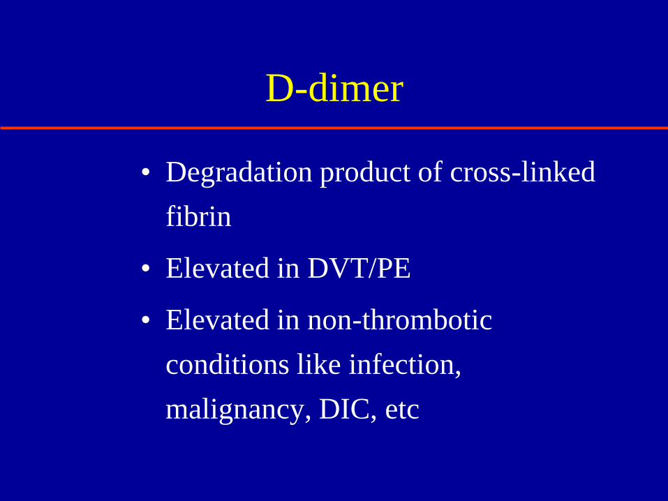
D-dimer
• Degradation product of cross-linked
fibrin
• Elevated in DVT/PE
• Elevated in non-thrombotic
conditions like infection,
malignancy, DIC, etc

D-dimer
• What is probability of obtaining a
negative D-dimer test if DVT/PE is
not present?

D-dimer
• A negative D-dimer test can be very
powerful

D-dimer
• A negative D-dimer test can be very
powerful
• Can be a “stand alone” test in
excluding PE in patients with low
pretest probability

A negative D-dimer can exclude
PE in low-risk patients?
1. True
2. False
1. 2.
0%0%
9

V/Q Scan

V/Q Scan
• Prospective Investigation of
Pulmonary Embolism Diagnosis
(PIOPED)

V/Q Scan
• Prospective Investigation of
Pulmonary Embolism Diagnosis
(PIOPED)
• Diagnostic accuracy greatest when
V/Q scan combined with clinical
probability

PIOPED – Incidence of PE (%)
Norm/near
norm VQ
Low
prob
VQ
Int
prob
VQ
High
prob
VQ
Low clin prob 2 4 16 56
Int clin prob 6 16 28 88
High clin prob 0 40 66 96
PIOPED, JAMA 1990; 263:2753

V/Q Scan
• High clinical probability with high-
probability V/Q scan – 95% likelihood
• Low clinical probability and low-
probability V/Q scan had only 4%
likelihood
• Normal V/Q scan excludes PE


45
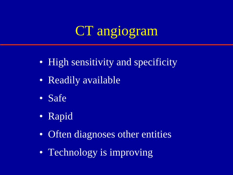
CT angiogram
• High sensitivity and specificity
• Readily available
• Safe
• Rapid
• Often diagnoses other entities
• Technology is improving

CTA vs V/Q in PE:
JAMA 298:2743-2753, 2007; 1400 patients
• CTA diagnoses more
patients with PE (19.2%)
• Sensitivity = 94.1%
• Specificity = 93.6%
• Positive PV = 95.5%
• Negative PV = 96.2%
• 1.4 to 31.8 mSv radiation
from 64 slice detector,
causing breast cancer in
1/143 20 year old women
• V/Q diagnoses fewer
patients with PE (14%)
• Sensitivity = 80.8%
• Specificity = 73.8%
• Positive PV = 95.5%
• Negative PV = 75.9%
• 0.28 to 0.9 mSv radiation
from V/Q scans but a
higher overall radiation in
pregnancy to the fetus

When to use V/Q scan?
• CT angio not available
• Morbid obesity
• Kidney disease
• Contrast allergy
• Marked discordance between clinical
probablilty and CT angio results

PE Algorithm

Algorithm if CT Angio not
available
• Well’s criteria (low, intermediate, high)
• D-dimer
• V/Q scan
• If answer unclear, pulmonary angiogram or
serial leg ultrasound

? Cardiac Echo
• Patient has hypotension or shock

? Cardiac Echo
• Patient has hypotension or shock
• CT angio shows bilateral PE

Right Ventricular Dilatation

Cardiac Echo in Massive PE
• size of RA or RV
• RV function
• Tricuspid regurgitation
• RV thrombus

Which of the following would you
not use as sole initial therapy in PE?
1. IV unfractionated
heparin
2. Rivaroxaban
3. Warfarin
4. Enoxaparin
5. Fondaparinux
1. 2. 3. 4. 5.
0% 0% 0%0%0%
9

57
Case 1
• 30 yo AAF
• 6 weeks of progressive SOB
• Worsening SOB on day she came to ED
• Pain in right medial thigh
• Not on OCP’s, no recent surgeries

58
Case 1
• Resting heart rate 128
• No hypoxemia, BP is normal
• Troponin I 0.02 ng/ml
• BNP 187 pg/ml
• D-dimer not done
• Bedside cardiac echo shows RV dilitation

Wells Prediction Score

A CT angiogram was performed

In addition, the CT shows extensive
DVT in right leg. What would you do?
1. Exoxaparin 1mg/kg
Q12 hours
2. Unfractionated heparin
by IV protocol
3. Place IVC filter
4. Lytic therapy
5. CT surgery consult
6. Strict bed rest 1. 2. 3. 4. 5. 6.
0% 0% 0%0%0%0%
9

63
ACCP Guideline 5.6.1.1
In patients with acute PE associated with
hypotension (e.g.systolic BP < 90), who do
not have a high bleeding risk, we suggest
systemically administered thrombolytic
therapy over no such therapy (Grade 2 C).

64
ACCP Guideline 5.6.1.2
In most patients with acute PE not
associated with hypotension, we
recommend against systemically
administered thrombolytic therapy (Grade
1C).
65
ACCP Guideline 5.6.1.3
In selected patients with acute PE not
associated with hypotension and with a low
bleeding risk whose initial clinical
presentation, or clinical course after starting
anticoagulant therapy, suggests a high risk
of developing hypotension, we suggest
administration of thrombolytic Rx (Grade 2
C)

66
Thrombolytic Therapy
• Accelerates the lysis of acute emboli
• Decreases PA pressures
• Improves RV function
• No clinical trial has been large enough to
conclusively demonstrate an improvement
in mortality

Family history is positive for multiple relatives
dying with PE. What would you do now?
1. Screen for
thrombophilia
2. Screen for occult
cancer
3. Anticoagulate for 6-12
months
4. Place a vena cava filter
5. Anticoagulate for life
1. 2. 3. 4. 5.
0% 0% 0%0%0%
9

Other Therapies for Massive PE
1. Catheter directed thrombolysis
2. Surgical embolectomy

Catheter Directed Thrombolysis

Other Therapies for Massive PE
1. Catheter directed thrombolysis
2. Surgical embolectomy

Case 2
• 67 yo WF came to ED with chest pain
• Pulse 120, RR 30, 100/78
• O2 sat 87% on RA
• Ultrasound shows large DVT right leg
• Bedside echo shows dilated RV

CT Angio

Case 2
• Pt. is on clopidogrel 75 mg per day
since stroke 2 years ago

What would you do next?
1. Lytic therapy with TPA
2. IV heparin by protocol
3. IVC filter
4. Stop clopidogrel
5. IV heparin + IVC filter
1. 2. 3. 4. 5.
0% 0% 0%0%0%
9

She was transferred to the MICU, heparin was
initiated and a vena cava filter was inserted
• Clopidogrel was stopped
• Initiation of warfarin was delayed in favor
of prolonged therapeutic anticoagulation
with unfractionated heparin
• She was kept at strict bedrest
• By the second ICU day her heart rate was
85, blood pressure was 130/80 mm Hg
and room air arterial saturation was 94%

What is best treatment duration
for PE provoked by surgery?
1. 3 months
2. 6 months
3. 12 months
4. Lifetime
1. 2. 3. 4.
0% 0%0%0%
9

80
ACCP Guideline 6.1
In patients with PE provoked by surgery,
we recommend treatment with
anticoagulation for 3 months over treatment
of shorter duration , treatment of longer
period (6-12 months), or extended therapy
(Grade 1B).

*

Responses remain anonymous!

1. Poor
2. Fair
3. Average
4. Good
5. Excellent
1. 2. 3. 4. 5.
0% 0% 0%0%0%
10

1. Poor
2. Fair
3. Average
4. Good
5. Excellent
Poor
Fair
Aver
age
Good
Excel
lent
0% 0% 0%0%0%
10

0%
0%
0%
10
1. Yes
2. No
3. N/A

1. Poor
2. Fair
3. Average
4. Good
5. Excellent
1. 2. 3. 4. 5.
0% 0% 0%0%0%
10

0%
0%
10
1. Yes
2. No