Pulmonary circulation
120
Pulmonary Circulation
-
Upload
uphar-gupta -
Category
Health & Medicine
-
view
64 -
download
3
Transcript of Pulmonary circulation

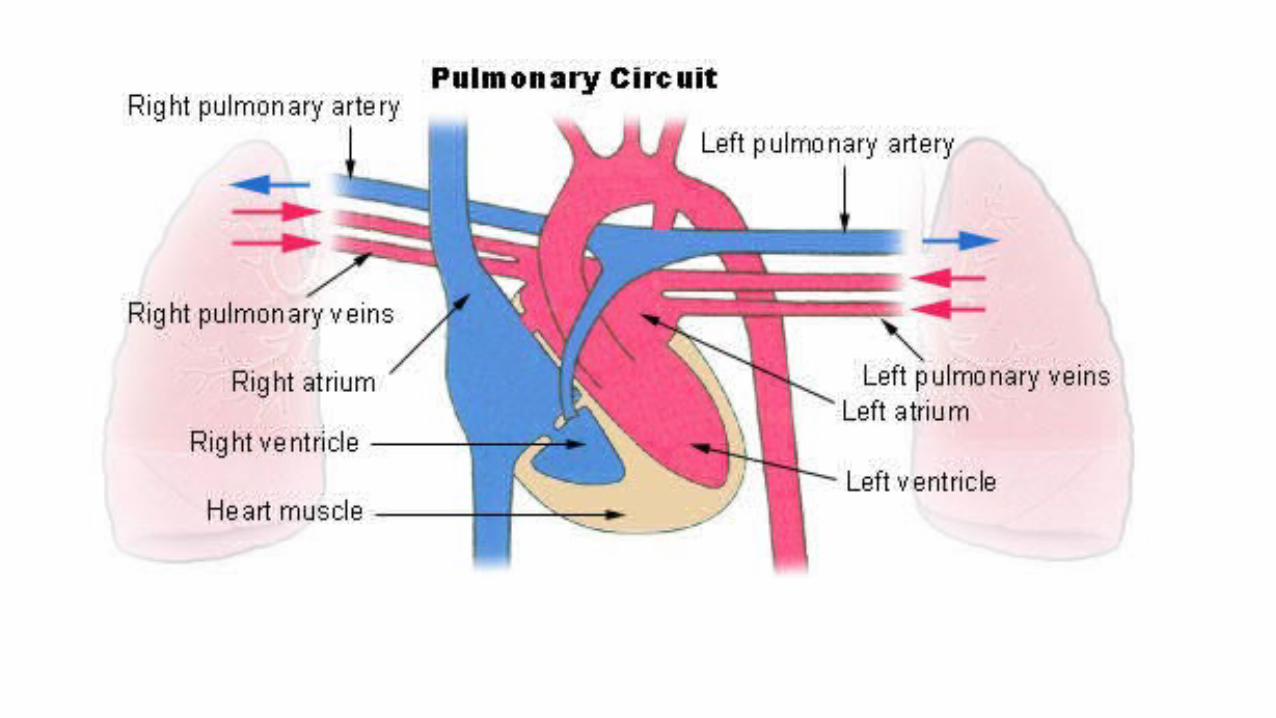
Pulmonary Circulation
Physiology
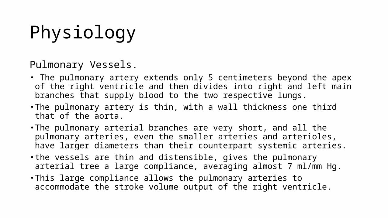
Pulmonary Vessels.• The pulmonary artery extends only 5 centimeters beyond the apex of the right
ventricle and then divides into right and left main branches that supply blood to the two respective lungs.
• The pulmonary artery is thin, with a wall thickness one third that of the aorta.• The pulmonary arterial branches are very short, and all the pulmonary arteries,
even the smaller arteries and arterioles, have larger diameters than their counterpart systemic arteries.
• the vessels are thin and distensible, gives the pulmonary arterial tree a large compliance, averaging almost 7 ml/mm Hg.
• This large compliance allows the pulmonary arteries to accommodate the stroke volume output of the right ventricle.

• The pulmonary veins, like the pulmonary arteries, are also short. • They immediately empty their effluent blood into the left atrium, to
be pumped by the left heart through the systemic circulation.
Bronchial Vessels
• small bronchial arteries that originate from the systemic circulation, amounting to about 1 to 2 per cent of the total cardiac output. • oxygenated blood• supplies the supporting tissues of the lungs, including the connective
tissue, septa, and large and small bronchi. • After this bronchial and arterial blood has passed through the
supporting tissues, it empties into the pulmonary veins and enters the left atrium, rather than passing back to the right atrium. • Therefore, the flow into the left atrium and the left ventricular output
are about 1 to 2 per cent greater than the right ventricular output.
Lymphatics.
• Lymph vessels are present in all the supportive tissues of the lung, beginning in the connective tissue spaces that surround the terminal bronchioles, coursing to the hilum of the lung, and thence mainly into the right thoracic lymph duct.• It drains • Particulate matter entering the alveoli• plasma protein leaking from the lung capillaries(prevent pulmonary edema).
Pressures in the Pulmonary System
• The systolic pressure in the right ventricle - 25 mm Hg,• the diastolic pressure averages about 0 to 1 mm Hg , values that are
only one fifth those for the left ventricle.
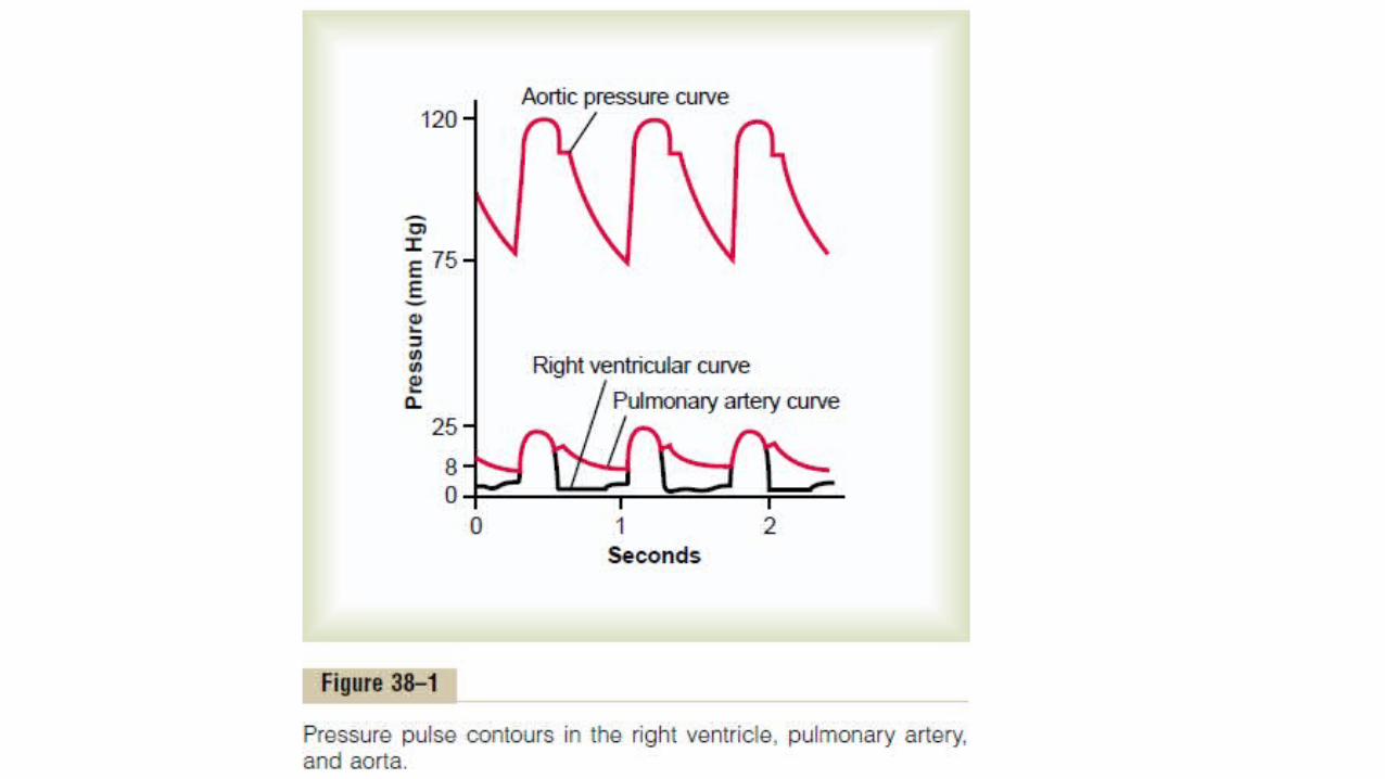

Pressures in the Pulmonary Artery.
• During systole, the pressure in the pulmonary artery is equal to the pressure in the right ventricle• After the pulmonary valve closes at the end of systole, the ventricular
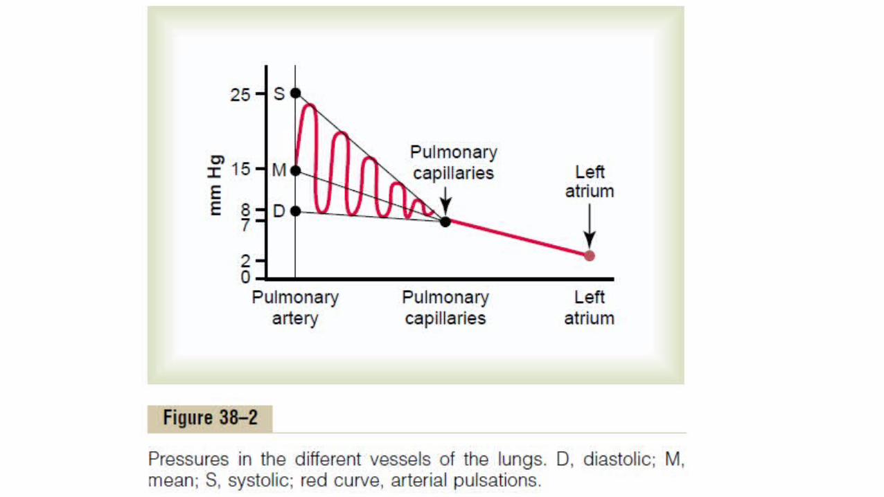
pressure falls precipitously, whereas the pulmonary arterial pressure falls more slowly as blood flows through the capillaries of the lungs.• The systolic pulmonary arterial pressure - 25 mm Hg• the diastolic pulmonary arterial pressure - 8 mm Hg • the mean pulmonary arterial pressure is 15 mm Hg.

Pulmonary Capillary Pressure.
• The mean - 7 mm Hg.

Left Atrial and Pulmonary Venous Pressures. • The mean pressure in the left atrium and the major pulmonary veins
averages about 2 mm Hg in the recumbent human being, varying from as low as 1 mm Hg to as high as 5 mm Hg. • The pressure measured through the catheter, called the “wedge
pressure,” is about 5 mm Hg.
• Because all blood flow has been stopped in the small wedged artery, and because the blood vessels extending beyond this artery make a direct connection with the pulmonary capillaries, this wedge pressure is usually only 2 to 3 mm Hg greater than the left atrial pressure.• When the left atrial pressure rises to high values, the pulmonary
wedge pressure also rises. • Therefore, wedge pressure measurements can be used to clinically
study changes in pulmonary capillary pressure and left atrial pressure in patients with congestive heart failure.

Blood Volume of the Lungs
• 450 millilitres (9 per cent of the total blood volume of the entire circulatory system ). • Approximately 70 milliliters of this pulmonary blood volume is in the
pulmonary capillaries, and the remainder is divided about equally between the pulmonary arteries and the veins.

Lungs as a Blood Reservoir.
• blood in the lungs can vary from as little as one half normal up to twice normal.• Loss of blood from the systemic circulation by hemorrhage can be
partly compensated for by the automatic shift of blood from the lungs into the systemic vessels.
Shift of Blood Between the Pulmonary and Systemic Circulatory Systems as a Result of Cardiac Pathology.
• Failure of the left side of the heart or increased resistance to blood flow through the mitral valve as a result of mitral stenosis or mitral regurgitation – damming up of blood in the pulmonary circulation - causing large increases in the pulmonary vascular pressures. • Because the volume of the systemic circulation is about nine times
that of the pulmonary system, a shift of blood from one system to the other affects the pulmonary system greatly but usually has only mild systemic circulatory effects.
Blood Flow Through the Lungs and Its Distribution
• The blood flow through the lungs is essentially equal to the cardiac output. • Therefore, the factors that control cardiac output—mainly peripheral
factors—also control pulmonary blood flow. • Under most conditions, the pulmonary vessels act as passive,
distensible tubes that enlarge with increasing pressure and narrow with decreasing pressure. • For adequate aeration of the blood to occur, it is important for the
blood to be distributed to those segments of the lungs where the alveoli are best oxygenated.
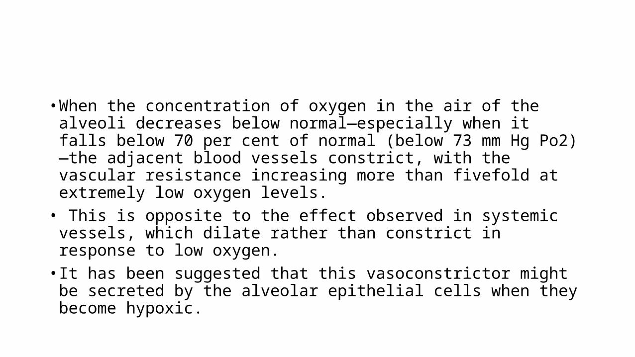
• When the concentration of oxygen in the air of the alveoli decreases below normal—especially when it falls below 70 per cent of normal (below 73 mm Hg Po2)—the adjacent blood vessels constrict, with the vascular resistance increasing more than fivefold at extremely low oxygen levels.• This is opposite to the effect observed in systemic vessels, which
dilate rather than constrict in response to low oxygen. • It has been suggested that this vasoconstrictor might be secreted by
the alveolar epithelial cells when they become hypoxic.
• to distribute blood flow where it is most effective. • This causes the blood to flow through other areas of the lungs that
are better aerated, thus providing an automatic control system for distributing blood flow to the pulmonary areas in proportion to their alveolar oxygen pressures.

Regional Pulmonary Blood Flow
• the blood pressure in the foot of a standing person can be as much as 90 mm Hg greater than the pressure at the level of the heart.• This is caused by hydrostatic pressure—that is, by the weight of the
blood itself in the blood vessels. • In the normal, upright adult, the lowest point in the lungs is about 30
centimeters below the highest point.

• This represents a 23 mm Hg pressure difference, about 15 mm Hg of which is above the heart and 8 below.• That is, the pulmonary arterial pressure in the uppermost portion of
the lung of a standing person is about 15 mm Hg less than the pulmonary arterial pressure at the level of the heart, and the pressure in the lowest portion of the lungs is about 8 mm Hg greater. • Such pressure differences have profound effects on blood flow
through the different areas of the lungs.

Zones of Pulmonary Blood Flow
• The capillaries in the alveolar walls are distended by the blood pressure inside them, but simultaneously, they are compressed by the alveolar air pressure on their outsides. • Therefore, any time the lung alveolar air pressure becomes greater
than the capillary blood pressure, the capillaries close and there is no blood flow.
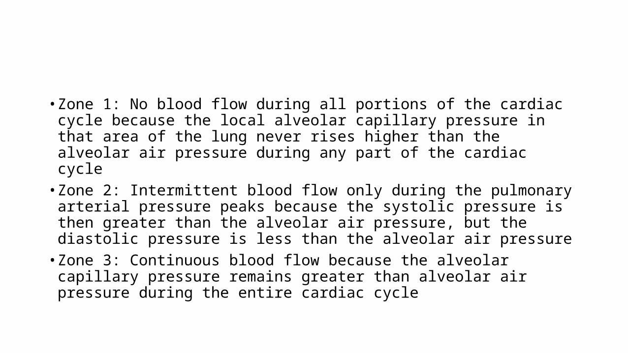
• Zone 1: No blood flow during all portions of the cardiac cycle because the local alveolar capillary pressure in that area of the lung never rises higher than the alveolar air pressure during any part of the cardiac cycle• Zone 2: Intermittent blood flow only during the pulmonary arterial
pressure peaks because the systolic pressure is then greater than the alveolar air pressure, but the diastolic pressure is less than the alveolar air pressure• Zone 3: Continuous blood flow because the alveolar capillary pressure
remains greater than alveolar air pressure during the entire cardiac cycle

• Normally, the lungs have only zones 2 and 3 blood flow—zone 2 (intermittent flow) in the apices, and zone 3 (continuous flow) in all the lower areas.• blood flow through the apical part of the lung is intermittent, with
flow during systole but cessation of flow during diastole; this is called zone 2 blood flow.

• In the lower regions of the lungs, from about 10 centimeters above the level of the heart all the way to the bottom of the lungs, the pulmonary arterial pressure during both systole and diastole remains greater than the zero alveolar air pressure. • Therefore, there is continuous flow through the alveolar capillaries, or
zone 3 blood flow. • Also, when a person is lying down, no part of the lung is more than a
few centimeters above the level of the heart. • In this case, blood flow in a normal person is entirely zone 3 blood
flow, including the lung apices.

• Zone 1 Blood Flow Occurs Only Under Abnormal Conditions. • Zone 1 blood flow, which is blood flow at no time during the cardiac
cycle, occurs when either the pulmonary systolic arterial pressure is too low or the alveolar pressure is too high to allow flow. • Seen in positive pressure ventilation and in severe blood loss.
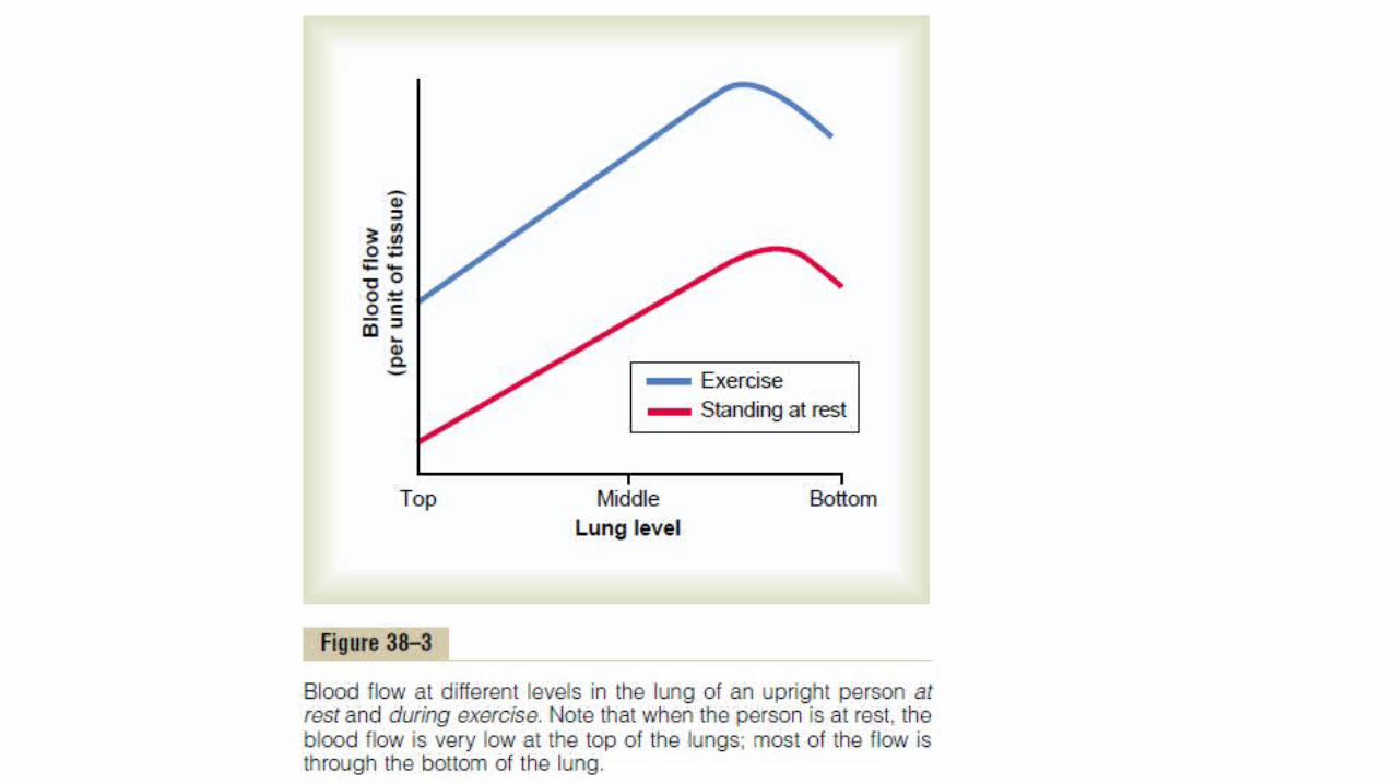
Effect of Exercise on Blood Flow
• blood flow in all parts of the lung increases during exercise.• The increase in flow in the top of the lung may be 700 to 800 per
cent, whereas the increase in the lower part of the lung may be no more than 200 to 300 per cent. • The reason for these differences is that the pulmonary vascular
pressures rise enough during exercise to convert the lung apices from a zone 2 pattern into a zone 3 pattern of flow.

Effect of Increased Cardiac Output
• During heavy exercise, blood flow through the lungs increases fourfold to sevenfold. • This extra flow is accommodated in the lungs in three ways:
(1) by increasing the number of open capillaries, sometimes as much as threefold; (2) by distending all the capillaries and increasing the rate of flow through each capillary more than twofold; and (3) by increasing the pulmonary arterial pressure.


• The ability of the lungs to accommodate greatly increased blood flow during exercise without increasing the pulmonary arterial pressure conserves the energy of the right side of the heart. • This ability also prevents a significant rise in pulmonary capillary
pressure, thus also preventing the development of pulmonary edema.

Function of the Pulmonary Circulation
• When the Left Atrial Pressure Rises as a Result of Left-Sided Heart Failure• The left atrial pressure in a healthy person almost never rises above +6
mm Hg, even during the most strenuous exercise. • These small changes in left atrial pressure have virtually no effect on
pulmonary circulatory function because this merely expands the pulmonary venules and opens up more capillaries so that blood continues to flow with almost equal ease from the pulmonary arteries.• When the left side of the heart fails, however, blood begins to dam up in
the left atrium.

• The left atrial pressure can rise up to 40 to 50 mm Hg. • The initial rise in atrial pressure, up to about 7 mm Hg, has very little effect
on pulmonary circulatory function. • But when the left atrial pressure rises to greater than 7 or 8 mm Hg, further
increases in left atrial pressure above these levels cause almost equally great increases in pulmonary arterial pressure, thus causing a concomitant increased load on the right heart. • Any increase in left atrial pressure above 7 or 8 mm Hg increases the
capillary pressure almost equally as much.• When the left atrial pressure has risen above 30 mm Hg, causing similar
increases in capillary pressure, pulmonary edema is likely to develop.
Length of Time Blood Stays in the Pulmonary Capillaries.
• From histological study of the total cross-sectional area of all the pulmonary capillaries, it can be calculated that when the cardiac output is normal, blood passes through the pulmonary capillaries in about 0.8 second.• When the cardiac output increases, this can shorten to as little as 0.3
second.• Thus, in only a fraction of a second, blood passing through the
alveolar capillaries becomes oxygenated and loses its excess carbon dioxide.
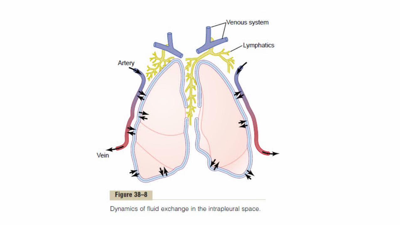
Fluid Dynamics
• The dynamics of fluid exchange across the lung capillary membranes are qualitatively the same as for peripheral tissues.• However, quantitatively, there are important differences, as follows:1. The pulmonary capillary pressure is low, about 7 mm Hg, in comparison with a considerably higher functional capillary pressure in the peripheral tissues of about 17 mm Hg.2. The interstitial fluid pressure in the lung is slightly more negative than that in the peripheral subcutaneous tissue.
3. The pulmonary capillaries are relatively leaky to protein molecules, so that the colloid osmotic pressure of the pulmonary interstitial fluid is about 14 mm Hg, in comparison with 7mmHg in the peripheral tissues.
4. The alveolar walls are extremely thin, and weak –it can be ruptured by any positive pressure in the interstitial spaces greater than alveolar air pressure (greater than 0 mm Hg), which allows dumping of fluid from the interstitial spaces into the alveoli.


• This filtration pressure causes a slight continual flow of fluid from the pulmonary capillaries into the interstitial spaces, and except for a small amount that evaporates in the alveoli, this fluid is pumped back to the circulation through the pulmonary lymphatic system.
Negative Pulmonary Interstitial Pressure • pulmonary capillaries and the pulmonary lymphatic system normally
maintain a slight negative pressure in the interstitial spaces• When extra fluid appears in the alveoli, it is sucked mechanically into
the lung interstitium through the small openings between the alveolar epithelial cells. • Then the excess fluid is either carried away through the pulmonary
lymphatics or absorbed into the pulmonary capillaries. • Thus, under normal conditions, the alveoli are kept “dry,” except for a
small amount of fluid that seeps from the epithelium onto the lining surfaces of the alveoli to keep them moist.
Pulmonary Edema
• Any factor that causes the pulmonary interstitial fluid pressure to rise from the negative range into the positive range will cause rapid filling of the pulmonary interstitial spaces and alveoli with large amounts of free fluid.• The most common causes of pulmonary edema are as follows: 1. Left-sided heart failure or mitral valve disease, with consequent great increases in pulmonary venous pressure and pulmonary capillary pressure and flooding of the interstitial spaces and alveoli.2. Damage to the pulmonary blood capillary membranes caused by infections such as pneumonia or by breathing noxious substances such as chlorine gas or sulfur dioxide gas –rapid leakage of both plasma proteins and fluid out of the capillaries and into both the lung interstitial spaces and the alveoli.

Safety Factor in Chronic Conditions
• When the pulmonary capillary pressure remains elevated chronically (for at least 2 weeks), the lungs become even more resistant to pulmonary edema because the lymph vessels expand greatly, increasing their capability of carrying fluid away from the interstitial spaces perhaps as much as 10-fold. • Therefore, in patients with chronic mitral stenosis, pulmonary
capillary pressures of 40 to 45 mm Hg have been measured without the development of lethal pulmonary edema.
Rapidity of Death in Acute Pulmonary Edema• When the pulmonary capillary pressure rises even slightly above the
safety factor level, lethal pulmonary edema can occur within hours, or even within 20 to 30 minutes if the capillary pressure rises 25 to 30 mm Hg above the safety factor level. • Thus, in acute left-sided heart failure, in which the pulmonary
capillary pressure occasionally does rise to 50 mm Hg, death frequently ensues in less than 30 minutes from acute pulmonary edema.

Fluid in the Pleural Cavity
• When the lungs expand and contract during normal breathing, they slide back and forth within the pleural cavity. • A thin layer of mucoid fluid lies between the parietal and visceral
pleurae.• The pleural membrane is a porous, mesenchymal, serous membrane
through which small amounts of interstitial fluid transude continually into the pleural space. • These fluids carry with them tissue proteins, giving the pleural fluid a
mucoid characteristic, which is what allows extremely easy slippage of the moving lungs.
• The total amount of fluid in each pleural cavity is normally slight, only a few milliliters.• Whenever the quantity becomes more than barely enough to begin
flowing in the pleural cavity, the excess fluid is pumped away by lymphatic vessels opening directly from the pleural cavity into
(1) the mediastinum, (2) the superior surface of the diaphragm, and (3) the lateral surfaces of the parietal pleura.
• Therefore, the pleural space—the space between the parietal and visceral pleurae—is called a potential space because it normally is so narrow that it is not obviously a physical space.

“Negative Pressure” in Pleural Fluid.
• A negative force is always required on the outside of the lungs to keep the lungs expanded. • This is provided by negative pressure in the normal pleural space.• The basic cause of this negative pressure is pumping of fluid from the
space by the lymphatics (which is also the basis of the negative pressure found in most tissue spaces of the body). • Because the normal collapse tendency of the lungs is about –4 mm
Hg, the pleural fluid pressure must always be at least as negative as –4 mm Hg to keep the lungs expanded.

• Actual measurements have shown that the pressure is usually about –7 mm Hg, which is a few millimetres of mercury more negative than the collapse pressure of the lungs.• Thus, the negativity of the pleural fluid keeps the normal lungs pulled
against the parietal pleura of the chest cavity, except for an extremely thinlayer of mucoid fluid that acts as a lubricant.

Pleural Effusion
• Pleural effusion means the collection of large amounts of free fluid in the pleural space.• The effusion is analogous to edema fluid in the tissues and can be
called “edema of the pleural cavity.”

• The causes of the effusion are the same as the causes of edema in other tissues • (1) blockage of lymphatic drainage from the pleural cavity; • (2) cardiac failure, which causes excessively high peripheral and pulmonary
capillary pressures, leading to excessive transudation of fluid into the pleural cavity;• (3) greatly reduced plasma colloid osmotic pressure, thus allowing excessive
transudation of fluid; and• (4) infection or any other cause of inflammation of the surfaces of the pleural
cavity, which breaks down the capillary membranes and allows rapid dumping of both plasma proteins and fluid into the cavity.

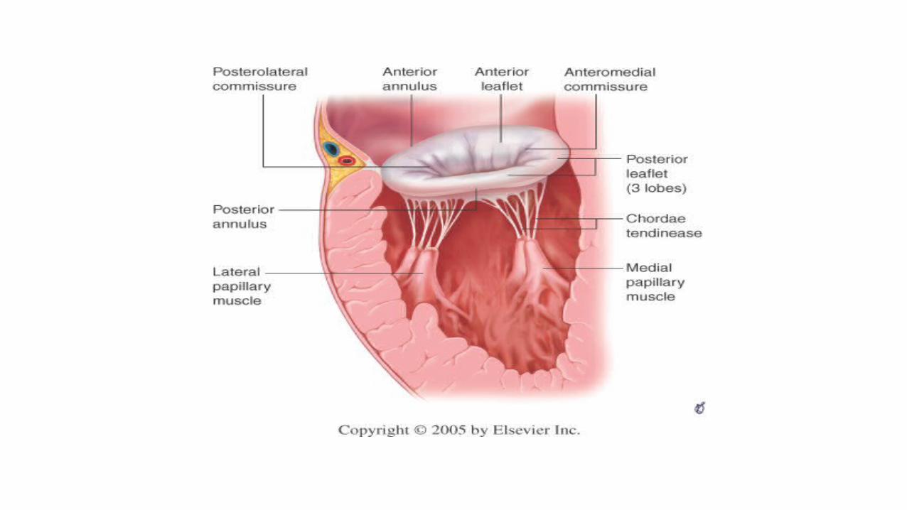
MITRAL REGURGITATION
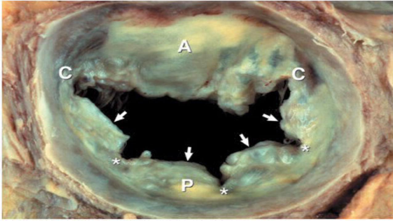
Anatomy
The mitral valve apparatus consists of • the anterior and the posterior leaflets ,• the mitral annulus (fixed and dynamic)• The chordae tendinae• Papillary muscles• Underlying myocardium
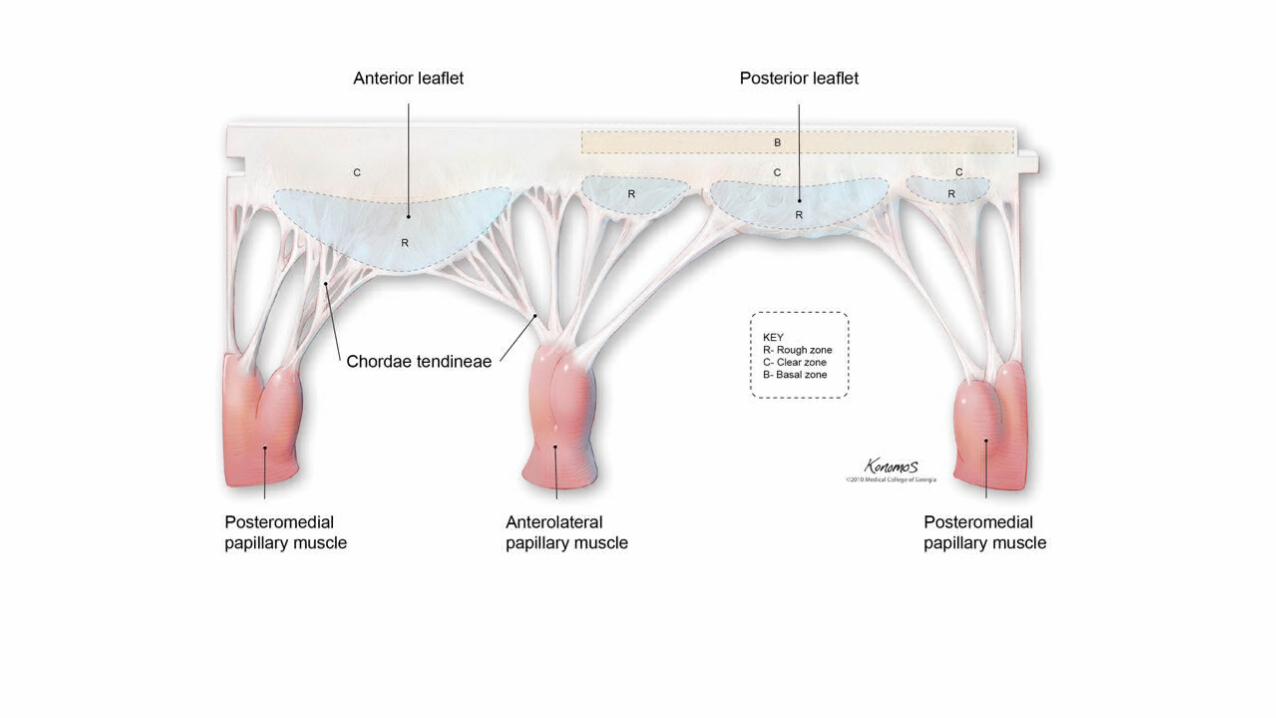

2- to 3-mm zone of overlap (the zona coapta).

Apical four-chamber view recorded in systole in a normal patient

Etiology
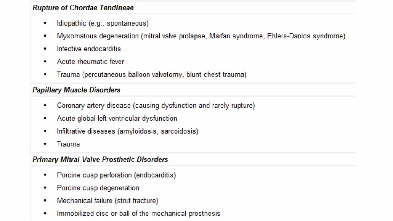
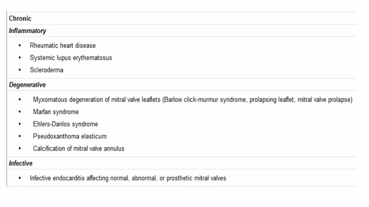

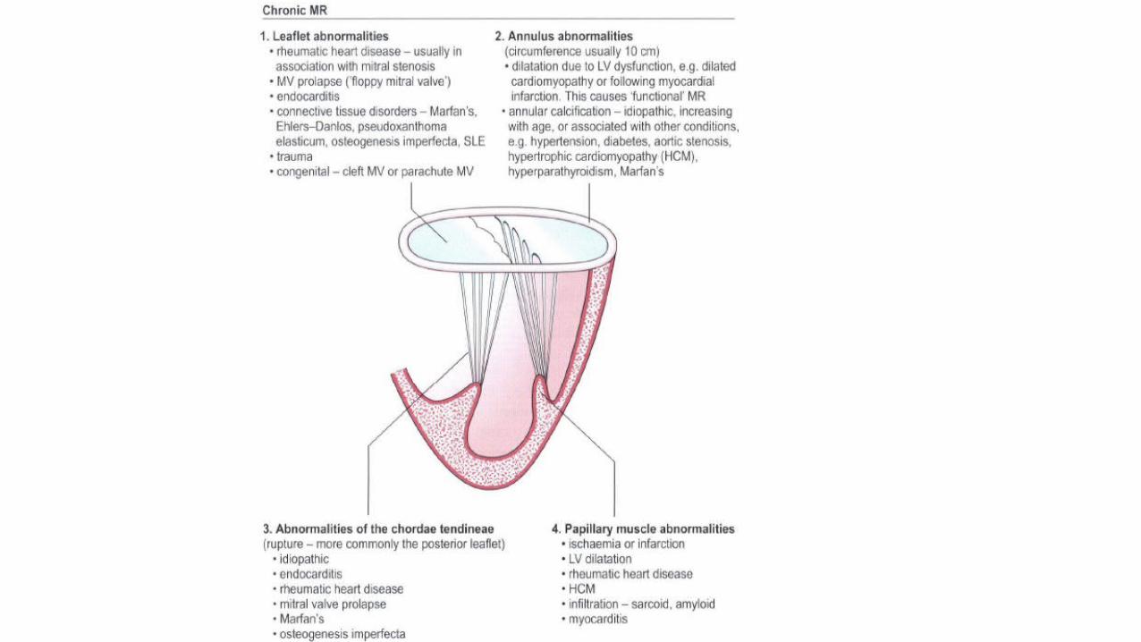

Rhuematic disease
• More common in men• consequence of shortening, rigidity, deformity, and retraction of one
or both mitral valve cusps• Associated with shortening and fusion of the chordae tendineae and
papillary muscles.

Degenerative
• Mitral annular calcification is common in the elderly population• accelerated by hypertension, AS, and diabetes, • Also associated with connective tissue diseases such as Marfan and
Hurler syndromes.
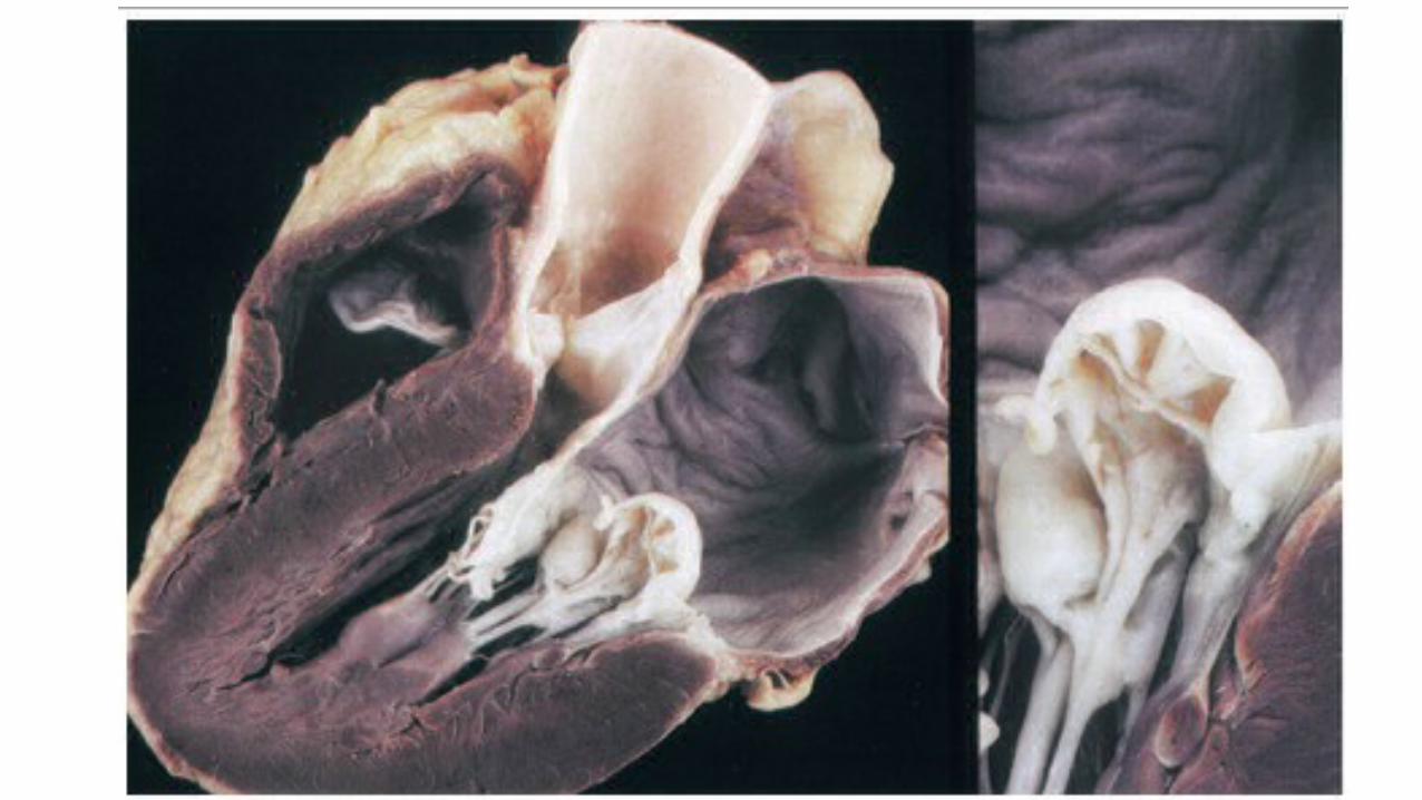
• associated with valve prolapse, • abnormal movement of the leaflets into the left atrium (LA) during
systole • caused by inadequate chordal support (elongation or rupture) and
excessive valvular tissue. • Myxomatous degeneration -characterized by redundant, floppy
leaflets and associated with progressive MR.

• 10 percent more water content and 30 to 150 percent higher glycosaminoglycan concentrations than normal.• chromosomal loci 11, 13, and 16 have been identified with mitral
valve prolapse

Infective Endocarditis
• approx. 5 %of cases of severe MR. • Vegetations may produce mild MR by interposition between leaflets.• Severe endocarditic MR is usually related to ruptured chordae and
less frequently to destruction of mitral tissue (edges or perforation)

Ischemic and Functional MR
• consequence of LV wall dysfunction (ischemia, scarring, aneurysm, cardiomyopathy, or myocarditis,)• The coaptation of intrinsically normal leaflets is incomplete. • The pattern of mitral valve (MV) deformation from the medial to the
lateral side was asymmetrical in ischemic MR, whereas it was symmetrical in nonischemic MR of cardiomyopathy.
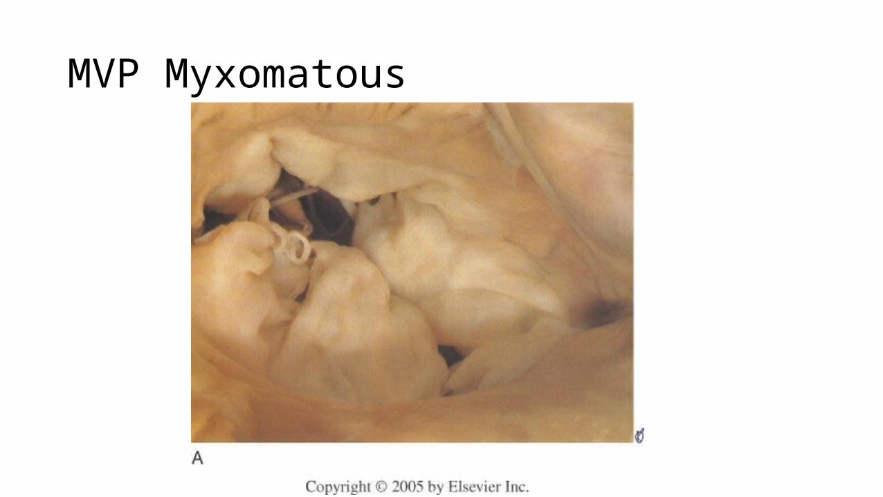
MVP Myxomatous
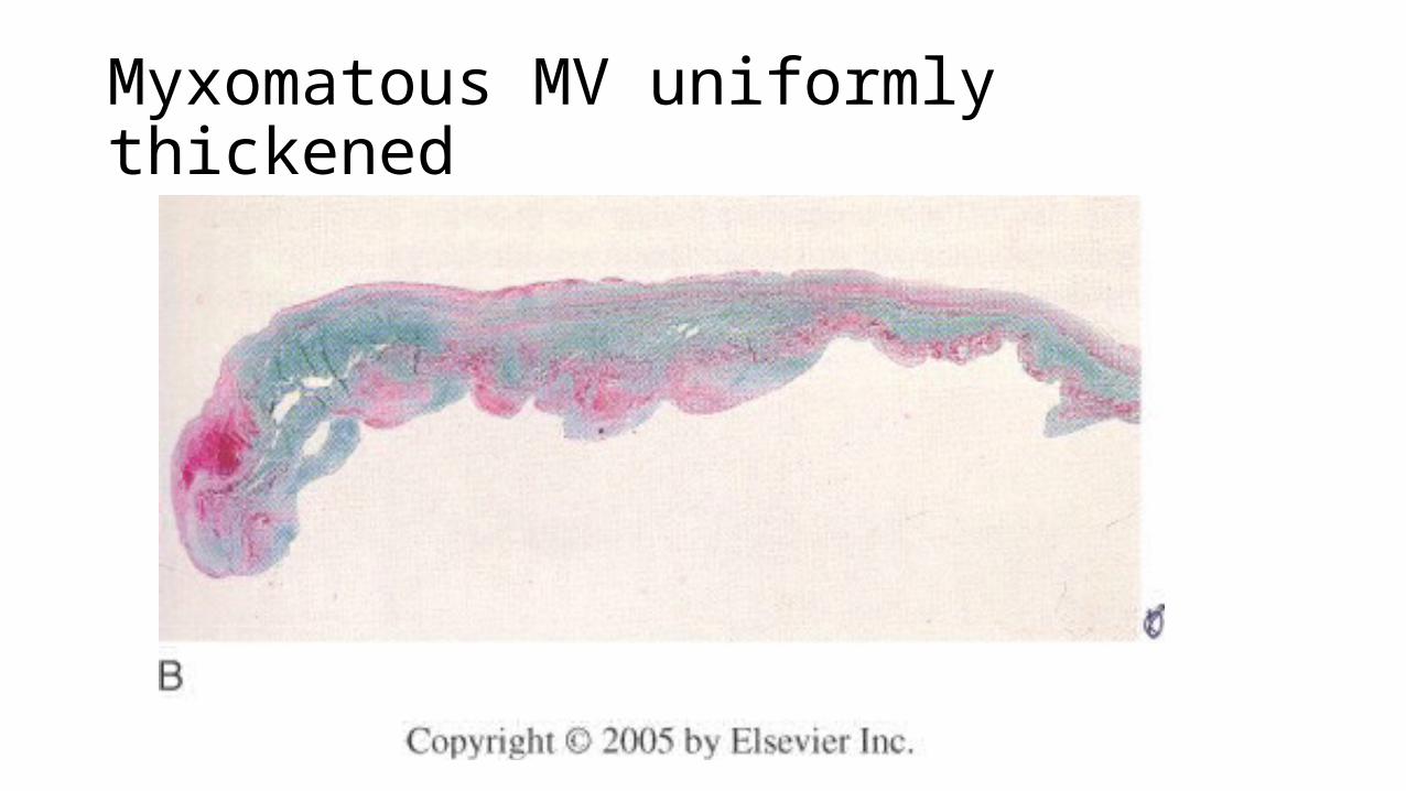
Myxomatous MV uniformly thickened

• usually responsive to vasodilators or improvement of ischemia-is a result of a reversal of adverse LV geometry.• Rupture of papillary muscle produces MR because of the flail leaflet, • in 80 percent of cases involves the posteromedial papillary muscle• Complete rupture is rapidly fatal without surgery, and partial- or
single-head rupture of the papillary allows emergency surgery


Acute phase
• sudden volume overload of both the left atrium and the left ventricle.• The combination of the forward stroke volume and the regurgitant
volume is known as the total stroke volume of the left ventricle.• In the acute setting, the stroke volume of the left ventricle is
increased (increased ejection fraction), this happens because of more complete emptying of heart. • However, as it progresses the LV volume increases and the contractile
function deteriorates and thus leading to dysfunctional LV and a decrease in ejection fraction.

• The increase in stroke volume is explained by the Frank–Starling mechanism, in which increased ventricular pre-load stretches the myocardium such that contractions are more forceful.• The regurgitant volume causes a volume overload and a pressure
overload of the left atrium. • The increased pressures in the left atrium inhibit drainage of blood
from the lungs via the pulmonary veins. • This causes pulmonary congestion.

Chronic phase
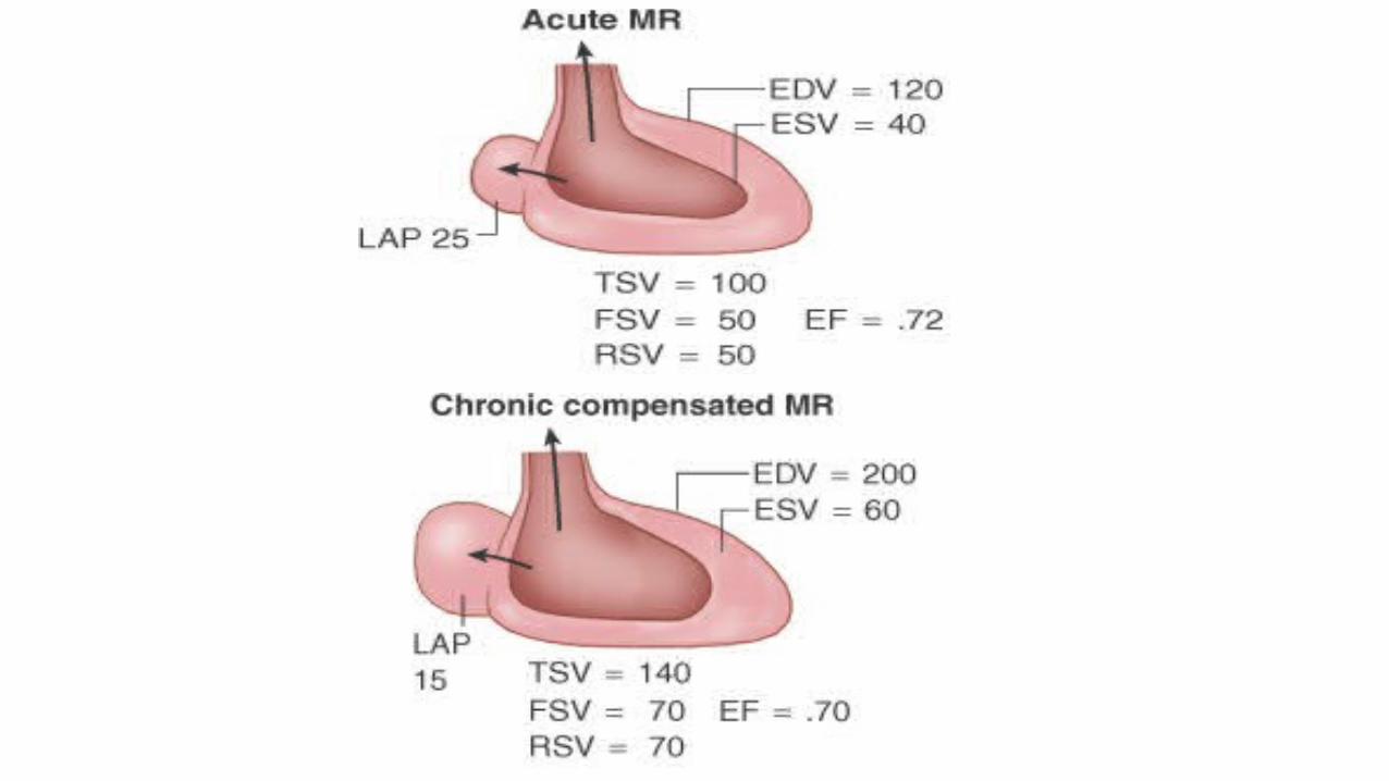
Compensated• If the mitral regurgitation develops slowly over months to years or if the acute
phase cannot be managed with medical therapy, the individual will enter the chronic compensated phase of the disease. • In this phase, the left ventricle develops eccentric hypertrophy in order to
better manage the larger than normal stroke volume. • The eccentric hypertrophy and the increased diastolic volume combine to
increase the stroke volume (to levels well above normal) so that the forward stroke volume (forward cardiac output) approaches the normal levels.• In the left atrium, the volume overload causes enlargement of the chamber of
the left atrium, allowing the filling pressure in the left atrium to decrease.
• This improves the drainage from the pulmonary veins, and signs and symptoms of pulmonary congestion will decrease.• These changes in the left ventricle and left atrium improve the low
forward cardiac output state and the pulmonary congestion that occur in the acute phase of the disease. Individuals in the chronic compensated phase may be asymptomatic and have normal exercise tolerances.

Decompensated
• develop left ventricular dysfunction• characterized by calcium overload within the cardiac myocytes.• In this phase, the ventricular myocardium is no longer able to contract
adequately to compensate for the volume overload of mitral regurgitation, and the stroke volume of the left ventricle will decrease. • The decreased stroke volume causes a decreased forward cardiac output
and an increase in the end-systolic volume. • The increased end-systolic volume translates to increased filling pressures
of the left ventricle and increased pulmonary venous congestion. • symptoms of congestive heart failure.

• The left ventricle begins to dilate during this phase.• This causes a dilatation of the mitral valve annulus, which may
worsen the degree of mitral regurgitation.

Hemodynamics
• The abnormal coaptation creates a regurgitant orifice during systole. • The systolic pressure gradient between the LV and LA is the driving
force of the regurgitant flow, which results in a regurgitant volume. • may be expressed as the regurgitant fraction. • induces a low-pressure form of volume overload as a result of
ejection into the LA. • Moderate MR when the regurgitant fraction is in the range of 30 to
50 %• severe MR - regurgitant fraction >50%

The degree of volume overload depends on three factors:
• the area of the regurgitant orifice,
• the regurgitant gradient, and
• the regurgitant duration.

• The volume overload is usually less severe in MR than in AR, despite a larger regurgitant gradient and orifice • due to shorter duration of regurgitation during the cardiac cycle in MR
than in AR• the LV is unloaded in both early and late systole by ejection into the
low-pressure LA; whereas in AR, the excess volume is ejected into the high-pressure aorta. • the lower LV mass and mass-to-volume ratio in patients with MR as
compared to AR.

• LA pressure is mainly determined by LA compliance.• In acuteMR, the LA is less compliant than it is in chronic MR and the
MR produces a marked increase in LA pressure. • decreases the ventriculoatrial gradient and tends to limit the
regurgitant volume. • In chronic MR the LA dilates, and does not limit the regurgitant
volume; • the LA pressure may be normal even with severe MR at rest
MR chronic decompensated stage
• muscle dysfunction has developed, impairing ejection fraction, diminishing both TSV and FSV. • EF, although still “normal,” has decreased to 0.55,• LAP is reelevated because less volume is ejected during systole,
causing a higher ESV.

• in chronic MR adrenergic system provides inotropic support (important mech. of compensation)• Norepinephrine release increases directly with increase in end systolic
volme.
Clinical presentation
• Usual symptoms -Fatigue and mild dyspnea on exertion (rapidly improved by rest.)• Severe dyspnea on exertion ,PND, frank pulmonary edema, or even
hemoptysis may be observed later • severe symptoms may be triggered by a new onset of AF, an increase
in degree of MR, the occurrence of endocarditis or ruptured chordae, or a change in LV compliance or function.
• Acute MR - dramatic symptoms-with pulmonary edema or ccf • progressively subside with administration of a diuretic, afterload
reduction, and increased LA compliance with time. • abrupt chordal rupture - sudden onset of atypical chest pain and
dyspnea • Rupture of papillary muscle in of a/c MI -dramatic presentation, with
cardiogenic shock or a severe pulmonary edema. • Pulmonary edema may also be observed in transient severe papillary
muscle dysfunction.

Physical examination
• BP is usually normal. Palpation • Carotid upstroke is brisk. • Palpation-laterally displaced, diffuse, and brief apical impulse with
enlarged LV. • An apical thrill is characteristic of severe MR. • Systolic expansion of the enlarged left atrium causes a late systolic
thrust in the parasternal region(,-confused with RV enlargement. )
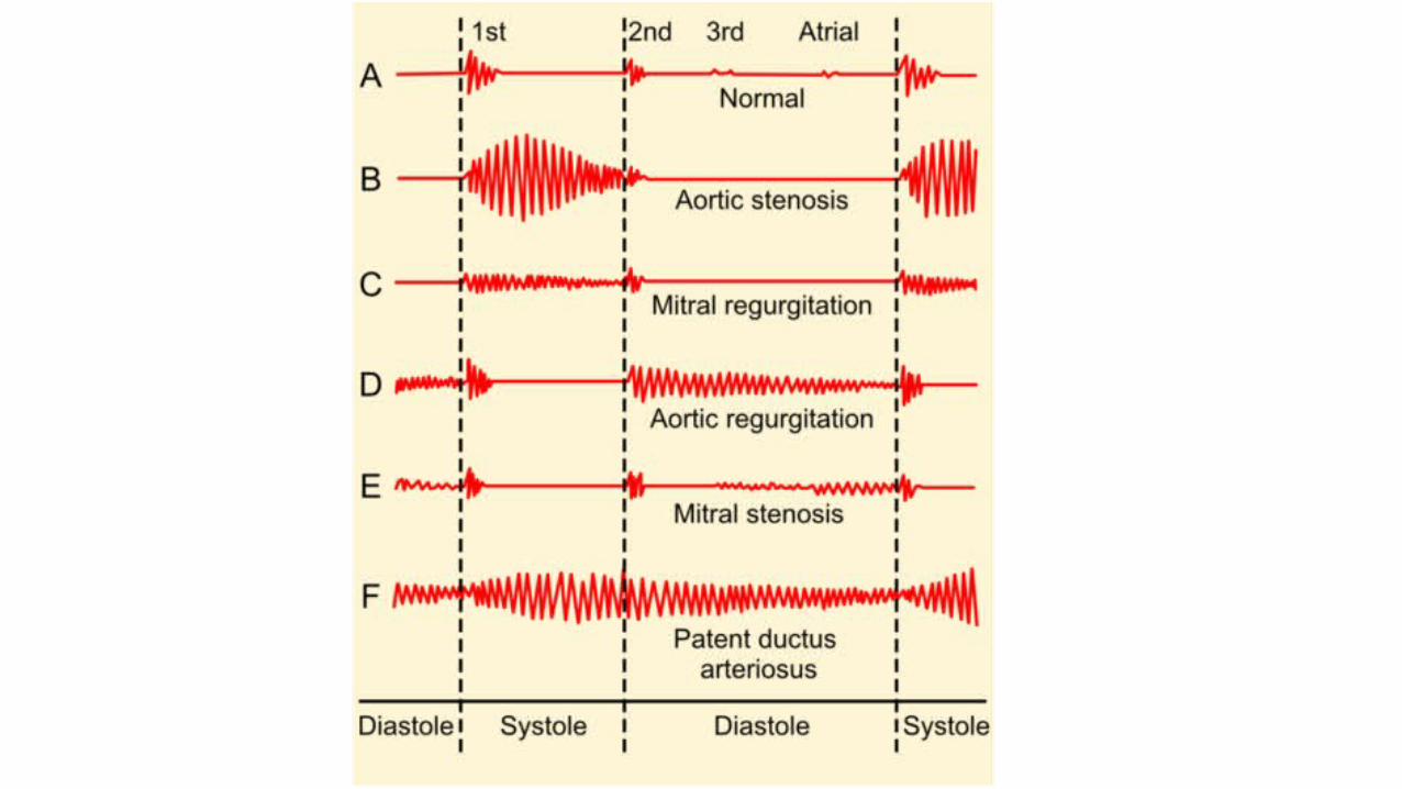
• S1 is included in the murmur and is usually normal but may be increased in rheumatic disease. • Wide splitting of S2 due to the shortening of LV ejection and an earlier A2
due to reduced resistance to LV ejection. • In patients with MR who have severe pulmonary hypertension, P2 is louder
than A2. • The abnormal increase in the flow rate across the mitral orifice during the
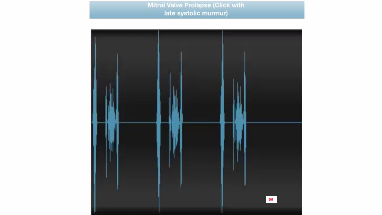
rapid filling phase is associated with an S3, (not due to heart failure) ; may be accompanied by a brief diastolic rumble.• Midsystolic clicks are markers of valve prolapse (see Mitral Valve Prolapse
Syndrome below).
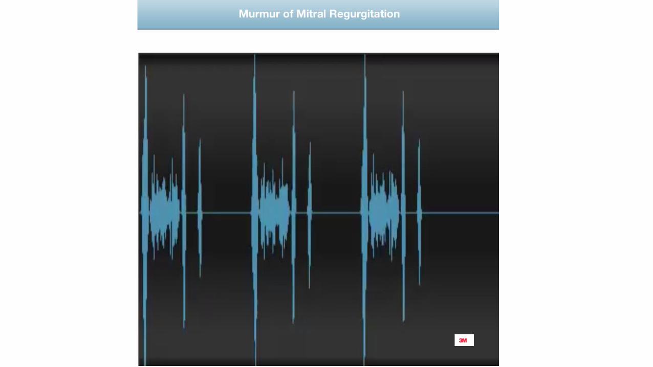

• systolic murmur, most often holosystolic, including 1st and 2nd heart sounds. • blowing type but may be harsh(MVP)• The maximum intensity is usually at the apex, and it may radiate to the axilla
in rheumatic or anterior leaflet prolapse, affecting primarily the anterior leaflet. • In posterior leaflet prolapse, the jet is usually superiorly and medially directed
and the murmur radiates toward the base of the heart. • in MI, severe MR may be totally silent. • Murmurs of shorter duration usually correspond to mild MR; they may be mid
or late systolic in mitral valve prolapse or early systolic in functional MR.
• Murmer is increased by squatting , isometric excercises• Decreased by amyl nitrite inhalation and valsalva
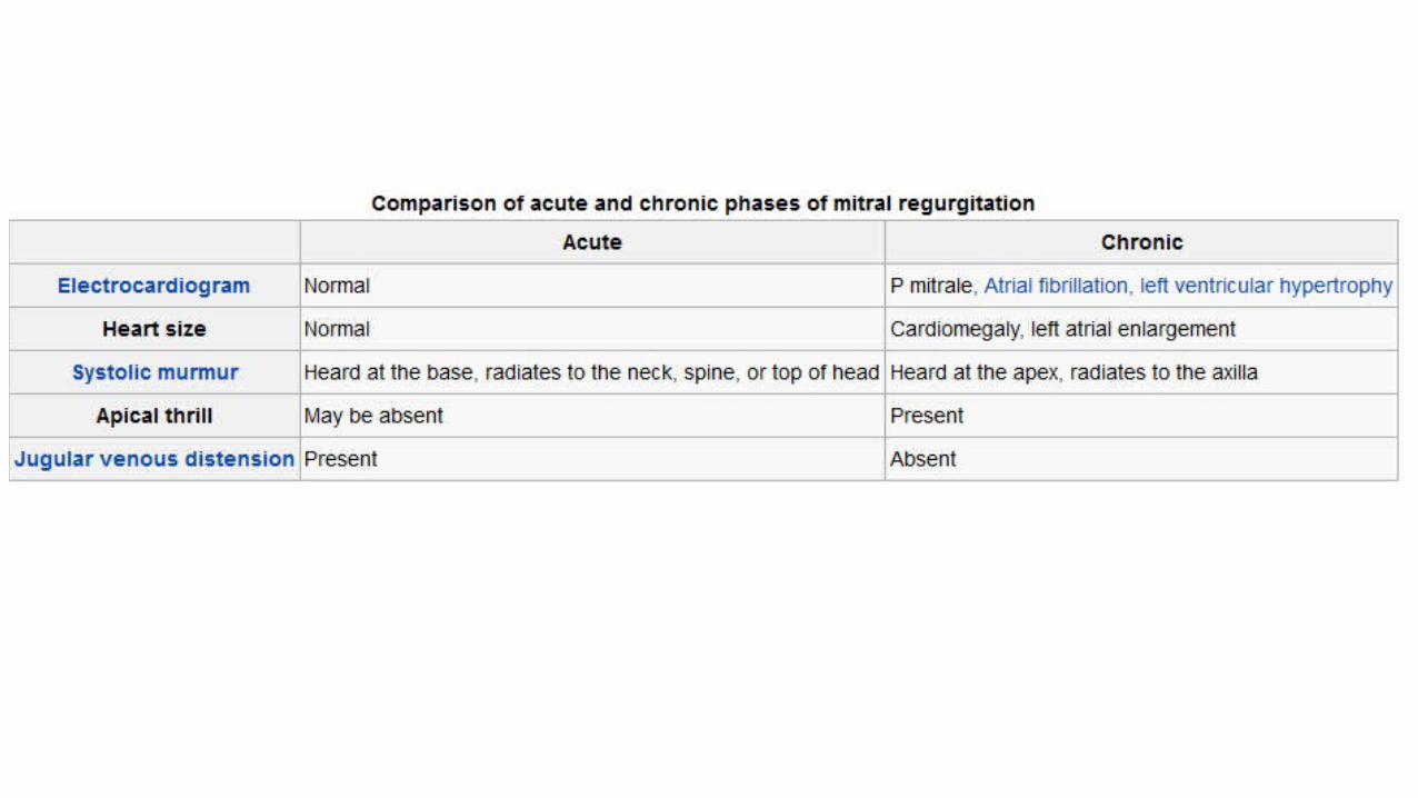
Ecg
The principal ECG findings are • left atrial enlargement and AF.• Wide notched P waves• ECG evidence of LV enlargement occurs in 1/3rd of patients with
severe MR. • Tall R in V5 V6 but S in V1 is small• 15 percent of patients -evidence of RV hypertrophy.

CXR
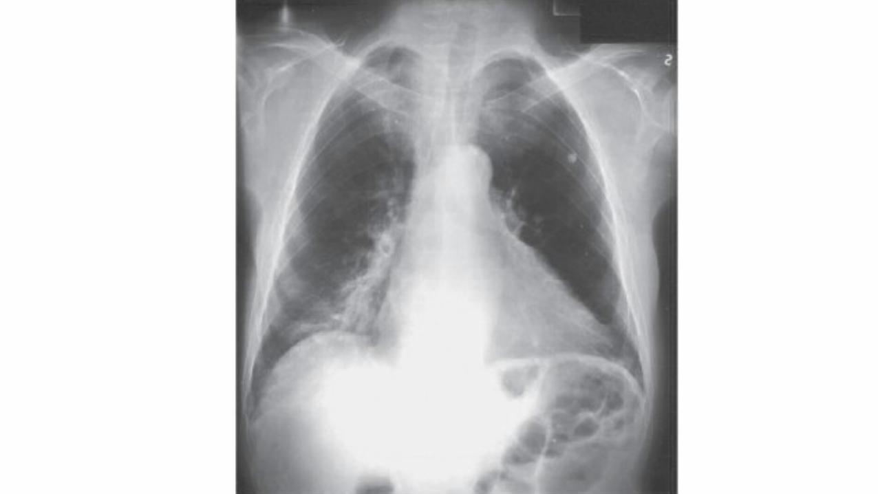
• Cardiomegaly may be present in chronic MR or in ischemic or functional MR . • LA body and appendage dilatation is frequent, but giant LA is rare. • annular calcification, seen as a C-shaped density below the posterior
leaflet, is frequent. • signs of pulmonary hypertension or pulmonary edema are rarely
observed.


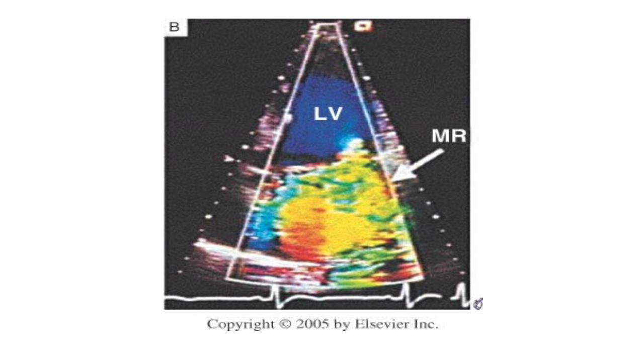
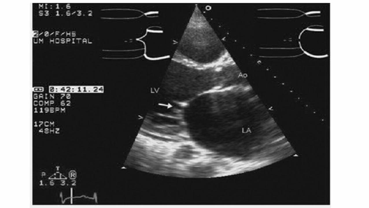
Doppler echocardiography
• high-velocity jet in the LA during systole• the severity of the regurgitation is a function of the distance from the
valve that the jet can be detected. • Area of the mitral jet >8 cm2 indicates severe MR.• Another method of determining MR severity uses the vena contracta
defined as the narrowest cross-sectional area of the regurgitant jet by color-flow Doppler.
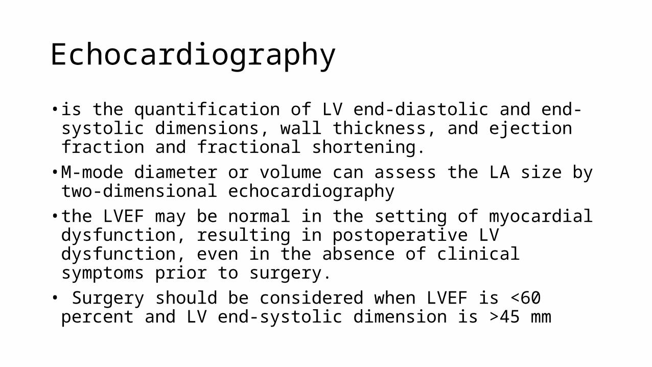
Echocardiography
• is the quantification of LV end-diastolic and end-systolic dimensions, wall thickness, and ejection fraction and fractional shortening.• M-mode diameter or volume can assess the LA size by two-
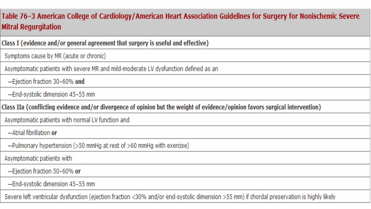
dimensional echocardiography• the LVEF may be normal in the setting of myocardial dysfunction,
resulting in postoperative LV dysfunction, even in the absence of clinical symptoms prior to surgery.• Surgery should be considered when LVEF is <60 percent and LV end-
systolic dimension is >45 mm
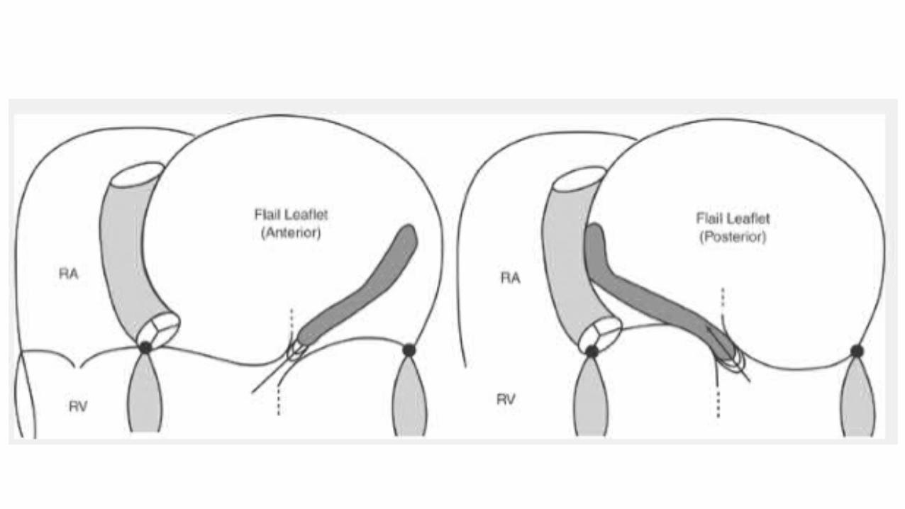

Flail MV

• Rheumatic MR is characterized by thickening of the leaflets and chordae. The posterior leaflet has reduced mobility, whereas the anterior leaflet may be doming if commissural fusion is associated. • degenerative MR, prolapse -the passage of valvular tissue beyond the
annulus plane in the long-axis view • The valve leaflets are diffusely thickened leaflets and excessive valvular
tissue and increased echogenicity of the annulus - mitral annular calcification. • Flail segments appear as complete eversion of the segment with or
without the small floating echo of ruptured chordae

• In ischemic or functional MR, - dilated annulus is nonspecific and annular descent is reduced.• The features of ischemic heart disease may be observed as regional
wall motion abnormalities. The leaflet tissue is normal. • mitral tenting caused by the abnormal traction by the principal
chordae on the anterior leaflet reduces the area of coaptation of the two leaflets, allowing for a central jet of MR. • With papillary muscle rupture, MR due to flail leaflet. • Transesophageal echocardiography provides superior imaging quality
to transthoracic echocardiography

Treatment (medical)
• Warfarin should be provided once AF intervenes with a target INR of 2–3. • Cardioversion - clinical context and left atrial size• Treatment of hert failure - diuretics, beta blockers, angiotensin-
converting enzyme (ACE) inhibitors, digitalis, and biventricular pacing (cardiac resynchronization therapy [CRT])• Asymptomatic patients with severe MR in sinus rhythm with normal LV
size and systolic function should avoid isometric forms of exercise.• IE prophylaxis• Rhuematic fever prophylaxis


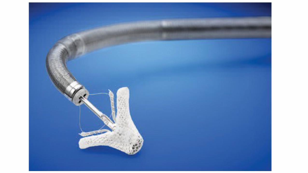
Percutaneous Mitral Valve Repair
• A transcatheter approach • deployment of a clip delivered via transeptal puncture that grasps the
leading edges of the mitral leaflets in their mid-portion (anterior scallop 2–posterior scallop 2 or A2-P2).
Post op outcome
• Mitral valve repair is better• Maintains continuity of valve with papillary muscles• Hence maintains LV geometry and maintains EF• Operative mortality 1-2 % (5-10 with MVR)• AF may persist - anticoagulation
References
• Textbook of Physiology – Guyton and Hall• Crofton and Douglas's Respiratory Diseases• Braunwald's Heart Diseases• Hurst Cardiology• Feigenbaum's Echocardiography

Thank you