
pufication.ppt
34
Protein sequencing The next step after the protein of interest has been purified and its molecular weight determined. To get information about Protein's function Evolutionary history First step is to determine the amino acid composition of the peptide
description
molecular biology
Transcript of pufication.ppt
Protein sequencing
The next step after the protein of interest has
been purified and its molecular weight
determined.
To get information about
Protein's function
Evolutionary history
First step is to determine the amino acid
composition of the peptide
Amino acid compositionHydrolyse peptide heating in 6 N HCl at
110°C for 24 hours..
Ion-exchange chromatography on
sulfonated polystyrene columns to separate
the hydrolysates
The amino acid is identified by its elution
volume
Amino acid composition Quantify amino acid by reaction with
Ninhydrin, detection limit 1g (10nmol); or
Fluorescamine, lower limit 1ng(10pmol).
Amino acids heated with ninhydrin yield
intense blue color
Except for proline, which gives a yellow
color
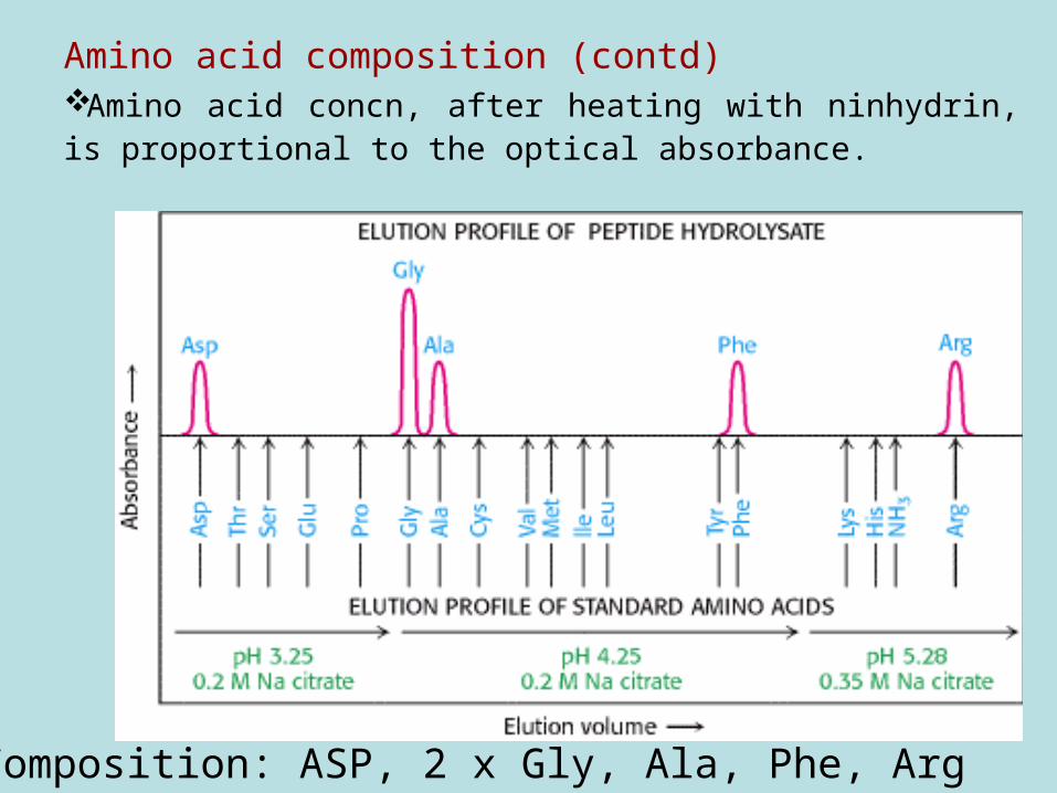
Amino acid composition (contd)Amino acid concn, after heating with ninhydrin, is proportional to the optical absorbance.
Composition: ASP, 2 x Gly, Ala, Phe, Arg
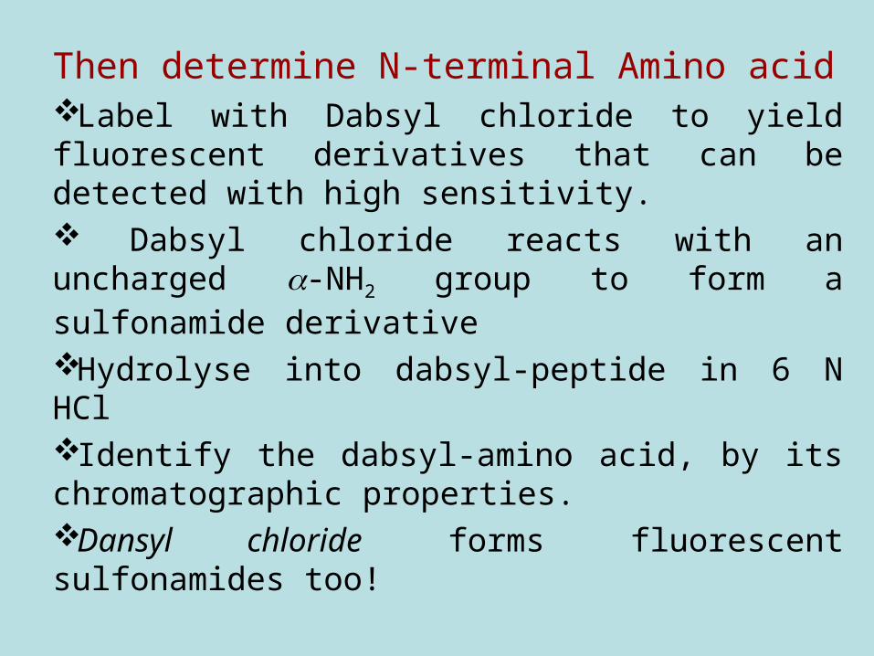
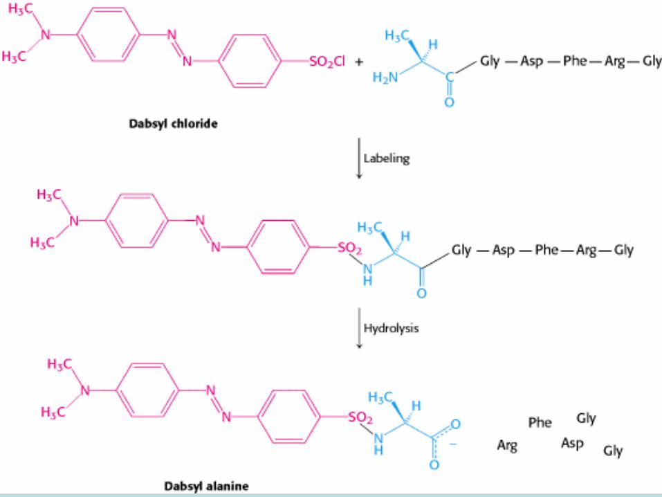
Then determine N-terminal Amino acid Label with Dabsyl chloride to yield fluorescent derivatives that can be detected with high sensitivity. Dabsyl chloride reacts with an uncharged -NH2 group to form a sulfonamide derivative
Hydrolyse into dabsyl-peptide in 6 N HClIdentify the dabsyl-amino acid, by its chromatographic properties. Dansyl chloride forms fluorescent sulfonamides too!
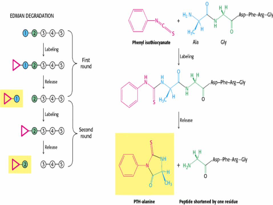
Sequencing: Edman degradation The dabsyl method leaves behind a totally
degraded peptide. In the Edman degradation,Phenyl isothiocyanate reacts with the
uncharged terminal amino group to form a phenylthiocarbamoyl derivative
Under mildly acidic conditions, a cyclic derivative of the terminal amino acid is liberated,
To leave an intact peptide shortened by one amino acid.
Sequencing: Edman degradation
The cyclic compound is a
phenylthiohydantoin (PTH)-amino acid,
Can be identified by chromatography
The Edman procedure is repeated on the
shortened peptide, yielding another PTH-
amino acid,
This is again identified by chromatography
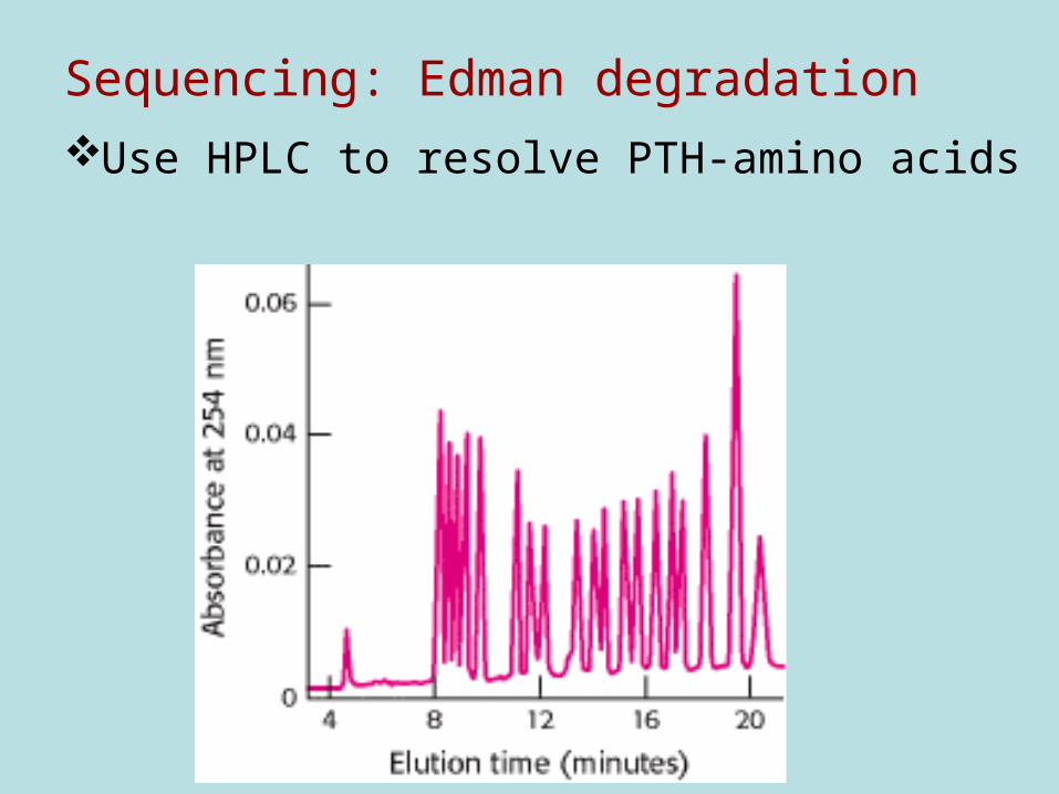
Sequencing: Edman degradation
Use HPLC to resolve PTH-amino acids
Edman degradation: LimitationsThe efficiency of PTH-amino acid release
declines with number of cycles
Upper limit for this method is 50 residues
Most proteins are N-terminally
blocked e.g with an acetyl group
So cannot be sequenced using the
Edman degradation methodology.
Edman degradation: SolutionsCleave peptide with proteases, such as trypsin, chymotrypsin, V8 protease
Edman degradation to sequence fragments,
Use Dabsyl labeling to determine the N-terminal segments of the protein
Use of BLAST or other database searching programs to identify the intact protein.
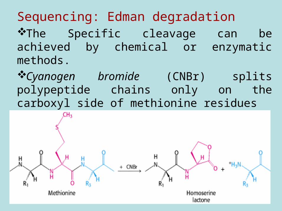
Sequencing: Edman degradationThe Specific cleavage can be achieved by chemical or enzymatic methods. Cyanogen bromide (CNBr) splits polypeptide chains only on the carboxyl side of methionine residues
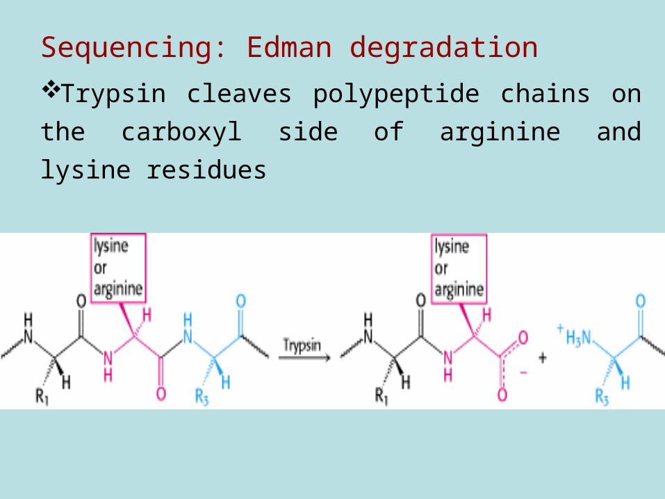
Sequencing: Edman degradation
Trypsin cleaves polypeptide chains on the
carboxyl side of arginine and lysine residues

Sequencing: Edman degradation
The peptides from specific cleavage are
separated by chromatography.
The sequence of each purified peptide
determined by the Edman method.
BUT
At this point, the order of the sequenced
segments is not defined.
Edman degradation: Order the peptides to
obtain the primary structure of the protein
The necessary additional information is
obtained from overlap peptides
Use another enzyme to split the polypeptide
chain at different linkages.
E.g. chymotrypsin cleaves preferentially on
the carboxyl side of aromatic and some other
bulky non-polar residues.
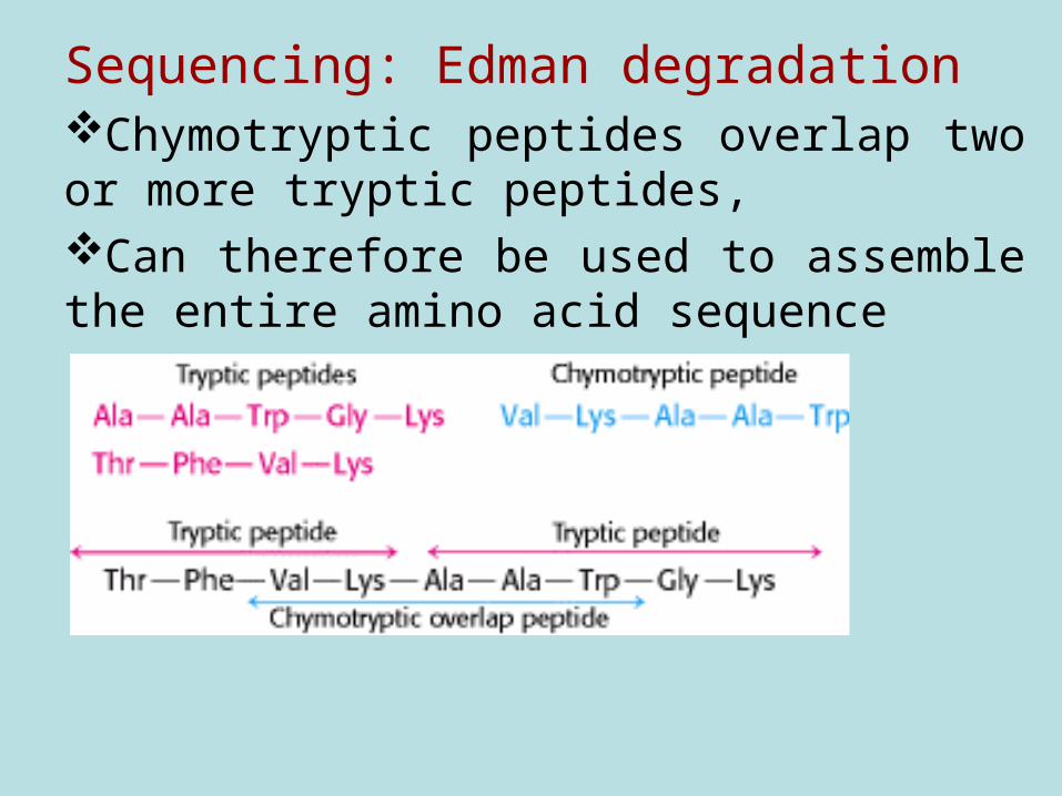
Sequencing: Edman degradationChymotryptic peptides overlap two or more tryptic peptides, Can therefore be used to assemble the entire amino acid sequence

Information obtained from Sequencing:
Infer the protein family/function by a BLAST
search
Establish evolutionary relationship:
Compare sequences of same protein from
different species
Presence of signal sequence as an
indicator of protein location e.g secretion
signal
Information obtained from Sequencing:
Sequence data provides a basis for
preparing antibodies specific for a protein of
interest.
In reverse genetics: DNA probes
corresponding to the amino acid sequence
can be constructed on the basis of the
genetic code.
Western BlottingBlotting involves transfer of separated proteins
onto nylon, nitrocellulose, or PVDF
membrane.
Why blot?
To make the protein more accessible to
probes,
Specific antibodies are preferred probes
Polyacrylamide is not particularly amenable
to the diffusion of large molecules.
Western BlottingThe attachment of specific antibodies to the
immobilised antigens can be readily
visualised by indirect enzyme
immunoassay techniques,
Usually a chromogenic substrate which
produces an insoluble product
Western BlottingOther types of probes include
Fluorescent labels (e.g. fluorescein)
Radioisotope labels (e.g. 125I)
Chemiluminescent substrates (highly
sensitive!)
Western Blotting: Antigen (blot) + antibodies
Then use secondary probes to detect the antibody binding
Conjugated anti-immunoglobulins (eg:
goat-anti-rabbit / human);
conjugated staphylococcal Protein A
( binds IgG of various species of animal); or
probes to biotinylated / igoxigeninylated
primary antibodies (eg: conjugated avidin /
streptavidin / antibody).
Western Blotting
During electroblotting,
Freshly-electrophoresed SDS-PAGE gels
are dipped into transfer buffer (0.025 M Tris-
HCl/20% (v/v) methanol, pH 8.3),
then laid flat on pre- wetted nitrocellulose
paper supported on three layers of wetted
filter paper resting on the ANODE
Western Blotting
Staining Proteins on Membranes:
(Nitrocellulose, Nylon, polyvinylidene
difluoride; PVDF)
Amido Black staining (not satisfactory);
India ink staining (OK),
Ponceau S (2 g Ponceau S + 30 g
trichloroacetic acid + 30 g sulfosalicyclic acid
+ water to 100 ml.) is highly useful.
Western Blotting: The Indirect enzyme
immunoassay,
Buffers and Solutions: Incubation/Blocking Buffer: 10 mM Tris-Cl/150
mM NaCl containing 1-5% non- fat milk powder and
0.05% Tween-20 or Triton X-100 (=Nonidet P-40),
pH 7.4.
Washing buffer: 10 mM Tris-Cl/150 mM NaCl pH
7.4 (OR SALINE) containing 0.05% Tween-20 or
Triton X-100.
Western Blotting: The Indirect enzyme immunoassay,
Buffers and Solutions contd: Substrate buffer: 100 mM Tris-Cl/100 mM NaCl/5 mM MgCl2 pH 9.5. Substrate stocks: Nitro Blue tetrazolium (NBT; Sigma), 75 mg/ml in 70% dimethyl formamide; 5-Bromo-4-chloro-3-indolyl phosphate (BCIP; (Sigma), 50 mg/ml in formamide (100%).
SUBSTRATE STOCKS IN 10 ML ALIQUOTS AT -20oC
Western Blotting: The Indirect enzyme immunoassay,Procedure:
Blocking: nitrocellulose blots are briefly rinsed in transfer buffer, then soaked for 1 hr at 37oC, or 2 hr at room temperature in blocking buffer. This procedure allows saturation of all non-specific protein binding sites on the blots.
Western Blotting: The Indirect enzyme immunoassay, Procedure contd
Attachment of specific antibodies to proteins: incubate blots in rabbit anti-sera diluted 1/10- 1/1000 in incubation buffer, in sealed boxes for 1 hr at room temperature on a shaking waterbath.
Washing: blots are washed by shaking in +100 ml/wash for 3x5 min., at room temperature.
Western Blotting: The Indirect enzyme
immunoassay, Procedure contdProbing of antibody binding:
is by means of suitably diluted alkaline
phosphatase - goat anti-rabbit globulins in
incubation buffer (eg 1/5000).
Incubation conditions and washing are the
same as for rabbit antiserum.
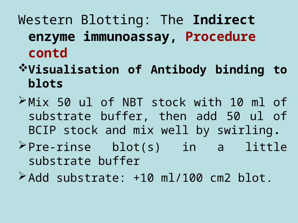
Western Blotting: The Indirect enzyme immunoassay, Procedure contd
Visualisation of Antibody binding to blots
Mix 50 ul of NBT stock with 10 ml of substrate buffer, then add 50 ul of BCIP stock and mix well by swirling.
Pre-rinse blot(s) in a little substrate bufferAdd substrate: +10 ml/100 cm2 blot.
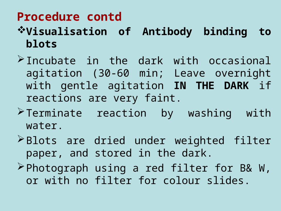
Procedure contdVisualisation of Antibody binding to blots
Incubate in the dark with occasional agitation (30-60 min; Leave overnight with gentle agitation IN THE DARK if reactions are very faint.
Terminate reaction by washing with water.Blots are dried under weighted filter paper,
and stored in the dark. Photograph using a red filter for B& W, or
with no filter for colour slides.
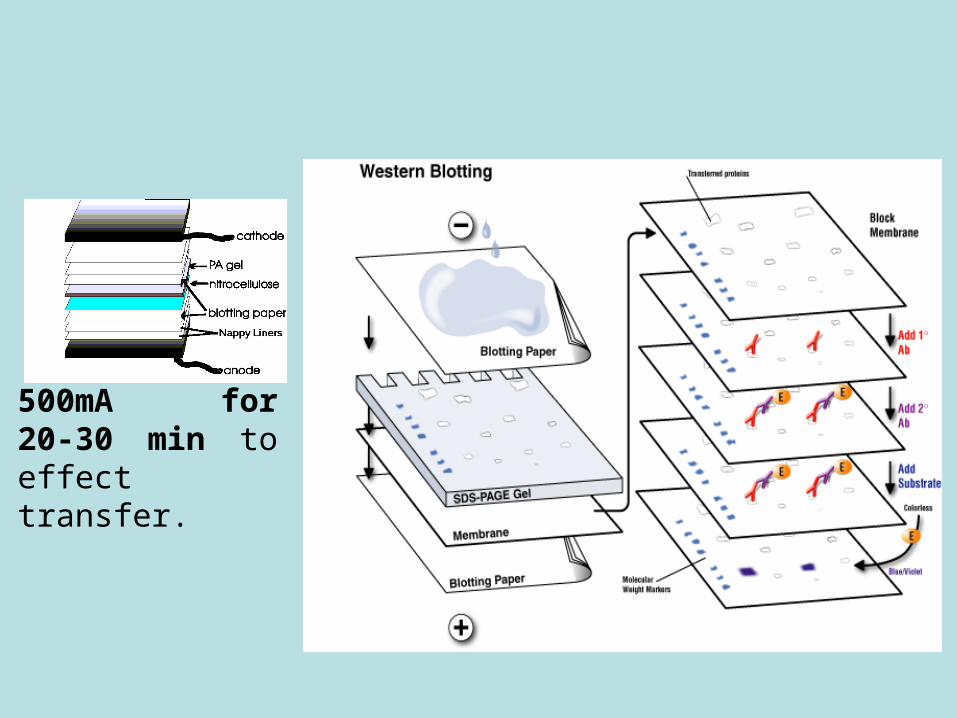
500mA for 20-30 min to effect transfer.
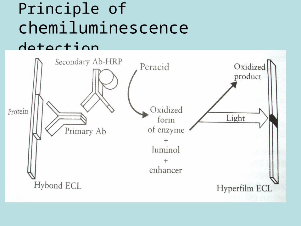
Principle of chemiluminescence detection


