
Published December 1, 1997 Accumulation of Muscle … · The characteristic shapes and positions of...
12
The Rockefeller University Press, 0021-9525/97/12/1231/12 $2.00 The Journal of Cell Biology, Volume 139, Number 5, December 1, 1997 1231–1242 http://www.jcb.org 1231 Accumulation of Muscle Ankyrin Repeat Protein Transcript Reveals Local Activation of Primary Myotube Endcompartments during Muscle Morphogenesis Anja Baumeister, Silvia Arber, and Pico Caroni Friedrich Miescher Institute, P.O. Box 2543, CH-4002 Basel, Switzerland Abstract. The characteristic shapes and positions of each individual body muscle are established during the process of muscle morphogenesis in response to pat- terning information from the surrounding mesen- chyme. Throughout muscle morphogenesis, primary myotubes are arranged in small parallel bundles, each myotube spanning the forming muscles from end to end. This unique arrangement potentially assigns a cru- cial role to primary myotube end regions for muscle morphogenesis. We have cloned muscle ankyrin repeat protein (MARP) as a gene induced in adult rat skeletal muscle by denervation. MARP is the rodent homologue of hu- man C-193 (Chu, W., D.K. Burns, R.A. Swerick, and D.H. Presky. 1995. J. Biol. Chem. 270:10236–10245) and is identical to rat cardiac ankyrin repeat protein. (Zou, Y., S. Evans, J. Chen, H.-C. Kuo, R.P. Harvey, and K.R. Chien. 1997. Development. 124:793–804). In denervated muscle fibers, MARP transcript accumu- lated in a unique perisynaptic pattern. MARP was also expressed in large blood vessels and in cardiac muscle, where it was further induced by cardiac hypertrophy. During embryonic development, MARP was expressed in forming skeletal muscle. In situ hybridization analy- sis in mouse embryos revealed that MARP transcript exclusively accumulates at the end regions of primary myotubes during muscle morphogenesis. This closely coincided with the expression of thrombospondin-4 in adjacent prospective tendon mesenchyme, suggesting that these two compartments may constitute a func- tional unit involved in muscle morphogenesis. Trans- fection experiments established that MARP protein ac- cumulates in the nucleus and that the levels of both MARP mRNA and protein are controlled by rapid degradation mechanisms characteristic of regulatory early response genes. The results establish the existence of novel regulatory muscle fiber subcompartments as- sociated with muscle morphogenesis and denervation and suggest that MARP may be a crucial nuclear cofac- tor in local signaling pathways from prospective tendon mesenchyme to forming muscle and from activated muscle interstitial cells to denervated muscle fibers. I n their physiological context in vivo, cells must inte- grate multiple signals from their environment to fulfill their specific roles within the organism. This crucial role of integration is particularly evident during develop- ment, when cells display coordinate migration, differentia- tion, and morphogenesis in response to a variety of spe- cific long-range and local signals. The formation of the skeletal musculature is an attractive model system in which to study how patterns of gene expression lead to the formation of a tissue. Due to major breakthroughs such as the establishment of differentiating myogenic cell lines, and the discovery of the converting activity of myogenic deter- mination factors, the early events in myogenesis, from pre- determined cells to terminally differentiated myotubes, are particularly well understood at the cellular level (Bucking- ham, 1992; Olson and Klein, 1994). In contrast, we know very little about the mechanisms that control muscle for- mation within the context of the developing organism. For example, the morphogenetic events that orchestrate the formation of the definitive muscle pattern are poorly un- derstood. These include specific successive splitting of pri- mordial muscle masses to generate all the different muscles, directed expansion of the newly formed muscles to gener- ate their unique shapes, sizes, and positions, and attachment of the muscles to their specific tendon organs (Lance-Jones, 1979; Schroeter and Tosney, 1991a, b; Hauschka, 1994). In the developing mouse limb, these events begin around em- bryonic day (E) 1 12.5, and the definitive muscle pattern is established by E16 (Lance-Jones, 1979). Throughout mus- Address all correspondence to Pico Caroni, Friederich Miescher Institute, P.O. Box 2543, CH-4002 Basel, Switzerland. Tel.: (41) 61-6973727. Fax: (41) 61-6973976. E-mail: [email protected] Silvia Arber’s present address is Howard Hughes Medical Institute, Department of Biochemistry and Molecular Biophysics, Columbia Uni- versity, New York, NY. 1. Abbreviations used in this paper: CARP, cardiac ankyrin repeat protein; E, embryonic day; MARP, muscle ankyrin repeat domain; MLP, muscle LIM protein; TSP-4, thrombospondin-4. on January 4, 2016 jcb.rupress.org Downloaded from Published December 1, 1997
-
Upload
nguyendiep -
Category
Documents
-
view
215 -
download
0
Transcript of Published December 1, 1997 Accumulation of Muscle … · The characteristic shapes and positions of...

The Rockefeller University Press, 0021-9525/97/12/1231/12 $2.00The Journal of Cell Biology, Volume 139, Number 5, December 1, 1997 1231–1242http://www.jcb.org 1231
Accumulation of Muscle Ankyrin Repeat Protein TranscriptReveals Local Activation of Primary MyotubeEndcompartments during Muscle Morphogenesis
Anja Baumeister, Silvia Arber, and Pico Caroni
Friedrich Miescher Institute, P.O. Box 2543, CH-4002 Basel, Switzerland
Abstract.
The characteristic shapes and positions of each individual body muscle are established during the process of muscle morphogenesis in response to pat-terning information from the surrounding mesen-chyme. Throughout muscle morphogenesis, primary myotubes are arranged in small parallel bundles, each myotube spanning the forming muscles from end to end. This unique arrangement potentially assigns a cru-cial role to primary myotube end regions for muscle morphogenesis.
We have cloned muscle ankyrin repeat protein (MARP) as a gene induced in adult rat skeletal muscle by denervation. MARP is the rodent homologue of hu-man C-193 (Chu, W., D.K. Burns, R.A. Swerick, and
D.H. Presky. 1995.
J. Biol. Chem.
270:10236–10245) and is identical to rat cardiac ankyrin repeat protein. (Zou, Y., S. Evans, J. Chen, H.-C. Kuo, R.P. Harvey, and K.R. Chien. 1997.
Development.
124:793–804). In denervated muscle fibers, MARP transcript accumu-lated in a unique perisynaptic pattern. MARP was also expressed in large blood vessels and in cardiac muscle,
where it was further induced by cardiac hypertrophy. During embryonic development, MARP was expressed in forming skeletal muscle. In situ hybridization analy-sis in mouse embryos revealed that MARP transcript exclusively accumulates at the end regions of primary myotubes during muscle morphogenesis. This closely coincided with the expression of thrombospondin-4 in adjacent prospective tendon mesenchyme, suggesting that these two compartments may constitute a func-tional unit involved in muscle morphogenesis. Trans-fection experiments established that MARP protein ac-cumulates in the nucleus and that the levels of both MARP mRNA and protein are controlled by rapid degradation mechanisms characteristic of regulatory early response genes. The results establish the existence of novel regulatory muscle fiber subcompartments as-sociated with muscle morphogenesis and denervation and suggest that MARP may be a crucial nuclear cofac-tor in local signaling pathways from prospective tendon mesenchyme to forming muscle and from activated muscle interstitial cells to denervated muscle fibers.
I
n
their physiological context in vivo, cells must inte-grate multiple signals from their environment to fulfilltheir specific roles within the organism. This crucial
role of integration is particularly evident during develop-ment, when cells display coordinate migration, differentia-tion, and morphogenesis in response to a variety of spe-cific long-range and local signals. The formation of theskeletal musculature is an attractive model system inwhich to study how patterns of gene expression lead to theformation of a tissue. Due to major breakthroughs such asthe establishment of differentiating myogenic cell lines, andthe discovery of the converting activity of myogenic deter-mination factors, the early events in myogenesis, from pre-
determined cells to terminally differentiated myotubes, areparticularly well understood at the cellular level (Bucking-ham, 1992; Olson and Klein, 1994). In contrast, we knowvery little about the mechanisms that control muscle for-mation within the context of the developing organism. Forexample, the morphogenetic events that orchestrate theformation of the definitive muscle pattern are poorly un-derstood. These include specific successive splitting of pri-mordial muscle masses to generate all the different muscles,directed expansion of the newly formed muscles to gener-ate their unique shapes, sizes, and positions, and attachmentof the muscles to their specific tendon organs (Lance-Jones,1979; Schroeter and Tosney, 1991
a
,
b
; Hauschka, 1994). Inthe developing mouse limb, these events begin around em-bryonic day (E)
1
12.5, and the definitive muscle pattern isestablished by E16 (Lance-Jones, 1979). Throughout mus-
Address all correspondence to Pico Caroni, Friederich Miescher Institute,P.O. Box 2543, CH-4002 Basel, Switzerland. Tel.: (41) 61-6973727. Fax:(41) 61-6973976. E-mail: [email protected]
Silvia Arber’s present address is Howard Hughes Medical Institute,Department of Biochemistry and Molecular Biophysics, Columbia Uni-versity, New York, NY.
1.
Abbreviations used in this paper
: CARP, cardiac ankyrin repeat protein;E, embryonic day; MARP, muscle ankyrin repeat domain; MLP, muscleLIM protein; TSP-4, thrombospondin-4.
on January 4, 2016jcb.rupress.org
Dow
nloaded from
Published December 1, 1997

The Journal of Cell Biology, Volume 139, 1997 1232
cle morphogenesis, the muscle pattern is reflected in theparallel arrangement of primary myotubes that span theforming muscles from end to end (Fig. 1) (Hilfer et al.,1973; Hauschka, 1974; Ontell and Kozeka, 1984; Duxsonand Usson, 1989; Sweeney et al., 1989). After completion ofmuscle morphogenesis, primary myotubes then serve asscaffolds for secondary muscle fibers, which are generatedthrough the proliferation and differentiation of intramus-cular precursor cells (Ross et al., 1987; Condon et al., 1990;Ashby et al., 1993).
Classical embryological experiments have established thatmyogenic precursor cells are not predetermined with re-spect to the particular muscles that they will contribute to(Chevallier et al., 1977). Potential instructive roles by form-ing nerve or vasculature patterns have been essentiallyruled out, and it is now clear that most morphogenetic in-formation is derived from the surrounding mesenchyme(Chevallier et al., 1977; Lanser and Fallon, 1987; Grim,1991; Ashby et al., 1993). How does patterned mesen-chyme induce and control muscle morphogenesis? Thefact that the spatial and temporal patterns of muscle split-ting and shaping are unique for each muscle and do not re-flect general gradients of limb differentiation (Grim, 1991;Schroeter and Tosney 1991
a
,
b
) argues for specific localsignaling processes. In principle, the end-to-end and paral-lel arrangement of primary myotubes within the formingmuscles assigns a unique potential role to myotube end re-gions for muscle morphogenesis (Fig. 1). Accordingly, one
possibility is that mesenchyme-to-muscle signaling may in-volve local regulation of myotube formation, orientation,and growth at the interface between myotube end regionsand adjacent mesenchyme. Such local signaling should bereflected in the expression of corresponding molecularmarkers in the mesenchyme and in the responding musclecells as well as in the existence of a spatially restricted sub-compartment at primary myotube end regions.
In a search for genes involved in the regulation of mus-cle gene expression and the formation of neuromuscularsynapses, we constructed and screened a cDNA library en-riched in messages induced in adult skeletal muscle bydenervation. As reported elsewhere, two clones coded fora striated muscle-specific positive regulator of myogenesis:muscle LIM protein (MLP; Arber et al., 1994) and throm-bospondin-4 (TSP-4; Arber and Caroni, 1995), whose ex-pression is induced in denervated muscle interstitial cells. Wereport here that a third clone codes for a nuclear proteinwith four ankyrin repeats that is expressed in striated mus-cle and vascular endothelial cells. Due to its predominantexpression in striated muscle we have called it MARP (mus-cle ankyrin repeat protein). During the course of this work,isolation of the homologous human protein (C-139; Chuet al., 1995) and of the identical rat cDNA (cardiac ankyrinrepeat protein [CARP]; Zou et al., 1997) were reported.MARP mRNA has degradation-promoting motifs charac-teristic of regulatory factors. Consistent with a possiblerole in signaling, MARP protein is rapidly degraded in thecell, and degradation can be substantially delayed by theaddition of short carboxyl-terminal extentions. The distri-bution of MARP mRNA in developing mouse skeletal mus-cle revealed a unique and highly localized pattern associ-ated with muscle morphogenesis. This was accompaniedby a complementary pattern of TSP-4 expression in adja-cent mesenchyme. The results establish the existence ofnovel muscle subcompartments associated with musclemorphogenesis and the perisynaptic region of denervatedmuscle and suggest that MARP may be a key nuclear co-factor in novel local signaling pathways to muscle.
Materials and Methods
Nucleic Acid Reagents
A partial clone coding for rat MARP was isolated from a subtracted plas-mid cDNA library enriched in transcripts induced in adult diaphragmmuscle 7 d after denervation. The construction of the library and thescreening procedure were as described (Arber et al., 1994). Subsequently,several additional clones, including a 1.7-kb putative full length clonewere isolated through rescreening of the same library. Mouse MARPcDNA clones were isolated from an adult mouse heart cDNA library, dueto their hybridization to a rat MARP cDNA probe. A genomic clone con-taining MARP proximal promoter sequences was isolated with mouseMARP cDNA probes from a mouse (strain 129Sv) genomic library (Strat-agene GmbH, Heidelberg, Germany). The promoter fragment shown inFig. 2
C
drives the expression of a reporter gene in transfected myogeniccell lines, indicating that it is a functional promoter (data not shown). cDNAsfor Northern blot analysis and in situ hybridization were from the follow-ing sources: rat MLP (Arber et al., 1994), rat TSP-4 (Arber and Caroni,1995), rat clone-16 (a cDNA isolated in our screen for denervation-induced genes; see Aigner et al., 1995), rat MyoD (kind gift from A. Buo-nanno, National Institutes of Health, Bethesda, MD), mouse tenascin-C(kind gift from R. Chiquet-Ehrismann, Friedrich Miescher Institute, Basel,Switzerland), mouse FGF6 (probe corresponding to 434 bp of 3
9
untrans-lated sequences, as in deLapeyriére et al., 1993; kind gift of D. Birnbaum,U119 Institut National de la Sante et de la Recherche Medicale, Marseille,
Figure 1. Schematic representation of the spatial arrangement ofmyogenic cells during muscle formation. Muscle morphogenesisproduces the definitive shape and arrangement of each individualmuscle and precedes the formation of secondary myotubes. Fromtheir first appearance, primary myotubes (shaded rectangles)span forming muscles in a parallel end-to-end arrangement. Dur-ing muscle morphogenesis, a substantial part of the muscle masscontains undifferentiated myoblasts (circles), and primary myo-tubes are arranged in parallel bundles of three to seven. Whenmorphogenesis is completed, primary myotube bundles dissoci-ate, and single primary myotubes serve as scaffolds along whichsecondary myotubes (white rectangles) fuse and elongate. Notethat due to the particular arrangement of the primary myotubes,muscle splitting and shaping may in principle be regulated bycontrolling the position and local elongation of primary myotubeend regions. The latter process may involve selective fusion ofmyoblasts to primary myotube end regions.
on January 4, 2016jcb.rupress.org
Dow
nloaded from
Published December 1, 1997
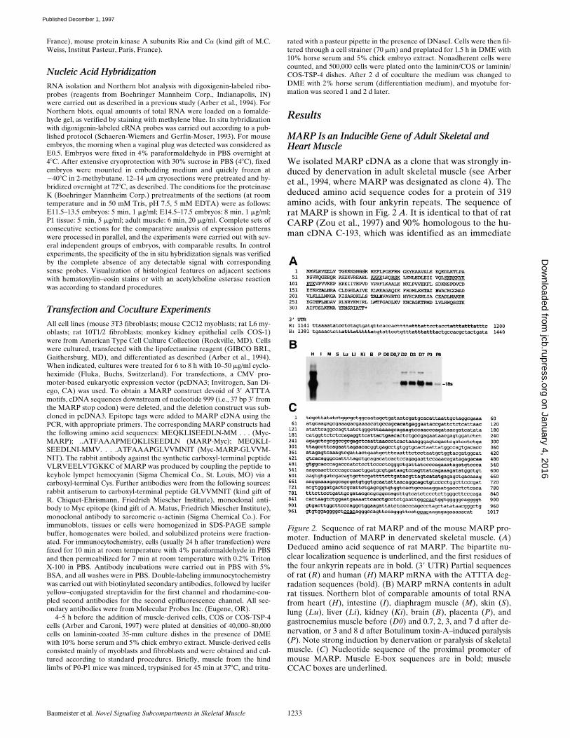
Baumeister et al.
Novel Signaling Subcompartments in Skeletal Muscle
1233
France), mouse protein kinase A subunits Ri
a
and C
a
(kind gift of M.C.Weiss, Institut Pasteur, Paris, France).
Nucleic Acid Hybridization
RNA isolation and Northern blot analysis with digoxigenin-labeled ribo-probes (reagents from Boehringer Mannheim Corp., Indianapolis, IN)were carried out as described in a previous study (Arber et al., 1994). ForNorthern blots, equal amounts of total RNA were loaded on a fomalde-hyde gel, as verified by staining with methylene blue. In situ hybridizationwith digoxigenin-labeled cRNA probes was carried out according to a pub-lished protocol (Schaeren-Wiemers and Gerfin-Moser, 1993). For mouseembryos, the morning when a vaginal plug was detected was considered asE0.5. Embryos were fixed in 4% paraformaldehyde in PBS overnight at4
8
C. After extensive cryoprotection with 30% sucrose in PBS (4
8
C), fixedembryos were mounted in embedding medium and quickly frozen at
2
40
8
C in 2-methybutane. 12–14
m
m cryosections were pretreated and hy-bridized overnight at 72
8
C, as described. The conditions for the proteinaseK (Boehringer Mannheim Corp.) pretreatments of the sections (at roomtemperature and in 50 mM Tris, pH 7.5, 5 mM EDTA) were as follows:E11.5–13.5 embryos: 5 min, 1
m
g/ml; E14.5–17.5 embryos: 8 min, 1
m
g/ml;P1 tissue: 5 min, 5
m
g/ml; adult muscle: 6 min, 20
m
g/ml. Complete sets ofconsecutive sections for the comparative analysis of expression patternswere processed in parallel, and the experiments were carried out with sev-eral independent groups of embryos, with comparable results. In controlexperiments, the specificity of the in situ hybridization signals was verifiedby the complete absence of any detectable signal with correspondingsense probes. Visualization of histological features on adjacent sectionswith hematoxylin–eosin stains or with an acetylcholine esterase reactionwas according to standard procedures.
Transfection and Coculture Experiments
All cell lines (mouse 3T3 fibroblasts; mouse C2C12 myoblasts; rat L6 my-oblasts; rat 10T1/2 fibroblasts; monkey kidney epithelial cells COS-1)were from American Type Cell Culture Collection (Rockville, MD). Cellswere cultured, transfected with the lipofectamine reagent (GIBCO BRL,Gaithersburg, MD), and differentiated as described (Arber et al., 1994).When indicated, cultures were treated for 6 to 8 h with 10–50
m
g/ml cyclo-heximide (Fluka, Buchs, Switzerland). For transfections, a CMV pro-moter-based eukaryotic expression vector (pcDNA3; Invitrogen, San Di-ego, CA) was used. To obtain a MARP construct devoid of 3
9
ATTTAmotifs, cDNA sequences downstream of nucleotide 999 (i.e., 37 bp 3
9
fromthe MARP stop codon) were deleted, and the deletion construct was sub-cloned in pcDNA3. Epitope tags were added to MARP cDNA using thePCR, with appropriate primers. The corresponding MARP constructs hadthe following amino acid sequences: MEQKLISEEDLN-MM . . . (Myc-MARP); ..ATFAAAPMEQKLISEEDLN (MARP-Myc); MEQKLI-SEEDLNI-MMV. . . .ATFAAAPGLVVMNIT (Myc-MARP-GLVVM-NIT). The rabbit antibody against the synthetic carboxyl-terminal peptideVLRVEELVTGKKC of MARP was produced by coupling the peptide tokeyhole lympet hemocyanin (Sigma Chemical Co., St. Louis, MO) via acarboxyl-terminal Cys. Further antibodies were from the following sources:rabbit antiserum to carboxyl-terminal peptide GLVVMNIT (kind gift ofR. Chiquet-Ehrismann, Friedrich Miescher Institute), monoclonal anti-body to Myc epitope (kind gift of A. Matus, Friedrich Miescher Institute),monoclonal antibody to sarcomeric
a
-actinin (Sigma Chemical Co.). Forimmunoblots, tissues or cells were homogenized in SDS-PAGE samplebuffer, homogenates were boiled, and solubilized proteins were fraction-ated. For immunocytochemistry, cells (usually 24 h after transfection) werefixed for 10 min at room temperature with 4% paraformaldehyde in PBSand then permeabilized for 7 min at room temperature with 0.2% TritonX-100 in PBS. Antibody incubations were carried out in PBS with 5%BSA, and all washes were in PBS. Double-labeling immunocytochemistrywas carried out with biotinylated secondary antibodies, followed by luciferyellow–conjugated streptavidin for the first channel and rhodamine-cou-pled second antibodies for the second epifluorescence channel. All sec-ondary antibodies were from Molecular Probes Inc. (Eugene, OR).
4–5 h before the addition of muscle-derived cells, COS or COS-TSP-4cells (Arber and Caroni, 1997) were plated at densities of 40,000–80,000cells on laminin-coated 35-mm culture dishes in the presence of DMEwith 10% horse serum and 5% chick embryo extract. Muscle-derived cellsconsisted mainly of myoblasts and fibroblasts and were obtained and cul-tured according to standard procedures. Briefly, muscle from the hindlimbs of P0-P1 mice was minced, trypsinised for 45 min at 37
8
C, and tritu-
rated with a pasteur pipette in the presence of DNaseI. Cells were then fil-tered through a cell strainer (70
m
m) and preplated for 1.5 h in DME with10% horse serum and 5% chick embryo extract. Nonadherent cells werecounted, and 500,000 cells were plated onto the laminin/COS or laminin/COS-TSP-4 dishes. After 2 d of coculture the medium was changed toDME with 2% horse serum (differentiation medium), and myotube for-mation was scored 1 and 2 d later.
Results
MARP Is an Inducible Gene of Adult Skeletal and Heart Muscle
We isolated MARP cDNA as a clone that was strongly in-duced by denervation in adult skeletal muscle (see Arberet al., 1994, where MARP was designated as clone 4). Thededuced amino acid sequence codes for a protein of 319amino acids, with four ankyrin repeats. The sequence ofrat MARP is shown in Fig. 2
A.
It is identical to that of ratCARP (Zou et al., 1997) and 90% homologous to the hu-man cDNA C-193, which was identified as an immediate
Figure 2. Sequence of rat MARP and of the mouse MARP pro-moter. Induction of MARP in denervated skeletal muscle. (A)Deduced amino acid sequence of rat MARP. The bipartite nu-clear localization sequence is underlined, and the first residues ofthe four ankyrin repeats are in bold. (39 UTR) Partial sequencesof rat (R) and human (H) MARP mRNA with the ATTTA deg-radation sequences (bold). (B) MARP mRNA contents in adultrat tissues. Northern blot of comparable amounts of total RNAfrom heart (H), intestine (I), diaphragm muscle (M), skin (S),lung (Lu), liver (Li), kidney (Ki), brain (B), placenta (P), andgastrocnemius muscle before (D0) and 0.7, 2, 3, and 7 d after de-nervation, or 3 and 8 d after Botulinum toxin-A–induced paralysis(P). Note strong induction by denervation or paralysis of skeletalmuscle. (C) Nucleotide sequence of the proximal promoter ofmouse MARP. Muscle E-box sequences are in bold; muscleCCAC boxes are underlined.
on January 4, 2016jcb.rupress.org
Dow
nloaded from
Published December 1, 1997

The Journal of Cell Biology, Volume 139, 1997 1234
early gene of vascular endothelial cells (Chu et al., 1995).Shared elements include a bipartite nuclear localizationmotif (residues 71–80 and 94–103), a PEST-like sequencecharacteristic of rapidly degraded proteins (residues 108–126; Rechsteiner and Rogers, 1996), and the four ankyrinrepeats (Blank et al., 1992; residues 152–184, 185–217, 218–250, and 251–283) that are flanked by a carboxyl-terminalextension of 36 residues. In addition, the MARP (rat, mouse)and C-193 (human) transcripts contain multiple ATTTAdegradation motifs (Shaw and Kamen, 1986) in their 3
9
un-translated sequences (Fig. 2
A
). Based on the high degreeof sequence homology and the conserved regulatory prop-erties of the transcripts and proteins (see below), we tenta-tively conclude that MARP is the rodent homologue ofC-193. As shown in Fig. 2
B
, adult rat heart contained highlevels of MARP mRNA, whereas very low levels were de-tected in lung and placenta, and either no or extremely lowsignals were detected in intestine, skeletal muscle, skin,liver, kidney, and brain. Denervation led to massive induc-tion of MARP mRNA in skeletal muscle (Fig. 2
B
). Com-parison with other denervation-sensitive transcripts showedthat MARP induction was particularly rapid (significantlyfaster than MLP and clone-16 and at least as fast as myo-genin; not shown). Finally, Fig. 2
C
shows the sequence ofthe proximal promoter of mouse MARP. Consistent withthe expression of MARP in skeletal and heart muscle, thissequence includes five muscle E-boxes and three CCAC-boxes that regulate transcription in skeletal and cardiacmuscle cells (Fig. 2
C
; Buckingham, 1992; Grayson et al.,1995).
Fig. 3
A
shows that no MARP transcript was detectablein adult medial gastrocnemius muscle, where strong ex-pression in the vicinity of acetylcholine esterase–positiveneuromuscular synapses was induced by denervation. Atcloser inspection, the MARP signal frequently highlightedstriations and accumulated in a longitudinal pattern alongthe inner face of muscle fibers, indicating that it was local-ized inside skeletal muscle fibers (Fig. 3
B
). This dramaticaccumulation pattern in denervated muscle had featuresunique to MARP. Thus: (
a
) MyoD and MLP transcriptsaccumulated throughout denervated muscle fibers, andthe signal for clone-16 mRNA only showed partial synap-tic enrichment (data not shown); (
b
) although the
a
sub-unit of the acetylcholine receptor displays some synapticenrichment, it is largely due to its synaptic expression ininnervated muscle, and synaptic accumulation is in factreduced in denervated muscle (Fontaine and Changeux,1989; Witzemann et al., 1991); (
c
) while typical synapse-associated transcripts such as the
a
subunit of the acetyl-choline receptor or the regulatory subunit RI
a
of proteinkinase A display local accumulation restricted to the sub-synaptic region (i.e., coincident with acetylcholine esterasereaction product; data not shown; see Witzemann et al.,1991; Imaizumi-Scherrer et al., 1996), MARP transcriptaccumulated in a much broader perisynaptic pattern, sug-gesting local regulation by signals in the vicinity of dener-vated synapses. Unlike these muscle fiber transcripts, andas reported previously, TSP-4 mRNA accumulated in mus-cle interstitial cells (Fig. 3
B
; see also Arber and Caroni,1995). In innervated muscle it was restricted to epimysium,whereas denervation induced expression in the endomy-sium (Arber and Caroni, 1995). Interestingly, induction
was distinctly more pronounced in perisynaptic regions,providing a first example of spatial correlation between theaccumulation of MARP mRNA in muscle fibers and thatof TSP-4 in adjacent cells (see below). The dramatic peri-synaptic accumulation of MARP transcript in denervatedmuscle is highly unusual. It suggests that this transcript issubject to local regulatory mechanisms and that even severaldays after removal of the motor nerve the perisynaptic regionof muscle is under the influence of distinct local signals.
Figure 3. Local patterns of MARP transcript induction in adultdenervated muscle and hypertrophic heart. (A) Perisynaptic ac-cumulation of MARP transcript in denervated muscle. Gastroc-nemius muscle of adult mice was collected 7 d after resection ofthe sciatic nerve. Note prominent MARP in situ hybridizationsignal at a broad region near to acetylcholine esterase reactionproduct (adjacent section on the right). As expected from the ab-sence of the corresponding mRNA on Northern blots, no MARPsignal was detected in control muscle (right). (B) High-magnifica-tion photographs of MARP (left) and TSP-4 (right) transcript lo-calization in the perisynaptic region of 7-d denervated gastrocne-mius muscle. Note MARP signal along the inner face of striatedmuscle fibers. Distribution of the TSP-4 transcript, which accu-mulates in a muscle interstitial cell pattern clearly distinct fromthat of MARP is shown for comparison. (C) Induction of MARPtranscript in the hypertrophic heart. (Left) In situ hybridizationfor MARP mRNA in adult mouse heart. In control heart MARPmRNA is expressed at high levels in atria and at low levels inventricles. In the hearts of MLP-deficient mice (2/2) with di-lated cardiomyopathy and hypertrophy (Arber et al., 1997),MARP transcript is strongly upregulated in ventricles. (Right)Selective upregulation of MARP mRNA in hypertrophic ventri-cles. The Northern blot was hybridized with a MARP probe andcomparable amounts of total RNA from adult ventricle (H) orgastrocnemius muscle (Mu) from control or MLP-deficient (2/2)mice were applied to the gel. Bar: (A) 70 mm, (B, MARP) 8 mm,(B, TSP-4) 16 mm.
on January 4, 2016jcb.rupress.org
Dow
nloaded from
Published December 1, 1997

Baumeister et al.
Novel Signaling Subcompartments in Skeletal Muscle
1235
Fig. 3
C
shows the localization of MARP transcript in acontrol and an MLP-deficient mouse heart with dilatedcardiomyopathy and hypertrophy (Arber et al., 1997). Incontrol hearts, MARP mRNA levels were very high inatria, whereas ventricles displayed a characteristic inho-mogeneous distribution. Interestingly, and similar to atrialnatriuretic factor (Chien et al., 1992), cardiac hypertrophyled to massive induction of MARP message in ventricularcardiomyocytes, whereas atrial levels were much less af-fected. Fig. 3
C
also shows that although by Northern blotMARP was expressed at substantial levels in the heart ofnormal adult mice (Fig. 2
B
), hypertrophic hearts con-tained markedly elevated levels of MARP transcript. Un-like MARP, the denervation-sensitive transcripts MLPand clone-16 were not elevated in hypertrophic heart(data not shown). MARP is therefore an inducible gene inboth adult skeletal and heart muscle.
MARP Is a Nuclear and Cytosolic Protein Subject to Tight Posttranscriptional Regulation
To investigate the cellular regulation of MARP mRNA weanalyzed its levels in non- and myogenic cell lines. As shownin Fig. 4
A
, the transcript could be detected in the myo-genic cell line C2C12, in 10T1/2 fibroblasts, but not in 3T3fibroblasts. Significantly, when C2C12 cells were trans-ferred to differentiation medium, MARP transcript levelswere selectively upregulated in myotubes (Fig. 4
B
). Myo-genic cell lines exhibited varying degrees of coupling be-tween MARP transcript levels and differentiation. Thus,while one batch of C2C12 cells displayed detectable ex-pression only upon differentiation (see Arber et al., 1994),a second C2C12 batch (Fig. 4
A
) and L6 cells (data notshown) already expressed significant levels under nondif-ferentiating conditions. In 10T1/2 cells, but much less soin C2C12 cells MARP mRNA levels were elevated in thepresence of the protein synthesis inhibitor cycloheximide(Fig. 4
A
). This is reminiscent of human vascular endothe-lial cells, where C-193 transcript has early response prop-erties in small vessel cells and is expressed at much higherlevels in large vessel cells (Chu et al., 1995).
The rat MARP transcript has five closely spaced ATTTAmotifs in its 3
9
untranslated sequence (Fig. 2
A
). Deletionof 3
9
untranslated sequences, including the ATTTA mo-tifs, resulted in dramatically higher transcript levels in trans-fected 3T3 cells, confirming that MARP mRNA carries func-tional degradation signals (Fig. 4
A
). Similar results wereobtained with transfected C2C12 and 10T1/2 cells (data notshown). To analyze the regulation and subcellular localiza-tion of MARP we generated an antiserum against the amino-terminal end of the protein. Surprisingly, in spite of thesubstantial corresponding Northern blot signals, MARP
Figure 4.
MARP has properties of a nuclear early response gene.(
A
) Regulation of MARP mRNA contents by myogenic differen-tiation, cycloheximide, and 3
9
untranslated sequences (
UTR
). TheRNA blots (equivalent amounts of total RNA for each blot) werehybridized with a rat MARP probe. The experiment on the leftwas carried out with a C2C12 subclone exhibiting MARP induc-tion during differentiation (days). The C2C12 subclone used forthe cycloheximide (
CHX
) induction experiment already expressedhigh levels of MARP in high-serum medium, whereas CHX in-
duced MARP transcript in 10T1/2 fibroblasts. (
2
and 1) RNAcollected after 6 h without or with CHX. For the experiments onthe right, Swiss 3T3 fibroblasts were transfected with MARP ex-pression constructs containing (1) or devoid (2) of 39 untrans-lated region sequences, and RNA was collected 2 d after transfec-tion. The presence of MARP 39 untranslated region sequencesled to greatly reduced transcript levels. (0) Nontransfected cells.(B) In differentiating C2C12 cells MARP transcript selectivelyaccumulates in multinucleated myotubes (arrows). In situ hybrid-ization of cultures 3 d after switch to differentiation medium.(Right) MARP sense probe as a negative control. The C2C12subclone was as in (A) (Left) Note homogeneous pattern ofMARP transcript accumulation in myotubes in vitro. (C) MARPprotein is rapidly degraded in transfected cells, and degradationcan be delayed by the fusion of short peptide sequences to thecarboxyl-terminal end of the protein. COS cells were transfectedwith MARP expression constructs, and MARP contents were de-termined on the immunoblot (anti-MARP antiserum). Equalamounts of cell homogenate protein were applied for each sam-ple (see background band at z31 kD). (N 1 C) myc-tag at theamino terminus and GLVVMNIT-tag at the carboxy terminus;(N) myc-tag at the amino terminus; (C) myc-tag at the carboxylterminus; (2) no epitope tags. For each MARP construct, cellswere incubated with (1) or without (2) CHX during the last 8 hbefore homogenization. (D) Nuclear and cytosolic accumulationof MARP in transfected COS cells. The epitope-tagged con-structs are defined as described in (C). (Top) Anti-myc labelingof amino- (left) and carboxyl-terminal–tagged (right) MARP.Note higher frequency of labeled cells and of cytosolic signal inthe presence of carboxyl-terminal tag sequence. Double-tagged(N 1 C) MARP was visualized by double labeling for the aminoterminus (n) and carboxyl terminus (c) epitope tag. Note nuclearaccumulation of undegraded MARP. Bar: (B) 15 mm; (D, singlelabelings) 9 mm; (D, double labeling) 6 mm.
on January 4, 2016jcb.rupress.org
Dow
nloaded from
Published December 1, 1997

The Journal of Cell Biology, Volume 139, 1997 1236
protein signals on immunoblots from heart homogenateswere very low, and no signals could be detected on immu-noblots of differentiated L6 and C2C12 cells (data notshown). A weak signal could be detected on immunoblotsof transfected COS (Fig. 4 C) and C2C12 cells. To excludeeffects due to the instability of MARP transcripts, alltransfection experiments were carried out with 39 untrans-lated region–deleted constructs (Fig. 4 A), and the pres-ence of comparable transcript levels was verified on corre-sponding Northern blots. To determine whether detectiondifficulties were due to poor antisera we analyzed immu-noblots of cells transfected with epitope-tagged MARPconstructs. These experiments led to the surprising findingthat the addition of short amino acid extensions to the car-boxyl-terminal end of MARP greatly protected this pro-tein against degradation. The analysis revealed that additionof any of two different epitope tags to the carboxyl-termi-nal end of MARP resulted in greatly elevated levels of thecorresponding protein. In contrast, amino-terminal epitopetags were ineffective (Fig. 4 C; note that all constructswere detected with antiserum against a sequence from theamino-terminal end of MARP). The fact that the car-boxyl-terminal tags also reduced the decline of MARPcontents in the presence of protein synthesis inhibitor (Fig.4 C) further argues for protection against protein degrada-tion. The data of Fig. 4 C were obtained with COS cells,but similar results were obtained in transfected C2C12 cells(data not shown).
MARP contains a classical nuclear localization sequence,suggesting that it may be a nuclear protein. Detection withMARP-specific antiserum revealed accumulation in the nu-cleus and in the cytosol of transfected cells (Chu et al.,1995; Zou et al., 1997). About two thirds of the labeledcells showed an exclusively nuclear pattern, whereas therest of the cells also displayed cytosolic signal (Fig. 4 D).Cytosolic MARP frequently accumulated in clump-likestructures, suggesting aggregation and/or degradation. Todetermine whether the entire MARP protein translocatesto the nucleus and whether stabilization against proteolyticdegradation may reveal selective accumulation in a cellu-lar compartment, we analyzed the subcellular localizationof epitope-tagged MARP constructs. When amino andcarboxyl-terminus-tagged protein was double labeled forboth epitopes, a majority of the labeled cells displayedcomparable distribution of the two epitopes (Fig. 4 D).These results indicate that a substantial proportion of ap-parently intact MARP accumulates in the nucleus (Chu et al.,1995; Zou et al., 1997). It is therefore likely that this highlyregulated early response protein exerts its function in thiscompartment. In addition, the experiments revealed thatbesides greatly elevating the number of cells with detect-able MARP signals (z20-fold increase), carboxyl-terminaltags also led to a substantially higher proportion of cytoso-lic MARP in the transfected cells (Fig. 4 D). A possible in-terpretation of these findings is that carboxyl-terminal epi-tope tags protect MARP from degradation in the cytosol.
The Spatial and Temporal Distribution of MARP mRNA in Embryonic Mouse Skeletal Muscle Coincides with Muscle Morphogenesis
In mouse embryos before E11.5, MARP mRNA was only
detectable in the heart and in large blood vessels. Both tis-sues expressed high levels of MARP transcript at all laterstages of embryogenesis. Prominent expression in the de-veloping and adult heart is consistent with the findings ofZou et al. (1997), and selective accumulation of MARPmRNA in large blood vessels is consistent with the consti-tutive expression of the human homologue of MARP (C-193) in endothelial cell lines derived from large, but notsmall blood vessels (Chu et al., 1995). Based on the tissueculture results with myogenic cell lines, the absence ofMARP mRNA in myogenic regions of E10.5 embryos wasunexpected, since in these embryos, determined myogeniccells already express MyoD, which in mouse skeletal mus-cle cells is the latest of the myogenic determination factors(Buckingham, 1992). At E11.5, MARP mRNA was detect-able in the myotome region (Fig. 5 A). Comparison withthe somitic expression patterns of MyoD, and of MLP,which is only expressed in terminally differentiated skeletalmuscle cells (Arber et al., 1994), revealed the presence ofMARP transcript in a subset of cells within MLP-positiveterritories (Fig. 5 A). In contrast, no MARP expression wasdetected in E11.5 forelimb, which contained MyoD-positive,but MLP-negative cells (Fig. 5 A). This coincidence be-tween MLP- and MARP-positive areas was consistentlyobserved between E11.5 and 16.5. The observation thatwithin MLP-expressing areas MARP-positive cells wereconsistently less numerous than those expressing MLPprompted us to analyze these cells in E14.5 embryos, wheremyotubes are easier to identify. As shown in Fig. 5 B,MLP-positive myotubes were arranged in small clusters ofprimary myotubes. These clusters were oriented parallelto each other and spanned the muscle from end to end(Fig. 5 B; see Fig. 7, left, for E12.5 data and Fig. 1 for aschematic representation of myotube arrangement duringmuscle morphogenesis). In contrast, MyoD-positive cells,which included myotubes and muscle precursor cells, dis-played a more homogeneous distribution. Hybridization forMARP mRNA revealed a striking pattern specifically as-sociated with the end regions of primary myotube clusters(Figs. 5 B, 6, and 7). The higher magnification photographsshow that MARP mRNA accumulated inside multinucle-ated striated muscle fibers (Fig. 6). Systematic analysis re-vealed that all MLP-positive muscles in these embryos con-tained MARP-positive muscle fibers.
The expression of MARP in embryonic skeletal musclewas restricted to early stages of muscle formation. As shownin Fig. 7, MARP transcript (left) was detectable in allMLP-positive muscles (right) between E12.5 and 15.5,with highest signal levels around E14.5. Throughout thisperiod, MARP mRNA was consistently restricted to mus-cle end regions. MARP mRNA levels started to decline af-ter E15.5, and by E17.5 signals were absent from mostmuscles (Fig. 7). Therefore, the temporal and spatial pat-tern of MARP expression coincides with splitting, shaping,and tendon attachment of developing muscles, i.e., withmuscle morphogenesis.
During Muscle Morphogenesis Expression of TSP-4 in Prospective Tendon Mesenchyme Is Adjacent to that of MARP-positive Skeletal Muscle Fibers
What signals are responsible for the striking accumulation
on January 4, 2016jcb.rupress.org
Dow
nloaded from
Published December 1, 1997

Baumeister et al. Novel Signaling Subcompartments in Skeletal Muscle 1237
of MARP transcript at the end regions of primary myo-tubes in forming muscles? Because of the role of mesen-chyme in muscle morphogenesis, we searched for tran-scripts expressed in regions adjacent to MARP-positivemuscle. Due to its association with tendon regions (Tuckeret al., 1994) and its upregulation in interstitial cells of de-nervated muscle (Arber and Caroni, 1995), TSP-4 was onepotential candidate. Before E12.5, TSP-4 mRNA was de-tected in the developing heart (data not shown). In addi-
tion, like in the chick (Tucker et al., 1995), TSP-4 mRNAaccumulated in chondrogenic regions of the mouse em-bryo. Close examination of the TSP-4 signal in the vicinityof developing cartilage revealed a striking complementar-ity to the localization of MARP mRNA (Fig. 8). The high-magnification photographs (Fig. 9 B) show that the MARPand TSP-4 signals precisely apposed each other, and thatTSP-4 expressing tissue was coextensive to muscle tissue.Exclusion of muscle transcripts from TSP-4–expressing endregions was also observed for MLP (Fig. 9) and MyoDmRNA. Comparable relative localizations of MARP andTSP-4 transcripts were detected from E13.5 to 17.5 (Figs. 8and 9). As shown in Fig. 9 B, spatial coincidence was notrestricted to regions adjacent to developing bone. Thecharacteristic arrangement of the TSP-4 signal with re-spect to forming muscle suggested that it was associatedwith prospective tendon mesenchyme. This conclusion wassupported by the fact that TSP-4–expressing tissue also ac-cumulated tenascin-C transcript (data not shown; Chiquetand Fambrough, 1984).
In addition to a close spatial correlation, MARP andTSP-4 transcripts were also correlated temporally: theyappeared together around E12.5, declined after E15.5, andwere nearly undetectable at most muscle end regions atE17.5 (Fig. 9 A). This was in contrast to tenascin-C mRNA,whose expression at the forming tendon persisted beyondthe period of muscle morphogenesis (data not shown).The tongue provided a striking example of the spatial andtemporal correlation of TSP-4 and MARP transcript accu-mulation. In this muscle-rich organ with no cartilage, mus-cle groups are linked by tendinous scaffold. Particularlyhigh levels of TSP-4 and MARP mRNA were detected in
Figure 5. During muscle formation in vivo, appearance of MARPtranscript coincides with terminal differentiation, and the tran-script selectively accumulates at muscle end regions. In situ hy-bridization of mouse embryo sections. MyoD is expressed in myo-genic cells before and after terminal differentiation; MLP is amarker for terminally differentiated striated muscle cells. Thehorizontal lines indicate consecutive sections (14 mm). (A) AtE11.5, MARP mRNA is detectable within MLP-positive myotome(left), whereas in the forelimb, MyoD-positive myogenic progeni-tor cells were negative for MLP and MARP transcripts. (B) AtE14.5, MARP transcript accumulated at end regions of MLP- andMyoD-positive muscles (top; the example is from foot muscles).A detail from the top panels (arrows) is shown in the middle pan-els. Note restriction of MARP transcript to edge of the indentationin the MLP-positive muscle. The bottom panels show an exampleof MARP transcript accumulation at the end of a MyoD-positivemuscle (between ribs). In the corresponding higher magnificationphotograph on the right, MARP transcript can be detected insidestriated cells, i.e., primary myotubes (arrows point to spared nu-clei). Bar: (A) 480 mm; (B, top) 240 mm; (B, middle) 30 mm; (B,bottom, left) 60 mm; (B, bottom, right) 15 mm.
Figure 6. Accumulation of MARP transcript at end regions ofprimary myotubes. Two high-magnification examples of MARPtranscript localization at end regions of facial muscles in an E14.5mouse embryo are shown. (Left) Nonmuscle region (top right) ismarked with an asterisk, and the three arrows delineate the non-labeled part of a primary myotube bundle (striations) that ex-tends towards the opposite end of the muscle; MARP signal is re-stricted to the end regions of the primary myotube bundles.(Right) In this example the muscle end is at the top left corner;multinucleated (arrows; nuclei are spared in the in situ hybridiza-tion labeling) primary myotube bundles are labeled from themuscle end, and signal intensity decreases towards the lower rightcorner of the figure, i.e., towards the opposite end of the muscle.Bar, 11 mm.
on January 4, 2016jcb.rupress.org
Dow
nloaded from
Published December 1, 1997

The Journal of Cell Biology, Volume 139, 1997 1238
the developing tongue, where the two transcripts accumu-lated at complementary locations. Unlike in other muscles,expression of both transcripts was maintained throughoutlate embryogenesis and was still detectable at postnatalday 1 (not shown). These observations demonstrate thatprospective tendon mesenchyme adajacent to sites of MARPmRNA accumulation in myotubes can be visualized by theexpression of TSP-4. The close temporal and spatial rela-tion between the two transcripts and the likely role of muscleattachment regions in muscle morphogenesis (Kardon, G.1996. Dev. Biol. 175:393a) suggest that accumulation ofMARP transcript may be a local response of primary myo-tubes to TSP-4–expressing mesenchyme.
What types of functional responses may be induced atmuscle end regions by prospective tendon mesenchyme?As mentioned above, one possibility is local induction of
muscle growth, including proliferation and fusion of myo-blasts to primary myotubes, myogenic differentiation, andmyotube extension. A previous in vitro study demon-strated that myoblasts adhere to a peptide correspondingto the cell-binding carboxyl-terminal end of TSP-4 (Adamsand Lawler, 1994). Accordingly, to determine whetherTSP-4 can promote myogenic differentiation we carriedout coculture experiments with primary myoblasts fromnewborn mice and TSP-4–expressing COS cells (COS-TSP-4; Arber and Caroni, 1995). The latter secrete substantialamounts of TSP-4, which was shown in a previous study toefficiently promote neurite outgrowth (Arber and Caroni,1995). COS-TSP-4 or naive COS cells were plated at lowdensity on laminin-coated culture dishes, and hind limbmuscle-derived cells were added 4–5 h later. After 2 d ingrowth-promoting medium the cultures were switched todifferentiation medium, and myotube formation was scored1 and 2 d later. As shown in Fig. 10, under these experi-mental conditions, myogenic differentiation and myotubeformation were markedly potentiated in the presence ofthe COS-TSP-4 cells. Muscle-derived cells, which werenoticably smaller and thus easily distinguishable from co-cultured COS cells, could be detected in comparableamounts in the two types of cultures. However, in the ab-sence of TSP-4 these cells only expressed low levels ofmuscle-specific a actinin and only rarely fused to myo-tubes (Fig. 10). The finding that TSP-4 can promote myo-genic differentiation under these in vitro conditions is con-sistent with the possibility that secretion of this extracellularmatrix protein by prospective tendon mesenchyme cells ispart of a local signaling process to couple muscle forma-tion to tendon morphogenesis.
Discussion
MARP Is a Nuclear Protein with Regulatory Properties of a Specialized Early Response Gene
The tight regulation of MARP at the posttranscriptionaland posttranslational levels is characteristic of a regulatoryearly response gene (McMahon and Monroe, 1992) and
Figure 7. Accumulation of MARP transcript at end regions ofskeletal muscle during muscle morphogenesis. In situ hybridiza-tion of consecutive sections labeled for MARP (left) and MLP(right) transcript. The sections display different groups of musclesbetween E12.5 and 17.5 (E12.5, face; E13.5 and 15.5, hindpaw;E17.5, front paw). Note consistent restriction of MARP tran-script to myotube end regions. MARP expression was stronglydownregulated at E17.5. Bar: (E12.5) 180 mm; (E13.5, 15.5, and17.5) 360 mm.
Figure 8. At muscle end regions, MARP and TSP-4 transcriptsaccumulate at adjacent locations. Consecutive sections (chestmuscles, above heart) from an E14.5 embryo were hybridized forMLP, MARP, and TSP-4 transcript, as indicated. Note similardistribution and complementarity of MARP and TSP-4 labelingpatterns (two examples are indicated by the arrowheads). In ad-dition to regions adjacent to muscle ends, TSP-4 transcript alsoaccumulated in chondrogenic areas (one example indicated bythe asterisk). Bar, 500 mm.
on January 4, 2016jcb.rupress.org
Dow
nloaded from
Published December 1, 1997

Baumeister et al. Novel Signaling Subcompartments in Skeletal Muscle 1239
suggests that MARP may be a control component in sig-naling to the nucleus (Chu et al., 1995). Thus the rat, mouse,and human mRNAs contain multiple ATTTA degrada-tion motifs in their 39 untranslated sequence; these motifsaffect mRNA levels in transfected cells, and MARP mRNAcontents are elevated in cycloheximide-treated cells. Thelatter property is a hallmark of early response genes whosetranscripts are subject to rapid degradation involving short-lived cellular proteins (McMahon and Monroe, 1992). Inaddition, possibly due to the presence of a PEST proteindegradation motif (Rechsteiner and Rogers, 1996), MARPwas difficult to detect in homogenates from tissues and celllines with substantial MARP mRNA contents. Transfec-tion of cDNA constructs devoid of ATTTA motifs led totransient accumulation of MARP protein with half-lives,30 min. Addition of short carboxyl-terminal peptide ex-tensions specifically retarded MARP degradation, sug-gesting the existence of regulatory mechanisms affectingMARP levels in the cell. Potential phosphorylation sites ofMARP have been noted adjacent to the PEST-like motifand near the carboxyl-terminal end of the protein (Chu etal., 1995). Accordingly, and in analogy to the regulateddegradation of other short-lived proteins, MARP stabili-zation may be regulated by phosphorylation.
Regulation of MARP levels has cell type- and signal-specific features. Thus a human cell line with properties oflarge vessel vascular endothelial cells expresses compara-tively high levels of MARP mRNA, whereas correspondingsmall vessel cells require activation with IL1, lysophospha-tidic acid, or cycloheximide for detectable MARP expression(Chu et al., 1995). Regulation of MARP mRNA levels in
vascular endothelial cells may have similar properties invivo, since in mouse embryos, corresponding in situ hy-bridization signals were only detected along subdomainsof large blood vessels (not shown). Similar cell-type spe-cific regulation was detected in myogenic and fibroblasticcell lines. In addition, cell type- and signal-specific regula-tion was detected in developing and adult mouse muscle invivo, where MARP expression was restricted to subtypesand subdomains of striated muscle cells under defined re-active conditions. The unique temporal and spatial patternsof MARP transcript accumulation suggest that MARPlevels may control specific signaling pathways to the nu-cleus. The observation that certain cell lines can containunexpectedly high levels of MARP mRNA may reflect theabsence or attenuation of physiological inhibitory mecha-nisms under tissue culture conditions. Similar consider-ations probably apply to primary myotubes from newbornhindlimb myoblasts, which expressed substantial levels ofMARP mRNA, although the corresponding muscle cells invivo contained no detectable MARP transcript (not shown).
What cellular processes may be affected by MARP? Al-though it accumulates in the nucleus, MARP does not ap-pear to bind DNA with high affinity (Chou et al., 1995). Arecent independent study focusing on the possible role ofCARP/MARP in heart-specific gene expression providedevidence that this protein is a nuclear cofactor that pro-motes the expression of certain muscle-specific genes incardiomyocytes (Zou et al., 1997). In that study CARP/MARP was isolated by a two-hybrid approach as a nuclearcofactor that interacts with the ubiquitous transcriptionfactor YB-1. Negative regulation of a YB-1–responsive
Figure 9. TSP-4 transcriptaccumulates at prospectivetendon mesenchyme regionsadjacent to MARP-positiveprimary myotube ends. Insitu hybridization experi-ments; horizontal lines indi-cate adjacent sections. (A)Spatial and temporal correla-tion of MARP and TSP-4transcript accumulation.Trunk muscles, cervical re-gion. Note TSP-4 signal adja-cent to MARP (e.g., at mus-cle indentations). Also notethat at E17.5 both MARPand TSP-4 signal were down-regulated. (B) MARP tran-script accumulates at theends of primary myotubes,adjacent to TSP-4–express-ing prospective tendon mes-enchyme. Consecutive sec-tions from E13.5 (top) andE15.5 (bottom) intercostalmuscle. Note coextensive ar-rangement of MARP- andTSP-4–expressing structures.H&E, Haematoxilin andeosin stain. Bar: (A) 1.1 mm,(B, top) 35 mm, (B, bottom)140 mm.
on January 4, 2016jcb.rupress.org
Dow
nloaded from
Published December 1, 1997

The Journal of Cell Biology, Volume 139, 1997 1240
minimal promoter construct suggested that CARP/MARPmay specifically regulate HF-1–dependent pathways forventricular muscle gene expression (Zou et al., 1997). Pos-sibly due to technical reasons, although a weak CARP/MARP signal was detected in that study on a Northernblot of adult skeletal muscle, no in situ hybridization signalwas detected in skeletal muscle at any stage of mouse em-bryogenesis (hence the name CARP; Zou et al., 1997). As-suming that the findings on YB-1 regulation can be ex-tended to skeletal muscle genes, then MARP may be anuclear cofactor in a signal transduction pathway for localcell activation in forming and denervated muscle.
Besides a classical bipartite nuclear localization sequenceand the PEST motif mentioned above, MARP has a clus-ter of four ankyrin repeats near its carboxyl-terminal end.Clusters containing different numbers of this 30-aminoacid motif have been found in a variety of apparently un-related proteins, where they are thought to mediate pro-tein–protein interactions (Blank et al., 1992). The molecu-lar size of MARP, the arrangement and number of itsankyrin repeats, and the fact that it is subject to degrada-tive regulation in the cell are most reminiscent of IkB pro-teins. These are negative regulators of NF-kB proteins, afamily of transcription factors mediating acute cellular re-sponses. To explore the possibility that MARP may func-tion as an IkB we carried out cotransfection experimentswith various NF-kB constructs, assaying for protein stabil-ity in the presence and absence of phorbol ester and fortranscriptional activation. So far, however, these experi-ments provided no positive indications (data not shown).Therefore, while it is tempting to speculate that MARP
may be involved in novel signal transduction pathwaysmediating local alterations in gene expression, elucidationof the actual nature of the processes affected by MARPwill require further experimentation.
Primary Myotube Endcompartments and Adjacent Prospective Tendon Mesenchyme May Constitute a Functional Unit Involved in Muscle Morphogenesis
A main finding of this study is that the accumulation ofMARP transcript visualizes a novel muscle subcompart-ment associated with muscle morphogenesis. MARP mRNA
Figure 11. The local accumulation of MARP mRNA visualizesnovel regulatory muscle subcompartments during muscle mor-phogenesis and in denervated muscle. (A) During muscle mor-phogenesis, MARP mRNA accumulates at primary myotube endregions, adjacent to TSP-4–expressing prospective tendon mesen-chyme. In contrast, MLP transcript accumulates throughout pri-mary myotubes, and that for MyoD is also detected in myoblasts.The arrows with question marks indicate the possible direction ofsignaling, from surrounding mesenchyme to tendon mesenchymeto primary myotube end regions. During muscle morphogenesis,extensive myoblast proliferation, fusion to preexisting primary myo-tubes, and myotube elongation may be restricted to muscle endregions under the control of prospective tendon mesenchyme.(B) In denervated adult skeletal muscle, MARP mRNA accumu-lates in the perisynaptic region of muscle fibers, in the vicinity ofactivated muscle interstitial cells. The arrow with question markindicates the possible direction of signaling from activated muscleinterstitial cells to the perisynaptic region of denervated muscle.Unlike MARP mRNA, transcripts for classical denervation-induced genes such as MyoD and MLP accumulate throughoutdenervated muscle fibers. In addition, subsynaptic muscle nucleiexpress distinct genes in innervated and denervated muscle.
Figure 10. Myogenic differentiation in the presence of COS-TSP-4cells. Cocultures of control (top; COS) or TSP-4–expressing (bot-tom; COS-TSP-4) COS cells with newborn hind limb muscle-derived cells. Culture dishes were precoated with laminin, and ex-perimental details were as described in Materials and Methods.The phase contrast photographs of living cells were taken 1 (left)and 2 (right) d after switch to differentiation medium. The photo-graphs on the right show immunocytochemistry of muscle-spe-cific a actinin 2 d after induction of differentiation. Note that inthe presence of naive COS cells, muscle-derived cells frequentlyformed chains (top, center) of cells that expressed low levels ofsarcomeric a actinin (top, right) but in most cases failed to fuse tomyotubes. In contrast, efficient formation of myotubes that ex-pressed high levels of sarcomeric a actinin was detected in thepresence of COS-TSP-4 cells (bottom). Bar: (Left and middle;phase-contrast) 24 mm; (right; immunofluorescence) 12 mm.
on January 4, 2016jcb.rupress.org
Dow
nloaded from
Published December 1, 1997

Baumeister et al. Novel Signaling Subcompartments in Skeletal Muscle 1241
accumulated at the ends of MLP-positive primary myo-tubes from about E12 to 16. Later in development, MARPskeletal muscle signal was restricted to the tongue, whereit could still be detected during the first postnatal week. Inaddition, denervation rapidly induced skeletal muscleMARP mRNA in the vicinity of vacated neuromuscularsynapses. Therefore, in developing and adult skeletal mus-cle, MARP transcript is under tight spatial and temporalregulation. Due to the unique size and shape of myotubesand skeletal muscle fibers, this narrow regulation of MARPmRNA reveals regulatory subcompartments within mus-cle fibers. The perisynaptic compartment of functionallydenervated skeletal muscle fibers may reflect signalingfrom activated muscle interstitial cells (Fig. 11; Connor andMcMahan, 1987; Gatchalian et al., 1989). The unique prop-erties of primary myotube end regions during muscle mor-phogenesis have not been detected before, and the possi-ble implications of these observations are discussed below.
Muscles form from initial masses of terminally differen-tiated primary myotubes through a series of stereotypedsplitting processes (e.g., Schroeter and Tosney, 1991a, b).These are followed by directed lateral extension to yieldthe final configurations of shapes, attachment sites, andpositions characteristic for each individual muscle. In themouse, this process of muscle morphogenesis is completedby E15–16 and precedes a second wave of myoblast prolif-eration and formation of secondary myotubes (Lance-Jones, 1979; Ross et al., 1987). Primary myotubes thenserve as scaffold for muscle growth (Ashby et al., 1993; Jel-lies, 1990; Hauschka, 1994). From the earliest time whenprimary myotubes could be detected, they were found tospan muscles from end to end in small parallel bundles(Hilfer et al., 1973; Hauschka, 1974; Duxson and Usson,1989). This configuration potentially assigns unique prop-erties to end regions of muscle during morphogenesis (Fig.11). Such unique properties may be crucial to the morpho-genesis process. Thus, (a) muscle splitting always proceedsin one defined direction, starting from one muscle end (ei-ther from the origin or the attachment site, depending onthe particular muscle; Schroeter and Tosney, 1991b), and(b) instead of involving cell migration, the morphogenesisof individual muscles appears to involve local orientationand growth through myoblast fusion at primary myotubeend regions (e.g., Hauschka, 1994). These regions are nowshown to accumulate MARP transcript in primary myo-tubes and TSP-4 mRNA in adjacent prospective tendonmesenchyme. The close temporal and spatial correlationin the expression of these two transcripts during musclemorphogenesis suggests that these domains at the ends offorming muscles constitute a functional unit involved inthe process of morphogenesis (Kardon, G. 1996. Dev. Biol.175:393a). This interpretation is consistent with the obser-vation that no obvious physical boundaries could be de-tected between adjacent TSP-4- and MARP-expressingtissue and that the two transcripts appeared to be ex-pressed in coextensive tissue (Fig. 9). Due to the fact thatit is the surrounding mesenchyme that provides the infor-mation for the formation and shaping of individual mus-cles, it seems likely that TSP-4- and MARP-expressingmuscle end regions respond to local patterning cues duringthe shaping process. One possibility is that local signals in-duce and guide the formation and shaping of TSP-4–express-
ing prospective tendon mesenchyme (Kardon, G. 1996.Dev. Biol. 175:393a). This in turn would signal to adjacentmuscle, as visualized by the accumulation of MARP tran-script and locally promote proliferation, fusion, and elon-gation of primary myoblasts and myotubes (Fig. 11).Within this context, extracellular matrix TSP-4 could con-tribute to a local environment promoting myogenesis. Inaddition, the muscle end properties of the MARP-express-ing subdomain of activated primary myotubes may alsolead to feedback signaling from muscle ends to muscle-associated and surrounding mesenchyme. One such signalmay be mediated by FGF-6. Thus this transcript preferen-tially accumulates at the ends of forming muscles betweenE12 and 16 (deLapeyriére et al., 1993). Although local ac-cumulation of FGF-6 mRNA was much less pronouncedthan that of MARP transcript, a spatial and temporal cor-relation between the two transcripts could be detected(data not shown).
In conclusion, this study has revealed the existence of anovel muscle subcompartment at the ends of primary myo-tubes and has provided molecular markers for that sub-compartment and prospective tendon mesenchyme duringmuscle morphogenesis. The spatial and temporal relationbetween the compartments defined by the expression ofMARP and TSP-4 suggests that they may define a func-tional unit involved in muscle morphogenesis. These find-ings should provide a molecular basis to elucidate novellocal signaling pathways leading to the specification andshaping of defined skeletal muscles. Likewise, the visual-ization of a perisynaptic subcompartment in denervatedskeletal muscle may provide a starting point to definenovel signaling pathways at denervated neuromuscularjunctions.
We are grateful to T. Jessell (Columbia University, New York, NY), R.Chiquet-Ehrismann, C. Hagios (Friedrich Miescher Institute) and M.Rüegg (Biocenter, Basel, Switzerland) for critically reading the manu-script. We thank M. Adam for substantial help during the initial phases ofthis project and M.-P. Chevron for help with TSP-4 experiments. Wethank K. Scheidereit (Max-Delbrück Center, Berlin, Germany) for gener-ously providing us with reagents and advice on I-kB proteins, M.C. Weissfor specific protein kinase A cDNA probes (RIa and Ca), and D. Birn-baum for a specific FGF6 cDNA probe. We are particularly grateful to G.Kardon (Duke University, Durham, NC) and C. Lance-Jones (Universityof Pittsburgh, Pittsburgh, PA) for precious information and insight aboutmuscle morphogenesis.
Received for publication 28 February 1997 and in revised form 20 August1997.
References
Adams, J.C, and J. Lawler. 1994. Cell-type specific adhesive interactions ofskeletal myoblasts with Thrombospondin-1. Mol. Biol. Cell 5:423–437.
Aigner, L., S. Arber, J.P. Kapfhammer, T. Laux, C. Schneider, F. Botteri, H.-R.Brenner, and P. Caroni. 1995. Overexpression of the neural growth-associ-ated protein GAP-43 induces nerve sprouting in the adult nervous system oftransgenic mice. Cell. 83:269–278.
Arber, S., and P. Caroni. 1995. Thrombospondin-4, an extracellular matrix pro-tein expressed in the developing and adult nervous system promotes neuriteoutgrowth. J. Cell Biol. 131:1083–1094.
Arber, S., G. Halder, and P. Caroni. 1994. Muscle LIM protein, a novel positiveregulator of myogenesis, promotes myogenic differentiation. Cell. 79:221–231.
Arber, S., J.J. Hunter, J. Ross, Jr., M. Hongo, G. Sansig, J. Borg, J.-C. Perriard,K.R. Chien, and P. Caroni. 1997. MLP-deficient mice exhibit a disruption ofcardiac cytoarchitectural organization, dilated cardiomyopathy, and heartfailure. Cell. 88:393–403.
Ashby, P.R., S.J. Wilson, and A.J. Harris. 1993. Formation of primary and sec-ondary myotubes in aneural muscles in the mouse mutant peroneal muscular
on January 4, 2016jcb.rupress.org
Dow
nloaded from
Published December 1, 1997

The Journal of Cell Biology, Volume 139, 1997 1242
atrophy. Dev. Biol. 156:519–528.Blank, V., P. Kourislky, and A. Israel. 1992. NF-kB and related proteins: Rel/
dorsal homologies meet ankyrin-like repeats. Trends Biochem. Sci. 17:135–140.Buckingham, M. 1992. Making muscle in mammals. Trends Genet. 8:144–149.Chevallier, A., M. Kieny, and A. Mauger. 1977. Limb-somite relationship: ori-
gin of the limb musculature. J. Embryol. Exp. Morphol. 41:245–258.Chien, K.R., K.U. Knowlton, H. Zhu, and S. Chien. 1992. Regulation of cardiac
gene expression during myocardial growth and hypertrophy: molecular stud-ies of an adaptive physiologic response. FASEB J. 5:3037–3046.
Chiquet, M., and D.M. Fambrough. 1984. Chick myotendinous antigen. I. Amonoclonal antibody as a marker for tendon and muscle morphogenesis. J.Cell Biol. 98:1926–1936.
Chu, W., D.K. Burns, R.A. Swerick, and D.H. Presky. 1995. Identification andcharacterization of a novel cytokine-inducible nuclear protein from humanendothelial cells. J. Biol. Chem. 270:10236–10245.
Condon, K., L. Silberstein, H.M. Blau, and W.J. Thompson. 1990. Developmentof muscle fiber types in the prenatal rat hindlimb. Dev. Biol. 138:256–274.
Connor, E.A., and U.J. McMahan. 1987. Cell accumulation in the junctional re-gion of denervated muscle. J.Cell Biol. 104:109–120.
deLapeyriére, O., V. Ollendorf, J. Planche, M.O. Ott, S. Pizette, F. Coulier, andD. Birnbaum. 1993. Expression of the Fgf6 gene is restricted to developingskeletal muscle in the mouse embryo. Development. 118:601–611.
Duxson, M.J., and Y. Usson. 1989. Cellular insertion of primary and secondarymyotubes in embryonic rat muscle. Development. 107:243–252.
Fontaine, B., and J.-P. Changeux. 1989. Localization of nicotinic acetylcholinereceptor a-subunit transcripts during myogenesis and motor endplate devel-opment in the chick. J. Cell Biol. 108:1025–1037.
Gatchalian, C.L., M. Schachner, and J.R. Sanes. 1989. Fibroblasts that prolifer-ate near denervated synaptic sites in skeletal muscle synthesize the adhesivemolecules tenascin(J1), N-CAM, fibronectin, and a heparan sulfate pro-teoglycan. J. Cell Biol. 108:1873–1890.
Grayson, J., R. Sanders Williams, Y.-T. Yu, and R. Bassel-Duby. 1995. Syner-gistic interactions between heterologous upstream activation elements andspecific TATA sequences in a muscle-specific promoter. Mol. Cell. Biol. 15:1870–1878.
Grim, M. 1991. Control of muscle morphogenesis and endplate pattern in limbmuscles of avian chimeras. In Developmental Patterning of the VertebrateLimb. J.R. Hinchliffe, J.M. Hurle, and D. Summerbell, editors. PlenumPress, New York. 293–297.
Hauschka, S.D. 1974. Clonal analysis of vertebrate myogenesis. 3. Develop-mental changes in the muscle-colony-forming cells of the human fetal limb.Dev. Biol. 37:345–368.
Hauschka, S.D. 1994. The embryonic origin of muscle. In Myology. Second Edi-tion. A.G. Engel and C. Franzini-Armstrong, editors. McGraw Hill, NewYork. 3–73.
Hilfer, S.R., R.L. Searls, and V.G. Fonte. 1973. An ultrastructural study of earlymyogenesis in the chick wing bud. Dev. Biol. 30:374–391.
Imaizumi-Scherrer, T., D.M. Faust, J.-C. Benichon, R. Hellio, and M.C. Weiss.1996. Accumulation in fetal muscle and localization to the neuromuscularjunction of cAMP-dependent protein kinase A regulatory and catalytic sub-units RIa and Ca. J.Cell Biol. 134:1241–1254.
Jellies, J. 1990. Muscle assembly in simple systems. Trends Neurosci. 13:126–131.Lance-Jones, C. 1979. The morphogenesis of the thigh of the mouse with spe-
cial reference to tetrapod muscle homologies. J.Morphol. 162:275–309.Lanser, H.E., and J.F. Fallon. 1987. Development of wing bud-derived muscles
in normal and wingless chick embryos: a computer assisted three-dimen-sional reconstruction study of muscle pattern formation in the absence ofskeletal elements. Anat. Rec. 217:61–85.
McMahon, S.B., and J.G. Monroe. 1992. Role of primary response genes in gen-erating cellular responses to growth factors. FASEB J. 6:2707–2715.
Olson, E.N., and W.H. Klein. 1994. bHLH factors in muscle development: deadlines and commitments, what to leave in and what to leave out. Genes Dev. 8:1–8.
Ontell, M., and K. Kozeka. 1984. The organogenesis of murine striated muscle:a cytoarchitectural study. Am. J. Anat. 171:133–148.
Rechsteiner, M., and S.W. Rogers. 1996. PEST sequences and regulation byproteolysis. Trends Biochem. Sci. 21:267–271.
Ross, J.J., M.J. Duxson, and A.J. Harris. 1987. Formation of primary and sec-ondary myotubes in rat lumbrical muscles. Development. 100:383–394.
Schaeren-Wiemers, N., and A. Gerfin-Moser. 1993. A single protocol to detecttranscripts of various types of expression levels in neural tissue and culturedcells: in situ hybridization using digoxigenin-labeled cRNA probes. His-tochemistry. 100:431–440.
Schroeter, S., and K.W. Tosney. 1991a. Spatial and temporal patterns of musclecleavage in the chick thigh and their value as criteria for homology. Am. J.Anat. 191:325–350.
Schroeter, S., and K.W. Tosney. 1991b. Ultrastructural and morphometric anal-ysis of the separation of two thigh muscles in the chick. Am. J. Anat. 191:351–368.
Shaw, G., and R. Kamen. 1986. A conserved AU sequence from the 39 untrans-lated region of GM-CSF mRNA mediates selective mRNA degradation.Cell. 46:659–667.
Sweeney, L.J., J.M. Kennedy, R. Zak, K. Kokjohn, and S.W. Kelley. 1989. Evi-dence for expression of a common myosin heavy chain phenotype in futurefast and slow skeletal muscle during initial stages of avian embryogenesis.Dev. Biol. 133:361–374.
Tucker, R.P., J.C. Adams, and J. Lawler. 1995. Thrombospondin-4 is expressedby early osteogenic tissues in the chick embryo. Dev. Dyn. 203:477–490.
Witzemann, V., H.-R. Brenner, and B. Sakmann. 1991. Neural factors regulateAChR subunit mRNAs at rat neuromuscular synapses. J. Cell Biol. 114:125–141.
Zou, Y., S. Evans, J. Chen, H.-C. Kuo, R.P. Harvey, and K.R. Chien. 1997.CARP, a cardiac ankyrin repeat protein, is downstream in the Nkx2-5 ho-meobox gene pathway. Development. 124:793–804.
on January 4, 2016jcb.rupress.org
Dow
nloaded from
Published December 1, 1997


![Arthur Veis1,4, Kevin Tompkins1, Keith Alvares1, Kuiru ... · A rat incisor tooth odontoblast-pulp cDNA library was screened using ... [8,11] by embryonic rat muscle fibroblasts (EMF).](https://static.fdocuments.in/doc/165x107/5e88b9b3a5a6643ec265d245/arthur-veis14-kevin-tompkins1-keith-alvares1-kuiru-a-rat-incisor-tooth-odontoblast-pulp.jpg)
