PTSD-Related Behavioral Traits in a Rat Model of Blast ... and Faden, 2014). The stimulation of...
15
Cognition and Behavior PTSD-Related Behavioral Traits in a Rat Model of Blast-Induced mTBI Are Reversed by the mGluR2/3 Receptor Antagonist BCI-838 Georgina Perez-Garcia, 1,2 Rita De Gasperi, 1,3 Miguel A. Gama Sosa, 1,3 Gissel M. Perez, 1 Alena Otero-Pagan, 1 Anna Tschiffely, 4 Richard M. McCarron, 4,5 Stephen T. Ahlers, 4 Gregory A. Elder, 1,2,3, and Sam Gandy 1,2,3, DOI:http://dx.doi.org/10.1523/ENEURO.0357-17.2018 1 Research and Development, James J. Peters Veterans Affairs Medical Center, Bronx, NY 10468, 2 Department of Neurology and NFL Neurological Care Center, Icahn School of Medicine at Mount Sinai, New York, NY 10029, 3 Department of Psychiatry and Alzheimer’s Disease Research Center, Icahn School of Medicine at Mount Sinai, New York, NY 10029, 4 Department of Neurotrauma, Operational and Undersea Medicine Directorate, Naval Medical Research Center, Silver Spring, MD 20910, and 5 Department of Surgery, Uniformed Services University of the Health Sciences, Bethesda, MD 20814 Visual Abstract Significance Statement Currently available therapies are only partially effective for the treatment of posttraumatic stress disorder (PTSD)- related symptoms that appear following blast injury. Treatment with the proneurogenic mGluR2/3 receptor antagonist BCI-838 reversed PTSD-related behavioral traits in a rat model of blast-related mild traumatic brain injury (mTBI). This study highlights BCI-838/BCI-632 and the mGluR2/3 pathway as potential leads in development of novel pharma- cological therapies for PTSD-related symptoms that follow blast injury. New Research January/February 2018, 5(1) e0357-17.2018 1–15
Transcript of PTSD-Related Behavioral Traits in a Rat Model of Blast ... and Faden, 2014). The stimulation of...
Cognition and Behavior
PTSD-Related Behavioral Traits in a Rat Model ofBlast-Induced mTBI Are Reversed by themGluR2/3 Receptor Antagonist BCI-838Georgina Perez-Garcia,1,2 Rita De Gasperi,1,3 Miguel A. Gama Sosa,1,3 Gissel M. Perez,1 AlenaOtero-Pagan,1 Anna Tschiffely,4 Richard M. McCarron,4,5 Stephen T. Ahlers,4 Gregory A. Elder,1,2,3,�
and Sam Gandy1,2,3,�
DOI:http://dx.doi.org/10.1523/ENEURO.0357-17.2018
1Research and Development, James J. Peters Veterans Affairs Medical Center, Bronx, NY 10468, 2Department of Neurology and NFLNeurological Care Center, Icahn School of Medicine at Mount Sinai, New York, NY 10029, 3Department of Psychiatry and Alzheimer’sDisease Research Center, Icahn School of Medicine at Mount Sinai, New York, NY 10029, 4Department of Neurotrauma, Operationaland Undersea Medicine Directorate, Naval Medical Research Center, Silver Spring, MD 20910, and 5Department of Surgery,Uniformed Services University of the Health Sciences, Bethesda, MD 20814
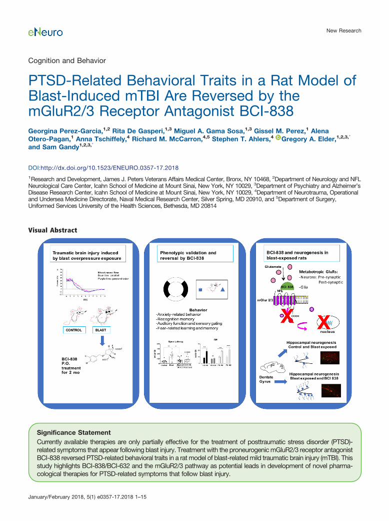
Visual Abstract
Significance StatementCurrently available therapies are only partially effective for the treatment of posttraumatic stress disorder (PTSD)-related symptoms that appear following blast injury. Treatment with the proneurogenic mGluR2/3 receptor antagonistBCI-838 reversed PTSD-related behavioral traits in a rat model of blast-related mild traumatic brain injury (mTBI). Thisstudy highlights BCI-838/BCI-632 and the mGluR2/3 pathway as potential leads in development of novel pharma-cological therapies for PTSD-related symptoms that follow blast injury.
New Research
January/February 2018, 5(1) e0357-17.2018 1–15

Battlefield blast exposure related to improvised explosive devices (IEDs) has become the most common cause oftraumatic brain injury (TBI) in the recent conflicts in Iraq and Afghanistan. Mental health problems are commonafter TBI. A striking feature in the most recent veterans has been the frequency with which mild TBI (mTBI) andposttraumatic stress disorder (PTSD) have appeared together, in contrast to the classical situations in which thepresence of mTBI has excluded the diagnosis of PTSD. However, treatment of PTSD-related symptoms thatfollow blast injury has become a significant problem. BCI-838 (MGS0210) is a Group II metabotropic glutamatereceptor (mGluR2/3) antagonist prodrug, and its active metabolite BCI-632 (MGS0039) has proneurogenic,procognitive, and antidepressant activities in animal models. In humans, BCI-838 is currently in clinical trials forrefractory depression and suicidality. The aim of the current study was to determine whether BCI-838 couldmodify the anxiety response and reverse PTSD-related behaviors in rats exposed to a series of low-level blastexposures designed to mimic a human mTBI or subclinical blast exposure. BCI-838 treatment reversed PTSD-related behavioral traits improving anxiety and fear-related behaviors as well as long-term recognition memory.Treatment with BCI-838 also increased neurogenesis in the dentate gyrus (DG) of blast-exposed rats. The safetyprofile of BCI-838 together with the therapeutic activities reported here, make BCI-838 a promising drug for thetreatment of former battlefield Warfighters suffering from PTSD-related symptoms following blast-induced mTBI.
Key words: BCI-838; blast; metabotropic glutamate receptor; mGluR2/3; posttraumatic stress disorder; trau-matic brain injury
IntroductionTraumatic brain injury (TBI) is a major cause of combat-
related disability (Gubata et al., 2014; Elder, 2015).Military-related TBIs occur through various mechanisms.Because of the widespread use of improvised explosivedevices (IEDs) in Iraq and Afghanistan blast-related mech-anisms have been the most common cause (Hoge et al.,2008; Tanielian and Jaycox, 2008). Further, as survivalafter battlefield trauma has improved, TBI has becomerecognized as a particularly common injury in the recentconflicts in Iraq and Afghanistan with estimates that asmany as 20% of returning veterans suffered a TBI duringdeployment (Hoge et al., 2008; Tanielian and Jaycox,2008). Initially, most attention focused on the moderate to
severe end of the injury spectrum, which was the type ofTBI that would be recognized acutely in theater. However,what soon became clear was that most TBIs being suf-fered in these conflicts were mild TBI (mTBI) with manygoing undocumented at the time of occurrence (Chase,2015).
Mental health problems occur often after TBI (Jorgeet al., 2014). Indeed, a striking feature in the most recentveterans has been the frequency with which posttrau-matic stress disorder (PTSD) has been seen followingblast-related mTBI (Elder et al., 2010). Studies in Iraqveterans have found that over one-third suspected ofhaving an mTBI-related postconcussion syndrome alsohave PTSD or depression (Hoge et al., 2008; Vasterlinget al., 2009). This dual diagnosis of PTSD and mTBIupends the conventional diagnostic separation of the twoentities. Indeed, the presence of mTBI has traditionallyexcluded the diagnosis of PTSD. While controversy re-mains over the separation of the two disorders clinically(Hoge et al., 2009; Bryant, 2011; Elder et al., 2014) thereis no doubt that mental health problems are commonfollowing blast-related mTBI.
BCI-838 (MGS0210), bicycle [3.1.0] hexane-2,6-dicarboxylicacid, 2-amino-3-[(3,4-dichlorophenyl)metoxy]-6-fluoro-,6-heptyl ester,(IR,2R,3R, 5R6R)-), is the prodrug for BCI-632(MGS0039), a Group II metabotropic glutamate receptor(mGluR2/3) antagonist. BCI-838 has been found to improvememory and reduce anxiety in an animal model of Alzheimer’sdisease (Kim et al., 2014). It has been tested in humansand found to be clinically well tolerated and orally bio-available. BCI-838 is currently in human clinical trials fordepression. As a prodrug BCI-838 is metabolized in theliver into BCI-632, which is the active compound deliveredto brain. In humans, daily oral dosing of BCI-838 results insteady-state levels in brain, which last for 22 h. mGluR2/3receptor antagonists are proneurogenic as evidenced bytheir stimulation of hippocampal neurogenesis in adultbrain (Yoshimizu and Chaki, 2004). In rodents, mGluR2/3receptor antagonists enhance learning and memory andthey also possess anxiolytic and antidepressive proper-
Received October 17, 2017; accepted January 11, 2018; First publishedJanuary 29, 2018.The authors declare no competing financial interests.Author contributions: G.P.-G., S.T.A., R.M.M., G.A.E., and S.G. designed
research; G.P.-G., R.D.G., M.A.G.S., G.M.P., A.O.-P., and A.T. performedresearch; G.P.-G., G.A.E., and S.G. analyzed data; G.P.-G., G.A.E., and S.G.wrote the paper.
This work was supported by the Department of Veterans Affairs, VeteransHealth Administration, Rehabilitation Research and Development ServiceAwards 1I01RX000684 (to S.G.) 1I01RX000996 (to G.A.E) and by DoD WorkUnit Number 0000B999.0000.000.A1503.
*G.A.E. and S.G. contributed equally to this work.Acknowledgements: The views expressed in this article are those of the
authors and do not necessarily reflect the official policy or position of theDepartment of the Navy, Department of Defense, nor the United States Gov-ernment. A.T., R.M.M., and S.T.A. are military service members (or employeesof the United States Government). This work was prepared as part of theirofficial duties. Title 17 U.S.C. §105 provides that copyright protection underthis title is not available for any work of the United States Government. Title 17U.S.C. §101 defines a United States Government work as a work prepared bya military service member or employee of the United States Government aspart of that person’s official duties.
Correspondence should be addressed to either of the following: Dr. SamGandy at the above address, E-mail: [email protected]; or Dr. Greg-ory A. Elder at the above address, E-mail: [email protected].
DOI:http://dx.doi.org/10.1523/ENEURO.0357-17.2018Copyright © 2018 Perez-Garcia et al.This is an open-access article distributed under the terms of the CreativeCommons Attribution 4.0 International license, which permits unrestricted use,distribution and reproduction in any medium provided that the original work isproperly attributed.
New Research 2 of 15
January/February 2018, 5(1) e0357-17.2018 eNeuro.org
ties (Higgins et al., 2004; Shimazaki et al., 2004; Yoshi-mizu et al., 2006; Campo et al., 2011). As a class, thesecompounds are regarded as promising for treatment of avariety of mental health and neurologic disorders includ-ing refractory major depression, suicidality, sleep-wakecycle disorders, and other psychiatric conditions in whichcognitive impairment is a prominent feature (Celanireet al., 2015).
Neurogenesis in the adult hippocampus affects highercognitive functions (especially memory) and influencesaffective behavior (Kempermann et al., 2015). Stimulationof hippocampal neurogenesis has been proposed as acentral mechanism underlying the action of antidepres-sant drugs (Chaki, 2017). TBI by cortical impact is re-ported to impair hippocampal neurogenesis (Rola et al.,2006; Shetty, 2014; Wang et al., 2016; Shapiro, 2017),raising the possibility that proneurogenic drugs might beeffective in modifying the course of latent manifestationsresulting from mTBI.
Modulation of other mGluRs has also been explored inexperimental models of TBI (Loane et al., 2009, 2013;Kabadi and Faden, 2014). The stimulation of mGluR5 hasemerged as one of the more promising approaches witheffects that include promoting reduced production of ni-tric oxide and tumor necrosis factor-� as well as limitingcaspase dependent apoptosis and intracellular genera-tion of reactive oxygen species (Loane et al., 2009). How-ever, none of these studies have explored modulation ofmGluRs in the context of blast injury.
The aim of this study was to investigate whether admin-istration of BCI-838 could modify the anxiety response andreverse PTSD-related behaviors while concomitantly en-hancing neurogenesis in the dentate gyrus (DG) in rats pre-viously found to exhibit a variety of chronic PTSD-relatedbehavioral traits (Elder et al., 2012; Perez-Garcia et al.,2016, 2018). BCI-838 treatment reversed PTSD-relatedtraits improving anxiety and fear-related behaviors, inaddition to long-term recognition memory. Hippocampalneurogenesis was also robustly increased in the DG ofdrug-treated blast-exposed rats. The present study high-lights the potential role for BCI-838, hippocampal neuro-genesis, and the mGluR2/3 pathway in the developmentof novel pharmacological therapies to help former Warf-ighters suffering from the dual diagnosis status whenPTSD-related symptoms coexist with blast-inducedmTBI.
Materials and MethodsAnimals
Adult male Long Evans Hooded rats (250–350 g; 10–12weeks of age; Charles River Laboratories International,Inc.) were used as subjects. All studies were approved bythe Institutional Animal Care and Use Committees of theJames J. Peters VA Medical Center and the Walter ReedArmy Institute of Research/Naval Medical Research Cen-ter. Studies were conducted in compliance with the PublicHealth Service policy on the humane care and use oflaboratory animals, the NIH Guide for the Care and Use ofLaboratory Animals, and all applicable Federal regulationsgoverning the protection of animals in research.
Blast overpressure exposureRats were exposed to overpressure injury using a shock
tube, which simulates the effects of air blast exposureunder experimental conditions (Ahlers et al., 2012). Theshock tube has a 0.32-m circular diameter and is a 5.94m-long steel tube divided into a 0.76-m compressionchamber that is separated from a 5.18-m expansion cham-ber. The compression and expansion chambers are sepa-rated by polyethylene terephthalate Mylar TM sheets (DuPont Co) that control the peak pressure generated. The peakpressure at the end of the expansion chamber was deter-mined with piezoresistive gauges specifically designed forpressure-time (impulse) measurements (Model 102M152,PCB, Piezotronics, Inc.).
Individual rats were anesthetized using an isofluranegas anesthesia system consisting of a vaporizer, gas linesand valves and an activated charcoal scavenging systemadapted for use with rodents. Rats were placed into apolycarbonate induction chamber, which was closed andimmediately flushed with 5% isoflurane mixture in air fortwo minutes. Rats were placed into a cone shaped plasticrestraint device and then placed in the shock tube. Move-ment was further restricted during the blast exposureusing 1.5 cm in diameter flattened rubber tourniquet tub-ing. Three tourniquets were spaced evenly to secure thehead region, the upper torso and lower torso while theanimal was in the plastic restraint cone. The end of eachtubing was threaded through a toggle and run outside ofthe exposure cage where it was tied to firmly affix theanimal and prevent movement during the blast overpres-sure exposure without restricting breathing. Rats wererandomly assigned to sham or blast conditions with thehead facing the blast exposure without any bodyshielding resulting in a full body exposure to the blastwave. The total length of time under anesthesia includ-ing placement in the shock tube and execution of theblast procedure was typically �3 min. Blast-exposedanimals received 74.5 kilopascal (kPa) exposuresequivalent to 10.8 pounds per square inch (psi). Oneexposure per day was administered for three consec-utive days. Sham exposed animals were treated iden-tically including receiving anesthesia and being placedin the blast tube but did not receive a blast exposure.Under the blast conditions used here blast-exposedrats recovered identically to controls and exhibited noloss of the righting reflex (Ahlers et al., 2012).
Animal housingAnimals were housed at a constant 70-72°F tempera-
ture with rooms on a 12/12 h light/dark cycle with lights onat 7 A.M. All subjects were individually housed in standardclear plastic cages equipped with Bed-O’Cobs laboratoryanimal bedding (The Andersons) and EnviroDri nestingpaper (Sheppard Specialty Papers). Access to food andwater was ad libitum. Subjects were housed on racks inrandom order to prevent rack position effects. Cageswere coded to allow maintenance of blinding to groupsduring behavioral testing.
New Research 3 of 15
January/February 2018, 5(1) e0357-17.2018 eNeuro.org

Drug administrationBCI-838 was dissolved in a solution of 5% carboxym-
ethylcellulose (CMC; Sigma Aldrich) and 0.3% 2 Nhydrochloric acid solution (Sigma Aldrich) at room tem-perature. The drug emulsion was prepared daily bysonication for 2 min to fully dissolve. Animals weredivided into four experimental groups: (1) sham ex-posed (placed in blast tube but did not receive blastexposure) treated with vehicle (5% CMC); (2) blastexposed treated with vehicle; (3) blast exposed treatedwith 4-mg/kg BCI-838 (low dose); and (4) blast exposedtreated with 10-mg/kg BCI-838 (high dose). The exper-iment was performed independently on two cohorts ofrats described in Extended Data Figures 1-1, 1-2.Doses were chosen based on previous work in otherrodent models (Kim et al., 2014). Bodyweight was re-corded weekly and doses were adjusted accordingly.
The drug was administered by oral gavage starting twoweeks after the last blast exposure. Administration wasconducted daily between 9 A.M. and 2 P.M. for 60 d bypersonnel experienced in the procedure. Restraint forgavage was performed similar to that described by Turneret al. (2012) except that a towel was used to firmly graspand gently immobilize the rat with the head and body heldvertically. A 7-cm straight stainless-steel gavage needlewith a 3-mm ball tip (Fischer Scientific) was used forgavage and wiped clean between animals.
Bromodeoxyuridine (BrdU) injectionsAll animals received once daily intraperitoneally injec-
tions of BrdU (150 mg/kg of body weight) for 8 d duringthe third week of drug treatment (five weeks after blastexposure). BrdU (Sigma) was dissolved in saline solution(0.9% NaCl in sterile H2O) warmed to 40°C and gentlyvortexed. The solution was allowed to cool to room tem-perature (25°C) before injection.
Behavioral testingBehavioral testing was begun at the end of the 60 d of
drug administration. All behavioral testing was performedby the same investigator (GPG). The following tests wereperformed.
Locomotor activity and open fieldGeneral locomotor activity and open field behavior was
examined in 40.6 � 40.6 cm Versamax activity cages(Accuscan), each outfitted with a grid of 32 infraredbeams at ground level and 16 elevated 7.6 cm aboveground level. Locomotor activity was recorded during 60min and analyzed with VersaData Software (Accuscan),which automatically calculates move time, move distanceand center time based on beam breaks. The center of thechamber was defined as a square of 25.4 � 25.4 cm (7.6cm from each side wall) and virtually drawn with Versa-Map software (Accuscan). Center entries and center resttime were defined based on the centroid of the rat beingin the center of the chamber with center rest time definedas time when the centroid was in the center of the cham-ber but during which no beam breaks were generated.Samples were recorded in 1-min bins and summed into5-min intervals for presentation.
Light/dark emergenceA light/dark emergence task was run in Versamax ac-
tivity cages with opaque black Plexiglas boxes enclosingthe left half of the interiors so that only the right sides wereilluminated. Animals began in the dark side and wereallowed to freely explore for 10 min with access to the left(light) side through an open doorway located in the centerof the monitor. Subject side preference and emergencelatencies were tracked by centroid location with all move-ment automatically tracked and quantified. Light-sideemergence latency, time to reach the center of the lightedside (light side center latency) and percentage total light-side duration were calculated from beam breaks. Allequipment was wiped clean between tests.
Elevated zero maze (EZM)The apparatus consisted of a circular black Plexiglas
runway 121.92 cm in diameter and raised 76 cm off thefloor (San Diego Instruments). The textured runway itselfwas 5.08 cm across and divided equally into alternatingquadrants of open runway enclosed only by a 1.27-cm lipand closed runway with smooth 15.24-cm walls. All sub-jects received a 5-min trial beginning in a closed arc of therunway. During each trial, subjects were allowed to movefreely around the runway, with all movement tracked au-tomatically by a video camera placed on the ceiling di-rectly above the maze. Data were analyzed by ANYMAZE(San Diego Instruments) yielding measures of total move-ment time and distance for the entire maze, as well astime spent and distance traveled in each of the individualquadrants. From the quadrant data, measures of totalopen and closed arc times, latency to enter an open arc,total open arm entries and latency to completely cross anopen arc between two closed arcs were calculated. Sub-ject position was determined by centroid location.
Novel object (NO) recognitionRats were habituated to the arena (90 cm length � 60
cm width � 40 cm height) for 20 min, 24 h before training.On the training day, two identical objects were placed onopposite ends of the empty arena, and the rat was al-lowed to freely explore the objects for 7 min. After a 1-hdelay, during which the rat was held in its home cage, oneof the two familiar objects (FOs) was replaced with a novelone, and the rat was allowed to freely explore the familiarand NO for 5 min to assess short-term memory (STM).After a 24-h delay, during which the rat was held in itshome cage, one of the two FOs was replaced with a novelone different from the ones used during the STM test. Therat was allowed to freely explore the familiar and NO for 5min to assess long-term memory (LTM). After a four-weekdelay (from training), during which the rat was held in itshome cage, one of the two FOs used during the LTMtesting was replaced with a novel one different from thoseused during either the STM or LTM tests. The rat wasallowed to freely explore the familiar and NO for 5 min toassess consolidation memory (CM). Raw explorationtimes for each object were expressed in seconds. Objectexploration was defined as sniffing or touching the objectwith the vibrissae or when the animal’s head was orientedtoward the object with the nose placed at a distance of
New Research 4 of 15
January/February 2018, 5(1) e0357-17.2018 eNeuro.org

�2 cm from the object. All sessions were recorded byvideo camera (Sentech) and analyzed with ANYMAZEsoftware (San Diego Instruments). In addition, offline anal-ysis by an investigator blind to the blast-exposed status ofthe animals was performed. Objects to be discriminatedwere of different size, shape and color and were made ofplastic or metal material. The objects consisted of a330-ml soda can, a metal box, a cup and a plastic tube.All objects were cleaned with 70% ethanol between trials.
Prepulse inhibition (PPI) and acoustic startleStartle magnitude and sensory gating were examined in
a 40-trial PPI assay (San Diego Instruments). Animalswere placed in isolation chambers inside closed Plexiglastubes, each of which was mounted on a platform restingon an accelerometer. Following a 5-min habituation pe-riod with 74-dB background white noise, each animalreceived 40 randomized trials separated by 20-30 s. Trialsconsisted of 10 each of background readings taken at 74dB, startle trials with readings following 40-ms 125-dBtones, PPI trials where the 125-dB tone was preceded100 ms earlier by a 20-ms 79-dB tone and control trialsconsisting of only the 20-ms 79-dB prepulse. On all trials,maximum magnitude of the animal’s startle (or other mo-tion) was automatically recorded in 500-ms windows byan accelerometer. The tubes were rinsed clean betweenanimals. Percentage PPI was calculated with the formula100 – (startle response on acoustic prepulse plus pulsestimulus trials/pulse stimulus response alone trials) � 100.The first startle response was compared among groups.
Contextual and cued fear conditioningSound-attenuated isolation cubicles (Coulbourn Instru-
ments) were used. Each cubicle was equipped with a gridfloor for delivery of the unconditioned stimulus (US) andoverhead cameras. All aspects of the test were controlledand monitored by the Freeze Frame conditioning andvideo tracking system (Actimetrics, Coulbourn Instru-ments). During training the chambers were scented withalmond extract, lined with white paper towels, had back-ground noise generated by a small fan and were cleanedbefore and between trials with 70% ethanol. The testerwore latex gloves. Each subject was placed inside theconditioning chamber for 2 min before the onset of aconditioned stimulus (CS; an 80 dB, 2-kHz tone), whichlasted for 20 s with a coterminating 2-s footshock (0.7 mA;US). Each rat remained in the chamber for an additional40 s following the CS-US pairing before being returned toits home cage. Freezing was defined as a lack of move-ment (except for respiration) in each 10-s interval. Minutes0–2 during the training session were used to measurebaseline freezing. Contextual fear memory testing wasperformed 24 h after the training session by measuringfreezing behavior during a 3-min test in the conditioningchamber under conditions identical to those of the train-ing session with the exception that no footshock or tone(CS or US) was presented. Animals were returned to theirhome cage for another 24 h, at which time cued condi-tioning was tested. To create a new context with differentproperties, the chambers were free of background noise
(fan turned off), lined with blue paper towels, scented withlemon extract and cleaned before and during all trials withisopropanol. In addition, the tester wore nitrile gloves andhabituated the rats pretesting in a different holding room.Each subject was placed in this novel context for 2 minand baseline freezing was measured, followed by expo-sure to the CS (20-s tone) at 120 and 290 s.
Tissue processing and immunohistochemistryAnimals were sacrificed at the conclusion of behavioral
testing. After deep anesthesia with a solution of 150mg/kg ketamine and 30 mg/kg xylazine, rats were eutha-nized by transcardial perfusion with cold 4% paraformal-dehyde in PBS. After perfusion, brains were removed andpostfixed in 4% paraformaldehyde for 48 h, transferred toPBS, and stored at 4°C until sectioning. Fifty-micrometer-thick coronal sections were cut through the entire extendof the hippocampus using a Leica VT1000 S Vibratome(Leica). The sections were stored at �20°C in a cryopro-tectant solution (25% ethylene glycol and 25% glycerinein 0.05 M PBS) until processing for immunofluorescence.
For stereologically based counting every 6th section ina series was processed for immunohistochemistry so thatthe interval between sections within a given series was300 �m. For BrdU staining, the sections from each brainwere treated with 50% formamide and 2 � SSC (0.3 MNaCl and 0.03 M sodium citrate) for 2 h, followed byincubation with 0.1 M boric acid buffer at pH 8.5 for 10min. After 4 � 5 min washes with PBS, they were incu-bated in blocking buffer (3% goat serum, 0.3% TritonX-100 in PBS) for 1 h and incubated overnight at 4°C in amixture of rat anti-BrdU (1:300, Abcam) plus rabbit anti-neuron-specific nuclear protein (NeuN, 1:500; Millipore)antibodies. The next day, sections were washed 4 � 5 minwith PBS and exposed for 2 h in the dark with Alexa Fluor568-conjugated donkey anti-rat IgG and with Alexa Fluor488-conjugated goat anti-rabbit IgG (Life Technologies).Both secondary antibodies were used at a dilution of1:300. To ascertain the effects of BCI-838 on cell prolif-eration and survival, a second series of sections fromeach animal was immunolabeled with doublecortin (DCX)and BrdU as described above using a goat monoclonalanti-DCX antibody (1:500 from Santa Cruz). All slices weremounted onto slides and covered under Fluoro-Gel (withTris Buffer from Electron Microscopy Sciences).
Image analysis and neurogenesis quantificationGiven the scarcity of BrdU- and DCX-immunostained
cells, the number of new cells was estimated using a mod-ified version of the optical fractionator method employing anexhaustive sampling scheme. All BrdU- or DCX-labeled cellswere counted on both sides of every 6th bilateral sectionthroughout the entire DG between coordinates �2.52 and5.40 mm relative to bregma. Immunostained cells were firstvisualized with a 40� objective. To ensure accurate com-parison between groups, we checked that section thick-nesses were similar for all the groups with the aid of amicrocator focused on immuno-fluorescence labeled nucleiat the border of the hilus and DG. The number of BrdU- orDCX-labeled cells per granule cell layer (GCL, including the
New Research 5 of 15
January/February 2018, 5(1) e0357-17.2018 eNeuro.org

SGZ) was estimated using the following formula:N � Q � (1/ssf), where Q is the total number of countedcells and 1/ssf is the reciprocal of the section samplingfraction (1/ssf � 12 in the present case).
For quantification of double-labeled BrdU/NeuN cells,11 bilateral slices per animal spanning the entire DG wereused to determine the frequency of BrdU-positive cellsexpressing NeuN. Eight to 12 optical sections (1 �m thick)were scanned from each area using the 40� objective.BrdU-labeled cells were scored as neurons when theNeuN labeling was unambiguously associated with aBrdU-positive nucleus in the stack of sections. The per-centages of BrdU-labeled cells that were also labeled withNeuN were calculated for each group.
Statistical analysisValues are expressed as mean � SEM. Extended Data
Figures 1-1, 1-2 contain the data structure, type of testused, observed power, and n for each figure. Each testincluded enough animals to reach a power close to orhigher than 0.8. Statistical tests were performed usingthe program GraphPad Prism 7.0 (GraphPad Software),IBM SPSS statistics 24, and G� Power (Heinrich-Heine-Universität Düsseldorf). To systematically test for normalitythe D’Agostino–Pearson and Shapiro Wilk tests were used.Depending on the behavioral test, multiple comparisonswere performed using one-way ANOVA for normally distrib-uted datasets followed by Tukey’s post hoc tests for multiplecomparisons when appropriate. The datasets used for two-way repeated measures ANOVA were normally distributedand were followed by post hoc Sidak’s test.
ResultsTreatment of blast-exposed rats with BCI-838
We studied a model of blast exposure using rats. Be-cause multiple blast exposures have been commonamong former Warfighters returning from Iraq and Af-ghanistan (Hoge et al., 2008; Tanielian and Jaycox, 2008;
Elder et al., 2010), we used a design in which rats receivedthree 74.5-kPa exposures delivered once per day on threeconsecutive days. Studies using this model have estab-lished that exposures up to 74.5 kPa (equivalent to 10.8psi), while representing a level of blast that is transmittedto brain, produce no gross neuropathological effects, andhistologic examination of the lungs show no hemorrhageor other pathology (Ahlers et al., 2012; Elder et al., 2012).Based on our experience with this model, we believe thatthese blast pressures mimic a low-level blast exposureequivalent to a human mTBI or subclinical blast exposure.
Since BCI-838 has a variety of potentially relevant neuro-psychiatric activities in other rodent models (Kim et al.,2014), we assessed its efficacy in modifying the behavioraltraits that follow blast injury. The time course of the experi-ments is shown in Figure 1. BCI-838 was administered attwo different doses (4 and 10 mg/kg/d) by oral gavagestarting two weeks after the last blast exposure and wascontinued for eight weeks. During the third week of drugtreatment, BrdU was administered daily for eight consecu-tive days. Gavage was stopped after week 10 post-blast andbehavioral testing was performed between 11 and 17 weeksafter blast exposure. Rats were sacrificed at the end ofbehavioral testing when the animals were 25 weeks old. Theexperiments were conducted on two cohorts of animals.Results from cohort 2 are primarily described below andpresented in Figures 2–6. The effect of BCI-838 treatment incohort 1 is discussed below and presented in ExtendedData Figures 2-1, 5-1. Extended Data Figures 1-1, 1-2,summarize effects in both cohorts.
BCI-838 reverses chronic anxiety in blast-exposedrats
Starting at week 11 post-blast, rats were tested in anopen field, an EZM, and a light/dark emergence task. Nodifferences in the open field were found among thegroups during the 60 min of testing (data not shown). Inthe light/dark emergence task (Fig. 2), blast-exposed rats
Figure 1. Experimental design and timing. Further description of both cohorts is contained in Extended Data Figures 1-1, 1-2.
New Research 6 of 15
January/February 2018, 5(1) e0357-17.2018 eNeuro.org

treated with vehicle exhibited an increased latency toreach the light center (Fig. 2B; one-way ANOVA, F(3,41) �6.486, p � 0.0011), made fewer light center entries (Fig.2C; F(3,41) � 8.585, p � 0.0002), and traveled less dis-tance on the light side (Fig. 2E; F(3,42) � 8.547, p � 0.0002)compared to vehicle-treated controls. Treatment withhigh-dose BCI-838 (10 mg/kg/d) reversed deficits in thelight center latency, light center entries and total distancetraveled on the light side.
Twenty four h after the light/dark emergence task, ratswere tested for 5 min in an EZM. Compared to vehicle-treated controls, blast-exposed rats treated with vehicletended to moved less (Fig. 3B; F(3,40) � 2.527, p � 0.071),showed an increased latency to reach an open arm (Fig.3C; F(3,40) � 5.080, p � 0.0045), made fewer open armentries (Fig. 3D; F(3,40) � 5.08, p � 0.0001), and spent lesstime in the open arms (Fig. 3E; F(3,40) � 3.39, p � 0.0189).They also exhibited an increased latency to cross be-tween two open arms (cross latency; Fig. 3F; F(3,40) �5.080, p � 0.0045). Treatment with 4 mg/kg and 10 mg/kgof BCI-838 reversed many of these effects. Results oftreatment in cohort 1 revealed similar effects with BCI-838reversing blast-associated anxiety in both light/darkemergence and the EZM (Extended Data Figs. 2-1A,B).Thus, blast-exposed rats exhibit signs of chronic anxietyin multiple tests that are reversed by treatment with BCI-838.
Enhanced PPI in blast-exposed rats is unaltered withBCI-838 treatment
Enhanced acoustic startle is an important characteristicof the hyperarousal found in PTSD. Startle magnitude andsensory gating were examined in a PPI assay. Results ofthe first startle reactions are shown in Figure 4A–C. Nodifferences were found between the groups whether ve-hicle or drug treated in background readings (pre; Fig. 4A;F(3,39) � 2.398, p � 0.0826), acoustic startle response(pulse; Fig. 4B; F(3,38) � 0.6367, p � 0.5960), or startlefollowing the prepulse (Fig. 4C; F(3,39) � 0.2161, p �0.8846). An increased response was found between blast-exposed rats treated with vehicle and vehicle-treatedcontrols when the first prepulse was subtracted from thefirst acoustic startle (pulse-prepulse; Fig. 4D). Blast-exposed rats treated with vehicle also exhibited an in-creased percentage of PPI versus vehicle-treated controls(Fig. 4E). Neither dose of BCI-838 affected startle magni-tude or PPI among groups. Results were similar in the firstcohort (Extended Data Fig. 2-1C). Thus, responses toauditory stimuli are altered following blast exposure butBCI-838 did not reverse these effects.
Altered fear responses in blast-exposed rats arereversed with high-dose BCI-838
Models of conditioned fear are regarded as relevant tothe study of the pathophysiological mechanisms of PTSD,where disordered fear regulation is observed (Mahan and
Figure 2. High-dose BCI-838 reverses anxiety in the light/dark emergence task. While the light edge latency (A) was unchanged, lightcenter latency (B) was increased in blast-exposed rats, which also made fewer entries (C), and traveled less distance on the light side(E). Treatment with high-dose BCI-838 (10 mg/kg) reversed these effects. Values significantly different from controls are indicated byasterisks (�p � 0.05, ��p � 0.01, ���p � 0.001, and ����p � 0.0001, Tukey’s or Sidak’s multiple comparisons tests). Values areexpressed as mean � SEM. Results from cohort one can be found in Extended Data Figure 2-1.
New Research 7 of 15
January/February 2018, 5(1) e0357-17.2018 eNeuro.org

Ressler, 2012). We examined blast-exposed rats in acued/contextual fear paradigm (Fig. 5). Freezing behaviorwas measured during minutes 0–2 of the training session(baseline), after the presentation of the tone and after thefootshock. Following the footshock, all groups showedincreased freezing but no differences were found amonggroups (Fig. 5A; repeated-measures ANOVA, F(2,42) �401.01, p � 0.001 baseline vs post-shock and F(2,42) �0.211, p � 0.888 for freezing among groups by condition).On day 2 in the contextual phase, freezing was similar inall groups in minutes 1 and 2 (Fig. 5B). However, in minute3, blast-exposed rats treated with low and high-doseBCI-838 showed less freezing compared to non-blast-exposed controls (one-way ANOVA, F(3,43) � 2.857, p �0.048 among groups min 3). On day 3, in the cued phase,blast-exposed rats treated with vehicle showed increasedfreezing in response to the second tone compared tovehicle-treated controls (Fig. 5C). Blast-exposed ratstreated with low-dose BCI-838 showed less freezingcompared with blast-exposed rats treated with vehicle inthe second tone period (one-way ANOVA, F(3,43) � 2.863,p � 0.0011 for freezing by condition tone 2). Moreover,blast-exposed rats treated with high-dose BCI-838 dis-played less freezing compared with blast-exposed ratstreated with vehicle in the intertone and tone 2 periods(one-way ANOVA, F(3,43) � 103.84, p � 0.0001 amonggroups for intertone and F(2,43) � 15.65, p � 0.0001 fortone 2).
In cohort 1, blast-exposed rats treated with vehicleshowed a tendency to increased freezing in the cuedphase of testing compared to vehicle-treated controls(Extended Data Fig. 5-1A). Enhanced freezing during thecued phase of fear conditioning training has been ob-served in multiple other cohorts of rats studied in the pastfollowing a similar blast exposure protocol (Elder et al.,2012 and data not shown). Blast-exposed rats treatedwith high-dose BCI-838 displayed a tendency to lessfreezing compared with blast-exposed rats treated withvehicle in the intertone and tone 2 (Extended Data Fig.5-1A). Thus, fear responses were chronically altered fol-lowing blast exposure and reversed by BCI-838 4 and 10mg/kg/d treatments in both of the cohorts.
Altered NO recognition in blast-exposed rats isreversed with BCI-838
Cognitive impairment is a significant component of TBIand PTSD. As a measure of cognitive functioning in blast-exposed rats and the effects of BCI-838, we performed aNO recognition task. During the training phase, blast-exposed rats treated with vehicle explored the objectsequally in each location but spent less total time in explo-ration (Fig. 6E) than all other groups (between-objectdiscrimination comparisons were made using unpaired ttests (Student’s), p � 0.855 for discrimination Ob1 vs Ob2controls; p � 0.7675 for blast exposed; p � 0.954 for blastexposed with BCI � LD and p � 0.724 for blast exposedwith BCI � HD; Fig. 6A). When presented a NO, the
Figure 3. Blast-induced anxiety is reversed in an EZM by BCI-838. While time in motion (A) did not differ, blast-exposed rats movedless (B) exhibited a longer latency to enter an open arm (C), made fewer open arm entries (D) as well as spent less time in the openarms (E) and exhibited an increased latency to cross between two open arms (cross latency; F). Treatment with both low- andhigh-dose BCI-838 reversed nearly all of these effects. Values significantly different between controls and blast-exposed rats areindicated by asterisks (�p � 0.05, ��p � 0.01, ���p � 0.001, Tukey’s or Sidak’s multiple comparisons tests). Values are expressedas mean � SEM.
New Research 8 of 15
January/February 2018, 5(1) e0357-17.2018 eNeuro.org

vehicle-treated control and blast-exposed rats spentmore time investigating the unfamiliar object, and theblast exposed again spent less total time in exploration(Fig. 6E) whether tested 1 h (STM; Fig. 6B; p � 0.0004 fordiscrimination FO vs NO controls; p � 0.0001 for discrim-ination FO vs NO blast exposed; p � 0.0002 for discrim-ination FO vs NO blast exposed with BCI �LD; and p �0.0001 for discrimination FO vs NO blast-exposed BCIHD) or 24 h (LTM; Fig. 6C) after training (p � 0.0001 fordiscrimination FO vs NO controls; p � 0.0001 for discrim-ination FO vs NO blast exposed; p � 0.0017 for discrim-ination FO vs NO blast-exposed BCI LD and p � 0.0001for discrimination FO vs NO blast-exposed BCI HD).Moreover, when an additional NO was presented fourweeks after training (CM; Fig. 6D), blast-exposed ratstreated with vehicle not only spent less time exploringboth objects (familiar and novel) compared with non-blast-exposed controls (Fig. 6E), they also failed to explore the NOmore than the familiar (p � 0.0083 for discrimination FOvs NO in controls; p � 0.5246 for discrimination FO vsNO blast exposed; p � 0.0003 for discrimination FO vsNO blast-exposed BCI LD and p � 0.0002 for discrimi-nation FO vs NO blast-exposed BCI HD). Effects on re-duced exploration time (Fig. 6E) as well as the late effectson CM (Fig. 6D) were reversed by low-dose (4 mg/Kg/d)and high-dose (10 mg/Kg/d) BCI-838. Results in cohort 1were similar in STM and LTM testing (Extended Data Fig.5-1B). Effects on CM at four weeks after training were notassessed in cohort 1.
Enhanced neurogenesis in blast-exposed ratstreated with BCI-838
mGluR2/3 receptor antagonists are known for their pro-neurogenic effects stimulating hippocampal neurogenesisin adult brain (Yoshimizu and Chaki, 2004). To determinethe effects of blast and BCI-838 treatment on hippocam-pal neurogenesis, we first evaluated BrdU labeling ofnewly generated hippocampal cells at 10 weeks after thefinal BrdU injection (Fig. 7). We found no difference be-tween numbers of BrdU-labeled cells in vehicle-treatedblast-exposed and control rats suggesting that there is noinherent effect of blast on neural progenitor proliferation.However, we found a statistically significant increase inthe number of BrdU-positive cells in blast-exposed ratstreated with high-dose BCI-838 compared with blast ex-posed treated with vehicle suggesting that drug treatmentinduced neurogenesis in the hippocampus (Fig. 9A; one-
Figure 4. BCI-838 treatment does not rescue enhanced PPI
Figure 4. continuedfound in blast-exposed rats. Startle magnitude and sensorygating were examined in a PPI assay. We analyzed acousticstartle and PPI of the first startle response. No differences werefound in background readings (pre; A), acoustic startle response(pulse; B), or startle following the prepulse (prepulse; C), but wefound an increased response when the prepulse was subtractedfrom the pulse (pulse-prepulse; D) and in the percentage PPI inblast-exposed rats versus the control group (E). Treatment withBCI-838 did not normalize either of these responses. Valuessignificantly different between controls and blast-exposed ratsare indicated by asterisks (�p � 0.05, Tukey’s multiple compar-isons test). Values are expressed as mean � SEM in all panels.
New Research 9 of 15
January/February 2018, 5(1) e0357-17.2018 eNeuro.org

way ANOVA, F(3,11) � 3.446, p � 0.0401 blast exposed vsblast exposed treated with high-dose BCI and p � 0.0793for treatment with low dose).
However, given that BrdU labeling was examined 10weeks after the last injection and not acutely, the analysisprovided an estimation of cell survival, and the questionremained as to whether BCI-838 treatment also stimu-lated cell proliferation. To assess this, a different set ofslices from each experimental group was immunolabeledwith antibodies against DCX and BrdU. In contrast toBrdU, which is a marker of cell proliferation since it isincorporated into DNA during the S-phase of the cellcycle, DCX is a microtubule-associated protein that in theadult brain labels immature neurons in the neurogenicniche (Brown et al., 2003) and is expressed specifically invirtually all migrating neuronal precursors of the develop-ing CNS. In the adult hippocampus, DCX visualizationgives a picture of the number of immature neurons. Inagreement with the results of BrdU staining, blast-exposed rats treated with high-dose BCI-838 exhibited anincreased number of DCX-labeled cells compared withblast exposed treated with vehicle (Figs. 8, 9B ). Thus,chronic treatment with BCI-838 does indeed increasehippocampal neurogenesis following blast injury.
DiscussionTBI involves damage to the brain from an external force
that can lead to direct tissue injury and hemorrhage aswell as to the activation of secondary injury cascades thatinclude inflammation and oxidative stress (Gennarelli andGrahm, 2005). TBI may also predispose to delayed neu-rodegeneration (DeKosky et al., 2010; Gandy et al., 2014;Elder, 2015). Postconcussion symptoms are often com-plicated by mental health problems including depression,anxiety and PTSD (Jorge et al., 2014). In particular, de-pression and PTSD have been common in former Warf-ighters returning from the recent conflicts in Iraq andAfghanistan (Hoge et al., 2008; Tanielian and Jaycox,2008; Elder et al., 2010).
Animal models of blast-related TBI have studied theeffects of differing blast pressures in the context of singleor multiple exposures to determine how blast affects thenervous system and possible associations with mentalhealth disorders including PTSD (Elder et al., 2010; Ko-beissy et al., 2013; Elder et al., 2014). Here, we studied amodel of blast exposure using rats that mimics a low-levelblast exposure equivalent to a human mTBI or subclinicalblast exposure (Ahlers et al., 2012; Elder et al., 2012). Inrodents, studies have often documented transient behav-ioral changes following blast exposure but typically didnot assess behavior beyond short-term acute effects (Ko-beissy et al., 2013; Elder et al., 2014). Rats exposed to theprotocol used here develop PTSD-related traits (includingstress and generalized anxiety), that are still present manymonths after blast exposure suggesting that blast inducesa chronic behavioral syndrome which may persist for thelifetime of the animal (Elder et al., 2012; Perez-Garciaet al., 2016, 2018). These traits include enhanced acousticstartle and anxiety in an EZM and light/dark emergencetask. Rats also exhibit an enhanced cued fear condition-ing response.
One striking feature of former Warfighters returningfrom the most recent conflicts has been the overlap ofmTBI with PTSD (Elder et al., 2014). The presence of bothdisorders has complicated diagnosis, since clinically dis-tinguishing a postconcussion syndrome from PTSD isoften difficult. Indeed, some have suggested that blast-induced mTBI has been overdiagnosed (Hoge et al., 2009;Elder et al., 2014), with many of the symptoms beingattributed to blast-related postconcussion syndrome bet-ter explained by PTSD (Hoge et al., 2009; Elder et al.,2014). However, it is intriguing that several case studieshave noted that PTSD can develop following TBI in vet-erans who did not recall the traumatic experiences (Bry-ant, 2011). From the studies presented here as well asprevious studies (Elder et al., 2012; Perez-Garcia et al.,2016, 2018), it appears that blast exposure per se can
Figure 5. High-dose BCI-838 reverses altered cued fear responses in blast-exposed rats. A, During the training phase, freezingbehavior was measured during minutes 0–2 of the training session (baseline), after the presentation of the tone and after thefootshock. All groups showed freezing and no differences were found among groups. B, The test for contextual fear memory wasperformed at 24 h in the same conditioning chamber. No differences were found between blast-exposed rats and non-blast-exposedcontrols. Blast-exposed rats treated with drug showed less freezing compared to non-blast-exposed controls. C, Cued fear memorywas tested another 24 h later. Blast-exposed rats showed increased freezing compared with non-blast-exposed controls after thesecond tone. Blast-exposed rats treated with high drug doses showed less freezing compared with blast exposed treated with vehicleand comparable freezing to non-blast-exposed controls. Asterisks indicate statistically significant differences (�p � 0.05, ��p � 0.01,���p � 0.001, one-way ANOVA, Tukey’s and Sidak’s multiple comparisons test). Values are expressed as percentage � SEM. Resultsfrom cohort one can be found in Extended Data Figure 5-1.
New Research 10 of 15
January/February 2018, 5(1) e0357-17.2018 eNeuro.org

induce PTSD-like traits in blast-exposed rats without anadded psychological stressor because blast exposuresoccurred under anesthesia. This is consistent with a se-ries of former Warfighters in whom a novel occult astro-glial scar at the junction of the cortical gray and whitematter was recently identified as the structural basis forpost-mTBI PTSD in a series of individuals (Shively et al.,2016).
The mechanism(s) underlying the development ofPTSD-related behavioral traits after blast exposure re-mains unclear. In patients, neurobiological (neurochemi-cal) and functional (neuroanatomical) abnormalities arecommonly observed. Among the neurotransmitters inbrain, amino acids like GABA and glutamate have a clearrelationship to psychiatric disorders. Glutamate inducesan excitatory synaptic signal and utilizes multiple recep-tors interacting and modulating cotransmitters in distinctregional brain areas associated with PTSD including thehippocampus, amygdala and cortex (Sherin and Nemer-off, 2011). mGluR2/3 receptors are distributed pre- andpostsynaptically in neurons and are found on astrocytesas well. They modify the activities of other neurotransmit-ter systems such as dopamine and serotonin as well asaffect glutamate signaling itself. Recently, selectivemGluR2/3 receptors antagonists have been developedthat increase synaptogenesis while at the same time mod-ifying serotonergic and dopaminergic signaling (Chaki,2017). Additionally, they exert antidepressant, anxiolytic,and procognitive effects in animal models.
Here, we show that BCI-838 can reverse multiplePTSD-related traits improving anxiety-related behaviors,fear responses, and long-term recognition memory. Amajor strength of the study is the replication of the effectsof BCI-838 in two independent cohorts. A limitation of thestudy is the lack of inclusion of a sham-exposed controltreated with drug although studies in mice have found thatBCI-838 administration to wild type mice does not affectbehavior (Kim et al., 2014). The fact that a mGluR2/3antagonist, BCI-838, can reverse multiple PTSD-relatedtraits in rats exposed to a blast overpressure injury whileconcomitantly enhancing neurogenesis in the DG indi-cates the involvement of glutamatergic components such
Figure 6. Reduced exploration time as well as late effects on
Figure 6. continuedrecognition memory in a NOR test were reversed by BCI-838.Blast-exposed rats that were not treated with drug spent lesstime than controls exploring the objects during the training ses-sion (A). All groups showed a preference for the NO comparedwith the FO when tested 1 h (B) or 24 h (C) later, suggesting thatblast does not affect STM or LTM in rats at five months of age.At four weeks after training (D), blast-exposed rats showedimpaired CM when exploring a FO and NO. This effect wasreversed by low dose and high dose of BCI-838. The othergroups showed a preference for the NO compared with the FO.Blast-exposed rats generally spent less time exploring the ob-jects than non-blast-exposed controls, an effect that was re-versed by both doses of BCI-838 (E). Values significantlydifferent from controls and blast-exposed are indicated by as-terisks (��p �0.01, ���p � 0.001, ����p � 0.0001, unpaired ttests, Student’s in A–D; Tukey’s multiple comparison’s test in E).Values are expressed as mean � SEM.
New Research 11 of 15
January/February 2018, 5(1) e0357-17.2018 eNeuro.org

as those in the hippocampus and cortex in the anxiety-related effects.
We evaluated primary anxiety in the light/dark emer-gence task (or light/dark box task), a test, which measuresaversion to light and open spaces. In addition to aversionto light, blast-exposed rats made fewer entries and trav-eled less distance on the light side suggesting anxiety tonovel and open spaces. The EZM is a measure of anxiety,which combines preference for closed versus openspaces with the added anxiety associated with elevationof the maze. It can also be interpreted as a cognitiveassessment of risk test (Cryan and Sweeney, 2011). Blast-exposed rats moved less, made fewer open arm entries,and spent less time in the open arms as well as exhibitedan increased latency to cross between two open arms(cross latency). Both light/dark emergence and EZM arebased on the approach-avoidance conflict betweenstress (light, open space and/or elevation), and the naturalexploratory tendency of rodents. In both, treatment withhigh-dose BCI-838 reversed (prevented) anxiety-relatedeffects.
The acoustic startle reflex is a basic response to strongexteroceptive stimuli, and humans with PTSD show anenhanced response to acoustic startle (Orr et al., 1995;
Morgan et al., 1996). While blast-exposed rats showedimpaired PPI compared to non-blast-exposed controlsneither dose of BCI-838 affected the abnormal responsesto the pulse and prepulse. The inferior colliculus andintralaminar nucleus are critical parts of the auditory path-way mediating PPI of acoustic startle (Winer et al., 2002).Interestingly, neither structure has been reported to ex-press mGluR2/3 receptors (Lu, 2014) suggesting a phar-macological explanation for the lack of BCI-838 effect.
In contrast, metabotropic glutamate receptor distribu-tion in hippocampus and amygdala is dense and essentialfor consolidation and extinction of fear conditioning inrodents. Individuals with PTSD typically show increasedsensitization to stress, overgeneralization of fear to irrel-evant stimuli, and impaired extinction of fear memories(Mahan and Ressler, 2012). Fear responses were attenu-ated by both the 4- and 10-mg/kg treatment doses ofBCI-838 suggesting participation of glutamatergic com-ponents in the hippocampus and cortex (Higgins et al.,2004; Li et al., 2011; Popoli et al., 2011) and in thehippocampus and amygdala which are essential for con-solidation and extinction of fear conditioning in rodents.Indeed, as shown in Figure 5, all groups in cohort 2responded with similar freezing during the first min of the
Figure 7. Neurogenesis is increased in blast-exposed rats stained for BrdU and the mature neuronal marker NeuN (BrdU-labeled cellsare indicated with arrows). Representative confocal images (A–D) of the DG stained for BrdU (red) and the mature neuronal markerNeuN (green) in vehicle-treated controls (A), vehicle-treated blast-exposed (B) and blast exposed treated with low-dose (C) orhigh-dose (D) BCI-838. Scale bar: 50 �m.
New Research 12 of 15
January/February 2018, 5(1) e0357-17.2018 eNeuro.org

contextual phase indicating that they remembered andinitially responded to the previously encountered contextwith a similar response. However, in minute 3, both drug-
treated groups froze less arguing that the main drug effectwas not on fear memory but on how the fear responsewas sustained. A similar conclusion can be drawn from
Figure 8. BCI-838 treatment increases the number of DCX-labeled cells in blast-exposed rats. Representative confocal images (A–D) of the DGstained for BrdU (green) and the young neuronal marker DCX (red) in vehicle-treated controls (A), vehicle-treated blast-exposed (B) and blastexposed treated with low-dose (C) or high-dose (D) BCI-838 (examples of DCX-labeled cells are indicated with arrows). Insets show examples oflabeled cells viewed at higher power. We did not detect double-labeled cells stained with BrdU and DCX. Scale bar: 50 �m (panels) and 10 �m(insets).
Figure 9. Quantification of neurogenesis in blast-exposed rats treated treated with vehicle or BCI-838. Data in A corresponds to themean � SEM of the total number of BrdU-labeled cells. Values significantly different from blast-exposed with vehicle and blast-exposed with drug are indicated by asterisks. Data in B corresponds to the mean � SEM of the total number of DCX-labeled cells.Values significantly different from blast exposed treated with vehicle and blast exposed treated with drug are indicated by asterisks(�p � 0.05, ANOVA; Sidak’s multiple comparisons test).
New Research 13 of 15
January/February 2018, 5(1) e0357-17.2018 eNeuro.org

the cued phase testing (Fig. 5C) in which group differ-ences were not seen in freezing to the initial tone butrather in the intertone and second tone periods where ratstreated with high-dose BCI-838 froze less. Similar trendswere seen in cohort 1 (Extended Data Fig. 5-1) reflected inless freezing in drug-treated rats in the third min of thecontextual phase and in the intertone and tone 2 intervalsin the cued phase. Collectively these results suggest thatBCI-838 does not directly affect fear memory but ratherproduces an habituation effect on the fear response.
Cognitive problems including deficits in attention, learn-ing, and memory are common in former Warfighters fol-lowing blast injury (Elder et al., 2010). NO recognition is atask dependent on extra hippocampal regions that have adense population of glutamatergic receptors. When blast-exposed rats were tested in a NO recognition task, theyspent less total time exploring whatever objects werepresented during training, STM (1 h) and LTM (24 h)testing. However, as did controls, blast-exposed rats dis-criminated the novel from the FO and spent more timeexploring the NO during the STM and LTM testing. Treat-ment with low and high doses of BCI-838 reversed low-ered exploration time, particularly the 10 mg/kg dose,which restored exploration time to the same magnitude ascontrols. When we conducted delayed testing four weeksafter initial training (and five months post-blast exposure),blast-exposed rats explored the NO no more than the FO,while controls retained the memory of the previously FOand explored the NO more. Treatment with low and highdoses of BCI-838 prevented this amnesic effect.
How BCI-838 exerts it beneficial effects on the behav-ioral changes that follow blast injury in rats remains in-completely understood. mGluR2/3 receptor antagonistsare known for their proneurogenic effects, stimulatinghippocampal neurogenesis in adult brain (Yoshimizu andChaki, 2004). Our studies herein provide the first quanti-tative demonstration of increased neurogenesis in the DGfollowing chronic BCI-838 administration in an animalmodel of blast-related TBI. We observed a significantincrease of proliferating cells (BrdU-positive) and an in-crease of immature neurons (DCX-positive) in BCI-838-treated animals. Other work has shown that the number ofDCX-expressing cells correlates with the level of cellularproliferation in the DG (Brown et al., 2003; Rao andShetty, 2004). These results demonstrate that chronicBCI-838 administration to blast-exposed rats is associ-ated with increased DG-cell proliferation, robustly in-creasing numbers of immature neurons, which appear toremain in a less differentiated state. Thus, increased neu-rogenesis could be one mechanism whereby BCI-838rescues the chronic PTSD-related behavioral phenotypedespite the fact that blast-exposed rats exhibited no def-icit per se in neurogenesis. However, the roles of mGluR2/3 receptors in neurons and glial cells are not fully knownand receptor blockade by BCI-838 may also exert neuro-protective actions through other mechanisms that aid inreversal of the phenotype.
Regardless of whatever the mechanism of action, weshow that BCI-838 is a promising drug to reverse PTSD-related traits in a rat model of mTBI improving anxiety-
related behaviors, fear responses, and long-term recognitionmemory in blast-exposed rats. Although BCI-838 increasedhippocampal neurogenesis in blast-exposed rats, this drugcould affect the glutamatergic system in other ways thatcontribute to its efficacy in treating PTSD-related traits. Aswith refractory major depression and suicidality, currenttherapies are only partially effective for treatment of PTSD-related symptoms following blast injury. The present studyhighlights BCI-838, hippocampal neurogenesis, and themGluR2/3 pathway as potential leads in the development ofnovel pharmacological therapies for former Warfighters suf-fering from PTSD symptoms. The blast protocol describedhere also provides a model to study the chronic and persis-tent behavioral effects of blast including the relationshipbetween PTSD and mTBI in dual diagnosis former Warfight-ers and a model to test new therapeutic strategies to relievethe PTSD symptoms in this population.
ReferencesAhlers ST, Vasserman-Stokes E, Shaughness MC, Hall AA, Shear
DA, Chavko M, McCarron RM, Stone JR (2012) Assessment of theeffects of acute and repeated exposure to blast overpressure inrodents: toward a greater understanding of blast and the potentialramifications for injury in humans exposed to blast. Front Neurol3:32. CrossRef
Brown JP, Couillard-Després S, Cooper-Kuhn CM, Winkler J, AignerL, Kuhn HG (2003) Transient expression of doublecortin duringadult neurogenesis. J Comp Neur 467:1–10. CrossRef Medline
Bryant R (2011) Post-traumatic stress disorder vs traumatic braininjury. Dialogues Clin Neurosci 13:251–262. Medline
Campo B, Kalinichev M, Lambeng N, El Yacoubi M, Royer-Urios I,Schneider M, Legrand C, Parron D, Girard F, Bessif A, Poli S,Vaugeois JM, Le Poul E, Celanire S (2011) Characterization of anmGluR2/3 negative allosteric modulator in rodent models of de-pression. J Neurogenet 25:152–166. CrossRef Medline
Celanire S, Sebhat I, Wichmann J, Mayer S, Schann S, Gatti S (2015)Novel metabotropic glutamate receptor 2/3 antagonists and theirtherapeutic applications: a patent review (2005 - present). ExpertOpin Ther Pat 25:69–90. CrossRef Medline
Chaki S (2017) mGlu2/3 receptor antagonists as novel antidepres-sants. Trends Pharmacol Sci 38:569–580. CrossRef Medline
Chase A (2015) Traumatic brain injury: structural changes can prog-ress for months after brain injury. Nat Rev Neurol 11:309. CrossRefMedline
Cryan JF, Sweeney FF (2011) The age of anxiety: role of animalmodels of anxiolytic action in drug discovery. Br J Pharmacol164:1129–1161. CrossRef Medline
DeKosky ST, Ikonomovic MD, Gandy S (2010) Traumatic brain injury:football, warfare, and long-term effects. N Engl J Med 363:1293–1296. CrossRef Medline
Elder GA (2015) Update on TBI and cognitive impairment in militaryveterans. Curr Neurol Neurosci Rep 15:68. CrossRef Medline
Elder GA, Mitsis EM, Ahlers ST, Cristian A (2010) Blast-induced mildtraumatic brain injury. Psychiatr Clin North Am 33:757–781. Cross-Ref Medline
Elder GA, Dorr NP, De Gasperi R, Gama Sosa MA, Shaughness MC,Maudlin-Jeronimo E, Hall AA, McCarron RM, Ahlers ST (2012)Blast exposure induces post-traumatic stress disorder-relatedtraits in a rat model of mild traumatic brain injury. J Neurotrauma29:2564–2575. CrossRef
Elder GA, Stone JR, Ahlers ST (2014) Effects of low-level blastexposure on the nervous system: is there really a controversy?Front Neurol 5:269. CrossRef Medline
Gandy S, Ikonomovic MD, Mitsis E, Elder G, Ahlers ST, Barth J,Stone JR, DeKosky ST (2014) Chronic traumatic encephalopathy:clinical-biomarker correlations and current concepts in pathogen-esis. Mol Neurodegener 9:37CrossRef Medline
New Research 14 of 15
January/February 2018, 5(1) e0357-17.2018 eNeuro.org

Gennarelli TA, Grahm DI (2005) Neuropathology. In: Textbook ofTraumatic Brain Injury (Silver JM, McAllister TW, Yudofsky SC,eds), pp 27-50. Arlington VA: American Psychiatric Publishing Inc.
Gubata ME, Packnett ER, Blandford CD, Piccirillo AL, Niebuhr DW,Cowan DN (2014) Trends in the epidemiology of disability relatedto traumatic brain injury in the US Army and Marine Corps: 2005 to2010. J Head Trauma Rehabil 29:65–75. CrossRef Medline
Higgins GA, Ballard TM, Kew JN, Richards JG, Kemp JA, Adam G,Woltering T, Nakanishi S, Mutel V (2004) Pharmacological manip-ulation of mGlu2 receptors influences cognitive performance in therodent. Neuropharmacology 46:907–917. CrossRef Medline
Hoge CW, McGurk D, Thomas JL, Cox AL, Engel CC, Castro CA(2008) Mild traumatic brain injury in U.S. Soldiers returning fromIraq. N Engl J Med 358:453–463. CrossRef Medline
Hoge CW, Goldberg HM, Castro CA (2009) Care of war veterans withmild traumatic brain injury–flawed perspectives. N Engl J Med360:1588–1591. CrossRef Medline
Jorge MS, Vasconcelos MG, Junior EF, Barreto LA, Rosa LR, deLima LL (2014) [Solvability of mental health care in the FamilyHealth Strategy: social representation of professionals and users].Rev Esc Enferm USP 48:1060–1068. CrossRef Medline
Kabadi SV, Faden AI (2014) Neuroprotective strategies for traumaticbrain injury: improving clinical translation. Int J Mol Sci 15:1216–1236. CrossRef Medline
Kempermann G, Song H, Gage FH (2015) Neurogenesis in the adulthippocampus. Cold Spring Harb Perspect Biol 7:a018812. Cross-Ref Medline
Kim SH, Steele JW, Lee SW, Clemenson GD, Carter TA, Treuner K,Gadient R, Wedel P, Glabe C, Barlow C, Ehrlich ME, Gage FH,Gandy S (2014) Proneurogenic group II mGluR antagonist im-proves learning and reduces anxiety in Alzheimer A� oligomermouse. Mol Psychiatry 19:1235–1242. CrossRef
Kobeissy F, Mondello S, Tümer N, Toklu HZ, Whidden MA,Kirichenko N, Zhang Z, Prima V, Yassin W, Anagli J, Chandra N,Svetlov S, Wang KK (2013) Assessing neuro-systemic & behavioralcomponents in the pathophysiology of blast-related brain injury.Front Neurol 4:186. CrossRef Medline
Li N, Liu RJ, Dwyer JM, Banasr M, Lee B, Son H, Li XY, AghajanianG, Duman RS (2011) Glutamate N-methyl-D-aspartate receptorantagonists rapidly reverse behavioral and synaptic deficitscaused by chronic stress exposure. Biol Psychiatry 69:754–761.CrossRef Medline
Loane DJ, Stoica BA, Pajoohesh-Ganji A, Byrnes KR, Faden AI(2009) Activation of metabotropic glutamate receptor 5 modulatesmicroglial reactivity and neurotoxicity by inhibiting NADPH oxi-dase. J Biol Chem 284:15629–15639. CrossRef Medline
Loane DJ, Stoica BA, Byrnes KR, Jeong W, Faden AI (2013) Activa-tion of mGluR5 and inhibition of NADPH oxidase improves func-tional recovery after traumatic brain injury. J Neurotrauma 30:403–412. CrossRef Medline
Lu Y (2014) Metabotropic glutamate receptors in auditory process-ing. Neuroscience 274:429–445. CrossRef Medline
Mahan AL, Ressler KJ (2012) Fear conditioning, synaptic plasticityand the amygdala: implications for posttraumatic stress disorder.Trends Neurosci 35:24–35.
Morgan CA 3rd, Grillon C, Southwick SM, Davis M, Charney DS(1996) Exaggerated acoustic startle reflex in Gulf War veteranswith posttraumatic stress disorder. Am J Psychiatry 153:64–68.CrossRef
Orr SP, Lasko NB, Shalev AY, Pitman RK (1995) Physiologic re-sponses to loud tones in Vietnam veterans with posttraumaticstress disorder. J Abnorm Psychol 104:75–82. Medline
Perez-Garcia G, Gama Sosa MA, De Gasperi R, Lashof-Sullivan M,Maudlin-Jeronimo E, Stone JR, Haghighi F, Ahlers ST, Elder GA
(2016) Exposure to a predator scent induces chronic behavioralchanges in rats previously exposed to low-level blast: implicationsfor the relationship of blast-related TBI to PTSD. Front Neurol7:176. CrossRef Medline
Perez-Garcia G, Gama Sosa MA, De Gasperi R, Lashof-Sullivan M,Maudlin-Jeronimo E, Stone JR, Haghighi F, Ahlers ST, Elder GA(2018) Chronic post-traumatic stress disorder-related traits in a ratmodel of low-level blast exposure. Behav Brain Res 340:117–125.CrossRef
Popoli M, Yan Z, McEwen BS, Sanacora G (2011) The stressedsynapse: the impact of stress and glucocorticoids on glutamatetransmission. Nat Rev Neurosci 13:22–37. CrossRef Medline
Rao MS, Shetty AK (2004) Efficacy of doublecortin as a marker toanalyse the absolute number and dendritic growth of newly gen-erated neurons in the adult dentate gyrus. Eur J Neurosci 19:234–246. CrossRef
Rola R, Mizumatsu S, Otsuka S, Morhardt DR, Noble-Haeusslein LJ,Fishman K, Potts MB, Fike JR (2006) Alterations in hippocampalneurogenesis following traumatic brain injury in mice. Exp Neurol202:189–199. CrossRef Medline
Shapiro LA (2017) Altered hippocampal neurogenesis during the first7 days after a fluid percussion traumatic brain injury. Cell Trans-plant 26:1314–1318.
Sherin JE, Nemeroff CB (2011) Post-traumatic stress disorder: theneurobiological impact of psychological trauma. Dialogues ClinNeurosci 13:263–278. Medline
Shetty AK (2014) Hippocampal injury-induced cognitive and mooddysfunction, altered neurogenesis, and epilepsy: can early neuralstem cell grafting intervention provide protection? Epilepsy Behav38:117–124. CrossRef Medline
Shimazaki T, Iijima M, Chaki S (2004) Anxiolytic-like activity ofMGS0039, a potent group II metabotropic glutamate receptorantagonist, in a marble-burying behavior test. Eur J Pharmacol501:121–125. CrossRef
Shively SB, Horkayne-Szakaly I, Jones RV, Kelly JP, Armstrong RC,Perl DP (2016) Characterisation of interface astroglial scarring inthe human brain after blast exposure: a post-mortem case series.Lancet Neurol 15:944–953. CrossRef Medline
Tanielian T, Jaycox LH, eds (2008) Invisible wounds of war: psycho-logical and cognitive injuries, their consequences, and services toassist recovery. Santa Monica, CA: Rand Corporation.
Turner PV, Vaughn E, Sunohara-Neilson J, Ovari J, Leri F (2012) Oralgavage in rats: animal welfare evaluation. J Am Assoc Lab AnimSci 51:25–30. Medline
Vasterling JJ, Verfaellie M, Sullivan KD (2009) Mild traumatic braininjury and posttraumatic stress disorder in returning veterans:perspectives from cognitive neuroscience. Clin Psychol Rev 29:674–684. CrossRef Medline
Wang X, Gao X, Michalski S, Zhao S, Chen J (2016) Traumatic braininjury severity affects neurogenesis in adult mouse hippocampus.J Neurotrauma 33:721–733. CrossRef Medline
Winer JA, Chernock ML, Larue DT, Cheung SW (2002) Descendingprojections to the inferior colliculus from the posterior thalamusand the auditory cortex in rat, cat, and monkey. Hear Res 168:181–195. Medline
Yoshimizu T, Chaki S (2004) Increased cell proliferation in the adultmouse hippocampus following chronic administration of group IImetabotropic glutamate receptor antagonist, MGS0039. BiochemBiophys Res Commun 315:493–496. CrossRef Medline
Yoshimizu T, Shimazaki T, Ito A, Chaki S (2006) An mGluR2/3antagonist, MGS0039, exerts antidepressant and anxiolytic effectsin behavioral models in rats. Psychopharmacology (Berl) 186:587–593. CrossRef Medline
New Research 15 of 15
January/February 2018, 5(1) e0357-17.2018 eNeuro.org