Psychiatric Times - Incarcerated Hernia - 2014-06-26
11
Incarcerated Hernia Published on Psychiatric Times (http://www.psychiatrictimes.com) Incarcerated Hernia Case Studies [1] | November 14, 2012 By Sushila Ladumor, MD [2] Incarcerated hernia: X-ray and CT scans evaluate inquinal swelling and and long-term swelling in elderly males. CASE 1: An elderly male patient with long term history of right inquinal swelling presented to emergency department with acute pain in abdomen along with nausea, vomiting, distension of abdomen and increase in swelling and tenderness in right inquinal region. Frontal erect abdominal X-ray shows only two dilated air-fluid level on right side. Air and fecal matter Page 1 of 11
-
Upload
beautifulbeast -
Category
Documents
-
view
231 -
download
0
description
gi pediatri incarcerated hernia
Transcript of Psychiatric Times - Incarcerated Hernia - 2014-06-26

Incarcerated HerniaPublished on Psychiatric Times(http://www.psychiatrictimes.com)
Incarcerated HerniaCase Studies [1] | November 14, 2012By Sushila Ladumor, MD [2]
Incarcerated hernia: X-ray and CT scans evaluate inquinal swelling and and long-term swelling inelderly males.
CASE 1: An elderly male patient with long term history of right inquinal swelling presented toemergency department with acute pain in abdomen along with nausea, vomiting, distension ofabdomen and increase in swelling and tenderness in right inquinal region.Frontal erect abdominal X-ray shows only two dilated air-fluid level on right side. Air and fecal matter
Page 1 of 11

Incarcerated HerniaPublished on Psychiatric Times(http://www.psychiatrictimes.com)
in colon. In view of history CT scan of abdomen recommended.
Axial CT with contrast images of abdomen shows hugely distended stomach and dilated small bowel.
Page 2 of 11

Incarcerated HerniaPublished on Psychiatric Times(http://www.psychiatrictimes.com)
Continuous axial CT with contrast images of abdomen shows dilated small bowel with right inguinalhernia containing dilated mildly thick wall small bowel and some fluid.
Page 3 of 11

Incarcerated HerniaPublished on Psychiatric Times(http://www.psychiatrictimes.com)
Continuous axial CT with contrast images of abdomen shows right inguinal hernia containing dilatedmildly thick wall small bowel and some fluid. Patient has penile prosthesis.
Coronal reformatted images of abdomen shows hugely distended stomach, dilated small bowel andright inguinal hernia containing dilated mildly thick wall small bowel and some fluid representincarcerated hernia causing small bowel obstruction.
Page 4 of 11

Incarcerated HerniaPublished on Psychiatric Times(http://www.psychiatrictimes.com)
Saggital and coronal reformatted images of abdomen shows hugely distended stomach, dilated smallbowel and right inguinal hernia containing dilated mildly thick wall small bowel and some fluidrepresent incarcerated hernia causing small bowel obstruction.Findings represent incarcerated hernia. Emergency physician informed immediately for furthermanagement.
CASE 2: An elderly male patient with previous history of abdominal surgery and long term swelling atthe site of surgical scar at left side of lower abdomen and now presented to emergency with acuteabdominal pain and distension along with nausea, vomiting and increase in swelling with tendernessat the site of swelling.Supine X-ray abdomen shows dilated small bowel indicating small bowel obstruction.
Page 5 of 11

Incarcerated HerniaPublished on Psychiatric Times(http://www.psychiatrictimes.com)
Erect X-ray abdomen shows multiple dilated air-fluid level in small bowel, indicating small bowelobstruction.
Page 6 of 11

Incarcerated HerniaPublished on Psychiatric Times(http://www.psychiatrictimes.com)
Axial images of CT abdomen with oral and intravenous contrast shows dilated proximal small bowelwith defect in left side of abdomen containing omental fat in upper aspect and bowel loop with somefluid and fat stranding in lower aspect, distal bowel loops are not dilated represent incarceratedhernia.
Page 7 of 11

Incarcerated HerniaPublished on Psychiatric Times(http://www.psychiatrictimes.com)
Axial images of CT abdomen with oral and intravenous contrast shows dilated proximal small bowelwith defect in left side of abdomen containing omental fat in upper aspect and bowel loop with somefluid and fat stranding in lower aspect with transitional zone, distal bowel loops are not dilatedrepresent incarcerated hernia.
Page 8 of 11

Incarcerated HerniaPublished on Psychiatric Times(http://www.psychiatrictimes.com)
Axial images of CT abdomen with oral and intravenous contrast shows dilated proximal small bowelwith defect in left side of abdomen containing omental fat in upper aspect and bowel loop with somefluid and fat stranding in lower aspect with transitional zone, distal bowel loops are not dilatedrepresent incarcerated hernia. Multiple non complicated colonic diverticulum.
Page 9 of 11
Incarcerated HerniaPublished on Psychiatric Times(http://www.psychiatrictimes.com)
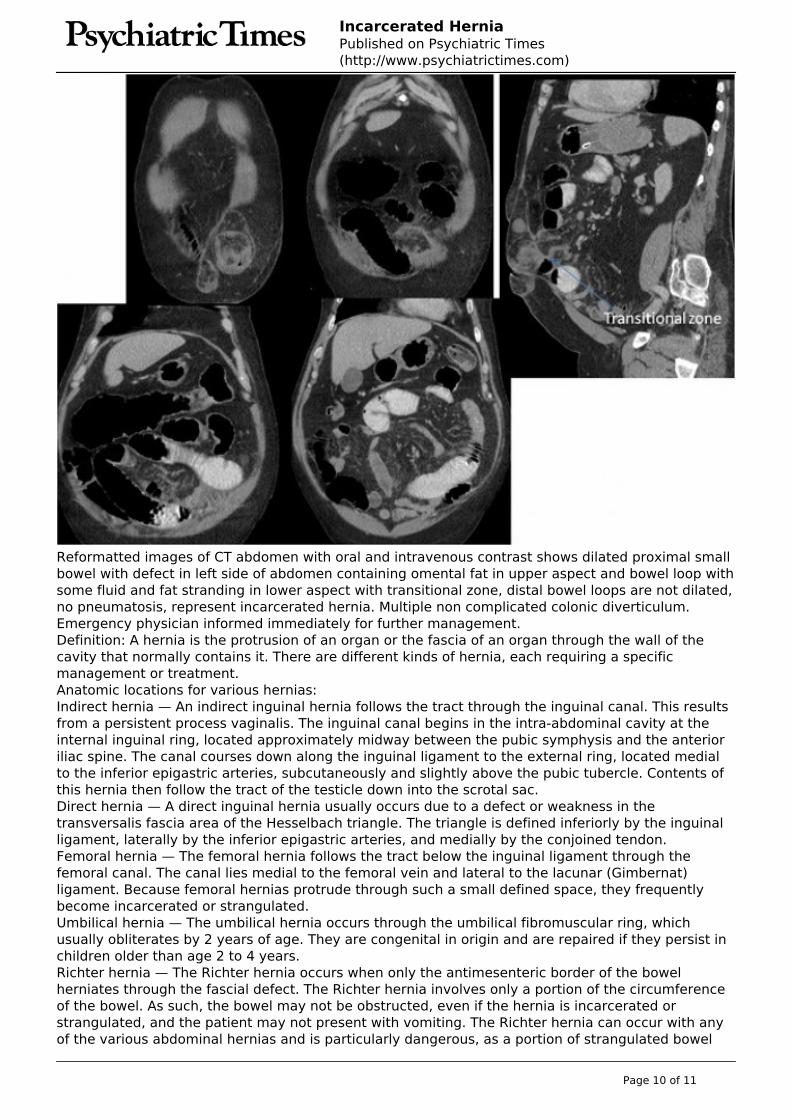
Reformatted images of CT abdomen with oral and intravenous contrast shows dilated proximal smallbowel with defect in left side of abdomen containing omental fat in upper aspect and bowel loop withsome fluid and fat stranding in lower aspect with transitional zone, distal bowel loops are not dilated,no pneumatosis, represent incarcerated hernia. Multiple non complicated colonic diverticulum.Emergency physician informed immediately for further management.Definition: A hernia is the protrusion of an organ or the fascia of an organ through the wall of thecavity that normally contains it. There are different kinds of hernia, each requiring a specificmanagement or treatment.Anatomic locations for various hernias:Indirect hernia — An indirect inguinal hernia follows the tract through the inguinal canal. This resultsfrom a persistent process vaginalis. The inguinal canal begins in the intra-abdominal cavity at theinternal inguinal ring, located approximately midway between the pubic symphysis and the anterioriliac spine. The canal courses down along the inguinal ligament to the external ring, located medialto the inferior epigastric arteries, subcutaneously and slightly above the pubic tubercle. Contents ofthis hernia then follow the tract of the testicle down into the scrotal sac.Direct hernia — A direct inguinal hernia usually occurs due to a defect or weakness in thetransversalis fascia area of the Hesselbach triangle. The triangle is defined inferiorly by the inguinalligament, laterally by the inferior epigastric arteries, and medially by the conjoined tendon.Femoral hernia — The femoral hernia follows the tract below the inguinal ligament through thefemoral canal. The canal lies medial to the femoral vein and lateral to the lacunar (Gimbernat)ligament. Because femoral hernias protrude through such a small defined space, they frequentlybecome incarcerated or strangulated.Umbilical hernia — The umbilical hernia occurs through the umbilical fibromuscular ring, whichusually obliterates by 2 years of age. They are congenital in origin and are repaired if they persist inchildren older than age 2 to 4 years.Richter hernia — The Richter hernia occurs when only the antimesenteric border of the bowelherniates through the fascial defect. The Richter hernia involves only a portion of the circumferenceof the bowel. As such, the bowel may not be obstructed, even if the hernia is incarcerated orstrangulated, and the patient may not present with vomiting. The Richter hernia can occur with anyof the various abdominal hernias and is particularly dangerous, as a portion of strangulated bowel
Page 10 of 11

Incarcerated HerniaPublished on Psychiatric Times(http://www.psychiatrictimes.com)
may be reduced unknowingly into the abdominal cavity, leading to perforation and peritonitis.Incisional hernia — This iatrogenic hernia occurs in 2 percent to 10 percent of all abdominaloperations secondary to breakdown of the fascial closure of prior surgery. Even after repair,recurrence rates approach 20 percent to 45 percent.Spigelian hernia — This rare form of abdominal wall hernia occurs through a defect in the spigelianfascia, which is defined by the lateral edge of the rectus muscle at the semilunar line (costal arch tothe pubic tubercle) The two subtypes are interstitial and subcutaneous, which are best defined usingCT and assist with optimizing the surgical approach when indicated.Obturator hernia — This hernia passes through the obturator foramen, following the path of theobturator nerves and muscles. Obturator hernias occur with a female-to-male ratio of 6:1, because ofa gender-specific larger canal diameter and predominately in the elderly. Because of its anatomicposition, this hernia presents more commonly as a bowel obstruction than as a protrusion of bowelcontents.Hernia symptoms and signs: The signs and symptoms of a hernia can range from noticing a painlesslump to the severely painful, tender, swollen protrusion of tissue that you are unable to push backinto theabdomen (an incarcerated strangulated hernia).Reducible hernia- It may appear as a new lump in the groin or other abdominal area.- It may ache but is not tender when touched.- Sometimes pain precedes the discovery of the lump.- The lump increases in size when standing or when abdominal pressure is increased (such ascoughing).- It may be reduced (pushed back into the abdomen) unless very large.Irreducible hernia- It may be an occasionally painful enlargement of a previously reducible hernia that cannot bereturned into the abdominal cavity on its own or when you push it.- Some may be chronic (occur over a long term) without pain.- An irreducible hernia is also known as an incarcerated hernia.- It can lead to strangulation (blood supply being cut off to tissue in the hernia).- Signs and symptoms of bowel obstruction may occur, such as nausea and vomiting.Strangulated hernia- This is an irreducible hernia in which the entrapped intestine has its blood supply cut off.- Pain is always present, followed quickly by tenderness and sometimes symptoms of bowelobstruction (nausea and vomiting).- The affected person may appear ill with or without fever.- This condition is a surgical emergency.Sushila Ladumor, MD, FRCR, consultant radiologist with multi-modality imaging experience, workingin Medical Imaging Department, King Abdulaziz Medical City, Riyadh, Saudi Arabia Source URL: http://www.psychiatrictimes.com/case-studies/incarcerated-hernia
Links:[1] http://www.psychiatrictimes.com/case-studies[2] http://www.psychiatrictimes.com/authors/sushila-ladumor-md
Page 11 of 11