PSRJournal Article Simplifying the Management of Caudal Septal ...
10
www.PRSJournal.com 379e A chieving a consistent functional and aes- thetic result when performing rhinoplasty demands intimate knowledge and under- standing of nasal anatomy and physiology. Cor- rection of the deviated nose is no exception. Accurate preoperative analysis and intraoperative diagnosis are essential to the surgeon’s success. Correction of these deformities poses an array of challenges to the rhinoplasty surgeon because of the dilemma of dealing with both functional and aesthetic problems at the same time. The anatomy of a deviated nose can involve asymmetries of the bony pyramid, a septal abnormality, or asymme- tries of the upper and lower lateral cartilages, but usually involves some combination of these prob- lems. This can result in nasal airway obstruction without external deviation, external deviation without nasal airway obstruction, or external devi- ation with nasal airway obstruction. In addition, facial asymmetries add complexity to the analysis. Classification systems exist for septal deformi- ties 1 and the deviated nose. 2 A multitude of tech- niques to address these deformities, including swinging door flaps, a variety of grafts including battens and spreaders, scoring, wedging, and extra- corporeal septoplasty, have been described. 1–20 Despite these various techniques, management of caudal septal deviation remains challenging. Building on concepts and techniques used to cor- rect posterior septal deviation, 2 this technique has evolved to include the management of the cau- dally deviated septum. Caudal septal deviation is common in patients seeking rhinoplasty. We performed a recent review of 100 consecutive patients in a single surgeon’s practice that showed a 44 percent prevalence of caudal septal deviation. In this article, our current technique for simplifying the management of the Disclosure: Dr. Rohrich receives book royalties from Quality Medical Publishing and instrument royal- ties from Micrins. The other authors have no finan- cial interest to declare in relation to the content of this article. Copyright © 2014 by the American Society of Plastic Surgeons DOI: 10.1097/PRS.0000000000000236 Fadi C. Constantine, M.D. Jamil Ahmad, M.D. Palmyra Geissler, M.D. Rod J. Rohrich, M.D. Dallas, Texas; and Mississauga, On- tario, Canada Summary: Correction of the deviated nose poses a challenge in even the most experienced hands. Frequently, the surgeon is faced with both a functional (airway obstruction) and an aesthetic problem that must be addressed con- jointly. Accurate preoperative analysis and intraoperative diagnosis are integral to good outcomes. Caudal septal deviation is frequently present in patients presenting for rhinoplasty. The authors’ current graduated technique for sim- plifying the management of the caudally deviated septum both aesthetically and functionally is described. If there is a persistent caudal septal deviation that has not been addressed by standard maneuvers, the caudal portion of the anterior septum is resected at the osseocartilaginous junction with the anterior nasal spine and maxillary crest and then sutured back to the periosteum of the anterior nasal spine with 5-0 polydioxanone. We have found this to be a safe and effective way of addressing the caudally deviated septum in the majority of cases. (Plast. Reconstr. Surg. 134: 379e, 2014.) From the Department of Plastic Surgery, The University of Texas Southwestern Medical Center; and The Plastic Sur- gery Clinic. Received for publication July 16, 2013; accepted December 12, 2013. Simplifying the Management of Caudal Septal Deviation in Rhinoplasty Supplemental digital content is available for this article. Direct URL citations appear in the text; simply type the URL address into any Web browser to access this content. Clickable links to the material are provided in the HTML text of this article on the Journal’s Web site (www. PRSJournal.com). SPECIAL TOPIC
Transcript of PSRJournal Article Simplifying the Management of Caudal Septal ...

www.PRSJournal.com 379e
Achieving a consistent functional and aes-thetic result when performing rhinoplasty demands intimate knowledge and under-
standing of nasal anatomy and physiology. Cor-rection of the deviated nose is no exception. Accurate preoperative analysis and intraoperative diagnosis are essential to the surgeon’s success. Correction of these deformities poses an array of challenges to the rhinoplasty surgeon because of the dilemma of dealing with both functional and aesthetic problems at the same time. The anatomy of a deviated nose can involve asymmetries of the bony pyramid, a septal abnormality, or asymme-tries of the upper and lower lateral cartilages, but usually involves some combination of these prob-lems. This can result in nasal airway obstruction without external deviation, external deviation without nasal airway obstruction, or external devi-ation with nasal airway obstruction. In addition, facial asymmetries add complexity to the analysis.
Classification systems exist for septal deformi-ties1 and the deviated nose.2 A multitude of tech-niques to address these deformities, including swinging door flaps, a variety of grafts including
battens and spreaders, scoring, wedging, and extra-corporeal septoplasty, have been described.1–20 Despite these various techniques, management of caudal septal deviation remains challenging. Building on concepts and techniques used to cor-rect posterior septal deviation,2 this technique has evolved to include the management of the cau-dally deviated septum.
Caudal septal deviation is common in patients seeking rhinoplasty. We performed a recent review of 100 consecutive patients in a single surgeon’s practice that showed a 44 percent prevalence of caudal septal deviation. In this article, our current technique for simplifying the management of the
Disclosure: Dr. Rohrich receives book royalties from Quality Medical Publishing and instrument royal-ties from Micrins. The other authors have no finan-cial interest to declare in relation to the content of this article.
Copyright © 2014 by the American Society of Plastic Surgeons
DOI: 10.1097/PRS.0000000000000236
Fadi C. Constantine, M.D.Jamil Ahmad, M.D.
Palmyra Geissler, M.D.Rod J. Rohrich, M.D.
Dallas, Texas; and Mississauga, On-tario, Canada
Summary: Correction of the deviated nose poses a challenge in even the most experienced hands. Frequently, the surgeon is faced with both a functional (airway obstruction) and an aesthetic problem that must be addressed con-jointly. Accurate preoperative analysis and intraoperative diagnosis are integral to good outcomes. Caudal septal deviation is frequently present in patients presenting for rhinoplasty. The authors’ current graduated technique for sim-plifying the management of the caudally deviated septum both aesthetically and functionally is described. If there is a persistent caudal septal deviation that has not been addressed by standard maneuvers, the caudal portion of the anterior septum is resected at the osseocartilaginous junction with the anterior nasal spine and maxillary crest and then sutured back to the periosteum of the anterior nasal spine with 5-0 polydioxanone. We have found this to be a safe and effective way of addressing the caudally deviated septum in the majority of cases. (Plast. Reconstr. Surg. 134: 379e, 2014.)
From the Department of Plastic Surgery, The University of Texas Southwestern Medical Center; and The Plastic Sur-gery Clinic.Received for publication July 16, 2013; accepted December 12, 2013.
Simplifying the Management of Caudal Septal Deviation in Rhinoplasty
Supplemental digital content is available for this article. Direct URL citations appear in the text; simply type the URL address into any Web browser to access this content. Clickable links to the material are provided in the HTML text of this article on the Journal’s Web site (www.PRSJournal.com).
Special Topic

380e
Plastic and Reconstructive Surgery • September 2014
caudally deviated septum both aesthetically and functionally is outlined.
OPERATIVE TECHNIQUEOpen rhinoplasty is the preferred approach
for accurately diagnosing and correcting all devi-ated structures. This exposure allows unprec-edented access to the entire septum and direct visualization of all structures.
Release of the MucoperichondriumA submucoperichondrial dissection is per-
formed using a Cottle elevator, beginning at the anterior septal angle. Bilateral mucoperichon-drial tunnels are dissected deep to the upper lat-eral cartilages, and a scalpel is used to separate the upper lateral cartilages from the dorsal sep-tum. The mucoperichondrial attachments must be widely released to allow any deviated portions of the septum to be returned to the correct posi-tion. In the case of caudal septal deviation, the mucoperichondrium must be released all the way to the anterior nasal spine. In some cases, the sep-tum will straighten after release of the mucoperi-chondrium if there are no intrinsic forces causing deviation of the cartilaginous or bony septum.
Creation of the L-StrutOnce the deforming extrinsic forces have
been released, the septum is assessed for intrinsic deviation. Septal reconstruction is performed to correct intrinsic septal deviation. Anterior septal deviation will lead to an external deformity. The anterior septum is separated from the posterior septum by creating an L-strut composed of the dorsal and caudal anterior septum. The dorsal and caudal L-strut should be at least 10 mm wide; however, this will depend on the strength of the septal cartilage, and in many instances, a width of 15 mm or more may be required to ensure long-term support. In addition, curving the transition points between the perpendicular plate of the eth-moid and the dorsal L-strut, and also between the dorsal and caudal L-strut, can help to strengthen the construct20 (Fig. 1).
Reconstruction of the Posterior SeptumThe anterior two-thirds and the inferior aspect
of the septum posteriorly can affect nasal airflow significantly when deviated from the midline.21 Anterior and inferior septal deviations tend to be poorly tolerated, as the cross-sectional area of the nasal airway is small compared with posterior in the nasal cavity.21 Septal reconstruction involves
returning the deviated septum to the midline. The principle of cartilage preservation is essential. Cartilaginous septum that is deviated or required for grafting should be removed. When address-ing the bony septum, the septum can be micro-fractured and returned to the midline. In cases of C- or S-shaped craniocaudal deviation, there is vertical excess of the septum, and removing the inferior aspect of the septum allows for microfrac-ture of the remaining septum and return to the midline. Microfracture should be performed in a careful and controlled manner to avoid uncon-trolled fractures into the superior nasal septum and cribriform plate. This is particularly impor-tant in posttraumatic cases where there may have been a prior septal fracture. Bony spurs of the septum can be removed using Takahashi forceps. Septal cartilage or bone should be removed with ease; if there is any resistance, residual soft-tissue attachments should be completely released.
Correcting Caudal Septal DeviationOnce the posterior septum has been recon-
structed, if there is a persistent caudal septal deviation, this is typically caused by vertical excess of the anterior septum (Fig. 2). The cau-dal portion of the L-strut is disarticulated from the osseocartilaginous junction with the ante-rior nasal spine and maxillary crest. The degree
Fig. 1. in many instances, the width of the dorsal and caudal l-strut should be 15 mm or more to ensure long-term support. curving the transition points between the perpendicular plate of the ethmoid and the dorsal l-strut and between the dorsal and caudal l-strut can help add strength.

Volume 134, Number 3 • Caudal Septal Deviation
381e
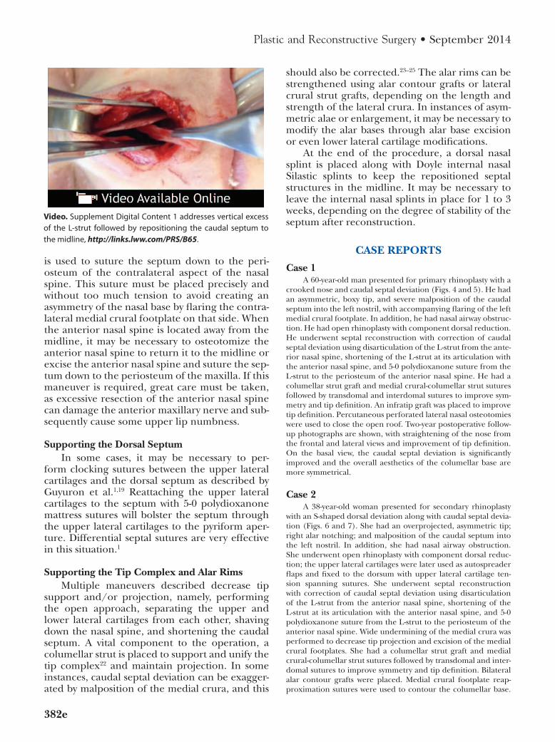
of vertical excess is assessed and excised to allow the previously deviated septum to be returned to the midline (Fig. 3). (See Video, Supplemen-tal Digital Content 1, which addresses vertical excess of the L-strut followed by repositioning the caudal septum to the midline, http://links.lww.com/PRS/B65.) Both excision of the caudal septum and release and excision of the septum at its articulation with the anterior nasal spine and maxillary crest have the potential to decrease tip projection. In many cases, only the portion of
the caudal L-strut that articulates with the ante-rior nasal spine and maxillary crest needs to be excised and the free edge of the caudal septum remains, whereas in other cases, both need to be altered. These maneuvers will alter tip projection and rotation in different ways, and their effects must be kept in mind when manipulating the tip complex and setting tip projection and rotation. In addition, suturing the caudal L-strut back to the anterior nasal spine must be accurate to rec-reate the articulation. A 5-0 polydioxanone suture
Fig. 2. (Left) cranial caudal septal deviation caused by vertical excess shown in red. (Center) Release of the cau-dal l-strut from the maxillary crest, with the amount of vertical excess shown in red. (Right) Repositioning of the caudal l-strut to the midline after excision of the excess caudal l-strut.
Fig. 3. intraoperative views showing correction of vertical excess of the septum (left) before release of the caudal l-strut and shortening followed by (right) repositioning to the midline by suturing to the anterior nasal spine.
382e
Plastic and Reconstructive Surgery • September 2014
is used to suture the septum down to the peri-osteum of the contralateral aspect of the nasal spine. This suture must be placed precisely and without too much tension to avoid creating an asymmetry of the nasal base by flaring the contra-lateral medial crural footplate on that side. When the anterior nasal spine is located away from the midline, it may be necessary to osteotomize the anterior nasal spine to return it to the midline or excise the anterior nasal spine and suture the sep-tum down to the periosteum of the maxilla. If this maneuver is required, great care must be taken, as excessive resection of the anterior nasal spine can damage the anterior maxillary nerve and sub-sequently cause some upper lip numbness.
Supporting the Dorsal SeptumIn some cases, it may be necessary to per-
form clocking sutures between the upper lateral cartilages and the dorsal septum as described by Guyuron et al.1,19 Reattaching the upper lateral cartilages to the septum with 5-0 polydioxanone mattress sutures will bolster the septum through the upper lateral cartilages to the pyriform aper-ture. Differential septal sutures are very effective in this situation.1
Supporting the Tip Complex and Alar RimsMultiple maneuvers described decrease tip
support and/or projection, namely, performing the open approach, separating the upper and lower lateral cartilages from each other, shaving down the nasal spine, and shortening the caudal septum. A vital component to the operation, a columellar strut is placed to support and unify the tip complex22 and maintain projection. In some instances, caudal septal deviation can be exagger-ated by malposition of the medial crura, and this
should also be corrected.23–25 The alar rims can be strengthened using alar contour grafts or lateral crural strut grafts, depending on the length and strength of the lateral crura. In instances of asym-metric alae or enlargement, it may be necessary to modify the alar bases through alar base excision or even lower lateral cartilage modifications.
At the end of the procedure, a dorsal nasal splint is placed along with Doyle internal nasal Silastic splints to keep the repositioned septal structures in the midline. It may be necessary to leave the internal nasal splints in place for 1 to 3 weeks, depending on the degree of stability of the septum after reconstruction.
CASE REPORTS
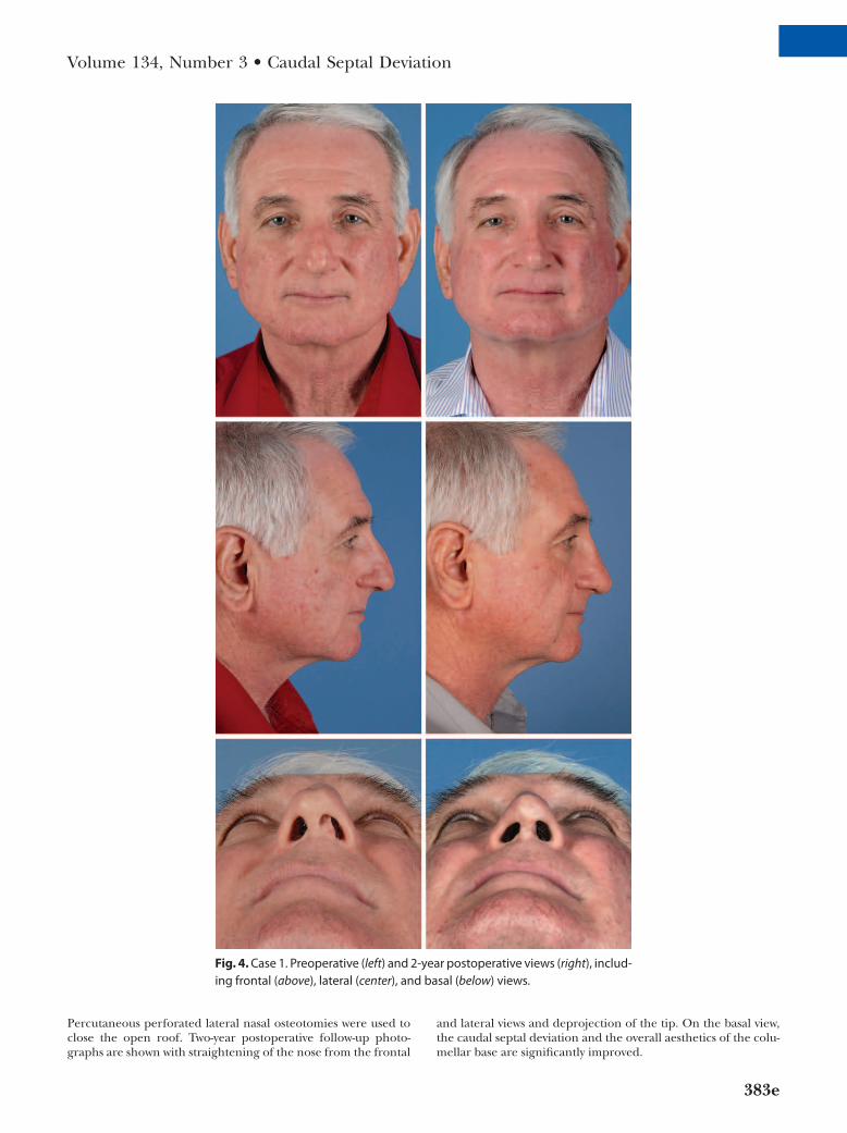
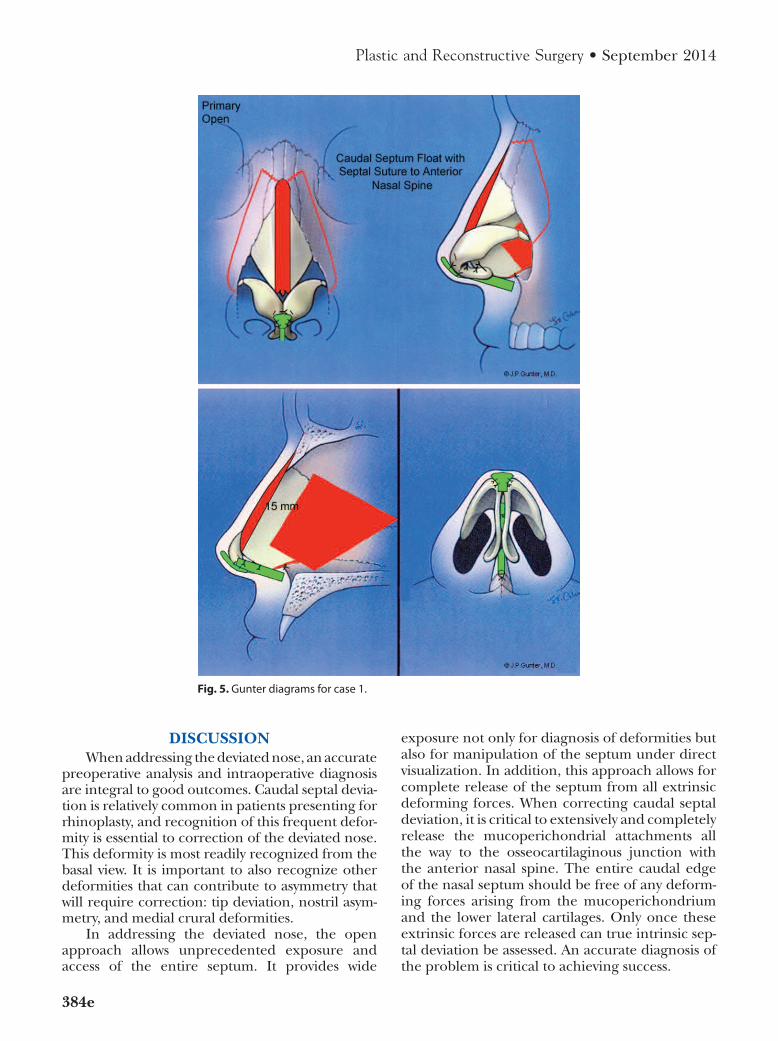
Case 1A 60-year-old man presented for primary rhinoplasty with a
crooked nose and caudal septal deviation (Figs. 4 and 5). He had an asymmetric, boxy tip, and severe malposition of the caudal septum into the left nostril, with accompanying flaring of the left medial crural footplate. In addition, he had nasal airway obstruc-tion. He had open rhinoplasty with component dorsal reduction. He underwent septal reconstruction with correction of caudal septal deviation using disarticulation of the L-strut from the ante-rior nasal spine, shortening of the L-strut at its articulation with the anterior nasal spine, and 5-0 polydioxanone suture from the L-strut to the periosteum of the anterior nasal spine. He had a columellar strut graft and medial crural-columellar strut sutures followed by transdomal and interdomal sutures to improve sym-metry and tip definition. An infratip graft was placed to improve tip definition. Percutaneous perforated lateral nasal osteotomies were used to close the open roof. Two-year postoperative follow-up photographs are shown, with straightening of the nose from the frontal and lateral views and improvement of tip definition. On the basal view, the caudal septal deviation is significantly improved and the overall aesthetics of the columellar base are more symmetrical.
Case 2A 38-year-old woman presented for secondary rhinoplasty
with an S-shaped dorsal deviation along with caudal septal devia-tion (Figs. 6 and 7). She had an overprojected, asymmetric tip; right alar notching; and malposition of the caudal septum into the left nostril. In addition, she had nasal airway obstruction. She underwent open rhinoplasty with component dorsal reduc-tion; the upper lateral cartilages were later used as autospreader flaps and fixed to the dorsum with upper lateral cartilage ten-sion spanning sutures. She underwent septal reconstruction with correction of caudal septal deviation using disarticulation of the L-strut from the anterior nasal spine, shortening of the L-strut at its articulation with the anterior nasal spine, and 5-0 polydioxanone suture from the L-strut to the periosteum of the anterior nasal spine. Wide undermining of the medial crura was performed to decrease tip projection and excision of the medial crural footplates. She had a columellar strut graft and medial crural-columellar strut sutures followed by transdomal and inter-domal sutures to improve symmetry and tip definition. Bilateral alar contour grafts were placed. Medial crural footplate reap-proximation sutures were used to contour the columellar base.
Video. Supplement Digital content 1 addresses vertical excess of the l-strut followed by repositioning the caudal septum to the midline, http://links.lww.com/PRS/B65.
Volume 134, Number 3 • Caudal Septal Deviation
383e
Percutaneous perforated lateral nasal osteotomies were used to close the open roof. Two-year postoperative follow-up photo-graphs are shown with straightening of the nose from the frontal
and lateral views and deprojection of the tip. On the basal view, the caudal septal deviation and the overall aesthetics of the colu-mellar base are significantly improved.
Fig. 4. case 1. preoperative (left) and 2-year postoperative views (right), includ-ing frontal (above), lateral (center), and basal (below) views.
384e
Plastic and Reconstructive Surgery • September 2014
DISCUSSIONWhen addressing the deviated nose, an accurate
preoperative analysis and intraoperative diagnosis are integral to good outcomes. Caudal septal devia-tion is relatively common in patients presenting for rhinoplasty, and recognition of this frequent defor-mity is essential to correction of the deviated nose. This deformity is most readily recognized from the basal view. It is important to also recognize other deformities that can contribute to asymmetry that will require correction: tip deviation, nostril asym-metry, and medial crural deformities.
In addressing the deviated nose, the open approach allows unprecedented exposure and access of the entire septum. It provides wide
expo sure not only for diagnosis of deformities but also for manipulation of the septum under direct visualization. In addition, this approach allows for complete release of the septum from all extrinsic deforming forces. When correcting caudal septal deviation, it is critical to extensively and completely release the mucoperichondrial attachments all the way to the osseocartilaginous junction with the anterior nasal spine. The entire caudal edge of the nasal septum should be free of any deform-ing forces arising from the mucoperichondrium and the lower lateral cartilages. Only once these extrinsic forces are released can true intrinsic sep-tal deviation be assessed. An accurate diagnosis of the problem is critical to achieving success.
Fig. 5. Gunter diagrams for case 1.

Volume 134, Number 3 • Caudal Septal Deviation
385e
Repositioning or resection of the deviated septum is referred to as “septal reconstruction,” as opposed to septoplasty or submucous resection.
Treatment of the posterior septum should include harvest of any septal cartilage for grafting, removal of any significantly deviated septum or
Fig. 6. case 2. preoperative (left) and 2-year postoperative views (right), including frontal (above), lateral (center), and basal (below) views.

386e
Plastic and Reconstructive Surgery • September 2014
spurs contributing to nasal airway obstruction that cannot be repositioned, and microfracture of any mildly deviated septum with reposition-ing to the midline. Preservation of cartilage is a key principle in modern rhinoplasty, and applies to septal reconstruction as well. If there is har-vested cartilage that remains unused at the end of the procedure, it can be banked posterior to the L-strut in the mucoperichondrial pockets. Releas-ing the L-strut/anterior septum from the poste-rior septum allows straightening of the L-strut by removing posterior deforming forces from the septal deviation. The literature is replete with the adage that only 8 to 10 mm of dorsal and caudal L-strut needs to be preserved to maintain its struc-tural integrity. Instead, the amount of L-strut that
should be preserved is highly dependent on the strength and quality of the anterior septum—in general, more is better; it is best to leave more than 10 mm caudal and dorsal and if possible 15 mm. In addition, curving the posterior edges of the L-strut at the superior osseocartilaginous junction or transition from the dorsal to caudal L-strut may contribute strength.20
If the L-strut remains deviated after the afore-mentioned maneuvers, it is most likely caused by vertical excess of the L-strut. The bony roof and floor of the pyriform aperture and nasal cavity are fixed structures. Vertical excess of the L-strut will contribute to craniocaudal C- or S-shaped devia-tion. The caudal L-strut can be disinserted at the osseocartilaginous junction with the anterior nasal
Fig. 7. Gunter diagrams for case 2.
Volume 134, Number 3 • Caudal Septal Deviation
387e
spine and maxillary crest and then shortened to remove the vertical excess. This is followed by suturing it back to the periosteum of the anterior nasal spine with 5-0 polydioxanone. Although these concepts and simplified approach to cor-rection of caudal septal deviation may work effec-tively in the majority of cases, in some instances, it is necessary to be more aggressive in correcting severe deformities of the anterior septum. As we described previously,2 scoring or partial-thickness wedge excisions coupled with the application of
splinting grafts may be required to establish a straight and stable L-strut. Similarly, deformities of the anterior septum stemming from previous fracture lines such as sharp angulations or overlap-ping segments also require more complex maneu-vers to first weaken or divide the anterior L-strut and then reinforce or reconstruct the L-strut into a straight construct. Techniques such as splinting grafts, tongue-in-groove grafts, drill-hole fixation, or medial crural footplate excision may be nec-essary for adequate correction of severe caudal septal deviations. These techniques may require more cartilage than present in the septum and necessitate ear or rib cartilage harvest.
Several other maneuvers are worth mention-ing when performing the aforementioned steps for correcting the caudally deviated septum. When extensive work has been performed on the caudal septum, it is usually necessary to place sev-eral through-and-through horizontal mattress 5-0 chromic gut sutures in the caudal septum to reap-proximate the caudal mucoperichondrial flaps to the midline.26 This allows the flaps to scar down in the midline position and provide extra long-term support. Along the same lines, it may be necessary to perform clocking sutures from the upper lat-eral cartilage to the dorsal septum to help reposi-tion the L-strut in the midline.
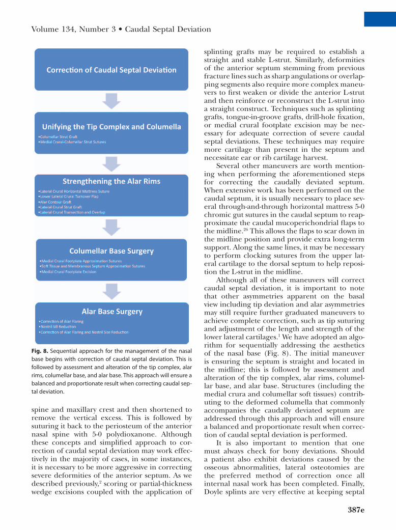
Although all of these maneuvers will correct caudal septal deviation, it is important to note that other asymmetries apparent on the basal view including tip deviation and alar asymmetries may still require further graduated maneuvers to achieve complete correction, such as tip suturing and adjustment of the length and strength of the lower lateral cartilages.1 We have adopted an algo-rithm for sequentially addressing the aesthetics of the nasal base (Fig. 8). The initial maneuver is ensuring the septum is straight and located in the midline; this is followed by assessment and alteration of the tip complex, alar rims, columel-lar base, and alar base. Structures (including the medial crura and columellar soft tissues) contrib-uting to the deformed columella that commonly accompanies the caudally deviated septum are addressed through this approach and will ensure a balanced and proportionate result when correc-tion of caudal septal deviation is performed.
It is also important to mention that one must always check for bony deviations. Should a patient also exhibit deviations caused by the osseous abnormalities, lateral osteotomies are the preferred method of correction once all internal nasal work has been completed. Finally, Doyle splints are very effective at keeping septal
Fig. 8. Sequential approach for the management of the nasal base begins with correction of caudal septal deviation. This is followed by assessment and alteration of the tip complex, alar rims, columellar base, and alar base. This approach will ensure a balanced and proportionate result when correcting caudal sep-tal deviation.
388e
Plastic and Reconstructive Surgery • September 2014
structures in the midline after septal reconstruc-tion as opposed to nasal packing. These are usu-ally placed with antibiotic ointment and secured using a 2-0 nylon suture. They are typically removed after 7 to 10 days.
CONCLUSIONSThe deviated nose is a common problem seen
by the rhinoplasty surgeon. We have described a simplified method of addressing caudal septal deviation through a graduated approach. If there is a persistent caudal septal deviation that has not been addressed by standard maneuvers, the cau-dal portion of the anterior septum is resected at the osseocartilaginous junction with the anterior nasal spine and maxillary crest and then sutured back to the periosteum of the anterior nasal spine with 5-0 polydioxanone. We have found this to be a safe and effective way of addressing the caudally deviated septum.
Rod J. Rohrich, M.D.Department of Plastic Surgery
The University of Texas Southwestern Medical Center 1801 Inwood Road
Dallas, Texas [email protected]
PATIENT CONSENTPatients provided written consent for the use of their
images.
REFERENCES 1. Guyuron B, Uzzo CD, Scull H. A practical classification of
septonasal deviation and an effective guide to septal surgery. Plast Reconstr Surg. 1999;104:2202–2209; discussion 2210.
2. Rohrich RJ, Gunter JP, Deuber MA, Adams WP Jr. The deviated nose: Optimizing results using a simplified clas-sification and algorithmic approach. Plast Reconstr Surg. 2002;110:1509–1523; discussion 1524–1525.
3. Constantian MB. An algorithm for correcting the asymmetri-cal nose. Plast Reconstr Surg. 1989;83:801–811.
4. Killian G. The submucous window resection of the nasal sep-tum. Ann Otol Rhinol Laryngol. 1905;14:363–416.
5. Dingman RO, Natvig P. The deviated nose. Clin Plast Surg. 1977;4:145–152.
6. Bernstein L. Submucous operations on the nasal septum. Otolaryngol Clin North Am. 1973;6:675–692.
7. Edwards N. Septoplasty: Rational surgery of the nasal sep-tum. J Laryngol Otol. 1975;89:875–897.
8. Converse JM. Corrective surgery of nasal deviations. AMA Arch Otolaryngol. 1950;52:671–708.
9. Fry H. Nasal skeleton trauma and the interlocked stresses of the nasal septal cartilage. Br J Plast Surg. 1967;20:146–158.
10. Gruber R, Lesavoy M. Closed septal osteotomy. Ann Plast Surg. 1998;40:283–286.
11. Johnson CM Jr, Anderson JR. The deviated nose: Its correc-tion. Laryngoscope 1977;87:1680–1684.
12. Byrd HS, Salomon J, Flood J. Correction of the crooked nose. Plast Reconstr Surg. 1998;102:2148–2157.
13. De la Fuente A, Martin del Yerro JL. Management of the sep-tum during rhinoplasty. Aesthetic Plast Surg. 1995;19:455–461.
14. Cannistrá C, Guerrieri L, Iannetti G. Deviated nose: Technical proposition for prevention of recurrences. Aesthetic Plast Surg. 1998;22:206–210.
15. Rees TD. Surgical correction of the severely deviated nose by extramucosal excision of the osseocartilaginous sep-tum and replacement as a free graft. Plast Reconstr Surg. 1986;78:320–330.
16. Gubisch W. The extracorporeal septum plasty: A technique to correct difficult nasal deformities. Plast Reconstr Surg. 1995;95:672–682.
17. Jugo SB. Total septal reconstruction through decortication (external) approach in children. Arch Otolaryngol Head Neck Surg. 1987;113:173–178.
18. Gunter JP, Rohrich RJ. Management of the deviated nose: The importance of septal reconstruction. Clin Plast Surg. 1988;15:43–55.
19. Guyuron B, Behmand RA. Caudal nasal deviation. Plast Reconstr Surg. 2003;111:2449–2457; discussion 2458.
20. Mau T, Mau ST, Kim DW. Cadaveric and engineering analysis of the septal L-strut. Laryngoscope 2007;117:1902–1906.
21. Howard BK, Rohrich RJ. Understanding the nasal airway: Principles and practice. Plast Reconstr Surg. 2002;109:1128–1146; quiz 1145–1146.
22. Rohrich RJ, Hoxworth RE, Kurkjian TJ. The role of the colu-mellar strut in rhinoplasty: Indications and rationale. Plast Reconstr Surg. 2012;129:118e–125e.
23. Rohrich RJ, Liu JH. Defining the infratip lobule in rhino-plasty: Anatomy, pathogenesis of abnormalities, and cor-rection using an algorithmic approach. Plast Reconstr Surg. 2012;130:1148–1158.
24. Rohrich RJ, Ahmad J. Rhinoplasty. Plast Reconstr Surg. 2011;128:49e–73e.
25. Cerkes N. The crooked nose: Principles of treatment. Aesthet Surg J. 2011;31:241–257.
26. Geissler PJ, Lee MR, Roostaeian J, Unger JG, Rohrich RJ. Reshaping the medial nostril and columellar base: Five-step medial crural footplate approximation. Plast Reconstr Surg. 2013;132:553–557.


![Review Article Nasal Septal Deviations: A Systematic ...downloads.hindawi.com/journals/psi/2016/7089123.pdf · caudal end (CE) of the septum nasi [ ]. ... septum as compared to an](https://static.fdocuments.in/doc/165x107/607b7a1f88bc8f23352b29c9/review-article-nasal-septal-deviations-a-systematic-caudal-end-ce-of-the.jpg)