PspoIIE gfp - Rudner Lab · 2020-02-10 · Supplemental Figure 1 (Marquis et al.) Figure S1...
13
-7˚::(tetO) 120 B -91˚::(tetO) 120 GFP membranes merge overexposed P spoIIE -gfp A Supplemental Figure 1 (Marquis et al.)
Transcript of PspoIIE gfp - Rudner Lab · 2020-02-10 · Supplemental Figure 1 (Marquis et al.) Figure S1...

-7˚::(tetO)120
B
-91˚::(tetO)120
GFP membranes merge
overexposed
PspoIIE -gfp A
Supplemental Figure 1 (Marquis et al.)

Supplemental Figure 1 (Marquis et al.)
Figure S1 TetR-GFP is stripped off the tetO array in sporulating cells that contain a wild-typecopy of SpoIIIE. In these experiments tetR-gfp was fused to a promoter (PspoIIE) that is activatedprior to polar septation in the predivisional cell. (A) A strain (BDR2128) harboring a PspoIIE-gfpfusion is shown early during sporulation. Transcription from the PspoIIE promoter initiates prior toasymmetric division resulting in an even distribution of cytoplasmic GFP at the time of polarseptation (red carets). After asymmetric division, transcription from this promoter persists in themother cell but is down-regulated in the forespore compartment (compare yellow and whitecarets). (B) Fluorescence images of strains harboring the tetR-gfp fusion under the PspoIIEpromoter. In the upper panel the tetO array was placed at an origin-proximal site (-7˚) (strainBKM986) and in the lower panel it was placed at an original-distal site (-91˚) (strain BKM1245).TetR-GFP synthesized in the predivisional cell binds to the tetO arrays and generates two foci ofequal intensity. Upon polar division, one of the -7˚ arrays is trapped in the foresporecompartment. Although synthesis of TetR-GFP continues in the mother cell, the signalintensities of the foci in the mother cell and forespore remain similar (yellow carets). Thissuggests that TetR-GFP saturates the tetO binding sites prior to polar division. The -91˚ arraysare both present in the mother cell at the time of polar division and at this time the two foci havesimilar intensity (yellow carets). However, when the array on the forespore chromosome istranslocated into the forespore the fluorescent signal is reduced (compare white and yellowcarets). We interpret this to mean that the fusion protein is stripped off the DNA duringtranlocation and then rebound by the sub-saturating levels of TetR-GFP present in the forespore.A rescaled (overexposed) image is shown to visualize the weak forespore foci. Scale bar, 1 µm.
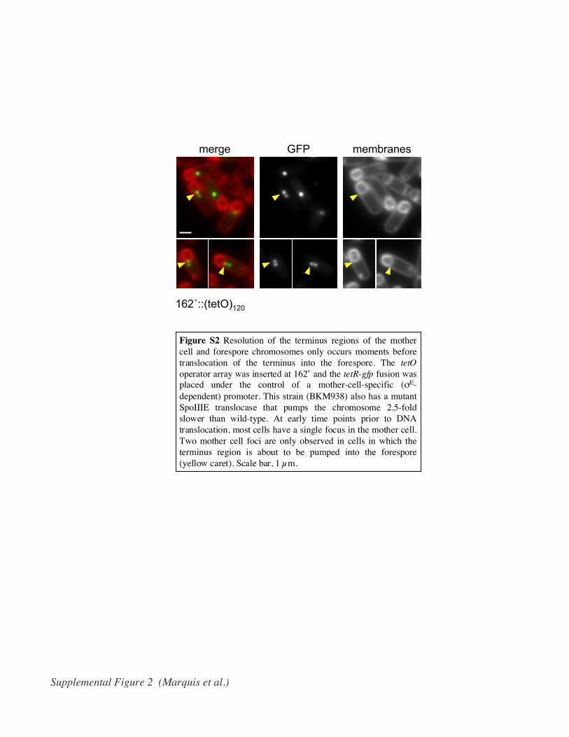
162˚::(tetO)120
merge GFP membranes
Supplemental Figure 2 (Marquis et al.)
Figure S2 Resolution of the terminus regions of the mothercell and forespore chromosomes only occurs moments beforetranslocation of the terminus into the forespore. The tetOoperator array was inserted at 162˚ and the tetR-gfp fusion wasplaced under the control of a mother-cell-specific (σE-dependent) promoter. This strain (BKM938) also has a mutantSpoIIIE translocase that pumps the chromosome 2.5-foldslower than wild-type. At early time points prior to DNAtranslocation, most cells have a single focus in the mother cell.Two mother cell foci are only observed in cells in which theterminus region is about to be pumped into the forespore(yellow caret). Scale bar, 1 µm.

free DNA
-NspI
+N
spI
IC
AT
P
SpoIII E
SpoIIIE
+A
TP
Supplemental Figure 3 (Marquis et al.)
Figure S3 SpoIIIE-dependent displacement of a transcriptioninitiation complex. E. coli RNA polymerase containing σ70
was incubated with a fluorescently-labeled DNA substratecontaining the λPR promoter to generate an initiation complex(IC). The complexes were then incubated with 3 mM ATP,120 nM SpoIIIE and 3mM ATP, or 120nM SpoIIIE andanalyzed by gel mobility shift on a native polyacrylamide gel.The shifted complex is an initiation complex because arestriction endonuclease (NspI) was unable to cleave the DNAat a site within the promoter region.
A
B
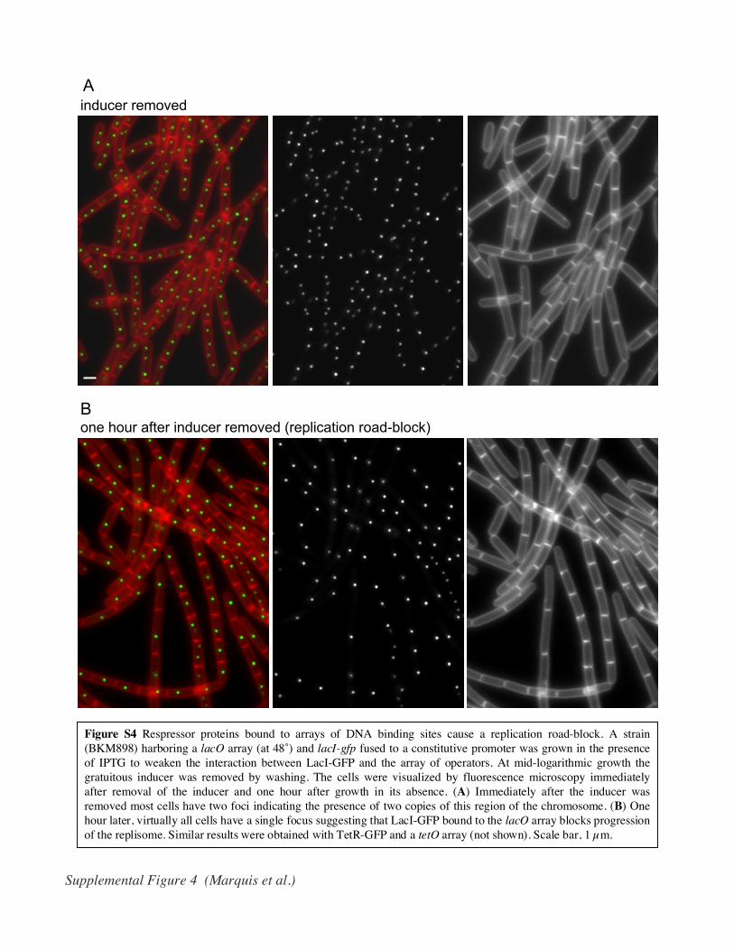
inducer removed
one hour after inducer removed (replication road-block)
Supplemental Figure 4 (Marquis et al.)
Figure S4 Respressor proteins bound to arrays of DNA binding sites cause a replication road-block. A strain(BKM898) harboring a lacO array (at 48˚) and lacI-gfp fused to a constitutive promoter was grown in the presenceof IPTG to weaken the interaction between LacI-GFP and the array of operators. At mid-logarithmic growth thegratuitous inducer was removed by washing. The cells were visualized by fluorescence microscopy immediatelyafter removal of the inducer and one hour after growth in its absence. (A) Immediately after the inducer wasremoved most cells have two foci indicating the presence of two copies of this region of the chromosome. (B) Onehour later, virtually all cells have a single focus suggesting that LacI-GFP bound to the lacO array blocks progressionof the replisome. Similar results were obtained with TetR-GFP and a tetO array (not shown). Scale bar, 1 µm.
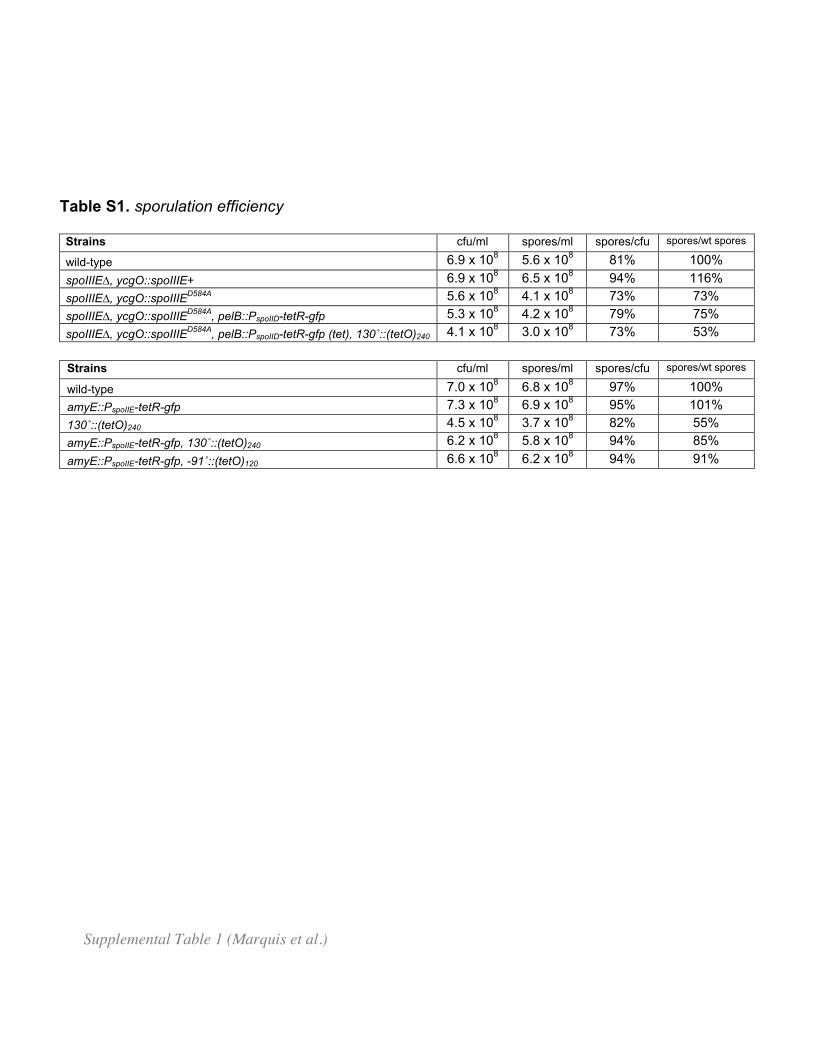
Table S1. sporulation efficiency
Strains cfu/ml spores/ml spores/cfu spores/wt spores
wild-type 6.9 x 10⁸ 5.6 x 10⁸ 81% 100%
spoIIIE∆, ycgO::spoIIIE+ 6.9 x 10⁸ 6.5 x 10⁸ 94% 116%
spoIIIE∆, ycgO::spoIIIED584A 5.6 x 10⁸ 4.1 x 10⁸ 73% 73%
spoIIIE∆, ycgO::spoIIIED584A, pelB::PspoIID-tetR-gfp 5.3 x 10⁸ 4.2 x 10⁸ 79% 75%
spoIIIE∆, ycgO::spoIIIED584A, pelB::PspoIID-tetR-gfp (tet), 130˚::(tetO)240 4.1 x 10⁸ 3.0 x 10⁸ 73% 53%
Strains cfu/ml spores/ml spores/cfu spores/wt spores
wild-type 7.0 x 10⁸ 6.8 x 10⁸ 97% 100%
amyE::PspoIIE-tetR-gfp 7.3 x 10⁸ 6.9 x 10⁸ 95% 101%
130˚::(tetO)240 4.5 x 10⁸ 3.7 x 10⁸ 82% 55%
amyE::PspoIIE-tetR-gfp, 130˚::(tetO)240 6.2 x 10⁸ 5.8 x 10⁸ 94% 85%
amyE::PspoIIE-tetR-gfp, -91˚::(tetO)120 6.6 x 10⁸ 6.2 x 10⁸ 94% 91%
Supplemental Table 1 (Marquis et al.)

Table S2. Strains used in this study
strain genotype reference
BDR1499 rpoC-gfp (spec) a gift from M. Fujita
BDR1522 rpoC-gfp (spec), spoIIGB::erm This work
BDR734 amyE::PxylA-gfp (cat) Rudner et al 2002
BDR1778 amyE::Pspac(c)-gfp (spec) Fujita and Losick 2002
BKM986 amyE::PspoIIE-tetR-gfp (spec), yycR::(tetO)120 (cat) This work
BKM1245 amyE::PspoIIE-tetR-gfp (spec), 269˚Ω(tetO)120 (kan) This work
BKM938 spoIIIE::neo, ycgO::spoIIIE(D584A)(cat), pelB::PspoIID-tetR-gfp (tet), 162˚Ω(tetO)120 (spec) This work
BKM898 thrC::Ppen-lacI∆11-gfp (erm), 48˚Ω(lacO)120 (cat) This work
BKM1016 spoIIIE::neo, ycgO::spoIIIE(D584A)(cat),pelB::PspoIID-tetR-gfp (tet), 130˚Ω(tetO)240 (spec) This work
Supplemental Table 2 (Marquis et al.)
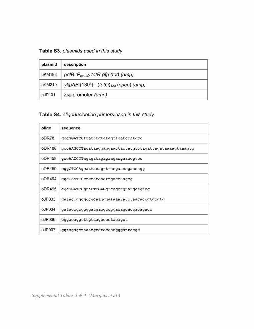
Table S3. plasmids used in this study
plasmid description
pKM193 pelB::PspoIID-tetR-gfp (tet) (amp)
pKM219 ykpAB (130˚) - (tetO)120 (spec) (amp)
pJP101 λPR promoter (amp)
Table S4. oligonucleotide primers used in this study
oligo sequence
oDR78 gccGGATCCttatttgtatagttcatccatgcc
oDR188 gccAAGCTTacataaggaggaactactatgtctagattagataaaagtaaagtg
oDR458 gccAAGCTTagtgatagagaagacgaaccgtcc
oDR459 cggCTCGAgcattacagtttacgaaccgaacagg
oDR494 cgcGAATTCctctatcacttgaccaagcg
oDR495 cgcGGATCCgtaCTCGAGgtccgctgtatgctgtcg
oJP033 gataccggcgccgcaagggataaatatctaacaccgtgcgtg
oJP034 gataccgcggggatgacgccggacagcaccacagacc
oJP036 cggacaggtttgttagcccctacagct
oJP037 ggtagagctaaatgtctacaacgggattccgc
Supplemental Tables 3 & 4 (Marquis et al.)
A
B
membranes GFP DNA merge
stripped translocated
β’-GFP
GFP
Figure 1 (Marquis et al.)
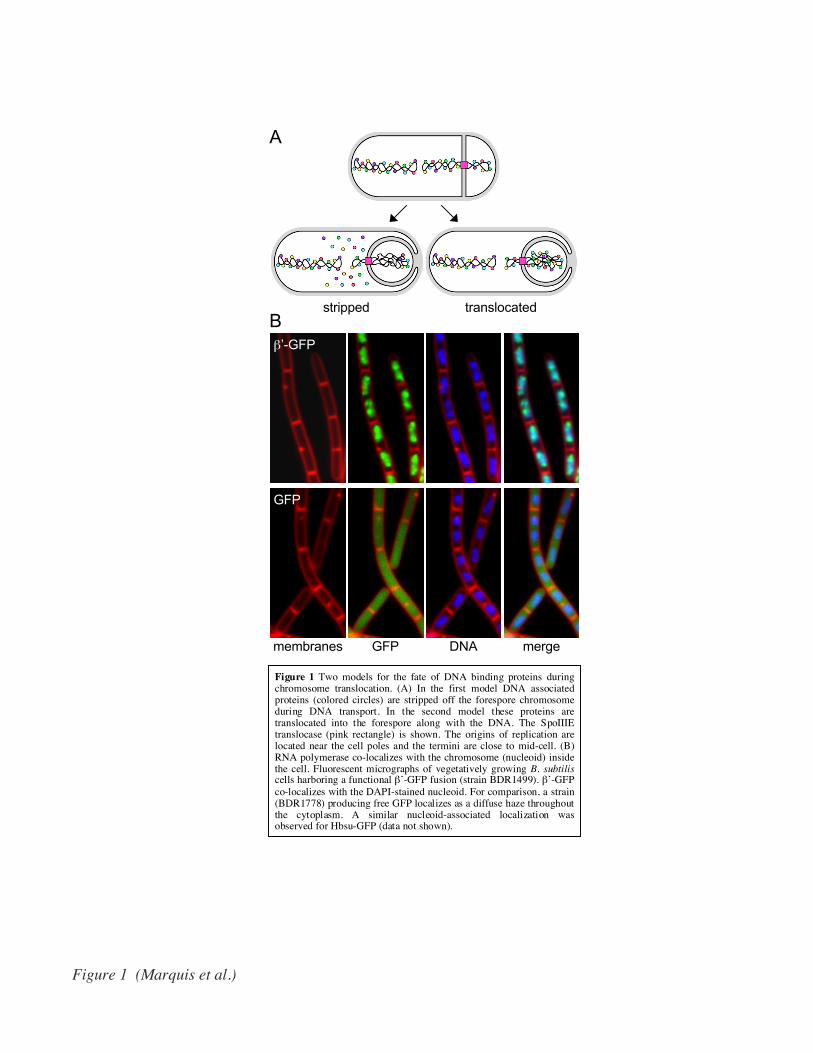
Figure 1 Two models for the fate of DNA binding proteins duringchromosome translocation. (A) In the first model DNA associatedproteins (colored circles) are stripped off the forespore chromosomeduring DNA transport. In the second model these proteins aretranslocated into the forespore along with the DNA. The SpoIIIEtranslocase (pink rectangle) is shown. The origins of replication arelocated near the cell poles and the termini are close to mid-cell. (B)RNA polymerase co-localizes with the chromosome (nucleoid) insidethe cell. Fluorescent micrographs of vegetatively growing B. subtiliscells harboring a functional β’-GFP fusion (strain BDR1499). β’-GFPco-localizes with the DAPI-stained nucleoid. For comparison, a strain(BDR1778) producing free GFP localizes as a diffuse haze throughoutthe cytoplasm. A similar nucleoid-associated localization wasobserved for Hbsu-GFP (data not shown).

wt
disporic
A
B
membranes GFPDNA
stripped translocated
Figure 2 (Marquis et al.)
Figure 2 RNA polymerase is stripped off the DNA during chromosome translocation. (A) Schematicdiagram of the experiment in B with the two possible outcomes. An abortively disporic mutant is shown withRNA polymerase (green balls) associated with the replicated chromosomes. If RNA polymerase is strippedoff the DNA, the fluorescent fusion protein will be present as a diffuse haze in the mother cell cytoplasm. IfRNA polymersase is translocated along with the DNA, the mother cell will be devoid of fluorescent signal.The SpoIIIE translocase (pink square) is shown. (B) β’-GFP is stripped off the chromosome duringtranslocation into the forespore. Fluorescent micrographs of sporulating B. subtilis cells harboring afunctional β’-GFP fusion. The upper panels show the co-localization of β’-GFP with the nucleoid in wild-type sporulating cells (strain BDR1499). The lower panels show β’-GFP in a disporic mutant (strainBDR1522). An abortively disporic sporangium that has not yet completed transport of the chromosomes intothe forespore compartments is shown (yellow caret). In this cell β’-GFP co-localizes with the DNA that isstill present in the mother cell compartment. Two abortively disporic sporangia that have finished DNAtranslocation are indicated (white carets). In these cells there is no DAPI signal in the mother cell and β’-GFPis visible as a diffuse haze in this compartment. The signal appears weaker because the protein is no longerconcentrated on the DNA. Quantitation of fluorescent intensity revealed that 85-90% of the mother cell signalremains after DNA transport. Similar results were obtained with Hbsu-GFP and RacA-GFP (data not shown).Scale bar, 1 µm.

A
Bstripped translocated
σE
tetR gfp
merge GFP membranes
overexposed
Figure 3 (Marquis et al.)
Figure 3 Transcriptional regulators are stripped off the DNA during translocation. (A) Schematic diagram of the experiment in B with thetwo possible outcomes. An array of tetO operators was inserted at a position near the teriminus (130˚) that is always present in the mothercell at the time of polar septation. In this strain, the tetR-gfp fusion was place under the control of a mother-cell-specific (σE-dependent)promoter. Finally, this strain has a mutant SpoIIIE translocase (pink square) that pumps the chromosome 2.5-fold slower than wild-type .Thus, at early time points after σE becomes active in the mother cell, two TetR-GFP foci should be visible in the mother cell. If TetR-GFPis stripped off the DNA one of these two foci will be missing when chromosome translocation is complete. If TetR-GFP is translocatedwith the chromosome, when DNA transport is complete, one TetR-GFP focus will be visible in the forespore and one in the mother cell.(B) TetR-GFP bound to the tetO array is stripped off the chromosome during translocation. Fluorescence micrographs of sporulating B.subtilis cells (strain BKM1016) at early (hour 2) and late (hour 3) time points. At hour 2, prior to the completion of DNA translocation twofluorescent foci are visible in the mother cell (yellow carets). At hour 3, when chromosome translocation is complete in most cells, a singlefocus is visible in the mother cell (yellow caret) and no detectable foci are seen in the forespore (white caret). No TetR-GFP foci are visiblein the forespore when the micrograph is rescaled (overexposed) to reveal weak signals. Scale bar, 1 µm.
B
-NspI
+N
spI
SD
S
AT
P
SpoIIIE
+A
TP
SpoIIIE
RNA
EC
-
1081 79 30 80% bound:
A
SD
S
AT
P
SpoIIIE
+A
TP
SpoIIIE
DNA
EC
-
Figure 4 (Marquis et al.)
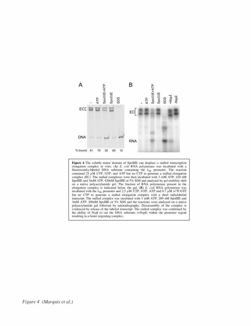
Figure 4 The soluble motor domain of SpoIIIE can displace a stalled transcriptionelongation complex in vitro. (A) E. coli RNA polymerase was incubated with afluorescently-labeled DNA substrate containing the λPR promoter. The reactioncontained 25 µM UTP, GTP, and ATP but no CTP to generate a stalled elongationcomplex (EC). The stalled complexes were then incubated with 3 mM ATP, 420 nMSpoIIIE and 3mM ATP, 420nM SpoIIIE or 5% SDS and analyzed by gel mobility shifton a native polyacrylamide gel. The fraction of RNA polymerase present in theelongation complex is indicated below the gel. (B) E. coli RNA polymerase wasincubated with the λPR promoter and 2.5 µM UTP, GTP, ATP and 0.7 µM α32P-GTPbut no CTP to generate a stalled elongation complex with a short radiolabeledtranscript. The stalled complex was incubated with 3 mM ATP, 200 nM SpoIIIE and3mM ATP, 200nM SpoIIIE or 5% SDS and the reactions were analzyed on a nativepolyacrylamide gel followed by autoradiography. Dissassembly of the complex isevidenced by release of the labeled transcript. The stalled complex was confirmed bythe ability of NspI to cut the DNA substrate (+NspI) within the promoter regionresulting in a faster migrating complex.

mergeGFP membranes
A
B
xylose
gfp
σA
Figure 5 (Marquis et al.)
Figure 5 Vegetative gene expression is down-regulated in theforespore after chromosome translocation. As a result of the wirestripping activity of SpoIIIE, much of σA associated RNApolymerase will be stripped off the forespore chromosome duringtranslocation. One prediction of this model is that σA-directedtranscription will be reduced in the forespore compared to the mothercell. (A) The xylose-inducible σA promoter PxylA was fused to gfp andplaced at an original-proximal position of the chromosome (strainBDR734) and synchronous sporulation was induced in the absenceof xylose. When ~40% of the sporulating cells had generated a polarseptum (hour 1) xylose was added (20mM final concentration) toinduce transcription from the PxylA promoter. (B) GFP fluoresencewas visualized 30 minutes later (hour 1.5). In sporulating cells inwhich DNA translocation had initiated or was complete, GFPfluorescence was reduced in the forespore (white caret) compared tothe mother cell (yellow caret). In cells that had just assembled anasymmetric septum, the signal in both compartments was similar(red caret). Cell-to-cell variability in the intensity of the GFP signalis due to the differences in stages of sporulation at the time of xyloseaddition. Scale bar, 1 µm.