Pseudomonas aeruginosa-Candida albicans …precursor, 5MPCA, was necessary and likely sufficient...
10
APPLIED AND ENVIRONMENTAL MICROBIOLOGY, Jan. 2009, p. 504–513 Vol. 75, No. 2 0099-2240/09/$08.000 doi:10.1128/AEM.01037-08 Copyright © 2009, American Society for Microbiology. All Rights Reserved. Pseudomonas aeruginosa-Candida albicans Interactions: Localization and Fungal Toxicity of a Phenazine Derivative Jane Gibson, Arpana Sood,† and Deborah A. Hogan* Department of Microbiology and Immunology, Dartmouth Medical School, Hanover, New Hampshire 03755 Received 8 May 2008/Accepted 8 November 2008 Phenazines are redox-active small molecules that play significant roles in the interactions between pseudo- monads and diverse eukaryotes, including fungi. When Pseudomonas aeruginosa and Candida albicans were cocultured on solid medium, a red pigmentation developed that was dependent on P. aeruginosa phenazine biosynthetic genes. Through a genetic screen in combination with biochemical experiments, it was found that a P. aeruginosa-produced precursor to pyocyanin, proposed to be 5-methyl-phenazinium-1-carboxylate (5MPCA), was necessary for the formation of the red pigmentation. The 5MPCA-derived pigment was found to accumulate exclusively within fungal cells, where it retained the ability to be reversibly oxidized and reduced, and its detection correlated with decreased fungal viability. Pyocyanin was not required for pigment formation or fungal killing. Spectral analyses showed that the partially purified pigment from within the fungus differed from aeruginosins A and B, two red phenazine derivatives formed late in P. aeruginosa cultures. The red pigment isolated from C. albicans that had been cocultured with P. aeruginosa was heterogeneous and difficult to release from fungal cells, suggesting its modification within the fungus. These findings suggest that intracellular targeting of some phenazines may contribute to their toxicity and that this strategy could be useful in developing new antifungals. Many diverse bacterial species secrete diffusible, redox-ac- tive phenazine compounds. Over 100 phenazine derivatives are produced by different bacterial species, with pseudomonads, streptomycetes, and Burkholderia spp. included among the best-known phenazine producers. Phenazines have antibiotic properties toward bacterial and eukaryotic species, and the side chain substituents on the phenazine backbone contribute to the biological activities of specific compounds. The produc- tion of phenazines has been shown to be important for antag- onistic interactions among microbes. For example, phenazine- 1-carboxylate (PCA) secreted by Pseudomonas fluorescens contributes to biocontrol activity against fungal phytopatho- gens such as Gaeumannomyces graminis (46, 47), and phena- zine-1-carboxamide produced by Pseudomonas chlororaphis PCL1391 is essential for inhibition of the fungus Fusarium oxysporum, which causes tomato root rot (6). Many toxic effects have been reported for different phenazines, and much of their toxicity depends on their redox activity and their ability to generate reactive oxygen species (21, 22, 30, 42). Pseudomonas aeruginosa, a common gram-negative soil bacte- rium and an opportunistic human pathogen, is well known for its ability to produce a blue phenazine, called pyocyanin, which is toxic to numerous bacteria and fungi and damages mammalian cells (21, 24, 35, 42, 52). P. aeruginosa culture supernatants also contain PCA, 1-hydroxyphenazine, and phenazine-1-carboxam- ide. In addition, P. aeruginosa can produce two red pigments, aeruginosins A and B (5-methyl-7-amino-1-carboxymethylphena- zinium betaine and 5-methyl-7-amino-1-carboxy-3-sulfo-methyl- phenazinium betaine, respectively), after prolonged incubation. Unlike the other phenazines produced by P. aeruginosa, aerugi- nosins A and B are highly water soluble, and their biological activities are much less well characterized (15, 20). Pseudomonads synthesize PCA from chorismate by the products of the genes within the phzABCDEFG operon (32, 33) (Fig. 1A). In the P. aeruginosa genome, there are two highly similar phzABCDEFG operons, phzA1 to phzG1 and phzA2 to phzG2 (45). The production of pyocyanin from PCA requires two additional enzymes, namely, PhzM, which cata- lyzes methylation at N-5, yielding the proposed intermediate 5-methyl-phenazine-1-carboxylate (5MPCA) (32), and PhzS, which catalyzes the conversion of the 1-carboxylate moiety to a hydroxyl group (32) (Fig. 1B). The phzM and phzS genes are adjacent to the phzA1-to-phzG1 operon (Fig. 1A) (45). While both its precursor, PCA, and its derivative, pyocyanin, are detected at near millimolar concentrations in culture superna- tants, the PhzM intermediate, proposed to be 5MPCA, has not been detected in supernatants and has been proposed to be unstable (4, 13, 39). In P. aeruginosa, phenazine production is controlled by a number of regulators, including those involved in cell density-dependent signaling, referred to as quorum sensing. Mutants that are unable to participate in signaling via C 4 -acylhomoserine lactone, synthesized by RhlI (3, 25), or the Pseudomonas quinolone signal (PQS) are defective in pyocya- nin production (12). Numerous reports indicate that P. aeruginosa and Candida albicans can coexist in a variety of different opportunistic in- fections (1, 10, 16, 36), and a number of different molecular interactions between these two organisms have been described (18, 19, 23, 24). Here we report the formation of a red pigment in P. aeruginosa-Candida albicans cocultures grown on solid medium. Through a combination of genetic, biochemical, and microscopic experiments, it was determined that a pyocyanin * Corresponding author. Mailing address: Dartmouth Medical School, HB7550 Vail Building 208, Hanover, NH 03755. Phone: (603) 650-1252. Fax: (603) 650-1318. E-mail: [email protected]. † Present address: Department of Molecular Biology and Microbi- ology, Tufts University, Boston, MA. Published ahead of print on 14 November 2008. 504
Transcript of Pseudomonas aeruginosa-Candida albicans …precursor, 5MPCA, was necessary and likely sufficient...
APPLIED AND ENVIRONMENTAL MICROBIOLOGY, Jan. 2009, p. 504–513 Vol. 75, No. 20099-2240/09/$08.00�0 doi:10.1128/AEM.01037-08Copyright © 2009, American Society for Microbiology. All Rights Reserved.
Pseudomonas aeruginosa-Candida albicans Interactions: Localizationand Fungal Toxicity of a Phenazine Derivative�
Jane Gibson, Arpana Sood,† and Deborah A. Hogan*Department of Microbiology and Immunology, Dartmouth Medical School, Hanover, New Hampshire 03755
Received 8 May 2008/Accepted 8 November 2008
Phenazines are redox-active small molecules that play significant roles in the interactions between pseudo-monads and diverse eukaryotes, including fungi. When Pseudomonas aeruginosa and Candida albicans werecocultured on solid medium, a red pigmentation developed that was dependent on P. aeruginosa phenazinebiosynthetic genes. Through a genetic screen in combination with biochemical experiments, it was found thata P. aeruginosa-produced precursor to pyocyanin, proposed to be 5-methyl-phenazinium-1-carboxylate(5MPCA), was necessary for the formation of the red pigmentation. The 5MPCA-derived pigment was foundto accumulate exclusively within fungal cells, where it retained the ability to be reversibly oxidized and reduced,and its detection correlated with decreased fungal viability. Pyocyanin was not required for pigment formationor fungal killing. Spectral analyses showed that the partially purified pigment from within the fungus differedfrom aeruginosins A and B, two red phenazine derivatives formed late in P. aeruginosa cultures. The redpigment isolated from C. albicans that had been cocultured with P. aeruginosa was heterogeneous and difficultto release from fungal cells, suggesting its modification within the fungus. These findings suggest thatintracellular targeting of some phenazines may contribute to their toxicity and that this strategy could beuseful in developing new antifungals.
Many diverse bacterial species secrete diffusible, redox-ac-tive phenazine compounds. Over 100 phenazine derivatives areproduced by different bacterial species, with pseudomonads,streptomycetes, and Burkholderia spp. included among thebest-known phenazine producers. Phenazines have antibioticproperties toward bacterial and eukaryotic species, and theside chain substituents on the phenazine backbone contributeto the biological activities of specific compounds. The produc-tion of phenazines has been shown to be important for antag-onistic interactions among microbes. For example, phenazine-1-carboxylate (PCA) secreted by Pseudomonas fluorescenscontributes to biocontrol activity against fungal phytopatho-gens such as Gaeumannomyces graminis (46, 47), and phena-zine-1-carboxamide produced by Pseudomonas chlororaphisPCL1391 is essential for inhibition of the fungus Fusariumoxysporum, which causes tomato root rot (6). Many toxic effectshave been reported for different phenazines, and much of theirtoxicity depends on their redox activity and their ability togenerate reactive oxygen species (21, 22, 30, 42).
Pseudomonas aeruginosa, a common gram-negative soil bacte-rium and an opportunistic human pathogen, is well known for itsability to produce a blue phenazine, called pyocyanin, which istoxic to numerous bacteria and fungi and damages mammaliancells (21, 24, 35, 42, 52). P. aeruginosa culture supernatants alsocontain PCA, 1-hydroxyphenazine, and phenazine-1-carboxam-ide. In addition, P. aeruginosa can produce two red pigments,aeruginosins A and B (5-methyl-7-amino-1-carboxymethylphena-zinium betaine and 5-methyl-7-amino-1-carboxy-3-sulfo-methyl-
phenazinium betaine, respectively), after prolonged incubation.Unlike the other phenazines produced by P. aeruginosa, aerugi-nosins A and B are highly water soluble, and their biologicalactivities are much less well characterized (15, 20).
Pseudomonads synthesize PCA from chorismate by theproducts of the genes within the phzABCDEFG operon (32,33) (Fig. 1A). In the P. aeruginosa genome, there are twohighly similar phzABCDEFG operons, phzA1 to phzG1 andphzA2 to phzG2 (45). The production of pyocyanin from PCArequires two additional enzymes, namely, PhzM, which cata-lyzes methylation at N-5, yielding the proposed intermediate5-methyl-phenazine-1-carboxylate (5MPCA) (32), and PhzS,which catalyzes the conversion of the 1-carboxylate moiety to ahydroxyl group (32) (Fig. 1B). The phzM and phzS genes areadjacent to the phzA1-to-phzG1 operon (Fig. 1A) (45). Whileboth its precursor, PCA, and its derivative, pyocyanin, aredetected at near millimolar concentrations in culture superna-tants, the PhzM intermediate, proposed to be 5MPCA, has notbeen detected in supernatants and has been proposed to beunstable (4, 13, 39). In P. aeruginosa, phenazine production iscontrolled by a number of regulators, including those involvedin cell density-dependent signaling, referred to as quorumsensing. Mutants that are unable to participate in signaling viaC4-acylhomoserine lactone, synthesized by RhlI (3, 25), or thePseudomonas quinolone signal (PQS) are defective in pyocya-nin production (12).
Numerous reports indicate that P. aeruginosa and Candidaalbicans can coexist in a variety of different opportunistic in-fections (1, 10, 16, 36), and a number of different molecularinteractions between these two organisms have been described(18, 19, 23, 24). Here we report the formation of a red pigmentin P. aeruginosa-Candida albicans cocultures grown on solidmedium. Through a combination of genetic, biochemical, andmicroscopic experiments, it was determined that a pyocyanin
* Corresponding author. Mailing address: Dartmouth MedicalSchool, HB7550 Vail Building 208, Hanover, NH 03755. Phone: (603)650-1252. Fax: (603) 650-1318. E-mail: [email protected].
† Present address: Department of Molecular Biology and Microbi-ology, Tufts University, Boston, MA.
� Published ahead of print on 14 November 2008.
504

precursor, 5MPCA, was necessary and likely sufficient for theformation of the red pigmentation. Further characterizationshowed that the red pigment accumulated within fungal cells,where it remained redox active, and that its formation corre-lated with decreased fungal viability. We propose that theintracellular accumulation of a 5MPCA-derived product withintarget cells may represent an important aspect of phenazine-mediated antagonism between P. aeruginosa and other species,including fungi.
MATERIALS AND METHODS
Strains and growth conditions. All strains used in these studies are included inTable 1. Fungal strains were grown at 30°C on YPD (2% peptone, 1% yeastextract, 2% glucose) solidified, when required, with 2% agar. Strains of Pseudo-monas spp. and Escherichia coli were grown on LB, also at 30°C. All clinicalisolates were obtained in compliance with federal guidelines and institutionalpolicies. Liquid cultures were aerated in a roller drum. For assessment of swim-ming motility, P. aeruginosa strains were inoculated into LB containing 0.3% agarfrom a freshly streaked LB-grown culture, followed by incubation at room tem-perature for 6 to 24 h. Pyocyanin production by P. aeruginosa transposon mutantswas determined by growth in LB medium for 16 h at 37°C with vigorous aeration.
P. aeruginosa-C. albicans cocultures. P. aeruginosa was inoculated onto pre-formed lawns of C. albicans SC5314 or a tup1/tup1 mutant, either by using a sharp
toothpick or spotting 10-�l drops of a 7 � 107-CFU/ml suspension onto thesurface of the plate-grown fungal culture. The C. albicans lawns were preparedby spreading 350 �l of a YPD-grown overnight C. albicans culture onto the plateby use of glass beads, followed by incubation at 30°C for 48 h. After inocu-lation with P. aeruginosa, the cocultures were incubated at 30°C for anadditional 24 to 96 h.
Complementation analysis was performed on filter-grown C. albicans culturesso that the C. albicans lawns could be transferred to fresh antibiotic-containingmedium (150 �g/ml tetracycline) to maintain selection for the complementingplasmids over the course of the experiment. In these assays, a sterile Nylon 66Plus Transphor transfer membrane was applied to the surface of a YPD plate,followed by inoculation with the C. albicans tup1/tup1 mutant as described above.Prior to inoculation with P. aeruginosa, the nylon filter was transferred to a freshYPD plate containing tetracycline. The C. albicans lawns were inoculated with10-�l drops of P. aeruginosa cultures diluted to an absorbance (optical density at600 nm [OD600]) of 0.01. Plates were incubated at 30°C for 48 h prior to beingphotographed.
To achieve larger amounts of red-pigmented cocultures, full-plate cocultureswere prepared by flooding established lawns of the C. albicans tup1/tup1 mutantwith a dilute bacterial suspension (OD600, 0.05), followed by incubation at 30°Cfor 24 to 48 h. To visualize the redox activity of the red pigment, growth from twofull-plate cocultures was scraped from the surface of the agar and resuspendedin 50 mM potassium phosphate buffer, pH 7. Fungal cells were sedimented bycentrifugation for 3 min at 3,000 � g and then washed free of bacteria byrepeating the procedure until the supernatant was no longer turbid. Bacteriawere sedimented from the first wash by centrifugation at 4,500 � g for 10 min. Tocompletely reduce or oxidize the suspension of fungal cells from the coculture, afew crystals of dithionite or 10 �l of a 3% solution of H2O2 was added to a 1-mlcell suspension.
Genetic screen for mutants defective in red pigment formation. A P. aerugi-nosa strain PA14 Tn5 mutant library containing �9,000 random insertion mu-tants (29) was screened on 2-day lawns of the filamentous C. albicans tup1/tup1mutant (2) grown on YPD. Inocula from frozen stocks stored in 96-well plateswere first grown on LB agar for 24 h at 30°C or 48 h at room temperature priorto transfer of the P. aeruginosa strains to the fungal lawns by use of a 48-pronginoculation device (Dan-Kar Corp.). After inoculation with P. aeruginosa, cocul-tures were incubated at 30°C. Mutants with altered zones of pigmentation wereretrieved from the master plate and retested at least three times in triplicate. Theidentities of the mutants were determined by arbitrary PCR as described previ-ously (38).
Pyocyanin production in single-species and mixed-species cultures. Pyocyaninlevels in C. albicans-P. aeruginosa cocultures were assessed by extracting the bluepigment from plates. In these assays, lawns of the C. albicans tup1/tup1 mutantwere grown on YPD for 48 h at 30°C as described above, except that one-half ofthe plate was covered with sterile cellophane before spreading of the fungalinoculum. The cellophane, together with cells growing on it, was peeled off aftergrowth of the fungus to provide an area of “conditioned” medium for measuringpyocyanin production in the absence of fungus. Three or four well-separateddrops of a suspension of P. aeruginosa PA14 were added to both the lawn and thelawn-free areas as described above. After different periods of growth, sections ofthe agar plate that encompassed the entire region of bacterial growth surround-ing the inoculation point (1.5 cm2), including the underlying agar, were removed,transferred to tubes, and vortexed vigorously in 2 ml of sterile water. Numbers ofviable P. aeruginosa cells were determined by plating serial dilutions of thissuspension on LB agar. To recover the pyocyanin, the agar suspension was thenmixed with 1 ml chloroform and incubated at 4°C in the dark until the agarfragments were no longer blue. The chloroform layer was recovered, and theaqueous layer was reextracted with 1 ml chloroform. Pyocyanin levels weredetermined spectrophotometrically at 690 nm (�mM at 690 nm � 4.2) (43) afterextracting the combined chloroform layers two times with 0.4 ml 50 mM HCl,combining the extracts, and neutralizing the suspension with 200 �l of 0.1 Mammonium acetate, pH 7.
Fungal viability assays. To assess the viability of C. albicans during coculturewith P. aeruginosa, established lawns of yeast-form C. albicans SC5314 grown for48 h were spotted with suspensions of the P. aeruginosa strains as described aboveand further incubated for various intervals. Agar cores (4-mm diameter) fromthe inoculated areas or control regions were taken in triplicate with invertedPasteur pipettes after defined times of incubation. For a given strain at a giventime point, the triplicate samples were taken from three distinct inoculationpoints. Each of the cores was vortexed vigorously in 1 ml sterile water, and thetotal number of fungal cells was determined microscopically using a hemocytom-eter. Numbers of viable C. albicans cells were determined by serial dilutionfollowed by plate counts, using YPD plates containing 150 �g/ml tetracycline to
FIG. 1. P. aeruginosa phenazine biosynthetic genes and structuresof pyocyanin and its immediate precursors. (A) P. aeruginosa has tworedundant operons encoding the enzymes necessary for PCA produc-tion (phzABCDEFG). phzM and phzS are present as single copies.(B) Proposed biosynthetic pathway modified from reference 32. The5MPCA intermediate has not been detected in P. aeruginosa cultures,while PCA and pyocyanin are readily detected in culture supernatants(4). Aeruginosin A has an amino substitution at position 7, and aerugi-nosin B has amino and sulfonate substitutions at positions 7 and 3,respectively.
VOL. 75, 2009 FUNGAL TOXICITY OF A PYOCYANIN PRECURSOR 505

inhibit bacterial growth. Viable bacterial cell counts were obtained by similarplating of cells on LB agar medium lacking antibiotics. For methylene bluestaining, similar suspensions of cocultures (2.5 �l) were mixed with 0.05% meth-ylene blue (2.5 �l) and incubated for 10 min prior to examination by bright-fieldor differential interference contrast microscopy.
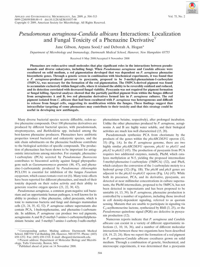
Epifluorescence microscopy of red-pigmented fungal cells. Cocultures of P.aeruginosa and C. albicans yeast cells were grown as described above for theviability assays. A small amount of the coculture growth was removed with amicropipette tip and resuspended in 500 �l of 50 mM phosphate buffer. Suspen-sions were incubated with 0.03% H2O2 for at least 20 min to ensure thatpigments were oxidized before being viewed with a Zeiss filter set 20 (excitation,550 nm; emission, 580 to 640 nm). Fixation of the yeast cells with 0.4% formal-dehyde did not interfere with fluorescence and stabilized the fluorescence formore than 3 days. All images were captured using a fixed capture time and lightintensity.
Preparation and spectral analysis of purified phenazines and fungus-associ-ated red pigment. PCA was prepared from spent culture supernatants of P.aeruginosa strains by published methods (32). The product of PhzM activity onPCA was prepared as described previously (32), by incubating PCA with E. coliDH5�/pUCP-M, which contains a plasmid carrying the phzM gene. Cells from 50ml of an overnight culture of this E. coli strain in LB with tetracycline (15 �g/ml)at 37°C were harvested by centrifugation, washed once in M63 medium (34)containing 0.2% glucose, and then resuspended in 5 ml of the same mediumamended with 1 mM methionine and 0.4 mM PCA. The suspension was shakenvigorously at 37°C for 6 to 7 h. As controls, E. coli cells containing the pUCP26vector and incubated with PCA and E. coli/pUCP-M cells incubated withoutPCA were treated identically. E. coli cells were removed by centrifugation, andthe supernatant was acidified to pH 5 with 2 M HCl before being extracted three
times with 3 ml chloroform to remove unreacted PCA. Recovery and quantita-tion of PCA in the chloroform fraction indicated that approximately 5 to 15% ofPCA had been consumed in E. coli/pUCP-M compared to that in control cul-tures. The aqueous fraction of the extracted medium was neutralized with 2 MNH4OH and concentrated under reduced pressure to a final volume of 200 �l.The solution containing the PhzM product was deep red, while the controlsolution was pale yellow.
Pyocyanin was extracted from 24-h LB-grown cultures of P. aeruginosa PA14by cycling three times between chloroform at neutral pH and 20 mM HCl (7).Aeruginosins A and B were recovered as water-soluble red pigments from 6-daysupernatants from P. aeruginosa PA14 cultures grown in Holliman’s medium(20). Approximately 200 ml of the culture supernatant was applied to chromato-graphic columns. Aeruginosin A bound weakly to a 2.5- by 15-cm column of theC18 hydrophobic interaction medium LRP-2 (Whatman) equilibrated with 20mM NH4HCO3, from which it was recovered by elution with 10% acetonitrile in20 mM NH4HCO3 in a volume of about 50 ml. Aeruginosin B, which passedthrough the hydrophobic interaction column, bound to a column (1.5 by 12 cm)of DEAE Sephadex in 20 mM NH4HCO3, from which it was eluted with agradient of NH4HCO3 (20 to 200 mM) in a volume of about 25 ml. Theaeruginosin-containing eluates were concentrated under reduced pressure. TheUV-visible spectra of both compounds corresponded to those previously re-ported (15, 20).
The fungus-associated red pigment was partially purified from the fungalfraction from full-plate cocultures (described above). The purification of thepigment is described in more detail in Results. The approximate concentration ofthe red pigment was determined using the extinction coefficient for aeruginosinsA and B (15, 20). The absorbance and fluorescence spectra were obtained using
TABLE 1. Bacterial and fungal strains used in this study
Strain Description DH no.a Source or reference
Pseudomonas aeruginosa strainsPA14 WT WT 123 41PA14 phzM::TnM TnM mutant, pyocyanin negative 693 28PA14 phzS::TnM TnM mutant, pyocyanin negative 698 28PA14 WT/pUCP26 WT with empty plasmid from reference 51 942 51PA14 phzM::TnM/pUCP26 Mutant with empty plasmid from reference 51 944 This studyPA14 phzM::TnM/pUCP-M Mutant complemented with the phzM gene on a
plasmid (32)945 This study
PA14 phzS::TnM/pUCP26 Mutant with empty plasmid from reference 51 946 This studyPA14 phzS::TnM/pUCP-S Mutant complemented with the phzS gene on a
plasmid (32)947 This study
PA14 �phz In-frame deletion mutant of phzA1 to phzG1 andphzA2 to phzG2
933 9
PA14 flgK::Tn5 Tn5 mutant, nonmotile 37PA14 pqsA::Tn5 Tn5 mutant, lacks PQS This studyPAO1 WT WT 20 45PAO1 �phzM Mutant lacking the phzM gene, pyocyanin negative 296 32PAO1 �phzS Mutant lacking the phzS gene, pyocyanin negative 295 32Clinical isolates Isolates from respiratory sputum 211 to 228 and 74 This study
Other Pseudomonas strainsPseudomonas fluorescens SWB25 245 G. O’Toole labPseudomonas putida KT2440 468 G. O’Toole labPseudomonas chlororaphis
PCL1391469 G. O’Toole lab
Fungal strainsC. albicans SC5314 WT 65 11C. albicans tup1/tup1 mutant BCa2-10; tup1/tup1 URA3/ura3 36 2Saccharomyces cerevisiae 1278b 1278b 347 F. Winston labS. cerevisiae BY4742 MAT� his3�1 leu2�0 lys2�0 ura3�0 195 F. Winston labS. cerevisiae BY4741 MATa his3�1 leu2�0 met15�0 ura3�0 196 F. Winston labEnvironmental yeast isolate 121 This study
Escherichia coli strainsDH5�/pUCP-M 32DH5�/pUCP26 32
a From our lab collection.
506 GIBSON ET AL. APPL. ENVIRON. MICROBIOL.

a 1-cm-path-length quartz cuvette in a SpectraMax M5 (Molecular Devices)spectrophotometer.
Treatment of yeast with the PhzM product. In order to determine if the E.coli-synthesized product of PhzM activity on PCA was sufficient to lead to redpigmentation and intracellular fluorescence of C. albicans SC5314 yeast cells, 50�l of aqueous extract (described above) was added to 0.5 ml of overnight C.albicans culture grown in liquid YPD (OD600 of 15). As controls, C. albicanscultures received equivalent volumes of aqueous extracts from E. coli/pUCP26incubated with PCA and E. coli/pUCP-M incubated without the PCA precursor.After 24 h at 30°C, the C. albicans suspensions were collected by centrifugation,washed two times with 50 mM phosphate buffer, pH 7, and resuspended in 100�l of 50 mM phosphate buffer for analysis by visual inspection, epifluorescencemicroscopy, and enumeration of the number of CFU.
RESULTS
Red pigment formation in P. aeruginosa-fungal cocultures.When P. aeruginosa strain PA14 was point inoculated ontoestablished lawns of C. albicans strain SC5314 yeast and fur-ther incubated at 30°C, a red-pigmented zone developedaround the inoculation point within 12 h and continued todevelop over 48 h (Fig. 2A). Red pigmentation also developedwhen P. aeruginosa was inoculated onto established lawns offilamentous C. albicans cells, as demonstrated using the con-stitutively filamentous C. albicans tup1/tup1 mutant (Fig. 2B)(2). Inoculation of P. aeruginosa onto a lawn of the filamentousC. albicans strain led to a larger zone of pigmentation than thatformed on C. albicans yeast (Fig. 2A and B). Neither P. aerugi-
nosa nor C. albicans produced any red pigmentation whengrown alone under similar conditions. While the red pig-mentation was observed regardless of whether the cocul-tures were grown on rich or minimal agar plates, i.e., YPDor M63 medium with 0.2% glucose, respectively (34), noobvious red pigmentation was observed when P. aeruginosaand C. albicans were grown together in liquid cultures of thesame medium composition.
To determine if the development of red pigmentation wasspecific to P. aeruginosa and C. albicans, other strains of P.aeruginosa, other Pseudomonas species, and other fungalstrains were analyzed in coculture assays. A similar, but paler,red pigmentation was observed when P. aeruginosa strain PA14was cultured on lawns of Saccharomyces cerevisiae BY4742,BY4741, and 1278b and three unidentified environmentalyeast-like fungi isolated from soil samples and plant material(data not shown). Upon coculture with the C. albicans tup1/tup1 mutant, P. aeruginosa strain PAO1 and 17 of 19 clinical P.aeruginosa isolates gave rise to red pigmentation to variousdegrees. No red pigmentation was observed upon incubationof Pseudomonas putida, Pseudomonas fluorescens, or Pseudo-monas chlororaphis on lawns of C. albicans (data not shown).
Identification of genes involved in coculture pigmentation.To identify P. aeruginosa genes involved in the formation of redpigmentation, strains from a collection of P. aeruginosa strainPA14 Tn5 transposon mutants (29) were screened, with 48mutants per plate, in the coculture plate assay described above.An initial screen of approximately 9,000 random Tn5 inser-tions found mutants with an altered pattern of pigmentation(more than 75 mutants), 5 mutants that lacked pigmentation,and 2 mutants with increased red pigmentation.
Role of swimming motility in coculture interactions. All ofthe P. aeruginosa mutant strains categorized as having an al-tered pattern of red pigmentation gave rise to a compact redring around the point of inoculation which was much smallerthan that formed by the wild-type (WT) strain on lawns of theC. albicans tup1/tup1 mutant. Mutants in this class includedflgK::Tn5 (Fig. 2C), fleN::Tn5, flgF::TnM, flgM::TnM, fliF::TnM, andfliD::TnM mutants (data not shown). All of these transposonmutants are predicted to lack a functional flagellum, and theydid not swim in a 0.3% agar swim assay (data not shown).When nonmotile P. aeruginosa mutants were tested in cocul-tures with C. albicans SC5314 yeast, there were no obviousdifferences between the mutants and the WT; all remained atthe point of inoculation (Fig. 2A and data not shown). Thesedata suggest that flagellar motility is necessary for P. aeruginosamovement across C. albicans filaments and that P. aeruginosa isincapable of this flagellum-dependent motility across lawns ofC. albicans in the yeast form. When P. aeruginosa mutantsdefective in type IV pilus-mediated twitching motility (pilB::Tn5or pilC::Tn5 mutant) or in the production of rhamnolipids(rhlA::Gm mutant), which are necessary for swarming motility onan agar surface, were assayed on lawns of the filamentous C.albicans tup1/tup1 mutant, the zones of red pigmentation wereindistinguishable from those formed by WT P. aeruginosa strainPA14 (Fig. 2B and data not shown) (37, 40).
To determine the relationship between the area of the P.aeruginosa colony on the C. albicans filaments and the size ofthe zone of the red pigment, samples from plate-grown fungalcocultures with either a WT or nonmotile (flgK) strain were
FIG. 2. Plate cocultures of C. albicans with P. aeruginosa strains.(A to C) C. albicans lawns were grown for 48 h at 30°C on YPDplates before point inoculation from LB agar-grown P. aeruginosastrains. Plates were photographed after coculture for 48 h. (A) C.albicans SC5314 with P. aeruginosa (P.a.) PA14 WT. (B) C. albicanstup1/tup1 mutant with P. aeruginosa PA14 WT. (C) C. albicanstup1/tup1 mutant with PA14 WT, flgK::Tn5, pqsA::TnM, phzS::TnM,and phzM::TnM strains. (D) Complementation of mutations inphzM::TnM and phzS::TnM strains in the coculture assay. P. aerugi-nosa strains were inoculated onto the C. albicans tup1/tup1 lawns as10-�l drops and incubated at 30°C. Vector controls for WT,phzM::TnM, and phzS::TnM/(pUCP26) strains were also included.(E) Redox activity of fungal-associated pigment. C. albicans tup1/tup1 cells from two plate-grown P. aeruginosa PA14 WT cocultureswere separated from the bacteria by centrifugation, resuspended in5 ml 50 mM phosphate buffer, pH 7, and divided into three tubes.The suspensions were photographed several minutes after aeration(center), after the addition of 20 �l 3% hydrogen peroxide (left),and after the addition of dithionite (right).
VOL. 75, 2009 FUNGAL TOXICITY OF A PYOCYANIN PRECURSOR 507

taken at 2-mm intervals from the point of inoculation andplated onto medium selective for P. aeruginosa. These assaysshowed that bacteria were present throughout the red-pig-mented zone and that no bacteria were detected outside thered-pigmented area, indicating that the red pigmentation de-veloped only in regions where the bacterial and fungal cellswere in close proximity and that the red pigment did not diffuseaway from the coculture region.
Involvement of phenazine-related genes in red pigment for-mation. Those mutants that lacked pigment production werealtered in quorum sensing (three independent hits in pqsA, onein its regulator, mvfR, and one in rhlR). The two mutants withincreased pigment production had insertions in phzF1 andphzS, which both encode enzymes involved in pyocyanin bio-synthesis. To further characterize the role of quorum-sensingregulation in phenazine production, mutants involved in quo-rum sensing and phenazine biosynthesis from the PA14 non-redundant mutant collection (28) were analyzed. Again, sev-eral mutants defective in signaling by PQS (pqsA::TnM andmvfR::TnM mutants) or C4-homoserine lactone (rhlR::TnMmutant), two quorum-sensing molecules, completely lackedred pigmentation upon coculture with C. albicans (Fig. 2C anddata not shown). A mutant with a disrupted rhlI gene had amore variable phenotype, and lasI and lasR mutants producedpigment on fungal lawns. None of these mutants producedpyocyanin in single-species P. aeruginosa cultures.
A P. aeruginosa phzM::TnM mutant, which lacks the phen-azine biosynthetic enzyme necessary for methylation of PCA(Fig. 1B), formed no color upon coculture with C. albicans(Fig. 2C). In contrast, the phzS::TnM mutant, which is defec-tive in reduction of the 1-carboxylate group of PCA to analcohol (Fig. 1B), produced more red pigmentation than theWT strain (Fig. 2C and D). Complementation analysis indi-cated that red pigmentation was largely restored to the phzMmutant by providing the phzM gene in trans and that the hy-perpigmentation phenotype of the P. aeruginosa phzS::TnMmutant was corrected by providing the phzS gene on a plasmid(Fig. 2D). Complementation of the phzS mutant also restoredthe production of pyocyanin in cocultures (Fig. 2D). The phzHmutant, which converts PCA to phenazine-1-carboxamide,produced slightly more red pigment than the WT strains did(data not shown) (32). While mutants defective in the phz-ABCDEFG biosynthetic genes involved in PCA biosynthesiswere not identified in our assays as being defective in pigmentproduction, likely due to the fact that there are redundantphzABCDEFG operons (Fig. 1A), the P. aeruginosa PA14�phzA1-G1 �phzA2-G2 mutant (�phz mutant) (9) did not giverise to red pigmentation upon coculture with C. albicans (datanot shown). Experiments with P. aeruginosa strain PAO1 sim-ilarly showed that the �phzM strain completely lacked produc-tion of the red pigment and that the PAO1 �phzS strain pro-duced more red pigment than the WT strain did (data notshown). Because the fungus-associated red pigment requiredfunctional phzABCDEFG genes and phzM, but not phzS orphzH, we hypothesized that this pigment was derived from thePhzM product, proposed to be 5MPCA.
Our genetic experiments indicated that red pigmentationwas dependent on a precursor to pyocyanin but not on pyocy-anin itself. To determine if pyocyanin formation was negativelyimpacted by growth with C. albicans, its levels were determined
in single-species and mixed-species cultures. Quantitative anal-ysis of pyocyanin levels in agar core samples from P. aerugi-nosa-C. albicans cocultures grown on plates for 24 h detected9.6 1 nmol pyocyanin per core. The amount of pyocyaninwas similar (9.7 1.2 nmol pyocyanin per core) for single-species P. aeruginosa cultures grown on medium conditionedby C. albicans but in the absence of fungal cells. The numbersof P. aeruginosa CFU per unit area based on the core diameterwere similar for both cultures (1.56 � 106 0.45 � 106/mm2
and 1.2 � 106 0.36 � 106/mm2, respectively) at the 24-h timepoint. After 48 h, the levels of pyocyanin were higher in co-cultures (106.8 8.5 nmol per core) than the amounts in P.aeruginosa single-species cultures (39.3 6.3 nmol per core),even though the numbers of P. aeruginosa CFU per unit areawere similar (1.65 � 106 1.6 � 106/mm2 and 1.64 � 106 0.66 � 106/mm2 in cocultures and single-species cultures, re-spectively). No red coloration was observed when P. aeruginosawas grown on C. albicans-conditioned medium without fungalcells. Because pyocyanin formation was similar or greater in P.aeruginosa-C. albicans cocultures than in single-species P.aeruginosa cultures, we propose that production of a 5MPCAproduct other than pyocyanin in cocultures is not likely duesolely to inhibition of the final step involved in pyocyaninproduction.
Redox activity of the red pigment. When P. aeruginosa-C.albicans cocultures were harvested from the surface of the agarmedium and resuspended in phosphate buffer, the cell suspen-sion was similarly red pigmented (Fig. 2E). The color of thecell suspension changed from red to buff upon standing atroom temperature for several minutes, and the red pigmentwas restored upon vigorous aeration. Similarly, the redness wasintensified upon the addition of dilute H2O2, and the cellsuspension converted from red to buff upon addition of a fewcrystals of dithionite (Fig. 2E). Color changes upon addition ofreducing and oxidizing agents could be observed repeatedlyusing the same coculture suspension, suggesting that the pig-ment could be reversibly oxidized and reduced.
Correlation between pigment production and fungal viabil-ity in coculture. To determine if the formation of red pig-mentation correlated with an altered viability of C. albicansSC5314, the survival of C. albicans was assessed by bothmethylene blue staining, which is indicative of a cell beingmetabolically inactive or membrane compromised, and com-paring the total number of yeast cells determined by micro-scopic direct counts to the numbers of CFU. In C. albicanscontrol cultures and C. albicans SC5314 cocultures with the�phz or phzM::TnM strain, �1% of the cells were stained bymethylene blue even after 48 h of coincubation (Fig. 3A).Yeast cells incubated with WT P. aeruginosa exhibited 14and 26% methylene blue staining at 24 and 48 h, respec-tively. Cocultures with the phzS mutant, which led to in-creased red pigmentation, contained approximately twice asmany methylene blue-stained cells (32 and 50% at 24 and48 h, respectively).
Viability determined by combining direct counts with deter-mination of CFU strongly supported the results from the meth-ylene blue staining experiments. In the absence of bacteria, C.albicans yeast viability was 86% at the 72-h time point, asmeasured by these means (Fig. 3B). When C. albicans wascocultured with the P. aeruginosa PA14 strains, the viability in
508 GIBSON ET AL. APPL. ENVIRON. MICROBIOL.

the presence of P. aeruginosa was reduced to 12% for WT P.aeruginosa and 3% for the phzS mutant. In contrast, C. albicanscocultured with the phzM strain had 76% viability. Analysis ofvariance found the differences between the WT and mutantstrains to be statistically significant (P � 0.05). The differencesin fungal killing were not due to differences in P. aeruginosagrowth, as the numbers of P. aeruginosa CFU were equivalent(9.6 � 107 to 10.4 � 107 CFU per core) in all of the coculturesincluded in this experiment. These data indicate that the pres-ence of the phzM gene, which leads to the formation of5MPCA, is required for much of the killing of C. albicans by P.aeruginosa in this assay.
Accumulation of red pigment within fungal cells. Severalpieces of evidence suggested that the red pigment that formed
in P. aeruginosa-C. albicans cocultures was present only inassociation with the fungal cells. First, while pyocyanin diffusedinto the agar below the P. aeruginosa-C. albicans coculture, thered pigmentation was observed only in the layer of cells atopthe agar medium in cocultures of up to 7 days old and did notdiffuse into the surrounding agar (data not shown). Second,centrifugal separation of bacteria from the fungal cells showedthat the red pigmentation was associated with the fungal pelletand was not present in the bacterium-containing fractions. Nored coloration was observed in water or phosphate bufferwashes of the fungal pellet, suggesting that the pigment wasnot capable of readily diffusing away from fungal cells (data notshown). The red pigmentation of the fungal cell fraction wasredox sensitive (Fig. 2E). Third, when an established lawn ofthe C. albicans tup1/tup1 mutant was covered with cellophaneprior to inoculation with P. aeruginosa, removal of the bacterialcolony after a 24-h growth period revealed red pigmentationonly of the fungal cells (data not shown). Culturing experi-ments confirmed that P. aeruginosa and C. albicans remainedseparate over the course of the cellophane separation experi-ment. Experiments describing the release and purification ofthe red pigment from fungal cells are described below.
Visualization of cells grown in coculture with P. aerugi-nosa by epifluorescence microscopy provided additional in-formation regarding the location of the red pigmentationthat formed in P. aeruginosa-C. albicans cocultures. After in-cubation of WT P. aeruginosa on lawns of C. albicans yeast, asmall amount of the coculture was resuspended in buffer andthen observed by epifluorescence microscopy. C. albicans thathad been incubated with WT P. aeruginosa exhibited brightintracellular fluorescence (Fig. 4A). The intracellular fluores-cence was evident at 24 h, and the percentage of fluorescingcells increased with time. C. albicans cells from cocultures withthe P. aeruginosa phzS::TnM strain, which led to increasedproduction of the red pigment, were also brightly fluorescent(Fig. 4A). In contrast, only a faint background fluorescencewas observed with C. albicans grown either in the absence of P.aeruginosa or in the presence of the P. aeruginosa phzM::TnMstrain, which does not support red pigmentation (Fig. 4A). Inmany C. albicans cells from cocultures with WT P. aeruginosa,the red fluorescence seemed brightest in the cytoplasm (Fig.4A), but in very brightly fluorescing cells, such as those de-tected in cocultures with the phzS::TnM strain, the fluores-cence appeared throughout the cell (Fig. 4A). The level offluorescence per cell was not even across the population. In-cubation of the constitutively filamentous tup1/tup1 mutantwith WT P. aeruginosa, but not the �phz or phzM mutant, alsoyielded brightly fluorescent cells (Fig. 4B). The addition ofdithionite, the reducing agent that changed the visual pigmen-tation of the culture from red to colorless (Fig. 2E), alsosuppressed fluorescence (data not shown).
Effects of PhzM product on red pigmentation, intracellularfluorescence, and viability of fungal cells. Because cocultureswith the P. aeruginosa phzM::TnM mutant formed no red pig-ment, while those with the phzS mutant overproduced the redpigment, we hypothesized that the PhzM product, proposed tobe 5MPCA, is responsible for the red pigmentation. To testthis hypothesis directly, the PhzM product was synthesized byfeeding PCA to resting cells of an E. coli strain carrying thephzM gene on a high-copy-number plasmid, using a previously
FIG. 3. Survival of C. albicans in coculture with strains of P. aerugi-nosa PA14. Established lawns of C. albicans SC5314 yeast were inoc-ulated with P. aeruginosa suspensions. Core samples from plates weretaken for counts of total cells by microscopy and of viable cells (CFU)after incubation for various times. (A) C. albicans methylene bluestaining after incubation alone or with P. aeruginosa PA14 WT, �phz(which lacks the genes necessary to synthesize PCA), phzM::TnM, andphzS::TnM strains. (B) Survival of C. albicans after 72 h alone (control)or in coculture with the P. aeruginosa PA14 WT, phzM::TnM, orphzS::TnM strain. Percent viability represents the fraction of the totalcells that gave rise to visually detectable colonies within 24 h. Theseexperiments were repeated as three completely separate experiments,with similar results each time.
VOL. 75, 2009 FUNGAL TOXICITY OF A PYOCYANIN PRECURSOR 509
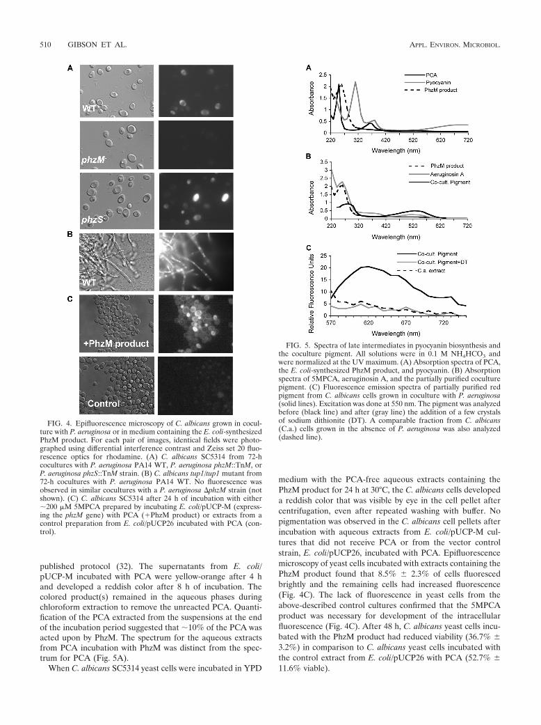
published protocol (32). The supernatants from E. coli/pUCP-M incubated with PCA were yellow-orange after 4 hand developed a reddish color after 8 h of incubation. Thecolored product(s) remained in the aqueous phases duringchloroform extraction to remove the unreacted PCA. Quanti-fication of the PCA extracted from the suspensions at the endof the incubation period suggested that �10% of the PCA wasacted upon by PhzM. The spectrum for the aqueous extractsfrom PCA incubation with PhzM was distinct from the spec-trum for PCA (Fig. 5A).
When C. albicans SC5314 yeast cells were incubated in YPD
medium with the PCA-free aqueous extracts containing thePhzM product for 24 h at 30°C, the C. albicans cells developeda reddish color that was visible by eye in the cell pellet aftercentrifugation, even after repeated washing with buffer. Nopigmentation was observed in the C. albicans cell pellets afterincubation with aqueous extracts from E. coli/pUCP-M cul-tures that did not receive PCA or from the vector controlstrain, E. coli/pUCP26, incubated with PCA. Epifluorescencemicroscopy of yeast cells incubated with extracts containing thePhzM product found that 8.5% 2.3% of cells fluorescedbrightly and the remaining cells had increased fluorescence(Fig. 4C). The lack of fluorescence in yeast cells from theabove-described control cultures confirmed that the 5MPCAproduct was necessary for development of the intracellularfluorescence (Fig. 4C). After 48 h, C. albicans yeast cells incu-bated with the PhzM product had reduced viability (36.7% 3.2%) in comparison to C. albicans yeast cells incubated withthe control extract from E. coli/pUCP26 with PCA (52.7% 11.6% viable).
FIG. 4. Epifluorescence microscopy of C. albicans grown in cocul-ture with P. aeruginosa or in medium containing the E. coli-synthesizedPhzM product. For each pair of images, identical fields were photo-graphed using differential interference contrast and Zeiss set 20 fluo-rescence optics for rhodamine. (A) C. albicans SC5314 from 72-hcocultures with P. aeruginosa PA14 WT, P. aeruginosa phzM::TnM, orP. aeruginosa phzS::TnM strain. (B) C. albicans tup1/tup1 mutant from72-h cocultures with P. aeruginosa PA14 WT. No fluorescence wasobserved in similar cocultures with a P. aeruginosa �phzM strain (notshown). (C) C. albicans SC5314 after 24 h of incubation with either�200 �M 5MPCA prepared by incubating E. coli/pUCP-M (express-ing the phzM gene) with PCA (�PhzM product) or extracts from acontrol preparation from E. coli/pUCP26 incubated with PCA (con-trol).
FIG. 5. Spectra of late intermediates in pyocyanin biosynthesis andthe coculture pigment. All solutions were in 0.1 M NH4HCO3 andwere normalized at the UV maximum. (A) Absorption spectra of PCA,the E. coli-synthesized PhzM product, and pyocyanin. (B) Absorptionspectra of 5MPCA, aeruginosin A, and the partially purified coculturepigment. (C) Fluorescence emission spectra of partially purified redpigment from C. albicans cells grown in coculture with P. aeruginosa(solid lines). Excitation was done at 550 nm. The pigment was analyzedbefore (black line) and after (gray line) the addition of a few crystalsof sodium dithionite (DT). A comparable fraction from C. albicans(C.a.) cells grown in the absence of P. aeruginosa was also analyzed(dashed line).
510 GIBSON ET AL. APPL. ENVIRON. MICROBIOL.

Comparison of 5MPCA-derived pigment within C. albicanscells to known P. aeruginosa phenazines. To determine thenature of the red-pigmented product observed within fungalcells, suspensions of C. albicans from P. aeruginosa-C. albicanscocultures were lysed and the red-pigmented products wereanalyzed. While the red product 5MPCA and derivatives syn-thesized from PCA by E. coli/pUCP-M are highly soluble inwater, the red pigment within fungal cells was poorly soluble ineither aqueous solutions (water or acidic or basic buffers) ororganic solvents (chloroform, ethyl acetate, or ethanol). Me-chanical or enzymatic and chemical disruption of cells, fol-lowed by centrifugation of the cell lysate at 10,000 � g, led tosedimentation of the majority of the pigment, yielding only apale pink supernatant. Conditions that promote yeast autoly-sis, including incubation of dense cell suspensions at 37°C for24 to 48 h, led to further, but far from complete, release ofpigment in a time-dependent manner. Heat inactivation of thecell suspension prior to incubation under autolysis conditionsprevented release of red-pigmented products into the aqueoussupernatant. The observations suggest that pigment is aggre-gated or polymerized inside the fungus but can be releasedslowly by the action of endogenous fungal enzymes.
Partial purification of red pigment from autolyzed suspen-sions of the C. albicans tup1/tup1 mutant from P. aeruginosacocultures could be achieved by size-exclusion chromatographyon Bio Gel P-2 (molecular weight, 200 to 2,000; Bio-Rad). Thered fractions emerged close to the void volume, whereas pyo-cyanin and vitamin B12 eluted in later fractions that corre-sponded to their predicted molecular weights of 210 and 1,355,respectively (49). These data suggest that the red pigmentmolecule(s) is of a substantially larger size than that of otherphenazines known to be produced by P. aeruginosa. For spec-trophotometric analysis, the red fractions were further purifiedusing a C18 hydrophobic interaction column (LRP-2; What-man) in 1 M NH4CO3, followed by elution with 10% acetoni-trile. The red fraction had a single broad absorbance maximumat �530 nm in the visible region (Fig. 5B) but lacked themaximum at �390 nm, which is characteristic of red P. aerugi-nosa phenazines aeruginosins A (Fig. 5B) and B (not shown),as reported previously by Holliman et al. (15, 20). The spec-trum of the red pigment isolated from fungal cells also differedfrom those of other known P. aeruginosa phenazines, includingPCA, pyocyanin, and the products of PhzM activity on PCAproduced in E. coli (Fig. 5).
To determine if the fluorescence observed within C. albicansyeast cells from cocultures by epifluorescence microscopycould be attributed to the red pigment that was partially puri-fied from C. albicans, the fluorescence spectrum of the partiallypurified material was measured with an excitation wavelengthof 550 nm (Fig. 5C). Red fluorescence was detected with amaximum at 620 nm when the sample was aerated. Consistentwith what was observed in the C. albicans whole-cell suspen-sions, the fluorescence was not observed in the same sampleafter reduction with dithionite (Fig. 5C). A comparable prep-aration from C. albicans cultures grown in the absence of P.aeruginosa yielded fractions with no detectable fluorescence(Fig. 5C).
While we were not able to determine the identity of thefungally associated pigment that formed upon incubation of C.albicans with the products of PhzM activity on PCA- or
5MPCA-producing strains, largely due to its insolubility, it isclear that it has properties that are distinct from those of thephenazines present in single-species P. aeruginosa cultures. Wecould detect this red pigment within C. albicans cells only aftercoculture with P. aeruginosa or upon incubation with the PhzMproduct, leading us to hypothesize that the pigment resultsfrom an activity on 5MPCA that occurs within the fungal cell.Incubation of fungal cells with the partially purified red pig-ment, aeruginosins A or B, PCA, or pyocyanin at concentra-tions of up to 1 mM did not give rise to red-pigmented fungalcells with intracellular fluorescence. It is important, however,that while treatment of the partially purified red pigment with1 mg/ml proteinase K, DNase, RNase, boiling, acid (1 M HCl),base (1 M NaOH), or sodium dodecyl sulfate (1%) did notliberate a homogenous low-molecular-weight species, we can-not rule out the alternative hypothesis which states that5MPCA or a derivative thereof is unaltered but aggregated insome way.
DISCUSSION
The studies reported here show that the P. aeruginosa prod-uct of PhzM activity on PCA, proposed to be 5MPCA, leads tothe accumulation of a red-pigmented, redox-active compoundwithin fungal cells. Like the C. albicans cells from cocultures,the partially purified pigment exhibited a red coloration thatwas colorless upon reduction and could readily be oxidized byaeration or the addition of hydrogen peroxide (Fig. 2E). Boththe pigmentation in association with fungal cells and the par-tially purified pigment exhibited a red fluorescence only in anoxidized state. P. aeruginosa PA14 and PAO1 mutants thatwere defective in phzM, the gene responsible for the methyl-ation of PCA to form 5MPCA, were incapable of red pigmentformation. Furthermore, pseudomonads such as Pseudomonasputida and Pseudomonas fluorescens, whose genomes do notcontain phzM (53), did not induce the accumulation of pigmenton C. albicans lawns. In contrast, deletion of the phzS gene,which encodes the enzyme that acts upon 5MPCA to formpyocyanin, led to hyperpigmentation (Fig. 2), suggesting thatthe formation of pyocyanin competed for the 5MPCA pool.Disruption of the phzH gene, which encodes an enzyme thatacts on PCA (32), also led to increased red pigment formation,suggesting that PhzH competes with PhzM for the PCA poolwithin cocultures.
The putative intermediate 5MPCA, which is responsible forthe coculture pigment, has not been studied extensively. Whileits immediate precursor, PCA, and its immediate derivative,pyocyanin, accumulate in P. aeruginosa cultures, 5MPCA hasnot been detected in supernatants (4, 14). Because pyocyaninlevels were higher in P. aeruginosa-C. albicans cocultures thanin P. aeruginosa cultures grown in the absence of C. albicansbut on C. albicans-conditioned medium, it does not appear thatthe release of the PhzM product was simply due to inhibitionof the last PhzS-dependent step in pyocyanin production by C.albicans. In vitro assays with purified PhzM and PhzS indicatethat PhzM activity is greatly enhanced or dependent on PhzS(39), suggesting that PhzM reaction kinetics are favorableonly when the product, 5MPCA, is consumed. Furthermore,5MPCA synthesized as a phenazinium chloride was found tobe stable in acid but not tractable at neutral pH (14). The fact
VOL. 75, 2009 FUNGAL TOXICITY OF A PYOCYANIN PRECURSOR 511

that the fungally associated red pigment formed only when P.aeruginosa and C. albicans were grown in close contact withone another leads us to speculate that C. albicans uptakeand subsequent modification of a small extracellular pool of5MPCA may increase PCA conversion to 5MPCA and theformation of 5MPCA-derived products. In a medium that sup-ports growth of P. aeruginosa on the surfaces of C. albicanshyphae (8), the fungal pellet develops a distinct pink colora-tion, and red fluorescent C. albicans cells were observed uponcoculture with WT P. aeruginosa and phzS mutant strains (datanot shown). The role that fungal cells play in promoting theformation and release of the 5MPCA intermediate is a subjectof our future research.
The exact chemical nature of this coculture red pigmentextracted from fungi remains elusive. Its UV-visible spectrumand solubility characteristics are distinct from those of phen-azines known to be produced by monocultures of P. aeruginosa,including PCA, pyocyanin, and aeruginosins A and B, andfrom the product in extracts from PCA-fed cultures of E. colicarrying the phzM gene (15, 20) (Fig. 5A and B). 5MPCA isconverted to a red phenazine, aeruginosin A, upon incubationwith late P. aeruginosa culture supernatants or through a chem-ical reaction with concentrated ammonia (14). A similar, sol-uble red phenazine accumulates in supernatants of phzS mu-tant cultures, as shown previously (32) and confirmed by ourlaboratory (data not shown). While aeruginosin A and the redpigment in phzS culture medium are very soluble in liquid andagar media, the red phenazine derivative formed in yeast waslocated exclusively in yeast cells and did not diffuse into thesurrounding medium. Incubation of C. albicans with purifiedaeruginosins did not give rise to red-pigmented yeast (data notshown). Nevertheless, the absorption spectrum of the cocul-ture pigment does resemble that of aeruginosin in that it has anabsorption maximum in the 520-nm range.
The concentration of the coculture pigment in the fungal cellfraction may indicate that fungal enzymes or other factors inyeast participate in the formation of the red pigment. At thistime, however, we know little about the factor or factors thatlead to the formation of the red product with poor solubilityand an apparent molecular weight that is significantly largerthan that of the 5MPCA precursor. Because formation of thered pigment is observed upon coculture with numerous fungi,including S. cerevisiae, any modifications are not unique to C.albicans. The red pigmentation that develops in ade2 mutantsof both S. cerevisiae and C. albicans grown in media withlimiting concentrations of adenine, due to the conjugation ofthe toxic adenine biosynthetic precursors with glutathione (5,44), is distinct from the pigment we observe in P. aeruginosa-C.albicans cocultures. The red pigment in ade2 mutants is notredox sensitive (50). The addition of adenine to the agar me-dium does not alter pigmentation of C. albicans in our exper-iments (data not shown).
Previous reports have demonstrated the toxicities of pyocy-anin and 1-hydroxyphenazine on C. albicans and S. cerevisiae,with MICs in the 100- to 500-�M range (24, 42). Our studiesindicate that the product of PhzM activity on PCA also leads todecreased fungal viability. Because the precise nature of the5MPCA product has not been described and purified prepa-rations are reported to be unstable at neutral pH (14), directquantitative comparison of its lethal effect on yeast to that of
other phenazines has not been performed. The phzS mutant,which leads to increased C. albicans red pigmentation andfluorescence compared to those of C. albicans cocultured withWT P. aeruginosa, is also more lethal for yeast than the WTstrain (Fig. 4). These data indicate that the putative product ofthe PhzM enzyme, 5MPCA, is more important for yeast killingthan pyocyanin when the two species are grown in close prox-imity to one another. While pyocyanin has previously beendemonstrated to play an important role in P. aeruginosa viru-lence (24, 42), previous studies by Lau et al. found that boththe phzM and phzS mutants were attenuated in lung infectionsin vivo. It is interesting, however, that the phzM mutant exhib-ited a greater defect, as determined by competitive index, thanthe phzS mutant in these in vivo assays (26). Experiments withboth partially purified pigment and whole C. albicans cellsshowed that the fungally associated red pigment retained redoxactivity within the fungus despite any modifications that mayoccur within the fungal cell (Fig. 2E and 5C). Reaction withoxygen is important for the toxicity of many different phen-azines toward a variety of species (31), and we hypothesize thatthe redox activity of the fungally associated pigment contrib-utes to the decreased C. albicans viability that we observe in itspresence.
While many phenazines have been shown to have toxic effectstoward a variety of species, including fungi, it is not yet known ifpreviously characterized phenazine antibiotics are modified orsequestered by other organisms (22, 27, 31, 48). One report de-scribes the production of an extracellular red pigment, with dif-ferent properties from the pigment described here, that forms incocultures of Aspergillus sclerotiorum and Pseudomonas chlorora-phis, suggesting that other fungi may be able to modify certainbacterially produced phenazines (17); the biological activity of themodified P. chlororaphis phenazine is not known. One can envi-sion many ways in which modification or aggregation of phen-azines after secretion by the producing microbe could enhance orreduce the toxicity of the antibiotic, and these processes may beimportant factors to consider in the design of phenazine-produc-ing biocontrol strains.
ACKNOWLEDGMENTS
This paper is dedicated to Jane Gibson, an inspirational scientist, forwhom this paper is published posthumously.
We thank Nicholas Jacobs for his important intellectual contribu-tions to this project. We thank Linda Thomashow and Dimitri Mavrodifor providing P. aeruginosa PAO1 �phzS and �phzM strains and thecomplementation plasmids. We thank Dianne Newman for providingthe PA14 �phzA1-G1�phzA2-G2 strain, Joseph Schwartzman and theDHMC clinical lab for providing P. aeruginosa clinical isolates, andGeorge O’Toole for use of the P. aeruginosa PA14 Tn5 mutant libraryand for providing strains of other Pseudomonas species. We acknowl-edge Nida Intarapanich for preparing and analyzing the comple-mented strains and Brittany Ciesluk for aiding in the viability studies.
This work was funded by the Pew Biomedical Scholars Program(D.A.H.) and the Cystic Fibrosis Foundation (D.A.H.).
REFERENCES
1. Bauernfeind, A., R. M. Bertele, K. Harms, G. Horl, R. Jungwirth, C. Peter-muller, B. Przyklenk, and C. Weisslein-Pfister. 1987. Qualitative and quan-titative microbiological analysis of sputa of 102 patients with cystic fibrosis.Infection 15:270–277.
2. Braun, B. R., and A. D. Johnson. 1997. Control of filament formation in Can-dida albicans by the transcriptional repressor TUP1. Science 277:105–109.
3. Brint, J. M., and D. E. Ohman. 1995. Synthesis of multiple exoproducts inPseudomonas aeruginosa is under the control of RhlR-RhlI, another set of
512 GIBSON ET AL. APPL. ENVIRON. MICROBIOL.

regulators in strain PAO1 with homology to the autoinducer-responsiveLuxR-LuxI family. J. Bacteriol. 177:7155–7163.
4. Byng, G. S., D. C. Eustice, and R. A. Jensen. 1979. Biosynthesis of phenazinepigments in mutant and wild-type cultures of Pseudomonas aeruginosa. J.Bacteriol. 138:846–852.
5. Chaudhuri, B., S. Ingavale, and A. K. Bachhawat. 1997. apd1�, a generequired for red pigment formation in ade6 mutants of Schizosaccharomycespombe, encodes an enzyme required for glutathione biosynthesis: a role forglutathione and a glutathione-conjugate pump. Genetics 145:75–83.
6. Chin, A. W. T. F., G. V. Bloemberg, I. H. Mulders, L. C. Dekkers, and B. J.Lugtenberg. 2000. Root colonization by phenazine-1-carboxamide-produc-ing bacterium Pseudomonas chlororaphis PCL1391 is essential for biocontrolof tomato foot and root rot. Mol. Plant-Microbe Interact. 13:1340–1345.
7. Cox, C. D. 1986. Role of pyocyanin in the acquisition of iron from transferrin.Infect. Immun. 52:263–270.
8. Cugini, C., M. W. Calfee, J. M. Farrow III, D. K. Morales, E. C. Pesci, andD. A. Hogan. 2007. Farnesol, a common sesquiterpene, inhibits PQS pro-duction in Pseudomonas aeruginosa. Mol. Microbiol. 65:896–906.
9. Dietrich, L. E., A. Price-Whelan, A. Petersen, M. Whiteley, and D. K. New-man. 2006. The phenazine pyocyanin is a terminal signalling factor in thequorum sensing network of Pseudomonas aeruginosa. Mol. Microbiol. 61:1308–1321.
10. El-Azizi, M. A., S. E. Starks, and N. Khardori. 2004. Interactions of Candidaalbicans with other Candida spp. and bacteria in the biofilms. J. Appl.Microbiol. 96:1067–1073.
11. Fonzi, W. A., and M. Y. Irwin. 1993. Isogenic strain construction and genemapping in Candida albicans. Genetics 134:717–728.
12. Gallagher, L. A., S. L. McKnight, M. S. Kuznetsova, E. C. Pesci, and C.Manoil. 2002. Functions required for extracellular quinolone signaling byPseudomonas aeruginosa. J. Bacteriol. 184:6472–6480.
13. Greenhagen, B. T., K. Shi, H. Robinson, S. Gamage, A. K. Bera, J. E. Ladner,and J. F. Parsons. 2008. Crystal structure of the pyocyanin biosyntheticprotein PhzS. Biochemistry 47:5281–5289.
14. Hansford, G. S., F. G. Holliman, and R. B. Herbert. 1972. Pigments of Pseudo-monas species. IV. In vitro and in vivo conversion of 5-methylphenazinium-1-carboxylate into aeruginosin A. J. Chem. Soc. (Perkin 1) 1:103–105.
15. Herbert, R. B., and F. G. Holliman. 1969. Pigments of pseudomonas species.II. Structure of aeruginosin B. J. Chem. Soc. 18:2517–2520.
16. Hermann, C., J. Hermann, U. Munzel, and R. Ruchel. 1999. Bacterial floraaccompanying Candida yeasts in clinical specimens. Mycoses 42:619–627.
17. Hill, J., and G. Johnson. 1969. Microbial transformation of phenazines byAspergillus sclerotiorum. Mycologia 61:452–467.
18. Hogan, D. A., and R. Kolter. 2002. Pseudomonas-Candida interactions: anecological role for virulence factors. Science 296:2229–2232.
19. Hogan, D. A., A. Vik, and R. Kolter. 2004. A Pseudomonas aeruginosa quo-rum-sensing molecule influences Candida albicans morphology. Mol. Micro-biol. 54:1212–1223.
20. Holliman, F. G. 1969. Pigments of Pseudomonas species. I. Structure andsynthesis of aeruginosin A. J. Chem. Soc. 18:2514–2516.
21. Kanthakumar, K., G. Taylor, K. W. Tsang, D. R. Cundell, A. Rutman, S.Smith, P. K. Jeffery, P. J. Cole, and R. Wilson. 1993. Mechanisms of actionof Pseudomonas aeruginosa pyocyanin on human ciliary beat in vitro. Infect.Immun. 61:2848–2853.
22. Kerr, J. R. 2000. Phenazine pigments: antibiotics and virulence factors.Infect. Dis. Rev. 2:184–194.
23. Kerr, J. R. 1994. Suppression of fungal growth exhibited by Pseudomonasaeruginosa. J. Clin. Microbiol. 32:525–527.
24. Kerr, J. R., G. W. Taylor, A. Rutman, N. Hoiby, P. J. Cole, and R. Wilson.1999. Pseudomonas aeruginosa pyocyanin and 1-hydroxyphenazine inhibitfungal growth. J. Clin. Pathol. 52:385–387.
25. Latifi, A., M. K. Winson, M. Foglino, B. W. Bycroft, G. S. Stewart, A.Lazdunski, and P. Williams. 1995. Multiple homologues of LuxR and LuxIcontrol expression of virulence determinants and secondary metabolitesthrough quorum sensing in Pseudomonas aeruginosa PAO1. Mol. Microbiol.17:333–343.
26. Lau, G. W., H. Ran, F. Kong, D. J. Hassett, and D. Mavrodi. 2004. Pseudo-monas aeruginosa pyocyanin is critical for lung infection in mice. Infect.Immun. 72:4275–4278.
27. Laursen, J. B., and J. Nielsen. 2004. Phenazine natural products: biosynthe-sis, synthetic analogues, and biological activity. Chem. Rev. 104:1663–1686.
28. Liberati, N. T., J. M. Urbach, S. Miyata, D. G. Lee, E. Drenkard, G. Wu, J.Villanueva, T. Wei, and F. M. Ausubel. 2006. An ordered, nonredundantlibrary of Pseudomonas aeruginosa strain PA14 transposon insertion mu-tants. Proc. Natl. Acad. Sci. USA 103:2833–2838.
29. Mah, T. F., B. Pitts, B. Pellock, G. C. Walker, P. S. Stewart, and G. A.O’Toole. 2003. A genetic basis for Pseudomonas aeruginosa biofilm antibioticresistance. Nature 426:306–310.
30. Mahajan-Miklos, S., M. W. Tan, L. G. Rahme, and F. M. Ausubel. 1999.Molecular mechanisms of bacterial virulence elucidated using a Pseudomo-nas aeruginosa-Caenorhabditis elegans pathogenesis model. Cell 96:47–56.
31. Mavrodi, D. V., W. Blankenfeldt, and L. S. Thomashow. 2006. Phenazine
compounds in fluorescent Pseudomonas spp. biosynthesis and regulation.Annu. Rev. Phytopathol. 44:417–445.
32. Mavrodi, D. V., R. F. Bonsall, S. M. Delaney, M. J. Soule, G. Phillips, andL. S. Thomashow. 2001. Functional analysis of genes for biosynthesis ofpyocyanin and phenazine-1-carboxamide from Pseudomonas aeruginosaPAO1. J. Bacteriol. 183:6454–6465.
33. Mavrodi, D. V., V. N. Ksenzenko, R. F. Bonsall, R. J. Cook, A. M. Boronin, andL. S. Thomashow. 1998. A seven-gene locus for synthesis of phenazine-1-car-boxylic acid by Pseudomonas fluorescens 2-79. J. Bacteriol. 180:2541–2548.
34. Neidhardt, F. C., P. L. Bloch, and D. F. Smith. 1974. Culture medium forenterobacteria. J. Bacteriol. 119:736–747.
35. Norman, R. S., P. Moeller, T. J. McDonald, and P. J. Morris. 2004. Effect ofpyocyanin on a crude-oil-degrading microbial community. Appl. Environ.Microbiol. 70:4004–4011.
36. Nseir, S., E. Jozefowicz, B. Cavestri, B. Sendid, C. Di Pompeo, F. Dewavrin,R. Favory, M. Roussel-Delvallez, and A. Durocher. 2007. Impact of antifun-gal treatment on Candida-Pseudomonas interaction: a preliminary retrospec-tive case-control study. Intensive Care Med. 33:137–142.
37. O’Toole, G. A., and R. Kolter. 1998. Flagellar and twitching motility arenecessary for Pseudomonas aeruginosa biofilm development. Mol. Microbiol.30:295–304.
38. O’Toole, G. A., L. A. Pratt, P. I. Watnick, D. K. Newman, V. B. Weaver, andR. Kolter. 1999. Genetic approaches to the study of biofilms. MethodsEnzymol. 310:91–109.
39. Parsons, J. F., B. T. Greenhagen, K. Shi, K. Calabrese, H. Robinson, andJ. E. Ladner. 2007. Structural and functional analysis of the pyocyaninbiosynthetic protein PhzM from Pseudomonas aeruginosa. Biochemistry 46:1821–1828.
40. Pukatzki, S., R. H. Kessin, and J. J. Mekalanos. 2002. The human pathogenPseudomonas aeruginosa utilizes conserved virulence pathways to infect thesocial amoeba Dictyostelium discoideum. Proc. Natl. Acad. Sci. USA 99:3159–3164.
41. Rahme, L. G., E. J. Stevens, S. F. Wolfort, J. Shao, R. G. Tompkins, andF. M. Ausubel. 1995. Common virulence factors for bacterial pathogenicity inplants and animals. Science 268:1899–1902.
42. Ran, H., D. J. Hassett, and G. W. Lau. 2003. Human targets of Pseudomonasaeruginosa pyocyanin. Proc. Natl. Acad. Sci. USA 100:14315–14320.
43. Reszka, K. J., Y. O’Malley, M. L. McCormick, G. M. Denning, and B. E.Britigan. 2004. Oxidation of pyocyanin, a cytotoxic product from Pseudomo-nas aeruginosa, by microperoxidase 11 and hydrogen peroxide. Free Radic.Biol. Med. 36:1448–1459.
44. Sharma, K. G., R. Kaur, and A. K. Bachhawat. 2003. The glutathione-mediated detoxification pathway in yeast: an analysis using the red pigmentthat accumulates in certain adenine biosynthetic mutants of yeasts revealsthe involvement of novel genes. Arch. Microbiol. 180:108–117.
45. Stover, C. K., X. Q. Pham, A. L. Erwin, S. D. Mizoguchi, P. Warrener, M. J.Hickey, F. S. Brinkman, W. O. Hufnagle, D. J. Kowalik, M. Lagrou, R. L.Garber, L. Goltry, E. Tolentino, S. Westbrock-Wadman, Y. Yuan, L. L.Brody, S. N. Coulter, K. R. Folger, A. Kas, K. Larbig, R. Lim, K. Smith, D.Spencer, G. K. Wong, Z. Wu, I. T. Paulsen, J. Reizer, M. H. Saier, R. E.Hancock, S. Lory, and M. V. Olson. 2000. Complete genome sequence ofPseudomonas aeruginosa PAO1, an opportunistic pathogen. Nature 406:959–964.
46. Thomashow, L. S., and D. M. Weller. 1988. Role of a phenazine antibioticfrom Pseudomonas fluorescens in biological control of Gaeumannomycesgraminis var. tritici. J. Bacteriol. 170:3499–3508.
47. Thomashow, L. S., D. M. Weller, R. F. Bonsall, and L. S. Pierson. 1990.Production of the antibiotic phenazine-1-carboxylic acid by fluorescentPseudomonas species in the rhizosphere of wheat. Appl. Environ. Microbiol.56:908–912.
48. Turner, J. M., and A. J. Messenger. 1986. Occurrence, biochemistry andphysiology of phenazine pigment production. Adv. Microb. Physiol. 27:211–275.
49. Watson, D., J. MacDermot, R. Wilson, P. J. Cole, and G. W. Taylor. 1986.Purification and structural analysis of pyocyanin and 1-hydroxyphenazine.Eur. J. Biochem. 159:309–313.
50. Weisman, L. S., R. Bacallao, and W. Wickner. 1987. Multiple methods ofvisualizing the yeast vacuole permit evaluation of its morphology and inher-itance during the cell cycle. J. Cell Biol. 105:1539–1547.
51. West, S. E., H. P. Schweizer, C. Dall, A. K. Sample, and L. J. Runyen-Janecky. 1994. Construction of improved Escherichia-Pseudomonas shuttlevectors derived from pUC18/19 and sequence of the region required for theirreplication in Pseudomonas aeruginosa. Gene 148:81–86.
52. Wilson, R., D. A. Sykes, D. Watson, A. Rutman, G. W. Taylor, and P. J. Cole.1988. Measurement of Pseudomonas aeruginosa phenazine pigments in spu-tum and assessment of their contribution to sputum sol toxicity for respira-tory epithelium. Infect. Immun. 56:2515–2517.
53. Winsor, G., R. Lo, S. Sui, K. Ung, S. Huang, D. Cheng, W. Ching, R.Hancock, and F. Brinkman. 2005. Pseudomonas aeruginosa genome databaseand PseudoCAP: facilitating community-based, continually updated, ge-nome annotation. Nucleic Acids Res. 33:D338–D343.
VOL. 75, 2009 FUNGAL TOXICITY OF A PYOCYANIN PRECURSOR 513


![GINGIVAL PIGMENTATION · Oral Pigmentation Oral pigmentation is a discolouration of the oral mucosa or gingiva associated with several exogenous and endogenous factors.[1-3] This](https://static.fdocuments.in/doc/165x107/5f82dc5ea46ef73d4a1ef172/gingival-pigmentation-oral-pigmentation-oral-pigmentation-is-a-discolouration-of.jpg)