Pseudoexfoliation syndrome: Clinical factors related to capsular rupture in cataract surgery
4
ACTA OP HTHA LM 0 LOG I CA 71 (1993) 181-184 Pseudoexfoliation syndrome: Clinical factors related to capsular rupture in cataract surgery Javier Moreno', Susana Duch2 and Jeronimo Lajara' Departamento de Oftalmologia'. Clinica Universitaria, Universidad de Navarra, Pamplona, Servicio de Oftalmologia2, Residencia Sanitaria de Bellvitge, Universidad de Barcelona, Espatia Abstract. Extracapsular surgery has a high frequency of capsular rupture or disinsertion in patients with pseu- doexfoliationsyndrome. Irido-phakodonesis is one of the factors that suggests this complication. Irido-phako- donesis acts as an 'all or nothing' mechanism, as it does not appear until there is a certain degree of zonular le- sion. We camed out a study on 330 eyes with pseudoex- foliation, relating irido-phakodonesisto 31 clinical signs. The results showed that irido-phakodonesisis related to cataracts, degree of mydriasis, presence of glaucoma, atrophy of the pupilar pigmentary ruff, and uniform pig- mentation of the trabecular meshwork. These results suggest that the signs which indicate the existence of zo- nular lesion during extracapsular surgery, include poor dilation, the presence of glaucoma and pigmentary alter- ations, whilst the pseudoexfoliation material deposited in the anterior segment does not appear to have a bear- ing on this complication. Key words: pseudoexfoliation syndrome - glaucoma - cap- sular rupture - extracapsular surgery - cataract - irido- phakodonesis. Pseudoexfoliation (PSX) syndrome is charac- terized by the presence of a white flaky substance which is deposited on various structures of the anterior segment of the eye, one of these being the zonula. Structural studies have shown that this de- posited substance damages the zonular fibers to the extent that in some cases, irido-phakodonesis (IPH) is produced (Garner & Alexander 1984). This zonular alteration is responsible for capsular rup- ture or disinsertion during extracapsular cataract surgery (ECCE)(Raitta 8c Setala 1986; Skuda et al. 1987; Coder 1988; Nauman et al. 1989),a rupture which according to Coder (1988) and Nauman et al. (1989), is 7 to 10 times more frequent than in eyes without PSX. IPH seems to follow an 'all or nothing' mechan- ism, i.e. it does not appear when there is little zo- nular lesion; however, when the zonular alteration is advanced, IPH is shown even to produce sublux- ation of the lens. In this report, we aim to evaluate the clinical signs that could lead us to suspect the existenceof zonular weakness, with the purpose of preventing capsular rupture or disinsertion dur- ing cataract surgery. Materialsand Methods A prospective study was carried out on 330 eyes with PSX, belonging to 191people with an average age of 73.8 years (range: 49-89 years). The study was carried out over a period of 24 months on con- secutive, non-selected patients who were then examined by the same investigator (JMM) using the same instruments and protocol. IPH was stu- died without pupil dilation in both eyes, even though PSX was only present in one. Criteria for exclusion were: a history of uveitis, herpetic ker- atitis, ocular trauma, laser photocoagulation, and any form of eye surgery. The clinical data contained in Table 1 were 181
-
Upload
javier-moreno -
Category
Documents
-
view
213 -
download
0
Transcript of Pseudoexfoliation syndrome: Clinical factors related to capsular rupture in cataract surgery
ACTA O P H T H A L M 0 L O G I C A 71 (1993) 181-184
Pseudoexfoliation syndrome: Clinical factors related to capsular rupture
in cataract surgery
Javier Moreno', Susana Duch2 and Jeronimo Lajara'
Departamento de Oftalmologia'. Clinica Universitaria, Universidad de Navarra, Pamplona, Servicio de Oftalmologia2, Residencia Sanitaria de Bellvitge, Universidad de Barcelona, Espatia
Abstract. Extracapsular surgery has a high frequency of capsular rupture or disinsertion in patients with pseu- doexfoliation syndrome. Irido-phakodonesis is one of the factors that suggests this complication. Irido-phako- donesis acts as an 'all or nothing' mechanism, as it does not appear until there is a certain degree of zonular le- sion. We camed out a study on 330 eyes with pseudoex- foliation, relating irido-phakodonesis to 31 clinical signs. The results showed that irido-phakodonesis is related to cataracts, degree of mydriasis, presence of glaucoma, atrophy of the pupilar pigmentary ruff, and uniform pig- mentation of the trabecular meshwork. These results suggest that the signs which indicate the existence of zo- nular lesion during extracapsular surgery, include poor dilation, the presence of glaucoma and pigmentary alter- ations, whilst the pseudoexfoliation material deposited in the anterior segment does not appear to have a bear- ing on this complication.
Key words: pseudoexfoliation syndrome - glaucoma - cap- sular rupture - extracapsular surgery - cataract - irido- phakodonesis.
Pseudoexfoliation (PSX) syndrome is charac- terized by the presence of a white flaky substance which is deposited on various structures of the anterior segment of the eye, one of these being the zonula. Structural studies have shown that this de- posited substance damages the zonular fibers to the extent that in some cases, irido-phakodonesis (IPH) is produced (Garner & Alexander 1984). This zonular alteration is responsible for capsular rup- ture or disinsertion during extracapsular cataract
surgery (ECCE) (Raitta 8c Setala 1986; Skuda et al. 1987; Coder 1988; Nauman et al. 1989), a rupture which according to Coder (1988) and Nauman et al. (1989), is 7 to 10 times more frequent than in eyes without PSX.
IPH seems to follow an 'all or nothing' mechan- ism, i.e. it does not appear when there is little zo- nular lesion; however, when the zonular alteration is advanced, IPH is shown even to produce sublux- ation of the lens. In this report, we aim to evaluate the clinical signs that could lead us to suspect the existence of zonular weakness, with the purpose of preventing capsular rupture or disinsertion dur- ing cataract surgery.
Materials and Methods
A prospective study was carried out on 330 eyes with PSX, belonging to 191 people with an average age of 73.8 years (range: 49-89 years). The study was carried out over a period of 24 months on con- secutive, non-selected patients who were then examined by the same investigator (JMM) using the same instruments and protocol. IPH was stu- died without pupil dilation in both eyes, even though PSX was only present in one. Criteria for exclusion were: a history of uveitis, herpetic ker- atitis, ocular trauma, laser photocoagulation, and any form of eye surgery.
The clinical data contained in Table 1 were
181

Table 1. Comparison of each sign with IPH.
Sign P
Cataract Degree of dilation Glaucoma Visual acuity Atrophy of the pupilar pigmentary ruff Iris colour
Uniform angle pigmentation Intraocular pressure Ciliary processes in the angle Pigment dispersion after dilation Depth of the anterior chamber Camerular angle aperture Ametropy Size of PSX material on lens periphery Degree of PSX material in the pupil CupJdisc ratio Sex Sampaolesi's Line Affected eye (O.D. or O.S.) Tjmdall in anterior chamber Degree of PSX material in periphery lens Pigment on corneal endothelium Location of less angle pigmentation Degree of PSX material in central lens Size of PSX material in central lens Degree of PSX material in camerular angle Degree of penmetric defects Degree of pigmentation in angle Location of more angle pigmentation Distance between 2 rings of PSX material
Age
0.0001 0.000 1 0.002 0.003 0.009 0.009 0.01 0.03 0.04 0.08 0.10 0.12 0.17 0.19 0.21 0.23 0.3 1 0.34 0.52 0.56 0.62 0.65 0.66 0.74 0.76 0.85 0.85 0.88 0.88 0.89 0.99
evaluated. The signs were divided into groups: a) Signs normally measured numerically, such as vis- ual acuity, ametropy, intraocular pressure (IOP), the central depth of the anterior chamber, the ver- tical diameter of the central and peripheral rings of PSX material deposited on the lens, and cup- /disc ratio. b) A second group of signs was qualita- tively classified into various grades from 0 to 3 ac- cording to intensity: Grade 0 (non-existent), grade 1 (slight), grade 2 (medium), grade 3 (intense). We had considered only the nuclear cataract which was classified in 4 stages: 0 (non-existent), 1 (inci- pient nuclear cataract), 2 (dense nuclear cataract), 3 (mature cataract), 4 (hypermature cataract). The Farrar scale was used to evaluate the perime-
182
tric defects (Farrar et al. 1989); the pupil dilation was classified as minimum (3 mm or less) medium (between 3 mm and 5 mm) and maximum (greater than 5 mm). c) A third group of signs was divided into 2 sub-groups (existent or non-existent), such as having glaucoma, aqueous humor tyndall before and after dilation, and uniform or non-uniform meshwork pigmentation. Eyes which presented IOP >20 mmHg with penmetric alterations, or IOP > 24 mmHg if-there had been no previous vis- ual fields or if IOP measurement was not reliable (limited collaboration, cataracts etc.) were con- sidered to be glaucomatous; eyes with IOP of 20 mmHg or less were considered to be normotense; finally, a group of eyes with IOP of between 21-25 mmHg and without visual fields testing, were con- sidered suspect cases and classified as cases with intraocular hypertension. d) Other data such as age, sex, the eye affected, iris colour, and the loca- tion of more/less pigmentation in the meshwork, were also evaluated.
All the data were statistically analysed using Stu- dent's t-test, chi-square tests and the analysis of va- riance.
Results
Thirty-five eyes (10.6%) presenting IPH were found. They corresponded to 16 right eyes and 19 left eyes, and belonged to 23 people (7 men and 16 women). Correlating the IPH with the different signs that were studied, 9 signs were found to be significantly related: degree of cataract dilation and atrophy of the pupilar pigmentary ruff, IOP, iris colour, age, glaucoma, diminished visual acuity and uniform angle pigmentation (Table 1).
IPH is more frequent in older age since all the 23 patients with IPH were of an average of 77.2 years old, as opposed to the normal cases who were 73.4 years old (p=O.Ol) there was no significant dif- ference in the ages of the patients with unilateral or bilateral IPH.
IPH appeared in 5.26% of the eyes with incipient nuclear cataract, increasing to 16.22% for those with mature cataract, and 29.5% for those with hypermature cataract (p = 0.0001); similar results were obtained in relation to visual acuity.
The eyes which presented the criteria for glau- coma had a frequency of IPH of 15.2%, whilst non- glaucomatous eyes showed a frequency of 4.7%

0 MINIMUM MYDRUSIS
MEDlUMMYDRlASlS
/ P I MAXlMUMMYDRlASE
>
5s P' Y
CATARACT
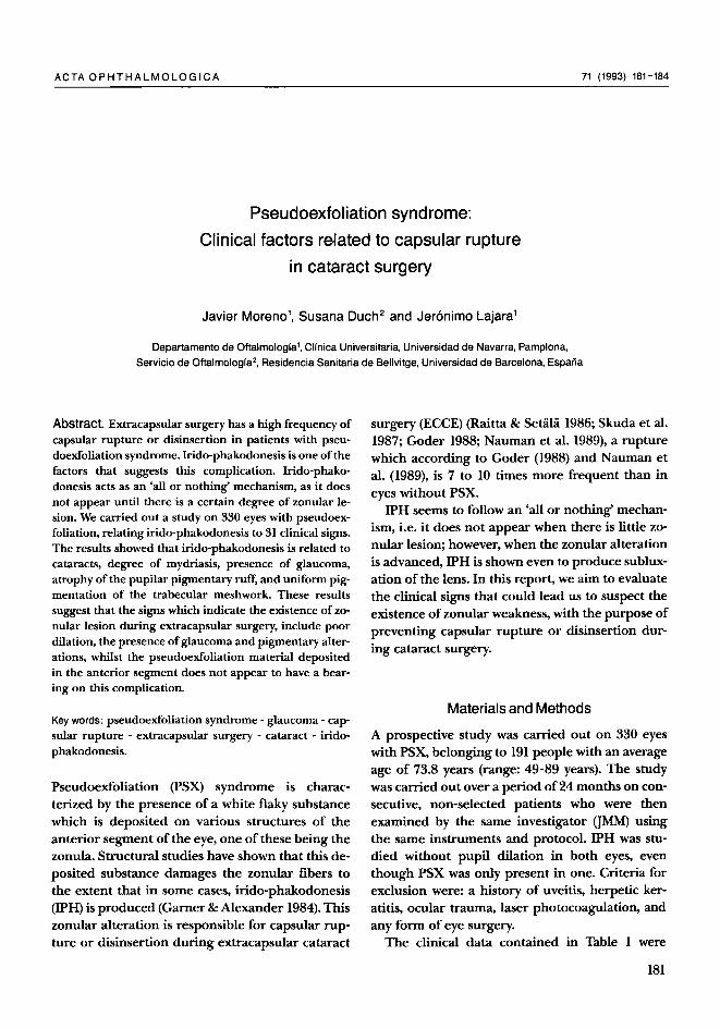
Fig. 1. IPH frequency according to cataract stage and degree of
dilation.
(p=0.002). Similarly, the mean IOP in the eyes with IPH was 27.3 f 9.3 mmHg, and in the eyes without IPH, it was 23.6 f 10.5 (p = 0.04); however, within the group of eyes with glaucoma, a higher IOP did not mean a higher occurrence of IPH.
The signs due to PSX material were not related to IPH, whilst a relation was found in some pro- duced by pigment. The study of atrophy of the pupilar pigmentary ruff by means of transillumi- nation, showed that IPH appears in 5.6% of the eyes with grade 1 atrophy, increasing to 9.6% with grade 2, and to 21.4% with grade 3 (p=0.009). In addition, the degree of uniform pigmentation in the camerular angle was signifcant, since the eyes with uniform pigmentation presented IPH in 13.2% of cases, as opposed to 6% in those without pigmentation (p = 0.03). Finally, IPH was present in 14.5% of eyes with light-coloured irises, and only in 5.6% of those with dark irises (p = 0.009).
After using midriatic drops the occurrence of IPH was 33.4% in eyes with no or minimal dilation, decreasing to 6.6% in those dilating normally (p=O.OOOl). One of the signs which complicates ECCE surgery is the poor dilation found in pa- tients with PSX. If the degree of dilation is associ- ated with cataracts, we find that the frequency of IPH was 50% in eyes with mature and hypermature cataracts which dilated minimally, compared to around 10% in eyes with mature cataracts and maximum dilation (p = 0.0001) (Fig. 1).
Finally, the relation between IPH's association with glaucoma and cataracts was studied due to the fact that these patients require a combined
WiUlaulGLAUCOMA
0 WilhGLAUCOMA P 40
/ STAGE 0 STAGE I STAGE 2 STAGE 3 STAGE 4
CATARACT
Fig 2. IPH frequency according to existence of glaucoma and
cataract stage.
ECCE-trabeculectomy intervention. The results are seen in Fig. 2: If a patient had a hypermature cataract and glaucoma, the occurrence of IPH was 41.6%, while it was only 15% in patients who only had a hypermature cataract with no signs of glau- coma; the frequency also increased with the stage of the cataract in both glaucomatous and non- glaucomatous eyes (p = 0.0001).
Discussion
IPH is a sign found in PSX, produced by damage to the zonular fibers brought about by the PSX ma- terial deposited on them. This zonular lesion is progressive to the point that, as IPH, it is visible by biomicroscopy. For this reason, although IPH func- tions as an 'all or nothing' mechanism, the zonular damage is a progressive alteration. In this report, our aim has been to analyse the different factors which would permit us to predict capsular rupture or disinsertion during cataract surgery; even though this report does not analyse the state of the zonular fibers, it is based on IPH in order to pro- vide an evaluation of zonular weakness.
The IPH frequency of 10.6% found in this study was similar to Futa & Furuyoshy (1989). Our results showed that IPH is not related to the quantity of PSX material deposited on the anterior lens cap- sule. However, some of the signs derived from pig- mentary alterations are related to IPH, such as: the uniform pigmentation of the camerular angle, atrophy of the pupillary pigmentary ruff, etc. In
183

addition, a previous study which we carried out References (Moreno et 2.1990) showed that in this syndrome glaucoma appears to be correlate with the quantity of pigment released by the iris and deposited on the meshwork, and not with PSX material. On the other hand, IPH is also linked to the existence of glaucoma, independently from glaucomatous IOP. Pignalosa et al. (1989) have stated that the fre- quency of capsular rupture during ECCE surgery is higher in eyes with PSX glaucoma than in those with PSX syndrome. These findings, together with the relation with old-age, could be due to the fact that glaucoma appears in more advance stages of PSX, and could explain that IPH is found in the later stages, being related to the signs which ap- pear later in this syndrome (age, the presence of glaucoma, the uniform pigmentation of the mesh- work etc.). Because of this, it would seem that IPH depends to a greater extent on the duration of the PSX syndrome than on the quantity of PSX ma- terial.
On the other hand, we wanted to evaluate the relationship between this sign and the existence of the cataract, both alone and glaucoma-associated. If the cataract is mature, IPH frequency is higher, especially when associated with poor pupil dila- tion, or glaucoma. In these cases combined surgery (ECCE-trabeculectomy) is required. Previous studies (Moreno et al. 1989, 1990), indicate that the cataract in PSX is not an additional condition, but that it is a sign of the PSX syndrome and evolves more quickly than in the senile cataract, it being likely therefore, that the cataract evolves over a shorter period of time to become mature or hyper- mature. The high frequency of IPH in cataracts as- sociated with glaucoma also indicates that this sign appears in the later stages of PSX.
Our results indicate that zonular weakness is greater in the advanced stages of PSX syndrome and especially in patients with hypermature cata- racts, poorly dilating pupils, glaucoma and uni- form pigmentation on the trabecular meshwork. The presence of these signs lead us to believe that the danger of capsular rupture or disinsertion dur- ing ECCE surgery is greater. Thus surgery should be planned more cautiously when cataract is asso- ciated with poor pupil dilation or glaucoma. Per- haps in eyes with PSX syndrome, in order to avoid capsular rupture, the cataract should be operated on in earlier stages than the senile cataract.
Farrar S M, Shields M B, Miller K N & Stoup Ch M (1989): Risk factors for the development and severity of glau- coma in the pigment dispersion syndrome. Am J Oph- thalmol 108: 223-229.
Futa R & Furuyoshy N (1989): Phakodonesis in capsular glaucoma: a clinical and electron microscopic study. Jpn J Ophthalmol33: 311-317.
Gamer A & Alexander R A (1984): Pseudoexfoliative dis- ease: histochemical evidence of an affinity with zonu- lar fibres. Br J Ophthalmol68 574-580.
Coder G J (1988): Our experiencies in planned extracap- sular extraction in the exfoliation syndrome. Acta Ophthalmol (Copenh), Suppl 184 126-128.
Moreno J, Alcolea A & Campos S (1989): Prevalence of pseudoexfoliation syndrome in the Northwest of Spain. Acta Ophthalmol (Copenh) 67: 383-385.
Moreno J, Alvarez A & Alcolea A (1990): Pseudoexfolia- tive glaucoma in patients with open angle glaucoma in the Northwest of Spain. Acta Ophthalmol (Copenh)
Moreno J, Alvarez A, Quinteiro A & Alcolea A (1990): Syndrome exfoliatif: etude clinique du angle irido-cor- neen. J Fr Ophthalmol 13: 183-188.
Naumann G 0, Kuchle M & Schonherr U (1988): Bseu- doexfoliationsyndrom als Risikofaktor f i r Glaskoper- verlust bei der extrakapsularen Kataraktextraktion. Fortschr Ophthalmol 86: 543-545.
Pignalosa B, Toni F & Liguori G (1989): Considerations on posterior chamber intraocular lens implantation in patients with pseudoexfoliation syndrome. Doc Oph- thalmol71: 49-53.
Raitta Ch & Setgla K (1986): Intraocular lens implanta- tion in exfoliation syndrome and capsular glaucoma. Acta Ophthalmol (Copenh) 6 4 130-133.
Skuda G L, Parrish I1 R K, Hodapp E, Forster R K et al. (1987): Zonular dialysis during extracapsular cataract extraction in pseudoexfoliation syndrome. Arch Oph- thalmol 105: 632-634.
68: 695-699.
Received on September 7th, 1992.
Author's address:
Dr Javier Moreno-Montaiies, Departamento de Oftalmologia, Clinica Universitaria, Universidad de Navarra, Apartado 192, 31080 Pamplona, Espana.
184