PROXIMAL FEMORAL FOCAL DEFICIENCY AND … · PROXIMAL FEMORAL FOCAL DEFICIENCY AND ITS TREATMENT1...
21
PROXIMAL FEMORAL FOCAL DEFICIENCY AND ITS TREATMENT 1 Thomas V. Rossi, M.D. 2 and Leon Kruger, M.D. 2 Proximal femoral focal deficiency (PFFD) is a relatively rare disorder involving the proximal femur and frequently the acetabulum. It occurs unilaterally more commonly than bilaterally. The problems seen in patients with PFFD are discrep- ancy in leg length, instability of the hip, contrac- tures of the hip and knee, unequal level of the knee, short stature, and associated anomalies. The associated anomalies increase the problems of treatment and prognosis significantly. Proximal femoral focal deficiency has been discussed at length in the proceedings of a sym- posium edited By Aitken (2,4,8,10). The present paper discusses the authors' experience with 38 patients, and suggests a classification of PFFD based on treatment considerations. Attempts are made to answer four questions: • When is amputation or turnplasty (Van Nes procedure) indicated? • When is knee fusion indicated? • When is valgus osteotomy indicated? • When is surgery contraindicated? Indications for hip stabilization are not dis- cussed because the authors' experience in this area is limited to two patients. The classification systems of Aitken (1, 2) and Amstutz (4), which are based primarily on x-ray data, are the best currently available. Aitken has divided PFFD into four types (A, B, C, and D). In both Type A and Type B a femoral head and an adequate acetabulum are present. In Type A, there is either a bony or cartilaginous connection between the head and neck fragments and the shaft at maturity, whereas in Type B there is no such connection. In Type A, often there is a sub- trochanteric pseudoarthrosis. X-rays of patients in Types C and D show no femoral head or acetabulum. These two types are differentiated by the presence of an ossified tuft at the proximal end of the shaft in Type C patients. The four types are depicted in Figure 1. Amstutz's method of classification is similar to Aitken's but includes coxa vara with bowing and further differentiation into subtypes. His five morphological types identified at birth are sub- typed usually by five years of age. The different types and subtypes are presented in Figure 2. 1 Presented at the annual meeting of Chiefs of Child Amputee Clinics sponsored by the Subcommittee on Child Prosthetics Problems of the Committee on Prosthetics Research and Development, National Academy of Sciences, held in Springfield, Mas- sachusetts, May 24-25, 1972. 2Shriners Hospital for Crippled Children, 516 Carew Street, Springfield, Massachusetts 01104. Fig. 1. Diagrammatic representation of four types of proximal femoral focal deficiency as proposed by Ait- ken (10).
Transcript of PROXIMAL FEMORAL FOCAL DEFICIENCY AND … · PROXIMAL FEMORAL FOCAL DEFICIENCY AND ITS TREATMENT1...
PROXIMAL FEMORAL FOCAL DEFICIENCY AND ITS TREATMENT1
Thomas V. Rossi, M . D . 2 and Leon Kruger, M . D . 2
Proximal femoral focal deficiency (PFFD) is a relatively rare disorder involving the proximal femur and frequently the acetabulum. It occurs unilaterally more commonly than bilaterally. The problems seen in patients with PFFD are discrepancy in leg length, instability of the hip, contractures of the hip and knee, unequal level of the knee, short stature, and associated anomalies. The associated anomalies increase the problems of treatment and prognosis significantly.
Proximal femoral focal deficiency has been discussed at length in the proceedings of a symposium edited By Aitken (2,4,8,10). The present paper discusses the authors' experience with 38 patients, and suggests a classification of PFFD based on treatment considerations. Attempts are made to answer four questions:
• When is amputation or turnplasty (Van Nes procedure) indicated?
• When is knee fusion indicated? • When is valgus osteotomy indicated? • When is surgery contraindicated?
Indications for hip stabilization are not discussed because the authors' experience in this area is limited to two patients.
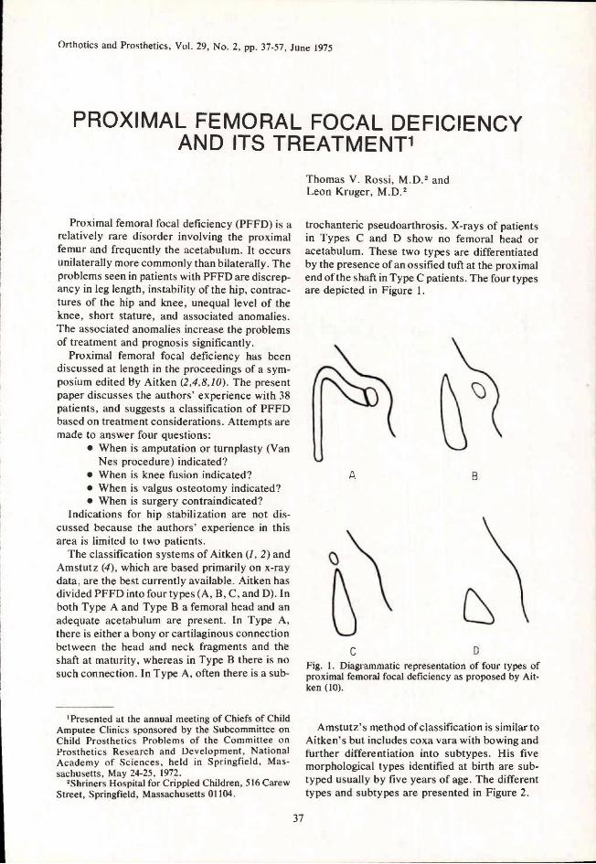
The classification systems of Aitken (1, 2) and Amstutz (4), which are based primarily on x-ray data, are the best currently available. Aitken has divided PFFD into four types (A, B, C, and D). In both Type A and Type B a femoral head and an adequate acetabulum are present. In Type A, there is either a bony or cartilaginous connection between the head and neck fragments and the shaft at maturity, whereas in Type B there is no such connection. In Type A, often there is a sub-
trochanteric pseudoarthrosis. X-rays of patients in Types C and D show no femoral head or acetabulum. These two types are differentiated by the presence of an ossified tuft at the proximal end of the shaft in Type C patients. The four types are depicted in Figure 1.
Amstutz's method of classification is similar to Aitken's but includes coxa vara with bowing and further differentiation into subtypes. His five morphological types identified at birth are sub-typed usually by five years of age. The different types and subtypes are presented in Figure 2.
1 Presented at the annual meeting of Chiefs of Child Amputee Clinics sponsored by the Subcommittee on Child Prosthetics Problems of the Committee on Prosthetics Research and Development, National Academy of Sciences, held in Springfield, Massachusetts, May 24-25, 1972.
2Shriners Hospital for Crippled Children, 516 Carew Street, Springfield, Massachusetts 01104.
Fig. 1. Diagrammatic representation of four types of proximal femoral focal deficiency as proposed by Ait-ken (10).
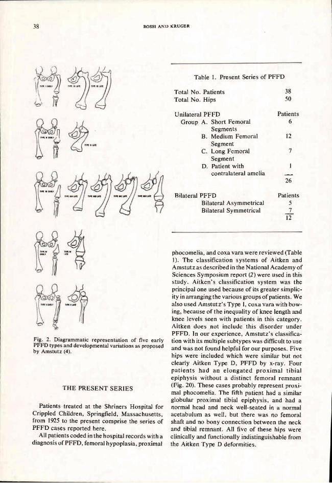
Fig. 2. Diagrammatic representation of five early PFFD types and developmental variations as proposed by Amstutz (4).
Table 1. Present Series of PFFD
THE PRESENT SERIES
Patients treated at the Shriners Hospital for Crippled Children, Springfield, Massachusetts, from 1925 to the present comprise the series of PFFD cases reported here.
All patients coded in the hospital records with a diagnosis of PFFD, femoral hypoplasia, proximal
phocomelia, and coxa vara were reviewed (Table 1). The classification systems of Aitken and Amstutz as described in the National Academy of Sciences Symposium report (2) were used in this study. Aitken's classification system was the principal one used because of its greater simplicity in arranging the various groups of patients. We also used Amstutz's Type I, coxa vara with bowing, because of the inequality of knee length and knee levels seen with patients in this category. Aitken does not include this disorder under PFFD. In our experience, Amstutz's classification with its multiple subtypes was difficult to use and was not found helpful for our purposes. Five hips were included which were similar but not clearly Aitken Type D, PFFD by x-ray. Four pat ients had an e l o n g a t e d prox imal tibial epiphysis without a distinct femoral remnant (Fig. 20). These cases probably represent proximal phocomelia. The fifth patient had a similar globular proximal tibial epiphysis, and had a normal head and neck well-seated in a normal acetabulum as well, but there was no femoral shaft and no bony connection between the neck and tibial remnant. All five of these hips were clinically and functionally indistinguishable from the Aitken Type D deformities.

Table 2. Associated Congenital Anomalies
A total of 50 hips in 38 patients are included in this series. There were 26 patients with unilateral involvement and 12 patients with bilateral involvement. The 12 patients affected bilaterally were further subdivided into bilateral symmetrical and bilateral asymmetrical categories. The criteria for differentiating the two groups were clinical leg-length measurements. Differences discernible and measurable in the roentgenographs were not used. The patient's functional status based on total leg-lengths and not his x-ray diagnosis was the primary concern. If both legs were equal functionally regardless of differences in the segments he was classified as a bilateral symmetrical PFFD. If he had significant leg-length discrepancies (3 inches or greater) from any cause, i.e., fibular hemimelia, proximal migration of the femoral remnant, or other deformity, he was classified as asymmetrical bilateral PFFD and his treatment was based on this consideration.
ASSOCIATED ANOMALIES
The data from this series confirmed the repeatedly reported findings of previous invest igators (5,8,10), namely , other congeni ta l anomalies are frequently associated with PFFD. Thirty-one of the 38 patients in the present series had other limb abnormalities (Table 2). The most frequent ly a s s o c i a t e d anomaly was fibular hemimelia which was found in 50% of the patients. Fourteen had ipsilateral fibular hemimelia and 5 had bilateral involvement.
CLASSIFICATION A N D TREATMENT
After careful study of the 38 patients, we concluded that while Aitken and Amstutz classifications are soundly based on the embryology and morphology of the condition, they do not indicate a therapeutic approach to the individual patient. On the basis of our experience, therefore, a new method of classification is suggested (Table 3). We submit that the basic division in these patients is between individuals with unilateral and bilateral PFFD. We would further subdivide the bilat
eral group into symmetrical and asymmetrical categories.
We suggest that the unilateral group be sub-classified according to the length of the remaining femoral segment. In order to express this in a meaningful manner we have used the technique of proportional measurement. The proportion, or percentage, is determined by dividing the length of the involved femur by the length of the normal femur and multiplying by 100:
The m e a s u r e m e n t s are made from the triradiate cartilage to the most distal border of the femur as viewed on the x-ray plate (Fig. 3). If the involved femur has migrated proximally so that it is superior to the triradiate cartilage we ignore the portion above our landmark. In an occasional patient the femoral remnant will be in an almost horizontal position due to hip and knee flexion deformities. It is then necessary to measure the entire length of the femoral remnant instead of using the triradiate cartilage as a fixed proximal point.
In the case of unilateral PFFD we believe that there are three therapeutic groups characterized by a short, medium, or long femoral segment. The short femoral segments measure less than 20% of the normal side. Those measuring 20 to 70% of the normal side were considered to be medium femoral segments, and those greater than 70% were designated as long femoral segments. While it was found that there was a tendency for the ratio or percentage of femoral length to change with time, in no case was this change great enough to put a patient into a different treatment group. Amstutz has stated that proportionality of growth was present in all patients by age five (4).
U N I L A T E R A L PFFD
Unilateral PFFD affects the largest group of patients. Of the 26 unilateral patients in our series, 19 underwent a total of 35 surgical procedures on the involved extremities. The two most common operations were amputation, which was performed 12 times, and valgus osteotomy in 7 patients (9 procedures). The next most common procedures were knee fusion in 4 patients, and contralateral epiphyseal stapling or epiphysiodesis in 3 patients. Other procedures involving at least one patient were rotation-plasty, os teosynthesis of the femoral neck, acetabulo-plasty, toe amputation, and open reduction and fusion of the hip with subfusion os teotomy. Seven patients did not receive any surgical treatment. Two of these were seen very early in the series, 2 were lost to follow-up, and 2 are yet too young for surgery. In the 7 patients for whom surgery was not indicated the leg-length discrepancy was no more than 1 1/2 in., and function and cosmesis were satisfactory otherwise.
Twenty-three unilaterally involved patients had a leg-length discrepancy of more than l 1/2 in. One patient had contralateral amelia, and 2 had coxa vara with bowing (Amstutz Type I PFFD) and leg-length discrepancies of less than 3 in. Twenty patients had leg-length discrepancies of 4 1/2 in. or greater at the time of amputation or when last seen. Twelve of these patients had an amputation. The youngest patient had 4 3/4 in. of shortening at the age of one year and the oldest had 4 1/2 in. of shortening at the age of 13 years. Of the remaining 8 patients with more than 4 1/2 in. of leg-length discrepancy who did not have amputa
tions, one had aturnplasty procedure, 2 were lost to follow-up, and the remaining 5 were doing well with either an extension prosthesis or a platform orthosis. Treatment of these 5 patients was carried out prior to 1955. On the basis of study of this group of patients we feel that patients in the short and medium femoral length groups will eventually come to amputation and above-knee-type prostheses.
Twenty-five patients (the twenty-sixth patient had contralateral amelia) were divided into three treatment groups according to femoral percentages as described (Table 3).
Table 3. Treatment Classification

Short Femoral Segments
Case History
Case 1, L.C. (Figs. 3 and 4), a white male, had a right proximal femoral focal deficiency, Aitken's Class D deformity, and bilateral upper-limb terminal transverse hemimelia. He had a right transtibial amputation at the age of nine years at which time the leg-length discrepancy was 11 in. His right ankle was at the level of the normal knee. Postoperatively, he was fitted with a nonstandard above-knee prosthesis which provided ischial weightbearing. As a result, this patient, who had
previously worn an extension prosthesis obtained an improvement in appearance and gait and an increase in hip stability. With modern concepts of amputation surgery, a foot disarticulation rather than a below-knee amputation might have been performed.
The treatment recommendations for the patients with the short femoral segments would be;
1. Foot disarticulation and ischial weightbearing above-knee prosthesis.
2. Van Nes turnplasty procedure and modified below-knee prosthesis.
3. N o surgery and an articulated extension prosthesis.
Fig. 3. The measurements of femoral length from triradiate cartilage to the end of the distal femoral epiphysis are demonstrated. The figure on the left shows a patient with an Aitken Class D deformity with a femoral segment less than twenty percent of the normal side. This patient in fact has a femoral segment ten percent of the normal side and represents what we would classify a short femoral segment. Note that the ankle on the affected side is above the normal knee level. In the right photograph notice again the measurement of the femoral length, and note that this patient demonstrates a left coxa vara with bowing with a femoral segment seventy-seven percent of the normal side. This represents a long femoral segment.

Fig. 4. Case 1; L.C. This six-year old boy presents with bilateral upper-limb transverse hemimelia had a right PFFD. His foot on the affected side is at the normal knee level (Fig. 3). At age nine the patient underwent transtibial amputation. The patient has been fitted with an A-K prosthesis with modified UCB quadrilateral socket and Silesian-type suspension. He is fully active and has gained hip stability through ischial bearing (11).
Medium Femoral Segments
There were 12 unilateral proximal femoral focal deficiency patients whose femoral segment ranged from 20 to 70% of the normal femur. The average shortening in this group was 6 1/2 in., with the foot lying both at the level of the opposite knee and, in approximately half of the patients, between the contralateral knee and ankle. Hip-and knee-flexion deformities are the rule unless the patient is seen very early in life and properly splinted or braced. Efforts at fitting these patients without surgery are rewarded by ambulation, but progressive knee- and hip-flexion deformities may occur. The longer the femoral segment, the easier it is to control knee flexion. In those patients with shorter femoral segments, 20-40%, the ship's ventilator or stovepipe type of socket is satisfactory, but good gait is difficult to achieve when hip- and knee-flextion deformities are present. The weightbearing line of the prosthesis will be anterior to (in front of) the body's weight line, and ischial weightbearing will be difficult to
achieve. The result is an exaggeration of the hip limp which the proximal femoral focal deficiency patients always demonstrate.
Perhaps the best way to correct this problem is by fusion of the knee in full extension. This provides a single-segment lever (9) to correct hip flexion and, when the distal limb is ablated, good ischial weightbearing is permitted through the prosthesis, thus diminishing the hip lurch. Knee fusion can be accomplished without damage to the distal femoral and proximal tibial epiphysis (Fig. 5). We have performed knee fusion as early as age five.
Only four of our patients had knee fusion while nine had amputations and one had a Van Nes rotation-plasty. The four patients with knee fusion had femoral segment ratios varying between 25 and 40%. On the basis of the results of these four, we feel that most patients in the group of patients with medium femoral segments do best with knee fusion and ablation. The functional and cosmetic appearance is superior to other known treatment regimes.

The amputation level for this group of patients depends upon the relationship of the involved foot to the normal knee. A transtibial amputation should be performed when the involved foot is below the normal knee. Ankle disarticulation is possible in those patients with a foot lying at or slightly above the normal knee. The object is to fit the patient with an AK prosthesis so that the prosthetic and normal knees are at the same level. We find that consultation with the prosthetist is of great benefit in making the final decision on level of amputation.
It is well to recall that in children the ideal ablation procedure is disarticulation, a procedure which permits end bearing by the resulting stump. This of course is valuable for proprioceptive feedback. It is also preferable because spiking as found in transtibial amputations, is prevented. When disarticulation leaves a stump that is excessively long, epiphysiodesis or bone shortening procedures should be considered in lieu of transtibial amputation.
Case Histories
The following cases illustrate the above points: Case 2, J.G. (Figs. 5 and 6), a white male, with a
left Aitken Class C proximal femoral focal deficiency, had associated upper-limb deficits. Present were terminal longitudinal adactylia bilateral of the fourth and fifth rays, and dislocation of the left radial head and syndactylism of the second and third fingers of the left hand. He had progressive shortening from 2 1/2 in. at age eight months to 7 in. at age seven years. When he was seven years of age a left Van Nes turnplasty procedure was performed. At the time of surgery the left ankle was 2 in. below the contralateral knee level. At the present t ime, he is wearing a modified below-knee prosthesis with his own ankle joint functioning as a knee joint. N o w , one-and-one-half years postoperatively, there has been no sign of spontaneous derotation of the turnplasty procedure, (5,7,8), and the ankle continues to provide good function as a knee joint.
The Van Nes procedure is an alternative to amputation and is probably indicated only when the ankle level of the affected limb can be expected to be at the knee level of the sound limb when maturity is reached. Some shortening, o f
course, can be gained at the time of osteotomy by removal of bone. We have considered this procedure as appropriate primarily for the male patient because the cosmetic appearance of the prosthesis is only fair and the appearance of the limb with the prosthesis off is poor. It is interesting to note that, when Van Nes (12) described his procedure in 1950, all three of the patients discussed were female and all three had knee fusion performed. In one patient, the rotation plasty was done through the knee.
Case 3 , L.S. (Figs. 7, 8, and 9), a white female with a left proximal femoral focal deficiency, Aitken Class A, had 7 1/2 in. of shortening at the age of eight years with the foot midway between the
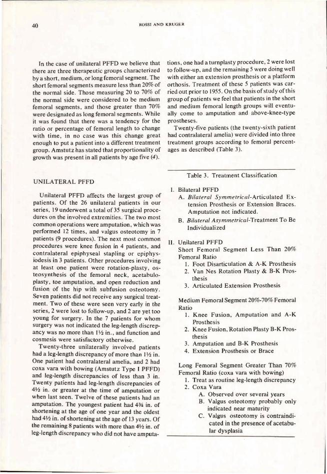
Fig. 5. Case 2; J.G. This boy age six with right PFFD had seven inches of shortening with an ankle level two inches below the normal knee. He had a poor gait with the extension prosthesis. At age seven he underwent a turnplasty procedure and received a B-K prosthesis. Note that the ankle level is still below the knee level. Ischial weightbearing will improve hip stability. Knee fusion in the future will simplify the prosthetic problem.
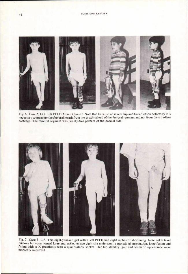
Fig. 6. Case 2; J.G. Left PFFD Aitken Class C. Note that because of severe hip and knee flexion deformity it is necessary to measure the femoral length from the proximal end of the femoral remnant and not from the triradiate cartilage. The femoral segment was twenty-two percent of the normal side.
Fig. 7. Case 3; L.S. This eight-year-old girl with a left PFFD had eight inches of shortening. Note ankle level midway between normal knee and ankle. At age eight she underwent a transtibial amputation, knee fusion and fitting with A-K prosthesis with a quadrilateral socket. Her hip stability, gait and cosmetic appearance were markedly improved.
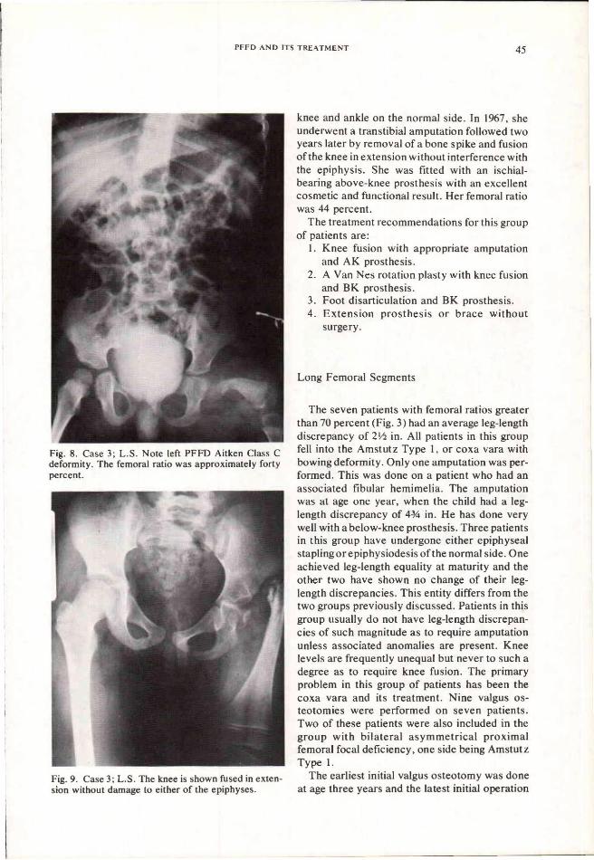
Fig. 8. Case 3; L.S. Note left PFFD Aitken Class C deformity. The femoral ratio was approximately forty percent.
Fig. 9. Case 3; L.S. The knee is shown fused in extension without damage to either of the epiphyses.
knee and ankle on the normal side. In 1967, she underwent a transtibial amputation followed two years later by removal of a bone spike and fusion of the knee in extension without interference with the epiphysis. She was fitted with an ischial-bearing above-knee prosthesis with an excellent cosmetic and functional result. Her femoral ratio was 44 percent.
The treatment recommendations for this group of patients are:
1. Knee fusion with appropriate amputation and AK prosthesis.
2. A Van Nes rotation plasty with knee fusion and BK prosthesis.
3. Foot disarticulation and BK prosthesis. 4. Extens ion prosthesis or brace without
surgery.
Long Femoral Segments
The seven patients with femoral ratios greater than 70 percent (Fig. 3) had an average leg-length discrepancy of 2 l/2 in. All patients in this group fell into the Amstutz Type 1, or coxa vara with bowing deformity. Only one amputation was performed. This was done on a patient who had an associated fibular hemimelia. The amputation was at age one year, when the child had a leg-length discrepancy of 4 3/4 in. He has done very well with a below-knee prosthesis. Three patients in this group have undergone either epiphyseal stapling or epiphysiodesis of the normal side. One achieved leg-length equality at maturity and the other two have shown no change of their leg-length discrepancies. This entity differs from the two groups previously discussed. Patients in this group usually do not have leg-length discrepancies of such magnitude as to require amputation unless associated anomalies are present. Knee levels are frequently unequal but never to such a degree as to require knee fusion. The primary problem in this group of patients has been the coxa vara and its treatment. Nine valgus os teotomies were performed on seven patients. Two of these patients were also included in the group with bilateral asymmetrical proximal femoral focal deficiency, one side being Amstutz Type 1.
The earliest initial valgus osteotomy was done at age three years and the latest initial operation

was at age seven. In two patients valgus osteotomies were repeated three years after the initial procedure. The neck-shaft angle ranged from 75 deg. in the youngest to 90 deg. in the oldest patient. The acetabular index at the time of surgery in all patients except three was approximately 20° which was very close to the opposite or normal acetabular index. All patients with acetabular indexes of close to 20° at the time of surgery had no significant change. The remaining three patients had acetabular indexes of 33, 30,
and 35°, respectively, at the time of surgery. All of these patients had an increase in the acetabular index.
The results of valgus osteotomy in these patients was generally unsatisfactory (Table 4). In four patients the results were rated as poor because of markedly short necks and frequently flattened femoral heads with limited motion (Figs. 10 and 11). Three patients had subluxation. One patient progressed from subluxation to eventual dislocation of the femoral head (Fig. 12). One patient rated as having a good result maintained a well-seated head without increase of the acetabular index and with improvement of the neck-shaft angle from 75° to 115°. The remaining patient is
Fig. 10. Case 5; C.M. Top, age two with right coxa vara with bowing. Note normal acetabular index. At age five years, middle, there is slight increase of coxa vara with short neck, base of neck vertical defect, and no definite proximal femoral epiphysis. At age twelve years, bottom, she has had a valgus osteotomy twice, at age five and seven. Note short neck, small head, well seated in the acetabulum.
Fig. 11a. Case 6; P.O. Note right coxa vara with bowing. At age three note the short laterally bowed femur with thickened medial cortex and coxa vara. There is a mid-neck vertical lucency and a normal acetabular index in the absence of a definite proximal femoral epiphysis.

one year post-valgus osteotomy and the result to date is considered good, with a neck-shaft angle of 120° and a rounded and well-seated femoral head.
In this series, 14 hips in twelve patients had coxa vara with bowing, but to date no hip has been followed long enough without surgery for the natural course of this deformity to be deter-
Fig. 11b. Case 6 at age seven. Note slight decrease in the neck-shaft angle and the base of neck defect.
Fig. 11c. Case 6 at age nine. A successful valgus osteotomy has been performed with a resulting neck-shaft angle of 125 deg. Note the small head and neck, absence of a proximal femoral epiphysis, and closure of mid-neck lucency.
Fig. 11d. Case 6 at age thirteen. Note the very short neck and small but rounded well-seated head.
Fig. 12a. Case 7, T.O., at age five prior to valgus osteotomy for her coxa vara with bowing deformity. Note the shallow acetabulum (33°) and the vertical lucency which is beginning to close.

Fig. 12b. Case 7, at age nine, four years after valgus osteotomy. Note the marked acetabular dysplasia, subluxed femoral head and very short femoral neck. This patient progressed to frank dislocation.
Table 4. Valgus Osteotomy In Patients With Coxa Vara With Bowing
mined. The treatment which is indicated for this group of patients is equali zation of leg-length discrepancy. Amputation may be indicated only when leg-length discrepancies are exaggerated by associated anomalies. Knee fusion is not indicated. This retrospective study suggests that these patients do not benefit from valgus osteotomy. If it is indicated it should not be performed until maturity has been attained. Valgus osteotomy is contraindicated in any patient with an acetabular index greater than 30°.
BILATERAL SYMMETRICAL PPFD
In the seven patients with bilateral symmetrical proximal femoral focal deficiency included in this

series, there was an extremely high incidence of associated anomalies, frequently multiple. Only one patient of the seven did not have an associated congenital anomaly.
The treatment of this group of patients was nonsurgical except for two. One patient, S.M. (Figs. 13 and 14), had a right ankle fusion. A second patient (T.B.) had a posterior release of the left ankle. No patient in this group had an amputation.
When they have upper limbs for balance assistance, patients in this group ambulate in spite of the severity of the lower-limb anomalies. We have fitted them with articulated limbs, without amputation, and they have done well. Our oldest patient is now thirty-two years of age and has been wearing articulated extension prostheses for eighteen years. We do not know whether or not she will be able to wear them at age forty or at age fifty. Therefore no amputation is anticipated.
This philosophy is shared by Hall (8), Aitken (2) and others. For those few patients in whom increasing deformities preclude continued ambulation without prosthesis, amputation is to be considered in order to provide the potential for prosthetic fitting. Of three patients with associated bilateral fibular hemimelia. two ambulate independently or with extension prostheses. The third patient is the only one with significantly limited ambulation (T.B.). He has bilateral proximal femoral focal deficiency, bilateral fibular hemimelia, and bilateral upper-limb amelia. It is the upper amelia and lack of balance assistance from arms that have precluded significant ambulation.
Fig. 13. Case 8, S.M., with bilateral symmetrica] PFFD, is shown with and without her articulated extension prostheses. There is marked improvement of cosmetic appearance and increase in height from 3 ft. 6 in. to 5 ft. 1 1/2 in. S.M. wears the prostheses full time, yet retains the ability to ambulate independently.
Fig. 14. X-ray of Case 8 showing bilateral symmetrical PFFD Aitken Class C deformities.

One patient was lost to follow-up at age seven. Five of the six remaining patients were walking independently albeit with a waddling gait when last seen. Two of the five patients use articulated extension prostheses and a third patient has been fitted with nonarticulated stubbies. At the age of twelve, the sixth patient (T.B.) can take no more than 12 steps without assistance.
Case Histories
Case 8, S.M. (Figs. 13 and 14), first seen at age fourteen, in 1953, was noted to have symmetrical bilateral proximal femoral focal deficiency of the Aitken Class C type with right intercalary paraxial fibular hemimelia and right terminal trans
verse distal humeral hemimelia plus a valgus deformity of the right foot with 1/2 in. shortening on the right side. Following fusion of her right ankle, she was fitted with articulated extension prostheses which increased her overall height from 3 ft. 6 in. to 5 ft. 1 1/2 in. After one year she was able to manage her prostheses without external support. At the age of thirty-two she has two normal children that she takes care of, and for the past eighteen years has worn her prostheses from the time she arises in the morning until she goes to bed at night.
Both hips are unstable and she has a 30° fixed-flexion deformity of the right knee and a 90° fixed-flexion deformity of the left knee. She walks with a waddling gait, both with and without her prostheses. This patient, as do all patients with unstable hips and weak musculature, ex-
Fig. 15. Case 9, T.B., with bilateral symmetrical PFFD and upper-limb anomalies, is shown in and out of braces. He is only able to take a few steps independently. The lack of upper limbs markedly decreased the potential for ambulation. The patient has associated scoliosis.

pends a great deal of energy in using the articulated extension prostheses. Balance is an added difficulty. Despite the extended period she has worn her prostheses it is difficult to say whether she will continue to wear them in another ten, twenty or thirty years.
Case 9, T.B. (Figs. 15 and 16), is a white male born with bilateral Aitken Class C proximal femoral focal deficiencies and bilateral terminal paraxial fibular hemimelia, bilateral upper-limb amelia, right congenital torticollis resolved, and a s lowly increasing right convex dorsolumbar scoliosis. The only surgical procedure was a left tendon Achilles lengthening and a posterior release for an equinovalgus deformity of the foot. He has 1 1/2 in. of apparent shortening on the right side. Independent ambulation is limited to twelve
Fig. 16. X-ray of Case 9, with bilateral symmetrical PFFD Aitken Class C deformities. Note the burrowing of the right femoral remnant into the ilium. He has pain on weightbearing and no passive or active motion on the right.
Fig. 17. Case 10, N.T. This patient with bilateral asymmetrical PFFD has both markedly short stature and progressive leg length discrepancy. At the time of this photo he was ten and a half years old with 3 1/2 in. of shortening on the right and was 3 ft. 5 in. in height. Extension prostheses increased his height to 4 ft. 5 l/2 in. He wears his prostheses in public, but for play and at home he removes them (11).

steps. This boy, who is of normal intelligence, relies on a CAPP electric cart to get about. Although his lower-limb involvement is no more severe than some of our other patients, he is markedly hampered by a balance problem owing to the absence of upper limbs.
BILATERAL ASYMMETRICAL PFFD
Five patients in the present series had bilateral asymmetrical proximal femoral focal deficiency. All had leg-length discrepancies of 3 1/2 in. or more. Three of these patients had amputations and were fitted with prostheses to provide equalization of leg lengths. One had a surgical fusion of the knee on the short side and valgus osteotomy of the contralateral side where coxa vara with bowing was present. A second also had an associated valgus osteotomy of a coxa vara deformity. Two patients were treated nonsurgically, the older being given an articulated extension prosthesis on the short side and an extension prosthesis on the long side. The fifth is currently only six months old.
T h e s e f ive pat ients do not represent a homogeneous entity; rather they tend to show characteristics of both unilateral and bilateral symmetrical proximal femoral focal deficiencies. M.V. (Case 11) and N.T. (Case 10) with their markedly shortened stature are very similar to the bilateral symmetrical proximal femoral focal deficiencies, yet they also have problems of leg-length discrepancy. They also had the problem of bilateral hip instability found in the bilateral symmetrical proximal femoral focal deficiencies. Three illustrative case reports follow.
Case Histories
Case 10, N.T. (Figs. 17 and 18), with bilateral asymmetrical proximal femoral focal deficiency (right Aitken Class D and left Aitken Class C deformities) had a non-segmentation of the right knee, associated scoliosis , and a hypoplastic mandible. His upper limbs were normal. He had a leg-length discrepancy which increased from 3 1/2
in. on the right at age ten, to 4 l/2 in. at age sixteen, at which time his height was 4 ft. 2 in. without prostheses. With bilateral extension prostheses this patient was the same height as his peers during school hours, but at home and after school he removed the prostheses and played without external support or other devices.
This case illustrates that while surgery might have been desirable because of significant leg-length discrepancy, it is not always necessary. A patient's short stature can be compensated for while his ability to get about without external aids is maintained.
Case 11, M.V. (Figs. 19 and 20), had bilateral asymmetrical proximal femoral focal deficiency (right proximal phocomelia and left Aitken Class
Fig. 18. Case 10; N.T. X-ray shows bilateral PFFD with a right Aitken Class D and left Aitken Class C deformities. Note proximal migration of right femoral remnant and non-segmentation of the knee which caused a functional leg length discrepancy of three and a half inches. Because of the functional and progressive discrepancy of over three inches we classified N.T. as a bilateral asymmetrical PFFD.

Fig. 19. Case 11, M. V. This boy has bilateral asymmetrical PFFD. At age seven he was 3 ft. 4 in. tall and had 4 1/2 in. of shortening on the right side, but could walk independently (Fig. 10 top). At age thirteen he was five years post right Boyd amputation (Fig. 3, right). His height was 3 ft. 9 1/2 in. with a right Syme's-type prosthesis. M. V. was fitted with a right A-K prosthesis and a left articulated extension prosthesis with increase in height to the level of his peers, but, for reasons of poor balance and marked effort, he returned exclusively to a right Syme's-type prosthesis.
C deformity). He also had an associated right terminal paraxial fibular hemimelia. The leg-length discrepancy of the right limb progressed from 3 in. at age five to 5 1/2 in. at age eight and a half, at which time a right Boyd amputation was performed, and a Syme's-type prosthesis was fitted.
At the age of thirteen years, M.V. was given a right above-knee prosthesis and a left extension prosthesis because of his short stature, then 3 ft. 9 1/2 in. He used these devices for approximately 2 1/2 years during school hours, but returned exclusively to his Syme's prosthesis. At the age of fifteen he elected to use the Syme's prosthesis
alone, because of the difficulty with balance and the physical effort involved in walking with the longer devices. He also had associated limb deficiencies, of a right upper terminal transverse hemimelia and a left upper terminal longitudinal incomplete paraxial ulna hemimelia.
M.V. was fully active with a right Syme's prosthesis and still had the ability to use his end-bearing stump. His height of 4 ft. 2 1/2 in. at the age of fifteen, lack of knee motion, and instability of the hip resulted in only fair cosmesis and function. This patient's ability to use his extension prosthesis because of lack of balance and the marked effort involved point out not only the

Fig. 20. Case 11; M.V. This x-ray illustrates bilateral asymmetrical PFFD with what is probably a proximal phocomelia on the right with associated fibular hemimelia. The left side was classified as Aitken Class C because of the lack of an acetabulum and definite femoral head.
importance of the upper limbs in ambulation but also the possible tragedy of bilateral amputation. M.V. was left with the option of being short but being able to walk independently. This option is not open to the patient with a bilateral amputation.
Case 12, M.G. (Figs. 21 and 22), a white male, had a left Aitken Class C proximal femoral focal deficiency and a right Amstutz Type I coxa vara with bowing deformity. He had 3 1/2 in. of shortening on the left side at age four and a half years, and without a lift or prosthesis he walked with a severe limp. At the age of five and a half years he had a right subtrochanteric valgus osteotomy followed by a left Boyd-type amputation. He was fitted with a Syme's-type prosthesis and ambu-
lated well but with marked inequality of knee levels and considerable instability of the left hip. At the age of eight years the right subtrochanteric valgus osteotomy was repeated. At the age of eleven years his left knee was fused and a transtibial amputation was performed above the level of the contralateral knee. He was fitted with a prosthesis incorporating a modified quadrilateral socket and hydraulic knee. He is now twelve years old, 4 ft. 9 in. high, and wears on the left s ide an a bo v e -knee prosthes i s with ischial weightbearing. His gait is considered to be excellent.
This case illustrates several points. This child had a marked leg-length discrepancy, which was 3 1/2 in. at age four with marked inequality of knee levels . Amputation improved his gait significantly but no improvement in inequality of knee levels or hip stability was achieved. Although this patient no longer has an end-bearing stump he wears a total-contact , ischial weightbearing socket which gives him increased hip stability and improved gait. M.G. also had contralateral coxa vara with bowing with moderate acetabular dysplasia and a conically shaped femoral head. This is the type that Amstutz would probably classify as a Type I B, and which he feels would do poorly with valgus osteotomy (4). As we can see from the x-rays, (Fig. 22) this child has undergone two valgus osteotomies without improvement. The head and neck are short and conical, the trochanter is above the hip joint and almost impinging on the ilium, and the acetabulum is dysplastic.
DISCUSSION
In this retrospective study of 38 patients with proximal femoral focal deficiency, we have proposed a new method of classification based on treatment considerations. For children with unilateral proximal femoral focal deficiency, classification would be on the basis of the femoral length ratios. It has been shown that while leg-length discrepancies almost always increase in patients with unilateral involvement, the growth ratios are constant. After five years of age, all patients show proportional growth (4). A patient with an ankle level opposite the normal knee at

Fig. 21. Case 12; M.G. Top left, this boy with bilateral asymmetrical PFFD had 3 1/2 in. of shortening at age four and a half years. Top, center, and right, at age 5 1/2 years he had a left Boyd amputation, and was fitted with an A-K prosthesis. Note the poor cosmetic appearance due to marked inequality of knee levels. His gait was poor because of the inequality of knee levels and hip instability. Bottom photographs, at age eleven he underwent a left knee fusion and B-K amputation. Now he has an A-K prosthesis with ischial weightbearing and hydraulic knee. His cosmetic appearance and gait are considered to be excellent.

Fig. 22. Case 12, M.G. At age seven his x-ray shows a left Aitken Class C deformity. On the right the patient is post valgus osteotomy for a coxa vara with bowing. Note the very short and conical head, acetabular dysplasia and probably early subluxation; also note the marked inequality of knee levels.
age 2-5 years will probably have a similar relationship at maturity unless there is marked migration of the femoral remnant, or increase in hip-and knee-flexion deformities.
Those with short femoral segments will either have a small nubbin of femur lying completely above the ischium or lower edge of the pelvis, or have a form of femoral phocomelia or proximal phocomelia. The patients with medium femoral segments almost all had knee levels below the ischium. This led us to calculate the ratios of the distance from triradiate cartilage to ischium, and compare this to the length of normal femora in twenty five patients (Fig. 3). The age range was from 2 months to 12 years and the ratios were all from 18-24%. Thus, a patient who has a unilateral proximal femoral focal deficiency with a knee level below the ischium on the involved side will usually have a femoral segment ratio of twenty percent or greater of the normal side.
Hip instability will be a problem in these patients. The above-knee prosthesis with ischial weightbearing will markedly improve hip stability. Patients treated with rotation-plasty and below-knee prostheses will also need added support to achieve hip stability. Bevan-Thomas (6) has suggested a pelvic corset and lateral hinge connected to the prosthes is , and an ischial weightbearing socket. We would emphasize the latter.
A C K N O W L E D G M E N T
We wish to express our appreciation for the kind assistance of Mr. Hector Kay in the preparation of this paper.
R E F E R E N C E S
1. Aitken, George T., Amputation as a treatment: for certain lower-extremity congenital abnormalities. J. Bone and Joint Surg., 41 - A: 7:1267-1285, October 1959.
2. Aitken, George T., Proximal femoral focal deficiency—Definition, classification, and management, pp. 1-6, in Proximal Femoral Focal Deficiency: A Congenital Anomaly. National Academy of Sciences Publication No. 1734, 1969.
3. Amstutz, Harlan C , and Philip D. Wilson, Jr., Dysgenesis of the proximal femur (coxa vara) and its surgical management. J. Bone and Joint Surg., 44-A: 1:1-24, January 1962.
4. Amstutz, Harlan C., The morphology, natural history, and treatment of proximal femoral deficiencies, pp. 50-57, in Proximal Femoral Focal Deficiency: A Congenital Anomaly. National Academy of Sciences Publication No. 1734, 1969.
5. Babb, F.S. , R. K. Ghormley, and C.C. Chatterton, Congenital coxa vara. J. Bone and Joint Surg., 31 A:115-131, January 1949.
6. Bevan-Thomas, W.H. , and E. A. Millar, A review of proximal focal femoral deficiencies. J. Bone and Joint Surg., 49-A:7:1376-1388, October 1967.
7. Hall, John E., Rotation of congenitally hypoplastic lower limbs to use the ankle joint as a knee. A preliminary report. Inter-Clin. Inform. Bull., 6:2:3-9, November 1966.
8. Hall, John E., and Dietrich Bochman, The surgical and prosthetic management of proximal femoral focal deficiency, pp. 77-81, in Proximal Femoral Focal

Deficiency: A Congenital Anomaly. National Academy of Sciences Publication No. 1734, 1969.
9. King, R . E . , Proving a single skeletal lever in proximal femoral focal deficiency. A preliminary case report. Inter-Clin. Inform. Bull., 6:2:23-28, November 1966.
10. King, Richard E., Some concepts of proximal femoral focal deficiency, pp. 23-27, in Proximal
Femoral Focal Deficiency: A Congenital Anomaly. National Academy of Sciences Publication No. 1734,1969.
11. Kruger, Leon M. Classification and prosthetic management of limb-deficient children. Inter-Clin. Inform. Bull., 7:12:1-25, September 1968.
12. Van Nes, C.P. , Rotation-plasty for congenital defects of the femur. J. Bone and Joint Surg., 32-3:1:12-16, February 1950.