anesth.utmb.eduanesth.utmb.edu/residents/documents/Emergency Protocols.docx · Web viewNo ETCO2 and...
34
UTMB DEPARTMENT OF ANESTHESIOLOGY EMERGENCY PROTOCOLS BY: Samip Sheth, Michelle Scanlon FACULTY MENTORS: Dr. Woodson, Dr. Przkora June 2014 Table of Contents 1 Unable to Deliver Set Tidal Volume 2 Delayed Emergence/Hypoxemia and No Train of Four 3 Local Anesthetic Toxicity 4 Amniotic Fluid Embolism 5 Anaphylaxis 6 Bronchospasm (Intubated Patient) 7 Delayed Emergence 8 Difficult Airway (Unanticipated) 9 Hemorrhage (Massive Transfusion) 10 Severe Hypotension
Transcript of anesth.utmb.eduanesth.utmb.edu/residents/documents/Emergency Protocols.docx · Web viewNo ETCO2 and...

UTMB DEPARTMENT OF
ANESTHESIOLOGY
EMERGENCY PROTOCOLS
BY Samip Sheth Michelle ScanlonFACULTY MENTORS Dr Woodson
Dr Przkora
June 2014
Table of Contents
1 Unable to Deliver Set Tidal Volume2 Delayed EmergenceHypoxemia and No
Train of Four3 Local Anesthetic Toxicity4 Amniotic Fluid Embolism5 Anaphylaxis6 Bronchospasm (Intubated Patient)7 Delayed Emergence8 Difficult Airway (Unanticipated)9 Hemorrhage (Massive Transfusion)10 Severe Hypotension11 Hypoxemia12 Malignant Hyperthermia13 Myocardial Ischemia14 Oxygen FailureO2 CrossoverPipeline
Failure15 Pneumothorax16 Power Failure17 Tachycardia ndash Stable SVT18 Total Spinal Anesthesia19 Transfusion Reaction20 Venous Air Embolism21 Post-Extubation Airway Compromise
22 Lost ETCO2 Tracing
(1) UNABLE TO DELIVER SET TIDAL VOLUMESigns include
1) Hypoxia and Hypercarbia 2) Desaturation3) Decreased minute ventilation4) Smell of volatile anesthetic
Rule Out
1) Cardiac Arrest (No ETCO2 tracing + hemodynamically unstable) CALL FOR HELP ACLS algorithm
2) No ETCO2 and hemodynamically stable Check both ends of CO2 tubing If connected Loss of ETCO2 algorithm
Management Ventilate with Ambubag and prepare to switch to TIVA technique
1) Check External Circuit Connections Elbow CO2 detector on patient and machine side InspiratoryExpiratory limbs at the machine InspiratoryExpiratory valves
2) Check ETT Cuffa Add air if necessaryb If ruptured notify surgeons and change ETT
with Cook catheter + DL3) Listen for bilateral equal breath sounds
a No Breath Sounds Rule out extubation DL to ensure ETT cuff past vocal cords If not past cords deflate cuff advance ETT inflate recheck for bilateral breath sounds secure ETT
b Unilateral breath sounds Withdraw ETT Deflate cuff Withdraw Reinflate Check BBS secure ETT
c ETT well-positioned + No breath sounds +- High PIP Suction ETT (mucus plug) Consider changing ETT
d Increased PIP + Bilateral expiratory wheezing Go to Bronchospasm andor Anaphylaxis algorithms
4) Check machine factors other than the circuit Call Biomed
a Vaporizer caps tightb CO2 absorber tightc Valvesd Scavenger system
5) If MajorInternalMachine Fault Ventilate patient with Ambubag Look for bilateral chest rise ETCO2 while on 100 O2 Switch to a TIVA technique while Biomed works on fixing the issue vs replacing the machine
(2) Delayed Emergence Hypoxemia AND NO TRAIN OF FOUR (Continued from Delayed Emergence or
Hypoxemia algorithms)
Signs include1) Respiratory insufficiencydistress (Minimal chest rise
Nominimal fogging of mask)2) Inability to move extremities or flailing around3) Somnolence (from CO2 narcosis)
High suspicion if1) Deep block (Intermediatelong-acting muscle relaxant +
short procedure 0 or 1 twitch prior to reversal No reversal given Older patient)
2) Drug interactions3) PatientFamily history of delayed emergence No prior
anesthetics
Management
A) If Patient Intubated1) Keep intubated with low-dose inhaled or IV
anesthetic PACU with full monitors Ambubag Ventilator in PACU
2) Recheck TOF or post-tetanic twitch at 2 locations
3) If No Twitches Wait until return of at least 1 twitch Give remainingfull reversal dose
4) Wait to meet extubation criteria and wean anesthetic (If extubating in PACU ensure all monitors airway equipment and suction present prior to extubation
B) If Patient Extubated1) Support ventilation with bagmask Notify
patient of situation if awake2) Check TOF If lt 4 strong twitches Give
remainingfull reversal3) Consider reintubationLMA placement if 0 or 1
twitch and start low dose anesthetic Monitor either in OR or PACU
C) If Patient Extubated in PACU1) Support breathing with bagmask Full
monitors Tell patient what is happening if awake
2) Check TOF If inadequate Call RT to bring ventilator Consider reintubation vs bagmask IV sedation
3) If not fully reversed reversal agent4) Consider ABG to determine CO2 level
ventilation status
(3) LOCAL ANESTHETIC TOXICITY
Symptoms1 Tinnitus Metallic Taste or Circumoral numbness2 Altered mental status3 Seizures4 Hypotension5 Bradycardia6 Ventricular arrhythmias7 Cardiovascular collapse
CALL FOR HELPCODE CART INTRALIPID KITINFORM TEAM
Treatment1 Stop LA injectioninfusion2 Get intralipid kit3 100 O2 high flow Consider ETT4 Treat seizure activity with BZDrsquos5 If signs persist or patient unstable
a Rapidly give 15 mLkg of 20 intralipid IV (70kg adult = 105 mL)
b Start infusion at 025 mlkgmin (18 mLmin)c If CV collapse repeat loading dose up to 3 total
dosesd If patient remains hypotensive Increase
infusion rate to 05 mLkgmin6 Monitor for hemodynamic instability treat hypotension
a Pulseless CPR and ACLS algorithmb Arrythmias ACLS algorithmsc If refractory to treatment Alert CT team for
possible cardiopulmonary bypass7 If attain circulatory stability
a Continue intralipid infusion for at least 10 mins Afterwards (Upper limit 10 mlkg over 1st 30 mins)
b Monitor in ICU for 12-24 hours after event
Notes1 Consider reducing epinephrine doses to lt 1mcgkg IV2 Avoid Vasopressin Beta blockers Calcium channel
blockers3 May require prolonged resuscitation
(4) AMNIOTIC FLUID EMBOLISM
Signs (Pregnant or Postpartum patient)1 Respiratory distress Decreased O2 saturation2 CV collapse Hypotension Tachycardia Arrythmias
Cardiac Arrest3 Coagulopathy +- DIC4 Seizures5 Altered mental status6 Unexplained fetal compromise
CALL FOR HELPCODE CARTINFORM TEAM
Treatment1 Place patient in LUD (left uterine displacement)2 100 O2 High Flow3 Large-bore IV access4 Support hemodynamics with IV fluid Vasopressors
(EpiNorepiPhenylephrine) and Inotropes (Dobutamine)
5 Prepare for emergent intubation (Propofol Succ Cricoid pressure)
6 If possible place arterial line and consider CVC7 Anticipiate massive hemorrhage and DIC Activate
massive transfusion protocol Go to Hemorrhage alogrithm
8 Consider circulatory support with CPBECMOIABP
Rule Out1 Eclampsia2 Hemorrhage
3 Air Embolism4 Aspiration5 Anaphylaxis6 Pulmonary embolism7 Anesthetic overdose8 Sepsis9 CardiomyopathyCardiac valvular abnormalityMI
(5) ANAPHYLAXIS
Signs1 Hypoxemia Difficulty breathing Tachypnea2 RashHives3 Hypotension (Maybe severe)4 Tachycardia5 BronchospasmWheezing6 Increase in PIP7 Angioedema
CALL FOR HELPCODE CARTINFORM TEAM
Prepare Epinephrine 10 ugmL or 100 ugmLConsider pausing surgeryIf patient becomes pulseless CPR Epinephrine 1 mg IV bolusses large volume IV fluid Go to PEA algorithm
Treatment1 Discontinue potential allergens Colloids Blood
products Latex products Antibiotics2 Discontinue volatile anesthetic if hypotensive and give
KetamineVersed3 100 O2 high flow4 IV fluid bolus (May require many liters)5 Epinephrine Start 10-100 ug and increase dose q2 mins
until clinical improvement May require large doses6 Vasopressin 2-4 units7 Treat bronchospasm with Albuterol or Epinephrine8 Give H1 antagonist (Diphenhydramine 25-50 mg IV)9 Consider steroids (Methylprednisolone 125 mg IV) to
decrease biphasic response
10 Consider early intubation to secure airway prior to angioedema
11 Consider additional PIV Arterial line
(6) BRONCHOSPASM (Intubated patient)
Signs1 Increased peak airway pressures2 Bilateral wheezing3 Increased expiratory time4 Increased ETCO2 with upsloping ETCO2 tracing5 Decreased tidal volumes if pressure controlled
CALL FOR HELPCODE CART
Treatment1 If patient hypotensive disconnect patient circuit (to
relieve air trapping)2 100 O2 High Flow3 Increase expiratory time4 Deepen volatile anesthetic5 Suction ETT6 Medications
a Give inhaled Albuterol +- Ipratropiumb If severe Epinephrine IV start 10 ug and
escalatec Ketamine 02 ndash 1 mgkg IVd Hydrocortisone 100 mg IVe Nebulized racemic epinephrine
7 Consider ABG
Rule Out1 Mainstem intubation2 Anaphylaxis Go to anaphylaxis algorithm
(7) DELAYED EMERGENCE
Management1 All anesthetics Off2 No residual muscle paralysis Go to Residual Paralysis
algorithm 3 HypoxemiaHypercarbia BagMask vs Intubation4 Hypothermia Warm Blankets and fluids Bare hugger
Warm the room5 ABG with electrolytes If CO2 narcosis Intubate
Correct electrolytes6 Rule out medication swap or dosing error See below7 If all of the above negativenormal consider STAT Head
CT and consult neurologyneurosurgery (If intubated look for pupils asymmetric movement gagging)
a If residual mental status abnormalities monitor in ICU with serial neurologic exams and repeat imaging as needed
If suspect medication overdose1 Opioid reversal Naloxone 40 ug IV Repeat q2 minutes
up to 5 doses (200 ug total)2 BZD reversal Flumazenil 02 mg IV Repeat q1 minutes
up to 5 doses (1 mg total)3 Scopolamine patch reversal Physostigmine 1 mg IV
(Have Atropine ready for potential cholinergic crisis)
(8) DIFFICULT AIRWAY UNANTICIPATED
After 1st failed DL Attempt Go to A or B
A) If Difficult Mask1 Call for Help and Difficult AW Cart2 Oral or Nasal airway3 2-handed mask4 LMA vs Intubating LMA vs Combitube
If above masking measures fail1 Call ENT STAT2 Get percutaneous cricothyrotomy kit ready vs
Transtracheal jet ventilation
B) If Easy mask1 If could not get a view
a Reposition patientb BURP external laryngeal manipulation (Back
Up Rt)c Call for CMAC andor FOBd Try again with different blade (ensure adequate
anesthetic on board) suction as needede If no view again CMAC FOB LMA Fastrach
LMA +- FOB2 If could not pass ETT
a Make sure patient adequately anesthetizedb Use a Bougiec Use a smaller ETTd If still unable to pass FOB
(9) HEMORRHAGE ndash MASSIVE TRANSFUSION
CALL FOR HELPCODE CART IN ROOM
Management1 Call blood bank to activate MTP
a Get TampC if not already done2 100 O2 High flow3 Turn downoff agent4 IV fluid bolus 5 Trendelenburg position vs Leg elevation6 Vasopressors temporarily
a Phenylephrineb Ephedrinec Epinephrine (double dilute)
7 Call for Belmont or Level 1 Cell Saver 8 Additional IV Access IO or EJ if needed + Hot line9 Communicate with surgeon and monitor blood loss
a Can transfuse O- blood if TampC not done yet10 Use Hotline and Bare hugger to keep pt warm11 Place arterial line if needed Ask nurse for foley if
needed12 Monitor ABGrsquos13 Call for cell saver
If continued blood loss Replace before labs1 If gt 1 blood volume lost For every 1 unit PRBC 1 unit
FFP and for every 6 units PRBC 1 apheresis unit of platelets
Components1 PRBC If Hgb lt 7-10 based on comorbidities and rate of
blood loss (1 unit raises Hgb 1 gdL)
2 FFP For PTINR or PTT gt 15X normal 10-15 mlkg 11 ratio with PRBCrsquos
3 Platelets For platelets lt 50-100K with ongoing bleeding 6 units PRBC 1 apheresis unit of platelets (1 unit raises 50KuL)
4 Cryoprecipitate For fibrinogen lt 80-100 mgdl (10 units raises fibrinogen 50mgdL)
EBL = EBV X Hct starting ndashHct measured Hct starting
(Blood Volume estimate = 4500 mL for 70kg person)
(10) SEVERE HYPOTENSION
CALL FOR HELPCODE CART
Management 1 Ensure pulse and regular rate and rhythm If not go
to ACLS2 Inspect field for blood loss or
a If non-bleeding cause Pause surgeryb If bleeding cause Hemorrhage algorithm
3 IV Fluid bolus4 Pressors
a Phenylephrine and Ephedrineb If severe Epinephrine 10-100 mcg Vasopressin
1-4 units5 100 O2 High flow6 Turn downoff anesthetic agent7 Talk to surgeon8 Go to Other management
Other management1 Algorithms for ACLS Anaphylaxis Hemorrhage
Hypoxemia Local anesthetic toxicity Myocardial ischemia Pneumothorax Total spinal anesthesia Transfusion rxn VAE
2 TEE3 More IV access and Arterial line4 Steroid for adrenal insufficiency (Hydrocortisone 100mg
IV)5 Send labs ABG Hgb Electrolytes TampC6 Foley catheter to monitor UOP
Differential DiagnosisMAP = CO X SVR and CO = SV X HRSV depends on preload contractility and afterload
1 darr Preload a Hemorrhage Hemorrhage algorithmb Hypovolemiac Auto-PEEP DC circuitd IVC Compression Decrease tidal volume
Reposition patiente Embolism (AirFatBloodAFE) Go to algorithmf Pneumothorax Pneumothorax algorithmg Pneumoperitoneum or Surgical manipulation
2 darr Contractilitya MIIschemia myocardial ischemia algorithmb Low EF Inotropec Valvular disease HOCMd Hypoxemiae Medicationsf Local anesthetic toxicityg Arrythmias
3 uarr Afterloada Pneumoperitoneum or surgical manipulationb Medications
4 darr HRa Vagal stimulus Pause surgery if needed
5 darr SVRa High volatile anesthetic b Neuraxial blockadec Anaphylaxis Go to algorithmd Medicationse Shock Sepsis Spinal Neurogenicf Endocrine abnormalities Steroids
(11) HYPOXEMIA
CALL FOR HELPCODE CART
Management1 100 O22 Check FiO2 ETCO2 PIP BP Pulse3 Hand-ventilate to check compliance4 Listen for bilateral breath sounds5 Suction ETT
Other Management1 Large recruitment breaths (unless hypotensive)2 Bronchodilators (Albuterol)3 Additional muscle relaxant4 Head up (to increase FRC)5 FOB to ro mainstem intubation or obstruction6 ABG andor CXR7 Terminate surgery if refractory
DDx1 Hypoventilation
a Signs High or Low ETCO2 Poor chest rise Decreased breath sounds Patient bucking
b Low TV or RR Change settingsc Circuit leakd ObstructedKinked ETTe High PIPf Residual NMBg Patient breathing asynchronously with
ventilatorh Bronchospasmi Pulmonary edema
j High spinalk Pain
2 Low FiO2a If Low FiO2 while on 100 O2 Go to O2
failure algorithm3 VQ mismatch or shunt
a Mainstem inubationb Atelectasisc Aspirationd Bronchospasm (Anaphylaxis)e Mucus plugf Pleural effusiong Rare causes Pneumothorax Hypotension
Embolism (Air blood fat amniotic fluid)4 Diffusion abnormality Usually chronic lung disease5 Increased metabolic O2 demand MH Thyrotoxicosis
Sepsis Hyperthermia6 Artifacts Confirm with ABG
a Poor waveform (probe malposition cold extremity light interference cautery)
b Dyes (methylene blue indigocarmine blue nail polish)
(12) MALIGNANT HYPERTHERMIA
Early Signs1 Increased ETCO2 2 Tachycardia 3 Tachypnea 4 Mixed Metabolic and Respiratory Acidosis (ABG) 5 Masseter spasm trismus 6 Sudden cardiac arrest in young person due to hyperkalemia (peaked T waves)
Late Signs 1 Hyperthermia 2 Muscle rigidity 3 Myoglobinuria 4 Cardiac Arrest
CALL FOR HELPCALL FOR MH CART START PREPARING DANTROLENEINFORM TEAM
Differential Diagnosis1 Light anesthesia 2 Hypoventilation 3 Over-heating (external) 4 Thyroid storm 5 Pheochromocytoma 6 Hypoxemia 7 Insufflation of CO2
Treatment 1 Discontinue anesthetic triggers (volatiles and succinylcholine) and increase fresh gas flow to 10 Lmin Do NOT change machine or circuit
2 Halt procedure If emergent continue with non-triggering anesthetic 3 Hyperventilate FiO2 100 high flow O2 4 Assign several people to prepare 25 mgkg IV Dantrolene bolus Dilute each 20 mg Dantrolene vial in 60 mL preservative-free sterile water (for 70kg person give 175 mg so prepare 9 vials of 20 mg Dantrolene each as above) 5 Rapidly administer Dantrolene Continue giving until patient stable (may give up to 10 mgkg) 6 Administer sodium bicarbonate 1-2 mEqkg for metabolic acidosishyperkalemia 7 Actively cool patient with ice packs lavage if open abdomen Stop cooling at 38degC 8 Arrhythmias are usually secondary to Hyperkalemia Go to ACLS algorithms as needed 9 Treat hyperkalemia with Calcium Chloride 1 g IV D50 1 Amp IV (25 g Dextrose) + Regular Insulin 10 units IV (monitor glucose) Sodium Bicarbonate 1 Ampule Avoid calcium channel blockers 10 Send labs for ABG CPK myoglobin PTPTT and lactic acid 11 Place foley catheter Monitor urine output Goal 2 cc kg per hour urine output Can give IV fluid and diuretics 12 Arrange ICU bed Mechanical ventilation usually required 13 Continue Dantrolene 1mgkg every 4-6 hours for 24-36 hours observe closely 24 hours Call MH hotline with questions
Additional Information
Contact the Malignant Hyperthermia Association of the United States (MHAUS hotline) at any time for consultation if MH is suspected 1-800-MH-HYPER (1-800-644-9737) or online at httpwwwmhausorg
(13) MYOCARDIAL ISCHEMIA
Signs1 Depression or elevation of ST segment 2 Arrhythmias conduction abnormalities unexplained tachycardia bradycardia or hypotension 3 Regional wall motion abnormalities or newworse mitral regurgitation on TEE 4 In an awake patient Chest pain SOB diaphoresis etc
CALL FOR HELPCALL FOR CODE CARTINFORM TEAM
Treatment1 Increase to 100 O2 high flow 2 Verify ischemia (expanded monitor view vs 12-lead EKG) 3 Treat hypotension or hypertension 4 Beta-blocker to slow heart rate Hold for bradycardia or hypotension 5 Consider aspirin rectal or PO or NGOG 6 Consult Cardiology - stat 7 Treat pain with narcotics (fentanyl or morphine) 8 Consider nitroglycerin infusion (hold until hypotension treated) 9 Place arterial line and send Labs ABG CBC Troponin 10 If Anemic treat with packed red blood cells 11 Consider TEE for monitoring volume status and regional wall motion abnormalities 12 Consider central venous access
13 If hemodynamically unstable consider Intra-Aortic Balloon Pump 14 Be Prepared for Arrhythmias and have Code Cart at Bedside
(14) OXYGEN FAILUREO2 CROSSOVERPIPELINE FAILURE
Signs1 Audible O2 failure alarm sounds2 Low FiO2 on gas analyzer while on 100 oxygen
Immediate Actions1 Disconnect the patient from the machine and ventilate with an AmbuTM bag on Room Air Do not connect the patient to auxiliary flowmeter on machine ndash comes from SAME central source 2 Open O2 tank on back of anesthesia machine (check not empty) and disconnect pipeline oxygen to force flow from tank into circuit Alternative Obtain full E cylinder of O2 with a regulator Ventilate with AmbuTM bag or Jackson Rees circuit attached to new O2 tank 3 Connect elbow adaptor to allow monitoring of respiratory gases Is the patient receiving 100 oxygen 4 Maintain anesthesia (if necessary) with IV drugs
CALL FOR HELPCONSIDER HAVING CODE CART AVAILABLEINFORM TEAM
Secondary Actions1 Reduce O2 flow rates to minimum needed to conserve oxygen 2 Obtain extra backup sources of O2 3 When patient more stable contact Bioengineers to alert them to problem and enlist help with machine diagnosis while you focus on patient
4 Inform OR leadership ICU hospital of potential large-scale O2 problem 5 Discuss with surgeon implications of O2 failure for this patients management and OR schedule
(15) PNEUMOTHORAX
Signs1 Increased peak inspiratory pressures 2 Tachycardia 3 Hypotension 4 Hypoxemia 5 Decreased or asymmetric breath sounds (breath sounds decreased or absent on ipsilateral side)6 Hyper resonance of chest to percussion 7 Tracheal deviation (late sign) 8 Increased JVDCVP 9 Have high index of suspicion for pneumothorax in trauma patients COPD patients or when a CVC has been recently placed in the jugular or subclavian veins
CALL FOR HELPCONSIDER HAVING CODE CART AVAILABLEINFORM TEAM
Treatment1 DO NOT WAIT FOR X-RAY TO TREAT IF HEMODYNAMICALLY UNSTABLE 2 Increase to 100 O2 high flow 3 Rule out mainstem intubation 4 Consider stat CXR or TTE to assess 5 Place 14 or 16 gauge needle mid clavicular line 2nd intercostal space on affected side should hear a whoosh of air if under tension 6 Immediately follow up needle decompression with thoracostomy (chest tube)
(16) POWER FAILURE
Immediate Actions1 Get additional light sources - Laryngoscopes cell phones flashlights etc 2 Open doors and shades to let in ambient light 3 Confirm ventilator is working and if not ventilate patient with Ambu bag and switch to TIVA 4 If monitors fail check pulse and manual blood pressure 5 Request Transport Monitor or defibrillator monitor 6 Confirm adequate backup O2 supply - Power failure may affect oxygen supply or alarms 7 Check extent of power failure - Call bio-med or engineering - Is the problem in one OR all ORs or hospital-wide - If only in your OR check if circuit breaker has been tripped
(17) TACHYCARDIA ndash STABLE SVT
Signs1 CHECK FOR PULSE If no pulse Go to PEA algorithm2 If Unstable Go to SVT ndash UNSTABLE and Prepare for
Synchronized Cardioversion UNSTABLE = SBPlt80 BP low for patient rapid BP decrease or acute ischemia
3 Sinus Tachycardia is NOT SVT May be compensatory Search for and treat underlying cause(s)
4 More likely SVT if any of a Rate gt 150 b Sudden onset c Irregular rhythm
CALL FOR HELPCONSIDER HAVING CODE CART AVAILABLEINFORM TEAM
Treatment1 Increase to 100 O2 high flow 2 Confirm adequate ventilation oxygenation 3 Consider 12-lead EKG or Print Rhythm Strip then treat per rhythm 4 If UNSTABLE at any point Go to Unstable SVT algorithm 5 If still STABLE Supraventricular Tachycardia
a Consider cardiology consult b Consider ABG with electrolytes
Narrow Complex and Regular 1 To convert Adenosine 6 mg IV push with flush May give 2nd dose 12 mg IV 2 If NOT converted may Rate Control Choose beta blocker or calcium channel blocker
a Esmolol Start 05 mgkg IV over 1 min May repeat after 1 min and may start infusion 50 1048577gkgmin b Metoprolol Start 1-25 mg IV May repeat or double after 25 min c Diltiazem 5-10 mg IV over 2 min May repeat after 5 min
3 Amiodarone 150 mg IV SLOWLY over 10 min May repeat once Start infusion 1mgmin for first 6 hours
Narrow Complex and Irregular 1 Choose beta blocker or calcium channel blocker
a Esmolol Start 05 mgkg IV over 1 min May repeat after 1 min and may start infusion 50 1048577gkgmin
b Metoprolol Start 1-25 mg IV May repeat or double after 25 min
c Diltiazem 5-10 mg IV over 2 min May repeat after 5 min
2 Amiodarone 150 mg IV SLOWLY over 10 min May repeat once Start infusion 1 mgmin for first 6 hours
Wide Complex and Regular 1 Amiodarone 150 mg IV SLOWLY over 10 min May
repeat once Start infusion 1 mgmin for first 6 hours 2 May also consider Procainamide or Sotalol
Wide Complex and Irregular (Likely Polymorphic VT) 1 Prepare to defibrillate 2 Go to Ventricular Tachycardia amp Ventricular Fibrillation
(VTVF) algorithm
(18) TOTAL SPINAL ANESTHESIA
Signs
AFTER NEURAXIAL ANESTHESIA BLOCK 1 Unexpected rapid rise in sensory blockade 2 Numbness or weakness in upper extremities 3 Dyspnea 4 Bradycardia 5 Hypotension 6 Loss of consciousness 7 Apnea 8 Cardiac arrest
If Cardiac Arrest Start CPR Immediate Epinephrine Go to PEA algorithm
CALL FOR HELPCALL FOR CODE CARTINFORM TEAM
Treatment1 Support ventilation and intubate if necessary 2 Treat significant bradycardia with immediate epinephrine (start 10-100 1048577g increase as needed go to appropriate ACLS algorithm) If mild consider atropine (05 mg - 1 mg) but progress quickly to epinephrine if needed 3 Administer IV fluid bolus 4 If parturient prepare for possible emergent C-section Left Uterine Displacement monitor fetal heart rate
(19) TRANSFUSION REACTION
Signs
Hemolytic Transfusion Reaction1 Tachycardia 2 Tachypnea 3 Hypotension 4 Disseminated Intravascular Coagulation 5 Dark Urine
Febrile Reaction1Fever
Anaphylactic Reaction1 Tachycardia 2 Wheezing 3 Urticaria Hives 4 Hypotension
CALL FOR HELPCONSIDER HAVING CODE CART AVAILABLEINFORM TEAM
Treatment1 Stop transfusion 2 Support blood pressure with IV fluids and vasoactive medications if needed 3 If anaphylactic reaction Go to anaphylaxis algorithm
4 Consider antihistamine and antipyretic (Diphenhydramine Acetaminophen) 5 For hemolytic reaction maintain urinary output with IV fluids diuretics renal dose dopamine 6 Monitor for and treat disseminated intravascular coagulation if hemolytic reaction 7 Monitor for TRALI (lung injury) and treat accordingly may require post operative ventilation 8 Notify blood bank of reaction They will need further blood samples May need to contact transfusion medicine MD
(20 VENOUS AIR EMBOLISM
Signs1 Sudden Decrease in blood pressure and ETCO2 2 Sudden Decrease in SpO2 3 Sudden Rise in CVP 4 Sudden Onset of dyspnea and respiratory distress in
awake patient 5 Air on TEE (if monitoring)
CALL FOR HELPCONSIDER HAVING CODE CART AVAILABLEINFORM TEAM
Treatment1 Increase to 100 O2 high flow 2 Flood surgical field with saline 3 Place surgical site below heart (if able) 4 Aspirate air from the central line if present 5 Give rapid fluid bolus to increase CVP 6Turn down or off volatile anesthetic 7 Give Epinephrine (start 10-100 1048577g) to maintain Cardiac Output 8 Start CPR if BP catastrophically low 9 Consider TEE to assess air amp RV function 10 Consider left lateral decubitus positioning11 If severe terminate procedure if able
(21) POST-EXTUBATION AIRWAY COMPROMISE
Differential Diagnosis
1 Inadequate reversal of neuromuscular blockadea Decreased Train-of Four and TOF ratio
2 Laryngospasma Inspiratory stridorb Inspiratory wheezingc Diminished stridor may represent total airway
obstructiond Decreased air movement
3 Airway obstructiona High index of suspicion for laryngeal edema
after prolonged intubation or in neonatesinfantschildren whose laryngeal diameter is already small
b Laryngeal edema may be caused by large ETT traumatic intubation excessive manipulation of the head and neck during surgery excessive coughing or bucking on ETT current or recurrent URIs
c Consider recurrent laryngeal nerve damage following thyroid resection and other neck surgeries
d Consider obstructive hematoma following neck and ENT surgeries
e Consider hypocalcemia 24-96 hours following parathyroidectomy (purposeful or accidental)
4 Post-obstructive Pulmonary Edemaa Initial post-extubation airway obstructionb Immediately followed by respiratory distressc Hemoptysisd Pink frothy sputum
e CXR changes consistent with pulmonary edema5 Acute Aspiration
a Coughingb Wheezingc Feverd Chest discomforte Visible gastric contents in the orpharynx
Treatment
1 Inadequate reversal of neuromuscular blockadea If zero twitches on TOF immediately reintubate
and mechanically ventilateb If patient with at least 1 twitch on TOF but
significantly weak immediately reintubatec If patient ventilating but weak may need
increased dose of reversal agents with close monitoring and emergency airway equipment prepared
d Maintain mechanical ventilation and close monitoring until patient is exhibiting full strength and meeting all criteria for extubation
e May need workup for pseudocholinesterase deficiency if prolonged Succinylcholine paralysis
2 Laryngospasma Mask ventilate with positive pressureb Increase positive pressure if original pressures
not effectivec Consider Propofol in order to deepen anestheticd Consider Succinylcholine (IM or IV) to relax
vocal cords3 Airway obstruction
a If significant laryngeal edema or tracheomalacia suspected consider FOB in order to make
definitive diagnosis Reintubate if significantly narrowed laryngeal diameter Administer steroids
b If recurrent laryngeal nerve damage is suspected may consider FOB or rigid bronchoscopy to make definitive diagnosis If bilateral vocal cord paralysis immediately reintubate and notify surgeon
c If expanding neck or peritracheal hematoma is visualized or suspected immediately reintubate for airway protection and notify surgeon
d If hypocalcemia suspected reiuntubate for airway protection Draw labs and replete Calcium
4 Post-obstructive Pulmonary Edemaa Immediately provide supplemental oxygenb Diuresis to relieve pulmonary edema unless
patient significantly hypovolemicc If obstruction is severe enough may require
reintubation and PPVd Prolonged PACU monitoring or ICU monitoring
in severe cases5 Acute Aspiration
a Place patient in head down position to prevent further migration of gastric contents into lung parenchyma
b Oropharyngeal suctioningc Consider reintubation if patient cannot protect
the airwayd Monitor in inpatient setting for at least 48 hourse Consider FOB to remove non-particulate matterf Currently steroids antibiotics or
bronchopulmonary lavage are not recommended initially
g Antibiotics may eventually be required if not improvement in gt 48 hours
(22) LOST ETCO2 TRACING
Differential Diagnosis
1 Problems with the Anesthesia Equipmenta Kinked ETTb Defective or disconnected CO2 analyzerc Disconnected circuit significant leakd Defective ventilator
2 Problems with the Patienta Esophageal intubation or unplanned extubationb Lost cardiac output (decreased or no delivery of
CO2 to lungs)c Severe Bronchospasm (minimal or no air
movement)d Embolic evente Severe hypotension (severe blood loss
anaphylaxis cardiac depression or cardiac dysfunction)
Anesthesia Equipment SourceOther vital signs usually unaffectedPatient unlikely in immediate danger
Patient SourceOther vital signs likely abnormal
Patient possibly in significant danger
1 Circuit disconnect-gt check circuit starting from patient and tracing back to ventilator
2 CO2 tubing or analyzer disconnected or malfunctioned -gt check capnograph with own expiratory air 3 Significant circuit leak -gt do high pressure leak test manually ventilate -gt airway pressures will likely be low4 Kinked or obstructed ETT
-gt visually inspect ETT consider FOB for inspection of internal lumen
-gt airway pressures will likely be elevated5 Ventilator malfunction
-gt do manual high pressure leak test disconnect from ventilator to complete test
-gt manual ventilation will be possible and effective
1 Esophageal intubation or accidental extubation-gt direct laryngoscopy to confirm ETT placement lung auscultation chest rise-gt hypoxemia may eventually cause PVCs desaturation hypotension bradycardia eventual PEA
2 Cardiac Arrest-gt usually accompanied by severe hypotension decrease in amplitude or complete loss of SpO2 waveform arrhythmias
-gt check pulses monitor VS3 Severe Bronchospasm
-gt lung auscultation will indicate little or no air movement
4 Pulmonary Embolism-gt usually accompanied by desaturation hypotension normal initial ECG with eventual PVCs from hypoxemia-gt lung auscultation usually non-contributory as problem is dead space ventilation
5 Severe Hypotension-gt significant EBL volume depletion anaphylaxis SIRSshock
Loss of ETCO2 WaveformDifferential Diagnosis
Anesthesia Equipment SourceOther vital signs usually unaffected Patient unlikely in immediate danger
Patient SourceOther vital signs likely abnormal
Patient possibly in significant danger
1 Circuit disconnect -gt reconnect circuit2 CO2 tubing or analyzer disconnected or malfunctioned -gt replace CO2 tubing or analyzer 3 Significant circuit leak -gt inflate ETT if possible -gt change ETT if torn cuff -gt consider manual ventilation with ambu bag 4 Kinked or obstructed ETT
-gt consider inserting bite block or repositioning patient-gt consider replacing ETT if significant internal obstruction
-gt consider exchanging to wire reinforced ETT5 Ventilator malfunction
-gt manually ventilate with ambu bag while troubleshooting machine
-gt call for replacement ventilator
1 Esophageal intubation or accidental extubation -gt reintubate patient via direct laryngoscopy -gt follow difficult airway algorithm if unable to reintubate2 Cardiac Arrest -gt call for help -gt initiate ACLS3 Severe Bronchospasm -gt deepen anesthetic plane with IV anesthetics -gt give Epinephrine 10-100mcg -gt give bronchodilators 4 Pulmonary Embolism -gt call for help -gt give Epinephrine 10-100 mcg -gt may need to initiate ACLS5 Severe Hypotension
-gt treat hypovolemia with blood products colloid or crystalloid
-gt trend PPV (may trend CVP) -gt give appropriate pressors
Loss of ETCO2 WaveformTreatment
REFERENCES
1 Barash Cullen Stoelting Cahalan amp Stock (Eds) (2009) Clinical Anesthesia Philadelphia PA Lippincott Williams amp Wilkins
2 Goldhaber-Fiebert Sara and Steven Howard ldquoImplementing Emergency Manuals Can Cognitive Aids Help Translate Best Practices for Patient Care During Acute Eventsrdquo Anesthesia-Analgesia 1175 (2013) 1149-1161 Print
3 Horlocker et al ldquoRegional Anesthesia in the Patient Receiving Antithrombotic or Thrombolytic Therapy American Society of Regional Anesthesia and Pain Medicine Evidence-Based Guidelines (Third Edition)rdquo Regional Anesthesia and Pain Medicine 351 (2010) 64-101 Print
4 Miller Ronald (Ed) (2010) Millerrsquos Anesthesia Philadelphia PA Churchill Livingstone
5 Runciman Kluger Morris Paix Watterson ampWebb ldquoCrisis Management during anesthesia the developmentof an anesthetic crisis management manualrdquo Qual Saf Health Care 141 (2005) Print
6 Stanford Anesthesia Cognitive Aid Group Emergency Manual Cognitive Aids for Perioperative Critical Events See httpemergency manualstanfordedu for latest version Creative Commons BY-NC-ND 2014 (Version 2)
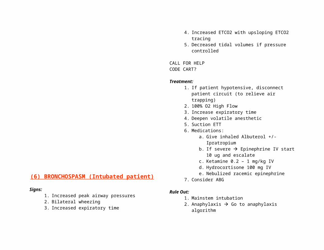
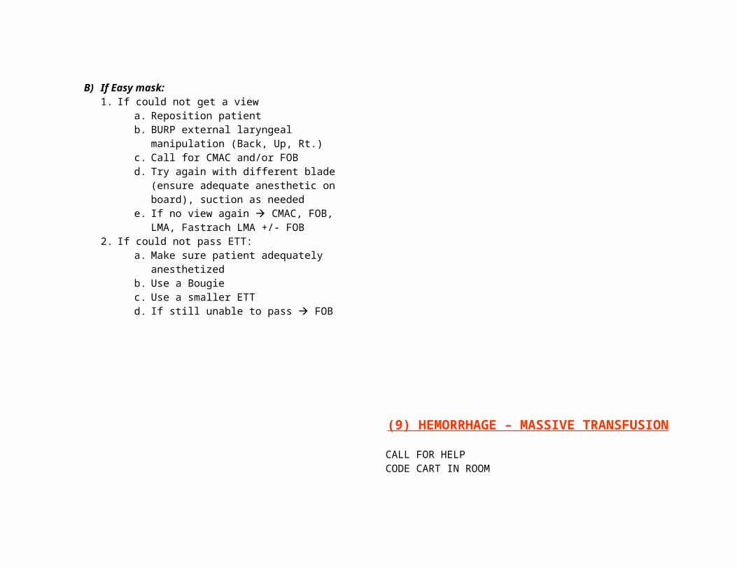
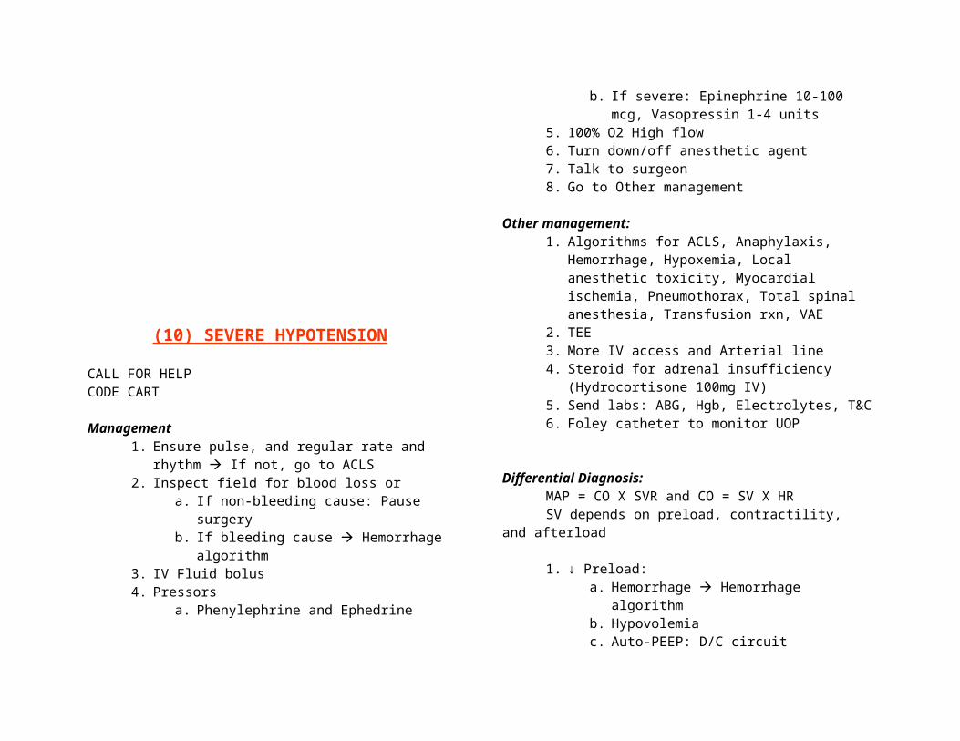
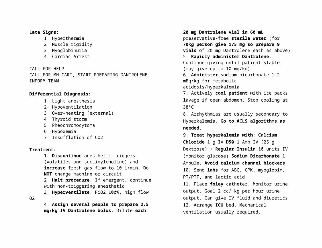