ProtoBlot II AP System with Stabilized Substrate/media/Files/Resources/Protocols/Technical...
22
Technical Manual ProtoBlot ® II AP System with Stabilized Substrate including Western Express ® Fast Blotting Protocol INSTRUCTIONS FOR USE OF PRODUCTS W3940, W3950 AND W3960. PRINTED IN USA. Revised 12/12 Part# TM026
Transcript of ProtoBlot II AP System with Stabilized Substrate/media/Files/Resources/Protocols/Technical...

T e c h n i c a l M a n u a l
ProtoBlot® II AP System with Stabilized Substrate including Western Express® Fast Blotting ProtocolINSTRUCTIONS FOR USE OF PRODUCTS W3940, W3950 AND W3960.
PRINTED IN USA.Revised 12/12 Part# TM026
tm026.1212:EIVD_TM.qxd 12/17/2012 12:44 PM Page a
Promega Corporation · 2800 Woods Hollow Road · Madison, WI 53711-5399 USA Toll Free in USA 800-356-9526 · Phone 608-274-4330 · Fax 608-277-2516 · www.promega.comPrinted in USA. Part# TM026Revised 12/12 Page 1
1. Description ..........................................................................................................1
2. Product Components and Storage Conditions ............................................3
3. General Considerations ....................................................................................3
4. Transfer of Antigen to Membrane .................................................................5A. Dot Blotting ...........................................................................................................5B. Transfer from Gels: Western Blotting ...............................................................6C. Plaque/Colony Lift Immunoscreening.............................................................6
5. Immunodetection of Antigens ........................................................................7A. Standard Protocol .................................................................................................7B. Western Express® Fast Blotting Protocol ............................................................8
6. Troubleshooting...............................................................................................13
7. Appendix ...........................................................................................................18A. Composition of Buffers and Solutions ............................................................18B. References ............................................................................................................18C. Related Products.................................................................................................19
1. Description
The ProtoBlot® II AP Systems with Stabilized Substrate improve upon thesimplicity of the original ProtoBlot® Western Blot AP Systems by incorporatingthe premixed, ready-to-use Western Blue® Stabilized Substrate for AlkalinePhosphatase into a convenient, reliable immunodetection system. TheProtoBlot® II System is designed for the rapid, sensitive detection of proteins orother macromolecular antigens immobilized on nitrocellulose or polyvinylidenefluoride (PVDF) membranes, either transferred from gels after electrophoresis(Western blots; 1,2) or bound directly from solution (“dot” blots).
Two protocols are provided: a standard immunodetection protocol and theWestern Express® Fast Blotting Protocol. The Western Express® Protocol is amodification of the standard protocol, which significantly reduces the timerequired to perform immunodetection to as little as 32 minutes. This protocol isdesigned to optimize detection time for the sensitivity of detection required.
ProtoBlot® II AP System withStabilized Substrate
All technical literature is available on the Internet at www.promega.com/tbs/ Please visit the web site to verify that you are using the most current version of this
Technical Manual. Please contact Promega Technical Services if you have questions on useof this system. E-mail [email protected].
tm026.1212:EIVD_TM.qxd 12/17/2012 12:44 PM Page 1

Applications of the Western Express® Protocol include:
• Fast Western and dot blots• Quick assays of column chromatography fractions on dot blots• Screening monoclonal antibodies
In general, 1ng of antigen on a Western blot and 200pg on a dot blot can bedetected using a 32-minute Western Express® Protocol. Using the standardprotocol, 50pg of antigen on a Western blot and 5pg on a dot blot can bedetected in 2.5–3 hours.
The ProtoBlot® II AP Systems with Stabilized Substrate are based on theenzyme-linked immunodetection of antigen-specific antibodies (supplied by theresearcher) using anti-IgG secondary antibodies conjugated to alkalinephosphatase (AP). Following incubations with the primary antibody andappropriate anti-IgG AP conjugate, Western Blue® Stabilized AP Substrate isapplied directly to the blot for color development. Sites of antigen localizationturn a dark purple color as the result of alkaline phosphatase activity.
Western Blue® Substrate contains NBT (nitro blue tetrazolium) and BCIP (5-bromo-4-chloro-3-indolyl-phosphate) in a proprietary buffer and is stable atroom temperature for up to one year. It is supplied premixed, fully diluted andready to use. Western Blue® Substrate brings a new level of convenience andreliability to this procedure while providing the same sensitive detection levelsseen with NBT/BCIP reagents, which require mixing before use.
Systems are available for the detection of human, mouse and rabbit antibodies.The blocking agents, bovine serum albumin (BSA) and Tween® 20, also areincluded with the systems.
Promega Corporation · 2800 Woods Hollow Road · Madison, WI 53711-5399 USA Toll Free in USA 800-356-9526 · Phone 608-274-4330 · Fax 608-277-2516 · www.promega.comPart# TM026 Printed in USA.Page 2 Revised 12/12
tm026.1212:EIVD_TM.qxd 12/17/2012 12:44 PM Page 2
2. Product Components and Storage Conditions
Product Cat.#ProtoBlot® II AP System with Stabilized Substrate, Human W3940ProtoBlot® II AP System with Stabilized Substrate, Mouse W3950ProtoBlot® II AP System with Stabilized Substrate, Rabbit W3960
Each system contains reagents sufficient to process twenty 10 × 15cm membranes. TheWestern Express® Fast Blotting Protocol is included. All secondary antibody APconjugates are affinity-purified from goat antisera. Includes:
• 100µl Anti-Human or Anti-Mouse IgG (H + L) or Anti-Rabbit IgG (Fc), AP Conjugate (1mg/ml)
• 300ml Western Blue® Stabilized Substrate for Alkaline Phosphatase• 5ml Tween® 20• 10g Blot-Qualified BSA• 1 Protocol
Storage Conditions: Store antibody conjugates at 4°C (undiluted). Store Western Blue® Substrate at room temperature (15–30°C).
3. General Considerations
In general, antigens immobilized on membranes are detected with antibodies ina three-step process (see Figure 1). First, the primary antibody, an IgG directedagainst the antigen in question, is added to bind potential antigenic sites. In thesecond step, a secondary antibody-enzyme conjugate, which recognizes generalfeatures of all IgGs (anti-IgG), is added to find locations where the primaryantibody bound. In the third step, AP, the enzyme conjugated to the secondaryantibody, catalyzes a colorimetric reaction when the appropriate substrate isadded, resulting in the deposition of colored product on the membrane at thereaction site. This color provides a visual indication of potential primaryantibody recognition.
Antigens can be immobilized (“blotted”) on nitrocellulose or PVDF membranesby one of several methods, depending on the requirements of the experiment.In all cases, the ability to detect a given antigen will depend on the amount ofantigen per unit area of the membrane and on the characteristics of the primaryantibody. Refer to Table 1 for detection sensitivies for dot blots [antigen spottedin 1µl (about 4mm)] and Western blots using this system and reasonably high-affinity, high-titer primary antibodies.
Promega Corporation · 2800 Woods Hollow Road · Madison, WI 53711-5399 USA Toll Free in USA 800-356-9526 · Phone 608-274-4330 · Fax 608-277-2516 · www.promega.comPrinted in USA. Part# TM026Revised 12/12 Page 3
tm026.1212:EIVD_TM.qxd 12/17/2012 12:44 PM Page 3
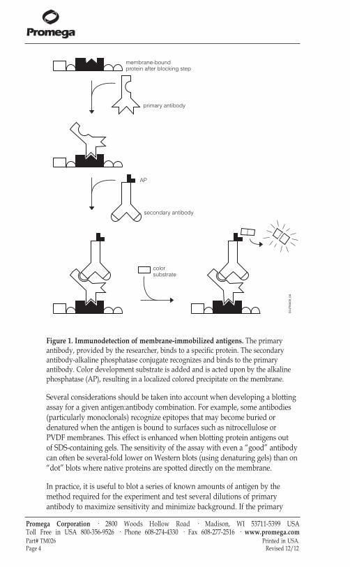
Figure 1. Immunodetection of membrane-immobilized antigens. The primaryantibody, provided by the researcher, binds to a specific protein. The secondaryantibody-alkaline phosphatase conjugate recognizes and binds to the primaryantibody. Color development substrate is added and is acted upon by the alkalinephosphatase (AP), resulting in a localized colored precipitate on the membrane.
Several considerations should be taken into account when developing a blottingassay for a given antigen:antibody combination. For example, some antibodies(particularly monoclonals) recognize epitopes that may become buried ordenatured when the antigen is bound to surfaces such as nitrocellulose or PVDF membranes. This effect is enhanced when blotting protein antigens out of SDS-containing gels. The sensitivity of the assay with even a “good” antibodycan often be several-fold lower on Western blots (using denaturing gels) than on“dot” blots where native proteins are spotted directly on the membrane.
In practice, it is useful to blot a series of known amounts of antigen by themethod required for the experiment and test several dilutions of primaryantibody to maximize sensitivity and minimize background. If the primary
Promega Corporation · 2800 Woods Hollow Road · Madison, WI 53711-5399 USA Toll Free in USA 800-356-9526 · Phone 608-274-4330 · Fax 608-277-2516 · www.promega.comPart# TM026 Printed in USA.Page 4 Revised 12/12
membrane-boundprotein after blocking step
primary antibody
secondary antibody
colorsubstrate
AP
0447MA09_0A
tm026.1212:EIVD_TM.qxd 12/17/2012 12:44 PM Page 4
antibody is used at too high a concentration, higher backgrounds result. At toolow a concentration, the positive signals will be weaker. In general, a range ofdilutions from 1:200 to 1:10,000 will reveal the optimal levels for most sera,ascites fluids and purified antibodies, whereas hybridoma tissue culture fluids(which have much lower antibody concentrations) usually require dilutionsfrom 1:10 to 1:100 for optimal performance.
4. Transfer of Antigen to Membrane
Bind the antigen to either nitrocellulose or PVDF membrane. Three differentmethods are described below.
Always wear gloves when handling membranes to avoid localizedbackground problems.
Materials to Be Supplied by the User(Solution compositions are provided in Section VII.A.)• Tris-buffered saline (TBS) • appropriate membranes• filter paper
4.A. Dot Blotting
Nitrocellulose Membranes
A solution containing the antigen (crude or purified) is spotted directly on adry or moist nitrocellulose membrane, usually in a 1µl volume.
1. To prepare a moist nitrocellulose membrane, float the membrane in acontainer of TBS (without Tween® 20) until it is evenly wet, submerge itbriefly, and place it on a piece of dry filter paper. Let the excess bufferdrain for approximately 5 minutes before use.
2. Dry membranes can be placed on top of filter paper or the backing paperincluded with the nitrocellulose membranes. It is useful to mark themembrane with a pencil or ball point pen at convenient intervals as a guidein spotting.
3. Prepare appropriate dilutions of the sample in TBS and apply in 1µlvolumes to the membrane using a micropipette. If more volume isrequired, add the sample in multiple applications over the same spot.Allow each spot to dry thoroughly before applying the next aliquot, so thespots remain compact. Allow all spots to dry before proceeding to theblocking step (Section V.A, Step 1, or V.B, Step 1).
4. An alternative method for making dot blots uses a filtration manifold,which allows multiple samples to be applied simultaneously in uniformspots. Several devices are commercially available for this purpose.
Promega Corporation · 2800 Woods Hollow Road · Madison, WI 53711-5399 USA Toll Free in USA 800-356-9526 · Phone 608-274-4330 · Fax 608-277-2516 · www.promega.comPrinted in USA. Part# TM026Revised 12/12 Page 5
!
tm026.1212:EIVD_TM.qxd 12/17/2012 12:44 PM Page 5
4.A. Dot Blotting (continued)
PVDF Membranes
PVDF is a hydrophobic membrane that must be prewet before use and notallowed to dry out before sample application. It is helpful to label sampleapplication positions with a pencil before wetting the membrane. Prewet thePVDF by floating the membrane in 100% methanol or ethanol and submergingit briefly. Transfer the membrane to a container of TBS (without Tween® 20),and submerge the membrane for 2 minutes.
1. To prevent the membrane from drying completely during spotting, placethe wet membrane on top of two sheets of filter paper wetted in TBS. Placethis wet sandwich on top of three sheets of dry filter paper (membrane up)to wick the surface moisture through the membrane.
2. Spot a solution containing the antigen (crude or purified) directly on themoist PVDF membrane, usually in a 1µl volume using a micropipette. Ifmore volume is required, apply the sample in multiple applications overthe same spot. Proceed to the blocking step (Section V.A, Step 1, or V.B,Step 1) while the membrane is still moist.
3. An alternative method for making dot blots is to use a filtration manifold,which allows multiple samples to be applied simultaneously in uniformspots. Several devices are commercially available for this purpose.
4.B. Transfer from Gels: Western Blotting
Antigens are transferred from polyacrylamide or agarose gels to themembrane by passive diffusion or electrophoresis. Usually Western blots aremade by electrophoretic transfer of proteins from SDS-polyacrylamide gels.Detailed procedures for electrophoretic blotting are usually included withcommercial devices and can also be found in references 1, 3, 4 and 5.
A general discussion of Western blotting with PVDF membranes is found inreference 6. PVDF membranes must be prewet in methanol or ethanol beforeequilibrating in transfer buffer prior to transfer.
A method also is available (7) for transferring gels that have already beenstained with Coomassie® brilliant blue after electrophoresis. Although thesensitivity of immunodetection is slightly lower using this method, it allowsone to perform immunodetection experiments on samples that mightotherwise be lost in a stained gel.
4.C. Plaque/Colony Lift Immunoscreening
A detailed procedure for screening λgt11 expression libraries with antibodiesis described in Chapter 4 of our Protocols and Applications Guide, Third Edition(8). Procedures for making colony lifts when immunoscreening plasmidlibraries have been described by Esen et al. (9) and Helfman et al. (10).
Promega Corporation · 2800 Woods Hollow Road · Madison, WI 53711-5399 USA Toll Free in USA 800-356-9526 · Phone 608-274-4330 · Fax 608-277-2516 · www.promega.comPart# TM026 Printed in USA.Page 6 Revised 12/12
tm026.1212:EIVD_TM.qxd 12/17/2012 12:44 PM Page 6
Note: After transfer, the nitrocellulose membranes can be stored at 4°C,wrapped in plastic wrap. If PVDF membranes dry out, they must be rewet inmethanol or ethanol followed by TBS before continuing with the blocking step.However, if PVDF membranes dry, there will be some loss of sensitivity.
5. Immunodetection of Antigens
For antibody incubations and the color development reaction, use just enoughsolution to submerge the membrane, protein side up. Usually, this volume isabout 0.1–0.15ml/cm2 of membrane surface (15ml for a 10 × 15cm membrane).Use at least twice this volume for blocking and washing steps.
Do not allow the membranes to dry out during any of the subsequent steps.Perform all of the washing and incubation steps at room temperature withgentle shaking. We recommend using a shallow container that is slightly largerthan the membrane.
Materials to Be Supplied by the User(Solution compositions are provided in Section VII.A.)• Tris-buffered saline (TBS)• TBST• 1% BSA in TBST• appropriate membranes
5.A. Standard Protocol
If rewetting stored, dry nitrocellulose membranes containing transferredantigens, float the membrane on TBST until it is evenly wet, submerge it, andrinse briefly in the same buffer. For dry PVDF membranes, first rewet inethanol or methanol followed by floating in TBS.
Blocking of Membranes
1. To saturate nonspecific protein binding sites, incubate the membrane inTBST + 1% Blot-Qualified BSA for 30 minutes for nitrocellulose membranesand 60 minutes for PVDF membranes.
Primary Antibody Binding
2. To bind primary antibody, replace the blocking solution (which can bereused several times) with TBST containing the appropriate dilution ofprimary antibody, and incubate for 60 minutes with gentle agitation. Note: Often a 30-minute incubation is sufficient.
3. To remove unbound antibody, wash the membrane three times in TBST for5–10 minutes each.
Promega Corporation · 2800 Woods Hollow Road · Madison, WI 53711-5399 USA Toll Free in USA 800-356-9526 · Phone 608-274-4330 · Fax 608-277-2516 · www.promega.comPrinted in USA. Part# TM026Revised 12/12 Page 7
!
tm026.1212:EIVD_TM.qxd 12/17/2012 12:44 PM Page 7

5.A. Standard Protocol (continued)
Secondary Antibody Binding
4. Transfer the membrane to TBST containing the appropriate dilution of anti-IgG AP conjugate and incubate for 30 minutes with gentle agitation.(1:5,000 dilution is recommended.)
5. Wash the membrane in TBST three times for 5–10 minutes each to removeunbound secondary antibody.
6. Rinse briefly in two changes of TBS to remove Tween® 20 from themembrane surface. Residual Tween® 20 can affect depositing of theprecipitated substrate and lead to smearing of bands. Deionized water can be substituted for TBS if it is neutral in pH.
Color Reaction
7. Start the color reaction by incubating the membrane in Western Blue®
Stabilized Substrate for Alkaline Phosphatase until the bands of interesthave reached the desired intensity. A 10 × 15cm blot requires about 15ml ofsubstrate solution. Protect the solution from strong light. Reactive areaswill turn purple, usually within 1–15 minutes. Color development willcontinue for at least 24 hours, although membranes left overnight tend tohave higher backgrounds.
8. When the color has developed to the desired intensity, stop the reaction bywashing the membrane in deionized water for several minutes, changingthe water at least once. The membrane can be photographed while stillmoist by placing it on top of a damp piece of filter paper on a glass plate.For storage, the membrane can be air-dried on filter paper. The bands andbackground will fade slightly upon drying but can be restored bymoistening with water. (PVDF must be rewet first with methanol orethanol.) Protect the membrane from light during prolonged storage.
5.B. Western Express® Fast Blotting Protocol
The Western Express® Protocol significantly reduces the time needed to performimmunodetection of blotted antigens. It can reduce the time required forimmunodetection with the ProtoBlot® II System to 30 minutes to 2 hours,depending on the detection sensitivity required. This protocol is amodification of the standard protocol described above.
The Western Express® Protocol should be used for applications in which speedis more important than obtaining maximum detection sensitivity. Suchapplications include fast Western and dot blots, quick assays of columnchromatography fractions using dot blots and screening monoclonalantibodies. Detection sensitivity varies depending on the protocol strategyemployed. Table 1 shows protocol variations and the detection sensitivitiesobtained. Although there is usually a loss in sensitivity with this protocol,small amounts of blotted antigen can still be detected using AP-conjugatedsecondary antibodies.
Promega Corporation · 2800 Woods Hollow Road · Madison, WI 53711-5399 USA Toll Free in USA 800-356-9526 · Phone 608-274-4330 · Fax 608-277-2516 · www.promega.comPart# TM026 Printed in USA.Page 8 Revised 12/12
tm026.1212:EIVD_TM.qxd 12/17/2012 12:44 PM Page 8
Strategy for Western Express® Protocol Optimization
The general strategy for optimizing the Western Express® Protocol is discussedbelow. The detection sensitivities obtained and the time needed for variousprotocol variations are shown in Table 1 at the end of this section. Optimizationof the protocol will vary depending on the primary antibody used. Note: The Western Express® Protocol was developed for use with high-qualitynitrocellulose and Immobilon®-P PVDF membranes. Results may vary whenusing other membranes.
• Decrease blocking time. The blocking step can usually be reduced to a 1-minute incubation with little loss in sensitivity or increase in background.Use dilute primary and secondary antibodies (1:3,000 to 1:5,000 dilution of a0.5–2mg/ml antibody) to minimize background.
• Reduce primary and secondary antibody incubation times. These incubationtimes can be reduced depending on the sensitivity required (see Table 1).The sensitivity decreases over a range of incubation times. The wash timesalso can be reduced; however washing less than 2 minutes may increasebackground.
• Combine the primary and secondary antibody incubation steps. If theprimary antibody is of high purity and specificity (monoclonal or affinity-purified polyclonal) and well diluted (1:5,000 dilution of a 2mg/mlantibody), the primary and secondary antibody incubation steps can becombined, however there will be some loss in detection sensitivity. Thisprocedure will not work with serum due to the high levels of nonspecificIgGs present.
If screening monoclonal antibodies to find one that will recognize denaturedantigens, perform a dot blot using antigens denatured in a diluted SDS samplebuffer (at least 1:20). (High levels of SDS in the sample buffer may blockbinding of the antibody.) Dilute the denatured antigens in transfer buffer anddot blot them onto a prepared PVDF membrane. We recommend making adilution series of the antigen and spotting samples in the range of 10ng to100pg. Also spot a dilution series of the native protein (in TBS) onto themembrane as a control. In some cases, native antigens will produce a positivesignal, whereas SDS-denatured antigens give a negative signal. Continue withthe Western Express® Protocol below.
If rewetting stored, dry nitrocellulose membranes containing antigens, float themembrane in TBST until it is evenly wet, submerge it, and rinse briefly in thesame buffer. For dry PVDF membranes, first rewet in ethanol or methanolfollowed by floating in TBS.
Promega Corporation · 2800 Woods Hollow Road · Madison, WI 53711-5399 USA Toll Free in USA 800-356-9526 · Phone 608-274-4330 · Fax 608-277-2516 · www.promega.comPrinted in USA. Part# TM026Revised 12/12 Page 9
tm026.1212:EIVD_TM.qxd 12/17/2012 12:44 PM Page 9
5.B. Western Express® Fast Blotting Protocol (continued)
Blocking of Membranes
1. To saturate nonspecific protein binding sites, incubate the membrane inTBST + 1% Blot-Qualified BSA for 1 minute. Note: If the primary and secondary antibodies are more concentrated thanrecommended (1:3,000 to 1:5,000 for a 0.5–2mg/ml antibody), backgroundmay increase. If this occurs, increase the blocking time.
Primary and Secondary Antibody Binding
Depending on the requirements of the experiment, the primary and secondaryantibody incubation steps may be combined or performed separately. If thesesteps are combined, the amount of secondary antibody AP conjugate usedshould be determined experimentally.
A 1:1 stoichiometry of primary to secondary antibody is a good starting point.Antibody incubation times need to be optimized for each antibody anddepend on the immunodetection sensitivity required.
We recommend diluting the antibodies in blocking buffer. This simplifies theincubation steps and allows blocking to continue during the antibodyincubations.
2.a. If performing separate antibody incubations: Bind the primary antibodyby diluting it directly into the blocking buffer. Incubate for 5–30 minuteswith gentle agitation. Then, to remove unbound antibody, wash themembrane three times for 2 minutes each in TBST.
b. If performing the primary and secondary antibody incubations together:Dilute the primary antibody into the blocking buffer as above, and alsodilute the secondary antibody into the same solution at the experimentallydetermined concentration. Incubate 5–30 minutes.Note: After incubation, the primary/secondary antibody mixture can bestored at 4°C in 3mM sodium azide for several weeks and reused, althougha slight reduction in detection sensitivity will be observed.
3.a. If performing separate antibody incubations: Transfer the membrane toblocking buffer containing the appropriate dilution of secondary antibodyand incubate 5–30 minutes. Then remove unbound antibody by washingthe membrane three times for 2 minutes each in TBST.
b. If performing the primary and secondary antibody incubations together:Remove unbound antibody by washing the membrane three times for 2 minutes each in TBST.
4. Rinse the membrane briefly in TBS or deionized water, changing the bufferonce, to remove residual Tween® 20.
Promega Corporation · 2800 Woods Hollow Road · Madison, WI 53711-5399 USA Toll Free in USA 800-356-9526 · Phone 608-274-4330 · Fax 608-277-2516 · www.promega.comPart# TM026 Printed in USA.Page 10 Revised 12/12
tm026.1212:EIVD_TM.qxd 12/17/2012 12:44 PM Page 10

Color Reaction
5. Start the color reaction by incubating the membrane in Western Blue®
Stabilized Substrate for Alkaline Phosphatase until the bands of interesthave reached the desired intensity. A 10 × 15cm blot requires about 15ml ofsubstrate solution. Protect the solution from strong light. Reactive areaswill turn purple, usually within 1–15 minutes. Color development willcontinue for at least 24 hours, although membranes left overnight tend tohave higher backgrounds.
6. When the color has developed to the desired intensity, stop the reaction bywashing the membrane in deionized water for several minutes, changingthe water at least once. The membrane can be photographed while stillmoist by placing it on top of a damp piece of filter paper on a glass plate.For storage, the membrane can be air-dried on filter paper. The bands andbackground will fade slightly upon drying but can be restored bymoistening with water. (PVDF must be rewet first with methanol orethanol.) Protect the membrane from light during prolonged storage.
Table 1. Sensitivity of Detection with Western Express® Protocol Variations.
Protocol Variations A B C D E F
Blocking 1 min 1 min 1 min 1 min 1 min 1 min
1st antibody incubation 60 min 30 min 15 min 5 min – –
Wash* 15 min 15 min 6 min 6 min 30 min 15 min
2nd antibody incubation 30 min 30 min 10 min 5 min – –
Wash* 15 min 15 min 6 min 6 min 6 min 6 min
Dot blot sensitivity 5pg 50pg 50pg 200pg 100pg 200pg
Western blot sensitivity 50pg 200pg 1ng 2ng 200pg 1ng
Total time** 130 min 101 min 48 min 33 min 47 min 32 min*Wash time equals the combined time of three separate washes.**Total time includes color development time. Color development was performed for 10 minutes with Western Blue® Substrate. The total time required for the standardprotocol is 190 minutes, and the detection sensitivity is 50pg on a Western blot and 5pg on a dot blot.
See next page for protocol variations in Table 1.
Promega Corporation · 2800 Woods Hollow Road · Madison, WI 53711-5399 USA Toll Free in USA 800-356-9526 · Phone 608-274-4330 · Fax 608-277-2516 · www.promega.comPrinted in USA. Part# TM026Revised 12/12 Page 11
tm026.1212:EIVD_TM.qxd 12/17/2012 12:44 PM Page 11
5.B. Western Express® Fast Blotting Protocol (continued)
Protocol Variations (Table 1)
A: Standard protocol, but with 1-minute blocking step. (Standard blockingtime is 60 minutes.)
B: Reduced 1st antibody incubation time.
C: Reduced 1st antibody incubation and 2nd antibody incubation times,reduced wash times.
D: Five-minute 1st and 2nd antibody incubations, reduced wash times.
E: 1st and 2nd antibody steps combined, 30-minute incubation followed by ashort wash.
F: 1st and 2nd antibody steps combined, 15-minute incubation followed by ashort wash.
Promega Corporation · 2800 Woods Hollow Road · Madison, WI 53711-5399 USA Toll Free in USA 800-356-9526 · Phone 608-274-4330 · Fax 608-277-2516 · www.promega.comPart# TM026 Printed in USA.Page 12 Revised 12/12
tm026.1212:EIVD_TM.qxd 12/17/2012 12:44 PM Page 12
6. Troubleshooting
For questions not addressed here, please contact your local Promega Branch Office or Distributor.Contact information available at: www.promega.com. E-mail: [email protected]
Symptoms Causes and Comments
Signal weak or absent Primary antibody lost activity during storage or is of low titer. Store antisera/ascites at –20°C in aliquots; avoid repeated freeze-thaw cycles. Use some other immunochemical assay, such as ELISA, immunoprecipitation, immunodiffusion, etc., to check reactivity toward the antigen. If positive, repeat the blot assay using higher concentrations of primary antibody.
Primary antibody is of low affinity. Low-affinity antibodies are more affected by buffer conditions, incubation times, and relative concentrations than are high-affinity antibodies. Eliminate Tween® 20 from the buffers, increase the incubation time and concentration of the primary antibody. If Tween® 20 is eliminated from the wash buffer, it may be replaced by 0.1% BSA.
Primary antibody binding is conformation-dependent. Some antibodies (particularly monoclonals) recognize epitopes involving secondary and/or tertiary structures that are altered when the antigen is applied to nitrocellulose and PVDF (see Section III). Test the antibody with dot blots made with native and denatured antigen. In some cases, native antigens will produce a positive signal, whereas SDS-denatured antigens will produce no signal. In other cases, an antibody that recognizes an antigen in solution (i.e., will immunoprecipitate the antigen) will not recognize the same antigen when it is bound to a surface, even under nondenaturing conditions. These antibodies generally produce no signal in ELISA as well as in blotting assays.
Inefficient transfer of antigen out of gel. Stain the gel with Coomassie® blue or silver stain after transfer to monitor the disappearance of antigen from the gel. If too much antigen remains in the gel, longer transfer times or higher voltages may be required. SDS (0.1%) can sometimes be added to the transfer buffer to improve the transfer efficiency of proteins over 100kDa.
Promega Corporation · 2800 Woods Hollow Road · Madison, WI 53711-5399 USA Toll Free in USA 800-356-9526 · Phone 608-274-4330 · Fax 608-277-2516 · www.promega.comPrinted in USA. Part# TM026Revised 12/12 Page 13
tm026.1212:EIVD_TM.qxd 12/17/2012 12:44 PM Page 13

6. Troubleshooting (continued)
Symptoms Causes and Comments
Signal weak or absent Incubation time with antibodies is too short. (continued) Increase the length of incubation with the
antibodies. If using the Western Express®
Protocol, there may not be enough antigen on the blot to detect. If detection is weak, reprobe the same blot using longer incubations, beginning at the primary antibody step.
Inefficient binding of antigen to membrane. This can be caused by insufficient contact or air bubbles between the gel and the membrane, or other conditions of transfer such as pH (more important to consider with native gels) or excess amounts of SDS in the gel or buffer. Proteins may be stained directly on nitrocellulose membranes with India ink (11) or with 0.1% Amido Black in 20% methanol, 10% acetic acid or colloidal gold. Proteins on PVDF can be stained with India ink or Coomassie® brilliant blue R-250 (0.2% Coomassie® brilliant blue R-250 in 45% methanol, 5% acetic acid for 30 seconds to 1 minute; destain in the same solution without stain). Alternatively, an extremely sensitive staining method involves derivatization of bound proteins with 1-fluoro-2,4-dinitrobenzene followed by incubation with rabbit anti-dinitrophenol antiserum, alkaline phosphataseconjugated goat anti-rabbit IgG and color development with NBT/BCIP (adaptation of method described in references 12 and 13).
If the concentration of Tween® 20 in the buffers was greater than 0.05%, proteins may have eluted from PVDF membranes. The sensitivities of dot blot assays and Western blots using the same antigen:antibody system often differ. This is not necessarily due to the amount of antigen bound but may reflect differences in the relative affinity of the antibody for different antigen conformations (see next page).
Promega Corporation · 2800 Woods Hollow Road · Madison, WI 53711-5399 USA Toll Free in USA 800-356-9526 · Phone 608-274-4330 · Fax 608-277-2516 · www.promega.comPart# TM026 Printed in USA.Page 14 Revised 12/12
tm026.1212:EIVD_TM.qxd 12/17/2012 12:44 PM Page 14
6. Troubleshooting (continued)
Symptoms Causes and Comments
Signal weak or absent Anti-IgG AP conjugate activity is too low. (continued) Conjugate stored improperly. Store at 4°C.
Avoid heat treatment and bacterial contamination. Test activity by adding 1ml of a 1:7,500 dilution in TBST to 1ml of color development solution. Intense purple color should appear within 5 minutes. (This is also a test for activity of the color development substrates.) Although unlikely, it is possible that some water may contain inhibitors. Use reagents of the highest quality available.
Improper blocking agent was used. Some blocking proteins may have effects on antigen recognition by certain antibodies. If this is suspected, try a blocking protein other than BSA (such as casein, gelatin or serum) to saturate excess binding sites. If an agent other than ProtoBlot® II System Blot-Qualified BSA is used, include controls omitting the primary antibody, the anti-IgG conjugate and both antibodies from the procedure to check for possible effects on background.
General purple background Color development reaction was too long. Stop throughout the membrane the reaction when the color has reached the (background too high) desired intensity.
Poor-quality nitrocellulose. Use high-quality 100% pure nitrocellulose membranes.
Poor-quality anti-IgG AP conjugates. Use ProtoBlot® II System secondary antibodies.
Improper blocking:• Use ProtoBlot® II System Blot-Qualified BSA.
The blocking step incubation time can beincreased to 1–2 hours, if necessary. Somealternative blocking agents such as nonfat drymilk may contain alkaline phosphataseactivity or IgGs that bind conjugates. Performcontrols by omitting the primary antibody andthe anti-IgG AP conjugate from the procedure.
• Elimination of Tween® 20 from the buffersalso can lead to increased background.Inclusion of 1% BSA in the antibodyincubation steps will often improve the signal-to-background ratio.
Promega Corporation · 2800 Woods Hollow Road · Madison, WI 53711-5399 USA Toll Free in USA 800-356-9526 · Phone 608-274-4330 · Fax 608-277-2516 · www.promega.comPrinted in USA. Part# TM026Revised 12/12 Page 15
tm026.1212:EIVD_TM.qxd 12/17/2012 12:44 PM Page 15
6. Troubleshooting (continued)
Symptoms Causes and Comments
General purple background Improper blocking (continued):throughout the membrane • We recommend first using a blocking buffer (background too high; continued) containing 1% BSA and 0.05% Tween® 20 in
TBS (1% BSA in TBST). If blocking is believed to be incomplete, try a blocking solution containing 5% BSA. (Millipore Corporation recommends blocking its Immobilon® PVDF membranes with 5% BSA.) We have found this works well. However, this method uses a large amount of BSA.
Anti-IgG AP conjugate concentration too high. Use the secondary antibody at a 1:5,000 dilution. Generally, the background will increase significantly at concentrations higher than 1:2,500.
Primary antibody binds nonspecifically even to blocked membranes. Perform the procedure using a blocked membrane carrying no bound antigen. A few IgGs and other immuno-globulins (particularly IgM) that will be recognized by the anti-mouse and anti-human AP conjugates are especially “sticky” and may be difficult to use for blotting.
Unexpected spots or bands Sample has alkaline phosphatase activity. Omit that appear to be positive the primary antibody and anti-IgG AP (localized background) conjugate incubations from the procedure. If
color development is significant, try to inactivate the alkaline phosphatase in the sample by either heating the blot at 80°C for 20 minutes or by incubating it in 0.1M acetic acid for 20 minutes followed by rinsing in TBST prior to the blocking step. If this is successful, test the effect of the treatment on primary antibody binding.
Promega Corporation · 2800 Woods Hollow Road · Madison, WI 53711-5399 USA Toll Free in USA 800-356-9526 · Phone 608-274-4330 · Fax 608-277-2516 · www.promega.comPart# TM026 Printed in USA.Page 16 Revised 12/12
tm026.1212:EIVD_TM.qxd 12/17/2012 12:44 PM Page 16
6. Troubleshooting (continued)
Symptoms Causes and Comments
Unexpected spots or bands that Primary antibody recognizes epitopes shared by appear to be positive (localized other species in the sample or contains a background; continued) mixture of antibodies with multiple specificities.
If the sample contains a mixture of antigens, the primary antibody may either contain IgGs that recognize molecules other than those being assayed, or the same IgG may cross-react with other molecules due to shared epitopes. It is sometimes difficult to distinguish between these possibilities, but several approaches are possible (for a more detailed discussion, see reference 14). The most straightforward is to design pre-adsorption experiments in which the primary antibody is incubated with various samples prior to being used in a blotting assay. For example, pre-incubate a crude sample not containing the antigen of interest with the antibody at various ratios, and then perform a blotting assay using the original crude antigen as the sample. If the antibody was hetero-geneous and contained IgG directed against other antigens, the pre-adsorption (at high ratios) should remove those specificities without affecting the IgG of interest. Therefore, samples on the blot should be positive, and the background should be decreased relative to unadsorbed antibody. However, if the antibody recognizes epitopes shared by other antigens, the pre-adsorption (at some ratio) should remove the IgG of interest since it is the same IgG that reacts with the antigen being assayed. In this case, the signal using the pre-adsorbed antibody will be decreased. In a reciprocal experiment, highly purified antigen could be used to pre-adsorb the antibody, and the results of the blotting assay should complement those using the crude mixture for adsorption.
Promega Corporation · 2800 Woods Hollow Road · Madison, WI 53711-5399 USA Toll Free in USA 800-356-9526 · Phone 608-274-4330 · Fax 608-277-2516 · www.promega.comPrinted in USA. Part# TM026Revised 12/12 Page 17
tm026.1212:EIVD_TM.qxd 12/17/2012 12:44 PM Page 17
7. Appendix
7.A. Composition of Buffers and Solutions
7.B. References
1. Towbin, H., Staehelin, T. and Gordon, J. (1979) Electrophoretic transfer of proteinsfrom polyacrylamide gels to nitrocellulose sheets: procedure and some applications.Proc. Natl. Acad. Sci. USA 76, 4350–4.
2. Burnette, W.N. (1981) “Western blotting”: electrophoretic transfer of proteins fromsodium dodecyl sulfate–polyacrylamide gels to unmodified nitrocellulose andradiographic detection with antibody and radioiodinated protein A. Anal. Biochem.112, 195–203.
3. Bittner, M., Kupferer, P. and Morris, C.F. (1980) Electrophoretic transfer of proteinsand nucleic acids from slab gels to diazobenzyloxymethyl cellulose or nitrocellulosesheets. Anal. Biochem. 102, 459–71.
4. Towbin, H. and Gordon, J. (1984) Immunoblotting and dot immunobinding—currentstatus and outlook. J. Immunol. Meth. 72, 313–40.
5. Bers, G. and Garfin, D. (1985) Protein and nucleic acid blotting andimmunobiochemical detection. BioTechniques 3, 276–88.
6. Pluskal, M.G. et al. (1986) Immobilon™ PVDF transfer membrane: A new membranesubstrate for Western blotting of proteins. BioTechniques 4, 272–82.
7. Thompson, D. and Larson, G. (1992) Western blots using stained protein gels.BioTechniques 12, 656–8.
8. Protocols and Applications Guide, Third Edition (1996) Promega Corporation, 55–72.
9. Esen, A., Conroy, J.M. and Wang, S.Z. (1983) A simple and rapid dot-immunobindingassay for zein and other prolamins. Anal. Biochem. 132, 462–7.
10. Helfman, D.M. et al. (1983) Identification of clones that encode chicken tropomyosinby direct immunological screening of a cDNA expression library. Proc. Natl. Acad. Sci.USA 80, 31–5.
11. Hancock, K. and Tsang, V.C.W. (1983) India ink staining of proteins on nitrocellulosepaper. Anal. Biochem. 133, 157–62.
12. Wojtkowiak, Z., Briggs, R.C. and Hnilica, L.S. (1983) A sensitive method for stainingproteins transferred to nitrocellulose sheets. Anal. Biochem. 129, 486–9.
Promega Corporation · 2800 Woods Hollow Road · Madison, WI 53711-5399 USA Toll Free in USA 800-356-9526 · Phone 608-274-4330 · Fax 608-277-2516 · www.promega.comPart# TM026 Printed in USA.Page 18 Revised 12/12
Tris-buffered saline (TBS)20mM Tris-HCl (pH 7.5)
150mM NaCl
1% BSA in TBST1% (w/v) Blot-Qualified BSA in
TBST
TBST20mM Tris-HCl (pH 7.5)
150mM NaCl0.05% Tween® 20
tm026.1212:EIVD_TM.qxd 12/17/2012 12:44 PM Page 18

Promega Corporation · 2800 Woods Hollow Road · Madison, WI 53711-5399 USA Toll Free in USA 800-356-9526 · Phone 608-274-4330 · Fax 608-277-2516 · www.promega.comPrinted in USA. Part# TM026Revised 12/12 Page 19
7.B. References (continued)
13. King, S.M., Otter, T. and Witman, G.B. (1985) Characterization of monoclonalantibodies against Chlamydomonas flagellar dyneins by high-resolution proteinblotting. Proc. Natl. Acad. Sci. USA 82, 4717–21.
14. Knecht, D.A., Mierendorf, R.C. and Dimond, R.L. (1983) Immunological recognitionof modifications on functionally related proteins. Methods Enzymol. 98, 159–66.
7.C. Related Products
Product Size Cat.#Anti-Human IgG (H + L) AP Conjugate 100µl S3821Anti-Mouse IgG (H + L) AP Conjugate 100µl S3721Anti-Rabbit IgG (Fc) AP Conjugate 100µl S3731Anti-Rat IgG (H + L) AP Conjugate 100µl S3831Donkey Anti-Goat IgG, AP 60µl V1151
Product Size Cat.#Blot-Qualified BSA 10g W3841Tween® 20 2.5ml W3831
Color Development Substrates
Product Size Cat.#Western Blue® Stabilized Substrate for Alkaline Phosphatase 100ml S3841BCIP/NBT Color Development Substrate 1.25ml/2.5ml S3771
tm026.1212:EIVD_TM.qxd 12/17/2012 12:44 PM Page 19
Promega Corporation · 2800 Woods Hollow Road · Madison, WI 53711-5399 USA Toll Free in USA 800-356-9526 · Phone 608-274-4330 · Fax 608-277-2516 · www.promega.comPart# TM026 Printed in USA.Page 20 Revised 12/12
© 2010, 2012 Promega Corporation. All Rights Reserved.ProtoBlot, Western Blue and Western Express are registered trademarks of Promega Corporation. Coomassie is a registered trademark of Imperial Chemical Industries, Ltd. Immobilon is a registered trademark of MilliporeCorporation. Tween is a registered trademark of ICI Americas,Inc.Products may be covered by pending or issued patents or may have certain limitations. Please visit our Web site for moreinformation.All prices and specifications are subject to change without prior notice.Product claims are subject to change. Please contact Promega Technical Services or access the Promega online catalog for themost up-to-date information on Promega products.
tm026.1212:EIVD_TM.qxd 12/17/2012 12:44 PM Page 20

Promega CorporationPromega Corporation • 2800 • 2800 WWoods Hollow Road oods Hollow Road Madison, WI 53711-5399 USA • Phone 608-274-4330
tm026.1212:EIVD_TM.qxd 12/17/2012 12:44 PM Page b