Proteomics: a new approach to the study of disease
9
Review Article Proteomics: a new approach to the study of disease George Chambers 1 , Laura Lawrie 2 , Phil Cash 3 and Graeme I. Murray 1 * 1 Department of Pathology, University of Aberdeen, Aberdeen, UK 2 Department of Molecular and Cell Biology, University of Aberdeen, Aberdeen, UK 3 Department of Medical Microbiology, University of Aberdeen, Aberdeen, UK * Correspondence to: Dr Graeme I. Murray, Department of Pathology, University of Aberdeen, Foresterhill, Aberdeen AB25 2ZD, UK. E-mail: [email protected] Received: 22 March 2000 Accepted: 5 June 2000 Abstract The global analysis of cellular proteins has recently been termed proteomics and is a key area of research that is developing in the post-genome era. Proteomics uses a combination of sophisticated techniques including two-dimensional (2D) gel electrophoresis, image analysis, mass spectrometry, amino acid sequencing, and bio-informatics to resolve comprehensively, to quantify, and to characterize proteins. The application of proteomics provides major opportunities to elucidate disease mechanisms and to identify new diagnostic markers and therapeutic targets. This review aims to explain briefly the background to proteomics and then to outline proteomic techniques. Applications to the study of human disease conditions ranging from cancer to infectious diseases are reviewed. Finally, possible future advances are briefly considered, especially those which may lead to faster sample throughput and increased sensitivity for the detection of individual proteins. Copyright # 2000 John Wiley & Sons, Ltd. Keywords: proteomics; gel electrophoresis; tumour; Alzheimer’s disease; cardiomyopathy Introduction The human genome project is providing a wealth of information about the sequences of individual genes, but as this project reaches completion, the focus of research is now moving to the immense task of identifying the structure, function, and interactions of the proteins produced by individual genes and their roles in specific disease processes. The global analysis of cellular proteins has recently been termed proteo- mics and is a key area of developing research in the post-genome era. Proteomics uses a combination of sophisticated techniques including 2D-gel electrophor- esis, image analysis, mass spectrometry, amino acid sequencing, and bio-informatics to resolve comprehen- sively, to quantify, and to characterize proteins [1]. There are several reasons for focusing on the analysis of proteins: the level of mRNA expression frequently does not represent the amount of active protein in a cell [2]; the gene sequence does not describe post- translational modifications, which may be essential for protein function and activity; and the study of the genome does not describe dynamic cellular processes [3]. The application of proteomics can be expected to have a major impact by providing an integrated view of individual disease processes at the protein level. Recent improvements in the techniques of protein analysis, and in particular the development of advanced bio-informatic databases and analysis soft- ware, have allowed the development of proteomics. The techniques used are now sufficiently robust and reliable to allow specific questions to be addressed regarding protein expression in individual diseases. In particular, proteomics offers pathologists the possibi- lity of identifying disease-associated protein markers to assist in diagnosis or prognosis and to select potential targets for specific drug therapy. The purpose of this review is to provide an outline of current proteomic techniques and to discuss the applications of proteomics to the study of individual disease processes. Proteomics techniques The key steps in proteomics are the separation and visualization of complex protein mixtures, most com- monly by 2D-gel electrophoresis, and the identification of proteins by mass spectrometry. The overall strategy for proteomics is outlined in Figure 1. Protein solubilization Effective 2D-gel electrophoresis requires that proteins be solubilized. The method used to do this must be compatible with iso-electric focusing (IEF) and should not introduce modifications to proteins that could affect mass spectrometry. The most common means of protein solubilization employed in proteomic studies is to lyse the tissue of interest, either chemically or mechanically. Often centrifugation is necessary to separate the solubi- lized protein from non-solubilized material. Sub-cellular fractions may also be prepared to permit the analysis of specific cellular compartments such as the nuclear or mitochondrial fractions. The extract can be incubated with nucleases to remove DNA or RNA molecules, which can interfere with electrophoresis. It is particu- larly difficult to solubilize hydrophobic and membrane- bound proteins. Sodium dodecyl sulphate (SDS) is the Journal of Pathology J Pathol 2000; 192: 280–288. Copyright # 2000 John Wiley & Sons, Ltd.
-
Upload
george-chambers -
Category
Documents
-
view
213 -
download
1
Transcript of Proteomics: a new approach to the study of disease

Review Article
Proteomics: a new approach to the study of disease
George Chambers1, Laura Lawrie2, Phil Cash3 and Graeme I. Murray1*1 Department of Pathology, University of Aberdeen, Aberdeen, UK2 Department of Molecular and Cell Biology, University of Aberdeen, Aberdeen, UK3 Department of Medical Microbiology, University of Aberdeen, Aberdeen, UK
*Correspondence to:Dr Graeme I. Murray,Department of Pathology,University of Aberdeen,Foresterhill, AberdeenAB25 2ZD, UK.E-mail: [email protected]
Received: 22 March 2000
Accepted: 5 June 2000
Abstract
The global analysis of cellular proteins has recently been termed proteomics and is a key area of
research that is developing in the post-genome era. Proteomics uses a combination of sophisticated
techniques including two-dimensional (2D) gel electrophoresis, image analysis, mass spectrometry,
amino acid sequencing, and bio-informatics to resolve comprehensively, to quantify, and to
characterize proteins. The application of proteomics provides major opportunities to elucidate
disease mechanisms and to identify new diagnostic markers and therapeutic targets. This review
aims to explain brie¯y the background to proteomics and then to outline proteomic techniques.
Applications to the study of human disease conditions ranging from cancer to infectious diseases
are reviewed. Finally, possible future advances are brie¯y considered, especially those which may
lead to faster sample throughput and increased sensitivity for the detection of individual proteins.
Copyright # 2000 John Wiley & Sons, Ltd.
Keywords: proteomics; gel electrophoresis; tumour; Alzheimer's disease; cardiomyopathy
Introduction
The human genome project is providing a wealth ofinformation about the sequences of individual genes,but as this project reaches completion, the focusof research is now moving to the immense task ofidentifying the structure, function, and interactions ofthe proteins produced by individual genes and theirroles in speci®c disease processes. The global analysisof cellular proteins has recently been termed proteo-mics and is a key area of developing research in thepost-genome era. Proteomics uses a combination ofsophisticated techniques including 2D-gel electrophor-esis, image analysis, mass spectrometry, amino acidsequencing, and bio-informatics to resolve comprehen-sively, to quantify, and to characterize proteins [1].
There are several reasons for focusing on the analysisof proteins: the level of mRNA expression frequentlydoes not represent the amount of active protein in a cell[2]; the gene sequence does not describe post-translational modi®cations, which may be essential forprotein function and activity; and the study of thegenome does not describe dynamic cellular processes [3].The application of proteomics can be expected to have amajor impact by providing an integrated view ofindividual disease processes at the protein level.
Recent improvements in the techniques of proteinanalysis, and in particular the development ofadvanced bio-informatic databases and analysis soft-ware, have allowed the development of proteomics.The techniques used are now suf®ciently robust andreliable to allow speci®c questions to be addressedregarding protein expression in individual diseases. Inparticular, proteomics offers pathologists the possibi-
lity of identifying disease-associated protein markers toassist in diagnosis or prognosis and to select potentialtargets for speci®c drug therapy.
The purpose of this review is to provide an outline ofcurrent proteomic techniques and to discuss theapplications of proteomics to the study of individualdisease processes.
Proteomics techniques
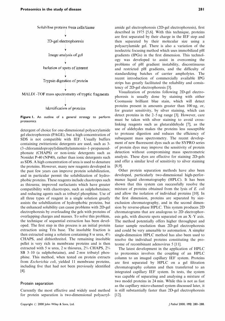
The key steps in proteomics are the separation andvisualization of complex protein mixtures, most com-monly by 2D-gel electrophoresis, and the identi®cationof proteins by mass spectrometry. The overall strategyfor proteomics is outlined in Figure 1.
Protein solubilization
Effective 2D-gel electrophoresis requires that proteins besolubilized. The method used to do this must becompatible with iso-electric focusing (IEF) and shouldnot introduce modi®cations to proteins that could affectmass spectrometry. The most common means of proteinsolubilization employed in proteomic studies is to lysethe tissue of interest, either chemically or mechanically.Often centrifugation is necessary to separate the solubi-lized protein from non-solubilized material. Sub-cellularfractions may also be prepared to permit the analysis ofspeci®c cellular compartments such as the nuclear ormitochondrial fractions. The extract can be incubatedwith nucleases to remove DNA or RNA molecules,which can interfere with electrophoresis. It is particu-larly dif®cult to solubilize hydrophobic and membrane-bound proteins. Sodium dodecyl sulphate (SDS) is the
Journal of PathologyJ Pathol 2000; 192: 280±288.
Copyright # 2000 John Wiley & Sons, Ltd.
detergent of choice for one-dimensional polyacrylamidegel electrophoresis (PAGE), but a high concentration ofSDS is not compatible with IEF. Usually bufferscontaining zwitterionic detergents are used, such as 3-(3-chloramidopropyl)dimethylammonio-1-propanesul-phonate (CHAPS) or non-ionic detergents such asNonidet P-40 (NP40), rather than ionic detergents suchas SDS. A high concentration of urea is used to denaturethe proteins. However, many new reagents developed inthe past few years can improve protein solubilization,and in particular permit the solubilization of hydro-phobic proteins. These reagents include chaotropes suchas thiourea; improved surfactants which have greatercompatibility with chaotropes, such as sulphobetaines;and reducing agents such as tributyl phosphine. Use ofall three types of reagent in a single solution greatlyassists the solubilization of hydrophobic proteins, butthe enhanced solubility can cause problems with 2D-gelelectrophoresis by overloading the gels with proteins ofoverlapping charges and masses. To solve this problem,the technique of sequential extraction has been devel-oped. The ®rst step in this process is an initial proteinextraction using Tris base. The insoluble fraction isthen extracted using a solution containing 8 M urea, 4%CHAPS, and dithiothreitol. The remaining insolublepellet is very rich in membrane proteins and is thenextracted with 5 M urea, 2 M thiourea, 2% CHAPS, 2%SB 3±10 (a sulphobetaine), and 2 mM tributyl phos-phine. This method, when tested on protein extractsfrom Escherichia coli, yielded 11 membrane proteins,including ®ve that had not been previously identi®ed[4].
Protein separation
Currently the most effective and widely used methodfor protein separation is two-dimensional polyacryl-
amide gel electrophoresis (2D-gel electrophoresis), ®rst
described in 1975 [5,6]. With this technique, proteins
are ®rst separated by their charge in the IEF step and
then separated by their molecular size using a
polyacrylamide gel. There is also a variation of the
isoelectric focusing method which uses immobilized pH
gradients (IPGs) in the ®rst dimension. This technol-
ogy was developed to assist in overcoming the
problems of pH gradient instability, discontinuous
and restricted pH gradients, and the dif®culty of
standardizing batches of carrier ampholytes. The
recent introduction of commercially available IPG
strips has greatly facilitated the reliability and consis-
tency of 2D-gel electrophoresis [3].Visualization of proteins following 2D-gel electro-
phoresis is usually done by staining with either
Coomassie brilliant blue stain, which will detect
proteins present in amounts greater than 100 ng, or,
for greater sensitivity, by silver staining, which can
detect proteins in the 2±5 ng range [3]. However, care
must be taken with silver staining to avoid cross-
linking reagents such as glutaraldehyde [7], as the
use of aldehydes makes the proteins less susceptible
to protease digestion and reduces the ef®ciency of
subsequent mass spectrometry. The recent develop-
ment of new ¯uorescent dyes such as the SYPRO series
of protein dyes may improve the sensitivity of protein
detection without compromising mass spectrometric
analysis. These dyes are effective for staining 2D-gels
and offer a similar level of sensitivity to silver staining
[8,9].Other protein separation methods have also been
developed, particularly two-dimensional high-perfor-
mance liquid chromatography (HPLC). It has been
shown that this system can successfully resolve the
mixture of proteins obtained from the lysis of E. coli
and allow the isolation of individual proteins [10]. In
the ®rst dimension, proteins are separated by size-
exclusion chromatography, and in the second dimen-
sion by reverse-phase HPLC. This system produces 2D
chromatograms that are analogous to 2D electrophor-
esis gels, with discrete spots separated on an X±Y axis.
The method potentially offers higher throughput and
faster sample resolution than 2D-gel electrophoresis
and could be very amenable to automation. A simpler
single-dimension HPLC method has also been used to
resolve the individual proteins constituting the pro-
teome of recombinant adenovirus 5 [11].The latest development in the application of HPLC
to proteomics involves the coupling of an HPLC
column to an imaged capillary IEF system. Proteins
are ®rst separated by HPLC on a gel ®ltration
chromatography column and then transferred to an
integrated capillary IEF system. In tests, the system
was capable of separating and analysing a mixture of
two model proteins in 24 min. While this is not as fast
as the capillary micro-channel system discussed later, it
is still substantially faster than 2D-gel electrophoresis
[12].
Figure 1. An outline of a general strategy to performproteomics
Proteomics in the study of disease 281
Copyright # 2000 John Wiley & Sons, Ltd. J Pathol 2000; 192: 280±288.

Image capture and analysis
The protein expression patterns generated from separa-tion techniques are usually complex and are bestanalysed by computer-based image analysis. In thisway, it is possible to create 2D databases. The ®rst forhuman cells included databases for transformedepithelial amnion cells, peripheral blood mononuclearcells, and embryonic lung MRC-5 ®broblasts [13±15],and 2D databases of a number of speci®c human tissuetypes and tumours have subsequently been developed.One of the best established is the SWISS-2D database,which was begun in 1993 [16]. The current versioncontains reference maps from human and mousetissues and also from micro-organisms, includingSaccharomyces cerevisiae and E. coli. A range ofother 2D databases are available, from human liver,to keratinocytes, to bladder carcinoma cells [17±19].Many 2D databases and proteomic resources areavailable on the World Wide Web (Table 1) [20].
A key part of many proteomic studies, especially inthe context of pathological investigations, is thecomparison of protein expression patterns betweennormal and abnormal tissue. Comparisons can bemade with established 2D databases if the sourcetissue is of the same type and if extraction and analysisconditions are similar. To facilitate such comparisons,it is necessary to use specialized image analysissoftware. While different 2D-gels are never perfectly
comparable, current gel analysis software can compen-
sate for this. Some systems such as ELISE and
HERMeS use a cluster-matching technique to compare
gels, whereas GELLABE relies on the operator
providing about 20 `landmark' matches which
`anchor' the two gel images under comparison. Soft-
ware systems also vary in the way their databases are
constructed and organized, from those using simple
models, like ELISE, to those using more complex ones,
like HERMes [21].The MELANIE system, ®rst introduced in 1988, is
based on correspondence analysis and ascendant
hierarchical classi®cation [22]. MELANIE II, the next
version of this system, and the one currently in most
widespread use, is capable of working on many
different operating systems. It is one of the latest and
most user-friendly 2D-gel analysis systems and can be
linked via the Internet to proteome databases available
on the World Wide Web [23]. These features have been
enhanced still further in MELANIE III, which has just
become available [24].Provided that electrophoresis conditions are similar,
good gel analysis software should be able to highlight
differences in protein expression between normal and
abnormal samples. Proteins of interest can be excised
directly from the gel or blotted onto a suitable
membrane [25±27] and they can then be subjected to
identi®cation techniques. Recent developments in
automated proteomic systems have allowed the possi-
Figure 2. A representative result of a proteomic investigation of colon carcinoma. Total cellular proteins from a colon carcinomahave been extracted using a CHAPS-based protein solubilization solution and then separated by 2D-gel electrophoresis. A singleprotein spot has been excised from the gel and characterized by MALDI-TOF mass spectrometry
282 G. Chambers et al.
Copyright # 2000 John Wiley & Sons, Ltd. J Pathol 2000; 192: 280±288.

bility of coupling spot-excising robots to imageanalysis software. The Proteomic Analyzer imageanalysis software system developed by GenomicsSolutions can be coupled to a Protein Picking2 roboticworkstation, which is capable of rapidly excisingthousands of protein spots from multiple gels andtransferring them to an automated protein digestionworkstation. This type of automated system is essentialfor high throughput proteomics.
Protein characterization and identi®cation
A variety of techniques are available to characterizeproteins. While 2D-gel electrophoresis can provide theisoelectric point and approximate mass of a protein,this information alone is currently insuf®cient toidentify the majority of proteins. One of the mostwidely used methods of identifying proteins is throughpeptide-mass ®ngerprinting [28,29]. The protein isdigested with a proteolytic enzyme, such as trypsin, toproduce a set of tryptic fragments unique to eachprotein. The masses of these peptides are thendetermined by mass spectrometry. The most commonsystem for doing this is matrix-assisted laser desorptiontime-of-¯ight mass spectrometry (MALDI-TOF). Thesystem is highly sensitive and can give molecularweight information at the attomole level (10x14 g fora 10 kD molecule) [30]. The tryptic masses are thenevaluated using a peptide-mass ®ngerprinting tool suchas MS-Fit, Mascot, or Peptide Search. These tools tryto `®t' a user's mass spectrometry data to a proteinsequence in an existing database and thus suggest theidentity of the protein. This form of protein identi®ca-tion will only be successful if the protein beinganalysed is represented in the databases. For proteinswhich have incomplete sequence information, it isnecessary to obtain sequence information for the
protein by Edman degradation [31] or by nano-electrospray mass spectrometry [32]. This sequenceinformation can then be used along with the massspectrometry information to interrogate expressedsequence tag (EST) databases.
Techniques for ensuring the analysis ofspeci®c cell types
Accurate proteomic analysis requires the isolation ofspeci®c morphologically or functionally uniform popu-lations of cells, since the presence of contaminatingcells within a sample is a major obstacle to meaningfulmolecular analysis. This is particularly important inthe analysis of pathological conditions. To minimize oreliminate any potential `interference' from other celltypes, cell enrichment is necessary. Several methods,including ¯ow cytometry, antibody puri®cation, andmore recently micro-dissection, have been used forobtaining speci®c cell types. The method chosendepends in part on the starting material and on theavailability of reagents such as antibodies for puri®ca-tion. Flow cytometric methods are ideally suited tohaematological cells. Antibody-linked magnetic beadscan be used to select cells of the desired type frommechanically disaggregated mixed cell populations [33].Protein expression pro®les of normal breast epithelialand myoepithelial cells have been recently established,the two cell types being separated using doubleantibody magnetic af®nity sorting [34]. However, theuse of such antibody puri®cation methods requires theavailability of antibodies speci®c for the cell type ofinterest and so might not always be feasible.
Micro-dissection can be used to isolate speci®c celltypes. Manual micro-dissection methods, which rely onthe removal of desired material by micro-manipulation
Table 1. Useful proteomic resources and databases available on the World Wide Web
Name Resource WWW address
Aberdeen University Proteomics Facility National proteomics research facility http://www.biochem.abdn.ac.uk/yjef/Protein_Lab.html
Aberdeen 2-D PAGE, Aberdeen University 2D-gel databases for Haemophilus in¯uenzae
and Neisseria meningitidis
http://www.abdn.ac.uk/ymmb023/2dhome.htm
EMBL protein and peptide group Useful information on mass spectrometry http://www.mann.embl-heidelberg.de/Default.htmlExPASy Molecular Biology Server Very extensive proteomic site, includes
SWISS-2DPAGE human 2D-gel database
http://www.expasy.ch/
HSC-2DPAGE (Heart Science Centre,
Hare®eld Hospital, UK)
2D-gel databases for heart (ventricle),
endothelial cells
http://www.hare®eld.nthames.nhs.uk/nhli/protein/
Human and mouse 2-D PAGE databases
(Danish Centre for Human Genomic
Research)
2D-gel databases for human keratinocytes,
bladder transitional cell carcinomas, bladder
squamous cell carcinomas, urine, ®broblasts
http://biobase.dk/cgi-bin/celis
Molecuar Anatomy Laboratory, Indiana
University, USA
2D-gel databases for liver, kidney, brain,
plasma, testis
http://iupucbio1.iupui.edu/frankw/molan.htm
Mycobacterium 2-DE databases, Max
Planck Institute for Infection Biology,Berlin, Germany
2D-gel databases for Mycobacterium bovis
and Mycobacterium tuberculosis
http://www.mpiib-berlin.mpg.de/2D-PAGE/
ProteinProspector Extensive web site with tools for protein
database searching including MS-®t
http://prospector.ucsf.edu/
SIENNA-2DPAGE 2D-gel database of human breast ductalcarcinoma cells
http://www.bio-mol.unisi.it/2d/2d.html
YPD yeast protein database 2D-gel databases for yeast Http://www.proteome.com/databases/index/html
Proteomics in the study of disease 283
Copyright # 2000 John Wiley & Sons, Ltd. J Pathol 2000; 192: 280±288.
with a probe or small-gauge syringe needle, areadequate for obtaining suf®cient tissue for polymerasechain reaction (PCR) analysis [35,36], but cannotprovide suf®cient tissue for proteomic studies andhave several disadvantages, including the necessity fora skilled operator and susceptibility to contamination.New methods based on laser capture techniques havehad a major impact on the micro-dissection of solidtissues and tumours. Laser capture is precise and rapidin comparison with other methods of micro-dissectionand has been used successfully in initial studies toselect speci®c cells for proteomic analysis [37±39].
Current limitations of proteomics
While proteomics offers a new approach to the studyof many pathological conditions, there are still severaltechnological limitations. From the pathologist's per-spective, one of the most important of these is thatformalin-®xed archival tissue cannot be used for 2D-gel electrophoresis, as the cross-linking of proteins byaldehyde ®xatives renders them insoluble. Anotherissue with proteomics is the time-consuming nature ofthe procedures, in particular the time taken to perform2D-gel electrophoresis.
There are also several speci®c limitations of 2D-gelelectrophoresis. It cannot adequately separate hydro-phobic proteins; Wilkins et al. [40] calculated thehydrophobicity values of 427 bacterial and yeastproteins and found that there was a clear hydro-phobicity cut-off point beyond which 2D-gel electro-phoresis was ineffective. Another serious drawback isthe inability of 2D-gel electrophoresis to detect lowabundance proteins; there is no equivalent in proteinbiochemistry of PCR in molecular biology and currentprotein staining techniques do not allow the visualiza-tion of low abundance proteins in 2D-gels. Abundantproteins, especially structural proteins such as b-actin,can also pose problems by obscuring other more minorproteins [41].
While 2D-gel electrophoresis can resolve post-transcriptionally or proteolytically modi®ed proteinsfrom their `parent' molecules, this can complicateprotein identi®cation. It has recently been suggestedthat up to a quarter of the protein spots visualized in a2D-gel are modi®ed proteins, so it is not uncommon toisolate what is effectively the same protein frommultiple spots on a gel [1]. The limitations posed by2D-gel electrophoresis may be overcome in the nearfuture by advances in other methods of proteinseparation, some of which are described later, butthey are still an important consideration when plan-ning research using proteomics.
Applications of proteomics in the study ofdisease
There is a great deal of current interest in theapplication of proteomics to the study of individual
diseases and research in these areas is continuallyexpanding.
Cancer research
Because of the potential of proteomics to identify newtumour markers, there is much current interest inapplying proteomics to the study of tumours, but todate only a limited number of studies have been carriedout.
Extensive 2D-gel electrophoresis of bladder tumourcells and urinary proteins has led to the developmentof a comprehensive 2D database for bladder carci-noma that includes pro®les of both transitional andsquamous cell carcinomas [42]. The same group hasalso developed a database of excreted proteins foundin the urine of bladder cancer patients. The database ismade up of scanned and annotated 2D-gels. Spots onthe gels which have de®nite protein identities foundfrom subsequent proteomic analysis are labelled oneach gel. The clinical results of these programmes are,however, somewhat disappointing, as only one candi-date marker for squamous cell carcinoma has beenproposed. This putative marker, psoriasin, is a lowmolecular weight calcium-binding protein which is alsoexcreted by cultured keratinocytes. It is excreted in theurine of squamous cell carcinoma patients and mayhave the potential to be developed as a diagnosticmarker for squamous cell carcinoma. Considerablework will be needed to evaluate this and there are somepotential problems. For example, there is a possibilitythat reversible benign metaplastic lesions may tem-porarily express psoriasin. Strati®ed squamous epithe-lium in the female trigone may also secrete psoriasin,which can thus be detected in the urine of some healthyfemales. An ELISA test for psoriasin is currently underdevelopment, and its use in large-scale studies may leadto a true evaluation of the usefulness of psoriasin as adiagnostic marker [43].
Initial protein pro®les have been de®ned in sometumours, but many of these studies are limited in theirusefulness, as no attempt has been made to purify thetumour cells. In the study of breast cancer, comparative2D-gel electrophoresis analysis of whole tissue sampleshas shown that decreased expression of cytokeratins andtropomyosins is associated with malignant progression[44]. In another study [45], 2D-gel electrophoresispro®ling of breast invasive ductal carcinomas found adistinctive pattern of 32 protein spots expressed stronglyby whole samples of breast ductal carcinomas, but notby whole samples of normal breast tissue. Comparisonof this gel to gel images contained in the SWISS-2DPAGE database allowed the identity of some of thespots to be determined.
A recent study of protein expression in renal cellcarcinoma has created a ®rst generation 2D-geldatabase for human kidney proteins. A variety oftechniques were employed including comparison of2D-gel images, amino acid analysis, N-terminal aminoacid sequencing, and immuno-detection, to identify 43
284 G. Chambers et al.
Copyright # 2000 John Wiley & Sons, Ltd. J Pathol 2000; 192: 280±288.
out of 2789 separated polypeptides. Comparison ofprotein expression patterns between whole samples ofnormal and tumour kidney tissues showed that fourpolypeptides synthesized by normal kidney were notpresent in renal carcinoma tissue. The techniquesemployed by the investigators were suf®cient toidentify only two out of the four proteins; one wasidenti®ed as ubiquinol cytochrome c reductase, whilethe other was mitochondrial NADH-ubiquinoneoxido-reductase complex I. Neither protein has a genelocus in a region known to be deleted in renal cellcarcinoma, so the loss of expression of these twoproteins may be due to unknown deletions, or tochanges in gene transcription or translation [46].
A study of lung tumours using 2D-gel electrophor-esis has shown that different histopathological tumourtypes display differences in their protein expressionpatterns and that an unknown protein is signi®cantlyoverexpressed in primary lung adenocarcinomas com-pared with other types of lung carcinoma and normaltissue [47]. Subsequently it has been found that thereare two variants of the protein, which differ slightly inisoelectric point but not molecular weight. A follow-upstudy has shown that both variants are expressed byprimary lung adenocarcinoma and that their expres-sion correlates closely with the degree of cellular atypiaand histological differentiation [48].
Proteomics is also being used in other areas ofcancer research, especially concerning tumour develop-ment and progression. One of the most importantareas is the study of apoptosis, since a failure of theapoptotic mechanism has been proposed as one of themajor factors in tumour growth and development. Aconsiderable body of research has been undertaken totry to understand the mechanism of apoptosis and toexplain how defects in that mechanism can lead to thedevelopment of cancer and the failure to respond toanti-cancer therapy. Studies of animal cell models haveshown two distinct patterns of proteolysis of proteinsassociated with apoptosis, one of which occurs at anearly stage and the other at a later stage. There is alsoan alteration in the phosphorylation pattern of certainproteins (including p53) in cells induced to undergoapoptosis [49,50]. One of the most recent studies ofprotein expression in apoptosis [51] has used proteo-mics to study nuclear protein expression in Burkittlymphoma BL60 cells. The BL60 cell line has beenused as a model for determining apoptosis-associatedproteins, by subtractive analysis of normal andapoptotic cells. The study examined 36 protein spotsfrom a Coomassie blue-stained gel and was able toassign identities to 33 of the proteins by a combinationof MALDI-TOF mass spectrometry and nano-electrospray mass spectrometry. The other threeproteins were not identi®ed in any of the availabledatabases. While some of the identi®ed proteins werecommon to normal cells, others were known to beinvolved in apoptosis. This study provides the basis forfurther work to construct a comprehensive database ofproteins expressed in apoptosis-induced cells.
Neuropathology
A 2D-gel electrophoresis database of normal brainproteins has recently been created from samples oftotal brain tissue from the parietal lobe cortex. In thedatabase, 400 identi®ed spots correspond to 180different brain proteins, mostly of cytoplasmic ormitochondrial origin. This should serve as a usefulreference against which gels prepared from brainextracts of abnormal individuals can be compared [52].
Proteomics has been used to investigate certainneuropathological disorders. In one study of proteinexpression in the brains of patients with Alzheimer'sdisease, comparative 2D-gel electrophoresis was usedto show differences from normal individuals. Fiveprotein spots were increased in the Alzheimer's group,28 were decreased, and nine were uniquely expressed.The study did not determine the protein identities butprovides a useful reference for future studies [53].
Another recent study of Alzheimer's disease andschizophrenia, also by comparative 2D-gel electrophor-esis, examined protein expression in post-mortemhippocampal tissue. While sample numbers wererelatively small (seven in each abnormal group andseven controls), eight proteins were found to beexpressed in decreased amounts in the schizophrenicpatients and eight in increased amounts. In theAlzheimer's disease patients, 35 proteins displayeddecreased expression and 73 displayed increasedexpression. Decreased expression of one particularprotein, diazepam binding inhibitor, was observed inboth schizophrenic and Alzheimer's patients; althoughthis may be due to the action of drugs prescribed forthese conditions, it is also possible that it represents aunique change [54]. Despite their limitations, bothstudies point the way to a greater understanding of themolecular basis of neuropathological disorders.
A proteomic approach has also been used toinvestigate demyelination in a mouse model. Theexpression of proteins was examined in the opticnerves of transgenic mice with a c-myc-induceddegenerative myelin disorder. There were markeddifferences in the amounts of cytoskeletal proteinsassociated with gliosis in the optic nerves of theabnormal mice, compared with the normal. Theexpression of several major myelin proteins wasmarkedly reduced in the transgenic mice, including®ve isoforms of myelin basic protein, four isoforms ofcyclic nucleotide 3k-phosphodiesterase, and myelin-associated glycoprotein. The study provides the basisfor future more detailed investigation and reveals anumber of candidate molecular markers for demyeli-nation [55].
Cardiovascular disease
In a recent study on cardiac hypertrophy, 2D-gelelectrophoretic patterns were compared from culturednormal rat cardiac myocytes and myocytes withphenylephrine-induced hypertrophy. Eleven proteinsdisplayed quantitative changes in expression in cardiac
Proteomics in the study of disease 285
Copyright # 2000 John Wiley & Sons, Ltd. J Pathol 2000; 192: 280±288.
hypertrophy, with three showing a decrease and eightan increase. Following peptide mass pro®ling and
database matching, all 11 spots were identi®ed. Five
were found to be isoforms of the myosin light chain, at
least one form of which is known to be associated with
cardiac hypertrophy. The others represented proteins
not previously known to be associated with the
condition [56].Application of proteomics to the study of dilated
cardiomyopathy has resulted in the establishment of a
human myocardial database containing 150 identi®edproteins. This was used as a baseline for comparisons
with protein pro®les from patients with dilated
cardiomyopathy. Signi®cant differential expression
was found for 25 proteins and it was possible to
identify 12 proteins [57].
Proteomics applied to microbiology
Many of the current applications of proteomic tech-
niques have been in microbiology, with much work
being carried out to link the genomes and proteomes of
a variety of bacterial and yeast species. The proteome
of E. coli has been particularly well investigated since
1983 and today has a 2D-gel database containingapproximately 1600 different protein spots. There are
data containing identi®cation information for many of
these proteins and also information on how their
expression patterns change according to different
bacterial growth conditions. Three hundred and ®fty
of the 4000 E. coli genes have now been matched to
approximately 400 protein spots (2D-gel electrophor-
esis will detect isomers as well as unique proteins) [58].
This type of analysis has paved the way for similar
work to be carried out on pathogenic micro-organisms.
Salmonella typhimurium, which is an enteric pathogenresponsible for many cases of gastroenteritis in
humans, is one of these pathogens whose proteome
has been investigated. In an analysis of S. typhimurium
cell envelope proteins, 53 proteins were identi®ed by
2D-gel electrophoresis followed by N-terminal sequen-
cing. Approximately 20% of these proteins had no
match in current sequence databases [59]. It is hoped
that a better understanding of the proteome of S.
typhimurium will identify which proteins are associated
with virulence.The immune response to infection with Helicobacter
pylori has been studied and 20 proteins have been
identi®ed which were reactive with the serum of
infected patients. All the proteins were identi®ed as
H. pylori proteins [60]. A follow-up study investigated
the possibility of using one of these proteins as an
antigen for generating an H. pylori vaccine, due to its
consistently high reactivity. Detailed analysis was
carried out using many proteomic techniques, includ-
ing 2D-gel electrophoresis and mass spectrometry.Endoprotease digestion was carried out, followed by
MALDI-TOF peptide sizing and sequence analysis. As
a result, it was possible to identify the protein as
corresponding to an open reading frame in the H.pylori genome [61].
The study of antibiotic resistance in pathogenicmicrobes is of great importance to medical microbiol-ogists and to specialists in infectious disease. A recentstudy has been carried out on erythromycin resistancein the pathogen Streptococcus pneumoniae. This bac-terium has developed widespread penicillin resistanceand the incidence of erthyromycin resistance is alsorising. In an effort to understand the mechanism oferythromycin resistance, proteomic methodologieswere used to examine protein expression in erythro-mycin-susceptible and -resistant strains. 2D-gel electro-phoresis was used to separate the proteins expressed byeach strain. Comparison of electrophoresis patternsshowed that a 38 500 D protein with an iso-electricpoint of 6.27 was expressed by the resistant strain, butnot by the susceptible strain. Following peptide massmapping, this protein was found to be glyceraldehyde-3-phosphate dehydrogenase. The particular resistantstrain was one displaying the M phenotype oferythromycin resistance, in which resistance is asso-ciated with an active ef¯ux process which reducesintracellular levels of erythromycin. Strains displayingan alternative mechanism of erythromycin resistance,where enzymatic methylation of ribosomal RNAblocks erythromycin binding to the ribosome (theMLS phenotype), expressed a protein pro®le nodifferent from that of the susceptible strains [62].
Research has also been carried out on yeast, as thisorganism offers a simple model for the integration ofgenomic and proteomic information in eukaryoticcells. Mass spectrometric identi®cation of yeast pro-teins by nano-electrospray tandem mass spectrometry,followed by database searching using multiplesequence tags, has been used to identify 150 individualyeast proteins as resolved by 2D-gel electrophoresis.More than 30 of these were novel and matchedpreviously uncharacterized open reading frames in theyeast genome [63]. Another study of yeast proteomics[64] has been used to construct a detailed proteomicmethodological outline which could easily be modi®edand employed in studies of other more complexeukaryotic cells. Research has continued in the ®eldof yeast proteomics with the development of a yeastprotein database. Efforts are now being undertaken tocarry out quantitative studies using 2D-gel electro-phoresis. One such recent study has been able to showa good correlation between protein abundance, mRNAabundance, and codon bias for identi®ed spots andrepresents a successful effort to integrate informationfrom proteomics with information from genomicresearch [65].
Future developments
At the moment, much research is being doneto improve the ef®ciency of proteomic techniques,especially with regard to automation, sample through-
286 G. Chambers et al.
Copyright # 2000 John Wiley & Sons, Ltd. J Pathol 2000; 192: 280±288.
put, and sensitivity. Since 2D-gel electrophoresis iscurrently the mainstay of protein separation inproteomic research, current developments have focusedon automated systems for excising individual spotsfrom 2D-gels and for protein digestion, followed bypeptide mass mapping using mass spectrometry withintegrated peptide identi®cation software [66]. Anothervery new and promising method is micro-channel-based separation, which has already been employed inthe rapid resolution of small amounts of nucleic acidmixtures. A methodology for micro-channel-basedSDS capillary gel electrophoresis of proteins has beendemonstrated [67] which offers high speed and resolu-tion as well as economy. The system utilizes a chipwith the micro-channels photo-etched onto it andproteins to be analysed labelled with ¯uorescein.Detection of individual proteins following electrophor-esis is by laser-induced ¯uorescence. The technique wasshown to resolve a mixture of six ¯uorescent-labelledproteins ranging in size from 9 to 116 kD within3.5 min. Such a rapid technique will allow a highthroughput of samples. There is also much researchbeing performed to develop protein `chips', which canbe considered to be analogous to DNA micro-arrays.
Conclusions
The development of proteomics represents an excitingnew way to examine pathological processes at themolecular level and is already leading to improvementsin the understanding of many conditions. It will beparticularly important to perform proteomic experi-ments with speci®c populations of cells, to generatesigni®cant information about cell-speci®c proteinexpression pro®les and how these change in differentdiseases. As the technology of proteomics analysiscontinues to improve, it will be possible in the nearfuture to combine genomic and proteomic informationto obtain a more comprehensive picture of manypathological conditions.
References
1. Blackstock WP, Weir MP. Proteomics: quantitative and physical
mapping of cellular proteins. Trends Biotechnol 1999; 17:
121±127.
2. Anderson L, Seilhammer J. A comparison of selected mRNA
and protein abundances in human liver. Electrophoresis 1997; 18:
533±537.
3. Humphrey-Smith I, Cordwell SJ, Blackstock WP. Proteome
research: complementarity and limitations with respect to the
RNA and DNA worlds. Electrophoresis 1997; 18: 1217±1242.
4. Herbert B. Advances in protein solubilisation for two-
dimensional electrophoresis. Electrophoresis 1999; 20: 660±663.
5. O'Farrel PH. High resolution two-dimensional electrophoresis of
proteins. J Biol Chem 1974; 250: 4007±4021.
6. Klose J. Protein mapping by combined isoelectric focusing and
electrophoresis of mouse tissues. A novel approach to testing for
induced point mutations in mammals. Humangenetik 1975; 26:
231±243.
7. Shevchenko A, Wilm M, Vorm O, Mann M. Mass spectrometric
sequencing of proteins from silver-stained polyacrylamide gels.
Anal Chem 1996; 68: 850±858.
8. Steinberg T, Jones LJ, Haugland RP, Singer VL. SYPRO ruby
and SYPRO red protein gel stains: one step ¯uorescent staining
of denaturing gels for detection of nanogram levels of protein.
Anal Biochem 1996; 239: 223±237.
9. Steinberg T, Jones LJ, Haugland RP, Singer VL. Applications of
SYPRO orange and SYPRO ruby gel stains. Anal Biochem 1996;
239: 238±245.
10. Opiteck GJ, Ramirez SM, Jorgenson JW, Moseley MA.
Comprehensive two-dimensional high performance liquid chro-
matography for the isolation of overexpressed proteins and
proteome mapping. Anal Biochem 1998; 258: 349±361.
11. Lehmberg E, Traina JA, Chakel JA, et al. Reverse-phase high-
performance liquid chromatographic assay for the adenovirus
type 5 proteome. J Chromatogr B Biol Appl 1999; 732: 411±423.
12. Tragas C, Pawliszyn J. On-line coupling of high performance gel
®ltration chromatography with imaged capillary isoelectric
focusing using a membrane interface. Electrophoresis 2000; 21:
227±237.
13. Celis JE, Ratz GP, Madsen P, et al. Comprehensive human
cellular protein databases and their implication for the study of
genome organisation and function. FEBS Lett 1989; 244:
247±254.
14. Celis JE, Honore B, Bauw G, Vandekerchove J. Comprehensive
computerised 2D gel protein databases offer a global approach
to the study of the mammalian cell. BioEssays 1990; 12: 93±97.
15. Celis JE, Leffers H, Rasmussen HH, et al. The master two-
dimensional gel database of human AMA cell proteins: towards
linking protein and genome sequence and mapping information
(update 1991). Electrophoresis 1991; 12: 765±801.
16. Appel RD, Sanchez JC, Bairoch A, et al. SWISS-2DPAGE: a
database of two dimensional gel images. Electrophoresis 1993; 14:
1232±1238.
17. Huges GJ, Frutiger S, Paquet N, et al. Human liver protein map:
update 1993. Electrophoresis 1993; 14: 1216±1222.
18. Rasmussen HH, Mortz E, Mann M, Roepstorff P, Celis JE.
Identi®cation of transformation sensitive proteins recorded in
human two-dimensional gel protein databases by mass spectro-
metric peptide mapping alone and in combination with micro-
sequencing. Electrophoresis 1994; 15: 406±416.
19. Rasmussen HH, Orntoft TF, Wolf H, Celis JE. Towards a
comprehensive database of proteins from the urine of patients
with bladder cancer. J Urol 1996; 155: 2113±2119.
20. Celis JE, Ostergaard M, Jensen NA, Gromova I, Rasmussen
HH, Gromov P. Human and mouse proteomic databases: novel
resources in the protein universe. FEBS Lett 1998; 430: 64±72.
21. Dunn MJ. In Computers in Biochemistry: A Practical Approach,
Bryce CFA (ed.). IRL Press: Oxford, 1992; 215±242.
22. Pun T, Hochstrasser DF, Appel RD, Funk M, Villars-
Augsburger V, Pellegrini C. Computerized classi®cation of two-
dimensional gel electrophoretograms by correspondence analysis
and ascendant hierarchical clustering. Appl Theor Electrophor
1988; 1: 3±9.
23. Appel RD, Palagri PM, Walther D, et al. Melanie II ± a third
generation software package for analysis of two-dimensional
electrophoresis images: I. Features and user interface. Electro-
phoresis 1997; 18: 2724±2734.
24. http//www.expasy.ch/melanie/ [8 March 2000].
25. Eckershorn C, Lottespeich F. Structural characterisation of
blotting membranes and the in¯uence of membrane parameters
for electroblotting and subsequent amino acid sequence analysis
of proteins. Electrophoresis 1993; 14: 831±838.
26. Patterson SD. From electrophoretically separated protein to
identi®cation: strategies for sequence and mass analysis. Anal
Biochem 1994; 221: 1±15.
27. Vestling MM, Feneslau C. Polyvinylidene di¯uoride (PVDF): an
interface for gel electrophoresis and matrix assisted laser
desorption/ionisation mass spectrometry. Biochem Soc Trans
1994; 22: 547±551.
28. Henzel W, Billeci TM, Stults JT, Wong SC, Grimley C,
Watanabe C. Identifying proteins from two-dimensional gels by
Proteomics in the study of disease 287
Copyright # 2000 John Wiley & Sons, Ltd. J Pathol 2000; 192: 280±288.
molecular mass searching of peptide fragments in protein
sequence databases. Proc Natl Acad Sci U S A 1993; 90:
5011±5015.
29. Mann M, Hojrup P, Roepstorff P. Use of mass spectrometric
molecular weight information to identify proteins in sequence
databases. Biol Mass Spec 1993; 22: 338±345.
30. McLafferty FW, Fridriksson EK, Horn DM, Lewis MA,
Zubarev RA. Biomolecule mass spectrometry. Science 19XX;
284: 1289±1290.
31. Vandekerckhove J, Bauw G, Puype M, VanDamme J, VanMon-
tague M. Protein-blotting on Polybrene-coated glass-®ber sheets:
a basis for acid hydrolysis and gas-phase sequencing of picomole
quantities of protein previously separated on sodium dodecyl
sulfate/polyacrylamide gel. Eur J Biochem 1985; 152: 9±19.
32. Wilm M, Mann M. Analytical properties of the nanoelectrospray
ion source. Anal Chem 1996; 68: 1±8.
33. Reymond MA, Sanchez JC, Hughes GJ, et al. Standardised
characterisation of gene expression in human colorectal epithe-
lium by two-dimensional electrophoresis. Electrophoresis 1997;
18: 2842±2848.
34. Page MJ, Amess B, Townsend RR, et al. Proteomic de®nition of
normal human luminal and myoepithelial breast cells puri®ed
from reduction mammoplasties. Proc Natl Acad Sci U S A 1999;
96: 12589±12594.
35. Whetsell L, Maw G, Nadon N, Ringer DP, Schaefer FV.
Polymerase chain reaction microanalysis of tumours from
stained histological slides. Oncogene 1992; 7: 2355±2361.
36. Zhuang Z, Bertheau P, Emmert-Buck MR, et al. A micro
dissection technique for archival DNA analysis of speci®c cell
populations in lesions <1 mm in size. Am J Pathol 1995; 146:
620±625.
37. Banks RE, Dunn MJ, Forbes MA, et al. The potential use of
laser capture microdissection to selectively obtain distinct
populations of cells for proteomic analysis ± preliminary
®ndings. Electrophoresis 1999; 20: 689±700.
38. Curran S, McKay JA, McLeod HL, Murray GI. Laser capture
microscopy. J Clin Pathol: Mol Pathol 2000; 53: 64±68.
39. Emmert-Buck MR, Gillespie JW, Pawletz CP, et al. An
approach to the proteomic analysis of human tumors. Mol
Carcinog 2000; 27: 158±165.
40. Wilkins MR, Gasteiger E, Sanchez JC, Bairoch A, Hochstrasser
DF. Two-dimensional gel electrophoresis for proteome projects:
the effects of protein hydrophobicity and copy number. Electro-
phoresis 1998; 19: 1501±1505.
41. Celis JE, Rasmussen HH, Gromov P, et al. The human
keratinocyte two-dimensional gel protein database (update
1995): mapping components of signal transduction pathways.
Electrophoresis 1995; 16: 2177±2240.
42. Celis JE, Ostergaard M, Rasmussen HH, et al. A comprehensive
protein resource for the study of bladder cancer: http//biobase.
dk/cgi-bin/celis. Electrophoresis 1999; 20: 300±309.
43. Ostergaard M, Wolf H, Orntoft TF, Celis JE. Psorasin (S100A7):
a putative urinary marker for the follow-up of patients with
bladder squamous cell carcinomas. Electrophoresis 1999; 20:
349±354.
44. Franzen B, Linder S, Alaiya AA, et al. Analysis of polypeptide
expression in benign and malignant human breast lesions: down
regulation of cytokeratins. Br J Cancer 1996; 73: 1632±1638.
45. Bini L, Magi B, Marzocchi B, et al. Protein expression pro®les in
human breast ductal cell carcinoma and histologically normal
tissue. Electrophoresis 1997; 18: 2832±2841.
46. Sarto C, Marocchi A, Sanchez JC, et al. Renal cell carcinoma
and normal kidney protein expression. Electrophoresis 1997; 18:
599±604.
47. Okuzawa K, Franzen B, Lindholm J, et al. Characterisation of
gene expression in clinical lung cancer materials by two-
dimensional polyacrylamide gel electrophoresis. Electrophoresis
1994; 15: 382±390.
48. Hirano T, Fujioka K, Franzen B, et al. Relationship between
TA01 and TA02 polypeptides associated with lung adenocarci-
noma and histocytological features. Br J Cancer 1997; 75:
978±979.
49. Robaye B, Doskeland AP, Suarez-Huerta N, Doskeland SO,
Dumont JE. Apoptotic cell death analysed at the molecular level
by two-dimensional gel electrophoresis. Electrophoresis 1994; 15:
503±510.
50. Maxwell SA, Roth JA, Mukhopadhyay T. Analysis of phos-
phorylated isoforms of the p53 tumour suppressor protein in
human lung carcinoma cells undergoing apoptosis. Electrophor-
esis 1996; 17: 1772±1775.
51. Muller EC, Schumann M, Rickers A , Bommert K, Whitmann-
Liebold B, Otto A. Study of Burkitt lymphoma cell line proteins
by high resolution two-dimensional gel electrophoresis and
nanoelectrospray mass spectrometry. Electrophoresis 1999; 20:
320±330.
52. Langen H, Berndt P, Roer D, Cairns N, Lubec G, Fountoulakis
M. Two dimensional map of human brain proteins. Electrophor-
esis 1999; 20: 907±916.
53. Tsuji T, Shimohama S, Kamiya S, Sazuka T, Ohara O. Analysis
of brain proteins in Altzheimer's disease using high resolution
two-dimensional gel electrophoresis. J Neurol Sci 1999; 166:
100±106.
54. Edgar PF, Sconberger SJ, Dean B, Faull RLM, Kydd R, Cooper
GJS. A comparative proteome analysis of hippocampal tissue
from schizophrenic and Alzheimer's disease individuals. Mol
Psychiatry 1999; 4: 173±178.
55. Jensen NA, Celis JE. Proteomic changes associated with
degeneration of myelin-forming cells in the central nervous
system of c-myc transgenic mice. Electrophoresis 1998; 19:
2014±2020.
56. Arnott D, O'Connel KL, King KL, Stults JT. An integrated
response to proteome analysis: identi®cation of proteins asso-
ciated with cardiac hypertrophy. Anal Biochem 1998; 258: 1±18.
57. Jungblut PR, Zimny-Arndt U, Zeindl-Eberhart E, et al. Proteo-
mics in human disease: cancer, heart, and infectious diseases.
Electrophoresis 1999; 20: 2100±2110.
58. VanBoglen RA, Abshire KZ, Moldover B, Olsen ER, Neidhart
FC. Escherichia coli proteome analysis using the gene±proteome
database. Electrophoresis 1997; 18: 1243±1251.
59. O'Connor CD, Farris M, Fowler R, Qi S-Y. The proteome of
Salmonella enterica serovar Typhimurium: current progress on its
determination and some applications. Electrophoresis 1997; 18:
1483±1490.
60. McAtee CP, Fry KE, Berg DE. Identi®cation of potential
diagnostic and vaccine candidates of Helicobacter pylori by
`proteome' technologies. Helicobacter 1998; 3: 163±169.
61. McAtee CP, Lim MY, Fung K, et al. Characterisation of a
Helicobacter pylori vaccine candidate by proteome techniques.
J Chromatogr B Biol Appl 1998; 714: 325±333.
62. Cash P, Argo E, Ford L, Lawrie L, McKenzie H. A proteomic
study of erythromycin resistance in Streptococcus pneumoniae.
Electrophoresis 1999; 20: 2259±2268.
63. Shevchenko A, Jensen ON, Podtelejnikov AV, et al. Linking of
genome and proteome by mass spectrometry: large scale
identi®cation of yeast proteins from two-dimensional gels. Proc
Natl Acad Sci U S A 1996; 93: 14440±14445.
64. Fey SJ, Nawrocki A, Larsen MR, et al. Proteome analysis of
Saccharomyces cerevisiae: a methodological outline. Electrophor-
esis 1997; 18: 1361±1372.
65. Futcher B, Latter GI, Monardo P, McLaughlin CS, Garrels JI.
A sampling of the yeast proteome. Mol Cell Biol 1999; 19:
7357±7368.
66. Traini M, Gooley AA, Keli O, et al. Towards an automated
approach for protein identi®cation in proteome projects. Electro-
phoresis 1998; 19: 1941±1949.
67. Yao S, Anex DS, Caldwell WB, Arnold DW, Smith KB, Schultz
PG. SDS capillary electrophoresis of proteins in microfabricated
channels. Proc Natl Acad Sci U S A 1999; 96: 5372±5377.
288 G. Chambers et al.
Copyright # 2000 John Wiley & Sons, Ltd. J Pathol 2000; 192: 280±288.