Proteomic Analysis of Plasmodium Merosomes: The Link ...
15
Proteomic Analysis of Plasmodium Merosomes: The Link between Liver and Blood Stages in Malaria Melanie J. Shears, † Raja Sekhar Nirujogi, ‡,∥ Kristian E. Swearingen, § Santosh Renuse, ‡,⊥ Satish Mishra, †,# Panga Jaipal Reddy, § Robert L. Moritz, § Akhilesh Pandey, ‡,⊥ and Photini Sinnis* ,† † Department of Molecular Microbiology & Immunology, Johns Hopkins Bloomberg School of Public Health, 615 North Wolfe Street, Baltimore, Maryland 21205, United States ‡ Department of Biological Chemistry, Johns Hopkins School of Medicine, 733 N. Broadway, Baltimore, Maryland 21205, United States § Institute for Systems Biology, 401 Terry Avenue, North Seattle, Washington 98109, United States ∥ Institute of Bioinformatics, International Tech Park, Bangalore 560 066, India * S Supporting Information ABSTRACT: The pre-erythrocytic liver stage of the malaria parasite, comprising sporozoites and the liver stages into which they develop, remains one of the least understood parts of the lifecycle, in part owing to the low numbers of parasites. Nonetheless, it is recognized as an important target for antimalarial drugs and vaccines. Here we provide the first proteomic analysis of merosomes, which define the final phase of the liver stage and are responsible for initiating the blood stage of infection. We identify a total of 1879 parasite proteins, and a core set of 1188 proteins quantitatively detected in every biological replicate, providing an extensive picture of the protein repertoire of this stage. This unique data set will allow us to explore key questions about the biology of merosomes and hepatic merozoites. KEYWORDS: malaria, merosomes, liver stages, exoerythrocytic stage, sporozoites, proteomics, PEXEL ■ INTRODUCTION Malaria remains one of the most important infectious diseases in the world, impacting approximately a third of the world’s population and causing an estimated 400 000 deaths annually. 1 The initial stage of malaria infection, comprised of infectious sporozoites inoculated by mosquitoes and the liver stages into which they develop, is clinically asymptomatic yet has been validated as a vaccine target in both rodent malaria models 2,3 and human Phase III clinical trials. 4,5 The low numbers of parasites during this pre-erythrocytic stage, combined with their decreased allelic diversity compared to blood stage parasites, explain, at least in part, why targeting this stage is advantageous [reviewed in ref 6]. However, the limitations of current pre-erythrocytic stage vaccines indicate that additional strategies or targets are still required. Building upon this success will necessitate identifying new pre-erythrocytic stage targets and expanding the repertoire of antigens to include late liver stage proteins. 7 In this study we focus on the final phase of the Plasmodium pre-erythrocytic stage, the merosomes, characterizing their proteome to obtain a better understanding of their biology and provide data to inform target selection for future interventions. Merosomes and the hepatic merozoites they contain are the link between the asymptomatic pre-erythrocytic stage and the clinically important erythrocytic stage of malaria infection. They are the product of an impressive replication: A single sporozoite that has successfully entered a hepatocyte can transform and give rise to 5000 to 10 000 hepatic merozoites in a matter of days. 8 Development of liver stage parasites occurs within a parasitophorous vacuole (PV), a parasite-derived structure that separates the newly invaded parasite from the host cell cytoplasm. 9 Within this vacuole, the parasite acquires nutrients from its host necessary for growth and replica- tion. 8,10,11 Once mature, the PV membrane ruptures, releasing the hepatic merozoites into the host cell cytoplasm. 12 Merozoites bud from the host hepatocyte in packets called merosomes, each containing 10 to 1000 hepatic merozoites and surrounded by modified host cell membrane. 13 These merosomes enter the bloodstream and release hepatic merozoites to initiate blood stage of infection. Iterative cycles of replication then give rise to erythrocytic merozoites, which are released and invade naı ̈ ve erythrocytes, leading to high parasite numbers and clinical disease. Hepatic and erythrocytic merozoites are morphologically similar, sharing specialized apical organelles, motility apparatus, and surface proteins necessary for erythrocyte invasion. Though frequently assumed to be identical, their different Received: May 18, 2019 Article pubs.acs.org/jpr Cite This: J. Proteome Res. XXXX, XXX, XXX-XXX © XXXX American Chemical Society A DOI: 10.1021/acs.jproteome.9b00324 J. Proteome Res. XXXX, XXX, XXX−XXX Downloaded via JOHNS HOPKINS UNIV on August 6, 2019 at 21:36:50 (UTC). See https://pubs.acs.org/sharingguidelines for options on how to legitimately share published articles.
Transcript of Proteomic Analysis of Plasmodium Merosomes: The Link ...

Proteomic Analysis of Plasmodium Merosomes: The Link betweenLiver and Blood Stages in MalariaMelanie J. Shears,† Raja Sekhar Nirujogi,‡,∥ Kristian E. Swearingen,§ Santosh Renuse,‡,⊥
Satish Mishra,†,# Panga Jaipal Reddy,§ Robert L. Moritz,§ Akhilesh Pandey,‡,⊥ and Photini Sinnis*,†
†Department of Molecular Microbiology & Immunology, Johns Hopkins Bloomberg School of Public Health, 615 North WolfeStreet, Baltimore, Maryland 21205, United States‡Department of Biological Chemistry, Johns Hopkins School of Medicine, 733 N. Broadway, Baltimore, Maryland 21205, UnitedStates§Institute for Systems Biology, 401 Terry Avenue, North Seattle, Washington 98109, United States∥Institute of Bioinformatics, International Tech Park, Bangalore 560 066, India
*S Supporting Information
ABSTRACT: The pre-erythrocytic liver stage of the malaria parasite,comprising sporozoites and the liver stages into which they develop,remains one of the least understood parts of the lifecycle, in partowing to the low numbers of parasites. Nonetheless, it is recognizedas an important target for antimalarial drugs and vaccines. Here weprovide the first proteomic analysis of merosomes, which define thefinal phase of the liver stage and are responsible for initiating theblood stage of infection. We identify a total of 1879 parasite proteins,and a core set of 1188 proteins quantitatively detected in everybiological replicate, providing an extensive picture of the proteinrepertoire of this stage. This unique data set will allow us to explore key questions about the biology of merosomes and hepaticmerozoites.
KEYWORDS: malaria, merosomes, liver stages, exoerythrocytic stage, sporozoites, proteomics, PEXEL
■ INTRODUCTION
Malaria remains one of the most important infectious diseasesin the world, impacting approximately a third of the world’spopulation and causing an estimated 400 000 deaths annually.1
The initial stage of malaria infection, comprised of infectioussporozoites inoculated by mosquitoes and the liver stages intowhich they develop, is clinically asymptomatic yet has beenvalidated as a vaccine target in both rodent malaria models2,3
and human Phase III clinical trials.4,5 The low numbers ofparasites during this pre-erythrocytic stage, combined withtheir decreased allelic diversity compared to blood stageparasites, explain, at least in part, why targeting this stage isadvantageous [reviewed in ref 6]. However, the limitations ofcurrent pre-erythrocytic stage vaccines indicate that additionalstrategies or targets are still required. Building upon thissuccess will necessitate identifying new pre-erythrocytic stagetargets and expanding the repertoire of antigens to include lateliver stage proteins.7 In this study we focus on the final phaseof the Plasmodium pre-erythrocytic stage, the merosomes,characterizing their proteome to obtain a better understandingof their biology and provide data to inform target selection forfuture interventions.Merosomes and the hepatic merozoites they contain are the
link between the asymptomatic pre-erythrocytic stage and theclinically important erythrocytic stage of malaria infection.
They are the product of an impressive replication: A singlesporozoite that has successfully entered a hepatocyte cantransform and give rise to 5000 to 10 000 hepatic merozoites ina matter of days.8 Development of liver stage parasites occurswithin a parasitophorous vacuole (PV), a parasite-derivedstructure that separates the newly invaded parasite from thehost cell cytoplasm.9 Within this vacuole, the parasite acquiresnutrients from its host necessary for growth and replica-tion.8,10,11 Once mature, the PV membrane ruptures, releasingthe hepatic merozoites into the host cell cytoplasm.12
Merozoites bud from the host hepatocyte in packets calledmerosomes, each containing 10 to 1000 hepatic merozoitesand surrounded by modified host cell membrane.13 Thesemerosomes enter the bloodstream and release hepaticmerozoites to initiate blood stage of infection. Iterative cyclesof replication then give rise to erythrocytic merozoites, whichare released and invade naıve erythrocytes, leading to highparasite numbers and clinical disease.Hepatic and erythrocytic merozoites are morphologically
similar, sharing specialized apical organelles, motility apparatus,and surface proteins necessary for erythrocyte invasion.Though frequently assumed to be identical, their different
Received: May 18, 2019
Article
pubs.acs.org/jprCite This: J. Proteome Res. XXXX, XXX, XXX−XXX
© XXXX American Chemical Society A DOI: 10.1021/acs.jproteome.9b00324J. Proteome Res. XXXX, XXX, XXX−XXX
Dow
nloa
ded
via
JOH
NS
HO
PKIN
S U
NIV
on
Aug
ust 6
, 201
9 at
21:
36:5
0 (U
TC
).Se
e ht
tps:
//pub
s.ac
s.or
g/sh
arin
ggui
delin
es f
or o
ptio
ns o
n ho
w to
legi
timat
ely
shar
e pu
blis
hed
artic
les.

tissue origins, together with the unique mechanism of hepaticmerozoite release, suggests these two merozoite populationsmay differ in biologically important ways. To date, only twostudies have examined the differences between these merozoitepopulations: Hepatic and erythrocytic merozoites were shownto express different members of the Py235 protein family,14
and more recently, the discovery that deletion of the cysteineprotease bergheipain-1 differentially impacts these distinctmerozoite populations.15 Whether other differences existbetween hepatic and erythrocytic merozoites is currentlyunknown, and questions also remain concerning the host-parasite interactions required for merosome formation andrelease.Our understanding of the biology of Plasmodium liver stages,
including the late-stage merosomes and hepatic merozoites,has been hampered by technical difficulties stemming fromtheir inaccessibility and low numbers of parasites. Rodentmalaria models such as P. berghei and P. yoelii offer easieraccess to liver stage parasites, and as such have been invaluablefor defining the biology of this malaria life cycle stage.16 Usingthe P. yoelii model, a transcriptomic and proteomic study of theliver stage has been reported, although it did not includemerosomes or hepatic merozoites.17 Thus, we still lack acomprehensive picture of the protein repertoire of the finalphase of the pre-erythrocytic stage of infection. Here we usethe rodent malaria parasite P. berghei to survey the proteome ofmerosomes and the hepatic merozoites within. By scaling upand modifying existing liver stage culture methods, wereproducibly generate large numbers of intact merosomes foranalysis. Using high-resolution Orbitrap mass spectrometry, weperform in-depth quantitative proteomic profiling of threebiological replicate samples. This data set provides animportant foundation for furthering our understanding of thebasic biology of merosomes and hepatic merozoites.
■ EXPERIMENTAL PROCEDURES
Production of P. berghei Sporozoites
Sporozoites were produced by allowing Anopheles stephensimosquitoes to blood feed on mice infected with P. bergheiANKA using standard procedures. All animal work wasconducted in accordance with the National Institutes ofHealth Guide for the Care and Use of Laboratory Animalsunder an approved Institutional Animal Care and UseCommittee protocol. Sporozoites were isolated at 20−21days postinfection by dissection of salivary glands into coldDMEM (Corning #10-013) containing 1000 IU Penicillin and1000 μg/mL Streptomycin (Corning #30-002) and 25 μg/mLAmphotericin B (Corning #30-003). Salivary glands werehomogenized and mosquito debris pelleted by centrifugation at100g for 2 min at 4 °C. Sporozoites were then transferred to anew tube, counted by hemocytometer, and incubated brieflyon ice until use.HepG2 Cell Culture and Infection
HepG2 human hepatoma cells were maintained at 37 °C and5% CO2 in complete medium, consisting of DMEM, 100 IUPenicillin and 100 μg/mL Streptomycin, 2 mM L-glutamine(Corning #25-005) and 10% heat inactivated fetal bovineserum (FBS; Biowest #S1520). Cells were plated in three 24-well plates precoated with collagen (Corning #354236) the daybefore infection at a density of 100 000 cells per well.Sporozoites were diluted in complete medium containing 1μg/mL mycamine (Astellas Pharma), and added to cells to give
200 000 to 400 000 sporozoites per well. Plates werecentrifuged at 310g for 3 min at room temperature (RT)and incubated at 37 °C. After 2 h, the medium containingunattached sporozoites was removed and wells were washedtwice with media as above before continued incubation.Infected cells were cultured for a total of 65 h with twice dailymedia changes, using media with 0.12−1 μg/mL mycamine forthe first 48 h, and media without mycamine thereafter.Merosome Isolation
Culture supernatants from infected HepG2 cells were collectedat 65 h postinfection and pooled into a 15 mL tube.Merosomes and detached cells were pelleted by centrifugationat 2000g for 5 min at 4 °C and the supernatant was removed.The pellet was washed twice in 10 mL of Hank’s BufferedSaline Solution (HBSS; Gibco #14025-029) with centrifuga-tion as above. The pellet was then resuspended in 100 μLHBSS, and 6 μL was taken for counting by hemocytometer andquantitative real time PCR (qPCR). All samples were snapfrozen in liquid nitrogen and stored at −80 °C until analysis.Experimental Design and Statistical Rationale
Three independent biological replicates were prepared asabove for proteomic analysis. This number was selected as wehave previously demonstrated that between one and sixreplicates provide sufficient proteomic coverage for untargetedanalysis of Plasmodium parasites.18,19
Merosome Immunofluorescence Staining for QualityAssessment
Merosomes were spun onto poly-L-lysine (Sigma #P8920)coated glass coverslips by centrifugation at 200g for 10 min atRT, then fixed in 4% paraformaldehyde in phosphate bufferedsaline (PBS) for 20 min at RT. Cells were permeabilized in ice-cold methanol for 1 h at −20 °C, washed with PBS, andincubated in 1% bovine serum albumin (BSA) in PBS for 1 hat RT. Primary antibody incubations were performed withrabbit anti-MSP1−19 (MR4 #MRA-23, kindly provided byScott Linder, Penn State, USA) or rabbit anti-UIS420 in 1%BSA/PBS for 1 h at RT. Secondary antibody incubations wereperformed with goat antirabbit AlexaFluor 488 (MolecularProbes #A11008) as above or overnight at 4 °C. Nuclei werestained with Hoechst 33342 in PBS for 5 min, and coverslipswere mounted using Prolong Gold Antifade mountant(Molecular Probes #P10144).Imaging for merosome quality assessment was performed
using a Zeiss AxioImager M2 fluorescence microscope fittedwith a Hamamatsu ORCA-R2 C10600 digital camera and 63xoil plan-APOCHROMAT objective. Image acquisition wasperformed using Volocity software version 6.3.1 (PerkinElm-er). Images were deconvolved using by iterative restorationalgorithm, and image registration, maximum projection andcontrast adjustments made using Volocity or FIJI ImageJsoftware.21
Immunofluorescence Staining for Localization of PHISTProtein
For staining liver stage parasites, HepG2 cells were plated ontocollagen-coated glass coverslips, infected with sporozoites andcultured for 60 h before fixation, permeabilization and blockingas above. For staining merosomes and free merozoites,merosomes were collected as described and spun onto poly-L-lysine coated glass coverslips, then fixed in 4% paraformalde-hyde, permeabilized and blocked as above. Cells were thenincubated with rabbit anti-UIS4 and goat antirabbit AlexaFluor
Journal of Proteome Research Article
DOI: 10.1021/acs.jproteome.9b00324J. Proteome Res. XXXX, XXX, XXX−XXX
B
488, followed by overnight incubation with rabbit anti-PHISTprotein [PBANKA_1145400]22 conjugated to TexasRed(Abcam Cat #ab195225). Nuclei were stained with Hoechst33342 and coverslips were mounted as above.Localization studies were performed using a Zeiss LSM 710
confocal microscope with a plan-apochromat 63×/1.40 oilobjective and Zen 2012 software. Imaging parameters were asfollows: a 680-pixel × 680-pixel × 30-slice Z-stack was taken ateach position, with averaging of 2 and voxel dimensions of 100nm × 100 nm × 460 nm. Image analysis was performed usingBitplane Imaris 8.3.1. Colocalization statistics for PHIST andUIS4 was calculated using the Imaris colocalization modulefrom images of 75 liver stage parasites from two independentexperiments.
Merozoite Genome Counting by Quantitative Real TimePCR
The number of individual merozoite genomes in each replicatewas quantified by qPCR using primers specific for a region ofthe HSP70 gene (primers 5′-TGCAGCAGATAATCA-AACTC-3′ and 5′-ACTTCAATTTGTGGAACACC-3′).Snap-frozen merosome sample aliquots were diluted 1:100 or1:1000 in deionized water and 4 μL was mixed with 21 μL ofPower SYBR Green PCR Mastermix (Applied Biosystems #4367659) containing 800 nM of each primer per reaction.PCR cycles were performed using a StepOnePlus instrument(Applied Biosystems) as follows: initial denaturation at 95 °Cfor 10 min, 40 cycles of denaturation at 95 °C for 10 s, primerannealing at 42 °C for 20 s, and extension at 60 °C for 40 s.Genome numbers in merosome samples were estimated bycomparison to a four-point parasite genomic DNA dilutionseries made from blood stage parasites, which was calibratedagainst a standard containing a fixed number of sporozoitegenomes. All samples were assayed in triplicate and thereproducibility of the standard dilution series was cross-checked between experiments.
Sample Preparation and Mass Spectrometry
Samples were resuspended in lysis buffer containing 9 M Ureain 50 mM triethylammonium bicarbonate pH 8.5 andsonicated for 30 s for 3 cycles. Protein lysates were centrifugedat 17 000g for 10 min and the cleared lysates were reduced byadding 5 mM dithiothreitol with incubation at 56 °C for 20min and then alkylated by adding 20 mM iodoacetamide andincubating at RT in the dark for 20 min. Proteins were digestedby adding sequencing grade trypsin (1:20 substrate:enzyme;Promega #5111) and incubating at 37 °C for 16 h. Thedigestion was then quenched by adding trifluoroacetic acid to afinal concentration of 1% (v/v).Peptides were fractionated using stage tip-based strong
cation exchange (SCX) fractionation as described.23 Briefly,the SCX stage tips were prepared using 20 mm syringe plungerfrom SCX disc (3 M Empore #2251), with three layers packedin 200 μL pipet tip. The SCX stage tips were activated byadding 100% acetonitrile (Fisher Scientific #A998) withcentrifugation at 1000g for 2 min. The acidified peptidedigests were loaded onto the tips and then cleaned twice with0.2% trifluoroacetic acid with centrifugation as above. Freshlyprepared elution buffers were added sequentially and theeluent was collected in a new tube by centrifugation at 400g for2 min. Fractions 1 to 5 were eluted by adding buffer containingX mM ammonium acetate, 20% (v/v) acetonitrile in 0.5% (v/v) formic acid, where “X” was 50 mM, 75 mM, 125 mM, 200mM and 300 mM, respectively. The sixth fraction was eluted
by adding 5% (v/v) ammonium hydroxide in 80% (v/v)acetonitrile. All eluted fractions were then vacuum-dried andstored at −80 °C until analysis.Fractions from the three biological replicates were analyzed
separately on an Orbitrap Fusion Lumos ETD massspectrometer interfaced with Easy-nanoLC 1200 nanoflowliquid chromatography system (Thermo Scientific). Peptidesfrom each fraction were reconstituted in 0.1% formic acid andloaded on a precolumn (PepMap C18, 100 μm × 2 cm,Thermo Scientific) at a flow rate of 5 μL per min. Peptideswere resolved on the analytical column (PepMap C18, 75 μm ×50 cm, 2 μ, Thermo Scientific) at 300 nL/min flow rate using astep gradient of 5% to 22% solvent B (0.1% formic acid in 95%acetonitrile) over 130 min, then 22% to 32% solvent B for 25min, followed by column wash and reconditioning to give atotal run time of 180 min. The EasySpray ion source wasoperated at 2.3 kV. Mass spectrometry data was acquired in adata-dependent manner with a survey scan in the range of m/z300−1500. Both MS and MS/MS were acquired and measuredusing Orbitrap mass analyzer. Full MS scans were measured ata resolution of 120 000 at m/z 200. Total scan cycle time wasset to 3.5 s. MS/MS fragmentation was carried out usinghigher-energy collisional dissociation (HCD) method withnormalized collision energy (NCE) of 32 and detected at amass resolution of 30 000 at m/z 200. Automatic gain controlfor full MS was set to 2 × 10⁵ ions and for MS/MS 5 × 10⁴
ions with a maximum ion injection time of 40 and 150 ms,respectively. Dynamic exclusion was set to 30 s and singlycharged ions and ions with unknown charge states wererejected. Internal calibration was carried out using lock massoption (m/z 445.120025) from ambient air.Peak picking followed by mass spectrometry data searching
was performed using MaxQuant’s (1.5.5.1 version) Andromedasearch engine against a combined database of P. berghei ANKA(PlasmoDB v26), H. sapiens (NextProt February 2016 release),B. taurus (NCBI Refseq 104), A. stephensi (Vectorbase Version2.1), and common contaminant proteins. This combineddatabase was compiled in-house using MaxQuant andcontained a total of 133 486 proteins. The search was set upby creating three replicate experiments with six SCX fractionsfor each, allowing for matching between runs within eachfraction and between replicates. Fully tryptic peptides with amaximum of two missed cleavages were considered, andpeptide lengths were set to a minimum of 7 and maximum of25 amino acids. Carbamidomethylation of Cys was set as afixed modification, and oxidation of Met, and phosphorylationof Ser, Thr, or Tyr set as variable modifications. First searchpeptide precursor tolerance was set to 20 ppm and main searchtolerance to 4.5 ppm, with a maximum charge of 7 allowed.MS/MS fragment ion mass tolerance was set to 7 ppm. Forpeptide spectrum matches (PSMs), peptides, and proteins, afalse discovery rate (FDR) of <0.01 was applied. This valuewas chosen as it is conservative for reliable identifications.24
The FDR was calculated using target-decoy search. Tomaximize the number of identifications, a second peptidesearch and Match Between Runs were performed with a 0.7and 20 min match time window and alignment time window,respectively. Label-free quantification (LFQ) and intensity-based absolute quantification (iBAQ) was carried out byenabling LFQ and iBAQ within the MaxQuant software suiteas described25 with a minimum of 1 ratio count for pairwisecomparisons. Mass spectrometry proteomics data have beendeposited to the ProteomeXchange Consortium via the
Journal of Proteome Research Article
DOI: 10.1021/acs.jproteome.9b00324J. Proteome Res. XXXX, XXX, XXX−XXX
C
PRIDE26 partner repository with the data set identifierPXD010559.
Data Analysis
Proteins originating from P. berghei, H. sapiens, B. taurus,A. stephensi, and common contaminant proteins were identifiedin all samples as expected from the choice of host cell, methodof culture, and media components. For all data analysis, wefocused on P. berghei proteins only. The percent P. bergheigenome coverage, percent of functionally annotated proteins,and percent of proteins with syntenic P. falciparum orthologswere determined using the predefined search strategies inPlasmoDB.27 Comparison to the published P. berghei rhoptryproteome28 was performed by converting proteins in the dataset to current PlasmoDB IDs and comparing to merosomeproteins with Venny (http://bioinfogp.cnb.csic.es/tools/venny/). Comparisons to published P. yoelii17 or P. falciparumproteomes29−33 were performed by identifying syntenicP. berghei orthologs for each data set and comparing tomerosome proteins as above.Gene Ontology (GO) term analysis was performed by
downloading GO term annotations for merosome proteins andtheir syntenic P. falciparum orthologs from PlasmoDB. GObiological process annotations for P. falciparum orthologs ofmerosome proteins were analyzed using GO Term Mapper34
(http://go.princeton.edu/cgi-bin/GOTermMapper) followedby manual curation to categorize proteins into groups ofinterest. All subsequent GO term analysis was performed usingthese P. falciparum homologue annotations. Comparison ofGO metabolic process annotations to published liver andblood stage proteomes were performed using current GO termannotations for P. falciparum proteomic data sets29−32 or datafor syntenic P. falciparum orthologs if the original data set wasfrom a rodent parasite species.17
Merozoite apical organelle proteins, merozoite surfaceproteins, merozoite proteases, and cytoskeleton or motilityproteins were identified using one of the following resources:PlasmoDB, ApiLoc (http://apiloc.biochem.unimelb.edu.au/apiloc), MEROPS (https://www.ebi.ac.uk/merops/), andselected publications.18,35−39 In each case, where localizationswere inferred from studies in species other than P. berghei,proteins were included only if found to have one-to-oneorthology to exclude potential issues with diverging proteinfunctions in multigene families.
Analysis of MSP4/5 Transcript Abundance and Splicing
Merosomes were cultured as above and blood stage schizontcontrols were produced as previously described.15 RNA wasisolated from samples using a PureLink RNA Mini Kit(Invitrogen #12183018A) and DNA was removed by on-column DNase digestion (Invitrogen #12185010). cDNAsynthesis was performed using random hexamers (Invitrogen#N8080127) and MuLV reverse transcriptase (RT; AppliedBioSystems #LSN8080018) with 100 ng RNA template perreaction. Samples without reverse transcriptase were alsoincluded to allow the presence of genomic DNA to be detectedin the subsequent PCR reactions.The relative total MSP4/5 transcript abundance in samples
was measured by qPCR using primers against the first exon ofthe gene, which were previously shown to detect both splicedand unspliced forms of the transcript40 (primers P1 5′-GAAAGCCGTAAATTACTTATCACTG-3′ and P2 5′-CCCTCATTTTGATTCGAACTAGTTG-3′). Relative abun-dance was calculated by comparison to the HSP70 transcript
(primers 5′-TGCAGCAGATAATCAAACTC-3′ and 5′-ACTTCAATTTGTGGAACACC-3′). Spliced and unsplicedforms of the MSP4/5 transcript were detected by PCR usingprimers that spanned the intron of the gene (primers P3 5′-GATAAAGCTGGAAGTGCTTC-3′ and P4 5′-ATCAT-CATCTTCATCATCTTCAG-3′). HSP70 primers were usedas a control for total starting RNA amount. Controls lackingreverse transcriptase were used to allow for the presence ofgenomic DNA contamination to be excluded.
Identification of Proteins with N-terminally ProcessedPEXEL Motifs
The identification of merosome proteins with N-terminallyprocessed protein export elements (PEXELs) was based on apreviously described method.41 Output files from massspectrometry analysis were converted to mzML format usingmsConvert version 3.0.600242 and searched with Cometversion 2015.02 rev.0.43 Spectra were searched against adatabase comprising P. berghei proteins [PlasmoDB v.33],27
H. sapiens proteins [UniRef reviewed reference proteome,44
proteins with 90% or greater redundancy collapsed into singleentries], and the common Repository of Adventitious Proteinsv.2012.01.01 (www.thegpm.org/cRAP). Decoy proteins withthe residues between tryptic residues randomly shuffled wereinterleaved among the real entries using a tool in the Trans-Proteomic Pipeline (TPP) version 5.0.0 Typhoon.45 Precursormass tolerance was ±10 ppm, fragment ions bins were set to atolerance of 0.02 m/z and a monoisotopic mass offset of 0.0m/z, and the use of flanking peaks for theoretical fragment ionswas enabled. Semitryptic peptides and up to two missedcleavages were allowed. Search parameters included a fixedmodification of +57.021464 Da at Cys for formation of S-carboxamidomethyl-Cys by iodoacetamide, and variablemodifications of +15.994915 Da at Met for oxidation,+17.026549 Da at peptide N-terminal Gln for deamidationfrom formation of pyroGlu, +18.010565 Da at peptide N-terminal Glu for loss of water from formation of pyroGlu,−17.026549 Da at peptide N-terminal Cys for deamidationfrom formation of cyclized N-terminal S-carboxamidomethylCys, and +42.010565 Da at Lys, at peptide N-termini, and atprotein N-termini (either at N-terminal Met or the N-terminalresidue after cleavage of N-terminal Met) for acetylation. TheMS/MS data were analyzed using the TPP, and peptidespectrum matches (PSMs) were assigned scores in Peptide-Prophet,46 employing accurate mass modeling to correct forsystematic mass error and using the Comet expect score as theonly contributor to the f-value for modeling. Localization ofvariable modifications was confirmed and/or corrected using adevelopment version of PTMProphet (source code available athttps://sourceforge.net/p/sashimi, SVN revision number7605). PSMs identifying decoy proteins were assignedprobabilities and used to estimate the FDR at a givenPeptideProphet probability.Putatively processed PEXEL motifs were identified by
selecting peptides bearing N-terminal acetylation after beingcleaved C-terminal to a Leu or Ile residue. Only PSMsidentified at a PeptideProphet probability corresponding to anFDR of 0.0% among all such cleaved and acetylated peptideswere taken for further analysis. (PeptideProphet probabilitycut-offs corresponded to FDRs of 0.21%, 0.12%, and 0.08%among all PSMs in replicates 1, 2, and 3, respectively). Cleavedand acetylated peptides were then examined in the context ofthe originating protein for a canonical PEXEL motif, i.e.,
Journal of Proteome Research Article
DOI: 10.1021/acs.jproteome.9b00324J. Proteome Res. XXXX, XXX, XXX−XXX
D

[K/R]x[L/I]x[D/E/Q] with cleavage between [K/R]x[L/I]and x[D/E/Q] and N-terminal acetylation of the resultingsemitryptic peptide beginning with x[D/E/Q]. AlternativePEXELs of [K/R]x[L/I]xx were also considered to allow forthe evolutionary divergence of P. berghei and P. falciparummotifs.47 Evidence for a cleaved and acetylated PEXEL wasconsidered “Strong” if it was identified in all three independentreplicates, “Fair” if it was identified by multiple PSMs in at leastone replicate, and “Weak” if it was identified from at most asingle PSM in any replicate (Supporting Table S4). Proteinswith “Strong” and “Fair” evidence for N-terminally processedPEXELs were considered confident identifications. These
proteins were analyzed for the presence signal peptides orsignal anchors using Signal P48 and TargetP.49
■ RESULTS
Merosome Production and Quality Assessment
To produce sufficient quantities of merosomes for proteomics,the routine method for P. berghei liver stage culture was scaledup and modified to reduce potential contaminants. Quality andintactness of the cultured merosomes was assessed using lightand immunofluorescence microscopy. Live light microscopyindicated samples contained a mixture of merosomes and
Figure 1. Overview of the core P. berghei merosome proteome. (A) Immunofluorescence analysis of merosome samples used for proteomics. Top:Staining with antibodies specific for MSP1 identifies hepatic merozoites within merosomes and confirms merosomes are intact. Middle and bottom:Staining with antibodies specific for the PV marker UIS4 demonstrates the vacuole is absent or fragmented in merosomes. DNA stained withHoechst. Scale bar 10 μm. (B) Overview of the P. berghei merosome proteome. Genomes refer to the number of hepatic merozoite genomesestimated by quantitative PCR. PSMs refers to peptide spectrum matches. Total IDs are proteins identified by at least one peptide. IDs ≥ 2 uniqueare proteins identified by at least two unique peptides. (C) Correlation among biological replicates. iBAQ values were log2 transformed to generatescatter plots showing the correlation among biological replicates. The Pearson correlation between replicates is indicated in each plot. Data shownare for proteins with >2 unique peptides and plots were generated using Perseus version 1.6.0.2078. (D) Concordance between protein IDs with≥2 unique peptides between replicates. We define the core merosome proteome as the 1188 proteins identified by ≥2 unique peptides in all threebiological replicates.
Journal of Proteome Research Article
DOI: 10.1021/acs.jproteome.9b00324J. Proteome Res. XXXX, XXX, XXX−XXX
E

detached cells as previously described,10 and indicated themajority of merosomes were spherical and intact. Immuno-fluorescence microscopy using antibodies specific for MSP1identified individual hepatic merozoites within merosomes,and staining for the PV marker UIS4 indicated the vacuolemembrane was fragmented or absent, confirming PV rupturehad occurred prior to merosome formation10 (Figure 1A).Three independent replicates were prepared for proteomic
analysis. Replicates one and two contained approximately100 000 merosomes, and replicate three over 250 000merosomes (Figure 1B). As merosomes may be of variablesize and contain variable numbers of merozoites,10 weadditionally quantified the number of hepatic merozoitegenomes by qPCR. All replicates contained similar numbersof merozoite genomes (Figure 1B), suggesting the highermerosome count for replicate three was balanced by a smallernumber of merozoites per merosome in that sample.
The Merosome Proteome
As expected, untargeted proteomic analysis of merosomesamples identified proteins from P. berghei, as well as from thehost hepatocyte, mosquito vector, media components, andcommon contaminant proteins (Supporting Table S1).Focusing our analysis on proteins of parasite origin, weidentified an average of 15 159 P. berghei PSMs per replicateand 1700 protein identifications per replicate (Figure 1B).Across all three replicates, we obtained a total of 1879 proteinsidentified by at least one peptide (Supporting Table S1). Todefine the core protein identifications in this set, we consideredonly those that were represented by at least two uniquepeptides in every biological replicate. We observed excellentconcordance between replicates in terms of protein abundance(Pearson correlation between 0.89 and 0.95; Figure 1C).Similarly, we observed strong concordance between replicatesin terms of protein identifications, with over 1331 proteinsidentified by at least two unique peptides in each replicate, anda core set of 1188 proteins identified by at least two peptides inevery replicate (Figure 1B,D). We define these 1188 proteinsas the core P. berghei merosome proteome (Supporting TableS1).To gain information about the relative abundance of
proteins in the merosome proteome, we used intensity-basedabsolute quantification (iBAQ) to rank proteins (SupportingTable S1). The most abundant proteins in the merosomeproteome included merozoite surface protein 1, elongationfactor alpha, actin, and common dual-function enzymes such asGAPDH and enolase.50 The PV resident protein exportedprotein 1 (EXP1) was also in this list, consistent with theobserved presence of vacuole membrane fragments in somemerosomes (Figure 1A). Other abundant proteins includedribosome components, histones, and heat shock proteins, aspreviously reported for other Plasmodium proteomes andtranscript analyses.28,51
The core merosome proteome corresponds to 24% ofpredicted protein coding sequences in the P. berghei genome.27
This depth of coverage is similar or superior to previouslypublished untargeted proteomes of other Plasmodium lifestages.17,18,41,52 Approximately 81% of merosome proteins haveannotated protein descriptions, while the remaining 19% arehypothetical proteins or proteins of unknown function.Importantly, the vast majority of proteins in the proteomewere found to have syntenic orthologs in P. falciparum,
indicating the protein repertoire of merosomes is broadlyconserved between rodent and human malaria parasite species.Global Comparison to Liver and Blood Stage Proteomes
Comparison of the core merosome proteome to previouslypublished Plasmodium liver and blood stage proteomes revealssignificant overlap with both of these stages (Figure 2 and
Supporting Table S2). Comparison to four differentP. falciparum blood stage schizont and erythrocytic merozoiteproteomes revealed 91.8% of merosome proteins were sharedwith at least one of these stages29−32 corresponding to 1091 ofthe 1188 identified proteins in the core merosome proteome(Supporting Table S2.1). The merosome proteome thereforebears very strong resemblance to published blood stageschizont and merozoite proteomes, consistent with observa-tions that liver stage merozoites comprise most of the volumeof merosomes10 and that these merozoites closely mirror theirblood stage counterparts.Merosomes also share considerable similarity to the P. yoelii
liver stage proteome, with 46.5% of merosome proteinspreviously identified in liver stage parasites,17 correspondingto 553 of the 1188 identified proteins in the core proteome(Figure 2 and Supporting Table S2.2). It is important to notethat since far fewer proteins were identified in the liver stageproteome,17 this comparison underestimates the true degree ofsimilarity between liver stage parasites and merosomes. Indeed,of the liver stage proteome (n = 664 with syntenic orthologs),83.3% of these proteins are identified in our core merosomeproteome, suggesting that when an updated liver stageproteome becomes available, the overlap will be in thisrange. Of note, we did observe a subset of proteins that wereuniquely shared between liver stages and merosomes, whichincluded known liver stage-specific proteins such as LISP1 andLISP2.51,53 The merosome proteome therefore likewise bearsconsiderable similarity to liver stages, consistent with thecontinued presence of liver stage proteins in merosomes afterPV breakdown and merosome budding from the hosthepatocyte.To explore the biological processes represented by the
proteins in this proteome, we combined GO term analysis with
Figure 2. Overlap between the core merosome proteome andpublished liver and blood stage proteomes. Venn diagram depictingthe overlap between our core merosome proteome, the P. yoelii liverstage proteome,17 and the combined proteomes of four Plasmodiumblood stage schizont or merozoite proteomes.29−32 Proteins in thepublished proteomes were converted to their syntenic P. bergheiorthologs for this comparison. The 73 proteins not overlapping withliver and blood stage proteomes were further analyzed. Proteinspreviously described in other life cycle stages or part of multigenefamilies were removed, and the remaining 28 proteins may be uniqueto merosomes. These proteins are presented in Table 2.
Journal of Proteome Research Article
DOI: 10.1021/acs.jproteome.9b00324J. Proteome Res. XXXX, XXX, XXX−XXX
F

manual curation of the proteome, using GO terms for theP. falciparum orthologs because of the relative paucity ofannotations for P. berghei (Supporting Figure S1 andSupporting Table S3.1). Consistent with the overall similarityof merosomes to liver and blood stage parasites, numerousmetabolic pathways were shared between stages. Commonprocesses included amino acid metabolism, carbohydratemetabolism, energy metabolism, cofactor metabolism, andlipid metabolism, most notably the fatty acid synthesisenzymes known to be upregulated in the liver stage relativeto the blood stage.17,54,55 Similarly, most of the annotatedmerosome metabolic proteins were also shared with bloodstage parasites.29−32 Therefore, at least for canonical metabolicpathways that are well annotated, merosomes share themajority of metabolic processes with liver and/or blood stageparasites.
Shared Liver and Blood Stage Merozoite Proteins
Apical Organelle Proteins. To identify individualproteins shared between hepatic and erythrocytic merozoites,we searched the merosome proteome for proteins known to be
involved in red blood cell invasion in erythrocytic merozoites.We identified forty-seven proteins that had previously beenlocalized to the apical organelles (rhoptries or micronemes),dense granules, or merozoite apex in erythrocytic merozoites(Table 1, Supporting Figure S2). Examining this list, weobserved similar abundance rankings of known complex-forming proteins RAP1 and RAP2/3, and RhopH2, RhopH3,and Clag, consistent with their predicted or known bindingratios in blood stage parasites.56,57 We detected one highlyabundant p235 reticulocyte binding protein and several lesserabundant p235s, mirroring findings from transcriptomicstudies of these proteins in the blood stage.58 However,because p235 family members do not have strict one-to-oneorthology across Plasmodium species, we could not determineif the most abundant merosome p235 protein was the closestortholog of the reported P. yoelii hepatic merozoite-specificPy235 transcript.14 We also identified all five components ofthe Plasmodium Translocon of Exported Proteins (PTEXtranslocon)59 (PTEX150, HSP101, EXP2, TRX2, andPTEX88), consistent with observed transfer of these
Table 1. Apical Organelle and Merozoite Surface Proteins in Merosomes
PlasmoDB ID protein description ranka
MicronemePBANKA_0702800 protein disulfide isomerase 27PBANKA_0915000 apical membrane antigen 1 (AMA1) 136PBANKA_1332700 duffy-binding protein 395PBANKA_0701900 GPI-anchored micronemal antigen
(GAMA), putative550
PBANKA_0933500 rhomboid protease (ROM1) 754PBANKA_1215100 Rh5 interacting protein, putative 774PBANKA_0514200 claudin-like apicomplexan microneme
protein, putative1096
Rhoptry NeckPBANKA_0501400 rhoptry neck protein 12 (RON12), putative 92PBANKA_0932000 rhoptry neck protein 4 (RON4) 156PBANKA_0100400 reticulocyte binding protein (Pb235),
putative209
PBANKA_0713100 rhoptry neck protein 5 (RON5), putative 261PBANKA_1315700 rhoptry neck protein 2 (RON2) 320PBANKA_0311700 rhoptry neck protein 6 (RON6), putative 519PBANKA_1003600 apical sushi protein (ASP), putative 709PBANKA_0721800 apical merozoite protein (Pf34), putative 740PBANKA_1464900 rhoptry neck protein 3 (RON3), putative 741PBANKA_0216761 reticulocyte binding protein (Pb235),
putative982
PBANKA_1400091 reticulocyte binding protein (Pb235),putative
1131
PBANKA_0316600 reticulocyte binding protein (Pb235),putative
1154
RhoptryPBANKA_1032100 rhoptry-associated protein 1 (RAP1),
putative53
PBANKA_1101400 rhoptry-associated protein 2/3 (RAP2/3) 55PBANKA_0919100 parasitophorous vacuolar protein 1 (PV1),
putative62
PBANKA_0830200 high molecular weight rhoptry protein 2(RhopH2), putative
150
PBANKA_0416000 high molecular weight rhoptry protein 3(RhopH3), putative
161
PBANKA_1400600 cytoadherence linked asexual protein(Clag), putative
191
PBANKA_1210600 rhoptry associated adhesin, putative 256PBANKA_0804500 rhoptry-associated membrane antigen
(RAMA), putative283
PlasmoDB ID protein description ranka
PBANKA_0619700 rhoptry-associated leucine zipper-likeprotein 1 (RALP1), putative
288
PBANKA_0716900 armadillo-domain containing rhoptryprotein, putative
301
PBANKA_0813000 inhibitor of cysteine proteases 563PBANKA_0111600 rhoptry protein ROP14, putative (ROP14) 996PBANKA_1330300 rhoptry protein, putative 1147
Dense GranulePBANKA_1008500 translocon component PTEX150
(PTEX150)196
PBANKA_0931200 heat shock protein 101 (HSP101) 249PBANKA_1334300 exported protein 2 (EXP2) 264PBANKA_0911700 subtilisin-like protease 2 (SUB2) 977
Apical RegionPBANKA_1002600 6-cysteine protein (p41) 218PBANKA_1107600 6-cysteine protein (p38) 316PBANKA_1358000 thioredoxin 2 (TRX2) 388PBANKA_0501200 early transcribed membrane protein (UIS4) 493PBANKA_0941300 translocon component PTEX88 593PBANKA_1433600 thrombospondin-related apical membrane
protein (TRAMP)766
PBANKA_1400800 early transcribed membrane protein (UIS3) 811PBANKA_1107100 subtilisin-like protease 1 (SUB1) 805PBANKA_0934500 protein tyrosine phosphatase 904PBANKA_0209100 thrombospondin related sporozoite protein
(TRSP)958
PBANKA_1321700 berghepain-1 (BP1) 1078Merozoite Surface
PBANKA_0831000 merozoite surface protein 1 (MSP1) 14PBANKA_1349000 MSP7-like protein (MSRP2) 21PBANKA_1349100 MSP7-like protein (MSP7) 41PBANKA_1102200 merozoite surface protein 8 (MSP8) 159PBANKA_1002600 6-cysteine protein (P41) 218PBANKA_0111000 6-cysteine protein (P12) 250PBANKA_1107600 6-cysteine protein (P38) 316PBANKA_1119600 merozoite surface protein 10, putative
(MSP10)721
aIntensity-based absolute quantification (iBAQ) ranking.
Journal of Proteome Research Article
DOI: 10.1021/acs.jproteome.9b00324J. Proteome Res. XXXX, XXX, XXX−XXX
G

components from the merozoite apical organelles to thedeveloping PV upon red blood cell invasion.60,61 Thus, therepertoire and relative abundance of known apical organelleproteins are largely conserved between hepatic and eryth-rocytic merozoites.A comparison with the available parasite organelle-enriched
proteomes indicated merosomes likewise share significantoverlap with these data sets (Supporting Table S2.4, S2.5, andS2.6). Merosomes contained approximately 70% of the 36proteins identified in the rhoptry-enriched fraction of bloodstage parasites,28 and a similar fraction of the ∼300 proteinsidentified in the microneme-enriched fraction ookinetes,62 themotile stage that infects the mosquito midgut. Merosomes alsohad orthologs of many proteins identified in P. falciparumblood stage extracellular vesicle fraction, which likely includesboth apical organelle and secreted proteins.33 While only asubset of these proteins have had their localizationsexperimentally confirmed,63 these findings nonetheless suggestthat hepatic merozoites may share tens to hundreds of apicalorganelle or cofractionating proteins with erythrocyticmerozoites and motile stages more broadly.Cytoskeleton, Motility, and Invasion Proteins. To
identify cytoskeleton and motility proteins shared betweenhepatic and erythrocytic merozoites, we searched themerosome proteome for proteins known to be involved inthese processes (Supporting Table S3.2). As observed in bloodstage parasites,29 we detected key components of the actin-myosin motor such as actin, myosin A, and myosin A taildomain interacting protein (MTIP) in considerable abun-dance, ranked 13th, 84th, and 198th in merosomes,respectively. Merosomes possessed numerous other importantmotility-related and cytoskeletal components such as actindepolymerizing factor 1, profilin, aldolase, GAP45, GAPM1,GAPM2, GAPM3, and tubulin.35,36 The actin nucleationprotein formin 164 was also detected in two of three merosomereplicates, suggesting it may be present at levels near the limitof detection (Supporting Table S1).Merozoite Surface Proteins and Proteases. To identify
merozoite surface proteins shared between hepatic anderythrocytic merozoites, we searched the merosome proteomefor proteins known to be surface-localized in erythrocyticmerozoites (Table 1, Supporting Figure S2). The knownmerozoite surface proteins (MSPs) found in merosomes wereMSP1, MSP7-like proteins, MSP8, MSP9, MSP10, and the 6-cysteine family members p12, p38, and p41.37,38 Consistentwith findings from blood stage parasites,65 MSP1 and MSP7-like proteins were among the most abundant proteins inmerosomes, ranked 14th, 21st, and 41st, respectively. Asproteolytic processing of surface and secreted proteins plays acritical role in egress and invasion,38 we also searched themerosome proteome for proteases (Supporting Table S3.3).We observed several proteases known to have roles duringmerozoite priming, egress and invasion, including members ofthe serine repeat antigen (SERA) family, the subtilisin-like(SUB) protease family, and the rhomboid (ROM) proteasefamily.38 Thus, for most characterized merozoite surfaceproteins and proteases, hepatic and erythrocytic merozoitesappear highly similar.
Unique Features of Merosomes and Liver StageMerozoites
Putatively Unique Merosome Proteins. While compar-ison to the published liver and blood stage proteomes suggests
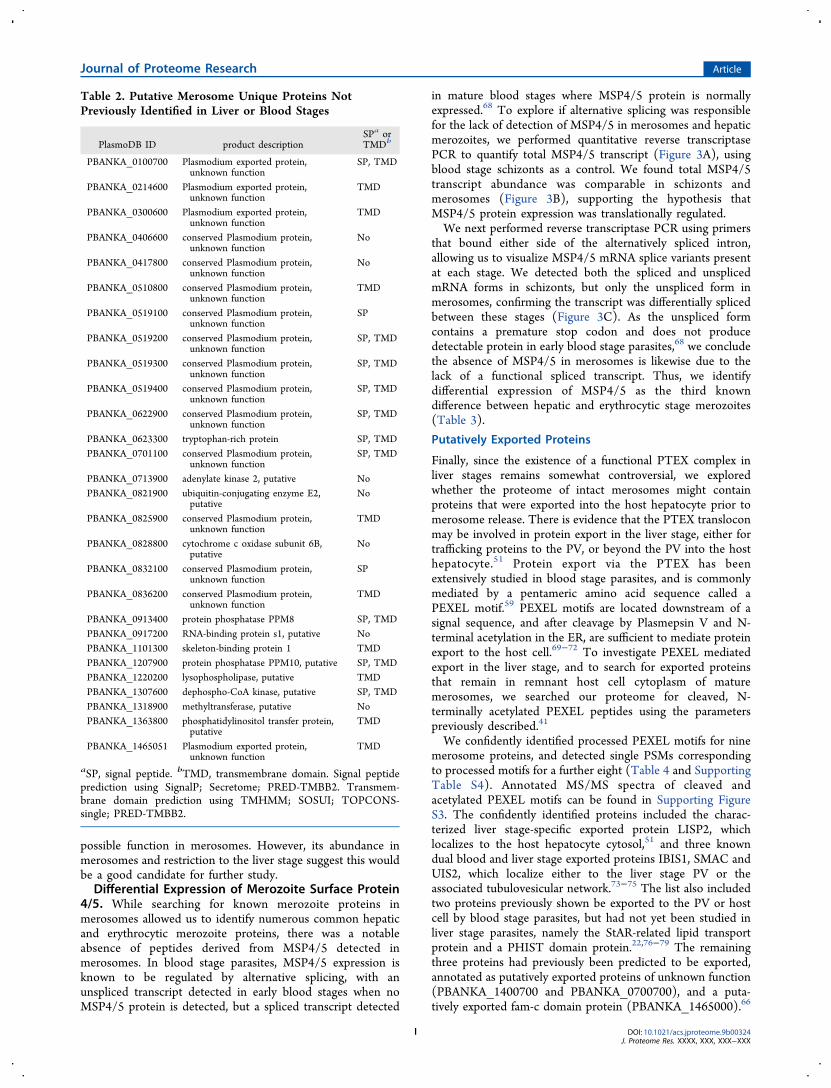
that the majority of merosome proteins are shared with one orboth of these stages,17,29−32 it also identified a small subset ofproteins that had not previously been detected or did not havesyntenic orthologs in these liver or blood stage proteomes.Specifically, we found 6.1% of the merosome proteome or 73proteins in this group (Figure 2 and Supporting Table S2.3).Closer examination of this list reveals that only a subset ofthese proteins is likely to be unique to merosomes. Indeed,several proteins are members of multigene families that do nothave strict one-to-one orthology across Plasmodium species,such as the p235 proteins, duffy binding proteins, and fam-a orfam-c proteins (Supporting Table S2.3). These proteins aretherefore not likely unique to merosomes, but were identifiedbecause we considered only proteins with clear orthologs inour global comparisons. Another group are liver stage proteinswhose orthologs were not observed in the only other publishedproteomic analysis of liver stages, performed on mid- and late-stage P. yoelii liver stage forms.17 As discussed, fewer proteinswere identified in that work compared to our proteome, in partbecause of interference from host proteins, and in part becausethe mass spectrometers available at the time were more limitedin sensitivity, duty cycle, and resolution than the instrumentused in our study. In light of this, many of the putativelyunique merosome proteins we identify may also turn out to bepresent in liver stages if such a study were revisited withmodern instrumentation. Finally, this subset also includesproteins previously detected in sporozoites,18,19 or organelle-fraction proteomes of various life cycle stages.62,63 Given thesemany caveats, we have manually annotated the list ofpotentially unique merosome proteins to highlight those 28proteins that may be unique to merosomes versus those thatwere prematurely labeled as unique for one of the reasonsoutlined above (Table 2 and Supporting Table S2.3). Weanticipate that future studies will confirm some of theseproteins to be truly unique to merosomes.
Abundant Hypothetical Proteins. Examining the mer-osome proteome as a whole, we noted several hypotheticalproteins were among the most abundant proteins in our dataset (Supporting Table S1). Since we suspected the uniquebiology of merosomes may be accompanied by unique ordivergent proteins, we undertook further analysis of theseproteins. The top 8 most abundant hypothetical proteins wereselected for analysis, as all were ranked among the top 100proteins in merosomes (Supporting Table S1). Examination ofavailable expression data for these proteins showed evidencefor expression at multiple other life stages for all except themost abundant, suggesting the majority do not have uniqueroles in the liver stage or merosomes (Supporting Table S3.4).Nonetheless, the most abundant hypothetical protein(PBANKA_0518900) was noteworthy because it appears tobe restricted to the late liver stage and merosomes based onavailable expression data. Clear homologues of the protein arefound only in rodent malaria parasites, so less data is availablethan for the proteins that have P. falciparum orthologs.Nevertheless, the available RNA-seq data indicate little or noexpression in blood stage parasites [PlasmoDB.org],66,67 andthe protein is conspicuously absent from the P. berghei bloodstage proteome,52 despite being detected in the P. yoelii liverstage proteome.17 Furthermore, our data indicate the protein ismore abundant in merosomes than the abundant merozoiteproteins MSP1 and actin (Supporting Table S1). As thishypothetical protein does not have any annotated domains orgenetic modification data to date, it is difficult to speculate its
Journal of Proteome Research Article
DOI: 10.1021/acs.jproteome.9b00324J. Proteome Res. XXXX, XXX, XXX−XXX
H
possible function in merosomes. However, its abundance inmerosomes and restriction to the liver stage suggest this wouldbe a good candidate for further study.Differential Expression of Merozoite Surface Protein
4/5. While searching for known merozoite proteins inmerosomes allowed us to identify numerous common hepaticand erythrocytic merozoite proteins, there was a notableabsence of peptides derived from MSP4/5 detected inmerosomes. In blood stage parasites, MSP4/5 expression isknown to be regulated by alternative splicing, with anunspliced transcript detected in early blood stages when noMSP4/5 protein is detected, but a spliced transcript detected
in mature blood stages where MSP4/5 protein is normallyexpressed.68 To explore if alternative splicing was responsiblefor the lack of detection of MSP4/5 in merosomes and hepaticmerozoites, we performed quantitative reverse transcriptasePCR to quantify total MSP4/5 transcript (Figure 3A), usingblood stage schizonts as a control. We found total MSP4/5transcript abundance was comparable in schizonts andmerosomes (Figure 3B), supporting the hypothesis thatMSP4/5 protein expression was translationally regulated.We next performed reverse transcriptase PCR using primers
that bound either side of the alternatively spliced intron,allowing us to visualize MSP4/5 mRNA splice variants presentat each stage. We detected both the spliced and unsplicedmRNA forms in schizonts, but only the unspliced form inmerosomes, confirming the transcript was differentially splicedbetween these stages (Figure 3C). As the unspliced formcontains a premature stop codon and does not producedetectable protein in early blood stage parasites,68 we concludethe absence of MSP4/5 in merosomes is likewise due to thelack of a functional spliced transcript. Thus, we identifydifferential expression of MSP4/5 as the third knowndifference between hepatic and erythrocytic stage merozoites(Table 3).
Putatively Exported Proteins
Finally, since the existence of a functional PTEX complex inliver stages remains somewhat controversial, we exploredwhether the proteome of intact merosomes might containproteins that were exported into the host hepatocyte prior tomerosome release. There is evidence that the PTEX transloconmay be involved in protein export in the liver stage, either fortrafficking proteins to the PV, or beyond the PV into the hosthepatocyte.51 Protein export via the PTEX has beenextensively studied in blood stage parasites, and is commonlymediated by a pentameric amino acid sequence called aPEXEL motif.59 PEXEL motifs are located downstream of asignal sequence, and after cleavage by Plasmepsin V and N-terminal acetylation in the ER, are sufficient to mediate proteinexport to the host cell.69−72 To investigate PEXEL mediatedexport in the liver stage, and to search for exported proteinsthat remain in remnant host cell cytoplasm of maturemerosomes, we searched our proteome for cleaved, N-terminally acetylated PEXEL peptides using the parameterspreviously described.41
We confidently identified processed PEXEL motifs for ninemerosome proteins, and detected single PSMs correspondingto processed motifs for a further eight (Table 4 and SupportingTable S4). Annotated MS/MS spectra of cleaved andacetylated PEXEL motifs can be found in Supporting FigureS3. The confidently identified proteins included the charac-terized liver stage-specific exported protein LISP2, whichlocalizes to the host hepatocyte cytosol,51 and three knowndual blood and liver stage exported proteins IBIS1, SMAC andUIS2, which localize either to the liver stage PV or theassociated tubulovesicular network.73−75 The list also includedtwo proteins previously shown be exported to the PV or hostcell by blood stage parasites, but had not yet been studied inliver stage parasites, namely the StAR-related lipid transportprotein and a PHIST domain protein.22,76−79 The remainingthree proteins had previously been predicted to be exported,annotated as putatively exported proteins of unknown function(PBANKA_1400700 and PBANKA_0700700), and a puta-tively exported fam-c domain protein (PBANKA_1465000).66
Table 2. Putative Merosome Unique Proteins NotPreviously Identified in Liver or Blood Stages
PlasmoDB ID product descriptionSPa orTMDb
PBANKA_0100700 Plasmodium exported protein,unknown function
SP, TMD
PBANKA_0214600 Plasmodium exported protein,unknown function
TMD
PBANKA_0300600 Plasmodium exported protein,unknown function
TMD
PBANKA_0406600 conserved Plasmodium protein,unknown function
No
PBANKA_0417800 conserved Plasmodium protein,unknown function
No
PBANKA_0510800 conserved Plasmodium protein,unknown function
TMD
PBANKA_0519100 conserved Plasmodium protein,unknown function
SP
PBANKA_0519200 conserved Plasmodium protein,unknown function
SP, TMD
PBANKA_0519300 conserved Plasmodium protein,unknown function
SP, TMD
PBANKA_0519400 conserved Plasmodium protein,unknown function
SP, TMD
PBANKA_0622900 conserved Plasmodium protein,unknown function
SP, TMD
PBANKA_0623300 tryptophan-rich protein SP, TMDPBANKA_0701100 conserved Plasmodium protein,
unknown functionSP, TMD
PBANKA_0713900 adenylate kinase 2, putative NoPBANKA_0821900 ubiquitin-conjugating enzyme E2,
putativeNo
PBANKA_0825900 conserved Plasmodium protein,unknown function
TMD
PBANKA_0828800 cytochrome c oxidase subunit 6B,putative
No
PBANKA_0832100 conserved Plasmodium protein,unknown function
SP
PBANKA_0836200 conserved Plasmodium protein,unknown function
TMD
PBANKA_0913400 protein phosphatase PPM8 SP, TMDPBANKA_0917200 RNA-binding protein s1, putative NoPBANKA_1101300 skeleton-binding protein 1 TMDPBANKA_1207900 protein phosphatase PPM10, putative SP, TMDPBANKA_1220200 lysophospholipase, putative TMDPBANKA_1307600 dephospho-CoA kinase, putative SP, TMDPBANKA_1318900 methyltransferase, putative NoPBANKA_1363800 phosphatidylinositol transfer protein,
putativeTMD
PBANKA_1465051 Plasmodium exported protein,unknown function
TMD
aSP, signal peptide. bTMD, transmembrane domain. Signal peptideprediction using SignalP; Secretome; PRED-TMBB2. Transmem-brane domain prediction using TMHMM; SOSUI; TOPCONS-single; PRED-TMBB2.
Journal of Proteome Research Article
DOI: 10.1021/acs.jproteome.9b00324J. Proteome Res. XXXX, XXX, XXX−XXX
I

These findings support the hypothesis that exported proteinscan be retained in the remnant host cell cytoplasm after PVbreakdown and merosome release, and suggests the as-yet-uncharacterized PEXEL-containing proteins may likewise beexported to the PV or host cell in the liver stage.Analysis of this group of proteins revealed several findings
consistent with the known mechanism of PEXEL-mediatedprotein export. We observed all but one protein had apredicted signal peptide or signal anchor, and in most cases thePEXEL motif matched the canonical “RxLxE” consensus59
(Table 4 and Supporting Table S4). We also found that mostof the PEXEL motifs were positioned in proximity to the signalsequence as expected.59 The exception was LISP2, a largeprotein in which the putative PEXEL motif RILAE appearsonce and a second putative PEXEL motif RLIAE appears 16times dispersed throughout a repeat region. In addition tofinding strong evidence for processing of the single RILAEmotif, we also detected processing of the RLIAE motif as thepeptide n42AEKESENNSENNSQDVK (n42 indicates wherethe PEXEL motif was cleaved and the protein was N-terminallyacetylated). This exact peptide sequence is repeated ninedifferent times in the protein, so it was not possible todetermine if the protein in the sample was processed at a single
site or multiple sites. However, we speculate this multiplicity ofPEXEL cleavage sites in LISP2 may explain the atypicalprocessing previously reported for this protein.51 Consistentwith these data, we identified both Plasmepsin V and theputative N-acetyltransferase in merosomes (Supporting TableS1), confirming the enzymes responsible for PEXELprocessing80 were present at this stage.To further validate these findings, we localized the PHIST
protein (PBANKA_1145400) by confocal immunofluores-cence microscopy in late liver stage parasites and merosomes.A previous study found this protein was highly expressed inP. berghei blood stage schizonts, and demonstrated it wasexported into the host red blood cell cytoplasm.22 In late liverstage parasites, we observed punctate staining for the PHISTprotein that partially colocalized with the PV marker UIS4,suggesting it was likewise exported at this stage (Figure 4A).To better quantify this partial colocalization, we performed 3Dimage analysis of z-stacks and calculated the percent of PHISTpositive pixels that were positive for UIS4. Analysis of 75individual parasites indicated over half of all PHIST positivepixels were UIS4 positive (mean colocalization 56.0% ± 15.1standard deviation). Thus, in late liver stage parasites a largefraction of the PHIST protein is trafficked to the PVmembrane, consistent with PEXEL-mediated export of theprotein.In merosomes, where the PV membrane is absent, we
observed PHIST staining associated with individual merozoites(Figure 4B). This finding was further supported by the patternof PHIST staining in free hepatic merozoites, where much ofthe “teardrop” shaped merozoite was stained (Figure 4C).Thus, it appears that the localization of the PHIST proteinchanges during liver stage development, being exported topuncta at or near the PV in late liver stages, then localizing onor within individual merozoites after merosomes are formed.
Figure 3. The absence of merozoite surface protein 4/5 in merosomes is associated with alternative splicing of the mRNA transcript. (A) Diagramof the P. berghei MSP4/5 gene structure, spliced mRNA, and unspliced mRNA. Unspliced mRNA contains a premature stop codon (red line).Primers used to quantify total MSP4/5 mRNA (P1 + P2) and detect splice variants (P3 + P4) are shown. (B) Quantitative reverse transcriptasePCR demonstrates relative abundance of total MSP4/5 mRNA is comparable in blood stage schizonts and merosomes. Relative abundancecalculated by comparison to HSP70. Relative abundance of MSP4/5 was normalized to 1.0 for schizonts. Error bars show mean ± standarddeviation. (C) Nonquantitative reverse transcriptase PCR demonstrates MSP4/5 is alternatively spliced in schizonts and merosomes.Abbreviations: Schiz, schizonts; Meros, merosomes; +RT, with reverse transcriptase; −RT, without reverse transcriptase.
Table 3. Known Differences between Hepatic andErythrocytic Merozoites
PlasmoDB ID protein difference ref
PBANKA_0304400 MSP4/5 Protein absent fromhepatic merozoites
here
PBANKA_1321700 berghepain-1 Knockout has more impacton hepatic merozoites
15
Multiple Py235 family Different members inhepatic vs erythrocyticmerozoites
14
Journal of Proteome Research Article
DOI: 10.1021/acs.jproteome.9b00324J. Proteome Res. XXXX, XXX, XXX−XXX
J

■ DISCUSSION
We describe the first proteomic analysis of Plasmodiummerosomes, the stage that initiates blood stage infection.Due to the relatively low numbers of hepatic merozoites andgreater barrier to accessibility in comparison to otherPlasmodium life cycle stages, few studies of merosomes andhepatic merozoites have been performed.16 Nonetheless,sensitive mass spectrometry techniques together with improve-ments to culture techniques have enabled us to identify a totalof 1879 P. berghei proteins from merosomes, and a core set of1188 proteins quantitatively detected across all replicates,producing the most comprehensive late liver stage data set todate.Comparison of the merosome proteome to published
Plasmodium liver and blood stage proteomes demonstratesthat merosomes share considerable similarity to both liver andblood stages. Considering merosomes are the final stage ofliver stage development and the initial stage of infection in theblood, this overlap is consistent with merosomes being poised
at this nexus. The similarity of merosomes to blood stagespoints to similar structural and functional features of hepaticand erythrocytic merozoites. Indeed, hepatic and erythrocyticmerozoites were found to share the vast majority of knownapical organelle proteins, cytoskeleton and motility proteins,and surface proteins. Merosomes also share considerablesimilarity to liver stage parasites, sharing known liver stage-specific proteins and a number of hypothetical proteins.Interestingly, this includes the highly abundant hypotheticalprotein (PBANKA_0518900), specific to liver stage parasitesand whose function remains unknown. Comparison of themerosome proteome to previously published proteomesfurther identified a subset of proteins potentially unique tomerosomes. Though a proportion of the proteins in this groupwould be expected to be liver stage proteins that were notidentified in the earlier liver stage proteome,17 some are likelyto be bona fide unique hepatic merozoite or merosomeproteins, and are high value targets for follow-up studies.
Table 4. Proteins with Processed PEXEL Protein Export Motifs
PlasmoDB ID protein peptidea SPb repsc PSMsd
PBANKA_0208900 StAR-related lipid transfer protein, putative RIL.EEALMDVENVKENLK Y 3 95PBANKA_1400700 Plasmodium exported protein RHL.AEYIPNISNPTGNVYHFSR N 3 56PBANKA_1003000 liver specific protein 2 (LISP2) RIL.AETQHENLVLSTDKDKER Y 3 23PBANKA_1003000 liver specific protein 2 (LISP2) RLI.AEKESENNSENNSQDVK Y 2 14PBANKA_1328000 serine/threonine protein phosphatase (UIS2) RVL.QEQNEDIKDDNDENDEGDEEDEYYSYLK Y 3 7PBANKA_1365500 exported protein IBIS1 RIL.SELDQNKDQNLGYK YR 3 7PBANKA_1465000 fam-c protein RSL.SEHSTEDKEETVTISDNK Y 2 4PBANKA_0700700 Plasmodium exported protein, unknown function RIL.SSMSNEKPYNILR* Y 2 4PBANKA_0100600 schizont membrane associated cytoadherence protein
(SMAC)RFL.VEYYDANIDQYNGNQSK Y 2 3
PBANKA_1145400 Plasmodium exported protein (PHIST) RNL.SETSVVNDNLSNNVNNLRDEPK Y 1 6aIdentified peptide sequence bearing a cleaved and acetylated PEXEL motif [K/R]X[L/I]X[D/E/Q] or possible PEXEL motif [K/R]X[L/I]XX(denoted by “*”). For both canonical and possible PEXEL motifs, the point of cleavage is denoted with “.”, and the three residues in the proteinsequence N-terminal to the peptide are shown. All displayed peptides were observed with mass spectral evidence for N-terminal acetylation. bSignalpeptide prediction; YR indicates a recessed signal peptide.73 cNumber of biological replicates in which the cleaved and acetylated PEXEL peptidewas identified. dTotal number of PSMs that identified the cleaved and acetylated PEXEL across all three biological replicates.
Figure 4. Localization of the PHIST protein in late liver stage parasites and merosomes. (A) Immunofluorescence staining of late liver stageparasites at 60 h postinfection indicates the PHIST protein (PBANKA_1145400) partially colocalizes with the parasitophorous vacuole markerUIS4. Colocalized pixels in white. DNA stained with Hoechst. (B,C) Immunofluorescence staining of merosomes and free merozoites at 65 hpostinfection demonstrates the PHIST protein associates with individual liver stage merozoites. No UIS4 staining was detected in the maturemerosome or free hepatic merozoite shown.
Journal of Proteome Research Article
DOI: 10.1021/acs.jproteome.9b00324J. Proteome Res. XXXX, XXX, XXX−XXX
K

Despite the overall similarity between erythrocytic andhepatic merozoites, previous studies focusing on particularproteins or protein families have demonstrated some differ-ences between these two populations of merozoites. P. yoeliihepatic merozoites were previously reported to express aunique Py235 rhoptry protein,14 and we had previously foundthat hepatic merozoites have a greater requirement for thecysteine protease berghepain-1 compared to erythrocyticmerozoites.15 Here we identify differential expression ofMSP4/5 as a third difference between these two merozoitepopulations. We found MSP4/5 was absent from merosomesdemonstrating that it is not expressed in hepatic merozoites, incontrast to erythrocytic merozoites where the protein is readilydetected.68 As reported for early blood stage parasites,68 lack ofthe MSP4/5 protein in merosomes correlated with alternativesplicing of the transcript, suggesting a common mechanism oftranslational regulation is conserved across both life stages.Why MSP4/5 might be expressed in erythrocytic merozoitesbut not hepatic merozoites is currently unclear, as its functionin blood stage parasites is not known.38 Since MSP4/5 hasbeen pursued as a vaccine antigen81−83 and vaccines targetingboth hepatic and erythrocytic merozoites would likely be moreeffective, it would be important to determine if the orthologousproteins are likewise differentially expressed in P. falciparum.Our findings also support a role for the PTEX translocon in
liver stage parasites. The identification of nine proteins withcleaved acetylated PEXEL motifs and concurrent detection ofthe enzymes responsible for their cleavage suggests thatPEXEL processing in liver stage parasites can occur as it doesin blood stages. Furthermore, the discovery of processedPEXELs in known liver stage exported proteins argues that thisprocessing is relevant to their trafficking. One of the exportedproteins we identified is the PHIST protein, PBANKA_1145400. P. berghei has two PHIST-domain proteins, and aprevious study characterizing these proteins in blood stageparasites found both were exported to the host erythrocytecytoplasm.22 Additionally, PBANKA_1145400 could not bedeleted and was predicted to be essential.22 Here we show thatthis PHIST protein partially localizes to the PV membrane inthe late liver stage, confirming that it is indeed exported at thisstage, and present at the host-parasite interface. Given therelatively low number of known PV proteins and the clearimportance of this structure for host parasite interactions in theliver stage,11 we propose further characterization of this PHISTprotein in the liver stage may be warranted.In conclusion, this work provides a valuable resource for one
of the least characterized stages of malaria parasites, themerosome. At the juncture between liver and blood stages,merosomes have a unique biology and initiate blood stageinfection with relatively low numbers of parasites, both factorsthat could be exploited in the generation of new drugs andvaccines. This data set and some of the insight gained from itshould help inform future studies and interventions.
■ ASSOCIATED CONTENT*S Supporting Information
The Supporting Information is available free of charge on theACS Publications website at DOI: 10.1021/acs.jproteo-me.9b00324.
Supporting Figure S1: GO Biological Process terms forproteins in the core merosome proteome; SupportingFigure S2: Scatterplot of iBAQ intensities for proteins in
the core merosome proteome, with apical and merozoitesurface proteins highlighted; Supporting Figure S3:Annotated representative MS/MS spectra identifyingputatively cleaved and acetylated PEXEL motifs (PDF)Supporting Table S1: Peptide and protein identifications(XLSX)Supporting Table S2: Comparison to other proteomes(XLSX)Supporting Table S3: GO term analysis and groups ofinterest (XLSX)Supporting Table S4 Proteins with processed PEXELmotifs (XLSX)
■ AUTHOR INFORMATIONCorresponding Author
*E-mail: [email protected]. Tel: 410 502 6918.ORCID
Melanie J. Shears: 0000-0002-8226-4354Robert L. Moritz: 0000-0002-3216-9447Akhilesh Pandey: 0000-0001-9943-6127Photini Sinnis: 0000-0003-2954-7547Present Addresses⊥Akhilesh Pandey, Santosh Renuse: Center for IndividualizedMedicine and Department of Laboratory Medicine andPathology, Mayo Clinic, Rochester, Minnesota 55905, UnitedStates; Manipal Academy of Higher Education (MAHE),Manipal 576104, Karnataka, India.#Satish Mishra: Division of Parasitology, CSIR-Central DrugResearch Institute, Lucknow 226031, India.Notes
The authors declare no competing financial interest.Mass spectrometry proteomics data have been deposited to theProteomeXchange Consortium via the PRIDE26 partnerrepository with the data set identifier PXD010559.
■ ACKNOWLEDGMENTSWe thank the Insectary and Parasitology Core Facilities at theJohns Hopkins Malaria Research Institute and in particular Dr.Abhai Tripathi, Dr. Godfree Mlambo, and Chris Kizito fortheir outstanding work. We are grateful to BloombergPhilanthropies for supporting these Core Facilities. We thankDr. Sean Prigge for his assistance with the merosome genomequantitative PCR experiments, and Dr. Scott Lindner forsharing antibodies. We thank the Johns Hopkins School ofMedicine Microscopy Facility for assistance with imagingexperiments, and Dr. Hoku West-Foyle for his guidance withconfocal microscopy and image analysis. This work wassupported by a Johns Hopkins Malaria Research Institute PilotGrant to Drs. Photini Sinnis and Akhilesh Pandey, NIH GrantR01 AI056840 (P.S.), a postdoctoral fellowship to Dr. MelanieShears from the Johns Hopkins Provost’s Office andBloomberg Philanthropies. This work was also funded in partby the National Institutes of Health, grants R01GM087221and 1S10RR027584 (R.M.), and K25AI119229 (K.S.).Funding for the Zeiss LSM 710 microscope was provided byNIH Grant S10 RR024550.
■ ABBREVIATIONSPV, parasitophorous vacuole; DMEM, Dulbecco’s ModifiedEagle’s Medium; FBS, fetal bovine serum; RT, room
Journal of Proteome Research Article
DOI: 10.1021/acs.jproteome.9b00324J. Proteome Res. XXXX, XXX, XXX−XXX
L

temperature; HBSS, Hank’s Buffered Saline Solution; qPCR,quantitative PCR; SCX, strong cation exchange; HCD, higher-energy collisional dissociation; NCE, normalized collisionenergy; PSM, peptide spectrum match; FDR, false discoveryrate; LFQ, label-free quantification; GO, Gene Ontology; RT,reverse transcriptase; PEXEL, protein export element; TPP,Trans-Proteomic Pipeline; iBAQ, intensity-based absolutequantification; EXP1, exported protein 1; PTEX translocon,Plasmodium Translocon of Exported Proteins; MTIP, myosinA tail domain interacting protein; MSP, merozoite surfaceprotein; SERA, serine repeat antigen; SUB, subtilisin-likeprotease; ROM, rhomboid protease.
■ REFERENCES(1) World Health Organization. World Malaria Report 2017; WHO,2017; pp 1−196.(2) Potocnjak, P.; Yoshida, N.; Nussenzweig, R. S.; Nussenzweig, V.Monovalent fragments (Fab) of monoclonal antibodies to asporozoite surface antigen (Pb44) protect mice against malarialinfection. J. Exp. Med. 1980, 151, 1504−1513.(3) Romero, P.; Maryanski, J. L.; Corradin, G.; Nussenzweig, R. S.;Nussenzweig, V.; Zavala, F. Cloned cytotoxic T cells recognize anepitope in the circumsporozoite protein and protect against malaria.Nature 1989, 341, 323−326.(4) The RTS,S Clinical Trials Partnership. First Results of Phase 3Trial of RTS,S/AS01 Malaria Vaccine in African Children. N. Engl. J.Med. 2011, 365, 1863−1875.(5) The RTS,S Clinical Trials Partnership. Efficacy and safety ofRTS,S/AS01 malaria vaccine with or without a booster dose in infantsand children in Africa: final results of a phase 3, individuallyrandomised, controlled trial. Lancet 2015, 386, 31−45.(6) Long, C. A.; Zavala, F. Malaria vaccines and human immuneresponses. Curr. Opin. Microbiol. 2016, 32, 96−102.(7) Butler, N. S.; Schmidt, N. W.; Vaughan, A. M.; Aly, A. S.; Kappe,S. H. I.; Harty, J. T. Superior antimalarial immunity after vaccinationwith late liver stage-arresting genetically attenuated parasites. Cell HostMicrobe 2011, 9, 451−462.(8) Prudencio, M.; Rodriguez, A.; Mota, M. M. The silent path tothousands of merozoites: the Plasmodium liver stage. Nat. Rev.Microbiol. 2006, 4, 849−856.(9) Spielmann, T.; Montagna, G. N.; Hecht, L.; Matuschewski, K.Molecular make-up of the Plasmodium parasitophorous vacuolarmembrane. Int. J. Med. Microbiol. 2012, 302, 179−186.(10) Sturm, A.; Amino, R.; van de Sand, C.; Regen, T.; Retzlaff, S.;Rennenberg, A.; Krueger, A.; Pollok, J.-M.; Menard, R.; Heussler, V.T. Manipulation of host hepatocytes by the malaria parasite fordelivery into liver sinusoids. Science 2006, 313, 1287−1290.(11) Nyboer, B.; Heiss, K.; Mueller, A.-K.; Ingmundson, A. ThePlasmodium liver-stage parasitophorous vacuole: A front-line ofcommunication between parasite and host. Int. J. Med. Microbiol.2018, 308, 107−117.(12) Graewe, S.; Rankin, K. E.; Lehmann, C.; Deschermeier, C.;Hecht, L.; Froehlke, U.; Stanway, R. R.; Heussler, V. Hostile takeoverby Plasmodium: reorganization of parasite and host cell membranesduring liver stage egress. PLoS Pathog. 2011, 7, e1002224.(13) Burda, P.-C.; Caldelari, R.; Heussler, V. T. Manipulation of theHost Cell Membrane during Plasmodium Liver Stage Egress. mBio2017, 8, e00139-17.(14) Preiser, P. R.; Khan, S.; Costa, F. T. M.; Jarra, W.; Belnoue, E.;Ogun, S.; Holder, A. A.; Voza, T.; Landau, I.; Snounou, G.; Renia, L.Stage-Specific Transcription of Distinct Repertoires of a MultigeneFamily During Plasmodium Life Cycle. Science 2002, 295, 342−345.(15) Hopp, C. S.; Bennett, B. L.; Mishra, S.; Lehmann, C.; Hanson,K. K.; Lin, J.-W.; Rousseau, K.; Carvalho, F. A.; van der Linden, W.A.; Santos, N. C.; Bogyo, M.; Khan, S. M.; Heussler, V.; Sinnis, P.Deletion of the rodent malaria ortholog for falcipain-1 highlightsdifferences between hepatic and blood stage merozoites. PLoS Pathog.2017, 13, e1006586.
(16) Prudencio, M.; Mota, M. M.; Mendes, A. M. A toolbox to studyliver stage malaria. Trends Parasitol. 2011, 27, 565−574.(17) Tarun, A. S.; Peng, X.; Dumpit, R. F.; Ogata, Y.; Silva-Rivera,H.; Camargo, N.; Daly, T. M.; Bergman, L. W.; Kappe, S. H. I. Acombined transcriptome and proteome survey of malaria parasite liverstages. Proc. Natl. Acad. Sci. U. S. A. 2008, 105, 305−310.(18) Lindner, S. E.; Swearingen, K. E.; Harupa, A.; Vaughan, A. M.;Sinnis, P.; Moritz, R. L.; Kappe, S. H. I. Total and Putative SurfaceProteomics of Malaria Parasite Salivary Gland Sporozoites. Mol. Cell.Proteomics 2013, 12, 1127−1143.(19) Swearingen, K. E.; Lindner, S. E.; Shi, L.; Shears, M. J.; Harupa,A.; Hopp, C. S.; Vaughan, A. M.; Springer, T. A.; Moritz, R. L.;Kappe, S. H. I.; Sinnis, P. Interrogating the Plasmodium SporozoiteSurface: Identification of Surface-Exposed Proteins and Demonstra-tion of Glycosylation on CSP and TRAP by Mass Spectrometry-BasedProteomics. PLoS Pathog. 2016, 12, e1005606.(20) Mueller, A. K.; Camargo, N.; Kaiser, K.; Andorfer, C.; Frevert,U.; Matuschewski, K.; Kappe, S. H. I. Plasmodium liver stagedevelopmental arrest by depletion of a protein at the parasite-hostinterface. Proc. Natl. Acad. Sci. U. S. A. 2005, 102, 3022−3027.(21) Schindelin, J.; Arganda-Carreras, I.; Frise, E.; Kaynig, V.;Longair, M.; Pietzsch, T.; Preibisch, S.; Rueden, C.; Saalfeld, S.;Schmid, B.; Tinevez, J.-Y.; White, D. J.; Hartenstein, V.; Eliceiri, K.;Tomancak, P.; Cardona, A. Fiji: an open-source platform forbiological-image analysis. Nat. Methods 2012, 9, 676−682.(22) Moreira, C. K.; Naissant, B.; Coppi, A.; Bennett, B. L.; Aime,E.; Franke-Fayard, B.; Janse, C. J.; Coppens, I.; Sinnis, P.; Templeton,T. J. The Plasmodium PHIST and RESA-Like Protein Families ofHuman and Rodent Malaria Parasites. PLoS One 2016, 11, e0152510.(23) Kulak, N. A.; Pichler, G.; Paron, I.; Nagaraj, N.; Mann, M.Minimal, encapsulated proteomic-sample processing applied to copy-number estimation in eukaryotic cells. Nat. Methods 2014, 11, 319−324.(24) Choi, H.; Nesvizhskii, A. I. False discovery rates and relatedstatistical concepts in mass spectrometry-based proteomics. J.Proteome Res. 2008, 7, 47−50.(25) Cox, J.; Hein, M. Y.; Luber, C. A.; Paron, I.; Nagaraj, N.; Mann,M. Accurate Proteome-wide Label-free Quantification by DelayedNormalization and Maximal Peptide Ratio Extraction, TermedMaxLFQ. Mol. Cell. Proteomics 2014, 13, 2513−2526.(26) Vizcaíno, J. A.; Csordas, A.; del-Toro, N.; Dianes, J. A.; Griss, J.;Lavidas, I.; Mayer, G.; Perez-Riverol, Y.; Reisinger, F.; Ternent, T.;Xu, Q.-W.; Wang, R.; Hermjakob, H. 2016 update of the PRIDEdatabase and its related tools. Nucleic Acids Res. 2016, 44, 11033−11033.(27) Aurrecoechea, C.; Brestelli, J.; Brunk, B. P.; Dommer, J.;Fischer, S.; Gajria, B.; Gao, X.; Gingle, A.; Grant, G.; Harb, O. S.;Heiges, M.; Innamorato, F.; Iodice, J.; Kissinger, J. C.; Kraemer, E.; Li,W.; Miller, J. A.; Nayak, V.; Pennington, C.; Pinney, D. F.; Roos, D.S.; Ross, C.; Stoeckert, C. J.; Treatman, C.; Wang, H. PlasmoDB: afunctional genomic database for malaria parasites. Nucleic Acids Res.2009, 37, D539−D543.(28) Sam-Yellowe, T. Y.; Florens, L.; Wang, T.; Raine, J. D.; Carucci,D. J.; Sinden, R.; Yates, J. R. Proteome Analysis of Rhoptry-EnrichedFractions Isolated from Plasmodium Merozoites. J. Proteome Res.2004, 3, 995−1001.(29) Florens, L.; Washburn, M. P.; Raine, J. D.; Anthony, R. M.;Grainger, M.; Haynes, J. D.; Moch, J. K.; Muster, N.; Sacci, J. B.;Tabb, D. L.; Witney, A. A.; Wolters, D.; Wu, Y.; Gardner, M. J.;Holder, A. A.; Sinden, R. E.; Yates, J. R.; Carucci, D. J. A proteomicview of the Plasmodium falciparum life cycle. Nature 2002, 419, 520−526.(30) Treeck, M.; Sanders, J. L.; Elias, J. E.; Boothroyd, J. C. Thephosphoproteomes of Plasmodium falciparum and Toxoplasmagondii reveal unusual adaptations within and beyond the parasites’boundaries. Cell Host Microbe 2011, 10, 410−419.(31) Pease, B. N.; Huttlin, E. L.; Jedrychowski, M. P.; Talevich, E.;Harmon, J.; Dillman, T.; Kannan, N.; Doerig, C.; Chakrabarti, R.;Gygi, S. P.; Chakrabarti, D. Global Analysis of Protein Expression and
Journal of Proteome Research Article
DOI: 10.1021/acs.jproteome.9b00324J. Proteome Res. XXXX, XXX, XXX−XXX
M
Phosphorylation of Three Stages of Plasmodium falciparum Intra-erythrocytic Development. J. Proteome Res. 2013, 12, 4028−4045.(32) Lasonder, E.; Green, J. L.; Grainger, M.; Langsley, G.; Holder,A. A. Extensive differential protein phosphorylation as intra-erythrocytic Plasmodium falciparum schizonts develop into extrac-ellular invasive merozoites. Proteomics 2015, 15, 2716−2729.(33) Abdi, A.; Yu, L.; Goulding, D.; Rono, M. K.; Bejon, P.;Choudhary, J.; Rayner, J. Proteomic analysis of extracellular vesiclesfrom a Plasmodium falciparum Kenyan clinical isolate defines a coreparasite secretome. Wellcome Open Research 2017, 2, 50.(34) Boyle, E. I.; Weng, S.; Gollub, J.; Jin, H.; Botstein, D.; Cherry, J.M.; Sherlock, G. GO::TermFinder–open source software for accessingGene Ontology information and finding significantly enriched GeneOntology terms associated with a list of genes. Bioinformatics 2004,20, 3710−3715.(35) Bullen, H. E.; Tonkin, C. J.; O’Donnell, R. A.; Tham, W.-H.;Papenfuss, A. T.; Gould, S.; Cowman, A. F.; Crabb, B. S.; Gilson, P. R.A novel family of Apicomplexan glideosome-associated proteins withan inner membrane-anchoring role. J. Biol. Chem. 2009, 284, 25353−25363.(36) Bargieri, D.; Lagal, V.; Andenmatten, N.; Tardieux, I.; Meissner,M.; Menard, R. Host cell invasion by apicomplexan parasites: thejunction conundrum. PLoS Pathog. 2014, 10, e1004273.(37) Cowman, A. F.; Berry, D.; Baum, J. The cellular and molecularbasis for malaria parasite invasion of the human red blood cell. J. CellBiol. 2012, 198, 961−971.(38) Beeson, J. G.; Drew, D. R.; Boyle, M. J.; Feng, G.; Fowkes, F. J.I.; Richards, J. S. Merozoite surface proteins in red blood cell invasion,immunity and vaccines against malaria. FEMS Microbiology Reviews2016, 40, 343−372.(39) Kuang, R.; Gu, J.; Cai, H.; Wang, Y. Improved prediction ofmalaria degradomes by supervised learning with SVM and profilekernel. Genetica 2009, 136, 189−209.(40) Kedzierski, L.; Black, C. G.; Coppel, R. L. Characterization ofthe merozoite surface protein 4/5 gene of Plasmodium berghei andPlasmodium yoelii. Mol. Biochem. Parasitol. 2000, 105, 137−147.(41) Silvestrini, F.; Lasonder, E.; Olivieri, A.; Camarda, G.; vanSchaijk, B.; Sanchez, M.; Younis Younis, S.; Sauerwein, R.; Alano, P.Protein export marks the early phase of gametocytogenesis of thehuman malaria parasite Plasmodium falciparum. Mol. Cell. Proteomics2010, 9, 1437−1448.(42) Kessner, D.; Chambers, M.; Burke, R.; Agus, D.; Mallick, P.ProteoWizard: open source software for rapid proteomics toolsdevelopment. Bioinformatics 2008, 24, 2534−2536.(43) Eng, J. K.; Jahan, T. A.; Hoopmann, M. R. Comet: an open-source MS/MS sequence database search tool. Proteomics 2013, 13,22−24.(44) UniProt Consortium, T. UniProt: the universal proteinknowledgebase. Nucleic Acids Res. 2018, 46, 2699−2699.(45) Deutsch, E. W.; Mendoza, L.; Shteynberg, D.; Slagel, J.; Sun, Z.;Moritz, R. L. Trans-Proteomic Pipeline, a standardized dataprocessing pipeline for large-scale reproducible proteomics infor-matics. Proteomics: Clin. Appl. 2015, 9, 745−754.(46) Keller, A.; Nesvizhskii, A. I.; Kolker, E.; Aebersold, R. Empiricalstatistical model to estimate the accuracy of peptide identificationsmade by MS/MS and database search. Anal. Chem. 2002, 74, 5383−5392.(47) Pick, C.; Ebersberger, I.; Spielmann, T.; Bruchhaus, I.;Burmester, T. Phylogenomic analyses of malaria parasites andevolution of their exported proteins. BMC Evol. Biol. 2011, 11, 167.(48) Petersen, T. N.; Brunak, S.; Heijne, v. G.; Nielsen, H. SignalP4.0: discriminating signal peptides from transmembrane regions. Nat.Methods 2011, 8, 785−786.(49) Emanuelsson, O.; Brunak, S.; von Heijne, G.; Nielsen, H.Locating proteins in the cell using TargetP, SignalP and related tools.Nat. Protoc. 2007, 2, 953−971.(50) Gomez-Arreaza, A.; Acosta, H.; Quinones, W.; Concepcion, J.L.; Michels, P. A. M.; Avilan, L. Extracellular functions of glycolytic
enzymes of parasites: Unpredicted use of ancient proteins. Mol.Biochem. Parasitol. 2014, 193, 75−81.(51) Orito, Y.; Ishino, T.; Iwanaga, S.; Kaneko, I.; Kato, T.; Menard,R.; Chinzei, Y.; Yuda, M. Liver-specific protein 2: a Plasmodiumprotein exported to the hepatocyte cytoplasm and required formerozoite formation. Mol. Microbiol. 2013, 87, 66−79.(52) Hall, N. A Comprehensive Survey of the Plasmodium LifeCycle by Genomic, Transcriptomic, and Proteomic Analyses. Science2005, 307, 82−86.(53) Ishino, T.; Boisson, B.; Orito, Y.; Lacroix, C.; Bischoff, E.;Loussert, C.; Janse, C.; Menard, R.; Yuda, M.; Baldacci, P. LISP1 isimportant for the egress of Plasmodium berghei parasites from livercells. Cell. Microbiol. 2009, 11, 1329−1339.(54) Yu, M.; Kumar, T. R. S.; Nkrumah, L. J.; Coppi, A.; Retzlaff, S.;Li, C. D.; Kelly, B. J.; Moura, P. A.; Lakshmanan, V.; Freundlich, J. S.;Valderramos, J.-C.; Vilcheze, C.; Siedner, M.; Tsai, J. H.-C.; Falkard,B.; Sidhu, A. B. S.; Purcell, L. A.; Gratraud, P.; Kremer, L.; Waters, A.P.; Schiehser, G.; Jacobus, D. P.; Janse, C. J.; Ager, A.; Jacobs, W. R.;Sacchettini, J. C.; Heussler, V.; Sinnis, P.; Fidock, D. A. The fatty acidbiosynthesis enzyme FabI plays a key role in the development of liver-stage malarial parasites. Cell Host Microbe 2008, 4, 567−578.(55) Shears, M. J.; Botte, C. Y.; McFadden, G. I. Fatty acidmetabolism in the Plasmodium apicoplast: Drugs, doubts andknockouts. Mol. Biochem. Parasitol. 2015, 199, 34−50.(56) Ghosh, S.; Kennedy, K.; Sanders, P.; Matthews, K.; Ralph, S.A.; Counihan, N. A.; de Koning-Ward, T. F. The Plasmodium rhoptryassociated protein complex is important for parasitophorous vacuolemembrane structure and intraerythrocytic parasite growth. Cell.Microbiol. 2017, 19, e12733.(57) Ling, I. T.; Florens, L.; Dluzewski, A. R.; Kaneko, O.; Grainger,M.; Yim Lim, B. Y. S.; Tsuboi, T.; Hopkins, J. M.; Johnson, J. R.;Torii, M.; Bannister, L. H.; Yates, J. R.; Holder, A. A.; Mattei, D. ThePlasmodium falciparum clag9 gene encodes a rhoptry protein that istransferred to the host erythrocyte upon invasion. Mol. Microbiol.2004, 52, 107−118.(58) Iyer, J. K.; Amaladoss, A.; Genesan, S.; Preiser, P. R. Variableexpression of the 235 kDa rhoptry protein of Plasmodium yoeliimediate host cell adaptation and immune evasion. Mol. Microbiol.2007, 65, 1596−1596.(59) de Koning-Ward, T. F.; Gilson, P. R.; Boddey, J. A.; Rug, M.;Smith, B. J.; Papenfuss, A. T.; Sanders, P. R.; Lundie, R. J.; Maier, A.G.; Cowman, A. F.; Crabb, B. S. A newly discovered protein exportmachine in malaria parasites. Nature 2009, 459, 945−949.(60) Bullen, H. E.; Charnaud, S. C.; Kalanon, M.; Riglar, D. T.;Dekiwadia, C.; Kangwanrangsan, N.; Torii, M.; Tsuboi, T.; Baum, J.;Ralph, S. A.; Cowman, A. F.; de Koning-Ward, T. F.; Crabb, B. S.;Gilson, P. R. Biosynthesis, localization, and macromolecular arrange-ment of the Plasmodium falciparum translocon of exported proteins(PTEX). J. Biol. Chem. 2012, 287, 7871−7884.(61) Matthews, K.; Kalanon, M.; Chisholm, S. A.; Sturm, A.;Goodman, C. D.; Dixon, M. W. A.; Sanders, P. R.; Nebl, T.; Fraser,F.; Haase, S.; McFadden, G. I.; Gilson, P. R.; Crabb, B. S.; de Koning-Ward, T. F. The Plasmodium translocon of exported proteins (PTEX)component thioredoxin-2 is important for maintaining normal blood-stage growth. Mol. Microbiol. 2013, 89, 1167−1186.(62) Lal, K.; Prieto, J. H.; Bromley, E.; Sanderson, S. J.; Yates, J. R.,III; Wastling, J. M.; Tomley, F. M.; Sinden, R. E. Characterisation ofPlasmodium invasive organelles; an ookinete microneme proteome.Proteomics 2009, 9, 1142−1151.(63) Sam-Yellowe, T. Y.; Banks, T. L.; Fujioka, H.; Drazba, J. A.;Yadav, S. P. Plasmodium yoelii: Novel rhoptry proteins identifiedwithin the body of merozoite rhoptries in rodent Plasmodium malaria.Exp. Parasitol. 2008, 120, 113−117.(64) Baum, J.; Tonkin, C. J.; Paul, A. S.; Rug, M.; Smith, B. J.;Gould, S. B.; Richard, D.; Pollard, T. D.; Cowman, A. F. A MalariaParasite Formin Regulates Actin Polymerization and Localizes to theParasite-Erythrocyte Moving Junction during Invasion. Cell HostMicrobe 2008, 3, 188−198.
Journal of Proteome Research Article
DOI: 10.1021/acs.jproteome.9b00324J. Proteome Res. XXXX, XXX, XXX−XXX
N

(65) Pachebat, J. A.; Kadekoppala, M.; Grainger, M.; Dluzewski, A.R.; Gunaratne, R. S.; Scott-Finnigan, T. J.; Ogun, S. A.; Ling, I. T.;Bannister, L. H.; Taylor, H. M.; Mitchell, G. H.; Holder, A. A.Extensive proteolytic processing of the malaria parasite merozoitesurface protein 7 during biosynthesis and parasite release fromerythrocytes. Mol. Biochem. Parasitol. 2007, 151, 59−69.(66) Otto, T. D.; Bohme, U.; Jackson, A. P.; Hunt, M.; Franke-Fayard, B.; Hoeijmakers, W. A. M.; Religa, A. A.; Robertson, L.;Sanders, M.; Ogun, S. A.; Cunningham, D.; Erhart, A.; Billker, O.;Khan, S. M.; Stunnenberg, H. G.; Langhorne, J.; Holder, A. A.;Waters, A. P.; Newbold, C. I.; Pain, A.; Berriman, M.; Janse, C. J. Acomprehensive evaluation of rodent malaria parasite genomes andgene expression. BMC Biol. 2014, 12, 86.(67) Yeoh, L. M.; Goodman, C. D.; Mollard, V.; McFadden, G. I.;Ralph, S. A. Comparative transcriptomics of female and malegametocytes in Plasmodium berghei and the evolution of sex inalveolates. BMC Genomics 2017, 18, 734.(68) Black, C. G.; Wang, L.; Hibbs, A. R.; Werner, E.; Coppel, R. L.Identification of the Plasmodium chabaudi homologue of merozoitesurface proteins 4 and 5 of Plasmodium falciparum. Infect. Immun.1999, 67, 2075−2081.(69) Chang, H. H.; Falick, A. M.; Carlton, P. M.; Sedat, J. W.;DeRisi, J. L.; Marletta, M. A. N-terminal processing of proteinsexported by malaria parasites. Mol. Biochem. Parasitol. 2008, 160,107−115.(70) Boddey, J. A.; Moritz, R. L.; Simpson, R. J.; Cowman, A. F.Role of the Plasmodium export element in trafficking parasite proteinsto the infected erythrocyte. Traffic 2009, 10, 285−299.(71) Boddey, J. A.; Hodder, A. N.; Gunther, S.; Gilson, P. R.;Patsiouras, H.; Kapp, E. A.; Pearce, J. A.; de Koning-Ward, T. F.;Simpson, R. J.; Crabb, B. S.; Cowman, A. F. An aspartyl proteasedirects malaria effector proteins to the host cell. Nature 2010, 463,627−631.(72) Russo, I.; Babbitt, S.; Muralidharan, V.; Butler, T.; Oksman, A.;Goldberg, D. E. Plasmepsin V licenses Plasmodium proteins forexport into the host erythrocyte. Nature 2010, 463, 632−636.(73) Ingmundson, A.; Nahar, C.; Brinkmann, V.; Lehmann, M. J.;Matuschewski, K. The exported Plasmodium berghei protein IBIS1delineates membranous structures in infected red blood cells. Mol.Microbiol. 2012, 83, 1229−1243.(74) Fougere, A.; Jackson, A. P.; Bechtsi, D. P.; Braks, J. A. M.;Annoura, T.; Fonager, J.; Spaccapelo, R.; Ramesar, J.; Chevalley-Maurel, S.; Klop, O.; van der Laan, A. M. A.; Tanke, H. J.; Kocken, C.H. M.; Pasini, E. M.; Khan, S. M.; Bohme, U.; van Ooij, C.; Otto, T.D.; Janse, C. J.; Franke-Fayard, B. Variant Exported Blood-StageProteins Encoded by Plasmodium Multigene Families Are Expressedin Liver Stages Where They Are Exported into the ParasitophorousVacuole. PLoS Pathog. 2016, 12, e1005917.(75) Schnider, C. B.; Bausch-Fluck, D.; Bruhlmann, F.; Heussler, V.T.; Burda, P.-C. BioID Reveals Novel Proteins of the PlasmodiumParasitophorous Vacuole Membrane. mSphere 2018, 3, 37.(76) van Ooij, C.; Tamez, P.; Bhattacharjee, S.; Hiller, N. L.;Harrison, T.; Liolios, K.; Kooij, T.; Ramesar, J.; Balu, B.; Adams, J.;Waters, A.; Janse, C.; Haldar, K. The Malaria Secretome: FromAlgorithms to Essential Function in Blood Stage Infection. PLoSPathog. 2008, 4, e1000084.(77) van Ooij, C.; Withers-Martinez, C.; Ringel, A.; Cockcroft, S.;Haldar, K.; Blackman, M. J. Identification of a Plasmodium falciparumPhospholipid Transfer Protein. J. Biol. Chem. 2013, 288, 31971−31983.(78) Khosh-Naucke, M.; Becker, J.; Mesen-Ramírez, P.; Kiani, P.;Birnbaum, J.; Frohlke, U.; Jonscher, E.; Schluter, H.; Spielmann, T.Identification of novel parasitophorous vacuole proteins inP. falciparum parasites using BioID. Int. J. Med. Microbiol. 2018,308, 13−24.(79) Pasini, E. M.; Braks, J. A.; Fonager, J.; Klop, O.; Aime, E.;Spaccapelo, R.; Otto, T. D.; Berriman, M.; Hiss, J. A.; Thomas, A. W.;Mann, M.; Janse, C. J.; Kocken, C. H. M.; Franke-Fayard, B.Proteomic and genetic analyses demonstrate that Plasmodium berghei
blood stages export a large and diverse repertoire of proteins. Mol.Cell. Proteomics 2013, 12, 426−448.(80) Boddey, J. A.; Cowman, A. F. Plasmodium Nesting: Remakingthe Erythrocyte from the Inside Out. Annu. Rev. Microbiol. 2013, 67,243−269.(81) Kedzierski, L.; Black, C. G.; Goschnick, M. W.; Stowers, A. W.;Coppel, R. L. Immunization with a combination of merozoite surfaceproteins 4/5 and 1 enhances protection against lethal challenge withPlasmodium yoelii. Infect. Immun. 2002, 70, 6606−6613.(82) Rainczuk, A.; Scorza, T.; Spithill, T. W.; Smooker, P. M. ABicistronic DNA Vaccine Containing Apical Membrane Antigen 1and Merozoite Surface Protein 4/5 Can Prime Humoral and CellularImmune Responses and Partially Protect Mice against VirulentPlasmodium chabaudi adami DS Malaria. Infect. Immun. 2004, 72,5565−5573.(83) Goschnick, M. W.; Black, C. G.; Kedzierski, L.; Holder, A. A.;Coppel, R. L. Merozoite Surface Protein 4/5 Provides Protectionagainst Lethal Challenge with a Heterologous Malaria Parasite Strain.Infect. Immun. 2004, 72, 5840−5849.
Journal of Proteome Research Article
DOI: 10.1021/acs.jproteome.9b00324J. Proteome Res. XXXX, XXX, XXX−XXX
O


![Life Sciences...76 3 Contribution of Natural Products to Drug Discovery in Tropical Diseases mosquito [2]. Plasmodium falciparum, Plasmodium vivax, Plasmodium ovale, Plasmodium malariae,andPlasmodium](https://static.fdocuments.in/doc/165x107/6049cbda4f3447749747f712/life-sciences-76-3-contribution-of-natural-products-to-drug-discovery-in-tropical.jpg)