proteins of SARS-CoV-2 Potential immune epitope map for ...
47
Page 1/47 Potential immune epitope map for structural proteins of SARS-CoV-2 Yengkhom Damayanti Devi Department of Molecular Biology and Biotechnology, Tezpur University Himanshu Ballav Goswami Department of Molecular Biology and Biotechnology, Tezpur University Sushmita Konwar Department of Molecular Biology and Biotechnology, Tezpur University Chandrima Doley Department of Molecular Biology and Biotechnology, Tezpur University Anutee Dolley Department of Molecular Biology and Biotechnology, Tezpur University Arpita Devi Department of Molecular Biology and Biotechnology, Tezpur University Chen Chongtham National Institute of Immunology, Aruna Asaf Ali Marg, Jawaharlal Nehru University Vashkar Biswa Department of Biotechnology, Bodoland University Latonglila Jamir Department of Environmental Science, Nagaland University (Central) Aditya Kumar Department of Molecular Biology and Biotechnology, Tezpur University Siddhartha Shankar Satapathy Department of Computer Science and Engineering, Tezpur University Suvendra Kumar Ray Department of Molecular Biology and Biotechnology, Tezpur University Ramesh Chandra Deka Department of Chemical Sciences, Tezpur University Robin Doley Department of Molecular Biology and Biotechnology, Tezpur University Manabendra Mandal Department of Molecular Biology and Biotechnology, Tezpur University Sandeep Das Department of Biotechnology, Bodoland University Ch. Shyamsunder Singh
Transcript of proteins of SARS-CoV-2 Potential immune epitope map for ...
Page 1/47
Potential immune epitope map for structuralproteins of SARS-CoV-2Yengkhom Damayanti Devi
Department of Molecular Biology and Biotechnology, Tezpur UniversityHimanshu Ballav Goswami
Department of Molecular Biology and Biotechnology, Tezpur UniversitySushmita Konwar
Department of Molecular Biology and Biotechnology, Tezpur UniversityChandrima Doley
Department of Molecular Biology and Biotechnology, Tezpur UniversityAnutee Dolley
Department of Molecular Biology and Biotechnology, Tezpur UniversityArpita Devi
Department of Molecular Biology and Biotechnology, Tezpur UniversityChen Chongtham
National Institute of Immunology, Aruna Asaf Ali Marg, Jawaharlal Nehru UniversityVashkar Biswa
Department of Biotechnology, Bodoland UniversityLatonglila Jamir
Department of Environmental Science, Nagaland University (Central)Aditya Kumar
Department of Molecular Biology and Biotechnology, Tezpur UniversitySiddhartha Shankar Satapathy
Department of Computer Science and Engineering, Tezpur UniversitySuvendra Kumar Ray
Department of Molecular Biology and Biotechnology, Tezpur UniversityRamesh Chandra Deka
Department of Chemical Sciences, Tezpur UniversityRobin Doley
Department of Molecular Biology and Biotechnology, Tezpur UniversityManabendra Mandal
Department of Molecular Biology and Biotechnology, Tezpur UniversitySandeep Das
Department of Biotechnology, Bodoland UniversityCh. Shyamsunder Singh
Page 2/47
Department of Paediatrics, Regional Institute of Medical SciencesPartha Pratim Borah
Department of Paediatrics and Neonatology, Pratiksha HospitalPabitra Nath
Department of Physics, Tezpur UniversityNima D. Namsa ( [email protected] )
Tezpur University, Assam, India
Research Article
Keywords: SARS-CoV-2, immune epitope mapping, in silico predictions, structural proteins, MHC class Iand class II binding epitopes
Posted Date: August 18th, 2020
DOI: https://doi.org/10.21203/rs.3.rs-47663/v1
License: This work is licensed under a Creative Commons Attribution 4.0 International License. Read Full License
Page 3/47
AbstractResearchers around the world are developing more than 145 vaccines (DNA/mRNA/whole-virus/viral-vector/protein-based/repurposed vaccine) against the SARS-CoV-2 and 21 vaccines are in human trials.However, a limited information is available about which SARS-CoV-2 proteins are recognized by human B-and T-cell immune responses. Using a comprehensive computational prediction algorithm and stringentselection criteria, we have predicted and identi�ed potent B- and T-cell epitopes in the structural proteinsof SARS-CoV and SARS-CoV-2. The amino acid residues spanning the predicted linear B-cell epitope inthe RBD of S protein (370-NSASFSTFKCYGVSPTKLNDLCFTNV-395) have recently been identi�ed forinteraction with the CR3022, a previously described neutralizing antibody known to neutralize SARS-CoV-2 through binding to the RBD of the S protein. Intriguingly, most of the amino acid residues spanning thepredicted B-cell epitope (aa 331-NITNLCPFGEVFNATRFASVYAWNRK-356, 403-RGDEVRQIAPGQTGKIADYNYKLPD-427 and aa 437- NSNNLDSKVGGNYNYLYRLFRKSNL-461) of the Sprotein have been experimentally veri�ed to interact with the cross-neutralizing mAbs (S309 and CB6) inan ACE2 receptor-S protein interaction independent-manner. In addition, we found that computationallypredicted epitope of S protein (370-395) is likely to function as both linear B-cell and MHC class II epitope.Similarly, 403-27 and 437-461 peptides of S protein were predicted as linear B cell and MHC class Iepitope while, 177-196 and 1253-1273 peptides of S protein were predicted as linear and conformationalB cell epitope. We found MHC class I epitope 316-GMSRIGMEV-324 predicted as high affinity epitope(HLA-A*02:03, HLA-A*02:01, HLA-A*02:06) common to N protein of both SARS-CoV-2 and SARS-CoV(N317-325) was previously shown to induce interferon-gamma (IFN-γ) in PBMCs of SARS-recoveredpatients. Interestingly, two MHC class I epitopes, 1041-GVVFLHVTY-1049 (HLA-A*11:01, HLA-A*68:01,HLA-A*03:01) and 1202-FIAGLIAIV-1210 (HLA-A*02:06, HLA-A*68:02) derived from SARS-CoV S proteinwith epitope conservancy between 85 to 100% with S protein of SARS-CoV-2 was experimentally veri�edusing PBMCs derived from SARS-CoV patients. We observed that HLA-A*02:01, HLA-A*02:03, HLA-A*02:06, HLA-A*11:01, HLA-A*30:01, HLA-A*68:01, HLA-A*68:02, HLA-B*15:01 and HLA-B*35:01 havebeen predicted to bind to the maximum number of MHC class I epitope (based on the criterion of allelepredicted to bind more than 30 epitopes) of S protein of SARS-CoV-2. Similarly, we observed that HLA-A*02:06, HLA-A*30:01, HLA-A*30:02, HLA-A*31:01, HLA-A*32:01, HLA-A*68:01, HLA-A*68:02, HLA-B*15:01 and HLA-B*35:01 are predicted to bind to the maximum number of MHC class I epitope of Nprotein of SARS-CoV-2. We found that HLA-DRB1*04:01, HLA-DRB1*04:05, HLA-DRB1*13:02, HLA-DRB1*15:01, HLA-DRB3*01:01, HLA-DRB3*02:02, HLA-DRB4*01:01, HLA-DRB5*01:01, HLA-DQA1*04:01,DQB1*04:02, HLA-DPA1*02:01, DPB1*01:01, HLA-DPA1*01:03, DPB1*02:01, HLA-DPA1*01:03,DPB1*04:01, HLA-DPA1*03:01, DPB1*04:02, HLA-DPA1*02:01, DPB1*05:01, HLA-DPA1*02:01, andDPB1*14:01 are predicted to bind to the maximum number of MHC class II epitope of S protein of SARS-CoV-2. Alleles such as HLA-DRB1*04:01, HLA-DRB1*07:01, HLA-DRB1*08:02, HLA-DRB1*09:01, HLA-DRB1*11:01, HLA-DRB1*13:02, HLA-DRB3*02:02, HLA-DRB5*01:01, HLA-DQA1*01:02, DQB1*06:02,DPB1*05:01 and HLA-DPA1*02:01 are found to interact with the maximum number of MHC class IIepitope of N protein of SARS-CoV-2. Using the IEDB tool we found the occurrence of HLA alleles withpopulation coverage of around 99% throughout the world. The �ndings of computational predictions of
Page 4/47
mega-pool of B- and T-cell epitopes identi�ed in the four main structural proteins of SARS-CoV-2 providesa platform for future experimental validations and the results of present works support the use of RBD orthe full-length S and N proteins in an effort towards designing of recombinant protein-based vaccine anda serological diagnostic assay for SARS-CoV-2.
IntroductionCOVID-19, a disease caused by a newly identi�ed strain of coronavirus called the ‘severe acute respiratorysyndrome coronavirus’ 2 (SARS‐CoV‐2) was responsible for pneumonia-like symptoms in Wuhan, Hubeiprovince, China in the early December of 2019. At a time while we communicate the �ndings ofcomputational work, the ongoing pandemic that �rst started in December 2019 in Wuhan, China andspread worldwide across 216 countries and territories as on May 23, 2020 as per the World HealthOrganization has now con�rmed cases of 50,61,476 and con�rmed deaths of 3,31,475. Phylogeny ofwhole genome sequences (~29,000 to 30,000 nucleotides) isolated from infected patients revealed thatSARS-CoV-2 was highly similar to the genome of RaTG13 with an overall identity of nearly 96.2%, butdistant from SARS-CoV BJ01 (about 79.6% sequence identity, GenBank accession number AY278488.2)and MERS-CoV (about 50% sequence identity, GenBank accession number NC_019843.3)1,2.
The genome of SARS-CoV-2 encodes for structural and nonstructural proteins (NSPs). The �rst ORFs(ORF1a/b) encode for 16 NSPs (NSP1‐16), except gamma Coronavirus that lacks NSP1. There is a -1frameshift between ORF1a and ORF1b leading to the production of two polypeptides (pp1a and pp1ab).These polyproteins are post-translationally processed by viral encoded chymotrypsin‐like protease(3CLpro) or main protease (Mpro) and one or two papain‐like proteases into 16 NSPs. The structuralproteins include spike glycoprotein (S), envelope protein (E), membrane protein (M) and nucleocapsidprotein (N). SARS-CoV (2003) and Middle East respiratory syndrome coronavirus (MERS-CoV) (2012)have caused world-wide outbreaks in the past two decades. The surface spike glycoprotein (S) has twofunctional subunits that mediate cell attachment (S1 subunit, existing of four core domains S1A throughS1D) and virus-host membrane fusion (S2 subunit). The S proteins of SARS-CoV-2 and SARS-CoV arephylogenetically closely related having an amino-acid sequence identity of around 77%1 and uses thesame cellular receptor called the angiotensin-converting enzyme 2 (ACE2) for entry into cells3.
The serum antibody response of patients with SARS have found that the antibody response to SARS-CoVappears to be dominated by antibodies to S and N proteins with fewer antibodies against E and Mproteins4,5,6. Currently, the main diagnostic test for SARS-CoV-2 relies on the detection of the SARS-CoV-2RNA genome using the real-time reverse-transcriptase polymerase chain reaction (RT-PCR). In silicoidenti�cation of B- and T-cell epitopes can predict potential candidate targets for immune responses andwould assist in the selection of speci�c target antigens for the development of a vaccine or diagnostictest for SARS-CoV-2. Here, we have used validated IEDB and online bioinformatic resources to predict B-and T-cell epitopes of four structural proteins (S, E, M and N) of SARS-CoV and SARS-CoV-2.
Page 5/47
Bioinformatic prediction has led to the identi�cation of potent common and viral-species speci�c B- andT-cell epitope that are likely to be recognized in humans and also determined the conservancy ofpredicted epitopes across different species of coronaviruses (CoVs).
ResultsPrediction of continuous B-cell epitopes of S, E, M and N proteins of SARS-CoV-2 and SARS-CoV
A recently published paper revealed that S proteins of SARS-CoV-2 and SARS-CoV are closely related withan amino-acid sequence identity of around 77%1 and SARS-CoV-2 uses ACE2 as the cellular receptor forentry into cells3. In this work, we have identi�ed and predicted potential B- and T-cell epitopes in thestructural proteins (S, E, M and N) of SARS-CoV and SARS-CoV-2 using the validated prediction tools(Fig.1). BepiPred8 has identi�ed and predicted around 22 B-cell epitope in SARS-CoV-2 S protein, 14 ofthese epitopes are likely to be antigenic as predicted by vaxigen (Table 1a). Similarly, Bcepred9 haspredicted around 27 B cell epitope in S protein of which 21 of them are likely antigenic epitopes. BepiPredhave predicted around 17 B-cell epitope in SARS-CoV-2 N protein which are likely to be antigenic aspredicted whereas Bcepred have predicted around 14 B-cell epitope in N protein of which 10 of them arelikely to be antigenic epitopes (Table 1a). Both Bepipred and Bcepred have predicted a similar number ofepitopes for S and N protein of SARS-CoV (Table 1a). As expected, a smaller number of B-cell epitopeswere predicted for E and M proteins of SARS-CoV-2 and SARS-CoV. The prediction results are suggestiveof a good degree of correlation between the size of proteins and total number of predicted epitopes. Aheat map was generated using R programming for the predicted B- and T-cell epitope. Both Bepipred andBcepred predicted linear B cell epitope showed the distribution of antigenic epitope in the S1 domain of Sprotein of SARS-CoV-2 (Fig.2a). As there are no known bioinformatic prediction server that could predictthe common epitopes, we have manually analyzed and sorted out B-and T-cell epitopes that are commonto SARS-CoV-2 and SARS-CoV. We have found a conserved epitope of S (407-VRQIAPGQTG-416, 421-YNYKLPD-427,1028-KMSECVLGQSKRVDFCGKGYHL-1049, 1254-CKFDEDDSEPVLKGVKLHYT-1273), N(173-AEGSRGGSQASSRSSSR-189 & 235-SGKGQQQQGQTVT-247), M (163-DLPKEITVATSRTLSYYKLG-182) and E (58-VYSRVKNLNSS-68) proteins that are common to both SARS-CoV and SARS-CoV-2 (Table1b).
The accessibility of linear B-cell epitopes of S, E, M and N proteins predicted as antigenic, non-allergenicand nearly conserved properties were visualized using BIOVIA Discovery Studio 2017 R2 and the resultsrevealed that epitopes are likely to be localized on the surface of 3D-structure of proteins (Table 1c andFig. 3a-e). The predicted linear B-cell epitopes were enriched in the S and N proteins, whereas lessenriched in the M and E proteins of SARS-CoV and SARS-CoV-2 (Table 1a & 1c). Most of the predictedepitope peptides showed good helical content as predicted based on Agadir score14,15. In addition toBepipred and Bcepred, we have predicted the presence of continuous (linear) B-cell epitope of S, E, M andN proteins of SARS-CoV-2 using the ABCpred server. ABCpred is used for prediction of B cell epitope in anantigen sequence with 65.93% accuracy using recurrent neural networks27. In this study, we applied a
Page 6/47
window length of 18 amino acid (aa) with a threshold setting of 0.7, the predicted epitopes were furtherveri�ed by VexiJen V2.0 to �nd out antigenic B-cell epitopes. A total of 52 antigenic linear B cell epitopeswere predicted for S, 18 for N, 7 for M and 3 for E proteins of SARS-CoV-2 (Table 1d), many of theseepitopes were also found to be predicted by Bepipred and Bcepred (Table 1c).
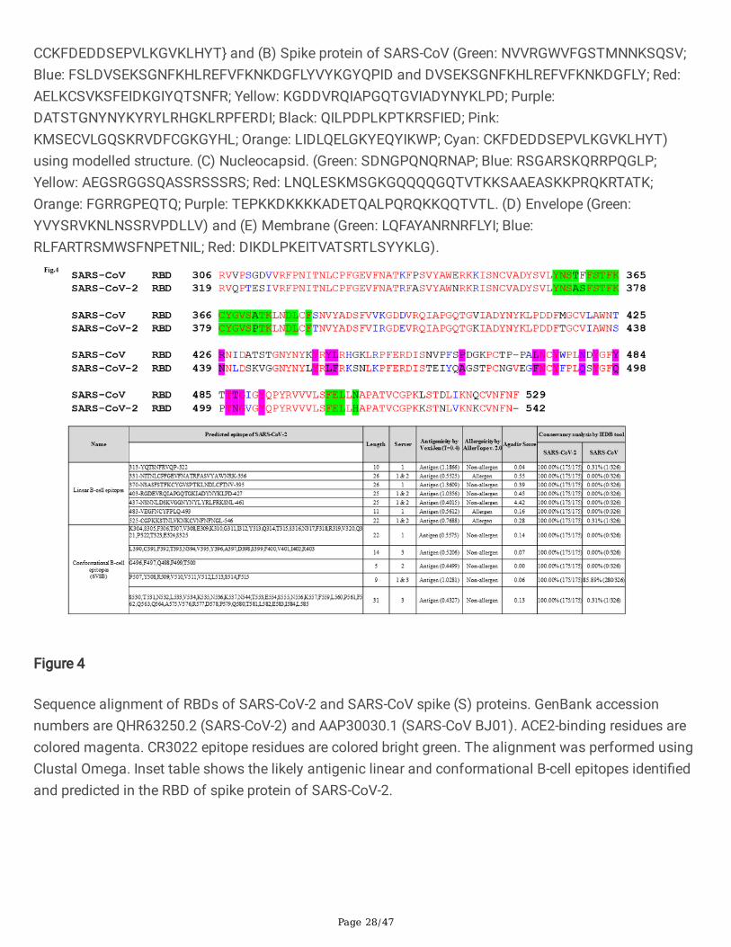
We have carried out sequence alignment of RBDs of S protein of SARS-CoV and SARS-CoV-2 usingclustal omega (Fig.4). Inset table of Fig.4. shows the linear and conformation B cell epitope predicted andidenti�ed in the RBD of the S protein of SARS-CoV-2 and notably, we have found a linear B-cell epitope ofS protein (370-NSASFSTFKCYGVSPTKLNDLCFTNV-395, inset table of Fig. 4) that was recently found tointeract with CR3022. CR3022 is a previously described neutralizing antibody isolated from aconvalescent SARS patient have now found to neutralize SARS-CoV-228,29. Co-crystallization of CR3022and RBD of the S protein of SARS-CoV-2 have identi�ed a conserved epitope residues enabling cross-reactive neutralization between SARS-CoV-2 and SARS-CoV without hampering ACE2 binding to RBD of Sprotein28. Many of the predicted linear and conformational B cell epitopes (Inset table of Fig.4) predictedin this work are found to be located in the RBD of S protein of SARS-CoV-2 (S1B residues 338-506) and areceptor-binding subdomain (residues 438-498). 47D11 is the �rst report of a human monoclonalantibody that neutralizes SARS-CoV-2 most likely by binding to the conserved core structure of the S1B
(residues 338-506) using a mechanism independent of receptor-binding inhibition30. Similarly, severalvirus-speci�c memory B cells recognizing the RBD of the S protein of SARS-CoV-2 have been identi�edand only two (of the total 206 SARS-CoV-2 RBD-speci�c monoclonal antibodies) antibodies were found toblock the viral entry which correlated with high competing capacity against ACE2 receptor31. The �ndingsare suggestive of virus species-speci�c antibody response to RBDs of SARS and cross-recognition targetregions outside the RBDs. Amanat et al (2020) have developed a serological assay using the S proteinexpressed in insect (iSpike/iRBD) and mammalian (mSpike/mRBD) expression system. They observedthat all COVID-19 plasma/serum samples reacted strongly to both RBD and full-length spike protein.Reactivity of COVID-19 sera was, in general, stronger against the full-length S protein than against theRBD. They have tested an additional 12 serum samples from patients with acute COVID19 disease, aswell as convalescent participants, for reactivity to mRBD and mSpike. All 12 samples reacted with bothRBD and spike protein32.
Prediction of conformational B-cell epitopes of S, E, M and N proteins of SARS-CoV-2 and SARS-CoV
The Cryo-EM structure of S protein of SARS-CoV-2 (PDB:6VSB3) is available from residues 1 to 1208 thathad missing 65 residues of C-terminal region of the protein. Similarly, the Cryo-EM solved structure(residues 1 to 1195) of S protein of SARS-CoV had missing residues of 60 amino acids towards the C-terminal end (PDB:5WRG33) due to limited electron density. We have overcome this problem using I-TASSER modelled structures of S protein of SARS-CoV and SARS-CoV-234. We compared modelledstructures with cryo-EM solved structure3,34 and observed high similarity with root mean square deviation(RMSD) of around 1.3 Å (SARS-CoV) and 2.0 Å (SARS-CoV-2) (data not shown). Similarly, the structuresof E, M and N proteins were modelled and used as input structures to identify potent conformational B-
Page 7/47
cell epitopes using CBTOPE, DiscoTope, ElliPro and EPSVR. We identi�ed a cluster of conformational B-cell epitopes predominately enriched in S and N proteins of SARS-CoV and SARS-CoV-2 (Table 2a & 2c).We have generated a heat map showing the distribution of antigenic epitopes in the S1 domain of Sprotein of SARS-CoV-2 (Fig.2b). Notably, we found conformational B-cell epitope of S, E, M and N proteinscommon to both SARS-CoV-2 and SARS-CoV (Table 2b). The �ndings of this work revealed that predictedconformational epitopes are likely to be localized on the accessible region of 3D-structure of proteins asvisualized by BIOVIA Discovery Studio 2017 R2 (Fig. 5a-e).
Prediction of MHC I binding epitopes of S, E, M and N proteins of SARS-CoV-2 and SARS-CoV
SARS-CoV is closely related human beta-CoV to SARS-CoV-2 and a previous data from SARS-CoVpatients in 2003-2004 has identi�ed CD8+ T and CD4+ T cell responses using the whole proteome35, alikely possibility has been that substantial CD4+ T cell, CD8+ T cell, and neutralizing antibody responsesdevelop to SARS-CoV-2 and all contribute to clearance of the acute infection and protective immunityagainst SARS-CoV-2 infection36,37. The availability of information of SARS-CoV-2 proteins and epitopesrecognized by human T-cell responses can greatly assist researchers in selecting potential epitopes ortarget proteins for the design of the candidate vaccine. All the four structural proteins (S, E, M and N) ofSARS-CoV-2 and SARS-CoV were screened and epitopes were predicted that are likely to be presented on27 HLA class I molecules (HLA-A and HLA-B) molecules using three different servers, proteasomalcleavage/TAP transport/MHC class I combined predictor17, MHC-NP18 and TepiTool16.
We selected the high affinity-ranked peptides and found nearly 683 MHC I epitope for S protein of SARS-CoV-2, as predicted using proteasomal cleavage/TAP transport/MHC class I combined predictor, 683 byMHC-NP and 341 by Tepitool (Table 3a). Similarly, we observed likely number of MHC I epitopes of Sprotein of SARS-CoV predicted using the proteasomal cleavage/TAP transport/MHC class I combinedpredictor (674), MHC-NP (673) and TepiTool (331). This observations give us some con�dence in theability of three independent servers with a varying operating algorithm to predict a consistent number ofunique MHC I epitopes across all four proteins (S, E, M & N) and corroborate with the overall highsequence similarity of SARS-CoV and SARS-CoV-2 (Fig. 6a). The predicted epitopes were further screenedfor their ability to elicit an IFN-γ response using the IFNepitope tool22 and the epitopes that are predictedas likely conserved across the species of CoVs, antigenic, non-allergenic have been shortlisted. It is alsoworth mentioning that the same epitope is being predicted by more than two servers employed in thisstudy. Notably, we observed and identi�ed 6 CD8+ T cell epitopes in S, 6 epitopes in N, 5 epitopes each inM and E proteins that are found common to both SARS-CoV-2 and SARS-CoV (Table 3b). Table 3c showsthe list of top MHC I binding epitopes that are antigenic, non-allergenic, a positive score for IFN-γ and themajority of the listed epitopes are predicted by more than two servers used in this study. A total of 41MHC I epitope was found for S protein, 19 each for N & M and 10 for E protein of SARS-CoV-2 (Table 3c).
We found that one epitope of N protein (316-GMSRIGMEV-324) common to both SARS-CoV-2 and SARS-CoV (N317-325, Table 3b) predicted as high affinity epitope (HLA-A*02:03, HLA-A*02:01, HLA-A*02:06)was previously known to induce interferon-gamma (IFN-γ) in PBMCs of SARS-recovered patients38.
Page 8/47
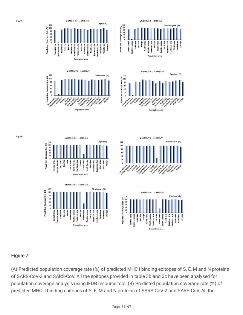
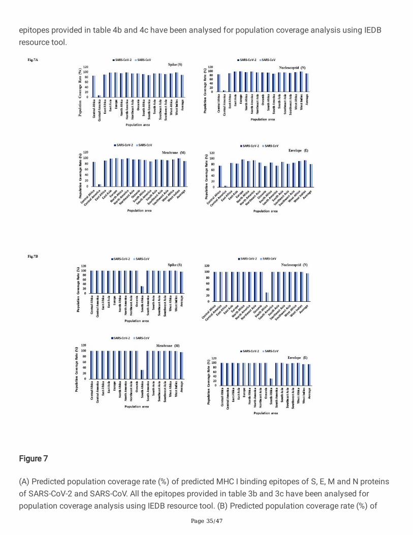
Interestingly, two epitopes, 1041-GVVFLHVTY-1049 (HLA-A*11:01, HLA-A*68:01, HLA-A*03:01) and 1202-FIAGLIAIV-1210 (HLA-A*02:06, HLA-A*68:02, Table 3c) derived from SARS-CoV S protein having epitopeconservancy between 85 to 100% with S protein of SARS-CoV-2 was previously reported as immunogenicin PBMCs derived from SARS-CoV patients38. CD4+ and CD8+T cells can recognize antigen only when itis presented by a self-MHC molecule and MHC molecules are extremely polymorphic. Selecting multiplepeptides with different HLA binding speci�cities will afford increased coverage of the patient populationtargeted by peptide-based vaccines and diagnostic methods. Using the IEDB tool we found theoccurrence of HLA alleles with population coverage of around 99% throughout the world except forcentral America where the population coverage of HLA allele was less than 10% (Fig.7a). We found thatHLA-A*02:01, HLA-A*02:03, HLA-A*02:06, HLA-A*11:01, HLA-A*30:01, HLA-A*68:01, HLA-A*68:02, HLA-B*15:01 and HLA-B*35:01 have been predicted to bind to the maximum number of epitope (based on thecriterion of allele predicted to bind more than 30 epitopes) of S protein of SARS-CoV-2 (Table S1a).Similarly, we observed that HLA-A*02:06, HLA-A*30:01, HLA-A*30:02, HLA-A*31:01, HLA-A*32:01, HLA-A*68:01, HLA-A*68:02, HLA-B*15:01 and HLA-B*35:01 are predicted to bind to the maximum number ofepitope (based on the criterion of allele predicted to bind more than 10 epitopes) of N protein of SARS-CoV-2 (Table S1a).
Prediction of MHC II binding epitopes of S, E, M and N proteins of SARS-CoV-2 and SARS-CoV
Helper T cells are required for adaptive immune responses and they help activate B cells to secreteantibodies and help activate cytotoxic T cells to kill infected target cells. NetMHCIIpan 4.019 andTepiTool16 have been used to predict and identify high-a�nity MHC II binding epitopes based on 23 HLAclass II molecules (HLA-DP, HLA-DQ and HLA-DR) molecules. We found nearly 884 and 519 MHC IIepitope for S protein of SARS-CoV-2 predicted using NetMHCIIpan 4.0 and TepiTool (Table 4a). A similarnumber of epitopes were predicted for S protein of SARS-CoV, the �ndings of computational predictionsare suggestive of reliability and good correlation. Interestingly, we found a total of eight (8) CD4+ T cellepitope for S, six (6) for N, two (2) for M and four (4) for E proteins that are common to both SARS-CoV-2and SARS-CoV with epitope conservancy of approximately between 80% to 100% (Table 4b). We haveselected the top list of MHC II epitopes based on antigenicity, non-allergenicity and conservancy and allthese epitopes were predicted to induce IFN-gamma as shown in Table 4c. The allele-wise distribution ofpredicted epitopes for S, E, M and N proteins of SARS-CoV-2 have been presented in a heat map (Fig.6b).Using the IEDB tool we found the occurrence of HLA alleles with population coverage of around 99%throughout the world except for South Africa where the population coverage of HLA class II allele wasaround 30% (Fig.7b).
We found that HLA-DRB1*04:01, HLA-DRB1*04:05, HLA-DRB1*13:02, HLA-DRB1*15:01, HLA-DRB3*01:01,HLA-DRB3*02:02, HLA-DRB4*01:01, HLA-DRB5*01:01, HLA-DQA1*04:01, DQB1*04:02, HLA-DPA1*02:01,DPB1*01:01, HLA-DPA1*01:03, DPB1*02:01, HLA-DPA1*01:03, DPB1*04:01, HLA-DPA1*03:01,DPB1*04:02, HLA-DPA1*02:01, DPB1*05:01, HLA-DPA1*02:01, and DPB1*14:01 are predicted to bind tothe maximum number of epitope (based on the criterion of allele predicted to bind more than 30 epitopes)of S protein of SARS-CoV-2 (Table S1b). Similarly, we observed that HLA-DRB1*04:01, HLA-DRB1*07:01,
Page 9/47
HLA-DRB1*08:02, HLA-DRB1*09:01, HLA-DRB1*11:01, HLA-DRB1*13:02, HLA-DRB3*02:02, HLA-DRB5*01:01, HLA-DQA1*01:02, DQB1*06:02, DPB1*05:01 and HLA-DPA1*02:01 are found to interactwith the maximum number of epitope (based on the criterion of allele predicted to bind more than 10epitopes) of N protein of SARS-CoV-2 (Table S1b). The �ndings of computational predictions aresuggestive that T-cell based immunity might be generated largely against S and N protein of SARS-CoV-2and could potentially be selected as target candidate for recombinant protein-based vaccine. On carefulobservations of computationally predicted epitopes (Table 5), we found epitope in S1B of spike protein(370-NSASFSTFKCYGVSPTKLNDLCFTNV-395) likely to function as both linear B-cell and MHC class IIepitope. 403-RGDEVRQIAPGQTGKIADYNYKLPD-427 and 437-NSNNLDSKVGGNYNYLYRLFRKSNL-461peptides of S protein were predicted as linear B cell and MHC class I epitope. 177-MDLEGKQGNFKNLREFVFKN-196 and 1253-CCKFDEDDSEPVLKGVKLHYT-1273 peptides of S proteinwere predicted as linear and conformational B cell epitope. Similar overlapping B- and T-cell epitopeswere predicted for E, M and N proteins of SARS-CoV-2 (Table 5).
Utilizing bioinformatic approaches a few researchers have identi�ed speci�c peptides in SARS-CoV-2 withincreased probability of being T-cell targets37,39 and have identi�ed circulating SARS-CoV-2-speci�c CD8+and CD4+ T-cells in approximately 70% and 100% of COVID-19 convalescent patients, respectively40.CD4+ T cell responses were observed predominately in the S protein and the robustness of T cellresponses was correlated with the magnitude of IgG and IgA titers of SARS-CoV-2. Among the structuralproteins, the S and M proteins were mainly recognized as possible targets for CD8+ T cells of SARS-CoV-240.
Identi�cation of potential B and T cell epitope of structural proteins for serological assay and multi-epitope-based vaccine
The spike protein of SARS-CoV-2 and SARS-CoV share an amino-acid sequence identity of around 77%and are phylogenetically closely related1. Previously characterized immune response of patients withSARS have found maximum neutralizing antibodies against the S and N proteins4,5,6 and the recent�ndings showed S protein as an immunodominant and highly speci�c target of antibodies in SARS-CoV-2patients32,41,42. Therefore, S and N proteins are the major targets for development of vaccine andserological assay because SARS-CoV-2 neutralization assays are time-consuming and require BSL-3containment facilities. In this study, we have designed a multi-epitope chimeric constructs by utilizing thecomputationally predicted putative and potent B- and T-cell epitopes of S, E, M and N proteins of SARS-CoV-2 (Table 6). In the multi-epitope constructs we have included MHC class I (CTL) and class II bindingepitopes (HTL) having the positive score for IFN-γ induction. CTL and HTL epitopes were linked togetherby AAY and KK cleavable linkers whereas the B- cell epitopes (linear and conformational) were linkedtogether with GGGGS �exible linker (Fig. 8a-f). Poly-Gly-rich linkers can be considered as independentunits and do not affect the function of the individual proteins. A total of six (6) multi-epitope chimericconstructs namely, (i) RBD of S protein (B- and T-cell epitopes), (ii) Full-length S protein (B- and T-cellepitopes), (iii) Structural protein construct (B- and T-cell epitopes of S, E, M and N proteins), (iv) Chimeric
Page 10/47
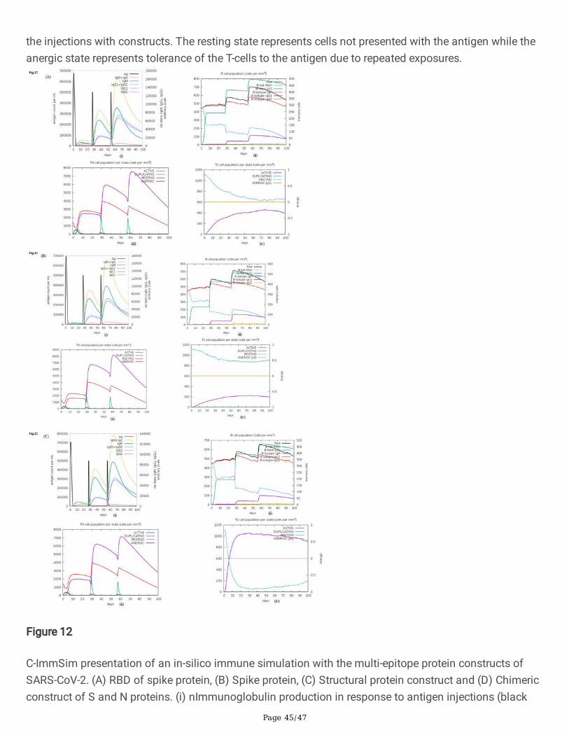
construct of S and N proteins (B- and T-cell epitopes of S and N), (v) S protein (B-cell epitopes of Sprotein) and (vi) Nucleocapsid protein (B-cell epitopes of N protein) were designed containing N-CTL, HTLand B-cell epitopes (Fig.8a-f). The structure of chimeric multi-epitope constructs has been modelled usingI-TASSER (Fig.9a-f) and validated by RAMPAGE server to generate a Ramachandran plot43. Most of theamino acid residues of epitope constructs were found in the favourable region (Fig.S1, Inset table) excepttwo constructs (RBD of S and nucleocapsid protein). Various physicochemical parameters that includednumber of residues, theoretical pI, molecular weight, aliphatic index and grand average of hydrophobicity(GRAVY) were analysed by ProtParam44. Based on the aliphatic index scores, the multi-epitope constructscould be considered moderately thermostable (Table S2). Gravy scores obtained were of a negative valuefor all the constructs indicating the likelihood of the chimeric multi-epitope antigen being globular andhydrophilic in nature. The secondary structure of multi-epitope constructs was predicted by online serverPSIPRED45 and helical and strand content of the constructs are provided in Table S2 and Fig.S2.Cathepsin and carboxypeptidase involved in proteolytic processing of endocytosed proteins in the MHCclass II pathway display preferential cleavage of dibasic (RR, KK, KR or RK) sites46. Proteases, whichprovide peptide ligands for the MHC class II antigenic presentation pathway display preferential cleavageof hydrophobic motifs (AAY). The cleavable linkers are required to be accessible for the proteasesassociated with MHC I and II antigen processing pathway. It is observed that the cleavable linker residues(AAY and KK) used in multi-epitope subunit vaccines were surface accessible based on computationalprediction algorithms indicating high probability of T-cell epitopes presentation by MHC molecules asvisualized by discovery studio (Fig.10a-f). The results of C-ImmSim server (http://150.146.2.1/C-IMMSIM/index.php) prediction revealed the ability of multi-epitope constructs to simulate IFN-γproduction following an immunization with the peptide (Fig. 11a-f). In-silico immune simulation of themulti-epitope constructs {(A) RBD of spike protein, (B) Spike protein, (C) Structural protein construct and(D) Chimeric construct of S and N proteins)} showed consistent results with the actual immune responsesas observed by the primary response of high levels of IgM. This is followed by marked increase in B-cellpopulations and levels of IgG1 + IgG2, IgM, and IgG + IgM antibodies as a part of the secondary andtertiary responses (Fig. 12, A-D (i-iv). A similarly high response was observed in the TH (helper) and TC
(cytotoxic) cell populations with corresponding memory development especially for constructs made ofstructural proteins (S, E, M and N) and chimeric construct of S and N proteins (Fig.12C & D).
DiscussionIn this study, using validated bioinformatics resources, we comprehensively screened and identi�edpotential B- and T-cell epitope predominately clustered in S and N proteins of SARS-CoV and SARS-CoV-2.This is signi�cant for the fact that knowledge of the innate and adaptive immunity to SARS-CoV-2 canprovide crucial information on the vaccine and serological assays for coronavirus disease 2019 (COVID-19) and its pathogenesis. Limited information is available about which SARS-CoV-2 proteins arerecognized by human T cell immune responses. Both B- and T-cells are the weapons of adaptive immuneresponse and computationally predicted epitopes are likely to provide information about the possibletargets of the immune response to SARS-CoV-2.
Page 11/47
Our bioinformatics analysis of structural proteins has revealed that most of the predicted B cell epitopeswere found in S and N proteins of SARS-CoV and SARS-CoV-2 (Table 1a & Table 2a). Importantly, �vehighly conserved amino acid (aa) regions were identi�ed, of which three are located in the RBD of Sprotein {aa 370-395 (linear B cell epitope and MHC II), aa 403-427 and aa 437-461 (linear B cell epitopeand MHC I)}, one in the N (aa 177-196) and C-terminal region of S protein (aa 1253-1273) were predictedand likely to function as both linear and conformational B-cell epitopes (Table 5). Similarly, �ve conservedamino acid regions were identi�ed for N protein, of which aa 32-46 and aa 366-394 were predicted aslinear and conformational B-cell epitopes and MHC I. The peptide aa 274-283 was predicted as linear andconformational B-cell epitopes and the peptide aa 173-190 and aa 227-266 were predicted as linear B-cellepitopes and MHC I. Three conserved amino acid regions (aa 35-48, aa 101-119 and aa 160-182) wereidenti�ed for M protein are likely to function as both linear and conformational B-cell epitope and MHC I.One conserved amino acid region (aa 57-75) was identi�ed for E protein which is likely to function asboth linear and conformational B-cell epitope. One epitope (aa1253-1273) for S, two (aa 173-190, aa 274-283) for N, two for M (aa 101-119, aa 160-182) proteins with high conservancy (~80% to 100%) andfound common to both SARS-CoV and SARS-CoV-2 (Table 5). These �ndings are in agreement with thatof Ju et al. (2020) who have isolated and characterized around 206 monoclonal antibodies speci�c toRBD of S protein derived from single B cells of eight SARS-CoV-2 infected individuals. Crystal structureanalysis of the RBD-bound antibody revealed that the neutralization activity correlates with theircompetitive capacity with ACE2 for RBD binding. The �ndings are suggestive that anti-RBD antibodies areviral species-speci�c inhibitors without cross-reactivity to RBDs of SARS-CoV and MERS-CoV31. In linewith our bioinformatic observations, Wang et al. (2020) have identi�ed a human monoclonal antibody(47D11) that neutralizes SARS-CoV-2 and SARS-CoV through binding to RBDs without hampering ACE2receptor interaction in cell culture model system30. Similarly, Amanat et al. (2020) have developed aserological assay using S protein expressed in insect and mammalian expression system and �ndingsindicated that the plasma/serum samples derived from COVID-19 patients reacted strongly to both RBDand full-length S protein32. We found a linear B-cell epitope in the RBD of S protein (370-NSASFSTFKCYGVSPTKLNDLCFTNV-395, inset table of Fig. 4) which was recently shown to interact withthe CR3022, a previously described antibody from the SARS patient and known to be neutralized bybinding to RBD of S protein of SARS-CoV and SARS-CoV-2 without hindering S protein interaction withthe cellular ACE2 receptor28,29. In a recent preprint study, Nanda et al. (2020) have developed a modi�edvaccinia Ankara (MVA) based-vaccines expressing either the full-length or secreted S1 domain of Sglycoprotein of SARS-CoV-2. The �ndings revealed that, although both immunogens induce strong IgGantibodies to puri�ed S protein, only the full-length S protein induced a strong neutralizing antibodyresponse against the SARS-CoV-247. In a similar MVA-based vaccine, the full-length S protein was foundto induce a broader spectrum of neutralizing antibodies in comparison with fragmented S protein ofSARS-CoV48.
Of the epitopes identi�ed in the RBD of S protein (Fig.4 inset table and Table 1c), the B cell peptidespanning the region (aa 403-RGDEVRQIAPGQTGKIADYNYKLPD-427, aa 437-NSNNLDSKVGGNYNYLYRLFRKSNL-461 and aa 483-VEGFNCYFPLQ-493) have recently been identi�ed as
Page 12/47
residues responsible for interaction between CB6 mAb (a human monoclonal neutralizing antibodyisolated from a convalescent COVID-19 patient) and the RBD of S protein of SARS-CoV-2. A co-crystallized structure has revealed that CB6 recognizes an epitope that overlaps with ACE2-binding sitesin the RBD of SARS-CoV-2 leading to disruption of the S-ACE2 receptor complex formation49. Intriguingly,most of the amino acid residues spanning the B-cell epitope (aa 331-NITNLCPFGEVFNATRFASVYAWNRK-356 and aa 437- NSNNLDSKVGGNYNYLYRLFRKSNL-461, Fig.4, inset table) identi�ed in the S protein have recently been shown to interact with the cross-neutralizing mAb, S309 (previously isolated fromSARS-CoV patient) in a ACE2 receptor-independent manner50. In a study, using the recombinant SARS-CoV-2 RBD antigen, a strong correlation between levels of RBD binding antibodies and SARS-CoV-2neutralizing antibodies have been observed in patients on day 9 after the onset of symptoms51. Similarinvestigations carried out using samples collected from SARS-CoV-2 patients of USA, Europe and HongKong have reported that the full-length S protein and the RBD performed well for speci�c and sensitiveantibody detection32,42,41. The �ndings of these works support the use of RBD or the full-length S proteinin serological diagnostic assays.
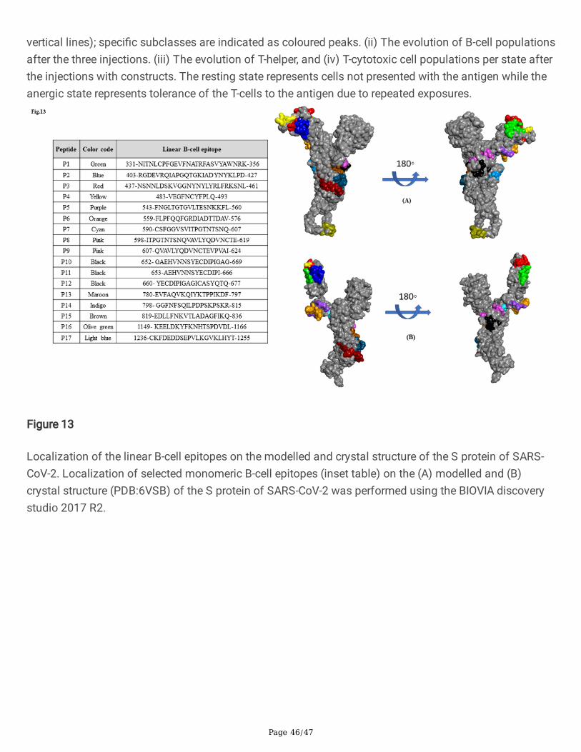
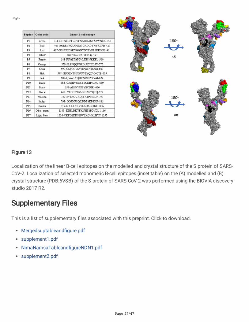
In a preprint by Farrera-Soler et al. (2020), the three linear epitopes (655-672, 787-822 and 1147-1158) ofS protein were most abundantly detected in all the SARS-CoV-2 positive samples tested, but each epitopeis detected in >40% of the positive patients. The 655-672 epitope was the most detected in the positivesamples and corresponds to a peptide that is not part of secondary structures. In our bioinformaticsprediction we have found the same linear B-cell epitope being predicted by both the server used- BepiPredand Bcepred and a poor Agadir score (0.03) collaborating a lack of helical content as reportedpreviously52. Similarly, these linear B-cell epitopes of S protein (aa 652- GAEHVNNSYECDIPIGAG-669, aa660- YECDIPIGAGICASYQTQ-677, aa 780-EVFAQVKQIYKTPPIKDF-797, aa 798- GGFNFSQILPDPSKPSKR-815, aa 819-EDLLFNKVTLADAGFIKQ-836, aa 1149- KEELDKYFKNHTSPDVDL-1166, Table 1d) was foundpredicted as potent B-cell epitopes using ABCpred server thereby collaborating with the recent �ndings ofdetection of the above epitope in the sera of SARS-CoV-2 positive patients52. Poh and colleagues (2020)have identi�ed and characterized neutralizing antibodies spanning the two immunodominant regions onS protein (aa 553-TESNKKFLPFQQFGRDIA-570, aa 809-PSKPSKRSFIEDLLFNKV-826) of SARS-CoV-2 thatare recognised by convalescent sera of COVID-19 patients53. Using the ABCpred server, we have identi�edthese B cell epitopes (aa 543-FNGLTGTGVLTESNKKFL-560, aa 559-FLPFQQFGRDIADTTDAV-576, aa 798-GGFNFSQILPDPSKPSKR-815, aa 819-EDLLFNKVTLADAGFIKQ-836, Table 1d) in the S protein of SARS-CoV-2. We further showed that these linear B-cell epitopes (inset table of Fig.13) are localized on theaccessible region using modelled and cryo-EM solved (PDB: 6VSB) structure of the S protein of SARS-CoV-2 (Fig.13A and B).
Among those B cell peptides identi�ed in S protein of SARS-CoV-2 (aa 590-CSFGGVSVITPGTNTSNQ-607,aa 607-QVAVLYQDVNCTEVPVAI-624, Table 1d) and SARS-CoV (aa 602-NCTDVSTAIHADQLTPAWRIYSTGNNVFQT-631, aa 1160- NIQKEIDRLNEVAKNLNESL-1179, aa 1179-LIDLQELGKYEQYIKWP-1195, data not show), of particular interest is the region of two tandem epitopes(S597-603 and S604-625) that showed a high level of serological reactivity and found to have the
Page 13/47
capacity to induce a long-term B-cell memory response with antisera from convalescent SARSpatients54,55,56. The linear B cell peptide corresponding to the region (aa 1236-CKFDEDDSEPVLKGVKLHYT-1255) of S protein is found conserved (~84%, Table 1b and 1c) and commonto both SARS-CoV and SARS-CoV-2. This peptide was earlier shown as one of the immunodominantantigenic peptides in the S protein of SARS-CoV, supporting further exploration of this highly conserved C-terminal region among the S protein of SARS-CoV-2 will be important55.
The nucleocapsid (N) protein is the most abundant and highly conserved structural protein across CoVsand the N protein of SARS-CoV-2 shares ~90% amino acid identity with N protein of SARS-CoVsuggesting that previously described antibodies against the N protein of SARS-CoV would likely recognizeand bind the N protein of SARS-CoV-2 as well. The earlier work by Leung et al. (2004) showed thatneutralizing antibodies were detected early and with high speci�city and predominantly against N proteinin patients with SARS-CoV5. The B-cell epitope predicted and identi�ed (aa 359-DAYKTFPPTEPKKDKKKKTDEAQPLPQRQKKQPTVTLLPAADMDD-403, Table 1c) in the N protein of SARS-CoV was previously shown to have the highest a�nity to form peptide-antibody complexes with SARSserum57,58. Notably, the same epitope was found localized on the surface of N protein of SARS-CoV-2(366-TEPKKDKKKKADETQALPQRQKKQQTVTL-394, Table 1c and Fig. 3c), thus providing scope for futureinvestigations and might contributes to the understanding of the immunogenicity and persistence ofSARS-CoV-2 coronavirus. Therefore, N371 (371-KDKKKKTDEAQPLPQRQKKQ-390) and N385 (N385-QRQKKQPTVTLLPAADMDDFSRQ-407) located at the C-terminal region of the N protein of SARS-CoVappear to be highly immunogenic and may be useful for serologic assays for SARS-CoV-2 given theconservancy of the previously characterized epitope of N protein57,59,58. Similarly, synthetic peptideM132-161 derived from the C-terminal epitope of M protein (aa 131-PLMESELVIGAVIIRGHLRM-150, Table1c) was found highly reactive using the convalescent-sera of SARS patients, suggesting the potentialapplication for serologic diagnosis of SARS60.
In this work we have identi�ed and listed the top CD8+ and CD4+ T cell epitopes based on antigenicity,conservancy, non-allergenicity and positive score for IFN-gamma, of which 41 MHC class I epitopes werepredicted for the S protein, 19 each for the N and M proteins, and 10 for the E protein of SARS-CoV-2(Table 3c), while we found 26 MHC class II epitopes predicted for the S protein, 8 epitopes for the Nprotein, 6 and 2 epitopes for the M and E proteins, respectively (Table 4c). A recent �nding showed thatthe S, M and N proteins of SARS-CoV-2 each accounted for 11-27% of the total CD4+ response, while forCD8+ T cells, S and M were recognized including some non-structural proteins40. In a recent preprintstudy, Takuya et al. (2020) have shown that SARS-CoV-2 elicits robust T-cell mediated immune responsesin both symptomatic and asymptomatic individuals, even in the absence of a positive antibody test61. Itis generally expected that B- and T-cell epitopes can be predicted from both structural and non-structuralproteins and in this study we have considered the four structural proteins (S, E, M and N) of SARS-CoV-2based on the existing knowledge of innate and adaptive immune responses to SARS-CoV. Analysis of T-cell epitopes has revealed that peptides corresponding to residues 1-30, 86-100, 306-320, and 351-365(Table S3 and S4) of N protein of SARS-CoV (isolate BJ01) have been reported earlier as
Page 14/47
immunodominant T-cell epitopes. It was previously observed that peptides corresponding to residues aa336-350 were capable of stimulating IFN-γ production in T-cell cultures derived from peripheral bloodmononuclear cells and the peptide spanning the region (aa 81-PDDQIGYYRRATRRV-95) have con�rmedthat YYRRATRRV is a very potent functional CD8+ T-cell epitope of N protein61. Intriguingly, thisfunctional CD8+ T-cell epitope (aa 86-GYYRRATRR-94) is found to bind with two alleles (HLA-A*31:01,HLA-A*33:0) is conserved to both N protein of SARS-CoV-2 and SARS-CoV (~85%, Table S3). Additionally,the epitope corresponding to aa 350-NVILLNKHIDAYKTF-364, aa 305- IAQFAPSASAFFGMS-319, aa 352-ILLNKHIDA-360 were found common to N protein of both SARS-CoV-2 and SARS-CoV (~80%conservancy, Table S3 and Table S4). Some of the present �ndings of B- and T-cell epitope predictedusing computational algorithms corroborate with the experimentally veri�ed immunogenic epitopes ofSARS-CoV and SARS-CoV-2. The potential for cross-protection also exists as the selected proteins andpredicted epitopes used in generating the chimeric multi-epitopes exhibited considerable conservationacross structural proteins of SARS-CoV and SARS-CoV-2.
Designing synthetic peptides for use as vaccines to induce both humoral and cell-mediated immunityrequires an understanding of the nature of T- and B-cell epitopes. Ideally, vaccines for inducing humoralimmunity should include peptides that form immunodominant B-cell epitopes. A successful vaccine mustalso generate a population of memory T cells; therefore the peptide should include immunodominant T-cell epitopes. Bioinformatic methods used for prediction of B-cell and MHC binding peptides are of veryhigh quality and predict epitopes with high accuracy. However, it remains to be investigated whether amega-pool of B- and T-cell epitopes identi�ed in this work can stimulate T-cells by measuring the amountof released cytokines both in vitro and in vivo experimental system, thus providing a platform for futureinvestigations.
Materials And MethodsStructural protein sequences of SARS-CoV-2 and SARS-CoV
We have used NCBI GenBank SARS-CoV-2 (isolate WIV02, accession number MN996527.11) and SARS-CoV (isolate BJ01, accession number AY278488.27) to download coding sequences for structuralproteins such as spike (S), nucleocapsid (N), membrane (M) and envelope (E) for prediction of B- and T-cell epitopes using bioinformatics methods. Sequence alignment of the RBDs of the S protein of SARS-CoV-2 (QHR63250.2) and SARS-CoV (QHR63300.2) was performed using Clustal omega.
B-cell epitope prediction for SARS-CoV-2 and SARS-CoV
BepiPred-2.08 and Bcepred9 servers were employed to predict linear B cell epitopes. BepiPred-2.0 is a web-based server that utilizes a random forest algorithm trained on epitopes annotated from antibody-antigenprotein structures. Bcepred predicts B-cell epitopes based on the physico-chemical properties such ashydrophilicity, �exibility, accessibility, polarity, exposed surface, turns and antigenic propensity. EPSVR,Discotope, CBTOPE and Ellipro servers were used to predict conformational B-cell epitopes. EPSVR uses a
Page 15/47
Support Vector Regression (SVR) method to predict antigenic B cell epitopes10. DiscoTope server predictsdiscontinuous B cell epitopes from protein-3D structures. The method utilizes calculation of surfaceaccessibility and a novel epitope propensity amino acid score11. CBTOPE server predicts conformationalB-cell epitope with an accuracy of more than 85% using antigen primary sequence in the absence of anyhomology with the known structures12. ElliPro13 predicts linear and discontinuous antibody epitopesbased on the protein structure and homology-based model of the amino acid sequence. The helicalbehaviour of predicted monomeric peptides was computed using Agadir server14,15.
T-cell epitope prediction for SARS-CoV-2 and SARS-CoV
TepiTool is an interactive and easy to use tool to predict potential peptides binding to MHC class I andclass II molecules. In Tepitool prediction we have used a panel of 27 most frequent alleles that aredistributed among populations worldwide16. Proteasomal cleavage/TAP transport/MHC class I combinedpredictor17 was used to identify MHC class I epitope based on proteasomal processing and TAP transportin the cell whereas MHC-NP predicts T cell peptides naturally processed by the MHC18. We selected thetop 2 % ranked epitopes as strong binders with 9-mer peptide length. NetMHCIIpan 4.0 server predictspeptide binding to any MHC II molecule of the known sequence using arti�cial neural networks19. Weapplied the threshold of top 2% ranked epitopes based on the prediction score as strong binders to HLAclass II molecules with 15-mer peptide length. All 9- and 10-mer peptides were predicted for their bindinga�nity to 27 MHC class I and 23 MHC class II molecules which account for 97% of HLA A and B allelicvariants in most ethnicities20. We used the Immune Epitope Database and Analysis Resource (IEDB)analysis resource tool to analyse population coverage of predicted MHC class I and class II epitopesusing allele frequency database of 115 countries and 21 different ethnicities grouped into 16 differentgeographical areas21. The ability of the predicted T-cell epitopes to induce interferon-gamma (IFN-γ)response was assessed using the IFNepitope server22. IFNepitope uses the algorithm based on threemodels such as motif-based, SVM based, and hybrid approach for prediction of IFN-γ inducing epitopes.
Antigenicity, allergenicity, and conservancy of predicted B- and T-cell epitope
The antigenicity and allergenicity of epitopes were predicted using VaxiJen v2.0 and AllerTop onlineservers. VaxiJen predict protective antigens and subunit vaccines based on an alignment-independentmethod23. AllerTop is a server for in silico prediction of allergens in a given protein or antigens24. We usedthe IEDB analysis resource epitope conservancy analysis tool to computes the degree of conservancy ofan epitope within a given protein sequence25.
Immune simulation
To determine the immunogenicity and immune response pro�le of the multi-epitope protein constructscontaining B- and T-cell epitopes, in-silico immune simulation was carried out using the C-ImmSim server.C-ImmSim server uses a position-speci�c scoring matrix and machine learning techniques for predictionof epitope and immune interactions. The server simulates three components of immune system found in
Page 16/47
mammals26: (i) the bone marrow, where hematopoietic stem cells are simulated and produce newlymphoid and myeloid cells; (ii) the thymus, where naive T-cells are selected to avoid auto immunity; and(iii) a tertiary lymphatic organ, such as a lymph node. All simulation parameters were set at default withtime steps set at 1, 84, and 168 (each time step is 8 hours and time step 1 is injection at time = 0).Therefore, three injections were given at four weeks apart without lipopolysaccharide (LPS). The Simpsonindex, D (a measure of diversity) was interpreted from the plot.
ConclusionOne of the limitations of the present work is the lack of information on B- and T-cell epitopes of non-structural proteins of SARS-CoV-2. In summary, the bioinformatics analysis of the structural proteins hasled to prediction and identi�cation of a pool of B- and T-cell epitopes, which are likely to facilitateresearchers to select appropriate epitope or region of proteins (especially the S and N proteins) in aneffort to develop a novel drugs, vaccines, and serological assays for the detection, treatment, andmanagement of SARS-CoV-2.
DeclarationsAcknowledgments
I as corresponding author had full access to all the data in this study and take responsibility for theintegrity of the data and the accuracy of the data analysis and had �nal responsibility for the decision tosubmit for publication. I have not been paid to write this article by any pharmaceutical company or otheragency.
Author contributions
YDD, HBG, SK, CD and AD contributed equally as �rst authors. YDD and HBG prepared �gures and tables.NDN conceived and designed the study and performed literature search. NDN supervised data collectionand analysis. YDD, HBG, SK, CD and AD collected data. YDD, HBG and NDN analysed and interpreted thedata. HBG have helped in reference formatting. AD and CC have helped to generate heat and surfacelocalization of predicted epitopes on modelled structures. VB and SD have helped to prepare �gures andtable and manuscript editing. LJ has edited the manuscript. AK, SSS, SKR, RCD, RD and MM have helpedin analysis of data. Ch. SS and PPB are a part of clinical collaborators. YDD and NDN wrote themanuscript. All authors contributed to reviewing and editing the manuscript.
Declaration of competing interests
We declare no competing interests. This part of work at present has no funding support. However, NDNhas submitted project on ‘development of multi-plex RT-PCR and serological assay for SARS-CoV-2’ toDST, SERB, Govt. of India for �nancial support.
Page 17/47
References1. Zhou, P. et al. A pneumonia outbreak associated with a new coronavirus of probable bat origin.
Nature 579, 270–273 (2020).
2. Lu, R. et al. Genomic characterisation and epidemiology of 2019 novel coronavirus: implications forvirus origins and receptor binding. Lancet 395, 565–574 (2020).
3. Wrapp, D. et al. Cryo-EM structure of the 2019-nCoV spike in the prefusion conformation. Science(80-. ). 367, 1260–1263 (2020).
4. Sui, J. et al. Potent neutralization of severe acute respiratory syndrome (SARS) coronavirus by ahuman mAb to S1 protein that blocks receptor association. Proc. Natl. Acad. Sci. U. S. A. 101, 2536–2541 (2004).
5. Leung, D. T. M. et al. Antibody Response of Patients with Severe Acute Respiratory Syndrome (SARS)Targets the Viral Nucleocapsid. J. Infect. Dis. 190, 379–386 (2004).
�. Tripp, R. A. et al. Monoclonal antibodies to SARS-associated coronavirus (SARS-CoV): Identi�cationof neutralizing and antibodies reactive to S, N, M and e viral proteins. J. Virol. Methods 128, 21–28(2005).
7. Wu, Q. et al. The E protein is a multifunctional membrane protein of SARS-CoV. Genomics,proteomics Bioinforma. / Beijing Genomics Inst. 1, 131–144 (2003).
�. Jespersen, M. C., Peters, B., Nielsen, M. & Marcatili, P. BepiPred-2.0: Improving sequence-based B-cellepitope prediction using conformational epitopes. Nucleic Acids Res. 45, W24–W29 (2017).
9. Saha, S. & Raghava, G. P. S. BcePred: Prediction of continuous B-cell epitopes in antigenic sequencesusing physico-chemical properties. Lect. Notes Comput. Sci. (including Subser. Lect. Notes Artif.Intell. Lect. Notes Bioinformatics) 3239, 197–204 (2004).
10. Liang, S. et al. EPSVR and EPMeta: Prediction of antigenic epitopes using support vector regressionand multiple server results. BMC Bioinformatics 11, (2010).
11. Kringelum, J. V., Lundegaard, C., Lund, O. & Nielsen, M. Reliable B Cell Epitope Predictions: Impacts ofMethod Development and Improved Benchmarking. PLoS Comput. Biol. 8, (2012).
12. Ansari, H. R. & Raghava, G. P. Identi�cation of conformational B-cell Epitopes in an antigen from itsprimary sequence. Immunome Res. 6, 6 (2010).
13. Ponomarenko, J. et al. ElliPro: A new structure-based tool for the prediction of antibody epitopes.BMC Bioinformatics 9, 1–8 (2008).
14. Muñoz, V. & Serrano, L. Elucidating the folding problem of helical peptides using empiricalparameters. II†. Helix macrodipole effects and rational modi�cation of the helical content of naturalpeptides. J. Mol. Biol. 245, 275–296 (1995).
15. Muñoz, V. & Serrano, L. Elucidating the folding problem of helical peptides using empiricalparameters: III. Temperature and pH dependence. J. Mol. Biol. 245, 297–308 (1995).
1�. Paul, S., Sidney, J., Sette, A. & Peters, B. TepiTool: A pipeline for computational prediction of T cellepitope candidates. Curr. Protoc. Immunol. 2016, 18.19.1-18.19.24 (2016).
Page 18/47
17. Tenzer, S. et al. Modeling the MHC class I pathway by combining predictions of proteasomalcleavage, TAP transport and MHC class I binding. Cell. Mol. Life Sci. 62, 1025–1037 (2005).
1�. Giguère, S. et al. MHC-NP: Predicting peptides naturally processed by the MHC. J. Immunol. Methods400–401, 30–36 (2013).
19. Reynisson, B. et al. Improved Prediction of MHC II Antigen Presentation through Integration and MotifDeconvolution of Mass Spectrometry MHC Eluted Ligand Data. J. Proteome Res. 19, 2304–2315(2020).
20. Sette, A. & Sidney, J. Nine major HLA class I supertypes account for the vast preponderance of HLA-Aand -B polymorphism. Immunogenetics 50, 201–212 (1999).
21. Bui, H. H. et al. Predicting population coverage of T-cell epitope-based diagnostics and vaccines.BMC Bioinformatics 7, 1–5 (2006).
22. Dhanda, S. K., Vir, P. & Raghava, G. P. S. Designing of interferon-gamma inducing MHC class-IIbinders. Biol. Direct 8, 1–15 (2013).
23. Doytchinova, I. A. & Flower, D. R. VaxiJen: A server for prediction of protective antigens, tumourantigens and subunit vaccines. BMC Bioinformatics 8, 1–7 (2007).
24. Dimitrov, I., Flower, D. R. & Doytchinova, I. AllerTOP - a server for in silico prediction of allergens. BMCBioinformatics 14, S4 (2013).
25. Bui, H. H., Sidney, J., Li, W., Fusseder, N. & Sette, A. Development of an epitope conservancy analysistool to facilitate the design of epitope-based diagnostics and vaccines. BMC Bioinformatics 8, 1–6(2007).
2�. Rapin, N., Lund, O., Bernaschi, M. & Castiglione, F. Computational immunology meets bioinformatics:The use of prediction tools for molecular binding in the simulation of the immune system. PLoS One5, (2010).
27. Saha S, R. G. Prediction of Continuous B-Cell Epitopes in an Antigen Using Recurrent Neural Network.Proteins Struct. Funct. Bioinforma. 65, 40–48 (2006).
2�. Yuan, M. et al. A highly conserved cryptic epitope in the receptor binding domains of SARS-CoV-2and SARS-CoV. Science (80-. ). 368, 630–633 (2020).
29. Tian, X. et al. Potent binding of 2019 novel coronavirus spike protein by a SARS coronavirus-speci�chuman monoclonal antibody. Emerg. Microbes Infect. 9, 382–385 (2020).
30. Wang, C. et al. A human monoclonal antibody blocking SARS-CoV-2 infection. Nat. Commun. 11, 1–6(2020).
31. Ju, B. et al. Human neutralizing antibodies elicited by SARS-CoV-2 infection. Nature. https://doi.org/10.1038/s41586-020-2380-z (2020).
32. Amanat, F. et al. A serological assay to detect SARS-CoV-2 seroconversion in humans. Nat. Med.https://doi.org/10.1038/s41591-020-0913-5 (2020).
33. Gui, M. et al. Cryo-electron microscopy structures of the SARS-CoV spike glycoprotein reveal aprerequisite conformational state for receptor binding. Cell Res. 27, 119–129 (2017).
Page 19/47
34. Roy, A., Kucukural, A. & Zhang, Y. I-TASSER: A uni�ed platform for automated protein structure andfunction prediction. Nat. Protoc. 5, 725–738 (2010).
35. Li, C. K. et al. T Cell Responses to Whole SARS Coronavirus in Humans. J. Immunol. 181, 5490–5500(2008).
3�. Guo, X. et al. Long-Term Persistence of IgG Antibodies in SARS-CoV Infected Healthcare Workers.medRxiv 2020.02.12.20021386; https://doi.org/10.1101/2020.02.12.20021386 (2020).
37. Grifoni, A. et al. A Sequence Homology and Bioinformatic Approach Can Predict Candidate Targetsfor Immune Responses to SARS-CoV-2. Cell Host Microbe 27, 671-680.e2 (2020).
3�. Tsao, Y. P. et al. HLA-A*0201 T-cell epitopes in severe acute respiratory syndrome (SARS) coronavirusnucleocapsid and spike proteins. Biochem. Biophys. Res. Commun. 344, 63–71 (2006).
39. Kiyotani, K., Toyoshima, Y., Nemoto, K. & Nakamura, Y. Bioinformatic prediction of potential T cellepitopes for SARS-Cov-2. J. Hum. Genet. 65, 569–575 (2020).
40. Grifoni, A. et al. Targets of T Cell Responses to SARS-CoV-2 Coronavirus in Humans with COVID-19Disease and Unexposed Individuals. Cell 181, 1489-1501.e15 (2020).
41. Perera, R. A. P. M. et al. Serological assays for severe acute respiratory syndrome coronavirus 2(SARS-CoV-2), March 2020. Eurosurveillance 25, (2020).
42. Okba, N. M. A. et al. Severe Acute Respiratory Syndrome Coronavirus 2-Speci�c Antibody Responsesin Coronavirus Disease 2019 Patients. Emerg. Infect. Dis. 26, (2020).
43. Ramachandran, G. N., Ramakrishnan, C. & Sasisekharan, V. Stereochemistry of polypeptide chaincon�gurations. J. Mol. Biol. 7, 95–99 (1963).
44. Gasteiger, E. et al. The Proteomics Protocols Handbook. Proteomics Protoc. Handb. 571–608doi:10.1385/1592598900 (2005).
45. Jones, D. T. Protein secondary structure prediction based on position-speci�c scoring matrices. J.Mol. Biol. 292, 195–202 (1999).
4�. Moudgil, K. D. et al. Antigen processing and T cell repertoires as crucial aleatory features in inductionof autoimmunity. J. Autoimmun. 9, 227–234 (1996).
47. Routhu NK, Gangadhara S, Cheedarla N, Shiferaw A, Rahman SA, Sahoo A, Shi PY, Menachery VD,Floyd K, Fischinger S, Atyeo C, Alter G, Suthar MS, A. R. Modi�ed Vaccinia Ankara Based SARS-CoV-2Vaccine Expressing Full-Length Spike Induces Strong Neutralizing Antibody Response. Preprint atbioRxiv. https://doi.org/10.1101/2020.06.27.175166 (2020).
4�. Chen, Z. et al. Recombinant Modi�ed Vaccinia Virus Ankara Expressing the Spike Glycoprotein ofSevere Acute Respiratory Syndrome Coronavirus Induces Protective Neutralizing Antibodies PrimarilyTargeting the Receptor Binding Region. J. Virol. 79, 2678–2688 (2005).
49. Shi, R. et al. A human neutralizing antibody targets the receptor binding site of SARS-CoV-2. Nature.https://doi.org/10.1038/s41586-020-2381-y (2020).
50. Pinto, D. et al. Cross-neutralization of SARS-CoV-2 by a human monoclonal SARS-CoV antibody.Nature. https://doi.org/10.1038/s41586-020-2349-y (2020).
Page 20/47
51. Premkumar, L. et al. The receptor binding domain of the viral spike protein is an immunodominantand highly speci�c target of antibodies in SARS-CoV-2 patients. Sci. Immunol. 5, 1–10 (2020).
52. Farrera-soler, L. et al. Identi�cation of immunodominant linear epitopes from SARS- CoV-2 patientplasma. Preprint at bioRxiv. https://doi.org/10.1101/2020.06.15.20131391 (2020).
53. Poh, C. M. et al. Two linear epitopes on the SARS-CoV-2 spike protein that elicit neutralisingantibodies in COVID-19 patients. Nat. Commun. 11, (2020).
54. Hu, H. et al. Screening and identi�cation of linear B-cell epitopes and entry-blocking peptide of severeacute respiratory syndrome (SARS)-associated coronavirus using synthetic overlapping peptidelibrary. J. Comb. Chem. 7, 648–656 (2005).
55. Chow, S. C. S. et al. Speci�c epitopes of the structural and hypothetical proteins elicit variablehumoral responses in SARS patients. J. Clin. Pathol. 59, 468–476 (2006).
5�. Wang, Q. et al. Immunodominant SARS coronavirus epitopes in humans elicited both enhancing andneutralizing effects on infection in non-human primates. ACS Infect. Dis. 2, 361–376 (2016).
57. Wang, J. et al. Assessment of Immunoreactive Synthetic Peptides from the Structural Proteins ofSevere Acute Respiratory Syndrome Coronavirus. Clin. Chem. 49, 1989–1996 (2003).
5�. Liu, S. J. et al. Immunological characterizations of the nucleocapsid protein based SARS vaccinecandidates. Vaccine 24, 3100–3108 (2006).
59. He, Y. et al. Mapping of antigenic sites on the nucleocapsid protein of the severe acute respiratorysyndrome coronavirus. J. Clin. Microbiol. 42, 5309–5314 (2004).
�0. He, Y., Zhou, Y., Siddiqui, P., Niu, J. & Jiang, S. Identi�cation of immunodominant epitopes on themembrane protein of the severe acute respiratory syndrome-associated coronavirus. J. Clin.Microbiol. 43, 3718–3726 (2005).
�1. Sekine, T., Perez-potti, A., Rivera-ballesteros, O. & Strålin, K. Robust T cell immunity in convalescentindividuals with asymptomatic or mild COVID-19. Preprint at bioRxiv.https://doi.org/10.1101/2020.06.29.174888 (2020).
TablesDue to technical limitations, full-text HTML conversion of the tables could not be completed. However, thetables can be downloaded and accessed as a PDF in the supplementary �les section.
Figures
Page 21/47
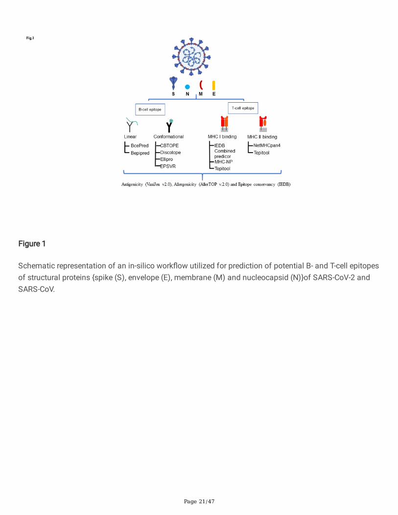
Figure 1
Schematic representation of an in-silico work�ow utilized for prediction of potential B- and T-cell epitopesof structural proteins {spike (S), envelope (E), membrane (M) and nucleocapsid (N)}of SARS-CoV-2 andSARS-CoV.

Page 22/47
Figure 1
Schematic representation of an in-silico work�ow utilized for prediction of potential B- and T-cell epitopesof structural proteins {spike (S), envelope (E), membrane (M) and nucleocapsid (N)}of SARS-CoV-2 andSARS-CoV.

Page 23/47
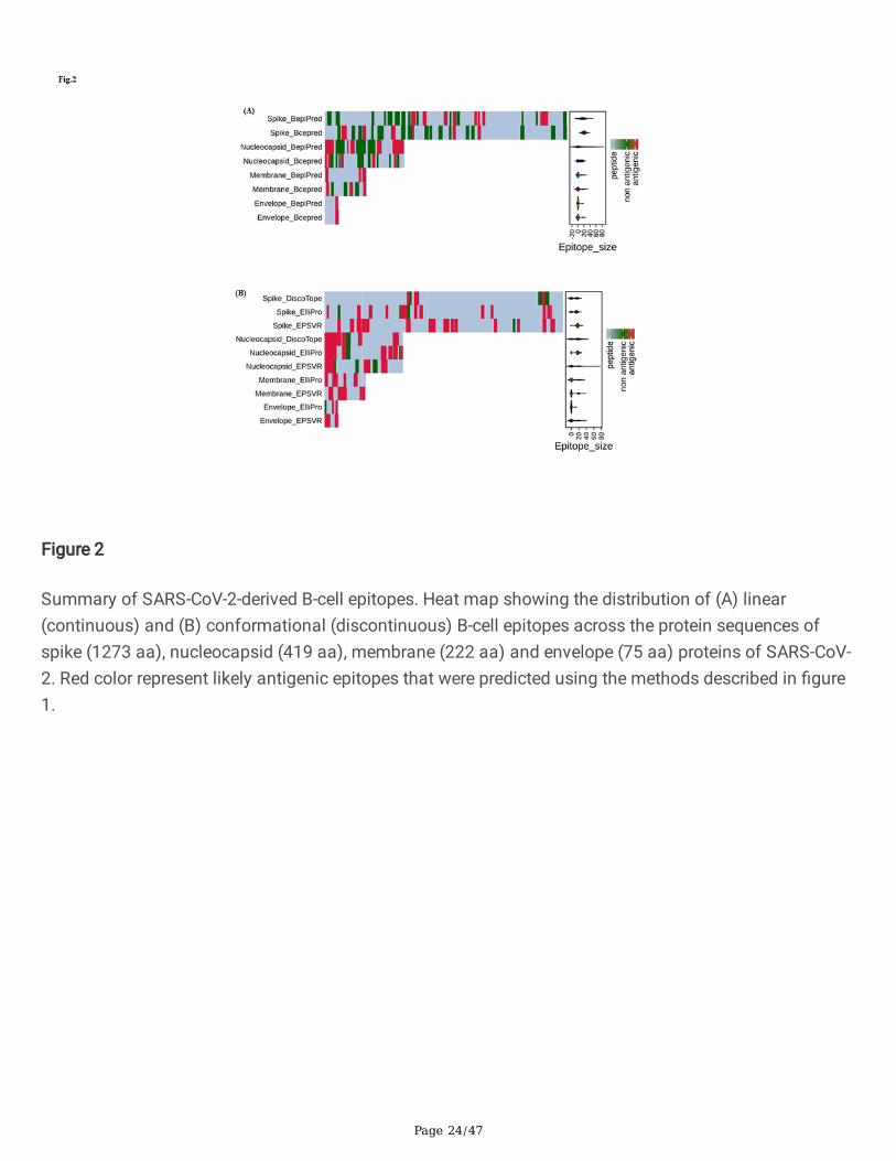
Figure 2
Summary of SARS-CoV-2-derived B-cell epitopes. Heat map showing the distribution of (A) linear(continuous) and (B) conformational (discontinuous) B-cell epitopes across the protein sequences ofspike (1273 aa), nucleocapsid (419 aa), membrane (222 aa) and envelope (75 aa) proteins of SARS-CoV-2. Red color represent likely antigenic epitopes that were predicted using the methods described in �gure1.
Page 24/47
Figure 2
Summary of SARS-CoV-2-derived B-cell epitopes. Heat map showing the distribution of (A) linear(continuous) and (B) conformational (discontinuous) B-cell epitopes across the protein sequences ofspike (1273 aa), nucleocapsid (419 aa), membrane (222 aa) and envelope (75 aa) proteins of SARS-CoV-2. Red color represent likely antigenic epitopes that were predicted using the methods described in �gure1.
Page 25/47
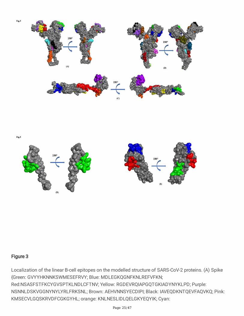
Figure 3
Localization of the linear B-cell epitopes on the modelled structure of SARS-CoV-2 proteins. (A) Spike{Green: GVYYHKNNKSWMESEFRVY; Blue: MDLEGKQGNFKNLREFVFKN;Red:NSASFSTFKCYGVSPTKLNDLCFTNV; Yellow: RGDEVRQIAPGQTGKIADYNYKLPD; Purple:NSNNLDSKVGGNYNYLYRLFRKSNL; Brown: AEHVNNSYECDIPI; Black: IAVEQDKNTQEVFAQVKQ; Pink:KMSECVLGQSKRVDFCGKGYHL; orange: KNLNESLIDLQELGKYEQYIK; Cyan:
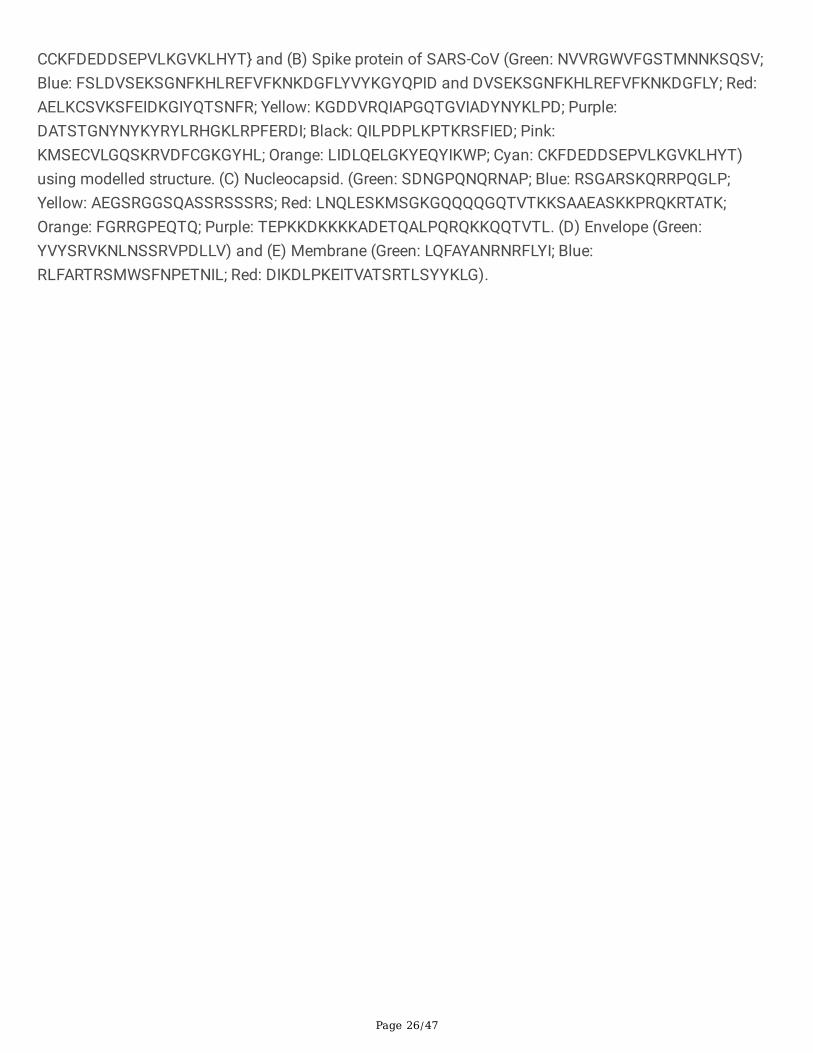
Page 26/47
CCKFDEDDSEPVLKGVKLHYT} and (B) Spike protein of SARS-CoV (Green: NVVRGWVFGSTMNNKSQSV;Blue: FSLDVSEKSGNFKHLREFVFKNKDGFLYVYKGYQPID and DVSEKSGNFKHLREFVFKNKDGFLY; Red:AELKCSVKSFEIDKGIYQTSNFR; Yellow: KGDDVRQIAPGQTGVIADYNYKLPD; Purple:DATSTGNYNYKYRYLRHGKLRPFERDI; Black: QILPDPLKPTKRSFIED; Pink:KMSECVLGQSKRVDFCGKGYHL; Orange: LIDLQELGKYEQYIKWP; Cyan: CKFDEDDSEPVLKGVKLHYT)using modelled structure. (C) Nucleocapsid. (Green: SDNGPQNQRNAP; Blue: RSGARSKQRRPQGLP;Yellow: AEGSRGGSQASSRSSSRS; Red: LNQLESKMSGKGQQQQGQTVTKKSAAEASKKPRQKRTATK;Orange: FGRRGPEQTQ; Purple: TEPKKDKKKKADETQALPQRQKKQQTVTL. (D) Envelope (Green:YVYSRVKNLNSSRVPDLLV) and (E) Membrane (Green: LQFAYANRNRFLYI; Blue:RLFARTRSMWSFNPETNIL; Red: DIKDLPKEITVATSRTLSYYKLG).

Page 27/47
Figure 3
Localization of the linear B-cell epitopes on the modelled structure of SARS-CoV-2 proteins. (A) Spike{Green: GVYYHKNNKSWMESEFRVY; Blue: MDLEGKQGNFKNLREFVFKN;Red:NSASFSTFKCYGVSPTKLNDLCFTNV; Yellow: RGDEVRQIAPGQTGKIADYNYKLPD; Purple:NSNNLDSKVGGNYNYLYRLFRKSNL; Brown: AEHVNNSYECDIPI; Black: IAVEQDKNTQEVFAQVKQ; Pink:KMSECVLGQSKRVDFCGKGYHL; orange: KNLNESLIDLQELGKYEQYIK; Cyan:
Page 28/47
CCKFDEDDSEPVLKGVKLHYT} and (B) Spike protein of SARS-CoV (Green: NVVRGWVFGSTMNNKSQSV;Blue: FSLDVSEKSGNFKHLREFVFKNKDGFLYVYKGYQPID and DVSEKSGNFKHLREFVFKNKDGFLY; Red:AELKCSVKSFEIDKGIYQTSNFR; Yellow: KGDDVRQIAPGQTGVIADYNYKLPD; Purple:DATSTGNYNYKYRYLRHGKLRPFERDI; Black: QILPDPLKPTKRSFIED; Pink:KMSECVLGQSKRVDFCGKGYHL; Orange: LIDLQELGKYEQYIKWP; Cyan: CKFDEDDSEPVLKGVKLHYT)using modelled structure. (C) Nucleocapsid. (Green: SDNGPQNQRNAP; Blue: RSGARSKQRRPQGLP;Yellow: AEGSRGGSQASSRSSSRS; Red: LNQLESKMSGKGQQQQGQTVTKKSAAEASKKPRQKRTATK;Orange: FGRRGPEQTQ; Purple: TEPKKDKKKKADETQALPQRQKKQQTVTL. (D) Envelope (Green:YVYSRVKNLNSSRVPDLLV) and (E) Membrane (Green: LQFAYANRNRFLYI; Blue:RLFARTRSMWSFNPETNIL; Red: DIKDLPKEITVATSRTLSYYKLG).
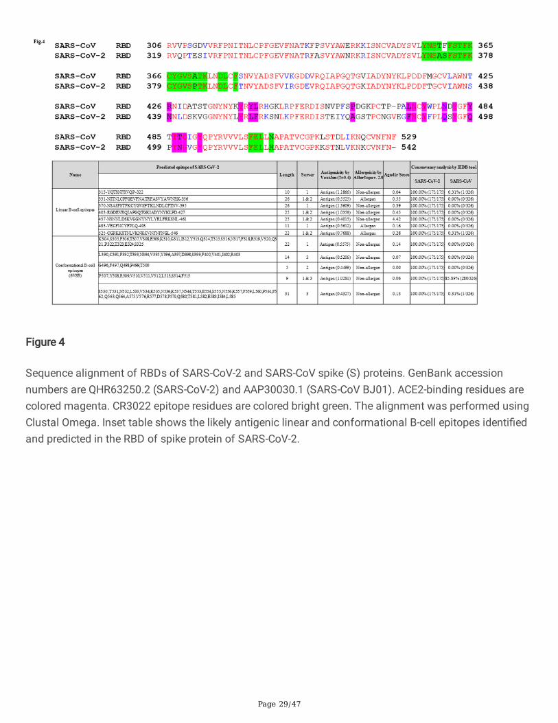
Figure 4
Sequence alignment of RBDs of SARS-CoV-2 and SARS-CoV spike (S) proteins. GenBank accessionnumbers are QHR63250.2 (SARS-CoV-2) and AAP30030.1 (SARS-CoV BJ01). ACE2-binding residues arecolored magenta. CR3022 epitope residues are colored bright green. The alignment was performed usingClustal Omega. Inset table shows the likely antigenic linear and conformational B-cell epitopes identi�edand predicted in the RBD of spike protein of SARS-CoV-2.
Page 29/47
Figure 4
Sequence alignment of RBDs of SARS-CoV-2 and SARS-CoV spike (S) proteins. GenBank accessionnumbers are QHR63250.2 (SARS-CoV-2) and AAP30030.1 (SARS-CoV BJ01). ACE2-binding residues arecolored magenta. CR3022 epitope residues are colored bright green. The alignment was performed usingClustal Omega. Inset table shows the likely antigenic linear and conformational B-cell epitopes identi�edand predicted in the RBD of spike protein of SARS-CoV-2.
Page 30/47
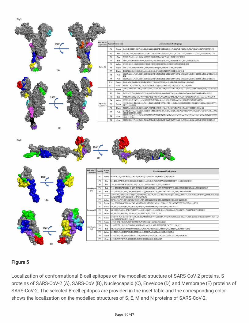
Figure 5
Localization of conformational B-cell epitopes on the modelled structure of SARS-CoV-2 proteins. Sproteins of SARS-CoV-2 (A), SARS-CoV (B), Nucleocapsid (C), Envelope (D) and Membrane (E) proteins ofSARS-CoV-2. The selected B-cell epitopes are provided in the inset table and the corresponding colorshows the localization on the modelled structures of S, E, M and N proteins of SARS-CoV-2.

Page 31/47
Figure 5
Localization of conformational B-cell epitopes on the modelled structure of SARS-CoV-2 proteins. Sproteins of SARS-CoV-2 (A), SARS-CoV (B), Nucleocapsid (C), Envelope (D) and Membrane (E) proteins ofSARS-CoV-2. The selected B-cell epitopes are provided in the inset table and the corresponding colorshows the localization on the modelled structures of S, E, M and N proteins of SARS-CoV-2.

Page 32/47
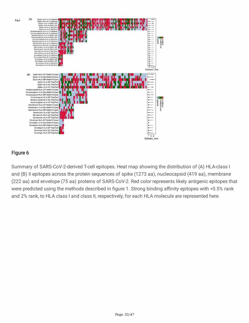
Figure 6
Summary of SARS-CoV-2-derived T-cell epitopes. Heat map showing the distribution of (A) HLA-class Iand (B) II epitopes across the protein sequences of spike (1273 aa), nucleocapsid (419 aa), membrane(222 aa) and envelope (75 aa) proteins of SARS-CoV-2. Red color represents likely antigenic epitopes thatwere predicted using the methods described in �gure 1. Strong binding a�nity epitopes with <0.5% rankand 2% rank, to HLA class I and class II, respectively, for each HLA molecule are represented here.
Page 33/47
Figure 6
Summary of SARS-CoV-2-derived T-cell epitopes. Heat map showing the distribution of (A) HLA-class Iand (B) II epitopes across the protein sequences of spike (1273 aa), nucleocapsid (419 aa), membrane(222 aa) and envelope (75 aa) proteins of SARS-CoV-2. Red color represents likely antigenic epitopes thatwere predicted using the methods described in �gure 1. Strong binding a�nity epitopes with <0.5% rankand 2% rank, to HLA class I and class II, respectively, for each HLA molecule are represented here.
Page 34/47
Figure 7
(A) Predicted population coverage rate (%) of predicted MHC I binding epitopes of S, E, M and N proteinsof SARS-CoV-2 and SARS-CoV. All the epitopes provided in table 3b and 3c have been analysed forpopulation coverage analysis using IEDB resource tool. (B) Predicted population coverage rate (%) ofpredicted MHC II binding epitopes of S, E, M and N proteins of SARS-CoV-2 and SARS-CoV. All the
Page 35/47
epitopes provided in table 4b and 4c have been analysed for population coverage analysis using IEDBresource tool.
Figure 7
(A) Predicted population coverage rate (%) of predicted MHC I binding epitopes of S, E, M and N proteinsof SARS-CoV-2 and SARS-CoV. All the epitopes provided in table 3b and 3c have been analysed forpopulation coverage analysis using IEDB resource tool. (B) Predicted population coverage rate (%) of
Page 36/47
predicted MHC II binding epitopes of S, E, M and N proteins of SARS-CoV-2 and SARS-CoV. All theepitopes provided in table 4b and 4c have been analysed for population coverage analysis using IEDBresource tool.
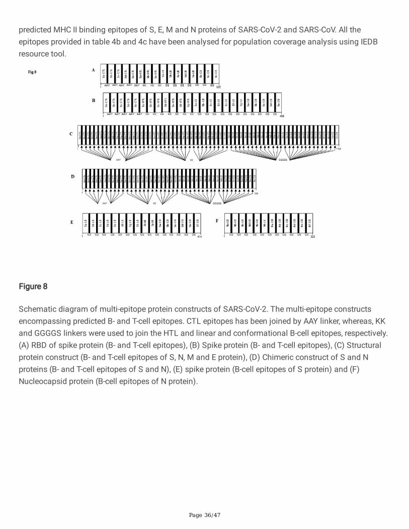
Figure 8
Schematic diagram of multi-epitope protein constructs of SARS-CoV-2. The multi-epitope constructsencompassing predicted B- and T-cell epitopes. CTL epitopes has been joined by AAY linker, whereas, KKand GGGGS linkers were used to join the HTL and linear and conformational B-cell epitopes, respectively.(A) RBD of spike protein (B- and T-cell epitopes), (B) Spike protein (B- and T-cell epitopes), (C) Structuralprotein construct (B- and T-cell epitopes of S, N, M and E protein), (D) Chimeric construct of S and Nproteins (B- and T-cell epitopes of S and N), (E) spike protein (B-cell epitopes of S protein) and (F)Nucleocapsid protein (B-cell epitopes of N protein).
Page 37/47
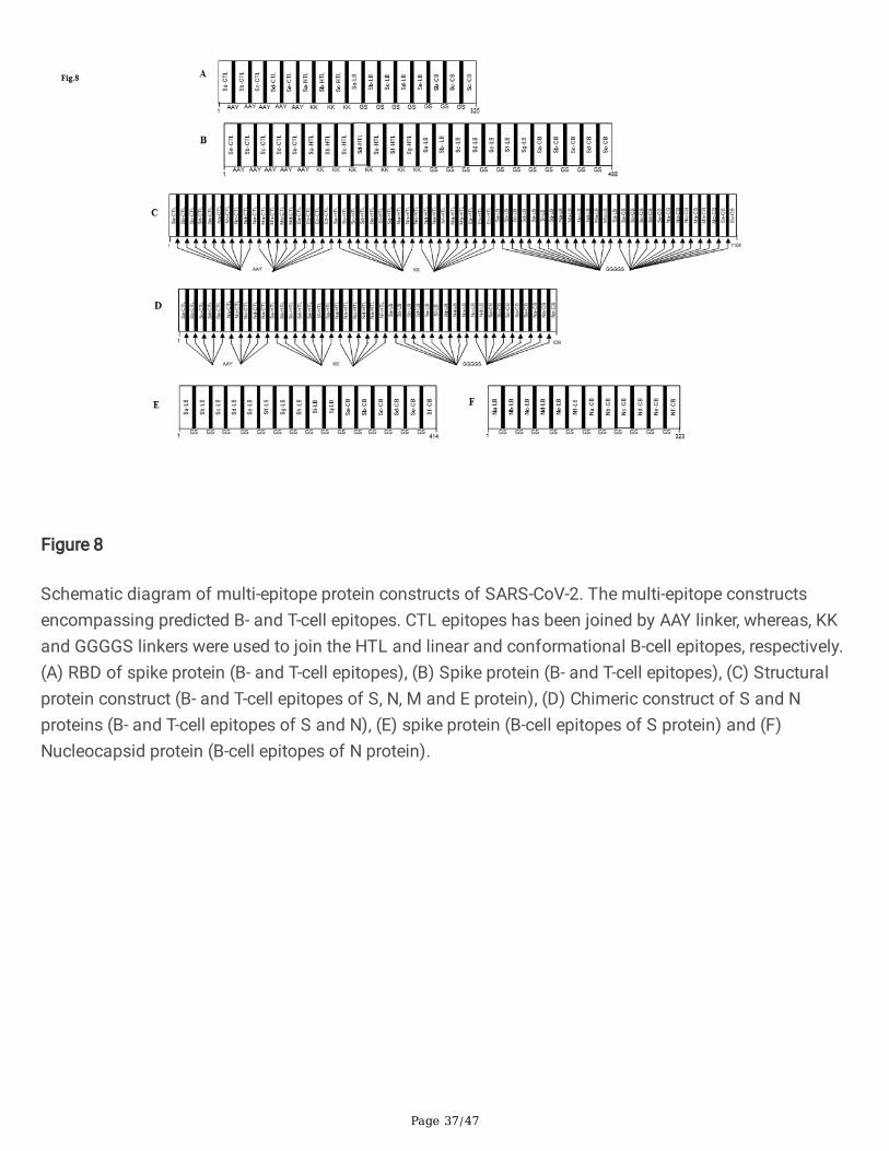
Figure 8
Schematic diagram of multi-epitope protein constructs of SARS-CoV-2. The multi-epitope constructsencompassing predicted B- and T-cell epitopes. CTL epitopes has been joined by AAY linker, whereas, KKand GGGGS linkers were used to join the HTL and linear and conformational B-cell epitopes, respectively.(A) RBD of spike protein (B- and T-cell epitopes), (B) Spike protein (B- and T-cell epitopes), (C) Structuralprotein construct (B- and T-cell epitopes of S, N, M and E protein), (D) Chimeric construct of S and Nproteins (B- and T-cell epitopes of S and N), (E) spike protein (B-cell epitopes of S protein) and (F)Nucleocapsid protein (B-cell epitopes of N protein).

Page 38/47
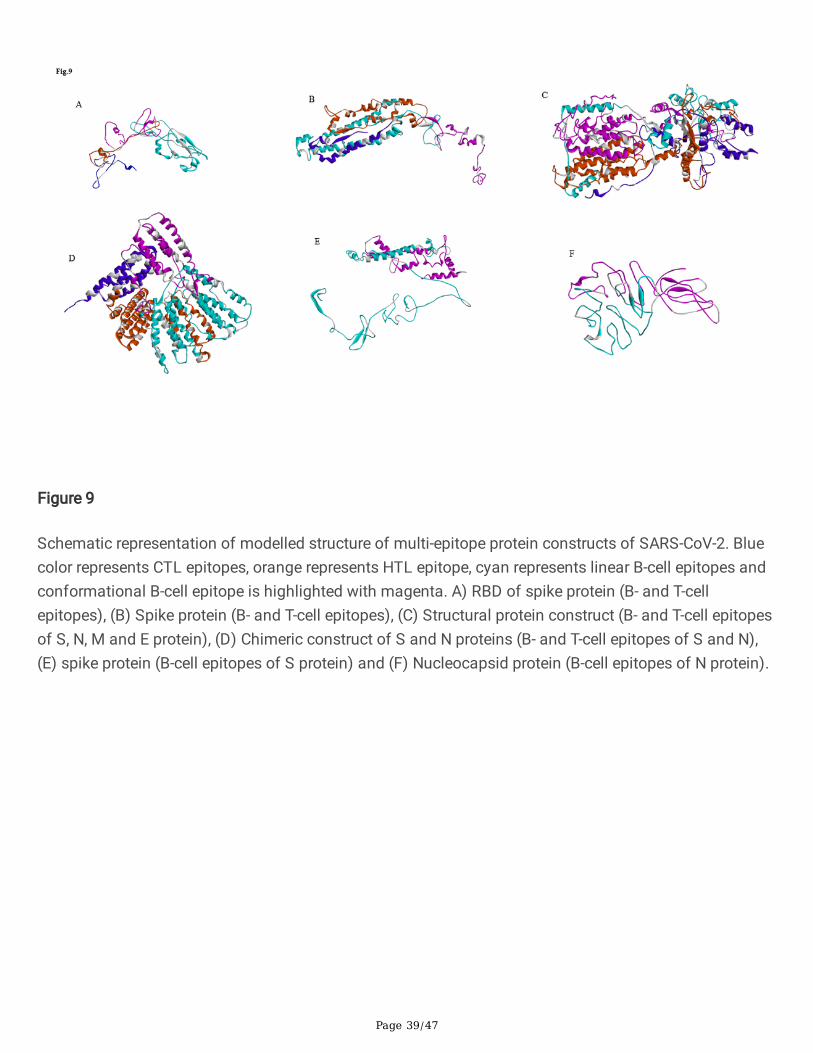
Figure 9
Schematic representation of modelled structure of multi-epitope protein constructs of SARS-CoV-2. Bluecolor represents CTL epitopes, orange represents HTL epitope, cyan represents linear B-cell epitopes andconformational B-cell epitope is highlighted with magenta. A) RBD of spike protein (B- and T-cellepitopes), (B) Spike protein (B- and T-cell epitopes), (C) Structural protein construct (B- and T-cell epitopesof S, N, M and E protein), (D) Chimeric construct of S and N proteins (B- and T-cell epitopes of S and N),(E) spike protein (B-cell epitopes of S protein) and (F) Nucleocapsid protein (B-cell epitopes of N protein).
Page 39/47
Figure 9
Schematic representation of modelled structure of multi-epitope protein constructs of SARS-CoV-2. Bluecolor represents CTL epitopes, orange represents HTL epitope, cyan represents linear B-cell epitopes andconformational B-cell epitope is highlighted with magenta. A) RBD of spike protein (B- and T-cellepitopes), (B) Spike protein (B- and T-cell epitopes), (C) Structural protein construct (B- and T-cell epitopesof S, N, M and E protein), (D) Chimeric construct of S and N proteins (B- and T-cell epitopes of S and N),(E) spike protein (B-cell epitopes of S protein) and (F) Nucleocapsid protein (B-cell epitopes of N protein).
Page 40/47
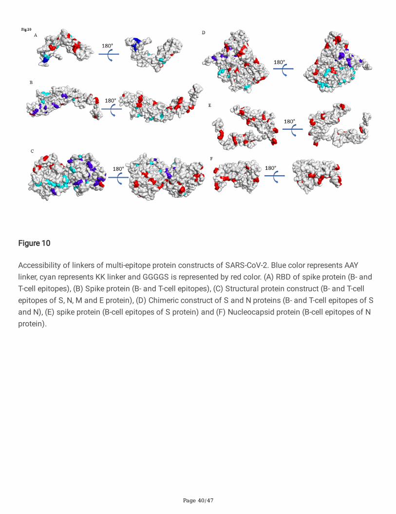
Figure 10
Accessibility of linkers of multi-epitope protein constructs of SARS-CoV-2. Blue color represents AAYlinker, cyan represents KK linker and GGGGS is represented by red color. (A) RBD of spike protein (B- andT-cell epitopes), (B) Spike protein (B- and T-cell epitopes), (C) Structural protein construct (B- and T-cellepitopes of S, N, M and E protein), (D) Chimeric construct of S and N proteins (B- and T-cell epitopes of Sand N), (E) spike protein (B-cell epitopes of S protein) and (F) Nucleocapsid protein (B-cell epitopes of Nprotein).

Page 41/47
Figure 10
Accessibility of linkers of multi-epitope protein constructs of SARS-CoV-2. Blue color represents AAYlinker, cyan represents KK linker and GGGGS is represented by red color. (A) RBD of spike protein (B- andT-cell epitopes), (B) Spike protein (B- and T-cell epitopes), (C) Structural protein construct (B- and T-cellepitopes of S, N, M and E protein), (D) Chimeric construct of S and N proteins (B- and T-cell epitopes of Sand N), (E) spike protein (B-cell epitopes of S protein) and (F) Nucleocapsid protein (B-cell epitopes of Nprotein).
Page 42/47
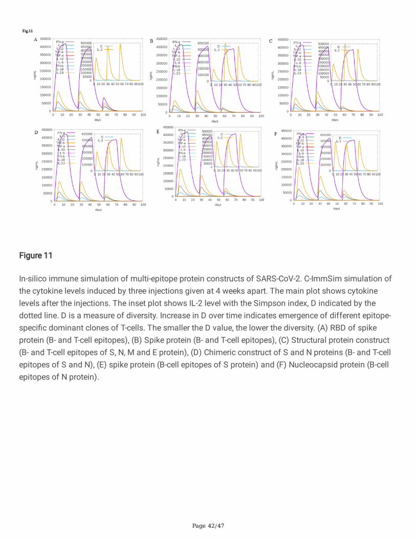
Figure 11
In-silico immune simulation of multi-epitope protein constructs of SARS-CoV-2. C-ImmSim simulation ofthe cytokine levels induced by three injections given at 4 weeks apart. The main plot shows cytokinelevels after the injections. The inset plot shows IL-2 level with the Simpson index, D indicated by thedotted line. D is a measure of diversity. Increase in D over time indicates emergence of different epitope-speci�c dominant clones of T-cells. The smaller the D value, the lower the diversity. (A) RBD of spikeprotein (B- and T-cell epitopes), (B) Spike protein (B- and T-cell epitopes), (C) Structural protein construct(B- and T-cell epitopes of S, N, M and E protein), (D) Chimeric construct of S and N proteins (B- and T-cellepitopes of S and N), (E) spike protein (B-cell epitopes of S protein) and (F) Nucleocapsid protein (B-cellepitopes of N protein).

Page 43/47
Figure 11
In-silico immune simulation of multi-epitope protein constructs of SARS-CoV-2. C-ImmSim simulation ofthe cytokine levels induced by three injections given at 4 weeks apart. The main plot shows cytokinelevels after the injections. The inset plot shows IL-2 level with the Simpson index, D indicated by thedotted line. D is a measure of diversity. Increase in D over time indicates emergence of different epitope-speci�c dominant clones of T-cells. The smaller the D value, the lower the diversity. (A) RBD of spikeprotein (B- and T-cell epitopes), (B) Spike protein (B- and T-cell epitopes), (C) Structural protein construct(B- and T-cell epitopes of S, N, M and E protein), (D) Chimeric construct of S and N proteins (B- and T-cellepitopes of S and N), (E) spike protein (B-cell epitopes of S protein) and (F) Nucleocapsid protein (B-cellepitopes of N protein).
Page 44/47
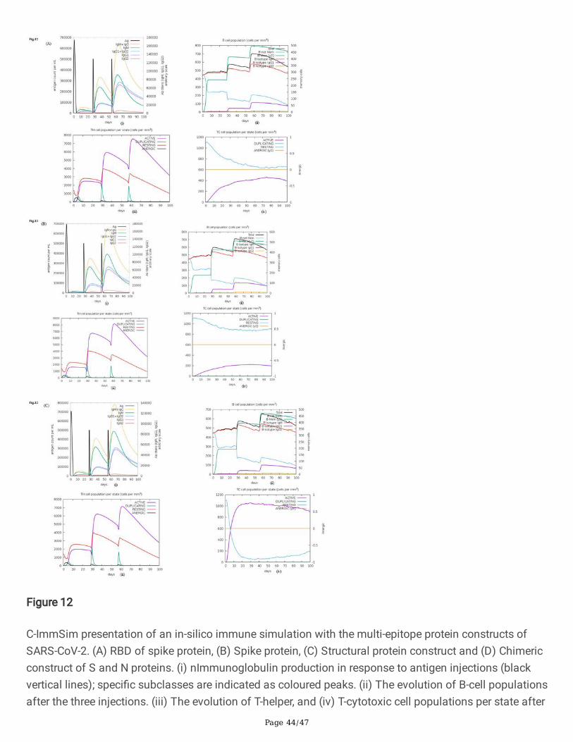
Figure 12
C-ImmSim presentation of an in-silico immune simulation with the multi-epitope protein constructs ofSARS-CoV-2. (A) RBD of spike protein, (B) Spike protein, (C) Structural protein construct and (D) Chimericconstruct of S and N proteins. (i) nImmunoglobulin production in response to antigen injections (blackvertical lines); speci�c subclasses are indicated as coloured peaks. (ii) The evolution of B-cell populationsafter the three injections. (iii) The evolution of T-helper, and (iv) T-cytotoxic cell populations per state after
Page 45/47
the injections with constructs. The resting state represents cells not presented with the antigen while theanergic state represents tolerance of the T-cells to the antigen due to repeated exposures.
Figure 12
C-ImmSim presentation of an in-silico immune simulation with the multi-epitope protein constructs ofSARS-CoV-2. (A) RBD of spike protein, (B) Spike protein, (C) Structural protein construct and (D) Chimericconstruct of S and N proteins. (i) nImmunoglobulin production in response to antigen injections (black
Page 46/47
vertical lines); speci�c subclasses are indicated as coloured peaks. (ii) The evolution of B-cell populationsafter the three injections. (iii) The evolution of T-helper, and (iv) T-cytotoxic cell populations per state afterthe injections with constructs. The resting state represents cells not presented with the antigen while theanergic state represents tolerance of the T-cells to the antigen due to repeated exposures.
Figure 13
Localization of the linear B-cell epitopes on the modelled and crystal structure of the S protein of SARS-CoV-2. Localization of selected monomeric B-cell epitopes (inset table) on the (A) modelled and (B)crystal structure (PDB:6VSB) of the S protein of SARS-CoV-2 was performed using the BIOVIA discoverystudio 2017 R2.
Page 47/47
Figure 13
Localization of the linear B-cell epitopes on the modelled and crystal structure of the S protein of SARS-CoV-2. Localization of selected monomeric B-cell epitopes (inset table) on the (A) modelled and (B)crystal structure (PDB:6VSB) of the S protein of SARS-CoV-2 was performed using the BIOVIA discoverystudio 2017 R2.
Supplementary Files
This is a list of supplementary �les associated with this preprint. Click to download.
Mergedsuptableand�gure.pdf
supplement1.pdf
NimaNamsaTableand�gureNDN1.pdf
supplement2.pdf