Protein Purification. Compartmentalization provides an opportunity for a purification step e Protein...
70
Protein Purification
-
Upload
angel-holland -
Category
Documents
-
view
219 -
download
1
Transcript of Protein Purification. Compartmentalization provides an opportunity for a purification step e Protein...

Protein Purification
Compartmentalization provides an opportunity for a purification step
e
Protein profile for compartments of gram-negative prokaryotes

Cell Disruption
Chemical: alkali, organic solvents, detergents
Enzymatic: lysozyme, glucanases, chitinase
Physical: osmotic shock, freeze/thawMechanical: sonication, homogenization,
wet milling, French press
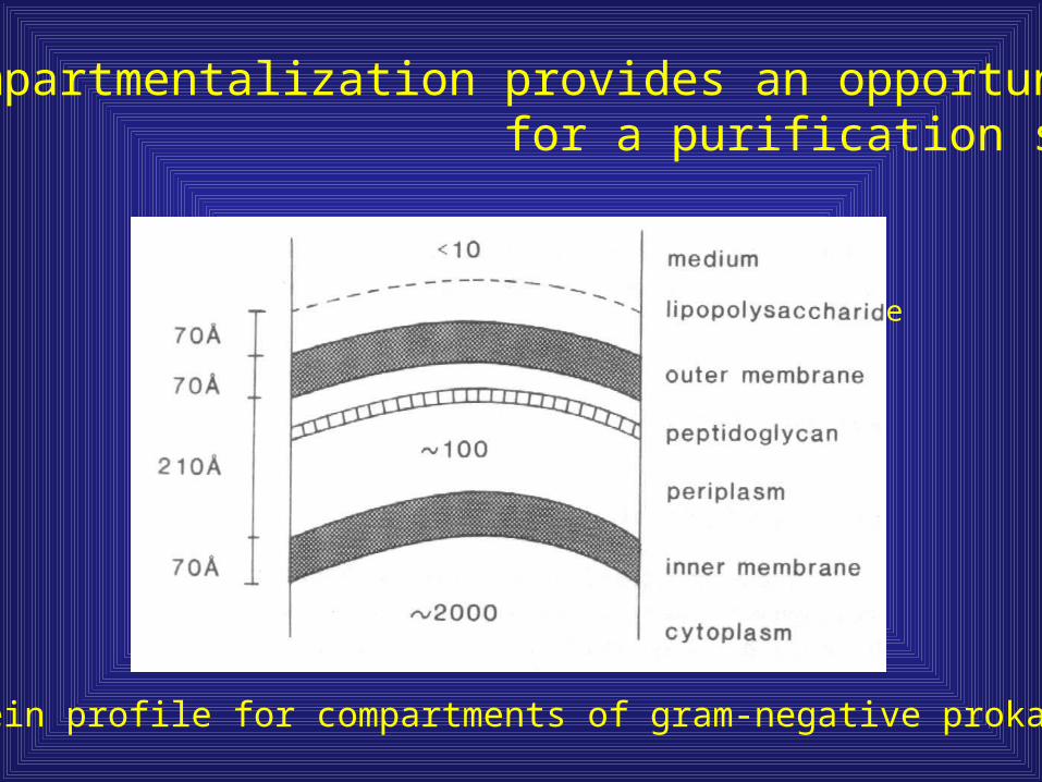
Chemical Disruption
Detergents such as Trition X-100 or NP40 can permeabilize cells by solubilizing membranes.
Detergents can be expensive, denature proteins, and must be removed after disruption
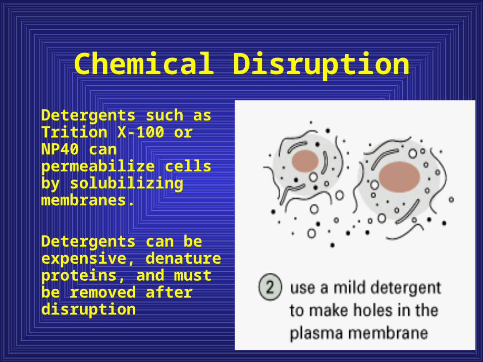
French Press
Cells are placed in a stainless steel container. A tight fitting piston is inserted and high pressures are applied to force cells through a small hole.
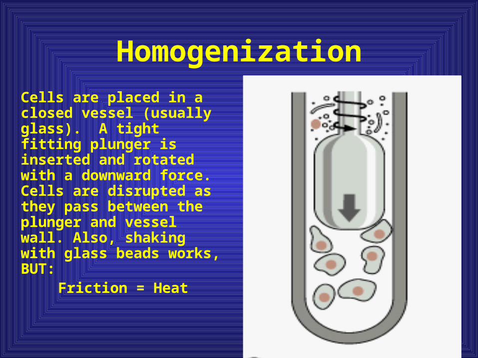
Homogenization
Cells are placed in a closed vessel (usually glass). A tight fitting plunger is inserted and rotated with a downward force. Cells are disrupted as they pass between the plunger and vessel wall. Also, shaking with glass beads works, BUT:
Friction = Heat
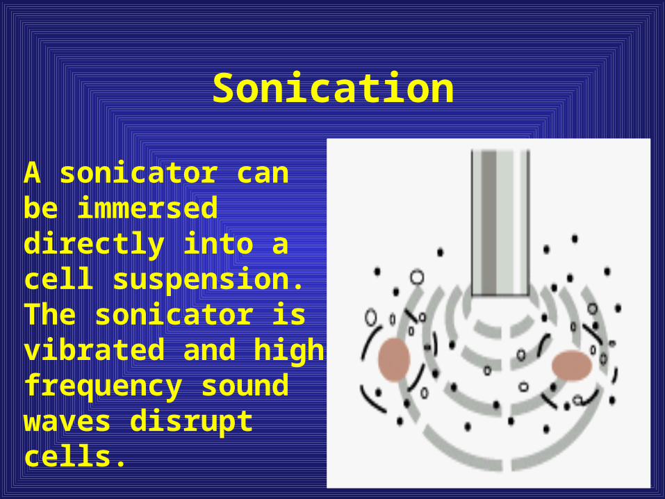
Sonication
A sonicator can be immersed directly into a cell suspension. The sonicator is vibrated and high frequency sound waves disrupt cells.

Differential centrifugation

Inclusion bodies provide a rapid purification step
Inclusion bodies providestorage space for protein,carbohydrate and lipid material in prokaryotes
However, proteins existas aggregates in inclusionbodies thus special precautions must be takenduring purification
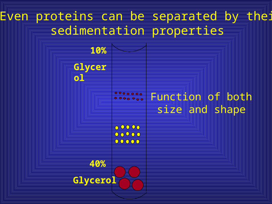
10%
Glycerol
40%
Glycerol
Even proteins can be separated by their sedimentation properties
Function of both size and shape

Proteins have unique properties resulting from their amino acid composition
Localization
Charge
Hydrophobicity
Size
Affinity for ligandsArbitrary protein
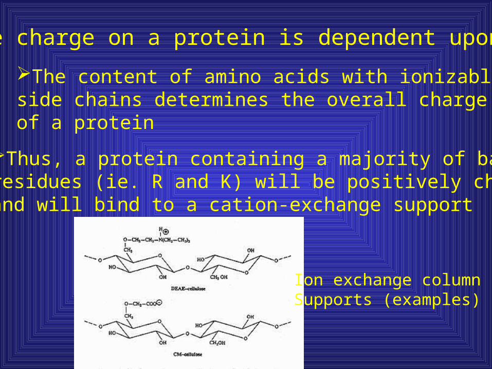
The charge on a protein is dependent upon pH
The content of amino acids with ionizableside chains determines the overall charge of a protein
Thus, a protein containing a majority of basicresidues (ie. R and K) will be positively chargedand will bind to a cation-exchange support
Ion exchange columnSupports (examples)

Separation based on surface charge
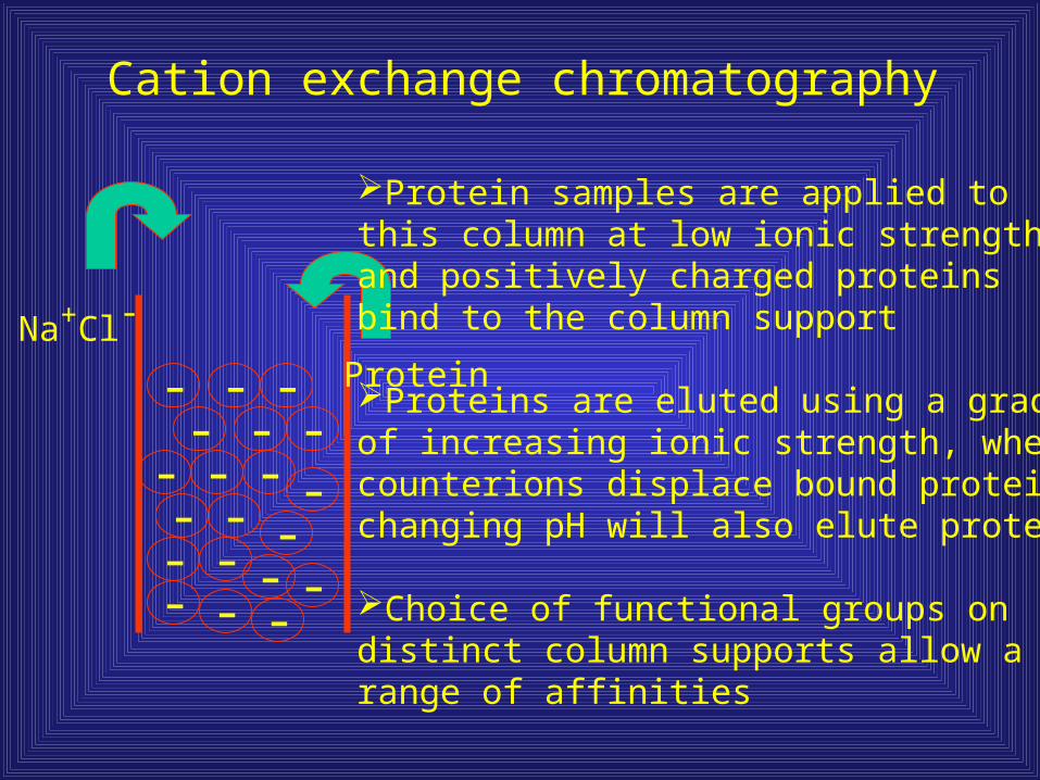
Cation exchange chromatography
Protein- -
--- - -
-- - ----
- --
--
-Na+Cl-
Protein samples are applied tothis column at low ionic strength,and positively charged proteinsbind to the column support
Proteins are eluted using a gradientof increasing ionic strength, wherecounterions displace bound protein, changing pH will also elute protein
Choice of functional groups ondistinct column supports allow arange of affinities
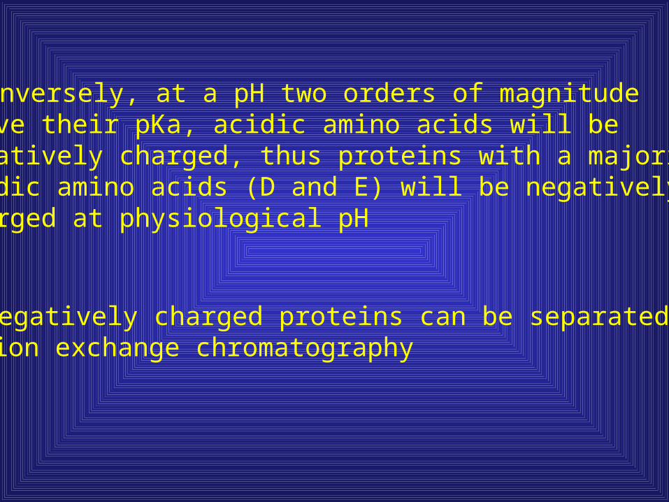
Conversely, at a pH two orders of magnitude above their pKa, acidic amino acids will benegatively charged, thus proteins with a majority ofacidic amino acids (D and E) will be negativelycharged at physiological pH
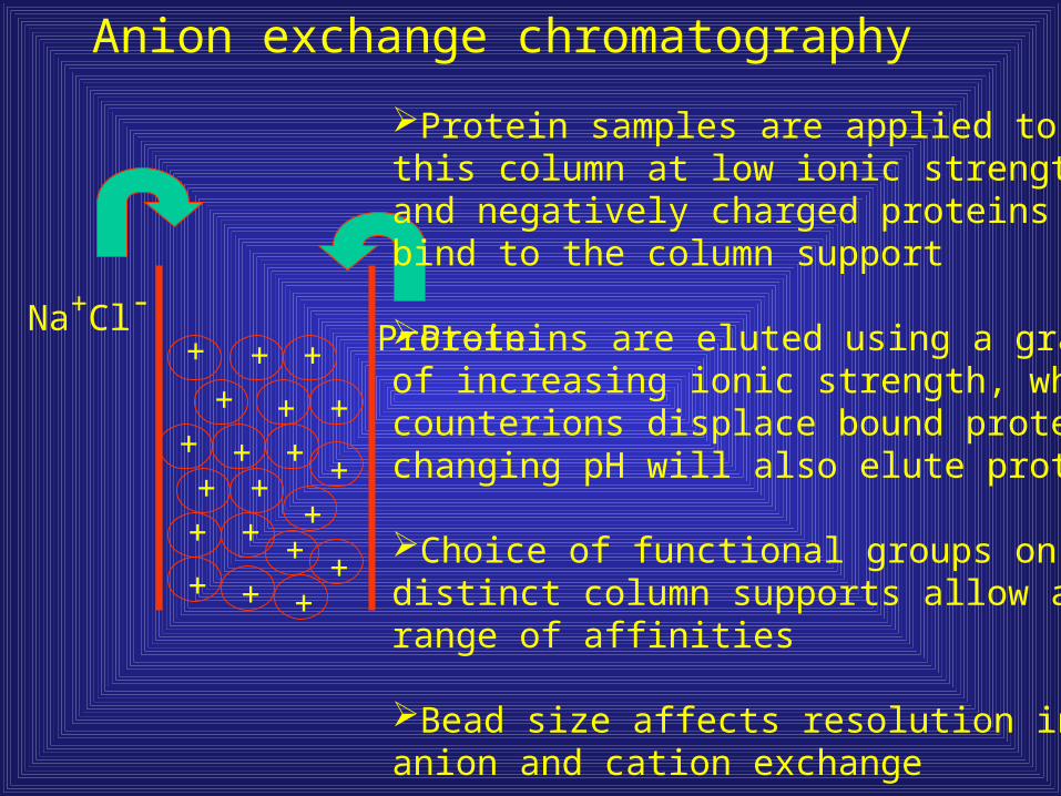
Negatively charged proteins can be separated usinganion exchange chromatography
Na+Cl-Protein
Protein samples are applied tothis column at low ionic strength,and negatively charged proteinsbind to the column support
Proteins are eluted using a gradientof increasing ionic strength, wherecounterions displace bound protein, changing pH will also elute protein
Choice of functional groups ondistinct column supports allow arange of affinities
Bead size affects resolution in bothanion and cation exchange
Anion exchange chromatography
+
+
+ +
++ +++ + + +
+++
+ ++++
Hydrophobic Interaction Chromatography
Although most hydrophobic amino acids are buried inthe interior of proteins, many proteins have hydrophobicsurfaces or patches which can be used for separation
A protein’s hydrophobic character is typically enhancedby addition of high salt concentrations
Proteins are eluted from HIC columns via a gradient ofhigh salt to low salt concentrations
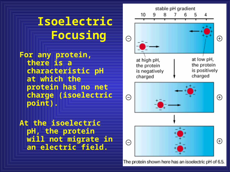
For any protein, there is a characteristic pH at which the protein has no net charge (isoelectric point).
At the isoelectric pH, the protein will not migrate in an electric field.
Isoelectric Focusing

Isoelectric focusing
Protein Precipitation
Precipitation is caused by changes that disrupt the solvating properties of water
Changes in pH, ionic strength, temperature, and the addition of solvents can cause precipitation (loss of solubility)
Most proteins have a unique set of conditions that result in precipitation
Precipitation with Salt
In practice, most procedures use the salt ammonium sulfate (NH4)2SO4 to precipitate proteins
The amount of salt required is directly related to the number and distribution of charged and nonionic polar amino acids exposed on the surface of the protein
At low ionic strengths, the charges on the surface of a protein attract counter ions, decreasing electrostatic free energy and increasing solubility. Addition of low concentrations of salt, then, increase solubility of proteins ("salting in"). At high salt concentrations, however, protein solubility decreases ("salting out"). This is due to electrostatic repulsion between the surface ions and the hydrophobic interior of the protein and to the avid interaction of salts with water. This disrupts the ordered water in the hydration layer. Salts vary in their ability to salt out proteins and generally follow the Hofmeister series:
Cations: NH4+ > K+ > Na+ > Mg++ > Ca++ > guanidium
Anions: SO4-- > HPO4-- > acetate > citrate > tartrate > Cl- > NO3-
Salt effects on protein solubility
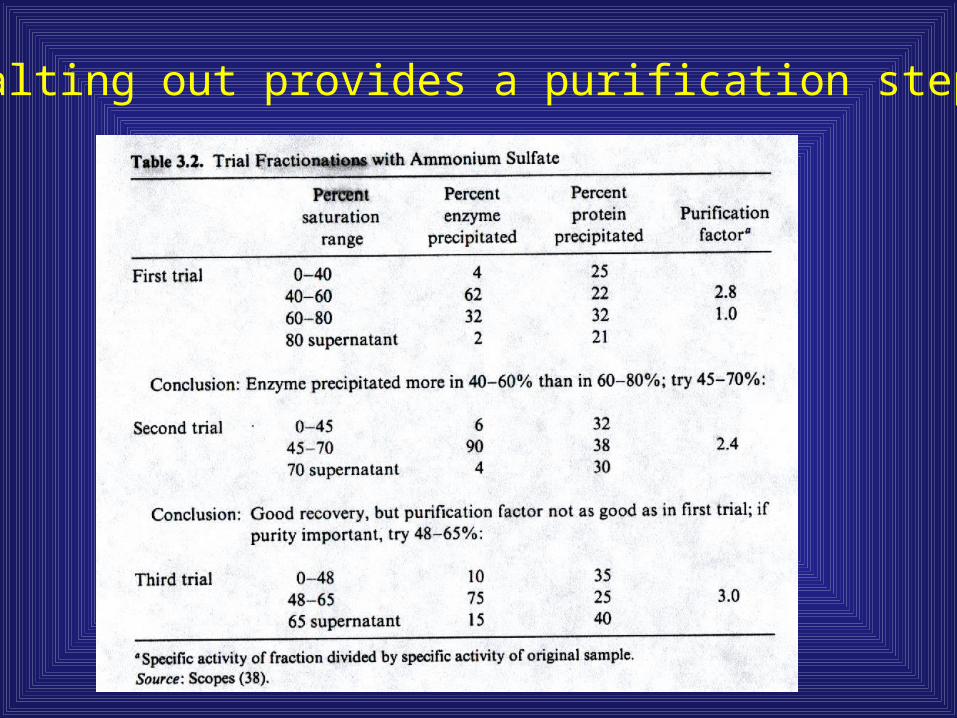
Salting out provides a purification step
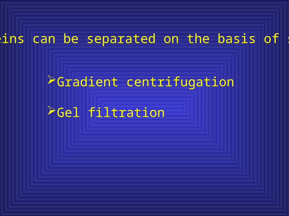
Proteins can be separated on the basis of size
Gradient centrifugation
Gel filtration

Gel Filtration provides a molecular sieve
Figures from Scopes, Protein Purificationon Reserve
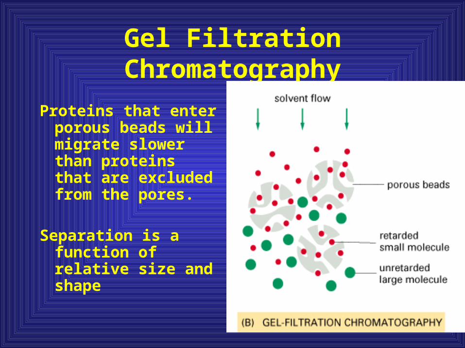
Gel Filtration Chromatography
Proteins that enter porous beads will migrate slower than proteins that are excluded from the pores.
Separation is a function of relative size and shape

Size exclusion can be used to determine oligomeric state
Vo = Void volume (the excluded volume surrounding the beads)
Ve = Intermediate volume (partially excluded)
Construct a standard curve using known proteins of known sizes

Gel Filtration Chromatography
LogMolWt
Ve - Vo

A protein’s substrate preference can be used in a very specific purification step
IntrinsicIf a protein binds ATP, put over a columnsupport that has ATP crosslinked on it, thusselecting for ATP-binding proteins (can be doneor a wide range of substrates such as sugars, Proteins, etc.)
AddedSpecific protein domains can be fused to proteinsof interest at the gene level to facilitate purification(ie. Fuse a maltose binding protein domain to any random protein, then it will bind specifically to amaltose containing column)
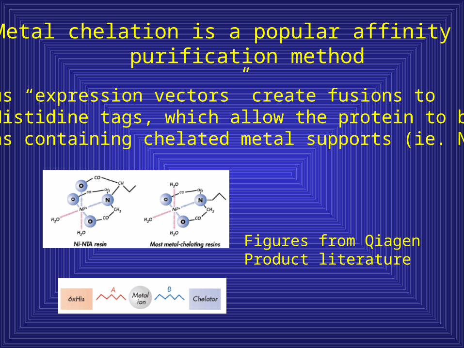
Metal chelation is a popular affinity purification method
Various “expression vectors” create fusions topoly-Histidine tags, which allow the protein to bind tocolumns containing chelated metal supports (ie. Ni+2)
Figures from QiagenProduct literature

Examining your purified protein

Use of SDS-PAGE vs. Native gel electrophoresis

Two dimensional gel electrophoresis
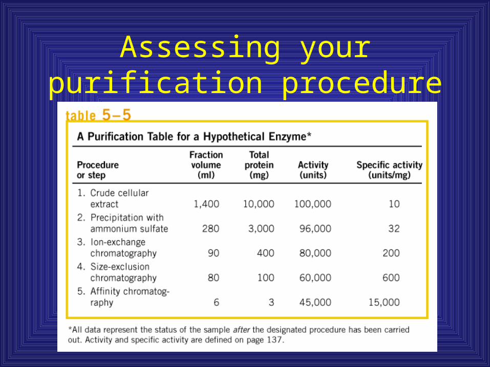
Assessing your purification procedure
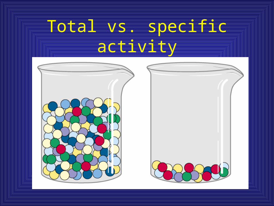
Total vs. specific activity
We can “control” protein expression
With the notable exception of proteins such asthose that compose the ribosome, many proteinsare found only in low abundance (particularlyProteins involved in regulatory processes)
Thus, we need to find ways to grow cells thatallow ample expression of proteins that would be interesting for biochemical characterization.
Find conditions for cell growth that enhance a protein’s expression
For example, cytochrome c2 is utilized by R.sphaeroidesfor both respiratory and photosynthetic growth; a slight increase in levels of this protein is observed under photosynthetic growth conditions.
However, Light-Harvesting complexes are only synthesizedunder photosynthetic growth conditions; obviously if you want to purify this protein you need to grow cells underphotosynthetic conditions

Molecular Biology allows us to manipulate genes
Understanding the basic mechanisms of gene expressionhas allowed investigators to exploit various systems for protein expression
Prokaryotic expression systems
Eukaryotic expression systemsYeastMammalian
Viral expression systemsBaculovirus and Insects

What do we need to produce a protein?
lamB
A gene
lamB
Promoter
Transcriptional unit
Terminator
lamB
Ribosome binding site
Translational unit

Molecular Biology presents an opportunity for useful genetic constructs
lamB
Promoter Terminator
bla
Plasmid
oriAntibiotic resistance gene Origin ofReplication
Can fuse gene to other sequences conferring affinity
Choice of promoter allows control over transcription levels
Intrinsic promoters can be sufficient for overexpressionin multi-copy plasmids
Constitutive promoters with high activity (ie. promoters forribosomal genes) can be useful for producing non-toxicproteins
Inducible promoters allow control of expression, one can “titrate” the promoter activity using exogenous agents

An expression system utilizing lactose and T7 RNA polymerase is a popular choice in prokaryotes
lamB
blaori
Plasmid
T7 polymerasedependent promoter
T7 polLactose-inducible promoter
Genome
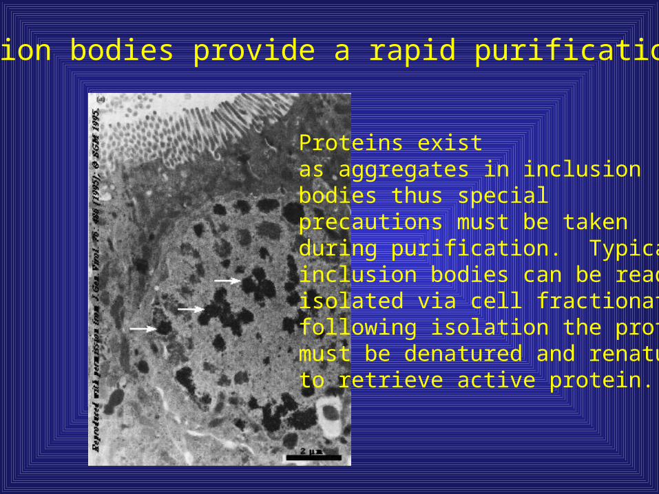
Inclusion bodies provide a rapid purification step
Proteins existas aggregates in inclusionbodies thus special precautions must be takenduring purification. Typically,inclusion bodies can be readilyisolated via cell fractionation.following isolation the proteinsmust be denatured and renaturedto retrieve active protein.
Additional concerns regarding protein expression
Modifications
Inclusion bodies
Codon usage

Cells exhibit nonrandom usage of codons
This provides a mechanism for regulation;however, genes cloned for purposes ofheterologous protein expression may contain“rare” codons that are not normally utilized bycells such as E. coli. Thus, this could limit protein production. Codon usage has been usedfor determination of highly expressed proteins.
Molecular Biology allows us to manipulate genes
Understanding the basic mechanisms of gene expressionhas allowed investigators to exploit various systems for protein expression
Prokaryotic expression systems
Eukaryotic expression systemsYeastMammalian
Viral expression systemsBaculovirus and Insects
Non-prokaryotic expression systems have emerged due toincreasing simplicity and the need for proper modifications.
Although you can express a eukaryotic cDNA in a prokaryote is the protein you purify, what the eukaryotic cell uses?
Invitrogen : www.invitrogen.com Gateway vectors
Novagen: www.novagen.com
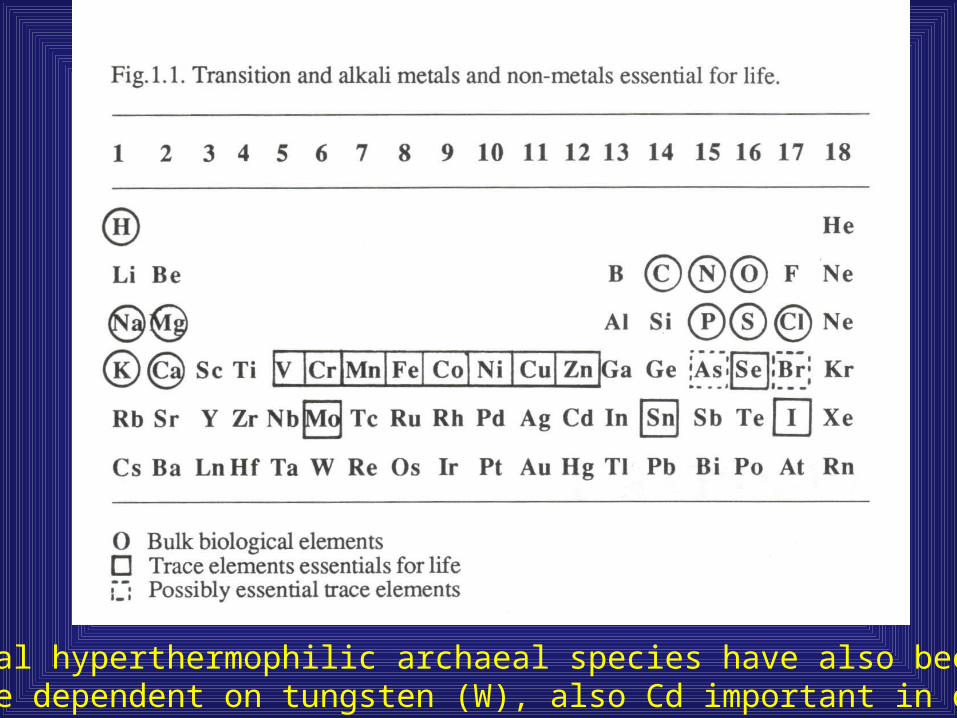
Several hyperthermophilic archaeal species have also been shown to be dependent on tungsten (W), also Cd important in diatoms

Fe is most abundant, followed by Zn
Metals in Biology
• Enzyme co-factorsRedox active centers in many enzymesFe: Electron transport, SOD, Cytochrome P450Zn: SODMg, Mn: PhotosynthesisCu: Electron transportCa: Cell signalingCa, Na, etc: Substrates in ion pumps
• Structural components of enzymes Fe: Hemoglobin, Cell structureZn: Zn fingers in transcription factorsCa: Bone structure, Cell structure
Metals and their biological effects
• Block essential function of biomolecules e.g. Ion pumps: Divalent metals inhibit Ca pumps
• Displace essential metal co-factorse.g. Cd can replace Cu in electron transport enzymes
• Modify configuration of biomolecules: Zn can be replaced Cd in Zn fingers

Metals and reactive oxygen species
• Redox potential of O2 ~ + 1 V; Extremely oxidizing• If there is a source of electrons:• O2 + e- O2
- + e- + 2 H+ H2O2 + e-
OH + OH- + e- + 2 H+ H2O • All but water are reactive oxygen species (ROS) and are
biologically damaging• In above order: superoxide, hydrogen peroxide, hydroxyl
radical• Biomolecules are a good source of reducing power: i.e.
electrons • Redox active metals can catalyze electron transfer from
biomolecules to O2
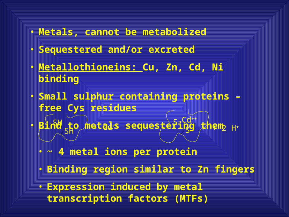
• Metals, cannot be metabolized
• Sequestered and/or excreted
• Metallothioneins: Cu, Zn, Cd, Ni binding
• Small sulphur containing proteins – free Cys residues
• Bind to metals sequestering them
SHSH + Cd++
S-
S- + 2 H+Cd++
• ~ 4 metal ions per protein
• Binding region similar to Zn fingers
• Expression induced by metal transcription factors (MTFs)
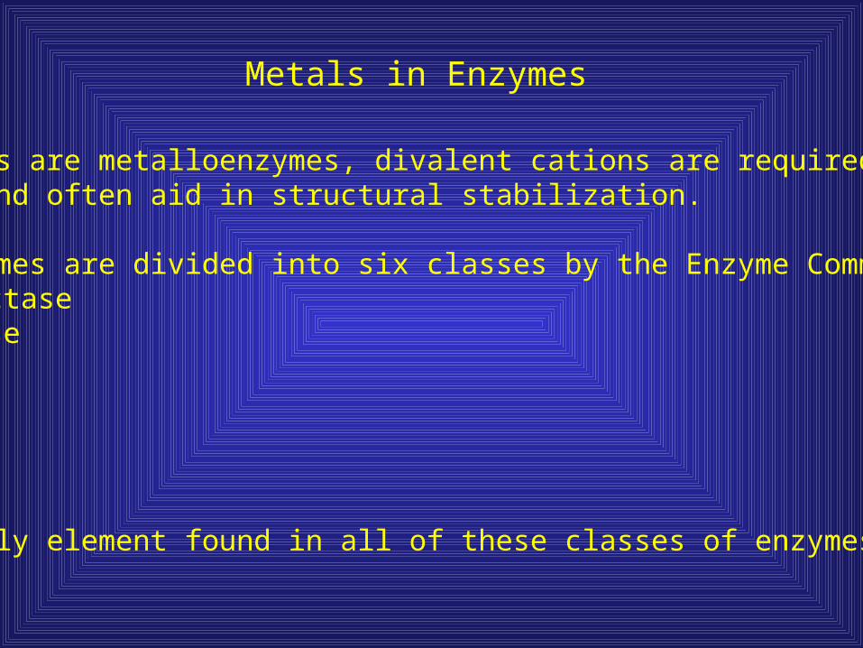
Metals in Enzymes
All ribozymes are metalloenzymes, divalent cations are required forchemistry, and often aid in structural stabilization.
Protein enzymes are divided into six classes by the Enzyme Commision:1. Oxidoreductase2. Transferase3. Hydrolase4. Lyase5. Isomerase6. Ligase
Zn is the only element found in all of these classes of enzymes.

Proteins bind metals based on size, charge, and chemical nature
Each metal has unique properties regarding ionic chargeionic radii, and ionization potential
Typically, metals are classified as “hard” or “soft” incorrelation with their ionic radii, electrostatics, andpolarization
Hard metals prefer hard ligands, soft prefer soft,Borderline metals can go either way.
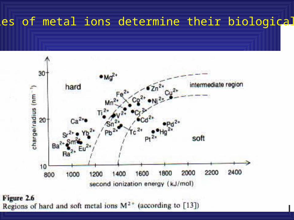
Properties of metal ions determine their biological utility

Soft
Hard
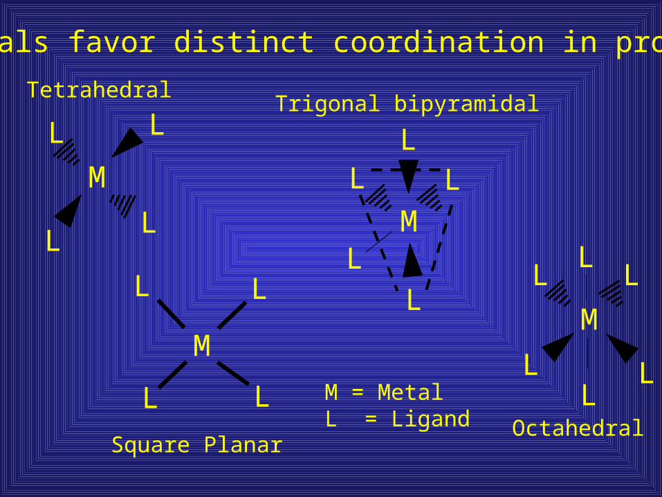
Metals favor distinct coordination in proteins
M
LL
L L
M
L L
LLM
L
LL
LL
LM
L
L L
L
L
Tetrahedral
Square Planar
Trigonal bipyramidal
Octahedral
M = MetalL = Ligand

Unsaturated coordination spheres usually have water as additional ligands to meet the favored 4 or 6 coordination
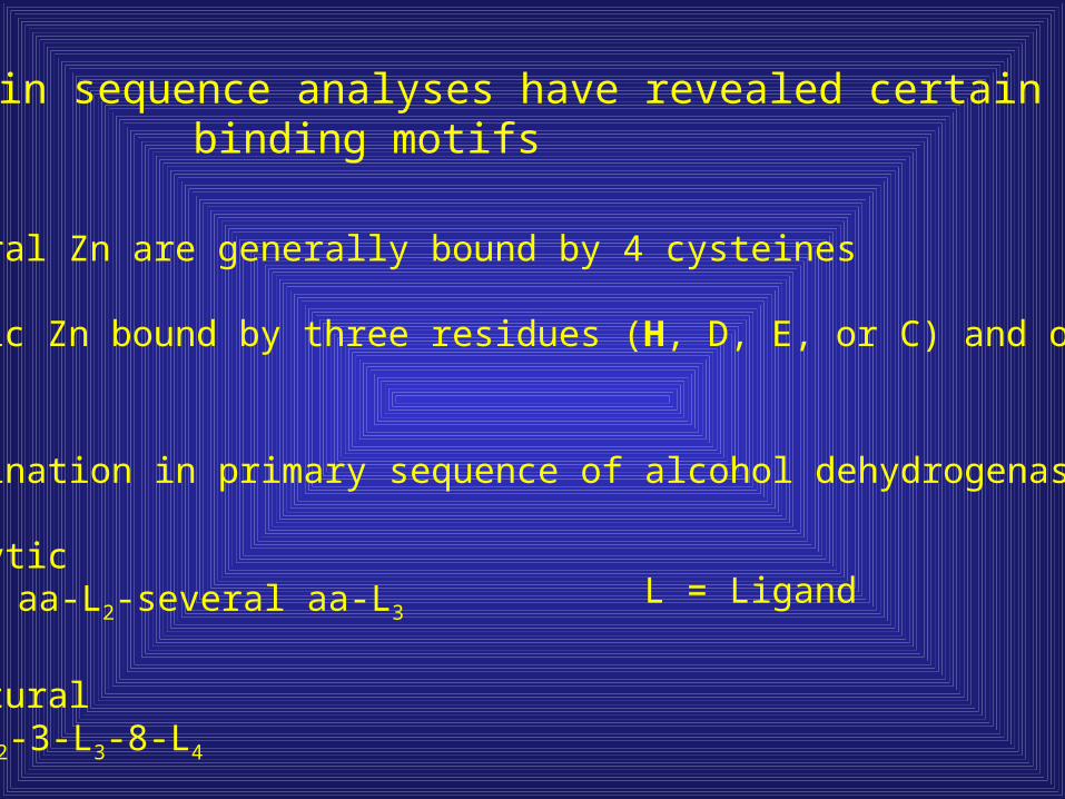
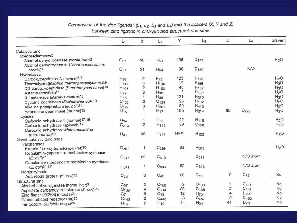
Protein sequence analyses have revealed certain metalbinding motifs
Structural Zn are generally bound by 4 cysteines
Catalytic Zn bound by three residues (H, D, E, or C) and one water
Coordination in primary sequence of alcohol dehydrogenase
CatalyticL1-few aa-L2-several aa-L3
StructuralL1-3-L2-3-L3-8-L4
L = Ligand

Biological roles of transition metals
Coordination Structure (protein and protein-substrate)Electrophilic catalysis Positive charge attracts electrons, polarize potential reactant, increase reactivityGeneral Acid – Base catalysisRedox reactionsMetalloorganic chemistry Free radicals
(not just limited to proteins*)

Carbonic Anhydrase catalytic mechanism
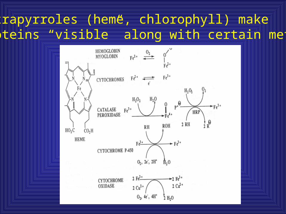
Tetrapyrroles (heme, chlorophyll) makeproteins “visible” along with certain metals
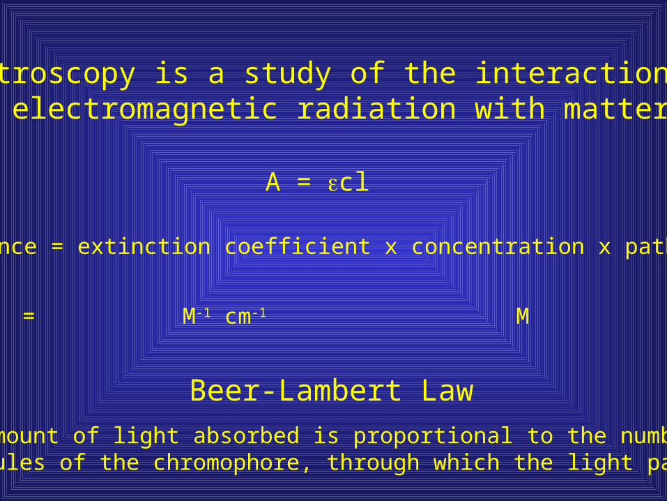
Spectroscopy is a study of the interaction of electromagnetic radiation with matter
A = cl
Absorbance = extinction coefficient x concentration x path length
Beer-Lambert Law
The amount of light absorbed is proportional to the number ofmolecules of the chromophore, through which the light passes
Units: None = M-1 cm-1 M cm
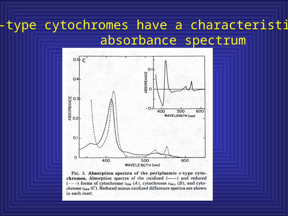
c-type cytochromes have a characteristic absorbance spectrum

Purification of GFP overview
Protein stability
Protein precipitation
Hydrophobic Interaction chromatography
Gel electrophoresis
Optical spectroscopy
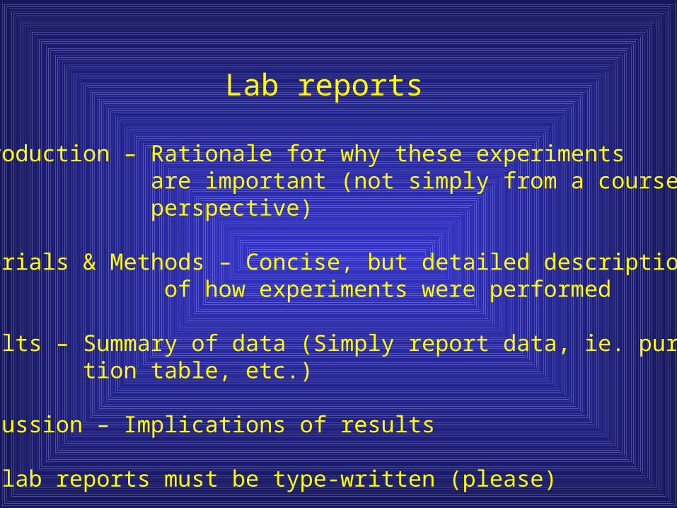
Lab reports
Introduction – Rationale for why these experimentsare important (not simply from a course workperspective)
Materials & Methods – Concise, but detailed description of how experiments were performed
Results – Summary of data (Simply report data, ie. purifica-tion table, etc.)
Discussion – Implications of results
All lab reports must be type-written (please)
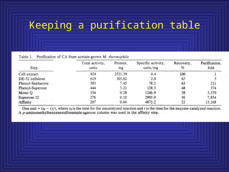
Keeping a purification table