Protein mediated membrane adhesion · There are a variety of membrane embedded proteins which form...
10
Protein mediated membrane adhesion Andreas Carlson and L. Mahadevan Citation: Physics of Fluids 27, 051901 (2015); doi: 10.1063/1.4919777 View online: http://dx.doi.org/10.1063/1.4919777 View Table of Contents: http://scitation.aip.org/content/aip/journal/pof2/27/5?ver=pdfcov Published by the AIP Publishing Articles you may be interested in Efficient elusion of viable adhesive cells from a microfluidic system by air foam Biomicrofluidics 8, 052001 (2014); 10.1063/1.4893348 Comment on “A method to measure cellular adhesion utilizing a polymer micro-cantilever” [Appl. Phys. Lett. 103, 123702 (2013)] Appl. Phys. Lett. 104, 236103 (2014); 10.1063/1.4882182 Spatial anisotropy and heterogeneity in contractility and adhesion distribution may contribute to cell steering during migration Appl. Phys. Lett. 104, 083705 (2014); 10.1063/1.4866797 Lipid membranes with transmembrane proteins in shear flow J. Chem. Phys. 132, 025101 (2010); 10.1063/1.3285269 Cytoskeleton mediated effective elastic properties of model red blood cell membranes J. Chem. Phys. 129, 065101 (2008); 10.1063/1.2958268 This article is copyrighted as indicated in the article. Reuse of AIP content is subject to the terms at: http://scitation.aip.org/termsconditions. Downloaded to IP: 128.103.149.52 On: Tue, 04 Aug 2015 01:28:00
Transcript of Protein mediated membrane adhesion · There are a variety of membrane embedded proteins which form...

Protein mediated membrane adhesionAndreas Carlson and L. Mahadevan Citation: Physics of Fluids 27, 051901 (2015); doi: 10.1063/1.4919777 View online: http://dx.doi.org/10.1063/1.4919777 View Table of Contents: http://scitation.aip.org/content/aip/journal/pof2/27/5?ver=pdfcov Published by the AIP Publishing Articles you may be interested in Efficient elusion of viable adhesive cells from a microfluidic system by air foam Biomicrofluidics 8, 052001 (2014); 10.1063/1.4893348 Comment on “A method to measure cellular adhesion utilizing a polymer micro-cantilever” [Appl.Phys. Lett. 103, 123702 (2013)] Appl. Phys. Lett. 104, 236103 (2014); 10.1063/1.4882182 Spatial anisotropy and heterogeneity in contractility and adhesion distribution may contribute to cellsteering during migration Appl. Phys. Lett. 104, 083705 (2014); 10.1063/1.4866797 Lipid membranes with transmembrane proteins in shear flow J. Chem. Phys. 132, 025101 (2010); 10.1063/1.3285269 Cytoskeleton mediated effective elastic properties of model red blood cell membranes J. Chem. Phys. 129, 065101 (2008); 10.1063/1.2958268
This article is copyrighted as indicated in the article. Reuse of AIP content is subject to the terms at: http://scitation.aip.org/termsconditions. Downloaded
to IP: 128.103.149.52 On: Tue, 04 Aug 2015 01:28:00

PHYSICS OF FLUIDS 27, 051901 (2015)
Protein mediated membrane adhesionAndreas Carlson1 and L. Mahadevan1,2,a)1School of Engineering and Applied Sciences and Wyss Institute for Biologically InspiredEngineering, Harvard University, Cambridge, Massachusetts 02138, USA2Departments of Physics, and Organismic and Evolutionary Biology, Kavli Institutefor Bionano Science and Technology, Harvard University, Cambridge,Massachusetts 02138, USA
(Received 30 November 2014; accepted 25 February 2015; published online 14 May 2015)
Adhesion in the context of mechanical attachment, signaling, and movement incellular dynamics is mediated by the kinetic interactions between membrane-embedded proteins in an aqueous environment. Here, we present a minimal theoret-ical framework for the dynamics of membrane adhesion that accounts for the kineticsof protein binding, the elastic deformation of the membrane, and the hydrodynamicsof squeeze flow in the membrane gap. We analyze the resulting equations usingscaling estimates to characterize the spatiotemporal features of the adhesive pattern-ing and corroborate them using numerical simulations. In addition to characterizingaspects of cellular dynamics, our results might also be applicable to a range ofphenomena in physical chemistry and materials science where flow, deformation,and kinetics are coupled to each other in slender geometries. C 2015 AIP PublishingLLC. [http://dx.doi.org/10.1063/1.4919777]
I. INTRODUCTION
Intercellular adhesion is critical for the formation, development, and maintenance of any multi-cellular organism, for it allows cells to make physical contact to communicate information inboth time and space. Adhesive interactions are also critically important for crawling cell move-ment, signaling, and recognition and enabled by the spatiotemporal patterning of the membraneembedded proteins.1–4 Previous work has focused on understanding the important biophysics ofintercellular interactions using models of adhesion statics,5,6 diffusion,7–9 membrane fluctuations,and stochastic protein kinetics.10–16 We complement these approaches and describe the time andlength scales associated with passive protein patterning, with a focus on the regime limited by theviscous fluid flow in the synaptic cleft. We do this by deriving a physiochemical continuum modelto couple membrane deformation, protein binding and clustering, and fluid flow in the membranegap, and to analyze it in certain prototypical settings. Our model is inspired by the transmem-brane protein dynamics during cellular adhesion3,4 but formulates a class of problems that broadlylinks binding kinetics, hydrodynamics, and interface deformation. These coupled processes oughtto be of relevance in a range of settings outside cellular dynamics in such situations as transientmechanical adhesion, physical chemistry, and problems in materials science to each other in slendergeometries.
II. MATHEMATICAL FORMULATION
In Figure 1, we schematize cell surface-to-surface adhesion mediated by trans-membrane pro-teins, limiting ourselves to one-dimension to emphasize the basic physiochemical processes inthe simplest geometry possible. The nucleation and growth of the protein domains are similar to
a)Electronic mail: [email protected]
1070-6631/2015/27(5)/051901/9/$30.00 27, 051901-1 ©2015 AIP Publishing LLC
This article is copyrighted as indicated in the article. Reuse of AIP content is subject to the terms at: http://scitation.aip.org/termsconditions. Downloaded
to IP: 128.103.149.52 On: Tue, 04 Aug 2015 01:28:00
051901-2 A. Carlson and L. Mahadevan Phys. Fluids 27, 051901 (2015)
FIG. 1. A schematic of membrane adhesion mediated by proteins with two different lengths l1, l2 (l1/l2= 15 nm/45 nm= 1/3) and concentrations (C1(x, t),C2(x, t)). As the two membranes come in contact, proteins form and break bonds, whichconsequently generates membrane deformation and fluid flow in the gap h(x, t)∼O(l2/h0= 1). The lateral scale x spans thecell size (L ∼ 10 µm).
that seen in two dimensions, with the main difference associated with dimensionality due to thedynamics of coarsening: protein domains can move around each other in 2D but not in 1D.
There are a variety of membrane embedded proteins which form and break bonds during theadhesion dynamics. Our specific model is inspired by the fact that the two most important adhe-sion proteins in the immune cell membrane3,4 have different lengths li, i = 1,2. These two proteintypes have concentrations given by Ci(x, t) that vary in space and time and their spring stiffnessesκi = κl1/li that are assumed to be inversely proportional to their lengths. When these protein bonds,modeled as Hookean springs,17,18 are compressed or stretched, the force per unit area scales asCiκi(h∗ − li) leading to a natural scaling for the pressure on the bilayer p = p∗
p0=
p∗
C0κl2, with C0 be-
ing the equilibrium concentration of the membrane embedded proteins per area. Here andelsewhere, we use the superscript ∗ to denote dimensional variables. The pressure deforms thebilayer membrane with a bending stiffness Bm =
EYb3
12(1−ν2) , where EY is the Young’s modulus, b themembrane thickness, and ν the Poisson ratio, and transverse force balance for the membrane thenyields the dimensionless equation for its height h(x, t) given by
p = Bhxxxx + C1(h − l1
l2) + 2l1
l2C2(h − 1), (1)
where x = Lx∗, with the lateral extent of the cell being L and ab = ∂a/∂b. Scaling the membranegap h with the longest protein bond (l2) h = h∗/l2 leaves us with two dimensionless parameters thatcharacterize the statics and geometry of the membrane: B = Bm
κC0L4 is the ratio of membrane bendingand protein deformation pressure, and l1/l2 = 1/3 is the ratio between the natural lengths of the twoproteins that are of comparable length.3,4
The relation linking pressure, membrane deformation, and protein concentration (1) needs tobe supplemented by noting that the entire process of cell adhesion occurs in an aqueous environ-ment, with the lateral dimension L ≫ l2. The small aspect ratio ϵ = l2/L ≪ 1 of the adhesion cleftimplies that the squeeze flow driven by adhesion can be very slow and is well described by anasymptotic theory for elastohydrodynamic lubrication. The thin film model couples fluid flow andthe membrane deformation that has been employed in other similar situations19,20 and reads, indimensionless form, as
1ϵ2 ht −
(h3
12px
)x
= 0, (2)
where we have rescaled time here and below by the viscous time τµ =µ
C0κl2, with µ the viscosity
and the pressure p is given by (1). To mimic an experimental setup of an anchored lipid-bilayer thatinteracts with a cell, the lower surface is considered as a rigid wall.
This article is copyrighted as indicated in the article. Reuse of AIP content is subject to the terms at: http://scitation.aip.org/termsconditions. Downloaded
to IP: 128.103.149.52 On: Tue, 04 Aug 2015 01:28:00
051901-3 A. Carlson and L. Mahadevan Phys. Fluids 27, 051901 (2015)
The dynamics of the trans-membrane proteins (Ci = C∗i /C0), which can react, diffuse, and moveby flow and are described by a dimensionless reaction-convection-diffusion equation that reads
Ci, t =l1
liPe−1Ci,xx + τKon
i (h)(1 − Ci) − τCi ×τon
τoff. (3)
Here, Ki(h)on are the Gaussian protein binding rates, which are described in detail below. Bychoosing τon/τoff = 1/3, we favor binding to unbinding, noting that although τon/τoff < 1 in manysystems,3,4 this ratio can vary substantially across biological systems. Since the membrane is muchmore viscous than the interstitial fluid, we neglect the influence of the advection velocity gener-ated by the squeeze flow and the diffusion coefficient Di is assumed to follow the Stokes-Einsteinrelation, which makes Di = (l1/li)D. Although membrane diffusivity can also be determined by itsanchors, our results are fairly insensitive to molecular diffusion, so that these alternate formulationslead to similar results. We see the appearance of two more dimensionless parameters, a Peclet num-ber Pe = L2 C0κl2
Dµthat describes the ratio between advection and diffusion where we note that LC0κl2
µ
is the viscous-spring velocity, and a dimensionless time τ =τµτk
that describe the ratio between thehydrodynamic time τµ and the kinetic rate coefficient τk. If τ > 1, bond formation is fast comparedto the fluid relaxation time and the dynamics is hydrodynamically limited. Conversely when τ < 1,the flow relaxation time is short and the dynamics is kinetically limited.
We assume that the protein kinetics can be described minimally in terms of first order ratesof binding. For diffusion limited reactions, the kinetics can be described in terms of a Kramersmean-first-passage-time over an energy barrier,21,22 determined by the equilibrium length of themolecules, and are therefore written in dimensionless form as
Koni (h) = exp
*..,−*.,
lil2− h
σonlil2
+/-
2+//-. (4)
Here, we have assumed that proteins of different lengths bind at different heights with a Gaussianform for Kon
i and a scaled width σon = 0.2 relative to the most probable length h(x, t) ∼ li. Wehave kept the width of the binding zone σon fixed in the numerical simulations. However, if σon isreduced protein binding would occur within a narrower range of h, leading to a smaller number ofattached proteins in the membrane that would generate slower dynamics. Increasing σon generates afaster dynamics with more bound transmembrane proteins, but different protein types would overlapwhich is unphysical. Our choice of σon = 0.2 and initial condition for h(x, t = 0) ≈ 0.5 ensure thatboth short and long proteins will bind. This scenario of having no “gaps” in the Gaussian distribu-tion of protein binding implies that very little of the membrane area will be free of bound proteins.Therefore, fluctuations will be damped. Contrariwise, reducing σon will lead to a larger “gap” inthe Gaussian distributions leading to a smaller number of bound proteins where fluctuations becomemore important.
We note that for hydrodynamically limited dynamics (τ ≫ 1), the local protein on-rate andoff-rate in (3) dominate over all gradient terms and by balancing the rate terms leads to rapidequilibration of transmembrane protein concentrations so that
Ceqi (h(x, t)) = Kon
i (h(x, t))Koni (h(x, t)) + τon
τoff
, (5)
similar in shape to Koni (h), Figure 2(b). Later we will use (5) below to set the Dirichlet boundary
conditions for Ci(x = 0, t) and Ci(x = 0, t) and to derive a simplified model for hydrodynamicallylimited adhesion.
Before proceeding to analyze the system, we note that including membrane tension is straight-forward via the additional Laplace-like term ∼ hxx in the expression for the pressure in Eq. (1); thissets another length scale for protein patterning. Since protein patterning occurs on nano-scales, ther-mal fluctuations may influence the membrane dynamics. They can be represented by a source termin (2) of the form Tf (h 2
3 N(x, t))x,23,24 where N(x, t) is the spatio-temporal Gaussian white noise
This article is copyrighted as indicated in the article. Reuse of AIP content is subject to the terms at: http://scitation.aip.org/termsconditions. Downloaded
to IP: 128.103.149.52 On: Tue, 04 Aug 2015 01:28:00

051901-4 A. Carlson and L. Mahadevan Phys. Fluids 27, 051901 (2015)
(a) (b)
FIG. 2. (a) Graphical description of the kinetic rates of protein binding correspond to (4). The protein binding rates are afunction of the membrane gap h(x, t), where binding is most probable as h(x, t)∼ li. We use σon= 0.2 in (4) to ensure thesame probability of binding for the initial condition h(0, x)≈ 0.5. Increasing σon generates a wider distribution, leading tooverlapping protein species which is unphysical. Reducing σon makes on the other hand the region for binding narrowerand would lead to a wider transition region between the two protein phases. (b) For hydrodynamically limited adhesionτ ≫ 1, concentration equation (3) can be reduced to (5) by assuming that the gradient terms are vanishingly small. Ceq
i (h)are illustrated graphically here, where the concentrations are similar in shape to kinetic binding rates (4), but with widerdistributions and shift in maxima.
with ⟨N(x, t)⟩ = 0,⟨N(x, t)N(x ′, t ′)⟩ = δ(x − x ′)δ(t − t ′), and Tf =kBT
(C0κh0)L3 is a dimensionless num-
ber representing the ratio of the thermal energy (kBT) and the protein spring energy ((C0κh0)L3).To simplify our description of the protein patterning, we focus here on the deterministic part ofthe equation and let Tf = 0. Linearization of (2) with respect to δ using h = 1 + δh in the absenceof spring pressure yields 12ht − ϵ2Bhxxxxxx = 0, with h(x, t) ∼ exp(ik x + σt) yields σ ∼ −Bϵ2k6,i.e., short wavelength fluctuations are strongly damped by viscosity. In contrast, long wavelengthfluctuations are damped out by springs. However, if the number of transmembrane proteins is low orif they are floppy, fluctuations can be important.
To complete the formulation of the problem, we need to prescribe some initial and boundaryconditions. The membrane is initialized with h(x, t = 0) ∼ 0.5 so that we have the same probabilityof binding both short and long proteins. This leads to an equilibrium configuration where the mem-brane is saturated with either short or long proteins. For boundary conditions, we assume that themembrane edge is pinned so that h(0, t) = h(1, t) = 0.5, and further that the membrane edge is freeof torques so that hxx(0, t) = hxx(1, t) = 0 and maintained at constant pressure p(0, t) = p(1, t) = 0,which allows for fluid flux into the cleft. Furthermore, for (3) and (4), we assume the equi-librium concentration at the boundary Ci(0, t) = Ci(1, t) = Ceq
i (h(0, t)) = Ceqi (h(1, t)), given by (5).
These boundary conditions correspond to the case where a cell interacts with an anchored lipid-bilayer. Alternative boundary conditions for a clamped membrane (h(0, t) = h(L, t) = 0.5,hx(0, t)= hx(L, t) = 0,px(0, t) = px(L, t) = 0) or a free edge (hxx(0, t) = hxx(L, t) = 0,hxxx(0, t)= hxxx(L, t) = 0,p(0, t) = p(L, t) = 0) that allow fluid in/out flux through the boundary do notaffect the results in any significant manner. However, assuming the edge to be a wall with apinned (hxx(0, t) = hxx(L, t) = 0) or clamped (hx(0, t) = hx(L, t) = 0) membrane (h(0, t) = h(L, t)= 0.5,px(0, t) = px(L, t) = 0) will naturally lead to stable protein clusters at steady-state due to massconservation.
III. RESULTS
The systems (1)-(4) describing the kinetics and elastohydrodynamics of membrane adhesioncan be characterized in terms of the dimensionless numbers ϵ, Pe, B, and τ that determine themagnitude of advection, elasticity, and hydrodynamics, respectively. In the absence of a generalanalytical solution to the nonlinear systems (1)-(4), we use a combination of scaling analysesand numerical simulations to understand the behavior of the simulation. Our simulations used asecond-order finite difference method for the spatial discretization (δx = 1/400 − 1/1600) and a
This article is copyrighted as indicated in the article. Reuse of AIP content is subject to the terms at: http://scitation.aip.org/termsconditions. Downloaded
to IP: 128.103.149.52 On: Tue, 04 Aug 2015 01:28:00
051901-5 A. Carlson and L. Mahadevan Phys. Fluids 27, 051901 (2015)
time-adaptive Gear method25 for time marching. Choosing the fluid viscosity µ ∼ 10−2 Pa · s, pro-tein stiffness κ ∼ 10−6 N/m,7,14 protein diffusion coefficient D ∼ 10−13 m/s2,26,27 thermal energykBT ∼ 10−21 J, and equilibrium membrane protein densities C0 ∼ 1014 m−2,3 we find that the hydro-dynamic time scale τµ =
µC0κl2
∼ 10−3 s, the bending moduli Bm ∼ (10−21 − 10−18) J,5,7,28 and the
inverse kinetic rate τk ∼ 10−5 − 10−1 s. Therefore, in all our simulations we use ϵ = 4.5 × 10⁻³,Pe = 5.4 × 10⁴ and let τ ∈ [10−3,30] and B ∈ [10−9,10−6] to understand the dependence of pro-tein patterning on the kinetics of protein binding and the scaled forces associated with membranebending.
To derive some scaling predictions for the characteristic spatiotemporal features of intercellularadhesion, we note that the micro-cluster size lc is determined by balancing the scaled bendingpressure (Bhxxxx) in the membrane with the scaled protein pressure (∼ C1(h − l1
l2)) in (1), which
gives the dimensionless cluster size
lc ≈ B14 ≈
(Bm
κC0L4
) 14
. (6)
In dimensional units, this reads l∗c =(BmκC0
) 14 , so that for B ∈ [10−9,10−6], the scaled cluster size
lc ≈ 0.005 − 0.05 or dimensionally lc∗ ≈(BmC0κ
) 14 = 50 nm − 500 nm, qualitatively consistent with
experimentally observed protein domain sizes.3,4
Similarly, the characteristic time scales are associated with the time for fluid drainage throughthe membrane gap and are determined by the balance inherent in (2). For micro-cluster formation ofsize lc, the dimensionless time scale TS then reads
TS ≈ 12 ×(
lcϵ
)2
≈ 12 × B12
ϵ2 , (7)
which in dimensional units is T∗S ≈12µB
12m
l32(κC0)
32
. For protein patterning on the size of the cell (L), the
dimensionless time scale reads
TL ≈12ϵ2 , (8)
which in dimensional units is T∗L ≈12µL2
C0κl32
. For the range of B ∈ [10−9,10−6], protein domains are
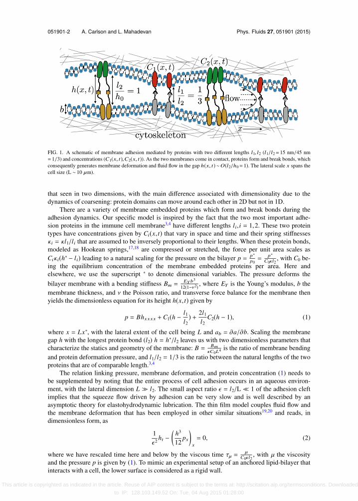
predicted to form at TS ≈ 25 − 500, i.e., in dimensional units TS ≈ 0.1 s − 2 s and the protein patternon the cell size to relax at TL ≈ 6 × 10⁵, i.e., in dimensional units TL ≈ 2400 s. Our numericalsimulations below corroborate these estimates, where micro-scale protein clusters form at shorttimes and coarsen on long length scales at much longer times as seen in Figure 3.
(a) (b) (c)
FIG. 3. Contour plots of the evolution of (a) membrane height h(x, t), (b) protein concentration C1(x, t), and (c) proteinconcentration C2(x, t) for two protein species i = 2 obtained by solving (1)-(4) for B = 2×10⁻⁸ and τ = 3. The membraneshape is initialized as h(t = 0, x)= 1+0.05× (tanh((10x−5))2−1.0), with protein concentrations corresponding to theirequilibrium values (Ceq
i (h(t = 0, x))). At short times, protein bonds nucleate on the membrane and are sorted into smallclusters. This generates membrane deformation and an inhomogeneous pressure that sets the intervening fluid in motion,causing the clusters to coarsen.
This article is copyrighted as indicated in the article. Reuse of AIP content is subject to the terms at: http://scitation.aip.org/termsconditions. Downloaded
to IP: 128.103.149.52 On: Tue, 04 Aug 2015 01:28:00
051901-6 A. Carlson and L. Mahadevan Phys. Fluids 27, 051901 (2015)
(a) (b)
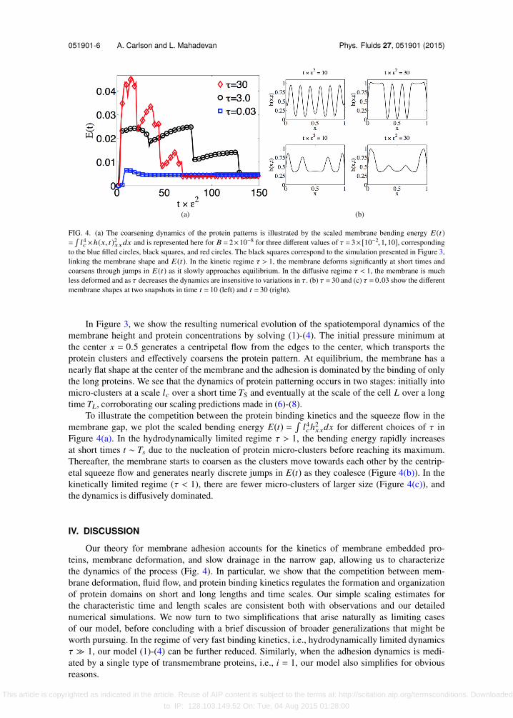
FIG. 4. (a) The coarsening dynamics of the protein patterns is illustrated by the scaled membrane bending energy E(t)=l4c×h(x, t)2xxdx and is represented here for B = 2×10⁻⁸ for three different values of τ = 3× [10−2,1,10], corresponding
to the blue filled circles, black squares, and red circles. The black squares correspond to the simulation presented in Figure 3,linking the membrane shape and E(t). In the kinetic regime τ > 1, the membrane deforms significantly at short times andcoarsens through jumps in E(t) as it slowly approaches equilibrium. In the diffusive regime τ < 1, the membrane is muchless deformed and as τ decreases the dynamics are insensitive to variations in τ. (b) τ = 30 and (c) τ = 0.03 show the differentmembrane shapes at two snapshots in time t = 10 (left) and t = 30 (right).
In Figure 3, we show the resulting numerical evolution of the spatiotemporal dynamics of themembrane height and protein concentrations by solving (1)-(4). The initial pressure minimum atthe center x = 0.5 generates a centripetal flow from the edges to the center, which transports theprotein clusters and effectively coarsens the protein pattern. At equilibrium, the membrane has anearly flat shape at the center of the membrane and the adhesion is dominated by the binding of onlythe long proteins. We see that the dynamics of protein patterning occurs in two stages: initially intomicro-clusters at a scale lc over a short time TS and eventually at the scale of the cell L over a longtime TL, corroborating our scaling predictions made in (6)-(8).
To illustrate the competition between the protein binding kinetics and the squeeze flow in themembrane gap, we plot the scaled bending energy E(t) =
l4ch2
xxdx for different choices of τ inFigure 4(a). In the hydrodynamically limited regime τ > 1, the bending energy rapidly increasesat short times t ∼ Ts due to the nucleation of protein micro-clusters before reaching its maximum.Thereafter, the membrane starts to coarsen as the clusters move towards each other by the centrip-etal squeeze flow and generates nearly discrete jumps in E(t) as they coalesce (Figure 4(b)). In thekinetically limited regime (τ < 1), there are fewer micro-clusters of larger size (Figure 4(c)), andthe dynamics is diffusively dominated.
IV. DISCUSSION
Our theory for membrane adhesion accounts for the kinetics of membrane embedded pro-teins, membrane deformation, and slow drainage in the narrow gap, allowing us to characterizethe dynamics of the process (Fig. 4). In particular, we show that the competition between mem-brane deformation, fluid flow, and protein binding kinetics regulates the formation and organizationof protein domains on short and long lengths and time scales. Our simple scaling estimates forthe characteristic time and length scales are consistent both with observations and our detailednumerical simulations. We now turn to two simplifications that arise naturally as limiting casesof our model, before concluding with a brief discussion of broader generalizations that might beworth pursuing. In the regime of very fast binding kinetics, i.e., hydrodynamically limited dynamicsτ ≫ 1, our model (1)-(4) can be further reduced. Similarly, when the adhesion dynamics is medi-ated by a single type of transmembrane proteins, i.e., i = 1, our model also simplifies for obviousreasons.
This article is copyrighted as indicated in the article. Reuse of AIP content is subject to the terms at: http://scitation.aip.org/termsconditions. Downloaded
to IP: 128.103.149.52 On: Tue, 04 Aug 2015 01:28:00

051901-7 A. Carlson and L. Mahadevan Phys. Fluids 27, 051901 (2015)
(a) (b)
FIG. 5. Comparison of full models (1)-(4) with simplified model (5), (9), and (10) for (a) the protein concentration C2(x, t)and (b) h(x, t) when B = 2×10⁻⁸ and τ = 30. (a) The steady-state membrane bound protein C2(x, t ×ϵ2= 15) is predicted by(1)-(4) and the analytical equilibrium concentration C
eq2 (h(x, t ×ϵ2= 15)) is predicted by (5) and agree well. h(x, t ×ϵ2= 15)
is used as input to (5). (b) The results of full models (1)-(4) are shown using solid lines and those using reduced models (5),(9), and (10) are shown using dashed lines at two different points in time t ×ϵ2= 7,28.
In the hydrodynamically limited regime of intercellular adhesion, clusters of membrane-boundproteins reorganize by domain coarsening similar to our observations.3 In this regime (τ ≫ 1), wecan reduce (3) and (4) by noting that the concentrations reach their equilibrium values quicklyso that Ci = Ceq
i (h(x, t)) in (5). To verify that this is a reasonable approximation, we compare theprediction from (5) and the numerical solution of the full model (1)-(4). In Figure 5(a), we show thatthis is indeed the case.
Next, we turn to the pressure in the membrane gap, which we obtain by combining (1) and (5),
peq = Bhxxxx + Ceq1 (h)(h − l1
l2) + 2l1
l2Ceq
2 (h)(h − 1). (9)
At equilibrium, peq = peq(x, t = ∞) = 0 and with (5), the equilibrium height field reduces to anordinary differential equation, which we have verified in additional simulations but not presentedhere. In fact, we can go beyond the equilibrium profiles by substituting p with peq in (2) to derivea simplified description for the spatiotemporal evolution of membrane shape and protein patterningthat is given by (5)-(9) and
1ϵ2 ht −
(h3
12peqx
)x
= 0. (10)
Direct comparison between the reduced mathematical model given by (5), (9), and (10) and fullmodel (1)-(4) for the membrane shape at different points in time are shown in Figure 5(b) and
(a) (b)
FIG. 6. (a) The three panels, starting from left to right, show the equilibrium membrane shapes as a function of B. Theseresults are obtained by solving (5), (9), and (10) numerically, assuming the membrane is pinned at the boundary (hxx
(x = 0, t)= hxx(x = 1, t)= 0,h(x = 0, t)= 0.5,h(x = 1, t)= 0.5) and with no fluid flux (px(0, t)= px(1, t)= 0). (b) To furthertest our scaling prediction for the characteristic membrane deformation lc ≈ B
14 , we systematically analyze the steady-state
membrane pattern as a function of B ∈ [10−10,10−5]. The dominant wavenumber q extracted from each simulation using afast-Fourier transform (square markers) corroborates our scaling prediction (6) with q ≈ 1
4π×lc (dashed line).
This article is copyrighted as indicated in the article. Reuse of AIP content is subject to the terms at: http://scitation.aip.org/termsconditions. Downloaded
to IP: 128.103.149.52 On: Tue, 04 Aug 2015 01:28:00

051901-8 A. Carlson and L. Mahadevan Phys. Fluids 27, 051901 (2015)
(a) (b)
FIG. 7. Contour plots of the evolution of (a) membrane height h(x, t), (b) protein concentration C1(x, t) for a singleprotein species i = 1, obtained by solving (1)-(4) for B = 2×10⁻⁸ and τ = 3. The membrane shape is initialized as h(t = 0, x)= 1+0.05× (tanh((10x−5))2−1.0), with protein concentrations corresponding to its equilibrium value (Ceq
1 (h(t = 0, x))).The membrane coarsening dynamics for a single type i = 1 of adhesion proteins is similar to the case with two protein typesi = 2 of different lengths as shown in Figure 3.
demonstrate that the reduced model gives an accurate representation of the spatiotemporal featuresin this limit.
To further test our scaling prediction for the characteristic membrane deformation lc in (6),we simulate the equilibrium membrane shape using reduced model (5), (9), and (10) and treat theboundaries as walls with no fluid flux, i.e., px(x = 0, t) = px(x = 1, t) = 0 and keeping the mem-brane pinned, i.e., hxx(x = 0, t) = hxx(x = 1, t) = 0, h(x = 0, t) = h(x = 1, t) = 0.5. The stationarymembrane shape in Figure 6(a) shows that the final pattern depends on B. A fast-Fourier transformof the equilibrium patterns is used to determine the characteristic wavenumber q and confirms ourscaling (6) q ≈ 1
lc(Figure 6(b)).
Another simplification of our model (1)-(4) arises by considering just one type of transmem-brane adhesion proteins, i.e., i = 1. In Figure 7, we show the results of our simulation for i = 1 andfind similar membrane coarsening dynamics as for the case with two protein types i = 2 of differentlengths (Figure 3); here too scaling relations (6)-(8) remain valid. However, we note that the timescale for the coarsening is much larger when i = 1, a consequence of a smaller driving force dueto a lower number of attached proteins. In addition, the averaged film height is smaller that alsogenerates a larger viscous resistance that slows the dynamics.
Our study has exposed a range of interesting phenomena that couple hydrodynamics to ki-netics and elastic deformations in thin films, problems that have a range of applications in biology,chemistry, and materials science. Natural next steps include quantifying the influence of thermalfluctuations, understanding the interplay between membrane tension and bending, and accountingfor the active dynamics of the membrane proteins linked to the cytoskeleton, while also accountingfor patterns that form when two-dimensional membranes adhere to a substrate.
ACKNOWLEDGMENTS
We acknowledge funding for this research provided by the Wyss Institute and Kavli In-stitute for BioNano Science and Technology. The computations in this paper were run on theOdyssey cluster supported by the FAS Division of Science, Research Computing Group at HarvardUniversity.
1 R. Parthasarathy and J. T. Groves, “Protein patterns at lipid bilayer junctions,” Proc. Natl. Acad. Sci. U. S. A. 101, 12798(2004).
2 J. T. Groves, “Bending mechanics and molecular organization in biological membranes,” Annu. Rev. Phys. Chem. 58, 697(2007).
3 A. Grakoui, S. K. Bromley, C. Sumen, M. M. Davis, A. S. Shaw, P. M. Allen, and M. L. Dustin, “The immunological synapse:A molecular machine controlling T cell activation,” Science 285, 221 (1999).
This article is copyrighted as indicated in the article. Reuse of AIP content is subject to the terms at: http://scitation.aip.org/termsconditions. Downloaded
to IP: 128.103.149.52 On: Tue, 04 Aug 2015 01:28:00
051901-9 A. Carlson and L. Mahadevan Phys. Fluids 27, 051901 (2015)
4 C. R. Monks, B. A. Freiberg, H. Kupfer, N. Sciaky, and A. Kupfer, “Three-dimensional segregation of supramolecularactivation clusters in T cells,” Nature 395, 82 (1998).
5 J. F. Allard, O. Dushek, D. D. Coombs, and P. A. Merwe, “Mechanical modulation of receptor-ligand interactions at cell–cellinterfaces,” Biophys. J. 102, 1265 (2012).
6 F. Y. Leong and K.-H. Chiam, “Adhesive dynamics of lubricated films,” Phys. Rev. E 81, 04923 (2010).7 S. Y. Qi, J. T. Groves, and A. K. Chakraborty, “Synaptic pattern formation during cellular recognition,” Proc. Natl. Acad.
Sci. U. S. A. 98, 6548 (2001).8 N. J. Burroughs and C. Wülfing, “Differential segregation in a cell–cell contact interface: The dynamics of the immunological
synapse,” Biophys. J. 83, 1784 (2002).9 N. J. Burroughs and P. A. van der Merwe, “Stochasticity and spatial heterogeneity in T-cell activation,” Immunol. Rev. 216,
69 (2007).10 T. R. Weikl, J. T. Groves, and R. Lipowsky, “Pattern formation during adhesion of multicomponent membranes,” EPL 59,
916 (2002).11 T. R. Weikl and R. Lipowsky, “Pattern formation during T-cell adhesion,” Biophys. J. 87, 3665 (2004).12 M. T. Figge and M. Meyer-Hermann, “Modeling receptor-ligand binding kinetics in immunological synapse formation,”
Eur. Phys. J. D 51, 153 (2009).13 M. J. Paszek, D. Boettiger, V. M. Weaver, and D. A. Hammer, “Integrin clustering is driven by mechanical resistance from
the glycocalyx and the substrate,” PLoS Comput. Biol. 5, e1000604 (2009).14 E. Reister-Gottfried, T. Bihr, U. Seifert, and A.-S. Smith, “Two intertwined facets of adherent membranes: Membrane
roughness and correlations between ligand-receptors bonds,” New J. Phys. 13, 025003 (2011).15 E. Reister-Gottfried, K. Sengupta, B. Lorz, E. Sackmann, U. Seifert, and A.-S. Smith, “Dynamics of specific vesicle-
substrate adhesion: From local events to global dynamics,” Phys. Rev. Lett. 101, 208103 (2008).16 T. Bihr, U. Seifert, and A.-S. Smith, “Nucleation of ligand-receptor domains in membrane adhesion,” Phys. Rev. Lett. 101,
258101 (2012).17 The protein stiffness can in certain instances be a function deformation/flow,18 but we use here the simplest description
with a constant protein spring stiffness.18 A. Salas, M. Shimaoka, A. N. Kogan, C. Harwood, U. H. von Andrian, and T. A. Springer, “Rolling adhesion through an
extended conformation of integrin αLβ2 and relation to α I and β I-like domain interaction,” Immunity 20, 1393 (2004).19 A. E. Hosoi and L. Mahadevan, “Peeling, healing, and bursting in a lubricated elastic sheet,” Phys. Rev. Lett. 93, 137802
(2007).20 A. Oron, S. H. Davis, and S. G. Bankoff, “Long-scale evolution of thin liquid films,” Rev. Mod. Phys. 69, 93 (1997).21 H. A. Kramer, “Brownian motion in a field of force and the diffusion model of chemical reactions,” Physica 7(4), 284 (1940).22 G. I. Bell, M. Dembo, and P. Bongrand, “Cell adhesion. Competition between nonspecific repulsion and specific bonding,”
Biophys. J. 45, 1051 (1984).23 B. Davidovitch, E. Moro, and H. A. Stone, “Spreading of viscous fluid drops on a solid substrate assisted by thermal
fluctuations,” Phys. Rev. Lett. 95, 244505 (2005).24 G. Grün, K. Mecke, and M. Rauscher, “Thin-film flow influenced by thermal noise,” J. Stat. Phys. 122, 1261 (2006).25 C. W. Gear, “Simultaneous numerical solution of differential–algebraic equations,” IEEE Trans. Circuit Theory 18, 89
(1971).26 C.-J. Hsu, W.-T. Hsieh, A. Waldman, F. Clarke, E. S. Huseby, J. K. Burkhardt, and T. Baumgart, “Ligand mobility modulates
immunological synapse formation and T cell activation,” PLoS One 7, e32398 (2012).27 B. Favier, N. J. Burroughs, L. Wedderburn, and S. Valitutti, “TCR dynamics on the surface of the living T cell,” Int. Immunol.
13, 1525–1532 (2001).28 R. Simson, E. Wallraff, J. Faix, J. Niewöhner, and G. Gerisch, “Membrane bending modulus and adhesion energy of
wild-type and mutant cells of dictyostelium lacking talin or cortexillins,” Biophys. J. 74, 514 (1998).
This article is copyrighted as indicated in the article. Reuse of AIP content is subject to the terms at: http://scitation.aip.org/termsconditions. Downloaded
to IP: 128.103.149.52 On: Tue, 04 Aug 2015 01:28:00


![Retinoic acid receptor gamma impacts cellular adhesion ......Integrin-mediated adhesion to the extracellular matrix stringently regulates cell cycle pro-gression [21, 22]. Integrins,](https://static.fdocuments.in/doc/165x107/5e92de562b69f522913c3786/retinoic-acid-receptor-gamma-impacts-cellular-adhesion-integrin-mediated.jpg)