Protein Bioinformatics Part I: Access to...
36
1 Protein Bioinformatics Part I: Access to information Jonathan Pevsner, Ph.D. [email protected] 260.655 March 30, 2010 Outline for today Introduction Accessing information Entrez Gene Accession numbers and RefSeq Protein Databases: UniProt, ExPASy Three genome browsers: NCBI, UCSC, Ensembl Four perspectives on individual proteins Perspective 1: Protein families (domains and motifs) Perspective 2: Physical properties (3D structure) Perspective 3: Localization Perspective 4: Function To provide students with the ability to analyze and understand data from high-throughput proteomics experiments. At the conclusion of the course the students will be able to: (a) Define protein physical properties and analyze protein structure. (b) Explain how proteins are studied experimentally and how data are generated in high-throughput experiments. (c) Describe the computational methods used to study protein structure and interactions. (d) Explain the algorithms, statistical techniques and software tools used to analyze high-throughput proteomics data. Course objectives
Transcript of Protein Bioinformatics Part I: Access to...

1
Protein Bioinformatics Part I: Access to information
Jonathan Pevsner, Ph.D. [email protected]
260.655 March 30, 2010
Outline for today
Introduction
Accessing information Entrez Gene Accession numbers and RefSeq Protein Databases: UniProt, ExPASy Three genome browsers: NCBI, UCSC, Ensembl
Four perspectives on individual proteins Perspective 1: Protein families (domains and motifs) Perspective 2: Physical properties (3D structure) Perspective 3: Localization Perspective 4: Function
To provide students with the ability to analyze and understand data from high-throughput proteomics experiments. At the conclusion of the course the students will be able to:
(a) Define protein physical properties and analyze protein structure.
(b) Explain how proteins are studied experimentally and how data are generated in high-throughput experiments.
(c) Describe the computational methods used to study protein structure and interactions.
(d) Explain the algorithms, statistical techniques and software tools used to analyze high-throughput proteomics data.
Course objectives

2
Syllabus (through April)
Tues 3/30 Protein bioinformatics I (Pevsner) Thurs 4/1 Protein bioinformatics II: Evolution (Pevsner)
Tues 4/6 Physical properties of amino acids (Prigge) Thurs 4/8 Protein structure essentials (Prigge)
Tues 4/13 How to visualize proteins (Prigge) Thurs 4/15 Why proteins fold (Prigge)
Tues 4/20 Structure determination and databases (Prigge) Thurs 4/22 Crystallography practicum (Prigge/Bosch)
Tues 4/27 Quantitative proteomics (Cole) Thurs 4/29 Proteomics and systems biology (Bosch)
Syllabus (through May)
Tues 5/4 Protein Structure: Databases & classification (Ruczinski)
Thurs 5/6 Protein secondary struct. prediction (Ruczinski)
Tues 5/11 Protein tertiary structure prediction (Ruczinski) Thurs 5/13 Protein structure prediction (CASP) (Ruczinski)
Tues 5/18 Review (Prigge/Ruczinski/Pevsner) Thurs 5/20 Final Exam + Practicum
Website
The course website is: http://www.biostat.jhsph.edu/~iruczins/teaching/
260.655/
(or Google “ingo teaching”)
3
Literature references
You are encouraged to read original source articles. They will enhance your understanding of the material. Readings are optional but recommended.
Computer labs
There are several computer labs (details to follow).
Grading
Grading is based on assignments and on a final exam.
4
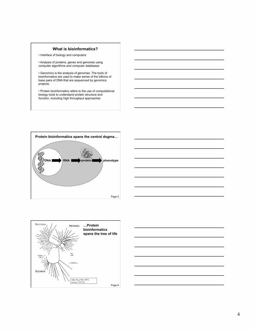
• Interface of biology and computers
• Analysis of proteins, genes and genomes using computer algorithms and computer databases
• Genomics is the analysis of genomes. The tools of bioinformatics are used to make sense of the billions of base pairs of DNA that are sequenced by genomics projects.
• Protein bioinformatics refers to the use of computational biology tools to understand protein structure and function, including high throughput approaches
What is bioinformatics?
DNA RNA phenotype protein
Page 5
Protein bioinformatics spans the central dogma…
After Pace NR (1997) Science 276:734
Page 6
…Protein bioinformatics spans the tree of life

5
Growth of GenBank + Whole Genome Shotgun (1982-November 2008)
Num
ber o
f seq
uenc
es
in G
enB
ank
(mill
ions
)
Bas
e pa
irs o
f DN
A in
Gen
Ban
k (b
illio
ns)
Bas
e pa
irs in
Gen
Ban
k +
WG
S (b
illio
ns)
0 20 40 60 80
100 120 140 160 180
200
1982 1992 2002 2008
Fig. 2.1 Page 15
Arrival of next-generation sequencing: approaching 100 terabases (100,000 gigabases) in 2009
Fig. 2.1 Page 15
DNA RNA
cDNA ESTs UniGene
phenotype
genomic DNA databases (infer protein sequences)
protein sequence databases
protein
Fig. 2.2 Page 18
6
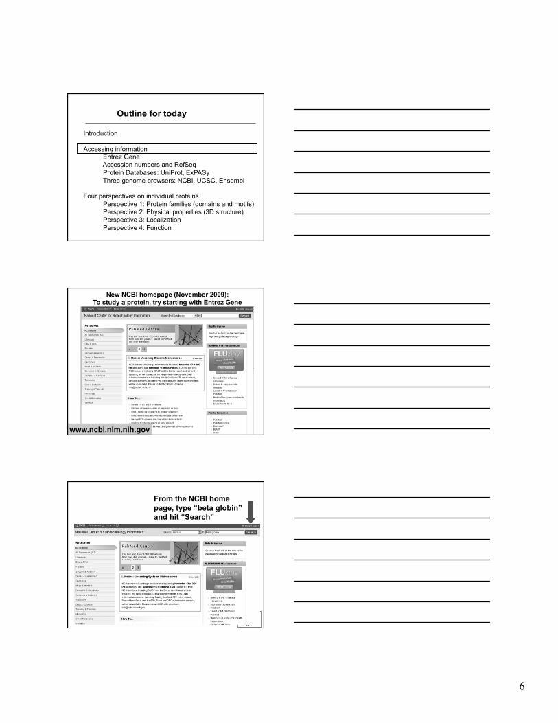
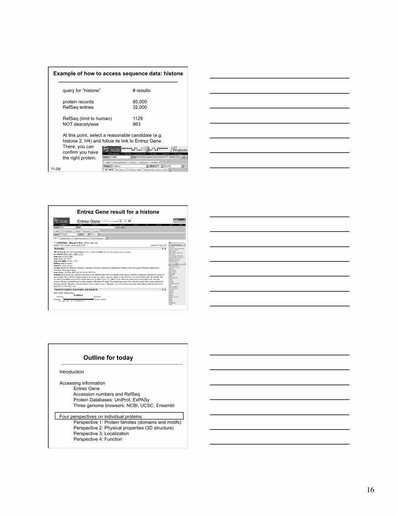
Outline for today
Introduction
Accessing information Entrez Gene Accession numbers and RefSeq Protein Databases: UniProt, ExPASy Three genome browsers: NCBI, UCSC, Ensembl
Four perspectives on individual proteins Perspective 1: Protein families (domains and motifs) Perspective 2: Physical properties (3D structure) Perspective 3: Localization Perspective 4: Function
www.ncbi.nlm.nih.gov
New NCBI homepage (November 2009): To study a protein, try starting with Entrez Gene
From the NCBI home page, type “beta globin” and hit “Search”
Fig. 2.5 Page 28

7
Fig. 2.5 Page 28
Follow the link to “Gene”
Entrez Gene is in the header Note the “Official Symbol” HBB for beta globin Note the “limits” option
Page 30
Entrez Gene (top of page)
Note that links to many other HBB database entries are available
8
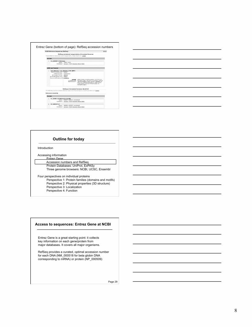
Entrez Gene (bottom of page): RefSeq accession numbers
Outline for today
Introduction
Accessing information Entrez Gene Accession numbers and RefSeq Protein Databases: UniProt, ExPASy Three genome browsers: NCBI, UCSC, Ensembl
Four perspectives on individual proteins Perspective 1: Protein families (domains and motifs) Perspective 2: Physical properties (3D structure) Perspective 3: Localization Perspective 4: Function
Access to sequences: Entrez Gene at NCBI
Entrez Gene is a great starting point: it collects key information on each gene/protein from major databases. It covers all major organisms.
RefSeq provides a curated, optimal accession number for each DNA (NM_000518 for beta globin DNA corresponding to mRNA) or protein (NP_000509)
Page 29

9
Accession numbers are labels for sequences
NCBI includes databases (such as GenBank) that contain information on DNA, RNA, or protein sequences. You may want to acquire information beginning with a query such as the name of a protein of interest, or the raw nucleotides comprising a DNA sequence of interest.
DNA sequences and other molecular data are tagged with accession numbers that are used to identify a sequence or other record relevant to molecular data.
Page 26
What is an accession number?
An accession number is label that used to identify a sequence. It is a string of letters and/or numbers that corresponds to a molecular sequence.
Examples (all for retinol-binding protein, RBP4):
X02775 GenBank genomic DNA sequence NT_030059 Genomic contig Rs7079946 dbSNP (single nucleotide polymorphism)
N91759.1 An expressed sequence tag (1 of 170) NM_006744 RefSeq DNA sequence (from a transcript)
NP_007635 RefSeq protein AAC02945 GenBank protein Q28369 SwissProt protein 1KT7 Protein Data Bank structure record
protein
DNA
RNA
Page 27
NCBI’s important RefSeq project: best representative sequences
RefSeq (accessible via the main page of NCBI) provides an expertly curated accession number that corresponds to the most stable, agreed-upon “reference” version of a sequence.
RefSeq identifiers include the following formats:
Complete genome NC_###### Complete chromosome NC_###### Genomic contig NT_###### mRNA (DNA format) NM_###### e.g. NM_006744 Protein NP_###### e.g. NP_006735
Page 27

10
Entrez Gene (bottom of page): non-RefSeq accessions (it’s unclear what these are, highlighting usefulness of RefSeq)
Fig. 2.8 Page 31
Entrez Protein: accession, organism, literature…
Fig. 2.8 Page 31
Entrez Protein: …features of a protein, and its sequence in the one-letter amino acid code

11
Page 31
Entrez Protein: You can change the display (as shown)…
FASTA format: versatile, compact with one header line
followed by a string of nucleotides or amino acids in the single letter code
Fig. 2.9 Page 32
Outline for today
Introduction
Accessing information Entrez Gene Accession numbers and RefSeq Protein Databases: UniProt, ExPASy Three genome browsers: NCBI, UCSC, Ensembl
Four perspectives on individual proteins Perspective 1: Protein families (domains and motifs) Perspective 2: Physical properties (3D structure) Perspective 3: Localization Perspective 4: Function

12
UniProt: a centralized protein database (uniprot.org)
Page 33
Fig. 2.10 Page 34
ExPASy: vast proteomics resources (www.expasy.ch)
Outline for today
Introduction
Accessing information Entrez Gene Accession numbers and RefSeq Protein Databases: UniProt, ExPASy Three genome browsers: NCBI, UCSC, Ensembl
Four perspectives on individual proteins Perspective 1: Protein families (domains and motifs) Perspective 2: Physical properties (3D structure) Perspective 3: Localization Perspective 4: Function
13
click human
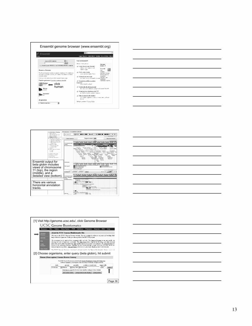
Ensembl genome browser (www.ensembl.org)
Ensembl output for beta globin includes views of chromosome 11 (top), the region (middle), and a detailed view (bottom).
There are various horizontal annotation tracks.
[1] Visit http://genome.ucsc.edu/, click Genome Browser
[2] Choose organisms, enter query (beta globin), hit submit
Page 36

14
[3] Choose the RefSeq beta globin gene
[4] The UCSC Genome Browser is an essential resource --choose which tracks to display --add custom tracks --the Table Browser is complementary
Example of how to access sequence data: HIV-1 pol
There are many possible approaches. Begin at the main page of NCBI, and type an Entrez query: hiv-1 pol
Page 36

15
11/09
Searching for HIV-1 pol: >130,000 nucleotide, protein hits
Searching for HIV-1 pol: using the command hiv-1[organism] limits the
output to just one entry
Try Taxonomy Browser to easily limit your query to your favorite organism(s). Example: NCBI home Taxonomy Taxonomy browser human protein to find a human protein
only 1 RefSeq
over 300,000 nucleotide entries for HIV-1
16
Example of how to access sequence data: histone
query for “histone” # results
protein records 85,000 RefSeq entries 32,000
RefSeq (limit to human) 1129 NOT deacetylase 863
At this point, select a reasonable candidate (e.g. histone 2, H4) and follow its link to Entrez Gene. There, you can confirm you have the right protein.
11-09
Entrez Gene result for a histone
Outline for today
Introduction
Accessing information Entrez Gene Accession numbers and RefSeq Protein Databases: UniProt, ExPASy Three genome browsers: NCBI, UCSC, Ensembl
Four perspectives on individual proteins Perspective 1: Protein families (domains and motifs) Perspective 2: Physical properties (3D structure) Perspective 3: Localization Perspective 4: Function

17
Perspective 1: Protein domains and motifs
Page 389
Definitions
Signature: • a protein category such as a domain or motif
Page 390
Definitions
Signature: • a protein category such as a domain or motif
Domain: • a region of a protein that can adopt a 3D structure • a fold • a family is a group of proteins that share a domain • examples: zinc finger domain immunoglobulin domain
Motif (or fingerprint): • a short, conserved region of a protein • typically 10 to 20 contiguous amino acid residues
Page 390
18
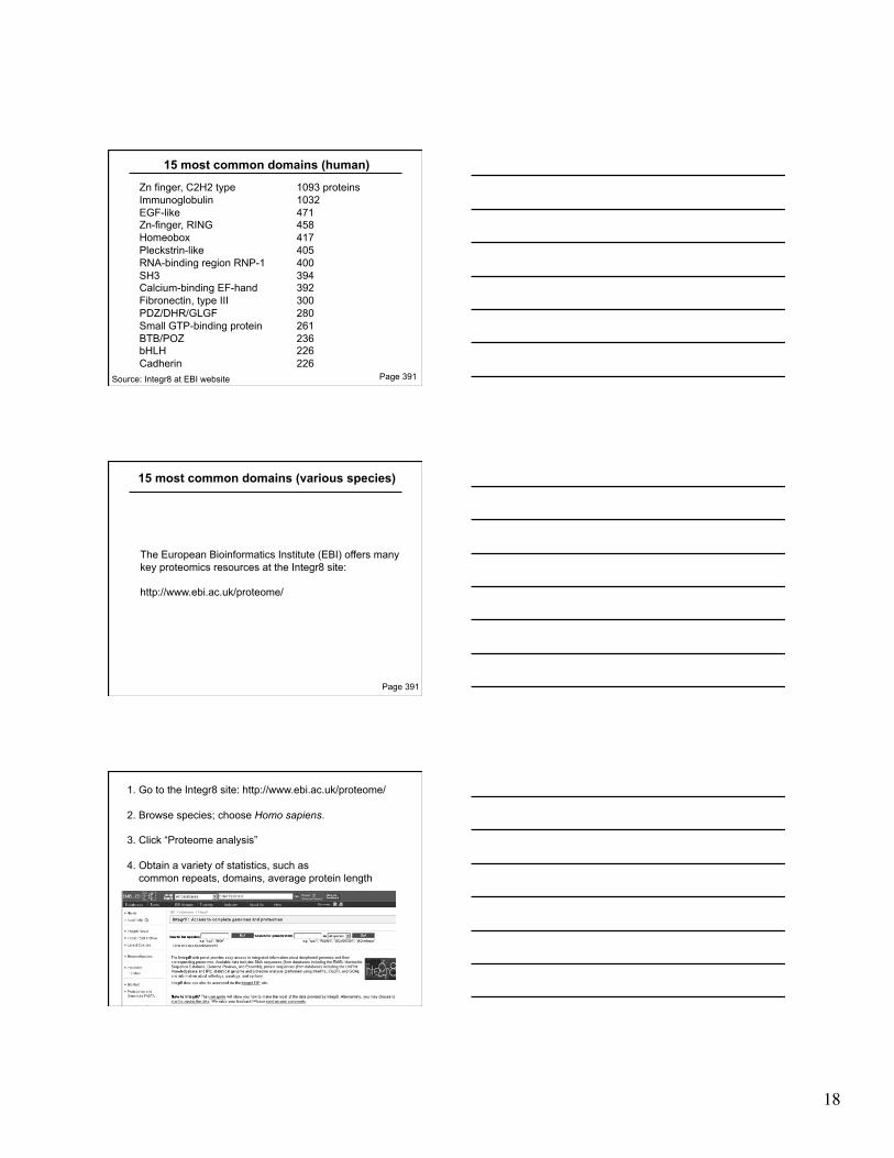
15 most common domains (human)
Zn finger, C2H2 type 1093 proteins Immunoglobulin 1032 EGF-like 471 Zn-finger, RING 458 Homeobox 417 Pleckstrin-like 405 RNA-binding region RNP-1 400 SH3 394 Calcium-binding EF-hand 392 Fibronectin, type III 300 PDZ/DHR/GLGF 280 Small GTP-binding protein 261 BTB/POZ 236 bHLH 226 Cadherin 226
Page 391 Source: Integr8 at EBI website
15 most common domains (various species)
The European Bioinformatics Institute (EBI) offers many key proteomics resources at the Integr8 site:
http://www.ebi.ac.uk/proteome/
Page 391
1. Go to the Integr8 site: http://www.ebi.ac.uk/proteome/
2. Browse species; choose Homo sapiens.
3. Click “Proteome analysis”
4. Obtain a variety of statistics, such as common repeats, domains, average protein length

19
Source: Integr8 at EBI website (updated 7/09)
amino acid
frequ
ency
Amino acid composition
Source: Integr8 at EBI website (updated 7/09)
Rel
ativ
e fre
quen
cy
protein length
Average protein length : 468+/- 522 amino acid residues Size range: 4 - 34350 amino acid residues
Definition of a domain
According to InterPro at EBI (http://www.ebi.ac.uk/interpro/):
A domain is an independent structural unit, found alone or in conjunction with other domains or repeats. Domains are evolutionarily related.
According to SMART (http://smart.embl-heidelberg.de):
A domain is a conserved structural entity with distinctive secondary structure content and a hydrophobic core. Homologous domains with common functions usually show sequence similarities.
Page 390

20
Varieties of protein domains
Page 393
Extending along the length of a protein
Occupying a subset of a protein sequence
Occurring one or more times
Example of a protein with domains: Methyl CpG binding protein 2 (MeCP2)
MBD
Page 393
TRD
The protein includes a methylated DNA binding domain (MBD) and a transcriptional repression domain (TRD). MeCP2 is a transcriptional repressor.
Mutations in the gene encoding MeCP2 cause Rett Syndrome, a neurological disorder affecting girls primarily.
Page 393
Result of an MeCP2 blastp search: A methyl-binding domain shared by several proteins
domain

21
Page 393
Are proteins that share only a domain homologous?
Proteins can have both domains and motifs (patterns)
Domain (aspartyl protease)
Domain (reverse transcriptase)
Motif (several residues)
Motif (several residues)
Page 396

22
Definition of a motif
A motif (or fingerprint) is a short, conserved region of a protein. Its size is often 10 to 20 amino acids.
Simple motifs include transmembrane domains and phosphorylation sites. These do not imply homology when found in a group of proteins.
PROSITE (www.expasy.org/prosite) is a dictionary of motifs (there are currently 1600 entries). In PROSITE, a pattern is a qualitative motif description (a protein either matches a pattern, or not). In contrast, a profile is a quantitative motif description. We will encounter profiles in Pfam, ProDom, SMART, and other databases.
Page 394
Summary of Perspective 1: Protein domains and motifs
A signature is a protein category such as a domain or motif.
You can learn about domains at Integr8, and at databases such as InterPro and Pfam.
A motif (or fingerprint) is a short, conserved sequence. You can study motifs at Prosite at ExPASy.
Perspective 2: Physical properties of proteins
Page 397

23
Page 398
Physical properties of proteins
Many websites are available for the analysis of individual proteins. ExPASy and ISREC are two excellent resources.
The accuracy of these programs is variable. Predictions based on primary amino acid sequence (such as molecular weight prediction) are likely to be more trustworthy. For many other properties (such as posttranslational modification of proteins by specific sugars), experimental evidence may be required rather than prediction algorithms.
Page 399
Page 399
Access a variety of protein analysis programs from the top right of the ExPASy home page

24
Page 400
Protein secondary structure
Protein secondary structure is determined by the amino acid side chains.
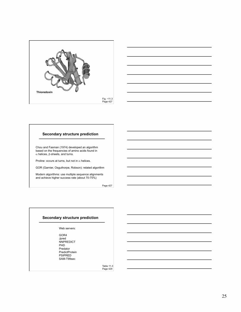
Myoglobin is an example of a protein having many α-helices. These are formed by amino acid stretches 4-40 residues in length.
Thioredoxin from E. coli is an example of a protein with many β sheets, formed from β strands composed of 5-10 residues. They are arranged in parallel or antiparallel orientations.
Page 425
Fig. 11.3 Page 427
Myoglobin (John Kendrew, 1958)
25
Thioredoxin
Fig. ~11.3 Page 427
Secondary structure prediction
Chou and Fasman (1974) developed an algorithm based on the frequencies of amino acids found in α helices, β-sheets, and turns.
Proline: occurs at turns, but not in α helices.
GOR (Garnier, Osguthorpe, Robson): related algorithm
Modern algorithms: use multiple sequence alignments and achieve higher success rate (about 70-75%)
Page 427
Secondary structure prediction
Web servers:
GOR4 Jpred NNPREDICT PHD Predator PredictProtein PSIPRED SAM-T99sec
Table 11-3 Page 429

26
Go to http://pbil.univ-lyon1.fr/, click “Secondary structure prediction” to access this prediction tool
Tertiary protein structure: protein folding
Main approaches:
[1] Experimental determination (X-ray crystallography, NMR)
[2] Prediction
► Comparative modeling (based on homology)
► Threading
► Ab initio (de novo) prediction
Page 430
Experimental approaches to protein structure
[1] X-ray crystallography -- Used to determine 80% of structures -- Requires high protein concentration -- Requires crystals -- Able to trace amino acid side chains -- Earliest structure solved was myoglobin
[2] NMR -- Magnetic field applied to proteins in solution -- Largest structures: 350 amino acids (40 kD) -- Does not require crystallization
Page 430

27
Steps in obtaining a protein structure
Target selection
Obtain, characterize protein
Determine, refine, model the structure
Deposit in repository Fig 11.5 page 431
The Protein Data Bank (PDB)
Page 434
• PDB is the principal repository for protein structures • Established in 1971 • Accessed at http://www.rcsb.org/pdb or simply http://www.pdb.org • Currently contains 64,000 structure entities
Updated 3/26/10
Fig. 11.7 Page 435

28
stru
ctur
es
Fig. 9.6 Page 281
year
Updated 3/26/10
PDB content growth (www.pdb.org)
1972 1980 1990 2000
yearly total
10,000
2010
50,000
30,000
PDB holdings (12/08)
50,621 proteins, peptides 2,225 protein/nucl. complexes 1,946 nucleic acids 33 other; carbohydrates 54,825 total
Table 11-4 Page 435
Fig. ~11.10 Page 436

29
Viewinghemoglobin(accession2H35)atPDB
ViewingstructuresatPDB:WebMol
Fig. 11.11 Page 437
Protein Data Bank
Swiss-Prot, NCBI, EMBL
CATH, Dali, SCOP, FSSP
gateways to access PDB files
databases that interpret PDB files
30
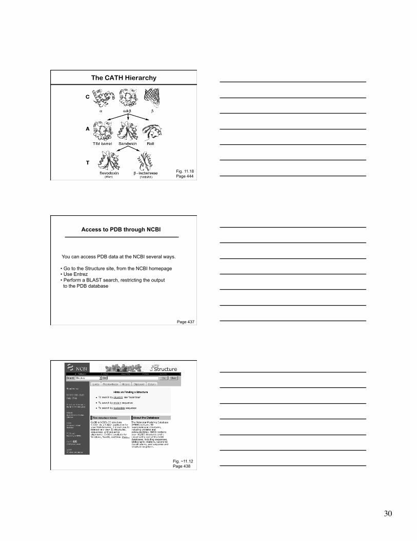
The CATH Hierarchy
Fig. 11.18 Page 444
Access to PDB through NCBI
Page 437
You can access PDB data at the NCBI several ways.
• Go to the Structure site, from the NCBI homepage • Use Entrez • Perform a BLAST search, restricting the output to the PDB database
Fig. ~11.12 Page 438

31
Do a blastp search; set the database to pdb (Protein Data Bank)
Structure links
Structure accession (e.g. 2JTZ)
Fig. 9.14 Page 289
32
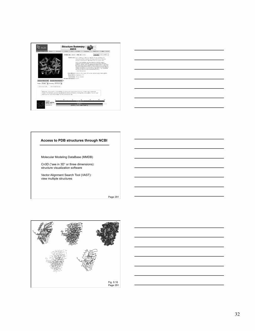
Access to PDB structures through NCBI
Page 291
Molecular Modeling DataBase (MMDB)
Cn3D (“see in 3D” or three dimensions): structure visualization software
Vector Alignment Search Tool (VAST): view multiple structures
Fig. 9.16 Page 291

33
Introduction to Perspectives 3 and 4: Gene Ontology (GO) Consortium
Page 237
The Gene Ontology Consortium
An ontology is a description of concepts. The GO Consortium compiles a dynamic, controlled vocabulary of terms related to gene products.
There are three organizing principles: Molecular function Biological process Cellular compartment
You can visit GO at http://www.geneontology.org. There is no centralized GO database. Instead, curators of organism-specific databases assign GO terms to gene products for each organism.
Page 237
Page 241
GO terms are assigned to Entrez Gene entries

34
Page 241
The Gene Ontology Consortium: Evidence Codes
IC Inferred by curator IDA Inferred from direct assay IEA Inferred from electronic annotation IEP Inferred from expression pattern IGI Inferred from genetic interaction IMP Inferred from mutant phenotype IPI Inferred from physical interaction ISS Inferred from sequence or structural similarity NAS Non-traceable author statement ND No biological data TAS Traceable author statement
Page 240
Perspective 3: Protein localization
Page 242

35
protein
Protein localization
Page 242
Protein localization
Proteins may be localized to intracellular compartments, cytosol, the plasma membrane, or they may be secreted. Many proteins shuttle between multiple compartments.
A variety of algorithms predict localization, but this is essentially a cell biological question.
Page 240

36
Page 242
Page 244