Protein bioelectronics: a review of what we do and do not know
158
Aug. 2017; to appear in Reports of Progress in Physics 1 Protein bioelectronics: a review of what we do and do not know Christopher D. Bostick, #1,2 Sabyasachi Mukhopadhyay, #3‡ Israel Pecht, 3 * Mordechai Sheves, 3 * David Cahen, 3 * David Lederman* 4 1 Dept. of Pharmaceutical Sciences, West Virginia University, Morgantown, WV 26506, USA 2 Inst. for Genomic Medicine, Columbia University Medical Center, New York, NY 10032, USA 3 Departments of Materials & Interfaces, of Organic Chemistry and of Immunology, Weizmann Institute of Science, Rehovot, Israel 76100 4 Department of Physics, University of California, Santa Cruz, CA 95060, USA # These authors contributed equally to the work * Corresponding authors, email addresses: [email protected], [email protected], [email protected], [email protected] Abstract We review the status of protein-based molecular electronics. First, we define and discuss fundamental concepts of electron transfer and transport in and across proteins and proposed mechanisms for these processes. We then describe the immobilization of proteins to solid-state surfaces in both nanoscale and macroscopic approaches, and highlight how different methodologies can alter protein electronic properties. Because immobilizing proteins while retaining biological activity is crucial to the successful development of bioelectronic devices, we discuss this process at length. We briefly discuss computational predictions and their connection to experimental results. We then summarize how the biological activity of immobilized proteins is beneficial for bioelectronic devices, and how conductance measurements can shed light on protein properties. Finally, we consider how the research to date could influence the development of future bioelectronic devices. ‡ Current affiliation - Department of Physics, School of Engineering & Applied Sciences, SRM University-AP, Amaravati, Andhra Pradesh, India - 522502
Transcript of Protein bioelectronics: a review of what we do and do not know

Aug. 2017; to appear in Reports of Progress in Physics
1
Protein bioelectronics: a review of what we do and do not know
Christopher D. Bostick,#1,2 Sabyasachi Mukhopadhyay,#3‡ Israel
Pecht,3* Mordechai Sheves,3* David Cahen,3* David Lederman*4
1 Dept. of Pharmaceutical Sciences, West Virginia University, Morgantown, WV 26506, USA
2 Inst. for Genomic Medicine, Columbia University Medical Center, New York, NY 10032, USA
3 Departments of Materials & Interfaces, of Organic Chemistry and of Immunology,
Weizmann Institute of Science, Rehovot, Israel 76100
4 Department of Physics, University of California, Santa Cruz, CA 95060, USA
# These authors contributed equally to the work
* Corresponding authors, email addresses: [email protected],
[email protected], [email protected], [email protected]
Abstract
We review the status of protein-based molecular electronics. First, we define and
discuss fundamental concepts of electron transfer and transport in and across proteins and
proposed mechanisms for these processes. We then describe the immobilization of
proteins to solid-state surfaces in both nanoscale and macroscopic approaches, and
highlight how different methodologies can alter protein electronic properties. Because
immobilizing proteins while retaining biological activity is crucial to the successful
development of bioelectronic devices, we discuss this process at length. We briefly discuss
computational predictions and their connection to experimental results. We then
summarize how the biological activity of immobilized proteins is beneficial for
bioelectronic devices, and how conductance measurements can shed light on protein
properties. Finally, we consider how the research to date could influence the development
of future bioelectronic devices.
‡ Current affiliation - Department of Physics, School of Engineering & Applied Sciences, SRM University-AP, Amaravati, Andhra Pradesh, India - 522502

Aug. 2017; to appear in Reports of Progress in Physics
2
Abstract ...................................................................................................................................................................... 1
1. Introduction .................................................................................................................................................... 6
2 Electron Transfer (ET) and Electron Transport (ETp) in Proteins .......................................... 8
2.1 Electron Transfer ............................................................................................................................... 13
2.2 Electron Transport ............................................................................................................................ 22
2.3 Protein Structure, Electron Transfer (ET) and Electron Transport (ETp) ................. 26
2.4 General Properties of Immobilized Proteins .......................................................................... 30
3. Methods of Protein Immobilization ........................................................................................................ 32
3.1 Direct Adsorption .................................................................................................................................... 33
3.2 Modified electrodes ................................................................................................................................ 35
3.3. Protein Tethering Through Linker .................................................................................................. 40
3.4 Break-junction, Nanoscopic techniques ......................................................................................... 42
3.5. Macroscopic and permanent contact measurement ................................................................ 46
3.6 Measuring protein adsorption and functionality on a surface.............................................. 48
3.6.1 Optical techniques ............................................................................................................................... 48
3.6.2 Scanning Probe Technique .............................................................................................................. 53
3.7 Artifacts in ET and ETp measurements .......................................................................................... 57
4. Electronic and structural properties of immobilized proteins................................................ 60
4.1 What can be expected experimentally from electronic and structural studies of
properties by computational methods? ................................................................................................ 60
4.2 Effects of immobilization on protein structure & electronic properties ..................... 66
4.2.1 Effects of choice of modified substrate surfaces on electron transfer ................. 66
4.2.2 Effects of modified substrate surfaces on protein conformation and electron
transport ........................................................................................................................................................ 70
4.3 Electronic Measurements: Interpreting results in terms of ET & ETp .............................. 73

Aug. 2017; to appear in Reports of Progress in Physics
3
4.3.1 Electron transfer and transport across immobilized proteins ..................................... 73
4.3.2 Biological relevance of electron transfer and transport measurements .................. 75
4.3.3 Protein structure and electron transport measurements .............................................. 79
4.3.4 Molecular vibronic contribution on electron transport measurements .................. 82
4.3.5 Opto-electronic properties of immobilized protein.......................................................... 85
4.3.6 Spintronic properties .................................................................................................................... 95
References ........................................................................................................................................................... 104

Aug. 2017; to appear in Reports of Progress in Physics
4
Glossary: AFM: Atomic force microscopy apo: protein without removed cofactor CP-AFM: Conductive Probe Atomic Force Microscopy CD: Circular Dichroism Cofactor: non-peptide organic or inorganic group or ion in protein, necessary for protein’s biological activity; can have/ be aromatic rings, conjugated chains, metal ions CPR: Cytochrome P450 Reductase CV: Cyclic Voltammetry CYP2C9: cytochrome P450 2C9 EC-STM: Electrochemical Scanning Tunneling Microscopy EDC: 1-Ethyl-3-[3-dimethylaminopropyl]carbodiimide Hydrochloride ET: Electron Transfer ETp: Electron Transport FAD: Flavine Adenine Dinucleotide FMN: Flavin Mononucleotide FR: Flickering Resonance GCE: Glassy Carbon Electrodes Hab: interaction Hamiltonian Holo: Fully intact protein, including cofactor HSA Human Serum Albumin Ka: Binding Association on rate KD: Binding Dissociation Constant Kd: Binding Dissociation off rate kET: Electron transfer rate LbL: Layer-by-Layer LOFO: Lift-Off Float-On Mb Myoglobin protein MUA: 11-mercaptoundecanoic acid NADPH: Nicotinamide Adenine Dinucleotide Phosphate NHE: normal hydrogen electrode NHS: N-hydroxysuccinimide P450: Cytochrome P450 PDMS Polydimethylsiloxane PFV: Protein Film Voltammetry PG: Pyrolytic Graphite Prosthetic group: tightly bound cofactor PSI: Photosynthetic Protein I QCM: Quartz Crystal Microbalance RCI: Photosynthetic Reaction Center I SAM: Self Assembled Monolayer SCE: Saturated calomel electrode SE: Superexchange

Aug. 2017; to appear in Reports of Progress in Physics
5
SERRS: Surface-Enhanced Resonance Raman Spectroscopy SPM Scanning Probe Microscopy SPR: Surface Plasmon Resonance STM: Scanning tunneling Microscopy STS: Scanning Tunneling Spectroscopy turnover rate: rate of protein biological activity for small molecule transformation
(exp. Oxidation or reduction) UV-Vis: Ultraviolet-Visible Spectroscopy YCC: yeast cytochrome c WT Wild Type, i.e., natural form of the protein
β: Distance decay factor in tunneling transport [ln (𝐼
𝐼0) ∝ −𝛽𝑑 ; 𝑑 is distance]
γCPR: Yeast Cytochrome P450 Reductase ϵ: Optical extinction coefficient

Aug. 2017; to appear in Reports of Progress in Physics
6
1. Introduction
Recent advances in the development of biocompatible electronics have been motivated
by applications where interfacing with sensory function in humans (e.g., artificial retinas)
is desired, some of which can have direct applications for improving human health (e.g., in
situ, real time monitoring of glucose or drug concentration in the blood). A fundamental
understanding of the properties of biomaterials at the nanoscale is essential for progress
towards interfacing with sub-cellular organelles or other nanoscale structures present in
living systems. Moreover, because many biologically relevant reactions occur at substrate
surfaces and interfaces, exploring interactions between individual biomolecules and
various substrate surfaces is of fundamental importance.
Proteins have evolved over billions of years into structures capable of precise
reactions, including highly specific substrate recognition, analyte binding, and facile and
directional electron tunneling.1 In addition, proteins are essential to many chemical
processes essential to life, such as photosynthesis, respiration, water oxidation, molecular
oxygen reduction, and nitrogen fixation.2–7 Due to their ability to catalyze a vast number of
reactions, there has been much interest in harnessing their abilities. One application is in
bio-catalysis in synthetic organic chemistry. The high chemo-, regio-, and stereo-selectivity
of enzymes negates the need for protecting groups, and thus reduces synthetic steps and
simplifies work flow.8 As such, the pharmaceutical industry is interested in proteins in
bioreactors (vessels in which chemical reactions are carried out by biological components)
for production of fine chemicals 9–13 and to filter out toxic or hazardous substances by
naturally occurring organisms.14 The high specificity of proteins also makes them ideal
candidates for use as sensors. In many sensor applications, proteins are immobilized onto a
solid support and act as transducers of optical15,16 or electronic17 signals that correlate with
specific protein-ligand interactions. Interfacing with solid state electronics has
demonstrated that this approach allows biosensing at low voltages with high sensitivities
and low detection limits, and offers long-term stability for measurements.18–20 Protein-
based biosensors are currently being developed for use in drug monitoring,21
environmental toxin detection,22,23 and early disease detection.24,25

Aug. 2017; to appear in Reports of Progress in Physics
7
Using single organic molecules as electronic components26 has been suggested as a
possible solution to the physical limitations of micro- and nano-scale electronic
development. Proteins could provide a way to realize this, because they are small (nm-
scale) and capable of undergoing chemo-mechanical, electromechanical, opto-mechanical,
and optoelectronic processes.27 Proteins have been studied for use in the design of bio-
electronic devices, including field effect transistors for sensing,28–32 bio-molecular
transistors for data storage,33 bio-molecular circuits,34 bio-hybrid solar cells,35 bio-
computers,36 and enzymatic biofuel cells.37–39 Biomedical applications exist in devices such
as implantable sensors for monitoring chemicals40,41 and diagnostics,42 as well as the
creation of artificial retinas43 and noses.44 There has been extensive research done on
electron transfer in proteins45–47 especially in well-characterized model systems such as
cytochrome c,48 plastocyanin49, and azurin.50–53 Other studies have explored the solid state
electronic transport properties of proteins whose mechanisms of action rely on electron
transfer, ET.54,55
Several books,28,54–59 recent special issues of journals (from which we cite results
published therein51,60), and reviews51,61,1,47,62,63 have appeared over the years, going back to
the early64 and late 1990s (1st edn. of reference 55). All deal with various aspects of the
topics considered here and, taken together, they reflect to some extent the evolution of the
idea of protein bioelectronics. Recent interest in possible applications of new protein
electronics have laid bare practical problems and stimulated interest in their fundamental
scientific issues.
In parallel, the area of ET has remained very active, and we rely heavily in this
review on our present understanding of ET to discuss what can be learned from protein
bioelectronics devices. We therefore start with a presentation of fundamental concepts
related to the electronic properties of proteins as they relate to ET and electron transport,
ETp, (cf. next section for an explanation of the difference between ET and ETp) and
compare the two processes. Because until now all ETp measurements involve immobilizing
proteins on an electronically conducting solid surface used as an electrode, we review the
current state of the field regarding protein immobilization techniques, including
characterization of the resulting biomolecular films. Subsequently, we discuss the results of
measurements on immobilized proteins and their impact on our fundamental

Aug. 2017; to appear in Reports of Progress in Physics
8
understanding of protein electronics. Finally, the implications of the research to date and
their possible implications for future protein bioelectronic devices are noted and reviewed.
2 Electron Transfer (ET) and Electron Transport (ETp) in
Proteins
Electron flow through a protein molecule involves intramolecular charge transport
and electron exchange with the surroundings.65 These two distinct, and widely studied
processes cannot be unambiguously separated since the interface of a molecule with an
adjacent electrode or ionic solution can have a profound influence on the molecular
properties, and hence on the molecule’s electrical conductance.66 The process of directed
electron motion, or electron flow, through molecular structures is termed electron transport or
electron transfer, depending on the environment in which it occurs. In the discussion below, we
denote as electron transfer (ET) the electron flow process with all or part of the protein in direct
contact with an electrolyte, which is ionically conducting and which can function as an electron
sink or source via a redox process. The electrolyte also provides charges, which screen the
change in electrical potential due to the electron flow. Electron flow in which an electrolyte is
absent, or does not participate in the electron flow process, with electrodes that are not ion
conductors, is termed electron transport (ETp). ETp is usually measured in a solid state
configuration.67 In ETp normally there is no possibility for charge balance via ions from an
electrolyte, while ET involves a surrounding medium with mobile ions which can provide such
charge balance.
Aug. 2017; to appear in Reports of Progress in Physics
9
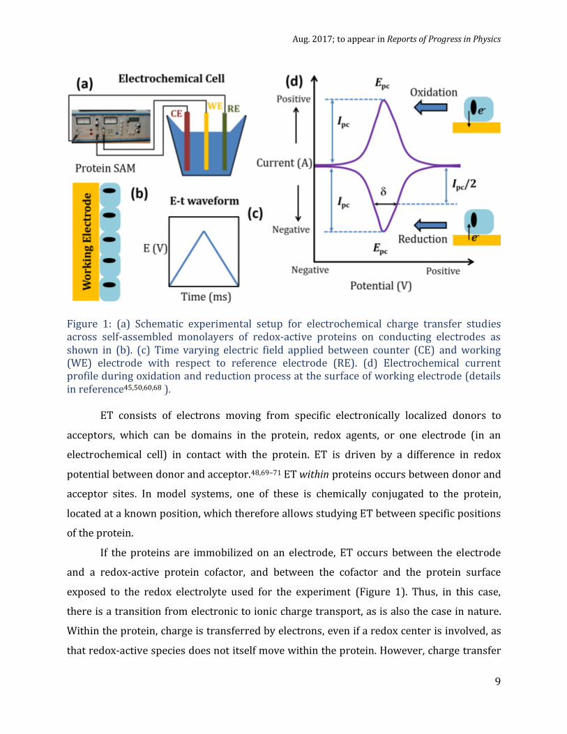
Figure 1: (a) Schematic experimental setup for electrochemical charge transfer studies across self-assembled monolayers of redox-active proteins on conducting electrodes as shown in (b). (c) Time varying electric field applied between counter (CE) and working (WE) electrode with respect to reference electrode (RE). (d) Electrochemical current profile during oxidation and reduction process at the surface of working electrode (details in reference45,50,60,68 ).
ET consists of electrons moving from specific electronically localized donors to
acceptors, which can be domains in the protein, redox agents, or one electrode (in an
electrochemical cell) in contact with the protein. ET is driven by a difference in redox
potential between donor and acceptor.48,69–71 ET within proteins occurs between donor and
acceptor sites. In model systems, one of these is chemically conjugated to the protein,
located at a known position, which therefore allows studying ET between specific positions
of the protein.
If the proteins are immobilized on an electrode, ET occurs between the electrode
and a redox-active protein cofactor, and between the cofactor and the protein surface
exposed to the redox electrolyte used for the experiment (Figure 1). Thus, in this case,
there is a transition from electronic to ionic charge transport, as is also the case in nature.
Within the protein, charge is transferred by electrons, even if a redox center is involved, as
that redox-active species does not itself move within the protein. However, charge transfer

Aug. 2017; to appear in Reports of Progress in Physics
10
out of the protein requires reduction /oxidation of a redox-active species in the electrolyte
that contacts the protein. This means that the redox-active species is now ionized
differently than before, and thus the charge will flow with this ionic species until the next
step in the overall process, as is the case in electron transport chains (discussed further
below). The change of charge state of the redox-active species in the electrolyte is
accompanied by a change in the protein’s electrical charge state. This change induces
electrical screening to reduce the electrostatic energy barrier for the subsequent steps. In
nature and in an electrochemical experiment, this can occur because of the surrounding
electrolyte. Naturally, the polarizability of the protein itself also affects the ET process.
Multiple ET processes occur in proteins, such as in respiration or photosynthetic
complexes, that involve electron transport chains.72 Proton-coupled electron transport
(PCET) can also occur, which is especially prevalent at metal cofactors that activate carbon,
oxygen, nitrogen, and sulphur atoms of enzyme substrate molecules.73 Protein cofactors
are organic or inorganic components, with conjugated chains, or metal ions that assist the
protein’s biological activity. As noted above, redox reactions with biomolecules such as
proteins can be studied with electrochemical experiments as shown in Figure 1, i.e., with
one electronically conducting, ionically blocking contact (the electrode) and one ionically
conducting, electronically blocking contact (the redox electrolyte). ETp, however, requires
electronic conduction across proteins set between two electronically conducting, ionically
blocking electrodes; it is defined as the flow of electrons across/through the protein that is
contacted by these electrodes (Figure 2).74
Electric charges in biological systems are generally transported by ions, and thus in
nature ET is always coupled to ionic transport where the redox processes serve as donor
(RedOx) and acceptor (Ox Red) electrical “contacts”. Thus, from a device perspective,
there is no a priori reason for electrons involved in (solid-state) ETp to use the same
conduction mechanism as those involved in ET processes. Nevertheless, there is
experimental evidence that a protein’s ETp characteristics are correlated with its ET and
redox potential,75,76 and therefore it is likely that there is a fundamental connection
between the ET and ETp processes and mechanisms.
From an experimental point of view (Figure 2), measuring ETp across bio-molecules
in dry, solid state condition is fundamentally different from measuring ET. In ETp, the

Aug. 2017; to appear in Reports of Progress in Physics
11
absence of a liquid electrolyte (with its above-noted ability for electrical screening) will
normally drive electrons from source to drain electrode, without intra-protein redox
process, via some electron transport mechanism (to be discussed later in this review).
ETp is measured on a single protein or on ensembles of proteins (such as
monolayers) between the two electronically conducting electrodes (cf. ref.77 for a
discussion of the differences between molecular junctions with single and many
molecules). While this type of junction can be thought of as a donor-bridge-acceptor
junction, the driving force of the transport is the electrical potential difference between the
electrodes (if the electrodes are made of the same material78) rather than a difference in
chemical potential of the electrons between two regions. In solution experiments, the
chemical potential differences of the redox-active ions need to be included.47
For practical purposes, the redox potential can be identified with the
electrochemical potential of the electron, μ̃e (≃ EF, Fermi level),79–83 where μ̃e = μe + qϕ, the
sum of the contribution of the electrical potential, ϕ, (q is the electron charge) and that of
the electron’s chemical potential, μe = μ0,e + kT ln ne. Here μ0,e is the standard chemical
potential of the electron, kT is the thermal energy, and ne is the electron concentration,
defined by the difference in activities of the reduced and the oxidized form of the redox
species. The latter can be identified as the difference in chemical potential of the (mostly
ionic) reduced and oxidized species, and it can drive ET, but not ETp (with electrodes of the
same materials, i.e., with the same electron chemical potential).
An additional important difference between ET and ETp is the fact that ET occurs in
an electrolyte solution, which allows for electrostatic screening around (parts of) the
protein. This is what makes the change in charge of the protein, which otherwise would
carry a very high electrostatic energy price, possible. Except for rare cases, no such change
in charge state occurs in ETp and electroneutrality is preserved. These differences allow
the characterization of ETp distances not considered possible in biological ET. Gray,
Winkler, Dutton, and co-workers have estimated the upper limit of D-A separation
distances in biological ET (pure quantum mechanical tunneling) to be ≤20 Å and ≤14 Å,
respectively.61,84 However, recent ETp measurements across proteins yield measurable
currents over electrode separations of up to 100 Å.85

Aug. 2017; to appear in Reports of Progress in Physics
12
Figure 2: Diagram representing solid state electron transport (ETp) and electron transfer (ET) measurements using spectroscopy and electrochemistry (see introduction to section 2 for more complete distinction between ET and ETp). The very important counter charges around the protein are not shown in the ET configurations.
Cofactors are responsible for the functional properties of proteins because they
enable redox activity, including the metal center of metalloproteins, such as Cu2+ in the blue
copper proteins, Fe3+ in the iron-sulfur proteins and cytochromes, or flavins found in
flavoproteins.64 Some redox-inactive proteins, such as the light-driven transmembrane
proteins bacteriorhodopsin and halorhodopsin, which contain a retinal cofactor, also
support ETp, which, remarkably, is at least as efficient as that of redox-active proteins.
In measurements of the differences in ETp observed between the holo- and apo-
forms of proteins, their electrical conduction properties were shown to depend strongly on
having the cofactors. Such experiments have been performed with the Cu redox protein
azurin,1,53 the heme-redox protein cytochrome C,86 the O2-binding protein myoglobin,32,87
and ferritin.88 Similarly, changes in transport mechanisms have been deduced from
temperature-dependent ETp studies on the proton or Cl¯ pumping photoactive membrane
proteins, bacteriorhodopsin, bR,89 or halorhodopsin, phR,90 and their derivatives lacking
retinal, for bR, or retinal and/or carotenoid for phR.

Aug. 2017; to appear in Reports of Progress in Physics
13
As noted above, multiple ET processes, often involving several proteins and other
redox molecules, carry electron transfer over relatively large distances. In the
respiratory91,92 and photosynthetic chains ( 50 Å),93–96, ET occurs sequentially across
several proteins (inter-protein, or protein-protein ET97,98); in hydrogenases (FeS clusters,
~ 52 Å),99 carbon monoxide-dehydrogenases 100,101 and other enzymes,102 the processes
are within one protein complex. This complicated and diverse behavior has made it clear
that it is important to develop a theoretical understanding of biological ET and its possible
implications to solid-state ETp.
In the following sub-sections, we review the mechanisms that have been developed
to explain ET essentially in terms of the Marcus model and various mechanisms that rely on
it. Then we briefly discuss ETp in terms of the Landauer one-dimensional conduction
model, which has been applied to molecular electronics, while keeping in mind that once
electrons enter the protein, ET-like mechanisms may play an important role. ETp is
discussed in more detail in terms of specific experiments in Section 4. We end this section
by discussing the effects of protein structure and protein immobilization on ET and ETp.
2.1 Electron Transfer
In 1956, Marcus presented a model describing ET reactions from a donor to an acceptor
that is close to it.103,104 The Marcus theory was originally developed to explain outer sphere
ET, where participating redox centers are not linked by any bridge, and electrons “hop”
from the reducing center to the acceptor. It has since been extended to cover inner sphere
ET, where two redox centers are linked covalently during transfer, and heterogeneous ET,
where an electron moves between a biomolecule (protein, small molecule, cofactor, etc.)
and an electrical contact. In the Marcus description, nuclear motions of the reactant and the
surrounding environment are approximated by a simple harmonic oscillator potential
along a reaction coordinate (Figure 3).105,106 The left parabola in Figure 3A represents the
Gibbs free energy surfaces for the nuclear motion of the reactants prior to ET, and the right
parabola represents this free energy for the nuclear motion of the products after ET. The
two driving parameters for the ET reaction to overcome the activation free-energy barrier
(-ΔG‡) are the driving force, characterized by the Gibbs free energy of activation (-ΔGᵒ),

Aug. 2017; to appear in Reports of Progress in Physics
14
determined from the difference in oxidation potentials of the donor and acceptor, and the
reorganization energy (λ) needed for the nuclear rearrangements that accompany ET.107
The rate of electron transfer, kET, depends on -ΔGᵒ relative to λ. The basic expression for kET
in the Marcus theory is
𝑘𝐸𝑇 = 2𝜋
ℏ𝐻𝐴𝐷
2 1
√4𝜋𝜆𝑘𝐵𝑇exp (
−(𝜆+∆𝐺ᵒ)2
4𝜆𝑘𝐵𝑇) (1)
Figure 3. (A) Schematic diagram of the Marcus model for electron transfer, showing the Gibbs free energy (y-axis) surface for the nuclear motion along a reaction coordinate (X-axis) of the donor and acceptor in the initial state, prior to ET (G, DA in blue), and post-ET from the donor to the acceptor (G, D+A-, green). ΔG‡, ΔG0, and λ are the activation energy, the free energy driving force, and the outer shell reorganization energy of the ET event, respectively. B) A diagram of the Gibbs free energy surface as in A) showing the effect of increasing ΔG0 on the ΔG‡. As ΔG0 increases, a decrease in ΔG‡ is seen until ΔG‡=0 (green curve). After this point increases in ΔG0 (from the green to red curve) lead to decreases in ΔG‡ as energy must be dissipated for an ET event, which is known as the inverted region. Note that λ does not change for the different scenarios in B), because the nuclear coordinate (X-axis) of the post-ET state does not change. Adapted with permission from reference 106
where HAD is the quantum mechanical electronic coupling between the initial and final
(donor and acceptor) states, kB is the Boltzmann constant, and T is the temperature. A
maximum, or optimal ET rate occurs for activation-less ET (-ΔGᵒ = λ) that decreases with
decreasing driving force. The Marcus theory also predicts an inverted region (-ΔGᵒ > λ)
where energy must be dissipated to allow ET and where the rate decreases with increasing

Aug. 2017; to appear in Reports of Progress in Physics
15
driving force. According to the semi-classical Marcus’ theory, electron transfer takes place
when a thermal fluctuation of the solvent or of the nuclear coordinate of the donor and
acceptor shifts the donor and acceptor electronic states into resonance. The quantity HAD in
eq. (1) originates from Fermi’s Golden Rule. HAD contains the interaction overlap integral
(matrix element) between the donor (reactant) and acceptor (product) electronic states
and the density of states, and thus depends on the distance between the states (cf. also eqn.
(2) below).72,108 The interaction between the donor and acceptor in a polarizable solvent is
known as the outer-sphere interaction. When electron transfer occurs within molecules
where it causes changes in bond lengths and local symmetries, a similar result is obtained,
although the origin of the interaction energy is more complicated. This result related to the
so-called inner-sphere interactions was developed by N. Hush,109, and the joint theory of
the inner- and outer-sphere interactions is sometimes called the Marcus-Hush theory
(section 4).
Over the years, several theoretical frameworks and computational tools have been
developed to explain the biological electron transfer process.110 In particular, there has
been much interest in understanding how the protein structure determines the protein
redox function, the implication being that enhanced comprehension could allow for control
and tailored biological electron transfer by targeted mutants. The field has evolved
significantly since the early ideas of bridge-mediated electron tunneling, first proposed by
Halpern et al. in the early 1960s, and the square barrier-tunneling model that Hopfield
proposed in the 1970s. 108,111
Table – 1: Summary of experimentally obtained ET rate constant for different
metalloproteins (see text for discussion)a
Protein
Electrochemical rate constant (ET)
kET (s-1)
References Spectroscopy rate constant (ET)
kET(s-1)
References
Cyt-C 0.2(b) 112 9.4105(b) 61
0.63(c) 112 2.7106(c) 61 Myoglobin 60 113,114 2.3×10⁶ 61
a The difference between the two entries for Cyt-C result from experiments with different donor-acceptor separations (different mutations), ~14 Å (b) and ~17 Å (c).

Aug. 2017; to appear in Reports of Progress in Physics
16
Many experimental studies have been carried out to understand how long-range
biological ET (over, at least, several direct bond distances) is accomplished.45,46,61,84,103,115
ET rate has been determined in cytochromes from line broadening, magnetization transfer,
or relaxation measurements in NMR.116,117 In addition, kET has been extracted from
electrochemistry of a monolayer of a protein (azurin, cytochrome C, myoglobin and other
metalloproteins) attached to a working electrode (Figure 1d).118 Until now, most conclusive
and systematic ET rate (kET) data have been obtained via electrochemical112–114,119 or
spectroscopic flash quench studies.45,61,65,97,119–121. Table 1 gives some experimental data
for two well-studied proteins, obtained by using mutated proteins to vary the donor-
acceptor separations.
Gray, Winkler and co-workers have used modified metalloproteins to understand
distance-dependent ET between a dye molecule and a metal ion that acts as quencher in
Azurin and cytochrome b562 proteins (Figure 4) using flash-quenching, where a foreign
dye is introduced on a protein surface and ET processes between redox centers of the
protein and dye molecules are monitored.122.65,102 By introducing ruthenium and rhenium
complexes as donors which are known to attach to certain histidine residues at known
distances from Cu (azurin), Fe (in the heme group in cytochrome) or Zn (modified
cytochrome) cofactors, they were able to show experimentally (Figure 4) that
𝑘𝐸𝑇 ∝ 𝑒−𝛽𝐿 , (2)
where L represents the donor-acceptor distance and 1/𝛽 is a characteristic decay distance.
Studies by Gray and Winkler123 demonstrated that out of nine kET values for histidine-
modified cyt b562 derivatives, seven were accurately fit by the tunneling model, but two
showed kET slower than predicted by Equation 2, as shown in Figure 4b. This implies that a
different transfer mechanism is at work for those residues. Surprisingly, protein electron
transfer rates deduced from electrochemical and spectroscopic methods were significantly
different (Table 1). This difference may indicate that protein immobilization on solid
surfaces used in the electrochemical methods constrains some of the electron transfer
pathways, while in spectroscopic methods performed in solution, more protein
conformations and transfer pathways are available. Alternatively, or additionally, the

Aug. 2017; to appear in Reports of Progress in Physics
17
switch from electronic to ionic conduction (assuming the redox center to solution step is
the rate-determining one) may affect the measured rate.
Another useful approach to investigate ET in proteins has been pulse radiolysis,
primarily those containing transition metal ions in the active sites. There were two main,
complementary objectives guiding these studies. One was attaining an understanding of the
electron-transfer process within the polypeptide matrix separating the redox centers and
defining the parameters controlling their rates. The second was resolving the detailed
mechanism of the function(s) performed by the protein, notably enzymes.122 Azurin has
been a model system for pursuing the first objective and was extensively investigated using
pulse radiolysis. Triggering an intramolecular ET from the residual ion to the Cu(II) site
over a 2.7-nm separation turned out to be useful for examining the impact of specific
structural differences introduced by mutations on ET rates. Parameters examined ranged
from changes in the medium separating the redox sites to those in the actual coordination
of copper site, which drastically affected its reorganization energy.124–127
Over the years, more sophisticated theoretical models have been developed to
understand ET in proteins, including bridge-mediated electron tunneling111 and square
barrier tunneling.108 These models establish the theory of ET between fixed sites within the
proteins through electron tunneling. In the case of redox proteins, multiple electron
transfer pathways often exist, and pathways can be highly dependent on protein structure
and the surrounding environment. Refinements by Beratan et al.128,129 of these early
models show that electrons can tunnel across proteins through favorable pathway(s),
which include mostly bonded groups, with less favorable non-bonded interactions being
important when the through-bond pathway is prohibitively long and a shorter through-
space path exists.130

Aug. 2017; to appear in Reports of Progress in Physics
18
Figure 4. (a) Ribbon diagram indicating the positions of the nine histidine sites on cyt b562, where a Ru complex was connected to different histidine groups in different mutants, to measure variations in electrochemical electron transfer rates between each of these sites and the heme group. (b) Seven ET rates follow the exponential distance dependency of Equation 1, whereas in two cases slower rates than predicted by Equation 1 were measured. (c) Chemical reaction to connect the Ru complex with different histidines in different mutants. Published with permission from ref. 131.
A square-barrier tunneling model, combined with a suitable decay constant, has
given a surprisingly good description for experimentally obtained biological ET (Figure
2).132 This model was subsequently refined by Beratan and co-workers, who proposed that
electrons tunnel along specific pathways, connecting electron donating and accepting
cofactors.132 The theory of ET in biochemical systems with several intermediary tunneling
(bridge) states was recently reviewed by Blumberger ,110 who summarized several models
that are currently considered viable for explaining electron transfer in biomolecules, all
based on the Marcus theory. These are the super-exchange (multi-step tunneling),
flickering resonance (proposed by Beratan, Skourtis, and coworkers 133; vide infra), and

Aug. 2017; to appear in Reports of Progress in Physics
19
hopping models, based on either hopping and/or tunneling as mechanisms of electron
transfer (Figure 5).
Figure 5: (a) Illustration of protein-mediated electron transfer models, tunneling (via super-exchange), flickering resonance and hopping, along a chain of 5 redox active sites (as
could be the case in a multi-heme or multi-copper protein), where D is the electron donor and A
is the acceptor (A), leaving three sites (1, 2, 3) between D and A. One-electron energy levels are
drawn as black lines for each site (cf. Fig. 5.2 for inclusion of vibrational broadening) The
electron that is transferring from D to A is shown as a Gaussian. In (a) thermal fluctuations bring
D and A levels into resonance (middle scheme), which allows tunneling from D to A. Also
during these process sites 1,2, 3 are non-resonant; they enhance tunneling but are not
significantly occupied by the tunneling electron at any time, i.e., there is no nuclear relaxation as
a result of electron occupation. In (b) ET occurs only when all levels are in resonance and the
tunneling electron transfers ballistically (with tunneling probability of 1). In (c) 133 D and nearest
neighbor site 1 become resonant, allowing efficient electron tunneling from D to 1, and so forth
till A. Adapted with permission from reference 110, Blumberger et al. Copyright © 2015,
American Chemical Society.
In the superexchange model, the donor and acceptor energy levels are brought into
resonance via bridge energy states, and then direct tunneling occurs, bypassing the

Aug. 2017; to appear in Reports of Progress in Physics
20
intermediate energy levels completely. Superexchange therefore provides a model for
direct tunneling, with a dependence of the electron transfer rate on distance between
donor and acceptor, R, given by
𝑘𝐸𝑇 = 𝐴𝑒−𝛽(𝑅−Δ𝑅) (3)
where Δ𝑅 is the distance between intermediate states, and 1/𝛽 is a characteristic decay
length. The proportionality constant 𝐴 depends on temperature as 𝐴 ∝ 𝑇−1/2exp(−𝑏/𝑘𝐵𝑇),
where b is a constant that has units of energy, but is independent of R and Δ𝑅, while 𝛽 ∝
1/Δ𝑅 and is temperature-independent.
The flickering resonance (FR) model, proposed by Beratan, Skourtis, and coworkers,
considers the dynamical nature of the system (cf. also discussion in section 4.1).133 For
flickering resonance to occur, the energies of the donor, acceptor, and intermediate energy
levels need all to be brought into resonance by thermal fluctuations to allow coherent
tunneling (hopping) between the donor and acceptor sites. In this case, the transfer rate
has the same distance dependence as in Eq. 3, with the same 𝛽 ∝ 1/Δ𝑅 dependence. The
proportionality constant 𝐴 has essentially the same temperature dependence as for the
superexchange mechanism, but with a 𝐴 ∝ (𝑅/Δ𝑅)−1 dependence for its upper bound
value. Although it has not been possible so far to unequivocally identify flickering
resonance in experimental data, this model might in principle be applicable to ETp in
biomolecules if it is sufficiently efficient over long distances (10s of nm to m). Moreover,
biological ET often involves multiple groups and redox cofactors in van der Waals contact
with each other, and coupling rates are highly sensitive to conformational fluctuations.
Thus, the flickering resonance model may be relevant to understand the electrical
conductance in bacterial nanowires and multiheme proteins, with strongly coupled
porphyrin arrays with closely packed (≲15 Å) redox groups.134,135
For larger distances, ET is assumed to occur primarily via hopping. In this mechanism,
the electron incoherently hops from site to site in a sequential fashion, with a certain
probability of hopping backwards and forwards. The transfer rate for this model is
expected to depend on R as
𝑘𝐻𝑜𝑝 = 𝐴/(𝑐 + 𝑅/Δ𝑅), (4)

Aug. 2017; to appear in Reports of Progress in Physics
21
where c is independent of temperature and 𝐴 ∝ exp(−𝑏/𝑘𝐵𝑇). The hopping mechanism is
thought to be especially relevant for longer biomolecules, such as membrane proteins
(Figure 6),51,61 where the electron transfer distance between donor and acceptor is more
than ~2 nm. The rate of hopping can also be described by the relation
𝑘𝐸𝑇 ∝ 𝑁𝑥 , (5)
where 𝑁 ∼ 𝑅/Δ𝑅 is the total number of steps and 𝑥 depends on whether the steps are
reversible or irreversible, and has a value between 1 and 2.107,136 It has been proposed that
in ribonucleotide reductases, responsible for catalyzing the conversion of nucleotides to
deoxynucleotides in all organisms, the long ET distance of 35 Å is covered via short
electron hopping steps between conserved aromatic amino acids, rather than a single super
exchange step (as illustrated in figure 7).137
Figure 6: Diagram comparing electron transport by superexchange and hopping across single α-helical peptide at nanoscopic configuration. Reprinted with permission from reference 138 with permission of the Royal Chemistry Society. (b) Diagram of electron transport process across dehydrated protein monolayer sandwiched between two macroscopic metal electrodes. SAM: self-assembled monolayer; linker: small molecule with terminal groups to bind to the protein and to the substrate. The protein shown is Azurin.

Aug. 2017; to appear in Reports of Progress in Physics
22
2.2 Electron Transport
The fundamental mechanisms of ETp via proteins are less understood than those of
ET processes. One important reason is that, as discussed above, ETp measurements are not
done in solution, and therefore there are no ionic charges in the medium surrounding the
protein to screen charging as the electron moves across the protein. Another reason is that
the interaction between the metal electrodes, which effectively act as initial and final donor
and acceptor states, are defined by the Fermi level (electrochemical potential) of the
electrons of the metals used, rather than by discrete molecular orbitals. Nevertheless, some
of the ET ideas and models discussed above (Figure 5) have been used as starting points to
describe ETp from a microscopic point of view. Some of the most widely accepted ETp
models based on ET ideas are illustrated in Figure 7. The top panel describes a scheme with
donor and acceptor states (as in ET) that can be the two electrodes of a molecular junction
in ETp. Different amino acid residues, which may also include a cofactor, affect the
potential that the electron encounters as it is transported through the protein. The lower
four panels in Figure 7 are highly schematic illustrations of one-dimensional modes of ETp
under an applied electrical potential between the right and left electrodes, according to
different proposed model. In the hopping model, transport between electrodes occurs via
intermediate sites that are potential wells for the electron, requiring activation energy to
escape, i.e., this process is always incoherent and dissipative. We note that this model is not
the same as the Hubbard model for the hopping mechanism in solid state physics, which
relies on an effective repulsive potential between sites.
Using the Marcus model as a starting point (Eq. 1), the conductance in the hopping
model is temperature dependent: ∝ 𝑇−1/2exp(−𝑏/𝑘𝐵𝑇). In the tunneling via superexchange
model, transport is coherent in a similar fashion to the superexchange model used in ET,
but because the Fermi levels of the electrodes are tuned by the externally applied electric
potential, it does not rely on a spontaneous thermal alignment of D-A levels. As a result, the
temperature dependence related to the D-A energies is different in ETp, and the tunneling
barrier is influenced by the energy levels of the intervening medium.

Aug. 2017; to appear in Reports of Progress in Physics
23
The term sequential tunneling has been and is used in different publications for
different processes and, as a result, its use is ambiguous. It has been stated that sequential
tunneling refers to hopping (cf. e.g., ref. 72) or that it is equivalent to resonant tunneling.139
Büttiker considered the issue, and concluded that tunneling probability through a barrier
can in general consist of coherent and an incoherent parts.140 The incoherent part, where
inelastic processes occur, destroys phase coherence. In a completely incoherent tunneling
process, the electron can scatter backwards or forwards. According to Büttiker, purely
sequential (incoherent) tunneling occurs if tunneling occurs solely through an inelastic
channel; on the other hand, if tunneling occurs through the coherent channel, coherent
resonant tunneling occurs. In general, a tunneling process with intervening energy states is
composed of both incoherent (sequential) and coherent tunneling channels. In this
situation, Büttiker has demonstrated that if the two channels are connected in series, they
are not independent of each other. In other words, increasing the sequential tunneling
probability affects the coherent channel probability.
Whether the transport process is coherent or incoherent depends mainly on the
residence time of the electron at the intermediate electronic states available for transport
in the medium between the electrodes. For example, if the electron spends enough time in
the resonant state so that a nuclear relaxation accompanies this process (a time 𝑡 ∼ 1/𝑓,
where 𝑓 is the characteristic frequency of the vibrational energy associated with state),
inelastic tunneling will occur. It is also important to note that while inelastic transport is
always incoherent, elastic transport can in general be coherent or incoherent.
Randomization of the phase of the electron during elastic, incoherent tunneling can occur,
for example, because of elastic scattering that randomizes the electron’s momentum
direction without changing its magnitude, as in a diffusive process. Another example is
when the resonant energy level is sufficiently narrow that the electron is effectively
localized for a significant amount of time during which memory of the phase is lost. Thus,
depending on what is meant by sequential tunneling, the process can be temperature
dependent (hopping) or not. Therefore, whenever the term “sequential tunneling” is
encountered, caveat lector!
The flickering resonance model was described in the ET discussion above and is
further discussed in section 4.1. The main difference between the FR and sequential

Aug. 2017; to appear in Reports of Progress in Physics
24
tunneling models is that flickering resonance is a coherent tunneling process involving
several intermediate resonant energy states, where the electron does not spend much time
within each intermediate energy state, and the entire process is elastic and is described by
a single wave function within the barrier.
In all four cases, the energy levels of the intervening states inside the protein are
generally considered to be thermally broadened, instead of the sharp resonance states used
for the ET models shown in Figure 5.
Aug. 2017; to appear in Reports of Progress in Physics
25
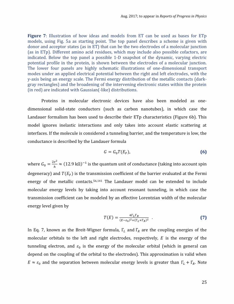
Figure 7: Illustration of how ideas and models from ET can be used as bases for ETp models, using Fig. 5a as starting point. The top panel describes a scheme is given with donor and acceptor states (as in ET) that can be the two electrodes of a molecular junction (as in ETp). Different amino acid residues, which may include also possible cofactors, are indicated. Below the top panel a possible 1-D snapshot of the dynamic, varying electric potential profile in the protein, is shown between the electrodes of a molecular junction. The lower four panels are highly schematic illustrations of one-dimensional transport modes under an applied electrical potential between the right and left electrodes, with the y-axis being an energy scale. The Fermi energy distribution of the metallic contacts (dark-gray rectangles) and the broadening of the intervening electronic states within the protein (in red) are indicated with Gaussian(-like) distributions.
Proteins in molecular electronic devices have also been modeled as one-
dimensional solid-state conductors (such as carbon nanotubes), in which case the
Landauer formalism has been used to describe their ETp characteristics (Figure 6b). This
model ignores inelastic interactions and only takes into account elastic scattering at
interfaces. If the molecule is considered a tunneling barrier, and the temperature is low, the
conductance is described by the Landauer formula
𝐺 = 𝐺0𝑇(𝐸𝐹), (6)
where 𝐺0 =2𝑒2
ℎ≈ (12.9 kΩ)−1 is the quantum unit of conductance (taking into account spin
degeneracy) and 𝑇(𝐸𝐹) is the transmission coefficient of the barrier evaluated at the Fermi
energy of the metallic contacts.56,141 The Landauer model can be extended to include
molecular energy levels by taking into account resonant tunneling, in which case the
transmission coefficient can be modeled by an effective Lorentzian width of the molecular
energy level given by
𝑇(𝐸) =4Γ𝐿Γ𝑅
(𝐸−𝜀0)2+(Γ𝐿+Γ𝑅)2 . (7)
In Eq. 7, known as the Breit-Wigner formula, Γ𝐿 and Γ𝑅 are the coupling energies of the
molecular orbitals to the left and right electrodes, respectively, 𝐸 is the energy of the
tunneling electron, and 𝜀0 is the energy of the molecular orbital (which in general can
depend on the coupling of the orbital to the electrodes). This approximation is valid when
𝐸 ≈ 𝜀0 and the separation between molecular energy levels is greater than Γ𝐿 + Γ𝑅. Note

Aug. 2017; to appear in Reports of Progress in Physics
26
that if 𝐸 = 𝜀0 and Γ𝐿 = Γ𝑅, 𝑇(𝐸) = 1, that is, perfect resonance occurs which results in
ballistic transport.
We reiterate that in this review we focus on describing protein ETp (electron
transport occurring in dry, solid state conditions) and on making further connections with
ET (electron transfer that occurs in conditions where electrolytes play a role in the
process) and protein biological function. It is important to note that in order to make real
ETp measurements, it is necessary to immobilize the protein (unlike in measurements
made in solution). This adds complexity to the problem because attachment of the protein
to an electrode can itself change its electronic structure. This can occur as a result of
hybridization with the metallic electrical contacts and/or the formation of Schottky
barriers and image charges in the contacts. These interactions can change the electronic
structure directly or by causing a configurational change that in many cases can render the
protein biologically inactive. Therefore, understanding the effects of immobilization on
proteins on their electronic structure is crucial to the interpretation of real experiments
where ETp is measured. We discuss the effects of protein structure and immobilization of
proteins in general on their ET and ETp properties below.
2.3 Protein Structure, Electron Transfer (ET) and Electron Transport (ETp)
Biological functions of proteins are dependent on their structure in a fundamental
way. As a result, preserving protein structure and chemical functionalities upon
immobilization on a solid surface is key for studying both ET and ETp. In this section, we
discuss some fundamental concepts related to protein structure that are relevant to ET and
ETp.
Protein structure can be understood at several levels, the first being the primary
structure, which is the linear sequence of amino acids, 2HN-(H)C(R)-CO(OH), held together
by covalent, amide (or peptide) bonds, -(H)N-C(O)-, forming a polypeptide chain. The next
level is the secondary structure, where the polypeptide chains form higher order three-
dimensional features (e.g. α-helices and β-sheets or turns) based on hydrogen bonding
between amino acid peptide bonds. Lastly, a tertiary structure is formed by additional
secondary structure elements that undergo the necessary folding by specific interactions,
including formation of salt bridges, hydrogen bonds, and disulfide bonds, to achieve free

Aug. 2017; to appear in Reports of Progress in Physics
27
energy optimization (lowest entropy) in the solvent, giving rise to a compact structure.
Proteins are generally amphiphilic, i.e., they contain both hydrophobic and hydrophilic
parts, with hydrophilic amino acids forming a protective shell via folding to minimize the
exposure of hydrophobic amino acids to the solvent. In the case of multi-subunit proteins,
where the sub-units are stabilized by the same interactions as the tertiary structure, a
quaternary structure refers to the global structure of the connected subunits. Multi-subunit
proteins are multi-protein complexes, with different complexes having different degrees of
stability over time to achieve a specific set of functions. A well-known example is
hemoglobin, which is a tetramer composed of four sub-units with a high degree of stability.
The oxidation-reduction potential of redox-active proteins is extremely sensitive to
the structure of the polypeptides. For example, a single point mutation or minimal
alteration of the secondary or tertiary structure of redox proteins can change the redox
potential by 100 mV or more.142 Conditions that break bonds or disrupt electrostatic
interaction within proteins may result in a loss of the structural order of proteins, a process
known as denaturation. Denatured proteins display some or complete loss of protein
function.
Enzymes are proteins that act as macromolecular biological catalysts, and thus
accelerate chemical reactions without themselves being altered by the reaction, often by
binding to small molecules. An example of how enzymes work can be seen in the
cytochrome family when discerning differences among members of the cytochrome P450s.
Cytochrome P450 is denoted by “CYP” followed by a number placing it in a gene family, a
letter that denotes a sub family. An individual gene receives a second number. Thus, the
name CYP2C9 represents a cytochrome P450 in the ‘2’ family, the ‘C’ subfamily, and
individual gene ‘9’. Both cytochrome c and cytochrome P450 are heme-containing
metalloproteins (with Fe as the metal ion) that have comparable redox potentials for ET.
P450s contain an active site that allows binding of small molecules, while cytochrome c
does not. Different small molecules bind specifically to different P450s, coordinated to the
heme group, and are known to alter the spin state of the heme Fe.143,144 Studies have shown
that the presence of small molecules in the active site alters the rate of ET in multiple
P450s144 and our research has demonstrated that changes both in ET rate and ETp

Aug. 2017; to appear in Reports of Progress in Physics
28
efficiencies of P450 from CYP2C9 occur when small molecules are bound inside the active
site.145–147
Electron transport across dry protein monolayers has been studied using solid-state
protein-based molecular junctions, with macroscopic and nanoscopic current-voltage (I-V)
methods.1,47,54,148,149 Results have been reported for several protein types, such as those
functioning as ET mediators, azurin (Az) and cytochrome c (CytC), or bacteriorhodopsin
(bR), a light-driven H+ pump protein, and proteins lacking any bound cofactor (bovine and
human serum albumin, BSA and HSA). Despite some general similarities, these proteins
differed in their ETp behavior. For instance, proteins with bound cofactors produce higher
currents than those that lack it. These observations indicate not only that the primary
sequence of the amino acid, which is mainly responsible for secondary structure is
important, but also that the presence of a cofactor creates relatively efficient current
transport paths and affects ETp in solid-state measurements.
The importance of the cofactor has also been demonstrated in studies of proteins for
which the apo form, from which the cofactors was removed, can be generated while
retaining structural integrity. Specific examples are azurin, myoglobin and CytC, which
have undergone rigorous studies in different forms. In the apo form, electrochemical redox
properties are absent for all metalloproteins, and electron conduction efficiencies are
reduced by only one to a two orders of magnitude compared to the holo-protein, when
both are measured in the dry solid state monolayer form.87 For CytC, removing the heme
group leads to denaturation, but it is possible to remove only the Fe (from the heme group),
and this was found NOT to affect ETp at all, in contrast to its effect on ET, which showed
loss of redox activity.150 Cofactors often serve as the elements that facilitate
superexchange-mediated tunneling for electronic charge carriers and protein-electrode
electronic coupling, and alterations of these groups can affect ETp significantly.151 Nanogap
measurements of single myoglobin molecules in solid state demonstrated resonant
tunneling through the heme group with a strong dependence on gate voltage only for the
holo- but not the apo-protein.32 In section 4.3.2 of the impact of cofactors on ETp is
discussed further.
For electrochemical studies (Figure 1), intramolecular electron transfer in redox
proteins specifically relates to the binding sites of redox prosthetic groups and redox-active

Aug. 2017; to appear in Reports of Progress in Physics
29
amino acids and their locations with respect to the working electrode, which influence the
electronic coupling between the redox-active center and the electrode contact
point.48,50,150,152 “Doping” human serum albumin protein with hemin (porphyrin,
protoporphyrin IX, with Fe3+; in heme, the metal ion is Fe2+) or retinoic acid enhances ETp
efficiencies by more than an order of magnitude in solid state molecular junctions; with
hemin, this protein also becomes redox-active in an electrochemical cell. The natural heme-
containing protein, Cyt C, shows a symmetric cyclic voltammogram with an ET rate
constant of ~18 s-1 (measured between the heme and the Au electrode), while the HSA–
hemin complex exhibits an asymmetric curve with a somewhat lower ET rate constant of ~
5 s-1.150,153 In case of redox proteins we note that the cofactors carry out the redox
reactions, which need not be so for solid-state ETp (details in Section 4). 154
In solid-state ETp across protein monolayers, electronic coupling (Γ, cf. eq. 7) to
electrodes dramatically influences its efficiencies and the electron flow mechanisms.155,156
Experimentally, the thermal activation energy for ETp is found to be governed by Γ for all
proteins that we have studied. This is illustrated by results on immobilized cytochrome c
junctions, for which the thermal activation energy is reduced by approximately a factor of
two, if it is covalently rather than electrostatically bound to the electrode.156 In the case of
metalloproteins, electronic coupling is mainly controlled by the relative position of the
redox cofactor w.r.t. the electrodes, and by its oxidation state.154,156 Covalent protein–
electrode binding (hybridization) increases Γ, in the tunneling transport regime (30 – 150
K). Proteins that are covalently bound to the electrode via a cysteine thiolate yield ten
times higher ETp efficiency than those that are electrostatically adsorbed on the
surface.47,86,156 Coordination changes of metal groups in different oxidation states also
affects ETp. For example, the Fe in the heme group in myoglobin and cytochrome P450
(P450) undergoes a change in coordination upon ET that leads to a change between Fe3+
and Fe2+ oxidation states, which have different spin states. This type of coordination change
is common in heme-containing proteins, and is known to have profound effects on both ET
and ETp.51 Another important consideration is how the cofactor is coupled in the ET
pathway. Prytkova et al.131 proposed that the anomalously slow ET rates seen by Gray and
Winkler in their histidine-modified cyt b562 derivatives (His 12 and His 73, see Figure 4)
could be explained by different coupling pathways to the heme group. In the case of these

Aug. 2017; to appear in Reports of Progress in Physics
30
two slow ET rates, there is a pathway coupled to the ligand, axial (perpendicular to) the
heme plane, as opposed to the other seven predicted rates with a pathway coupled to the
heme edge.
An increase in transport efficiency was observed across a myoglobin monolayer on
silicon if Mb was oriented by chemical binding of its hemin group to the substrate (via
protein reconstitution) compared to a monolayer of Mb that was randomly oriented, i.e., its
heme group was randomly oriented with respect to the substrate and, thus, with respect to
the contacts.87 This ability to change ET and ETp by binding and orientation could allow
use of proteins with different properties in a precise way in bioelectronics.
One method for achieving desired characteristics is the design of metalloproteins de
novo157 or by utilizing native protein scaffolds.3 De novo design has allowed the creation of
metalloproteins with ET, redox-coupled proton exchange,158 hydroxylase,159 peroxidase,160
and oxygenase161 activity. Metalloprotein design, using native protein scaffolds, has also
been useful because the current incomplete understanding of protein folding mechanisms
limits de novo design capabilities. Using techniques such as site-directed mutagenesis162–
164, it is possible to introduce new functions, metal specificity, and substrate specificity into
existing metalloproteins.165–169 A better understanding of protein function will allow
greater control of protein design for use in bioelectronics.
2.4 General Properties of Immobilized Proteins
The ability to immobilize proteins allows for a direct study of their electrical
transport properties, including ETp (measured in solid state conditions).47 Protein
immobilization is a powerful tool for controlling protein assembly and allows single
molecule analysis. Immobilization entails physically localizing proteins in a region of space,
usually on a solid-state substrate, while retaining their function for useful ETp studies.
Factors that determine protein function after immobilization include amino acid
composition of their surface, physical and chemical properties of the solid substrate, and
the type of interface between the protein and the substrate (“coupling”),170 all of which can
affect the structure, orientation, and (average) conformation of the immobilized protein.133
A variety of immobilization techniques have been used to link proteins to electronic
components. For redox-active proteins immobilization should result in the part that is

Aug. 2017; to appear in Reports of Progress in Physics
31
redox-active and the electronic component onto which the protein is immobilized should
be appropriately positioned for efficient electronic coupling. For photoactive proteins,
immobilization should minimize fluorescence quenching by non-radiative energy transfer
to the substrate.
Many proteins spontaneously adsorb on solid surfaces through hydrophobic or
electrostatic interactions (physi-sorption), but uncontrolled and likely undesirable
orientations of physisorbed proteins lead to multiple types of contacts and interactions
with the surface, thus compromising the proteins’ inherent functionality, which, for redox
proteins often leads to a substantially slower electron transfer than in solution.170–172 For
the creation of bioelectronic devices, achieving controlled immobilization via direct
chemisorption of functional proteins on metal or semiconductor surfaces remains an
important task. The challenge is to achieve direct ET between the protein and electrode.
Metalloproteins, such as azurin, plastocyanin and cytochrome c, adsorbed on the
surface of solid substrates, have been extensively characterized by a combination of
techniques, as assemblies or at the single molecule level. These techniques include
measurements of their topography, as well as spectroscopic, and ET properties.28,173–179
Atomic force microscopy (see section 3.6.2) and scanning tunneling microscopy
(STM) have been employed to investigate morphological properties, ETp, and redox
activity of individual metalloproteins, chemisorbed on gold substrates.2,32,49,85,149,180–186 The
arrangement and orientation of the proteins on a gold substrate, and their structural and
dynamic properties, have been simulated using molecular dynamics.62,181,187,188 Conductive
probe AFM (CP-AFM; see section 3.6.2) experiments and scanning tunneling spectroscopy
(STS) have probed ETp across adsorbed proteins. Redox functionality of azurin,
cytochrome c, and myoglobin, immobilized on gold, were found to be preserved by cyclic
voltammetry measurements of ET.50,113,118,189–193 Electrochemical STM (EC-STM, see section
3.6.2) allows studying ET in solution with a variable electrochemical potential difference
between sample and a reference electrode. In STM-based molecular junctions, redox-active
cytochrome b562 was engineered by introducing a thiol group, allowing for controlled
binding to gold electrodes.181,194 Various studies using a combination of spectroscopic and
scanning probe techniques have found that immobilized proteins retain their structure
partially over a range of electrochemical over-potential differences, as shown, for example,

Aug. 2017; to appear in Reports of Progress in Physics
32
by the measurement of the ET properties of azurin immobilized on a gold
substrate.177,179,195–198
Proteins can be absorbed on surfaces through electrostatic interactions of charged
surface amino acids and the solid surface,171 hydrophobic interactions if there are such
exposed regions, or through tethering with a linker molecule.199–201201,202 Direct
immobilization to the substrate not only gives poor control of the protein orientation, as
noted above, but also often results in inactive protein conformations.
Proteins are classified into two groups in terms of rigidity, which is a measure of the
protein’s ability to resist conformational changes upon adsorption to a surface.171 The
terms “hard” and “soft” are used to characterize flexibility of a protein, as deduced from its
molecular adiabatic compressibility.203 Protein rigidity can affect the viability of the protein
if attached to a solid-state surface. Hard proteins, such as horseradish peroxidase,
lysozyme, and ribonuclease A, generally go through minimal conformational changes upon
adsorption.204 In contrast, soft proteins, such as bovine serum albumin, myoglobin, and
hemoglobin, are usually more susceptible to interaction with the surface onto which they
are adsorbed and can show marked changes in secondary and tertiary structure upon
adsorption.205 Strong interactions with a solid substrate can even denature soft proteins
that lack a rigid structure.204 The nominal surface coverage for maximum protein activity
correlates with the rigidity of the protein.206 Although adsorption on a solid surface can
lower protein activity due to denaturation, in some instances it increases stability of
proteins, and actually enhances activity. Several studies have demonstrated an
enhancement of lipase activity if immobilized to hydrophobic supports.207,208 Enhancement
of protein activity and increased stability were also demonstrated on nano-scale platforms,
where a possible reason may be nano-structuring, i.e., binding between substrate and
protein promotes more active and/or stable conformations than is the case for the
unbound protein.20,209–212
3. Methods of Protein Immobilization
Immobilizing proteins is critical for studying electron flow across proteins,
especially those related to ETp, where electronically conducting contacts are required. For

Aug. 2017; to appear in Reports of Progress in Physics
33
ETp studies to be biologically relevant, immobilized proteins must be in their native
conformation with structural and biological activities preserved. For electrical conduction
studies and other applications, historically the first strategy employed was protein
immobilization on bare metal electrodes or metal electrodes modified with small molecules
to promote protein adsorption. Direct absorption was found to be ineffective because of
loss of biological function, and thus electrodes modified with organic overlayers were
developed to address this problem. In this section, we review these attachment techniques
and the ways in which the attached proteins can be characterized.
3.1 Direct Adsorption
Polycrystalline metals, conducting polymers, semiconductors, especially silicon, or
metal oxides, and carbon electrodes have all been used for direct immobilization (cf. ref.213
for a recent review of semiconductors as substrates/electrodes for molecular
electronics).214–219 As mentioned previously, proteins are immobilized non-covalently by
passive adsorption onto the surface through hydrophobic or electrostatic interactions
(Figure 8).
Redox-active proteins can be studied by protein film voltammetry (PFV), pioneered
by Armstrong, where the protein is adsorbed directly on to the electrode surface and
probed electrochemically with techniques such as cyclic voltammetry (Figure 1).220,221 In
PFV it is important to choose an electrode surface that allows adsorption of the protein in
an electroactive conformation, usually with the redox center near the electrode (Figure 9).
While electrodes like pyrolytic graphite (PG), which can be polished to add oxide
functionalities making it hydrophilic, have a distinct advantage over metals in allowing
multiple and varied interactions with protein surfaces, metal electrodes are better suited
for chrono-amperometric and impedance measurements69,222,223 due to the absence of slow
charging issues with graphite.224
Utilizing PFV, Hill, Kuwana, and co-workers demonstrated reversible diffusion-
controlled voltammetry of adsorbed cytochrome c.225,226 This approach was further
extended to other proteins, including azurin, to demonstrate electron-exchange
studies.220,224 Direct adsorption of multi-heme proteins, such as cytochrome c nitrite

Aug. 2017; to appear in Reports of Progress in Physics
34
reductase,227,228 fumurate reductase,229–231 and flavocytochrome c3,232 reveal distinct redox
peaks for each heme group. These studies enabled interrogation of ET in flavin adenine
dinucleotide (FAD) cofactors, covalently attached to the redox enzyme fumurate reductase,
and non-covalently attached to flavocytochrome c3. FAD is particularly prominent in
voltammetry because of its two-electron transfer center, which can be detected
electrochemically easily, compared to other methods like UV-visible spectroscopy, where it
is obscured by the intense bands from heme groups.233
Figure 8. Enzyme immobilization on different interfaces and possible effects on the enzyme orientation/conformation. High charge density (above left) or hydrophobic surface (above right) are possible causes of enzyme conformational changes and inactivation. Enzyme co-immobilized with hydrophilic polymers (middle, left) or tethered (middle, right) can reduce unfolding and inactivating support–enzyme interactions. Incorrect orientation (below, left) and multilayer formation (below, right) may cause reduction of specific activity. Reproduced in part from reference 171 with permission of The Royal Society of Chemistry.
In situ vibrational (Raman and IR) spectroscopy measurements have been carried out
to examine the retention of proteins structural and conformational after direct
immobilization via adsorption or covalent attachment to electrode surfaces.234–236 ET
activity, as measured by electrochemistry, is often hindered by the large distance of redox-
active groups (several nm) from the electrode surface upon adsorption (Figure 9). The

Aug. 2017; to appear in Reports of Progress in Physics
35
spectroscopy data, together with large changes in the electrochemical potentials, also have
shown that direct immobilization results in denaturation, such as a loosening of the helix
packing, including partial dissociation of a histidine ligand in the ferrous state.
3.2 Modified electrodes
Indirect adsorption of proteins using a chemical linker or cross-linked chemistry238,239
on modified electrodes allows for better protein structure than direct adsorption. Thiol-
containing molecules allow for the formation of self-assembled monolayers (SAMs) on
metal surfaces, which provide a convenient and simple system with which to tailor the
interfacial properties of electrodes.214,240–243 Proteins with a natural dipole moment (such as
membrane proteins) are electrostatically adsorbed to a polar head (carboxylate, amine)
group of a SAM, which in turn is attached to an electrode. Successful surface modifiers are
often those that bind the protein in a fashion similar to that of known redox factors, i.e.,
small molecules or proteins, which mediate ET from a donor to an acceptor, and preferably
at the same location.244 Extensive characterization has revealed that binding of proteins to
their redox partners often involves hydrophobic and electrostatic interactions,245–249 and
thus tailoring the choice of surface modifier to mimic natural interaction can position and
Figure 9. Effect of orientation of immobilized protein (blue) on direct electron transfer with electrode. On the left the redox group (green) is far away from the electrode (yellow), and electron transfer cannot be observed. On the right, the redox group is close to the electrode allowing for efficient electron transfer. Adapted with permission of reference237

Aug. 2017; to appear in Reports of Progress in Physics
36
adjust the (average) protein conformation133 for optimal electron transfer properties upon
adsorption.
A number of surface modifiers have been evaluated for enhancement of heterogeneous
ET of immobilized cytochrome c, and it has been proposed that successful modifiers allow a
cytochrome c conformation with the prosthetic heme group close to the electrode.250,251
Later work demonstrated that electronic coupling greatly impacts ETp, with covalent
attachment doubling the current density compared to electrostatic adsorption. No
correlation could be found between measured current and electrode-heme distances,
although close electrode-heme distances generally displayed more efficient ETp.156
Electron transfer of immobilized cytochrome c has been accomplished via various small
organic linker molecules, especially those containing thiol groups.252–258
Chemically functionalized long-chain molecules, such as silanes or thiols, can
spontaneously assemble on metal, glass, and carbon surfaces to form stable covalent bonds
between their terminal functional group and the activated metal, metal oxide, or silicate.
Densely packed, organized molecular layers assemble approximately orthogonal to the
surface via extended hydrocarbon chain linkers.240 Early examples include monolayers of
long chain alcohols on carbon glass,214,259,260 long chain amines on platinum,261 alkyl
trichlorosilanes on silicon and mica,262 and long chain thiols, thioesters, thiones and alkyl
disulfides263–265 on gold.
Amphiphilic molecules, such as siloxanes, alkanethiols and carboxylic acids, form well-
ordered mono-layers on metal and metal oxide surfaces.240,266–268 Long chain thiols such as
the 11 carbon units long 11-mercapto-undecanoic acid (MUA; ~ 1.1.nm), shorter ones such
as the 3 carbon unit long mercaptopropionic acid, and others have been used to immobilize
proteins.269 On metal electrodes, the long-chain thiols form a dense self-assembled layer
that is effective as a promoter, i.e., an electro-inactive surface modifier that enhances ET of
immobilized proteins,270,271. Linker length47,272 and composition266 have been shown to
affect the electron transfer rate.
The control of protein stability and orientation through surface modification has led to
the development of many different techniques to enhance ET rates and ETp efficiency
across proteins. In addition to the small molecules mentioned above, polymers,113,201,273–276
surfactants,188,254,277,278 clay composites,279–283 and nanoparticles284–289 have been used to

Aug. 2017; to appear in Reports of Progress in Physics
37
modify electrode surfaces to achieve effective coupling between the electrode surface and
immobilized redox proteins. These compounds help to enhance protein immobilization
through their entrapment in thin films. Thin films provide a favorable microenvironment
for proteins, similar to biomembranes,290 allowing them to retain their native
conformations while enhancing the electron transfer to/from the electrode with respect to
systems where the proteins are closer/tighter bound to the substrate. As the adsorbed
protein is strongly bound to the film, much lower protein concentrations are required for
SAM-modified surface methods than for direct immobilization, where the protein binds
reversibly to the surface.291
Electrodes modified with surfactants and/or lipids can mimic the membrane
environment where proteins are found in nature. This is especially true for enzymes which
function in nature while bound to, or embedded in bi-layer lipid membranes (Figure 10).292
These surfactant/lipid films are generally water insoluble, have polar head groups, and
contain one or more relatively long hydrocarbon chains. Lipids and surfactants can form
stable multilayers through electrostatic interactions with electrodes maintaining the native
conformation. 278,290,293 Thus, the goal in this approach is to create an environment on the
electrode that allows proteins to be immobilized in a manner that mimics their natural,
membrane-bound environment and maintains their native conformation that facilitate
electron transfer across protein-based junctions.
Figure 10. Example of protein interactions with lipid membranes in nature, which are dictated by the proteins’ hydrophobic and hydrophilic surface groups. Membranes consist of amphiphilic phospholipids with hydrophilic phosphate heads and hydrophobic tails. Proteins can be embedded within the lipid environment as depicted for integral membrane proteins. They can be bound to lipid head groups or exterior surfaces of embedded proteins as seen for peripheral membrane proteins, or they can be linked to the membrane

Aug. 2017; to appear in Reports of Progress in Physics
38
via a hydrophobic tail group that buries itself in the lipid membrane.
Work by Murray and co-workers is the first report of reversible ET of a protein
incorporated in a polymer film on an electrode.294 In their work, cyclic voltammograms for
cytochrome c, incorporated in gel coatings of poly-acrylamide and polyethylene oxide,
yielded reversible peaks, indistinguishable from cytochrome c in aqueous solution. The
success of this study led to the investigation of different polymers and methodologies for
direct electron transfer studies. Amphiphilic polymers, polymers containing both polar and
non-polar groups, have been demonstrated to allow immobilization of myoglobin and
hemoglobin, in their native conformations.295,296 In addition to amphiphilic polymers, there
has also been increasing interest in using naturally created biopolymers as immobilization
matrices for proteins because of their biocompatibility and non-toxicity, and a variety of
natural supports have been used to immobilize proteins successfully.297 Ultra-thin clay
coatings, such as sodium montmorillonite, kaolinite, talc, goethite, and ochre, have also
been demonstrated to promote the direct electrochemistry of heme-containing
proteins.281–283,298 An example is sodium montmorillonite, which is used to immobilize
redox proteins and enzymes onto glassy carbon electrodes (GCE) for sensitive chemical and
gas sensor applications.299
Polyelectrolytes, which are polymers with negative charges (polyanions) or positive
charges (polycations), have also been used to immobilize proteins on a substrate surface,
using layer-by-layer (LbL) deposition (Figure 11). First proposed for the synthesis of
charged colloidal particles and proteins,300 later work established that adsorption at every
stage of polyanion or polycation assembly leads to multilayer formation and a reversal in
the terminal charge.301 In LbL growth, multiple layers of oppositely charged films of
proteins, which have natural surface charges, and polyelectrolytes of opposite charge, are
built up on the electrode surface, providing several active layers of entrapped protein
(Figure 11). Layers can be formed by successive submersion/wash/submersion cycles of a
substrate in solutions of the relevant polyelectrolytes, each at a pH suitable for the desired
ionization; greater layer control and reproducibility can be achieved through
electrochemical deposition.275 The ability to incorporate multiple layers on an electrode
increases the effective protein concentration on the surface, and could thus enhance

Aug. 2017; to appear in Reports of Progress in Physics
39
downstream applications such as biosensing sensitivity. It, also enhances control over the
film architecture when compared to solution-casting.293
Figure 11. Representation of layer-by-layer (LbL) formation on a substrate using polyanions (negatively charged polymers, shown in red) and polycations (positively charged polymers, shown in blue). Layers are formed by immersion of the substrate in alternating solutions of polycations and polyanions. This allows the creation of a film to entrap a desired protein based on its surface charges, as shown on the right for the positively charged protein. Adapted with permission from Macmillan Publishers Ltd: Nature Mater 273 Copyright © 2009.
In addition to polyelectrolytes, nanoparticles (NPs) have also been investigated to
generate ultrathin protein films.302–304 Inorganic NPs have large surface areas, can have
good biocompatibility, and provide a favorable microenvironment for electron transfer
from electrodes to proteins. Using NPs in conjunction with polyelectrolytes leads to protein
films on electrodes that are more electroactive than what is obtained by layer formation
with polyelectrolytes alone. This is evidenced by an increase in how thick a protein layer
remains electroactive, when formed with LbL immobilization: from ~1.4 electroactive
layers for polyelectrolyte - myoglobin films on gold113 to 7 electroactive layers of
polyelectrolyte - myoglobin films on pyrolytic graphite,274 and up to 10 such layers with
polycations and manganese oxide nanoparticles.305 He and Hu also demonstrated that LbL
films of myoglobin, hemoglobin, and horseradish peroxidase with silicon dioxide
nanoparticles retained their electroactivity of samples up to 6 layers thick, and such
immobilized proteins can electrochemically catalyze reactions with their respective bound

Aug. 2017; to appear in Reports of Progress in Physics
40
ligands.287
3.3. Protein Tethering Through Linker
Proteins attached to substrate surfaces through an organized monomolecular layer
with site-specific immobilization should provide better reproducibility and better control
over electron transfer and transport measurements than randomly immobilized proteins,
by eliminating unpredictable orientations and improving uniformity of protein
conformation. Indeed, covalent attachment of proteins to an electrode via a tether has
become a robust method to control protein adsorption.53,184,195,306 This allows for a greater
degree of control over protein orientation on the SAM surface, for an appropriate choice of
linker and for controlled coupling chemistry.217
To overcome the slow ET rates reported in studies of electrostatically adsorbed
proteins on modified electrodes, researchers have immobilized unmodified proteins
through covalent attachment to SAMs on an electrode. There are various techniques that
have been used to covalently link proteins to SAMs.307 One of these uses chemical activation
with reagents (“EDC-NHS” method) to bind a protein to a carboxylate (COO-)-terminated
SAM via a surface-exposed amine (NH2) group of the protein. In this method, a covalent,
amide bond (-(H)N-C(O)-) is formed 308 by activating the carboxyl group of the thiol linker
molecule to allow bond formation with the amine (-NH2) group of the protein.87 If the
substrate is reflective, then Fourier transform Polarization-Modulation InfraRed
Reflection-Absorption Spectroscopy, PM-IRRAS, can serve to confirm bond formation.309
The EDC-NHS method has been used successfully to covalently attach different proteins to
a gold surface.310 For example, immobilized cytochrome P450(CYP2C9) was shown to
retain its enzymatic activity in the presence of its redox partners cytochrome P450
reductase (CPR), and nicotinamide adenine dinucleotide phosphate (NADPH, a biological
redox agent)147 A cyclic voltammetry scan of the Au/Linker/CYP2C9 electrode revealed
quasi-reversible reduction and oxidation peaks in the range of known redox and oxidation
potentials for P450s (Figure 12a).311 CYP2C9-modified electrodes were also used to sense
small molecules. This is possible as the current (I) - concentration (C) curves agree with the
Michaelis-Menten equation,312

Aug. 2017; to appear in Reports of Progress in Physics
41
𝐼 = 𝑖𝑚𝑎𝑥 𝐶
𝐾𝑀 𝑎𝑝𝑝
± 𝐶, (8)
where 𝑖max is the maximum current, C is the concentration of CYP2C9, and KMapp is the
Michaelis constant which describes how well a small molecule (ligand) binds to the enzyme
(Figure 12a). The work showed that concentration-dependent increases in current were
observed only when the full P450 reaction cycle was completed, in the presence of both O2
and small molecule binder. More recently, Fantuzzi et al. showed rapid determination of
small molecule binding to various cytochrome P450s using a SAM linked P450-electrode
integrated into a high-throughput plate format (Figure 13).313,314 Taken together, these
results demonstrate successful use of a linker to preserve innate protein functionality,
which allow for biologically-relevant ET studies and suggest possible biosensing platforms.
Figure 12. (a) Electrochemical Current-Voltage curves, Cyclic Voltammograms, of CYP2C9, covalently immobilized to Au(111) via a linker SAM at a scan rate of 1.6 V/s. Curves were obtained in the absence (curves a, b) and presence of warfarin (curve c), a small molecule that binds CYP2C9 and coordinates with its heme group. In the absence of O2 (curve a), the reaction stops at the conversion of Fe(3+) to Fe(2+) and there is no reduction. (b) Reduction (of P450) current dependence on the -concentration (of small molecule that binds to protein). The solid line is the non-linear regression fit of the oxygen-saturation data, and the dashed line is the linear regression line of the nitrogen-saturation data (saturation was not observed with the nitrogen-saturation data). Reprinted with permission from reference 147.
Non-redox proteins were also successfully attached on Au(111) surface using
carboxylic acid-terminated thiol (-SH) molecules, where the thiol group binds to the Au
surface following the EDC-NHS reactions discussed above.315,316,87

Aug. 2017; to appear in Reports of Progress in Physics
42
Figure 13. Schematic of a high-throughput drug screening plate with eight parallel cytochrome P450-electrode containing wells. The P450 was covalently immobilized through coupling between a sulfur atom of the linker molecules and the gold electrode (yellow). Each well also contains a Pt counter electrode (black) and carbon reference electrode (gray). Current was measured by a potentiostat as drug is added in increasing concentrations to determine current maxima. Reproduced with permission from reference314 Copyright © 2011 American Chemical Society.
Protein immobilization by itself does not guarantee that individual proteins can be
placed exclusively on specific sites on a device. To achieve placement, multifaceted
approaches have been implemented that combine different techniques. For example, a
regular array of Au bottom nanowells (100 nm diameter), fabricated using nanosphere
lithography, was modified by covalent immobilization of biologically active cytochrome
P450 (CYP2C9) via coupling to a SAM of octanethiol.317 An array of 10 nm diameter Au
nanopillars, functionalized with suitable SAMs, allowed reliable determination of β value
(cf. Eq. 2) for alkanethiols that agreed with previous studies.318 With a similar approach,
Bostick and co-workers used an indexed array of gold nanopillars as individual pixels for
protein immobilization to which CYP2C9 could be linked using a SAM comprised of a mixed
thiol layer.145,146 Indexing of the array allowed multiple studies on the same P450 enzyme
exposed to different substrates.
3.4 Break-junction, Nanoscopic techniques
To help understand protein structure – function relations, and explore the use of

Aug. 2017; to appear in Reports of Progress in Physics
43
protein properties for bioelectronics, the ability to study single proteins is an important
complement to the monolayer studies discussed above. Measurements of monolayers
provide average values of a large ensemble of proteins in various conformations, and thus
the data represent a statistical average over all proteins that may hide specific transport
features. In single molecule break-junction studies, a conductance trace across a single
protein captures a time-averaged statistical histogram of conductance distributions over
many conformations ( 10,000 traces, depending on the temperature and how fast the
data are acquired). Thus, the method can provide information on conductance variations
over protein conformation fluctuations.
There have been several investigations of ET and ETp properties of single proteins. One
method involves the use of electrochemical scanning tunneling microscopy (EC-STM; see
section 3.6.2) to investigate electron transport across a single azurin protein.179,198,319,320 An
advantage of this method is the high spatial resolution of EC-STM that allows visualization
of single proteins that are measured (Figure 14a). Although EC-STM allows confirmation of
the existence of single proteins, it does not provide a means of controlling aggregation.
Another disadvantage of this method is that it does not allow multiple measurements to be
conducted on the same protein after removing the sample, which prevents systematic
studies to be performed under different conditions. The recently reported possibility to use
mechanical controlled break junctions in aqueous environment321 opens the possibility of
forming such junctions with proteins, which may improve the control over the force
applied to this floppy system, something that is a problem in other single molecule junction
methods (Figure 14b).

Aug. 2017; to appear in Reports of Progress in Physics
44
Figure 14. (a) Experimental electrochemical tunneling microscope, EC-STM, set-up for single protein junctions consisting of Azurin-bridged between a gold substrate and the probe of an EC-STM and (b) 2D I-V histogram showing two populations of curves in Azurin following its orientation in EC--STM junction configuration. (c) Schematic of the mechanically controlled break junction (MCBJ) with a liquid cell and scanning electron microscope images of electrodes in MCBJ devices. (d) Schematic of vertical nanogap device with protein molecules assembled in the gap (drawing is not to scale); (e) and (f): corresponding scanning electron microscope images. Images are reproduced with permission from references 31,320–322
Another nanoscale technique for studying single proteins involves the creation of nano-
gap electrodes through mechanical breaking323,324 or electromigration techniques.325
Electromigration has been used to create electrodes with 5 nm gaps for protein entrapment
between Pt source and drain electrodes deposited on Si oxide/Si substrates.32 This narrow
gap allowed isolation and interrogation of molecular energy levels of apo-myoglobin or
myoglobin single proteins.29,32
The availability of apo-myoglobin (myoglobin without the heme group) allows
performing controlled experiments to determine the relevance of the heme group to ETp.
The role of the heme group was studied explicitly by Li et al. using Pt break junctions
deposited on a SiO2/doped Si substrate. A drop of the protein solution was placed on the Pt
junction, which was then cooled to 6 K and broken by applying a large voltage
(electromigration).32 In a successful sample, a single protein would fall into the ~5 nm gap,
and using the Pt contacts as source and drain electrodes, and a bottom contact on the Si
substrate as a gate electrode, a transistor structure was formed (Figure 15a). Coulomb
blockade triangles were observed at 6 K in the drain-source vs gate voltage conductance
maps for myoglobin samples, indicating the presence of single electron transistor (SET)
behavior (Figure 15b), but not in any apo-myoglobin samples. During the tunneling process
in a SET, the Coulomb repulsion between a tunneling electron and electrons at the contacts
precludes simultaneous tunneling of more than one electron at a time, and the transport is
characterized by Coulomb blockade triangles in the differential conductance data. The
presence of Coulomb triangles in the myoglobin data proved that ETp across the protein
can proceed via energy levels that allow resonant tunneling within the protein. We note,
however, that the conductivities measured were an order of magnitude larger than those
observed in semiconductor quantum dot SETs.326–328 Inelastic electron tunneling

Aug. 2017; to appear in Reports of Progress in Physics
45
spectroscopy (IETS), where the second derivative of the conductance data is measured to
determine if vibrational levels are involved in the tunneling process (IETS is discussed
further in Section 4.3.4), indicated that these resonant energy levels are associated with
electronic energy levels of the heme group (Figure 15c).32 The results of this work are
consistent with ETp composed of an incoherent sequential tunneling process (cf. section
2.2) through the protein with resonant energy levels associated with the iron heme. The
key role played by the heme group energy levels in the enhancement of the (ETp)
conductivity in metalloproteins is consistent with the work of Raichlin et al.87 on myoglobin
and apo-myoglobin, and with some of the theoretical models discussed in Section 2 above,
which rely on the alignment of internal energy levels for efficient ET and ETp.
Figure 15. (a) Sketch of a three-terminal device and AFM image of a bare Pt junction broken by electromigration. The AFM image indicates that the gap is on the order of 5 nm wide. (b) Differential conductance (dI/dV) data from a Mb sample [gray scale from 0 (black) to 5.0 × 10⁻⁶ S (white)] at T = 6 K. The red arrows point to vibration-assisted (IETS) conduction lines. (c) dI/dV (black) and d2I/dV2 (red) spectra at VG = 11 V for another myoglobin sample at T= 6 K. The vertical arrows correspond to inelastic tunneling peaks (see section 4.3.4). Adapted with permission of reference 32.
In more recent work, ETp in cytochrome c was characterized with a vertical nanogap
device.322 The devices were comprised of all silicon contact and represent the first
approach towards nanometer-spaced silicon contacts to proteins. The work demonstrated
the ability to conduct nanoscopic solid-state ETp measurements via a protein, revealing
thermal activation only above 140 K.
The success of these studies on heme-containing myoglobin and cytochrome c is
proof that nanogaps can be applied to study other (metallo) proteins. However, nanogap
contact configurations have several drawbacks. Creation of working junctions is inefficient
(low yields) and, if not performed correctly, can result in probing of the characteristics of

Aug. 2017; to appear in Reports of Progress in Physics
46
non-molecular components of the junction. For example, if metal nanoscale islands remain
in the gap during the electromigration process, they could dominate the measurements
instead of the molecule of interest.32,329 In addition, to date it has been not possible to
control the orientation nor to determine the conformation of the protein on the electrodes.
More details about the nanoscopic techniques are discussed in Section 3.6.2.
3.5. Macroscopic and permanent contact measurement
Studies on the single molecule level are of great scientific importance, but reaching
conclusions is limited by the statistics and reproducibility that can be achieved. Ron et al.
established a broad experimental basis for studying ETp across proteins in a dry state, and
thus, undoubtedly, at least a partially dehydrated solid-state configuration.63,330 Measuring
the collective electrical properties of an ensemble of molecules, in the form of a monolayer,
led to highly reproducible results, which indicated electrical behavior of these
macromolecules, comparable to that of organic molecules. Surprisingly, the ETp efficiencies
(measured as currents at a given voltage) were more like conjugated than saturated
molecules.63 One advantage of using macroscopic electrical contacts on protein monolayers
over nanoscopic methods is that protein monolayers can be characterized in situ by various
spectroscopic measurements before a top contact is made (IETS, described in Section 4.3.4,
is an exception). Along with obtaining information about protein structure upon
assembling proteins into a device configuration, it is possible to verify protein orientation
with respect to the electrodes and provide information about in situ structural alterations
during electron transport. A systematic alteration of protein-electrode combinations allows
to obtain an understanding of how protein/contact interactions can affect ETp processes.
For macroscopic contacts, a dense protein monolayer is prepared on conducting
substrates, such as a highly doped Si wafer, or template-stripped gold/silver (rms
roughness 1 nm), which can serve as substrates and bottom electrode of protein-based
junctions.89,90,151,219 Low roughness is important for macroscopic junctions, because
otherwise the probability for electrical shorts is too high, leading to questionable results
and/or low junction yields. Common top contacts are Hg or In-Ga eutectic (E-GaIn)
electrodes.258,331,332 The former has the advantage of being semi-noble, and with a surface
that will thus be much better defined and cleaner than those of most other contacts, with

Aug. 2017; to appear in Reports of Progress in Physics
47
the possible exception of evaporated Pb (see below). Its main disadvantage (in addition to
its toxicity) is its relatively high vapor pressure, which allows for ready amalgamation with
Au, unless an extra molecular layer is adsorbed on it
In-Ga was used recently, to fill PDMS micro-channels, resulting in a contact
configuration with a reportedly better defined junction contact area than in earlier
experiments, allowing recycling these junctions with reproducible measurements.85,148
Another method for top contact formation is floating ready-made thin metal pads onto
monolayers (Lift-Off Float-On, LOFO). The pads (circular Au films with > 0.1 mm diameter)
are then transferred onto protein monolayers in a non-destructive way, serving as a top
electrical contact. 90,151 This protocol helps avoid possible thermal damage to the proteins
and metal penetration through the monolayer, both of which are likely to occur using direct
thermal evaporation of most metals. An exception is thermal evaporation of Pb, which can
be done under very mild conditions and which was recently shown to allow good contact to
bR monolayers (cf. figure. 33), following earlier work with organic molecules.333,334 These
contacting schemes also shield the protein layer from the surrounding atmosphere, which
is advantageous because it protects the proteins. The down side, however, is that the
proteins are not accessible for chemical interactions with other molecules, which is
possible in the nanoscopic contacting schemes discussed above.
To obtain junctions that are intermediate between macroscopic and AFM-type
nanoscopic ones, photo-lithographically patterned Au substrates and gold nanowires can
serve to form solid-state molecular junctions with protein monolayers.335 Such junctions
are also of interest for monitoring optical plasmonic effects.336–340,335 Gold nanowires can
be trapped on molecularly modified electrodes by AC dielectrophoresis.341,342 As an
alternative geometry, devices with electrode gaps of 10- to 50 nm, developed via complex
(electron-beam) lithography have been used to trap proteins so as to allow transport
measurements while still allowing the proteins to interact with the surrounding.30,32,343
Both macroscopic and nanogap junctions demonstrate possible avenues toward the
fabrication of permanent electrical junctions, where proteins can be wired without
harming them. Such a development will allow studying transport mechanisms via these
junctions in detail, combining different experimental techniques, interaction with the
environment, and using biological activity as an active electronic function.

Aug. 2017; to appear in Reports of Progress in Physics
48
3.6 Measuring protein adsorption and functionality on a surface
3.6.1 Optical techniques
Linking ETp measurements across proteins, especially enzymes, to their biological
electron transfer function requires studies of functional immobilized proteins that are in
conformations similar to those of their native states. To interpret results obtained from
immobilized proteins, it is necessary to know the protein layer thickness, density of
adsorbed proteins, their orientation, secondary and tertiary structure, protein stability, and
most importantly, the effect of adsorption on their biological function.172 Many studies of
immobilized proteins use electrochemical techniques, in particular cyclic voltammetry, to
ensure their electroactivity after immobilization. These studies can show if immobilized
proteins are still capable of catalytic turnover, i.e., small molecule transformation. An
example can be seen in studies of currents through [Fe-Fe]-hydrogenase, immobilized on
Au (111), after adding its specific bound ligand, H2 in this case. The current obtained was
directly proportional to the protein’s catalytic turnover rate and to the electrode
potential.344 Using this approach, the turnover rate has been determined for various
hydrogenases.345–347 The finding that the immobilized protein still retains its biological
activity can also be supported via measurement of products, if electrochemical techniques
are coupled to standard detection techniques such as high performance liquid
chromatography and liquid chromatography-coupled mass spectrometry.348
Although these characterization methods offer no information on the orientation of the
immobilized proteins, information can be obtained on the homogeneity, in terms of
orientation and conformation of the adsorbed proteins, from the width of voltammogram
peaks. For example, peak widths wider than 90 mV, the theoretical value for a typical single
electron wave of an adsorbed protein,349 point to heterogeneous protein orientation.147
Circular dichroism (CD) spectroscopy is a well-recognized technique for examining the
secondary structure of proteins, especially the degree of helicity (essentially the presence
of α-helices).350 While solid state CD was applied to molecular monolayers recently,
intermolecular interactions in the solid state do not give rise to large discrepancies
between the L/R absorption spectra.351,352 Thus, no CD data that provide convincing
information on the conformation of the proteins that make up the monolayer have been

Aug. 2017; to appear in Reports of Progress in Physics
49
reported. To overcome this limitation, CD measurements in solution of nanoparticles onto
which proteins are adsorbed as monolayers are sometimes used to examine possible
structural alterations after immobilization method..353,354
Some properties, such as heme spin state and binding of substrates to heme proteins,
can be obtained readily with UV-Vis spectroscopy in solution.355,356 For example
myoglobin’s optical absorption spectrum is characterized by its very intense Soret band
(molar attenuation coefficient ϵ ≈ 200 mM−1 cm−1) at 409 nm357,358 that can be detected
even in a single monolayer with an absolute photoluminescence quantum yield
spectrometer,359 and which can be used to resolve protein aggregates by monitoring peak
shifts.360 Another example is that of cytochrome P450, which exhibits a Soret band at 450
nm (ϵ ≈ 91 mM−1 cm−1) in its reduced form bound to carbon monoxide.361 This peak shifts
to 420 nm (ϵ ≈ 176.5 mM−1 cm−1) if the protein is denatured, and can be used to estimate
which fractions of the protein are native or denatured.362
A very sensitive technique for measuring attachment of ultra-thin films of atoms or
molecules, including proteins, is that of a quartz crystal microbalance (QCM). The QCM
technique measures changes in oscillating frequency of the substrate upon mass load to
determine layer thickness.172 Electrodes covering the QCM piezoelectric crystal can be
coated with practically any desired thin film, allowing tailoring to the relevant
immobilization scheme.
Optical techniques are widely used for monitoring protein structure, conformation and
specific functions.363,364 Proteins with photo-induced conformational changes (bR,
Halorhodopsin, photoreceptor-YtvA, Dronpa,) show shifts in the optical absorption band
when converted between light- and dark-adapted states, not only in buffer solution but also
after immobilization on quartz as a monolayer.90,365–367 In a few cases, a small shift in the
optical absorption has been observed, which has been explained by the absence of
hydration shells surrounding the optically active components upon monolayer
preparation.365 Recently, photoswitching was reported for Sn-substituted Cyt C and
attributed to photoexcited hole transport.258
Different optical techniques are employed to determine monolayer surface coverage on
different substrate and protein-electrode interactions. The thickness and quality of protein
monolayers, prepared by chemi- or physisorption on different solid substrate surfaces,

Aug. 2017; to appear in Reports of Progress in Physics
50
have been routinely monitored with spectroscopic ellipsometry (SE). SE is typically used
for films with thickness in the range of less than a nm up to a μm or more, allowing for
single protein layer resolution, and can be tailored by changing the wavelength of incoming
light. However, using optical absorption often yields thickness values, that are less than the
actual protein dimensions obtained from crystallography.63,330 The sensitivity of
spectroscopy ellipsometry techniques is approximately 5 Å, such that changes of protein
monolayer thickness ( ~ 18 - 90 Å) on a different substrate surface can be monitored even
after multiple layer applications.367
Other useful techniques are the earlier-mentioned PM-IRRAS and grazing angle
attenuated total reflection (GATR)–FTIR spectroscopy, which can be used to characterize
protein structure and orientation in thin films or monolayers on metal substrates. Surface-
enhanced infrared absorption spectroscopy (SEIRA) can be used to probe a protein
monolayer’s functionality, such as label-free in situ study of biomolecular interactions.368,369
All of these techniques can be used to determine if protein conformation and activity are
affected by immobilization. Surface-enhanced resonance Raman spectroscopy
(SERRS)370,371 can also be used to study protein conformation and interactions by analyzing
various vibrational modes of amino acids and cofactors.372,373 Raman spectroscopy has
been used to study conformation, and cofactor coordination of heme proteins,374–376 and
non-redox proteins.377–379 SERRS can also yield information about protein stability and
retention of native function. An example is a SERRS study conducted on a CY(101)
immobilized on a SAM on Ag electrodes which showed that both the ferric (Fe3+) and
ferrous (Fe2+) states of the protein were in an inactive form.380 A more recent study
demonstrated that cytochrome P450 2D6 (CYP2D6) immobilized via a SAM on Ag
electrodes was able to reversibly bind its small molecule ligand, dextromethorphan, and
retained its native structure.381 However, Raman shifts were also observed, indicative of the
inactive form, and it was not possible to reduce the protein heme group. This result is
interesting given recent work, which demonstrated that biologically active CYP2D6 can be
adsorbed via a SAM on Au,382 and may reveal surface-specific differences in immobilized
P450 function. Surface plasmon resonance (SPR) spectroscopy has been successfully used
to study multi-layer /aggregation formation during immobilization (Figure 16).383–386 The

Aug. 2017; to appear in Reports of Progress in Physics
51
surface plasmon resonances are highly sensitive to the electrons’ local environment.
Attachment of molecules to the surface changes the local dielectric constant, and thus alters
the resonance angle and finally the shape of the SPR absorption/scattering spectra.
Figure 16. Diagram of surface plasmon resonance, SPR, spectroscopy with a cross-linked enzyme system. The black bar in the detector represents the resonance angle where the light is absorbed by the electrons in the metal creating the SPR dip. Adapted with permission from reference 387. SPR studies in conjunction with electrochemical studies are used to determine
electroactivity characteristics in protein assemblies. Jin et al. measured layer formation of
alternating ds-DNA and Mb layers, and found that full electroactivity is seen only in the first
layer with electron transfer occurring mainly through hopping.386 In-depth studies by
Advincula et al. using simultaneous SPR, AFM, and electrochemical measurements allowed
for collection of dielectric, surface morphology, and electrical current data (Figure
17).388,389 Together with electrochemical surface plasmon resonance (EC-SPR), the same
group demonstrated protein adsorption of a soluble polypyrrole-terminated with the
glucose oxidase (GOx) enzyme (Figure 17B). When exposed to glucose, GOx is reduced and
glucose in turn is oxidized and converted to gluconolactone and hydrogen peroxide. Of note
in this study is the difference in glucose sensing, determined optically (Figure 17C) and
electrochemically (Figure 17D), revealing that the choice of layer composition should be

Aug. 2017; to appear in Reports of Progress in Physics
52
based on the relevant downstream application. In the present case, to create an optical
sensor, an undoped polymer would allow for glucose sensing with higher sensitivity,
whereas a doped polymer allows for enhanced electrochemical sensing. In other words,
even when biological viability upon attachment is demonstrated, the details are important
when a bioelectronic device is the desired end product.
Figure 17: A) Setup of EC-SPR, with multilayer of water-soluble polypyrrole poly(N, N-
dimethylethyl-3-(1H-pyrrol-1-yl)propan-1-ammoniumchloride) or (PPy), Poly(3,4-
ethylene dioxythiophene) (PEDOT), and glucose oxidase (GOx) immobilized onto a
dielectric SPR platform (Au on glass. Buffer is phosphate-buffered saline (PBS). B) Real-
time SPR plot showing changes in reflectivity upon adsorption of layers. Inset shows the
change in SPR absorption dip, due to layer formation. C) SPR glucose sensing as a function
of glucose concentration for doped and de-doped states of PPy. D) Amperometric glucose
sensing as a function of glucose concentration for doped and de-doped states of PPy.
Adapted with permission, from reference 389 Copyright © 2010, American Chemical Society.

Aug. 2017; to appear in Reports of Progress in Physics
53
3.6.2 Scanning Probe Technique
Scanning probe techniques have revealed a great deal of information about immobilized
proteins due to their high spatial resolution. Scanning tunneling microscopy (STM) has a
lateral resolution down to the atomic level, allowing single protein measurements, and in
principle can also provide information on adsorbed protein structure. The high spatial
resolution has also been exploited for a better understanding of immobilized protein
orientation.31,180,198,320
Electrochemical STM (EC-STM) combines conventional STM and cyclic voltammetry.179
The conducting substrate and STM tip act as working electrodes in an electrolyte solution,
allowing measurement of ET. Reference and counter electrodes are located in the solution
on opposite sides of the tip. The reference electrode is often an Ag wire, whose potential is
measured to obtain a measurement that referred to the standard hydrogen electrode
potential. A bipotentiostat allows independent control of the potential of the two working
electrodes, the substrate and the tip, with respect to the reference electrode, and their
potential difference represents the STM’s tunneling bias. The counter electrode allows the
current to flow.
In EC-STM, electrons tunnel between the working electrodes via the protein due to
alignment of energy levels of the protein with the Fermi levels of the contact electrodes.
This alignment can be obtained through application of a bias voltage or nuclear fluctuations
of the protein.319,390 The first single molecule studies of a redox-active molecule by EC-STM
were conducted by Tao, who investigated the oxidized (hemin) form of the redox-active
prosthetic group of heme proteins, Fe(III)-protoporphyrin (FePP), and the redox-inactive
protoporphyrin (PP) which lacks the Fe ion.391 Using PP as a reference, Tao demonstrated
the apparent STM-measured height of FePP with respect to PP as a function of substrate
potential that gave resonant tunneling-like behavior. In addition, in an STM image FePP had
a larger apparent height than the reference PP at a substrate potential near the redox
potential of FePP, resulting from the increase in tunneling current of the FePP,
demonstrating the usefulness of this technique.391
Most extensive work by Ulstrup and co-workers combined cyclic voltammetry and STM
to study a diheme protein, cyt c4.392 STM demonstrated a controlled adsorption of cyt c4

Aug. 2017; to appear in Reports of Progress in Physics
54
(adi-heme protein, i.e., with one heme on each side) on a SAM in an orientation
perpendicular to the substrate, with the heme at the C-terminal (i.e., the carboxyl group
peptide’s chain end) adjacent to the electrode and the heme at the N-terminal above it, and
observed an interheme kET of 8,000−30,000 s−1. These values are much higher than those
previously obtained by the same group using standard cyclic voltammetry (1,600 s−1), a
result that the authors explain was plagued with experimental error due to diffusion of the
adsorbed protein, and lower current density due to an inability to generate well-oriented
monolayers.393
EC-STM measurements have also provided mechanistic information about ET in redox
proteins and changes in redox potential and conformation. For example, there have been
extensive studies of Azurin using EC-STM, which have yielded mechanistic information on
ET.179 Gorostiza and co-workers used EC-STM to monitor changes in the barrier height for
transport in relation to the copper redox state, and to calculate β from current distance
measurements (cf. eq. (3) in section 2.1).320 EC-STM experiments were performed also on
other classes of immobilized proteins, including hydrogenases and oxidases.345,394,395
Madden et al.345 measured the rate of the reduction of a single H2 molecule by a
hydrogenase, CaHydA, immobilized as SAM onto Au(111), as a function of the applied
potential.
Atomic force microscopy (AFM) is another scanning probe technique extensively used
in protein surface characterization.396 AFM measures the topography of a surface using a
tip consisting of a small probe attached to a cantilever. Depending on the AFM method, the
tip either makes direct contact with the surface (contact mode), or oscillates while driven
close to its resonance frequency near the surface so that van der Waals forces cause
changes in its oscillation phase and amplitude (tapping mode). AFM has been used to
monitor protein interaction with lipid bilayers,397,398 to determine protein surface
coverage,310,397,399,400 and to demonstrate protein-protein interactions.401,402
Conductive probe AFM (CP-AFM) is used to measure the local conductivity of a sample
in contact mode. In CP-AFM, an electric potential is applied between the tip and the sample
(generally via a conducting substrate for the sample), and the current that flows can then
be used to measure ETp locally. Current images can also be obtained if the imaging mode is
used. An important feature of CP-AFM is its ability to conduct electrical measurements as a

Aug. 2017; to appear in Reports of Progress in Physics
55
function of applied force, which can be used not only to change the distance between the
electrodes, but also to measure the effect of compression on the measured object. Proteins
are relatively soft and their stability under compressive (and tensile) stress can affect
ETp.182,186 For example, force-dependent I-V studies have shown that holo-Azurin, i.e., with
the Cu redox group present, retains higher stability with increasing force (until 30 nN)
than the protein from which the Cu has been removed, apo-Azurin. However, even for holo-
Az plastic deformation sets in at an applied force above ~ 10 nN.186,182,88,53 For a more
complex protein such as bR, oriented with its long axis perpendicular to the substrate, and
in ether one of the two possible remaining orientations, junction conductance linearly
increases in the 3 – 10 nN applied force range reversibly, confirming retention of its native
conformation. Under higher applied forces ( 10 nN), the protein exhibits structural
deformations and the change in conductance with force is no longer reversible, i.e., plastic
deformation occurs (Figure 18).365 Force-dependent ETp investigations of Az, bR, CYP2C9,
and other proteins after immobilizing onto gold substrates, have demonstrated added
protein stability in the presence of cofactors or small molecule binders.146
The response to an applied force of the conductance of an Az monolayer under cyclic
mechanical loading (with CP-AFM) resembled memristive behavior, which could be
modeled empirically using the viscoelastic property of the protein. Importantly, the Az
conductance was found to depend not only on the force magnitude, but also on loading
time. A possible general conclusion could be that conductance, measured by CP-AFM, is
process-related and dominated by the time integral of the applied force.403

Aug. 2017; to appear in Reports of Progress in Physics
56
Nanoscopic ETp using CP-AFM measurements showed high reproducibility if applied to
the indexed nanopillar structure discussed in section 3 (Figure 19a) even on different days
after P450 protein immobilization. The ability to complete repeated studies on the same
enzyme demonstrated that ETp correlates with rates of metabolism, i.e., the biological
function of the cytochrome P450, and that competitive inhibitors not only block the active
site, but also inhibit transfer of electrons through the protein (Figure 19b).145,146 This
exciting technique, which combines the high spatial resolution of CP-AFM with the high
reproducibility of the indexed nanopillar architecture, allows for precise measurement of
effects of small molecule interactions with proteins on ETp.
Figure 18. (a) Variation in ETp efficiency expressed by I-V characteristics, with applied force in protein-based conducting AFM junctions. (b) Resistance of bR-protein based CP-AFM junction with applied force (along with additive force of 3 nN). Inset shows same variation with proteins of different secondary structures. Adapted with permission, from reference 365 Copyright © 2014, American Chemical Society

Aug. 2017; to appear in Reports of Progress in Physics
57
Figure 19. (a) Height distributions (fitted with a Gaussian model) for Au pillars before (blue squares) and after (red circles) cytochrome P450 (CYP2C9) attachment via tethering to a SAM of linker molecules. Inset: AFM height image of array with 421 Au pillars (20 - 40 nm diameter), used for protein attachment. The increased peak width indicates an increase in surface heterogeneity.146 (b) Measured I-V curves (with standard error) for one pillar with (1) depicting nanopillar with attached CYP2C9, (2) representing measurement of the same protein in (1) exposed to sulfaphenazole, a small molecule that specifically binds and inhibits its function, and (3) the same protein in (1) exposed to flurbiprofen + dapsone, small molecules that specifically bind to this protein and coordinate with its heme group. A wash step with doubly deionized water was used to remove small molecules in between exposures. Adapted with permission, from reference 146.
3.7 Artifacts in ET and ETp measurements
Artifacts are associated with electron transfer and transport measurements, both in
solution electrochemistry and solid-state experimental configurations. Artifacts in STM and
CP-AFM measurements generally originate from protein denaturation and conformational
rearrangements during the force application by sharp probes.182,186 In some cases it is
possible to detect changes in protein conformation in situ by IR-absorption measurements,
Raman scattering, fluorescence and/or electrochemical characterization.404,405 Gold
substrate topography influences ET rates (investigated with interfacial electrochemical
method) across cytochrome c, when adsorbed on carboxylic acid-terminated self-
assembled monolayers.406,407 With smoother topography, SAMs exhibit an increased ability

Aug. 2017; to appear in Reports of Progress in Physics
58
to block a solution probe molecule, which decreases spurious signals from direct contact
between the electrode and solution molecules.406 The extent of physisorption and the
magnitude of the electrochemical response of chemisorbed redox-proteins decrease
significantly with decreasing substrate roughness. Roughness limits the electrostatically
driven adsorption of proteins onto a SAM-covered surface and the effectiveness of the
protein's electronic coupling to the SAM-covered substrate.406 Substrate roughness also
leads to artifacts in experimental electrochemical studies from topography-dependent
acid/base properties of the SAMs.
In macroscopic transport studies, most of the artifacts originate from defects of the
self-assembled monolayer, including pinholes and the random orientation of proteins in
monolayers. In general, current density (at high bias) across the chemisorbed linker
monolayer is ~ 103 times larger than that across a monolayer of proteins that is connected
to the substrate surface via a monolayer of a < 1 nm long linker molecule, or directly.
Considering simple tunneling transport (Eq. 2), a decay length constant of β 0.12 –
0.3 Å-1 is obtained from solid-state electron transport (ETp) measurements across protein
monolayers.330 We note that this value is several times smaller than that obtained for
ET.65,97,121 Conduction through pinholes in a protein monolayer can only compete with
direct transport via the proteins if the gap between the two electrodes is ~3 Å. Given that
this will be a vacuum-, air- or maybe (adventitious) hydrocarbon-filled gap, this is
extremely unlikely with protein monolayers of 2.5 - 7 nm thickness considering the
mechanical stiffness of the top electrode or the surface tension of Hg (inset in Figure 20), as
explained in the next paragraph.
In the lower inset of Fig. 20 we show a representation of a monolayer-covered
surface, which as an empirical grid, where each pixel represents a unit of a tunneling
element, proteins (white) or pinholes (red). This is illustrated more graphically in the
upper-right inset of the Figure. The figure itself shows the tunneling currents per pixel, as a
function of the tunneling distance (x-axis), where the pinholes are filled with hydrocarbon,
i.e., the medium with the smallest tunneling decay constant, compared to those of vacuum
or air (β ~ 1 Å-1). The plots start at tunneling lengths of 3 Å for pinholes with hydrocarbon
impurity (red-white arched region, defined by a range of possible β values of 0.6 – 1 Å-1)

Aug. 2017; to appear in Reports of Progress in Physics
59
and 20 Å for protein (black-white arced region, defined by β values of 0.12 -0.3).63 It is clear
that currents through hydrocarbon-filled pinholes in a protein monolayer (red-arced) will
not be significant for a ~ 30 - 60 Å deep pinhole, compared to the currents through the
proteins (black-arced). Thus, most of the current must flow through protein domains
rather than through small pinholes with impurities in protein SAMs. If the pinholes are
filled with air or vacuum, the probability decreases even further.
Figure 20: (a) Calculated approximate current (in normalized form) decay profiles over varying tunneling distances (defined by electrode separation) for pinholes and proteins. The y-axis is in units of amperes. Lower Inset shows an empirical grid where each pixel represents a unit of tunneling elements. A red filled pixel represents a pinhole pixel whereas a white one is a protein pixel. Upper Inset shows a schematic illustration of a pinhole as deduced from high resolution AFM scans. Adapted with permission from reference 90 Copyright © 2015, American Chemical Society.
It is relatively straightforward to calculate that the ability of liquid Hg (used for
electrical contacts) to penetrate pinholes with diameters of 15 nm between ~ 100 nm
protein domains (as found from high-resolution AFM) is negligible compared to the few nm
protein monolayer thickness, because of Hg’s high surface tension. With solid contacts,
such penetration is impossible because of their stiffness.

Aug. 2017; to appear in Reports of Progress in Physics
60
The next issue to consider for possible conduction artifacts that bypass the proteins
is the roughness of the substrate (cf. section 3.5). Here rms roughness is not a good
indicator, but rather, maximum roughness information is needed over μm2 to tenths of mm2
areas. To assure minimal roughness, highly polished doped Si and template stripped noble
metals are desirable conducting substrates.
4. Electronic and structural properties of immobilized
proteins
4.1 What can be expected experimentally from electronic and structural
studies of properties by computational methods?
The ETp mechanisms discussed in Section 2.1 and illustrated in Figure 7 suggest that a
variety of combined paths may exist in a given protein structure that connects donor and
acceptor residues in an ET process. In computational models, after defining donor and
acceptor residues in a protein structure, an algorithm is used to calculate the attenuation
factors for a large number of paths that connect these two specific residues. The path with
the smallest attenuation factor is selected as the preferred path. In this pathway method,
protein structure and composition are computationally analyzed to determine electron
transfer with exponential length attenuation factors that take into account the atomic
structure of the protein. The method is derived from the protein physics: electronic
interactions decay much more rapidly through vacuum than through chemical bonds, and
the protein is viewed as fully coarse-grained, described by way of a dielectric permittivity
of the medium and local environment. The simplest structured protein models employ
tunneling pathway and average packing density analyses to identify specific tunneling
paths and transport-mediating protein residues; the outcomes can be experimentally
tested by protein mutation studies.408
Another interesting question is whether electron tunneling paths, selected by evolution
are defined solely by donor-acceptor separation distance, or by other factors.
Electrochemical studies on smaller bio-molecules, such as peptides and cysteine-linked
peptide nucleic acids (PNA) immobilized on conducting electrodes, demonstrate that

Aug. 2017; to appear in Reports of Progress in Physics
61
peptide backbones, and even via amino acid side chains, affect electron transfer processes.
Experiments have verified that backbone rigidity and electron-rich side chains increase ET
efficiency across peptide monolayers, supporting the idea that electronic state localization
occurs on amino acid side chains.409 The difference between the Fermi level of the gold
electrodes and the nearest peptide orbital energy, determined from density functional
theory (DFT) gas phase computations, has been interpreted as a transport barrier height
for ETp studies across different homopeptide junctions. A direct comparison was made
between a DFT-computed transport barrier and experimental results from a solid state
system (monolayer on Au surface) obtained from ultraviolet photoelectron spectroscopy
(UPS).409,410 Detailed ETp studies with a nanowire junction configuration (cf. Fig. 27, below)
showed that ETp efficiency across a series of different homopeptides directly correlated
with the computed electronic barrier and UPS observations.
Preferred pathways for ET (in biologically-relevant, electrolyte solution conditions) via
the peptide backbone might exist, depending on the peptide’s secondary structure.411 The
effect of secondary structure on electronic transport has been experimentally
demonstrated with molecular junctions of extended helical Ala and Lys homopeptide
monolayers. Monolayers of 20-mer alanine and 20-mer lysine (neutral), which have a high
propensity to form a helix, are only 4 Å longer than the respective extended hepta-peptides.
Nevertheless, ETp comparisons showed that conduction via the helices was substantially
higher than via the extended peptides.155,410 In further work, coupling to the electrode
contact was found to be critical for ETp efficiency across Au/7-peptide/Au junctions, as
shown by comparing ETp between peptides with tryptophan (an amino acid with an
aromatic indole side chain) in different positions along the chain.403
In most biological ET processes, the cofactor of the protein is thought to enhance the
electron tunneling probability with respect to vacuum tunneling. Protein cofactor-
mediated tunneling is generally described by superexchange, with the assumption that
electron tunneling between donor and acceptor is mediated by unoccupied energy levels
(for electron tunneling) or occupied states (for hole tunneling) of the cofactor, but the
electrons that tunnel this way do not have any significant residence time on the bridge at
any time. Similar to the pathway model, important amino acids or cofactors between

Aug. 2017; to appear in Reports of Progress in Physics
62
donors and acceptors need to be selected to model superexchange-mediated tunneling in
protein ET. This ET mechanism was developed assuming that the energy gaps between
highest occupied/lowest unoccupied electronic energy levels of the bridging units and the
redox-active donor and acceptor levels are large compared to the electronic coupling
energy between donor/acceptor and the bridge.57 Naturally, charge transfer can also occur
via hole transfer (HT) superexchange. As an example, a bridge site close to the oxidized
acceptor donates an electron to the acceptor and the resultant hole tunnels through
occupied bridge states to the donor. In early studies involving periodic alkane bridges,
Beratan and Hopfield showed that both electron- and hole-mediated propagation could be
explored to model distance-dependent transfer decay.412
Theoretical models of ET processes (in solution) can be used to simulate ETp, in a solid
state configuration, across proteins that connect two electronically conducting electrodes.
Single proteins or monolayers of azurin, cytochrome-c, and myoglobin were modeled by a
square potential barrier with an effective barrier height.47,87,151,330 Tunneling can describe
protein-mediated solid state ETp over length scales up to a few tens of Å, but it is not clear
how it can serve to describe electron transport over longer distances ( 50 Å).413 Very long-
range ( 100 Å) ET in biological systems occurs via redox-active cofactors using chains
with close cofactor spacing of typically 15 - 20 Å. Transport across photosynthetic and
mitochondrial membranes includes protein-protein ET, and, within a protein or protein
complex ,is generally described as multi-step hopping between multiple redox cofactors, or
via multiple aromatic residues.97,137 Metal-containing cofactors, protein residues of
tryptophan, tyrosine, or cysteines may also act as electron relays, in e.g., ribonucleotide
reductase, photosystem II, DNA photolyase, and cytochrome c/cytochrome c peroxidase.65
Identifying the transition between coherent single-step transport (tunneling) and
incoherent multistep tunneling theoretically and experimentally remains a great challenge.
Recent computational studies on charge recombination between hemes in the cytochrome
c/cytochrome c peroxidase complexes explored the tunneling vs. hopping transition as
function of transport distance.414 The analysis indicates that even for moderate donor–
acceptor separation, hopping involving a tryptophan (residue 191) governs ET.

Aug. 2017; to appear in Reports of Progress in Physics
63
Experimental ET studies of complex mutants with varying donor–acceptor distance
indicate that coupling pathways and reorganization energies dictate ET kinetics and also
reveal how hopping through aromatic residues can accelerate ET.415
Open questions remain concerning electron transport in biology over much larger
distances. As a rather ad hoc speculation, band-like coherent conduction was proposed as
an alternative to hopping for transport along biological nanowires (see Figure 21).416 Such
an idea is problematic using an electronic band model, because electronic bands originate
in solid-state physics for systems that obey the Bloch condition of periodicity, and is
described in its simplest (1-D) form by the Kronig-Penney model. Possibly models like
those used for liquid metals and semiconductors may need to be revisited, but to the best of
our knowledge, that has not yet been done for extra long-range ETp in biology.417 A very
recent report argues, based on experimental evidence, that the mechanisms for all long-
range transport reports are electrochemical nature.418
Flickering resonance (FR), mentioned in sections 2.2 and 2.2, and in the discussions of
Figs. 5 and 7, has been used to describe multistate biological electron transport across a
chain of biological redox active units with donor, acceptor, and bridge levels that are
similar in energy. The transport efficiencies expected from this model were compared with
those of the established superexchange (SE) and hopping models.110,134,135 To understand
FR, we note that site vibrational energies in biological systems can broaden electronic
energy levels on a scale of 100s of meV. Thus, short-lived (fs scale) coherences could form
among multiple electronic states and create band-like states. Such transient situations
would contribute to ET charge transport rates with a soft exponential distance dependence
(small values). This is the process that lies at the heart of the flickering resonance idea
and could be important in the 1-10 nm range, before incoherent hopping becomes the more
efficient transport process. In the FR model, the medium between donor and acceptor is
not only a bridge enhancing electronic coupling (as in the superexchange model), but is
also a chain of redox sites, each of which can participate in electron or hole transport when
in resonance. Long-range transport is assumed to take place when thermal fluctuations
bring the redox-active energy levels of donor, bridge, and acceptor sites simultaneously in
resonance. In contrast to charge hopping, where the carrier moves sequentially from one

Aug. 2017; to appear in Reports of Progress in Physics
64
site to the next with nuclear relaxation at each step, charges in flickering resonance are
assumed to move through the ‘in resonance intermediate-states’ without further nuclear
relaxation until they reach the acceptor. (Figures 5 and 7).110
The FR model accounts for both exponential distance dependence and partial excess
charge location on the bridge. It has been successfully implemented to analyze
experimental results obtained for short-range transport in DNA instead of the
superexchange model.
The riddle of transport across bacterial nanowires (essentially parts of the
Shewanella oneidensis membrane)416 and across pili (hair-like bacterial structures)419 of
Geobacter sulfurreducens, inspired a flickering resonance model to explore the physical
constraints under which high currents could flow across such wires (Figure 21).419,420 It
was found that a combination of thermal fluctuations and hopping-like transport could
reproduce the experimental I−V curves, with plausible values of reaction free energies,
reorganization energies, and a packing density for redox cofactors (e.g., hemes), consistent
with that of multi-heme proteins found on the bacterial cell surface.133,135,421,422 However,
one could argue that FR should be less relevant for ET along heme wires in proteins
because of the relatively small electronic coupling between heme cofactors. While clearly
the last word on ET (or ETp?) across bacterial nanowires and pili has not been written,416
419,420 we paraphrase Blumberger110 and note that it is highly desirable to design
experiments, motivated and guided by theory, that can elucidate the true mechanism(s) of
electron flow in proteins.
There is no a priori reason to think that nuclear relaxation could not occur during
resonant tunneling processes itself. As an example, consider the resonant tunneling in a
metalloprotein discussed in Section 3.4 above. If tunneling occurs via resonance with a
heme group, when the electron reduces the Fe atom during the tunneling process, a
structural change could occur, depending on how fast is the tunneling process. If the
resonant tunneling process is slow, a nuclear configurational change could occur which
changes the energy level of the resonance. This is analogous to Franck-Condon transitions
in molecules in which electronic transitions (induced optically, for example) result in a
change in the nuclear configuration and an excited vibronic state. In ETp, if the occupation

Aug. 2017; to appear in Reports of Progress in Physics
65
of the excited electronic state by the tunneling electron is long enough, this causes the
nuclear configuration change. Li et al. have shown that theoretically this could be identified
in a single electron transistor (SET) measurement by a gap in the SET Coulomb triangles,
that is, there would be a vertical separation between Coulomb triangles at the point where
they join to normally give a non-zero differential conductance at zero bias (see Figure
15).32 However, the experimental data are somewhat unclear about this,32 and so far this
Franck-Condon-like effect in tunneling phenomena in proteins has not been
unambiguously identified.
Figure 21. (a) ETp studies across bacterial nanowire (extensions of the membrane of the bacterium) from Shewanella oneidensis MR-1 cell with Pt electrodes as macroscopic contact. (b) Cartoon of possible efficient ETp process in bacterial nanowires via hopping sites (star shapes) which mainly originates from closely spaced heme group. Used with permission from ref 135. Copyright 2012 Royal Society of Chemistry.
Recently, solid state conduction efficiency and existence of multiple transport
pathways across bacteriorhodopsin, bR, were investigated theoretically using the extended
Marcus−Hush theory.423 The computed electronic charge mobilities of bR (1.3 × 10⁻² and
9.7 × 10⁻⁴ cm2/(Vs) for holes and electrons, respectively) based on a hopping mechanism
were remarkably similar to the experimentally obtained average electron mobility of 9 ×
10−4 cm2/(Vs) for a system consisting of bR, bound to a TiO2 nanowire.424
Overall, it seems that only a combination of models described in Section 2 and
earlier in this section can quantitatively describe some of the experimental ETp data well,
which indicates that our fundamental understanding is incomplete. Further control

Aug. 2017; to appear in Reports of Progress in Physics
66
experiments, together with sophisticated computational work, are needed to improve our
understanding of ETp in proteins. Obtaining detailed, microscopic information about the
protein’s structure in the experiments may be necessary to determine which of the models
fits the electrical conductivity data best. The ideal experiment should use samples where
the protein structure is well understood, the protein is not denatured, and the protein
binding configuration can be measured and varied independently of the ETp
measurements. While the various conditions have been met separately, realizing such an
experiment remains one of the great challenges of the field. In this section, we describe
various efforts aimed at dealing with these problems.
4.2 Effects of immobilization on protein structure & electronic properties
Protein immobilization on metal surfaces appears to be advantageous for the study
and use of protein’s biological activities under solid-state conditions. In most cases,
proteins retain their biological functionality when assembled on metal electrodes
functionalized with self-assembled monolayers (SAMs) via electrostatic and hydrophobic
interactions as well as by covalent cross-linking, and only a few studies report
conformational transitions and redox potential shifts of redox active proteins. For example,
the redox potential of the electrostatically adsorbed P450 protein decreases upon
shortening of the length of the carboxyl-terminated SAMs, i.e., upon increasing the strength
of the local electric field.380 Thus, it is important to summarize how immobilization affects
protein structure and electrical properties. Upon immobilization, two competing effects
modulate the redox potential of the adsorbed protein. First, the increased hydrophobicity
of the redox environment brought about by immobilization on the SAM tends to increase
the redox potential by stabilizing the formally neutral form. Second, increasing the electric
field tends to stabilize the positively charged oxidized form, thus reducing the redox
potential. Unlike solution measurements, immobilization affects solid-state ETp processes.
From the device perspective, here we summarize ongoing research of immobilization
effects on protein’s redox and electrical properties.
4.2.1 Effects of choice of modified substrate surfaces on electron transfer
Few proteins directly adsorb on a solid-state substrate without substrate or protein
Aug. 2017; to appear in Reports of Progress in Physics
67
modification. As discussed above, direct binding offers poor control of protein
conformation and often results in reduced biological activity due to denaturation, and in
the case of enzymes, by blocking active sites. These issues not only hinder protein
functionality, but also hamper analysis of results due to the ambiguous nature of the
protein state on the electrode surface.
A commonly used method allowing some protein conformational control is the direct
immobilization on bare gold using surface-exposed cysteine groups of the protein via the
formation of Au-S bonds. This has been used successfully implemented in yeast cytochrome
c (YCC) to achieve electroactivity of immobilized species, as measured by cyclic
voltammetry on a polycrystalline bare gold wire.192,425 Studies of cytochrome c immobilized
on Au (111) via Cys102 (cysteine at position 102 in the protein amino acid sequence) 426
yielded a kET =1.8×10³ s-1; work on the immobilization of cytochrome C555m via Cys18
yielded a kET =1.4×10⁴ s-1. 427 Thus, extremely fast ET, with a large kET, is possible through
direct electron transfer between the electrode and the covalently immobilized protein.
Azurin, which has a surface disulfide (Cys3-Cys26) that is suitable for covalent linking
to Au (111), has been immobilized on Au via these disulfide groups while retaining its
structure.50,118 This has allowed azurin to be probed electrochemically by both CP-
AFM182,186 and EC-STM.179 However, several studies have shown that immobilization of
azurin on Au (111) by this method produces an electrochemically-inactive protein with kET
values (30 s-1 and 300-400 s-1)118,428 that are significantly slower than that of azurin
adsorbed onto pyrolytic edge-plane graphite, for which kET values are as high as 5000 s-1.224
Davis and co-workers have demonstrated the importance of the orientation by measuring
electrochemically active azurin on Au (111) with the cysteine residues that bind to Au on
different parts of the protein’s surface, with limited perturbation of the native
structure.428,429 Of note is that the fastest kET =300-570 s-1, obtained for an Az mutant with a
cysteine near the copper center (S118C), is still an order of magnitude slower than for
azurin, adsorbed to pyrolytic edge-plane graphite electrodes. These observations indicate
that, along with protein orientation, adsorption modes on specific substrates do effect
ET.118
Direct absorption of cytochrome P450 proteins, first reported by Hill and co-workers
who immobilized CYP101 to edge-plane pyrolytic graphite (PG) electrodes,430 has been less

Aug. 2017; to appear in Reports of Progress in Physics
68
successful. The study concluded that electrostatic interactions by positively charged amino
acids (Arg-72, Arg-112, Lys-344, and Arg-364) located near the redox center with the
negative charges on edge-plane PG allowed for electrochemically driven catalysis yielding
substrate potentials. Work by Fantuzzi et al.431 demonstrated that P450 CYP2E1 adsorbed
directly to bare glassy carbon electrodes is more electrochemically active, but with very
slow kET (5 s-1). In a recent study, the electrochemical (redox) activity of WT CYP101 was
compared to that of mutants with all surface cysteines replaced by inert alanines (surface
cysteine-free, SCF) and a mutant containing only one surface cysteine near to the redox
center (SCF-K334C).432 The single cysteine mutant yielded anodic (-50 mV vs. SCE) and
cathodic (-170 mV vs. SCE) peaks in cyclic voltammograms (Figure 1), while no difference
from background was observed in the WT and SCF mutant. The result indicates that
multiple inactive conformations of the WT P450 protein may exist, and that the enhanced
electroactivity of SCF P450 may originate from the differences in P450 orientation. In
general, protein immobilization in a controlled orientation helps to bypass difficulties such
as electroactive prosthetic groups buried within the protein, denaturation of proteins upon
adsorption, and unfavorable protein conformations/orientation at electrodes.221,290,433
Studies by Waldeck and co-workers have demonstrated that the kET of cytochrome c
varies from ≤10-4 to ∼10-1 cm/s, depending on the type of SAM used.244,434 Using
alkanethiol linker molecules to tether the protein to the Au electrode increased kET because
the orientation of the protein was changed so that the redox center faced the gold electrode
upon adsorption. In contrast, when the protein is adsorbed on bare gold, the redox center
is close to the protein side opposite to the gold electrode. In addition, by changing the
alkanethiol length, the interfacial kET could be controlled.50,118,435Hydrophobic surfaces,
obtained from adsorption of methyl-terminated hexanethiol on gold, have been exploited
to mimic the interaction of a biological redox partner, binding to the protein, thus
preserving native conformations and enhancing conduction.436

Aug. 2017; to appear in Reports of Progress in Physics
69
Figure 22. Schematic presentation of Cyt C (circles) adsorbed to a Au electrode without modifiers (Type I), co-adsorbed with 4,4'- bipyridyl (purple, Type II), and co-adsorbed with bis(4-pyridyl) disulfide (orange, Type III). In Type I and Type II adsorption denaturing of the protein (depicted as flattening of the circle that represents the protein) is seen, due to interaction with the electrode. Red depicts highly denatured Cyt C, while the blue depicts partially denatured Cyt C. In Type II we see some, and in Type III complete preservation of the native conformation (green) through interaction with the modifier on top of the electrode. Denaturing of Cyt C strongly affects the protein’s redox potential. Adapted with permission, from reference 437 Copyright © 1990, American Chemical Society.
Using various 4-pyridyl derivatives as linker molecules, it was confirmed that two
scenarios exist, one in which the protein adsorbs on top of the surface modifier and retains
its native confirmation, and another where the protein adsorbs both on the modifier and on
the electrode surface437. Therefore, denaturation of the protein can be mitigated by
modifying the electrode, and a proper choice of modifier is necessary to retain full protein
functionality. Also, changes in the redox potential are linked to the amount of unfolding of
cytochrome c caused by adsorption (Figure 22).25 While the kET of myoglobin on bare PG
has been shown to be so small and irreversible that it is almost impossible to measure,277,438
the reversible ET rate in myoglobin on PG, modified by a positively charged polymer
(poly(ester sulfonic acid)), was sufficiently fast to be measured (kET = 52 ± 6 s-1).296 Using
Nafion cast from aqueous mixtures on to the basal plane pyrolytic graphite, reversible CV
peaks were measured for cytochrome c551, cytochrome b5, and azurin films.439 This
demonstrates that surfaces can be specifically tailored through modification to promote
effective conduction in different proteins.
With glassy carbon electrodes (GCE), immobilized CYP101 has a positive shift of the
redox potential (-361 mV vs Ag/AgCl) in comparison to CYP101 in solution (-525 mV vs
Ag/AgCl). Immobilization on hydrophobic substrate causes partial dehydration of CYP101,
excluding water from the heme pocket changing the coordination of the heme iron
following shift from low to high spin.68 Sodium montmorillonite modified GCE with

Aug. 2017; to appear in Reports of Progress in Physics
70
immobilized myoglobin also yielded redox-active protein with quasi-reversible CV peaks,
and the ability to detect nitric oxide concentration through changes in heme
coordination.280 The examples discussed above demonstrate heterogeneous values of kET,
which were not found to be influenced by the co-adsorption of the second heme protein,
leading to the possibility of developing biosensors capable of simultaneously measuring
different analytes.
A quartz crystal microbalance method is another important technique to demonstrate
regular and reproducible layer formation via alternating layers of proteins utilizing
polyions such as cationic poly(ethyleneimine) (PEI) or anionic poly(styrenesulfonate)
(PSS) on gold.113,440 It has been experimentally demonstrated that styrene epoxidation
enhances turnover on a carbon cloth electrode when functionalized with protein
multilayer, which could be used as sensitive protein multilayer devices.
4.2.2 Effects of modified substrate surfaces on protein conformation and electron
transport
Two fundamental issues for the field of bioelectronics are (i) how to order proteins
on the electrodes with respect to their conformation, position of active sites and (ii) how to
affirm electrical contact area between the protein and the electrode. Immobilized protein
orientation and electrical contacts significantly alter electron transport mechanisms and
junction properties in ETp measurements. Macroscopic and nanoscopic measurements of
photosynthesis proteins immobilized on metal substrates show different electrical
conductivities based on protein orientation.85,441,442 Control over the orientation of the
photosystem I (PSI) protein complex and especially of its reaction center (RC I; this is the
protein part without the majority of chlorophyll molecules, which is used for light-
harvesting) on surfaces can be achieved by its direct covalent attachment. This control can
be achieved by mutations which provide an exposed cysteine amino acid (has an SH group
available for binding) and/or by modification of the surface with functional groups that can
bind to exposed amino acids or interact electrostatically with different parts of the protein
complex.441,443

Aug. 2017; to appear in Reports of Progress in Physics
71
Figure 23: (a) I–V curves of bacterial reaction centers of photosystem I, PSI, deposited onto Au substrates modified with different linker molecules including 2-mercaptopyridine (2MP), 2-mercaptoethanol (2ME), 4-mercaptopyridine (4MP), 2-mercaptoacetic acid (MAC); error bars indicate the variability between different sets of I–V curves. (b) Schematic of possible orientations of PSI on chemically modified Au surfaces. Semi log plots of current (c) and current density (d) versus voltage for junctions measured using CP-AFM (c) and EGaIn (d) for SAMs of PSI on sodium 3-mercapto-1-propanesulfonate, MPS (purple) and 2ME (black), respectively. Black arrows in inset schematic indicate the two different orientations of the PSI complexes on the surface. Reprinted with permission, from references 85,442,444. (a) Copyright © 2008 American Scientific Publishers.
ETp has been measured across the RC from Rhodobacter sphaeroides on SAM-modified
Au (111) substrates using CP-AFM. Orientation-dependent ETp efficiency was determined
using different linkers to the Au substrate, as shown in Figure 23a and 23b. The large
tunneling distance (~7.5 nm) rules out direct tunneling as an ETp mechanism, and the
variations in the tunneling current thus support the existence of alternative electron
transport pathways and electronic states (localized or delocalized over the entire
structure) across the proteins complex. Slight rectification (at negative bias range) in the

Aug. 2017; to appear in Reports of Progress in Physics
72
cases of the mercaptoethanol SAM-modified Au (111) electrode (Figure 23a and 23c)
reflects preferential adsorption of the RC such that electrons from the Au-coated AFM probe
flow to the chemically modified Au(111).442 The variations in transport efficiency with PSI
and RC orientation mainly originate from electronic interactions between protein
complexes and substrate energy levels, which affect the electron injection barrier.85 For
efficient electron injection into proteins, the work function of the electrode has to match
the energy level of the lowest unoccupied molecular orbital (LUMO) or highest occupied
molecular orbital (HOMO) of the conducting species,445 taking into account that the work
function of the electrode will be affected by the SAM. In these studies, electrode
modification was carried out only on the conducting substrates, which seems to contradict
the effect of orientation on ETp. Orientation-dependent conductance effects need to be
determined by measurements that combine modified-Au (111) substrates and modified-
Au-coated probes, which eliminate the effect of lowering carrier injection barrier only at
the substrate-protein interfaces.
An investigation of CytC protein monolayers in a sandwich configuration indicates
that covalent protein–electrode binding significantly enhances electronic interactions, as
currents across CytC mutants bound covalently to the electrode via a cysteine thiolate are
orders of magnitude higher than those through electrostatically adsorbed CytC.150,156 Seven
different mutants of CytC, where a cysteine (Cys) residue were incorporated at different
locations on the protein’s surface and bound covalently to the electrode, forming seven
different orientations of CytC with distinct electrode–heme distances (Figure 24). The
differences in electrical current magnitude efficiency with different CytC mutants indicate
that ETp mostly depends on electrode–heme distance rather than on the separation
distance between electrodes.446,447 A configuration with small electrode–heme distance
provides efficient electronic interaction/coupling, allowing for more efficient sequential
tunneling from heme to one of the contacts at low T (80 – 130 K) and thermally activated
transport dominated via electrode (silicon wafer)-protein coupling at higher T (>
150K).86,446

Aug. 2017; to appear in Reports of Progress in Physics
73
Figure 24: Molecular schemes of the different CytC mutants along with WT azurin, bound to the bottom electrode where the position and orientation of the heme cofactor is systemically modified with respect to the bottom electrode. The calculated distances between where the Cys thiolate is bound to the bottom electrode to the Fe in the heme cofactor, and from there to the other end of the protein (in contact with the top electrode), are indicated. Reprinted with permission, from ref. 156.
4.3 Electronic Measurements: Interpreting results in terms of ET & ETp
4.3.1 Electron transfer and transport across immobilized proteins
The ability to conduct controlled measurements of ETp on immobilized proteins has
allowed mechanistic interrogation of ETp in a variety of proteins with different
functionalities. Studies ranging from nanometer to macroscopic scales have demonstrated
that ETp occurs mainly by tunneling at low temperatures (< 150-200 K), and can switch to
thermally activated transport originating primarily in electrode-protein coupling at higher
temperatures (>200 K).47 By combining these ETp measurements with the structural
characterization of immobilized proteins, it is possible to determine at least some of the
crucial factors that modulate the electronic transport behavior of these proteins. Aside
from the cases discussed in Section 4.2.2 above, another example is work by Rinaldi et al.197
which demonstrates that the orientation of azurin immobilized on a SiO2 substrate strongly
affects the measured current intensity. Oriented azurin was approximately ten times more
conducting than randomly oriented azurin, as depicted in Figure 25. Remarkably, electrical
conductance was measured over a huge 60-100 nm gap. We suggest that this might be

Aug. 2017; to appear in Reports of Progress in Physics
74
explained by the at least 50% relative ambient humidity. Parallel orientations of the azurin
Cys3-Cys26 bridge result in a distribution of parallel dipoles that favors conduction, while
random electrostatic interactions on the non-oriented layer result in random dipole
orientations, and thus a reduced total dipole field and a lower ETp efficiency.197
Figure 25. I-V curves of non-oriented azurin bound by electrostatics (sample A) and oriented azurin covalently bonded by surface-exposed cysteins (sample B). Reprinted with permission from reference 197.
In STM-based molecular junctions, the orientation of cytochrome b562 was
engineered by introducing two thiol groups spanning different axes of the protein for
controlled binding to gold electrodes, which yielded a larger conductance than the wilt type
(WT) protein immobilized through electrostatic interactions.181,194 In macroscopic
configurations, covalent attachment to both electrodes offers higher conduction and
reduced thermal activation energy compared to one-side chemical attachments or
electrostatically bound proteins.156 As in the Cyt C experiments described above, these
studies indicate that currents, i.e. ETp efficiencies, are mostly affected by heme-electrode
distance rather than by electrode’s separation distance, illustrating the importance of

Aug. 2017; to appear in Reports of Progress in Physics
75
redox cofactors in ETp.156,194 These and the previous results also demonstrate
experimentally a connection between ETp and ET (pathways).
Given the importance of redox-active prosthetic groups in ETp, studies of modified
proteins which replace redox cofactors with non-redox place fillers, or which remove
prosthetic groups/cofactors entirely, have been carried out to understand the role of these
metal centers in metalloproteins.86,150,154,156 Findings from these studies have
demonstrated that redox centers are not only important for maintaining protein
conformation, but also for mediating the ET and ETp mechanism. Mukhopadhyay and co-
workers conducted solid-state electron transport through holo- and apo-ferritin as well as
three metal core-reconstituted ferritins including Mn(III)-ferritin, Co(III)-ferritin, and
Cu(II)-ferritin using CP-AFM.149 The study concludes that the metal core has a direct effect
on electronic conductivities of the ferritins.149 In further work, they concluded that the
ferritins behave as n-type semiconductors with variable bandgaps (0.8 -2.6 eV) determined
from differential conductance data, with the largest gap found in apo-ferritin.219 ETp
studies across holo- and apo-ferritin by the Davis group and Nijhuis group also show
increased conductivity of the holo protein over the apo-protein.88,148 Macroscopic and
break junction experiments on apo- and holo- heme-proteins such as CytC and myoglobin
conclude that ETp depends on the relative position of the heme group with respect to the
electrodes.29,32,87 In the solid-state, the porphyrin ring of the heme-proteins (with and
without iron ion) contributes significantly to determining the protein’s ETp characteristics,
perhaps acting as a tunneling resonant center. In this scenario, solid-state transport
processes differ significantly from electrochemical charge transfer mechanism, which
center only on the metal ion for redox reactions. We discuss this issue in more detail in the
next section.
4.3.2 Biological relevance of electron transfer and transport measurements
Theoretical studies of biological electron transfer aim to understand the effect of
molecular and solvent properties on observed rates and yields, mostly following Marcus
theory (section 2.1). The connections between molecular charge transfer (ET) kinetics and
molecular conductance (ETp) in protein-based molecular junctions may be unravelled by a

Aug. 2017; to appear in Reports of Progress in Physics
76
fundamental understanding of electronic conduction channels (ETp pathways in the
pathway model) across macromolecular structures.
ETp in proteins shares common mechanisms with ET, such as superexchange
mediated tunneling or hopping via protein residues and/or cofactors (sections 2.1 and 4.1).
A hypothetical relation between protein ET and ETp could be obtained by comparing WT
and modified forms of proteins with/without their biological activities. ET and ETp of holo-
and apo-proteins have been studied in cytochrome c,86,150 azurin,53,154 and myoglobin,87
with increased conductance (G)/turnover rates in the holo proteins. Other relevant results
include those from the study of macroscopic ETp of holo-CytC compared to CytC without its
Fe ion (porphyrin-CytC), and apo-CytC (without heme).86
Chemical “doping” of human serum albumin (HSA) with hemin reveals similar
electrochemical and solid-state ETp as CytC.150 While removal of Fe from heme eliminates
ET activity in both CytC and hemin-doped HSA, it has almost no effect on solid-state ETp
measurements, demonstrating a clear difference between ETp and ET.150 A likely reason is
that ET requires redox activity of the protein which depends on electrochemical potentials,
reflecting the thermodynamic and kinetic dispersion of protein response, whereas ETp
does not require it. We compared ET rates with ETp currents (also rates!) after correcting
for ~ 103 – 104 ratios between actual and geometric contact area in macroscopic
measurements and for protein lengths, solid-state ETp measurements with different
proteins overlap, covering a range of rates.47 The obtained ETp rate is at least an order of
magnitude larger than those determined from spectroscopic ET data. Experimentally
obtained electrochemical ET rate data are even lower, likely because of the additional
electronic to ionic transport step at the protein/solution interface.
Temperature-dependent (~10 -360 K) transport studies can be a powerful tool to
identify possible transport mechanisms. Such studies were carried out for ET (mostly on
frozen solutions, in a glassy state) some time ago and were surprisingly independent of
temperature in some cases.448,449 Temperature-dependent ETp studies are relatively
straightforward if suitable device structures that are stable under temperature cycling are
used. Two-electrode, sandwich-configuration devices have been used for such
measurements on azurin,151 cytochrome C,156 human and bovine serum albumin,89,153
ferritin,148 and on membrane proteins such as bacteriorhodopsin,89 halorhodopsin (phR)90

Aug. 2017; to appear in Reports of Progress in Physics
77
and PS-1.85 Temperature–independent transport over a wide temperature range (160-
320K and 200-300K) was found for PS-I, azurin, phR and ferritin proteins,53,85,90,148,151
although ref.219 suggests ferritin is a semiconductor, which would be incompatible with
temperature- independent transport. Indeed, temperature-independent transport
behavior is surprising because while azurin is a relatively small protein (~ 3.5 nm along its
largest dimension, some 2-2.5 nm between electrodes because of tilt), all others are large
(≥ 6 nm) and form monolayers that are well over 5 nm thick. These distances exceed the
maximum length over which ETp across conjugated organic molecules was found to be
temperature-independent (via tunneling).450,451
Recent data have shown that it is the coupling of the protein to the electrodes that
determines if there is temperature dependence, likely because of the absence/presence of
an electrostatic barrier.452 Thus, strong electrode-protein coupling in azurin and
halorhodopsin junctions could allow superexchange-mediated tunneling, which is
temperature-independent, except for the small temperature dependence due to thermal
broadening of the Fermi-Dirac distribution of the electrons in the contacts (although ref.
453 argues to the contrary for strong temperature dependence). This hypothesis is
supported by the study of ETp in different CytC mutants (discussed earlier in this review)
with different electrode-protein couplings, due to different orientations between the
protein and the electrodes imposed by the location of the binding cysteine.156
In addition to the redox protein examples, given above, modifications of the non-
redox photoactive proteins bacteriorhodopsin89 and halorhodopsin90,365 have
demonstrated the importance of cofactors for the temperature-dependent ETp, and by
inference, for the dominant electron transport mechanism. ETp efficiency decreases for
bacteriorhodopsin (bR)-based junctions upon retinal removal, similar to what happens in
azurin upon Cu removal. Replacement with retinal oxime (RO) results in a different
temperature dependence, suggesting a different dominant ETp mechanism than in WT
bR.89 Halorhodopsin (phR) contains two cofactors, which are chromophores, and which
undergo a conformational change following visible light absorption. These chromophore
cofactors are directly responsible for halorhodopsin’s biological function as a chloride ion
pump. In halorhodopsin the removal of conjugated cofactors led to a change of ETp from a
temperature independent to a temperature dependent behavior.89,90 The alterations of the

Aug. 2017; to appear in Reports of Progress in Physics
78
ETp pathway upon removal of cofactors were not the result of conformational changes, as
CD measurements showed minimal changes in secondary structure, but, in view of the
experimentally found role of electrode-protein coupling for ETp temperature
(in)dependence in the above-mentioned recent data, it is likely that the surface charge
density distribution changed. In the case of phR, it was necessary to have both cofactors for
maximal ETp efficiency, and to retain temperature-independent transport.90
In conclusion, macroscopic ETp measurements down to low temperatures with
different protein monolayers show that temperature-independent transport always
dominates < 150 K, though junction currents differ markedly in their magnitude for
different proteins.47,87,89,156 As the temperature increases, currents via different types of
protein converge to a similar current magnitude.154 Temperature-dependent ETp is in
keeping with hopping as the dominant mechanism for transport and can occur if a barrier
exists at the electrode-contact interface. Thus, given temperature-independent coupling to
the electrodes, ETp across proteins is temperature-independent, a conclusion that will
require careful experimental scrutiny, as it challenges our present understanding of
electron transport.
One can view the α-helices and β-sheets that make up about ¾ of proteins as
possible bridges with multiple hopping sites, each site with vibronic broadening. As the
temperature increases, the phonon-electron coupling will increase, as well as the level
broadening. Thus, coupling between adjacent sites can be strong enough to create efficient
conduction paths that do not involve the cofactor moiety. The convergence of the currents
at room temperature implies that at elevated temperatures the proteins interact strongly
with the electrodes, which dominates the measurements, and there is no dependence on
the protein’s exact composition. At low temperatures, the molecular vibrations are weak,
and therefore other pathways can dominate ETp. Judging from the differences in currents
at lower temperature, we presume that those pathways depend strongly on the presence of
the cofactors. Cofactors with delocalized electronic states can be viewed as a “dopant” of
the peptide skeleton. Such a picture is consistent with super-exchange-mediated off-
resonance tunneling as a mechanism for temperature-independent ETp, where the average
energy states (less populated) available for tunneling in proteins and the electrodes’ Fermi
levels is negligible.

Aug. 2017; to appear in Reports of Progress in Physics
79
At low temperatures, ETp efficiency across a dry protein monolayer that is
covalently bound to an electrode is significantly higher if the protein is oriented with
respect to the electrode and higher than if it is randomly oriented.87 This is apparently due
to the stronger protein-electrode electronic coupling via covalent bonding of the protein to
the substrate, and via cofactors for the case of oriented proteins that may allow highly
efficient protein-electrode electronic coupling.
4.3.3 Protein structure and electron transport measurements
Conformational changes, especially if they affect a protein’s active site, can have
large effects on ETp. Stabilization of protein active site prior to immobilization on a
substrate is necessary to retain their substrate binding specificity, metabolic functionality,
and activity levels comparable to their solution metabolism.147,310,454 This is important
because immobilizing the protein has been shown to alter functionality even while
stabilizing the protein’s native structure.310 The method of an indexed array (of gold
nanopillars) allows sequential measurements of the same protein under varied conditions
(addition and removal of small molecules) when a protein is covalently tethered to the
pillars.145,146 It was demonstrated that the addition of small molecules greatly impact ETp
in immobilized P450 CYP2C9 through analysis of I-V plots fit using the Poole-Frenkel (PF)
emission model (Figure 26).145,146 The PF model consists of electrons conducting from one
localized state to another within an insulating layer,455 and in this sense is akin to the
hopping mechanism discussed in Section 2.1 (Figure 5). In the PF model, the current I is
described by
𝐼 = 𝐶𝑉 exp [−𝑞 (𝛷𝐵−√𝑞𝑉/𝜋𝑑𝜀0𝜀𝑠
𝑘𝑇)] , (9)
where V is the applied (bias) voltage, q is the charge of an electron, ΦB is the effective
voltage barrier that the electron must overcome to move from one localized state to
another, d is the distance across which the voltage is applied, εο is the permittivity of free
space, and εs is the relative permeability of the material (immobilized protein) at high
frequencies, assuming that there is no local polarization induced, k is Boltzmann’s constant,
T is the absolute temperature, and C is a constant that depends on the intrinsic mobility of

Aug. 2017; to appear in Reports of Progress in Physics
80
the charge carriers, the effective area of the electrical contact, and the effective distance d
across which V is applied. Taking the natural log of both sides results in
ln (𝐼
𝑉) = ln 𝐶 −
𝑞Ф𝐵
𝑘𝑇+
𝑞
𝑘𝑇(
𝑞
𝜋𝑑𝜀0𝜀𝑠)
1/2𝑉1/2, (10)
so that a plot of ln(𝐼/𝑉) as a function of V1/2 yields a straight line with an intercept
component proportional to ΦB (Figure 26). Using this model, changes in ΦB were measured
in the same CYP2C9 protein after exposure to different small molecules with known impact
on CYP2C9 functionality. These studies demonstrated ETp is enhanced by known CYP2C9
small molecule ligands, and reduced by a known CYP2C9 inhibitor. Furthermore, ETp
results correlated well with the known biological activity of the protein for activators
compared to non-activators.
Figure 26. (A) I−V curves measured for two nanopillars through the course of an experiment. Curve (a) shows the bare gold nanopillars prior to any attachment, curve (b) is after attachment of thiol SAM (MUA: OT), curve (c) is after covalent bonding of CYP2C9 to SAM. The remaining curves represent covalently tethered CYP2C9 exposed with small molecule binders flurbiprofen (d), dapsone (e), with both flurbiprofen and dapsone simultaneously (f), and inhibitor aniline (g). The gold and SAM data are not shown for nanopillar 2 for clarity. (B) shows Poole-Frenkel plot for the data obtained in A) for positive bias voltages using the same color scheme. The symbols are the data, and the lines

Aug. 2017; to appear in Reports of Progress in Physics
81
are fits to a straight line. Reprinted with permission from reference 145 Copyright © 2013, American Chemical Society.
We previously noted that ETp need not be limited to the same pathways as ET, and
thus it is possible that ET and ETp phenomena are not correlated. However, in this study
there was a clear correlation of ET efficiency and ETp as related to biological function of the
measured protein. This is especially relevant in the case of flurbiprofen and dapsone, two
small molecules that are readily transformed by CYP2C9, and which have different effects
on the spin state of the heme iron. Flurbiprofen is known to promote a high spin state of
the heme iron, while dapsone has no effect on the spin state, but no correlation is observed
between ETp and spin state (cf. section 4.3.6 for spintronic effects in proteins). These small
molecules have, however, been shown to bind simultaneously and greatly promote CYP2C9
activity,456 which correlates well with the observed changes in ΦB. The conductance related
to the addition of the small molecules themselves cannot explain differences in the
observed electrical conduction, leading to the assumption that induced conformational
changes of the protein or heme may lead to altered ETp pathways. Evidence for this comes
from I-V studies as a function of tip force which reveal an apparent phase transition at high
force (32 nN) in proteins not containing a small molecule within the active site, but absent
in the presence of a small molecule possibly due to protein stabilization (Figure 27A).146
Using the Poole-Frenkel model, changes in the ETp pathway as a function of force can be
estimated using a very low force as a baseline. From this analysis, an increase in the
pathway length with increasing force was deduced, possibly explained by a zig-zag
pathway upon compression of the protein (Figure 27B).
Lastly, these studies demonstrated for the first time that two known inhibitors,
thought to function only by blocking the entrance into the protein’s active site, both lower
ETp conductance.145,146 This may provide mechanistic insight on how CYP2C9 is inhibited,
and could also delineate key structural features of small molecules that keep them from
being transformed, which is of great importance in drug design.
Taken together, also these results suggest that functional groups within proteins
play an important part in determining mechanisms and pathways for ETp, and those ETp
characteristics correlate with protein biological function. Protein conformation is a crucial

Aug. 2017; to appear in Reports of Progress in Physics
82
factor for determining electron transport pathways. When proteins are in known active
conformations, studies have shown higher conductivities. This implies that there is some
correlation between conformations with higher ETp and conformations for biological
activity. The above work is another example of that cofactors impact electron transport
with minimal changes in secondary structure, showing that cofactors play a larger role than
just stabilizing conformations. The ability to study holo- and apo-proteins, or proteins with
known small molecule ligands, will allow better correlation of ETp measurements with
biological functionality in the future. These studies provide a glimpse of how results from
ETp measurements can inform on mechanisms of protein activity for proteins whose
functionality is dependent upon ET.
Figure 27. I-V curves for two nanopillars with CYP2C9 immobilized via thiol SAM. A) Curves for a nanopillar with CYP2C9 alone (Pillar A) and CYP2C9 exposed to small molecule binder flurbiprofen (Pillar B). All curves taken on the same pillar and therefore the same CYP2C9 protein. B) Changes in barrier height and d/d0 as a function of force for nanopillars in A) showing decreasing barrier height with force, but increasing d/d0. Reprinted with permission from reference146.
4.3.4 Molecular vibration contribution to electron transport measurements
Molecular vibrations within proteins, which are sensitive to the protein’s atomic
conformations, sometimes alter the tunneling matrix element during charge
transport.457,458 When a molecular vibrational mode with a characteristic frequency 𝑓𝑣𝑖𝑏 =
𝜔/2𝜋 is involved in the transport, depending on the applied bias, the electron can lose or

Aug. 2017; to appear in Reports of Progress in Physics
83
gain a quantum of energy 𝐸𝑣𝑖𝑏 = ℏ𝜔 to (de)excite the vibrational mode during transport
(Figure 28a). This opens an inelastic tunneling channel when the energy difference
between the Fermi levels of the electrodes is greater than ℏ𝜔, which increases the total
conductance. Thus, the total junction-current shows a kink as a function of the applied bias
for |𝑒𝑉𝑏𝑖𝑎𝑠| > ℏ𝜔. This kink results in a step-like signal in the differential conductance
(dI/dV) plot, and becomes a peak in a d2I/dV 2 plot. Since only a small fraction of electrons
are transported inelastically, the conductance step is small. A phase-sensitive (“lock-in”)
second harmonic detection technique can be used to directly measure the d2I/dV 2 peaks
with more sensitivity than taking numerical derivatives of I(V) data.
Figure 28: (a, b) Schematic of metal-molecule-metal junctions and energy band diagram with a vibrational mode of frequency ω localized inside the junction. ‘a’ represents the elastic tunneling process; ‘b’ is the inelastic tunneling process. (c) Corresponding I(V), dI/dV, and d2I/dV2 characteristics. Reprinted with permission from reference 459
Early IETS research included studies of biologically important molecules such as
RNA and DNA, and even myoglobin, by Hansma and co-workers.460 A porous (inorganic)
insulator, doped with the (bio)organic molecule of interest, was used in a metal-insulator-
metal junction configuration. While indeed IETS measurements on molecular junctions

Aug. 2017; to appear in Reports of Progress in Physics
84
provide information on the vibrational modes of the molecules involved in transport, the
ill-defined coverage and random orientations of molecules in the junctions of these
pioneering studies made them act as dopants of the main (inorganic) tunneling barrier.
Thus, it was difficult to draw conclusion about the ETp properties of the molecules.
Recently, the IETS of an azurin-based molecular junction was measured where the most
prominent inelastic transport peak was the C-H stretching mode around 2,900 cm-1.341
Because of the special conformation of the junction (azurin between two Au electrodes; cf.
Figure 29a) this could be a double junction, in which case the applied voltage would be
divided and cause the C-H stretching peak to appear at a higher applied voltage of 3000 cm-
1. Instead, IETS peaks around 1,600 cm-1 were observed, corresponding to amide I and II
vibrational modes with significant amplitudes, i.e., the intensity ratio is quite different from
that seen in the IR (infrared) spectrum of a monolayer on Au (using PM-IRRAS). 341The
other peaks could be assigned to modes of the side groups of amino acids, such as the NH2
in-plane bending and C=C or CN bonds in aromatic side groups present in tyrosine,
phenylalanine, and tryptophan. These findings confirm that the side groups of the amino
acids play a role in the inelastic part of the transport (which is at most a few percent of the
total current in the case of the azurin junction) across proteins.
Figure 29: (a) Scheme of the azurin junction fabricated by trapping nanowires by ac electric field and I-V and IETS measurement. (b) IETS spectrum for the azurin junction (c) IETS

Aug. 2017; to appear in Reports of Progress in Physics
85
spectrum of azurin junction at around 5.5 K with 6 mV ac modulation amplitude. Tentative assignments of the peaks are based on previously reported IR spectra of the protein. Reprinted with permission from reference 341 Copyright © 2015, American Chemical Society.
4.3.5 Opto-electronic properties of immobilized protein
The development of optoelectronic biomaterials has been driven by the
convergence of biochemical techniques for peptide/protein engineering. Multilayers of
photoactive rhodopsin families of (mutated) proteins have been explored over the last
three decades as optical sensors and memory devices.58 To date, much of protein-based
optoelectronics research has focused on the photoresponse characteristics of the light-
sensitive protein bacteriorhodopsin (bR) with the aim of developing light switches, or
modulators using semiconductor-based photonic crystals.461 No membrane protein has
been studied as extensively as bR, where the reversible photocycle is triggered by light-
induced trans → cis retinal double bond isomerization, as occurs in human visual pigments.
The configuration alteration of the retinal chromophore, which is covalently bound to the
protein and located at the center of the protein, results in a conformational change of the
surrounding protein and the proton pumping action (Figure 30a).462 Transient (photo-
voltage/-current) and steady-state photoconduction with bR, acting as the light-sensitive
material across an indium tin oxide (ITO)/bR/Au/GaAs heterostructures, has been
reported (Figure 30).463 Light-induced optical memory elements have been realized with a
genetically engineered or chemically modified bR, with long excited state lifetimes (
minutes).464 Photoinduced surface potential alteration of bR patches has been employed as
electrostatic memory using scanning surface potential microscopy (Figure 30d and
30e).465,466 bR-FET-VCSEL monolithically integrated bio-photo-receivers and photo-
transceiver arrays have been successfully designed, fabricated, and characterized for
standard communication applications.467 The large electro-optic effect in bR films can in
principle be used in other novel all-optical light switches in bio-hybrid semiconductor
photonic crystals.468

Aug. 2017; to appear in Reports of Progress in Physics
86
Figure 30: (a) Conformation alteration of retinal cofactor in bacteriorhodopsin upon light absorption. (b) Schematic device configuration for ITO/bR/Au/GaAs heterostructure showing terminal connection for measuring photoconduction; (c) Measured photoresponse with 630nm light modulation for devices above under ambient humidity conditions. Reprinted with permission from reference 463. (d) Time trace of contact potential difference (CPD) from bR layer on solid support (Si/Al/AlOx/APS/bR) with green light modulation. (e) Empirical energy level diagram explains the results from CPD measurement, where the polarity of the dipole for each layer shown above the diagram. Dashed lines indicate experientially observed shifts after irradiation with green light.
Monolayers of bR could be more suitable for optoelectronic transport studies than
multilayers because protein structure and orientation in monolayers is better defined. The
transient photocurrent of bR monolayers in an aqueous environment has been measured in
a photo-electrochemical cell where a bR monolayer was created at the interface between a
transparent conductive electrode and an aqueous electrolyte gel.469,470 Systematic
optoelectronic transport studies (at 293 K and 40% RH) were reported with monolayers of
bR that were prepared as follows: bR was extracted from its natural purple membrane and
reconstituted with a detergent to form membranes with the detergent, containing ~ 80 %
bR in terms of area. Such membranes close on themselves to form spherical vesicles. These
vesicles were self-assembled on a suitable solid support, such as surface-modified silicon or
gold, and spontaneously opened when incubated in water (pH - 7). By using positively

Aug. 2017; to appear in Reports of Progress in Physics
87
charged silicon surfaces as electrically conducting supports with semi-transparent (30 – 50
%) gold-pads as top electrodes, planar junction structures were fabricated, as shown
schematically in the inset of Figure 31b.471 Those junctions were then used to test if the so-
called photocycle, i.e., the photochromic activity of bR that is central to its biological
proton-pumping action, is preserved in a dry solid-state monolayer configuration.
Figure 31: (a) Schematic of Au/WT-bR (oriented)/APTMS/AlOx/Al junction, with geometric area 2 × 10⁻³ cm2. Measurements were carried out upon illumination with green light ( > 550 nm; black), and after varying times in the dark, after this illumination, (red, blue, green) (b) Reversible I–V curves upon illumination at > 550 nm and upon illumination with (380 nm< < 440 nm) light. Inset represents differential-adsorption spectra of bacteriorhodopsin monolayers. The black curve shows the irradiated–dark measurements; the green curve shows the (dark after illumination)–irradiated measurements. Reprinted with permission from references 471,472.
As shown in Figure 31, the electrical current could indeed be optically modulated in
a dry, solid-state bR monolayer junction with electrostatically favorable protein
orientations, following the photocycle of the protein. The origin of photo-induced current
enhancement was further confirmed via uv-visible absorption of bR monolayer in the
presence of green light (abs. maximum around ~ 560 nm). Upon green light (> 495 nm)
illumination the 560 nm absorption peak of bR disappeared and a new band appeared at ~
420 nm, indicating the formation of the so-called M-state, which results from the retinal in
the bR undergoing the trans-cis isomerization that triggers the photocycle. (Figure 31b,
inset).

Aug. 2017; to appear in Reports of Progress in Physics
88
The photochemically induced intermediate M-state thermally decayed to the ground
state in a few msec, as deduced from the re-appearance of the 560 nm band and the
disappearance of the 420 nm band. The enhancement, induced by green light of the
junction conduction can then be directly associated with the bR photocycle, implying more
efficient conduction via the photochemically induced M intermediate state protein
conformation which occurs upon irradiating the junction with green light. M-state
involvement in photoconduction was further confirmed through control experiments with
apo-retinal bR, where no photocurrent enhancement was obtained, which at least
correlates with the known lack of M-state formation in the apo-form of bR.472
Davis, Watts and coworkers studied largely lipid-free (25% of endogenous lipid
remaining) bR and reported reproducible, robust and controlled interfacial assembly.473–475
They explored both partial delipidation and single-site mutation of bacteriorhodopsin
(bRcys) where one of the amino acids, methione (Met-163), located on the cytoplasmic side
of bR (i.e., the side exposed to the bacteria’s inside cytoplasm in the purple membrane) ,
was replaced with a cysteine. This allows for direct binding of the protein to Au via a Au-S
bond, resulting in a unidirectional uniform orientation of the protein on a gold substrate
(Figure 31). Along with the photo-cycle, proton flux movement across protein structure-
induced potential changes was mapped out by scanning probe microscopy.466 The
magnitude of the surface potential switch upon illumination is consistently larger for bRcys
(delipidated or PM embedded) than with the WT-type bR. Strikingly, the photoinduced
surface potential switch was solely unidirectional (positive direction), which confirms the
designed and confined surface molecular orientation (Figure 32).

Aug. 2017; to appear in Reports of Progress in Physics
89
Figure 32: (a) Surface potential cross-section of wild-type purple membrane, PM, patches, immobilized on gold, as mapped by KPFM, in the dark (red line) and under illumination of 495 nm (black line). (b) Comparison of light-induced surface potential modulation for wild-type bR in PM, delipidated wild-type bR and delipidated bR with cysteine mutation. Reprinted with permission from reference 474.
The study concludes that the process of freeing the protein from its associated lipid
and introducing a reactive surface cysteine residue preserves the protein’s photophysical
characteristics, while enabling a more intimate interfacial sampling on gold electrodes
compare to purple membrane. The modulated, wavelength-specific irradiation of a bRcys
monolayer generates current transients similar to those naturally observed during the
native membrane’s proton pumping activities.43 The chemical binding of functional,
delipidated bRcys onto solid surfaces allowed analysis of potential photoswitching, which
may be relevant to the development of derived nanoscale photoresponsive devices (the
“molecular pixel”).473
To enhance photoconduction across bR-containing monolayers, ETp was probed
across higher protein concentrations in partially delipidated WT-bR, where ∼75% of the
PM lipids were removed.365 bR was immobilized as a monolayer, taking advantage of
interactions of the WT surfaces, which are normally in contact with lipids, with the surface
of highly ordered pyrolytic graphite (HOPG) used as a conducting substrate (Figure 33a).
The hydrophobic interaction induces bR in ‘lying down’ configuration rather than ‘standing
up’ like on gold or silicon substrate. This junction configuration reduces tunneling barrier
height from 6 nm, as for bRcys, to 2.5 nm, thus enhancing junction current and light-induced
conductance variations with electro-optical modulation.
(a) (b)

Aug. 2017; to appear in Reports of Progress in Physics
90
The conductance of bR monomer junctions was significantly enhanced under green
light illumination compared to that measured in the dark, as long as junctions were probed
with an applied force within the elastic force range (2-10 nN) (Figures 33a and 33b). In the
elastic force regime, the relative photo-conductance, ΔG/Gdark, induced by green
illumination does not depend on tip force and Ggreen/Gdark = 2 (i.e., ΔG/ Gdark = 1).
Figure 33: (a) Schematic of electron transport measurement across a bR momoners with conducting probe atomic force microscopy. (b) Schematic of bR photocycle in dry dehydrated condition, (c) Typical light-induced conduction modulation of a dLbR junction. I-V characteristics of a dLbR junction in the dark (black), under green illumination (green), and with blue+green illumination (blue). (d) Conductance modulation of a dLbR junction (numbers on top of bars, in nS) under different illumination conditions (shown at bottom). Reprinted with permission from reference 365 Copyright © 2014, American Chemical Society.
At higher applied forces (> 10 nN), in the plastic regime, the change in the relative
photo-induced conductance decreases, and ΔG/Gdark reduces to as little as 0.2, decreasing
monotonically with force (see section 3.6.2). This behavior can be interpreted as follows: at
low forces, there is efficient conversion of the ground state to the M-like intermediate

Aug. 2017; to appear in Reports of Progress in Physics
91
under green illumination and minor structural perturbation of the bR monomer (Figure
33b). At higher forces, in the plastic range, bR monomers are under higher applied force,
which hinders the light-induced conformational changes and impedes M-like state
formation (Figure 33b), leading to a decrease in photoconductance. During successive
green and (blue + green) illumination cycles, a reproducible conductance variation was
observed over typically three to four full cycles (Figures 33c and 33d). These results rule
out junction heating upon illumination as the cause of the conductance changes and
strongly support the interpretation of photochemically induced M-like intermediate
accumulation during green light illumination, which increases the conductance (refer to
Figure 31a). Exposure to blue light, on top of the green light, decreases the fraction of M-
like intermediate in the mixture by accelerating its conversion back to the initial dark bR
state, which further decreases the conductance (Figure 33c and 33d). Humidity-dependent
light responses at two distinct applied forces, in the elastic (9 nN) and in the plastic (23 nN)
regimes, demonstrate relative current enhancement with increasing humidity followed by
saturation above 70% relative humidity.
Like bacteriorhodopsin, a dry photosystem I protein (PS I) monolayer on a linker
modified gold surface retains its fundamental optoelectronic properties, as deduced from
the optical absorption spectra and the surface photovoltage spectral response (SPV).445
However, the SPV spectrum was broad and slightly blue-shifted (~ 10 - 20 nm) when
compared to the absorption spectrum, which can be due to electronic coupling of PS I to the
electronic energy states of the gold, i.e. substantial mixing between the molecular and
substrate wave functions.476 Illumination of the PS I monolayer with 632 nm (some 50 nm
below the peak, but within the absorption band of PS I) caused a dramatic reversible
increase of ~ 0.45 V in the contact potential difference (Figure 34). This photovoltage
modification was explained by the light-induced charge separation that drives electron
transfer across the reaction center, resulting in the appearance of a negative charge at the
reducing end of the protein away from the gold surface. The explanation implies that PS I
protein was highly orientated on the linker modified gold substrate, possibly due to the
presence of intrinsic dipole moment of protein itself. In the dark, charge recombination
takes place and the photovoltage nulls to original levels.

Aug. 2017; to appear in Reports of Progress in Physics
92
Figure 34: (a) Structure of photosynthetic reaction center I (RC I) based upon crystallographic data (PDB 2O01). (b, c) scanning Kelvin probe images of the PS I monolayer on a gold surface and corresponding topography images of oriented PS I monolayer. Reprinted with permission from reference 445 Copyright © 2007, American Chemical Society. (d) Schematic energy level diagram of the gold-PS I junction which were experimentally defined through contact potential difference. Reprinted with permission from references 477 Copyright © 2000, American Chemical Society.
Scanning Kelvin force probe microscopy was used to study the electrostatic
potentials generated at single PS I reaction centers (RC I), immobilized on liker modified
atomically flat gold (Figure 35).477 The photovoltage at the central region of the PS I
domains was more positive than at the peripheral envelop area by 240 – 360 meV. This
energy difference suggests a possible mechanism following the Calvin-Benson cycle,
whereby negatively charged ferredoxin, the soluble electron carrier from RC I, is anchored
and positioned at the reducing end of RC I for electron transfer. Under illumination, the
electric potentials of RC I acceptors develop a negative voltage following electron capture,
whereas in the dark, the potential is positive.
(a)
(b) (c) (d)

Aug. 2017; to appear in Reports of Progress in Physics
93
Figure 35: Schematic illustration of KPFM technique used for photovoltage measurement of single reaction centers RC I. Dual measurements employed (a) tapping mode AFM for topographic imaging and (b) lift-mode KFM for voltage imaging. (c) Topographic and (d) electric potential image maps (500 500 nm) of the same set of isolated and oriented PS I reaction centers on a 2-mercaptoethanolmodified gold surface. (d) Demonstrates a clear light-induced PS I electric potential reversal from positive voltage to negative upon illumination. The scanning directions for each raster of the constructed images were from left to right and top to bottom. Reprinted with permission from reference 477 Copyright © 2000, American Chemical Society.
Unidirectional photocurrent of RC I on π- system-modified graphene electrodes has
been pursued in the quest for developing functional photobiohybrid systems(Figure 36).478
Naphthalene derivatives provide a suitable surface for the adsorption of RC I, and their
combination yield, already at open-circuit potential, a high cathodic photocurrent output of
4.5±0.1 μA/cm2, which is significant for bio-hybrid device applications.
Figure 36: (a) Construction of graphene-based biohybrid light-harvesting architectures consisting of RC I adsorbed onto π-system-modified graphene interfaces. (b) Photochronoamperometric examination of a graphene·dicarboxylic-anthracene-PSI electrode, recorded with different light intensities (2, 10, 20, 40, 60, 100 mW cm–2). Cathodic photocurrents were shown in figure; all measurements have been performed under air saturation. Reprinted with permission from reference 478 Copyright © 2015, American Chemical Society.
What were likely close to monolayers of cytochrome c, with Sn instead of Fe, were found
to act as reversible and efficient photo-switches in hexane immersed solid-state junctions.
Excitations in either the longer wavelength (Q-)band, @ 535 nm, or in the Soret band @ 405 nm
les to significant on/off ratios of up to 25. While no mechanistic studies were done, it was

Aug. 2017; to appear in Reports of Progress in Physics
94
suggested that intramolecular excitations, rather than photo-induced conformational changes,
opened new transport channels,.258
Conductance switching in the photoswitchable protein dronpa (similar to GFP,
green fluorescent protein) self-assembled onto gold substrates (111), was measured using
scanning tunneling microscopy (STM) and scanning tunneling spectroscopy (STS) methods
before and after the protein was reversibly switched to a non-fluorescent dark state and to
the fluorescent bright state in a cyclic fashion using 488 and 405 nm light (Figure 37).479
Figure 37: (a, b) Structure of bright state and dark state of Dronpa (PDB ID: 2Z1O and 2POX) with N-terminus highlighted in blue. The chromophore is representing in white, resides at different position of the β-barrel in different state, c) Averaged STS I−V spectra of His-tagged Dronpa as the protein is cycled through dark and bright states. Inset – showing Ohmic region (−0.1 V to +0.1 V) of I−V spectra. An increase in conductance is visible upon switching from the bright state to the dark state. Reprinted with permission from reference 479 Copyright © 2012, American Chemical Society.
ETp studies on photoreceptor proteins containing a flavin chromophore have shown
that chromophore-protein interactions, and the electronic environment (which mostly
originates from protein residues) around the chromophore, plays an important in light-
induced transport enhancements.366
Theoretical and experimental studies of optoelectronic properties of bio-molecule
based junctions have become an active field of research over the past decade and a half.
(a)
(b)
(c)

Aug. 2017; to appear in Reports of Progress in Physics
95
Proteins appear quite suitable for biomolecular optoelectronics, based on results from
electron transport by solid-state electronic conduction (ETp) measurements of monolayers
of “dry” proteins, i.e., with only tightly bound water retained, so that they keep their natural
conformation. Although the specific mechanism of photo-induced conduction
enhancements across protein monolayers is still being debated, the modulation of
transport efficiency upon illumination indicates an alteration of protein structure,
conformation, and charge distribution that directly impact their ETp properties. Alteration
in conformation could be also be associated with alteration of the hydrogen-bond network,
including water rearrangement in case of photoactive membrane proteins.
To use proteins as tunable soft-matter building blocks for optoelectronic
applications, they must be modified to generate properties beyond their native biological
functions, which is possible using biochemistry and via mutations. A further level in
functionality can be reached by combining proteins, as indeed occurs in several cases in
nature (e.g., in the photosynthetic and mitochondrial electron transport chains). An
example, demonstrated in an electrochemical set-up by Lee et al.,480 is an artificial
photosynthetic cascade, where a monolayer of the redox-active electron transfer, ET
protein CytC is attached, by chemical modification, to a monolayer of the redox-active ET
protein azurin. Future work may allow to extend this approach to the solid state by
sandwiching the protein bilayer between an electron- and a transparent hole-selective
electrode. In their natural environment, protein-protein docking enables electron transfer
between these two proteins.
Until now, the challenge in bio-opto-electronics has been to be able to choose and
chemically modify proteins to get predictable ETp modulation characteristics with
illumination. To that end, the origin of dominant ETp modulation mechanisms must be
identified and parameters crucial for tailoring the optoelectronic properties of these
biomolecules, such as the relations between ETp and protein structure and composition,
will have to be determined.
4.3.6 Spintronic properties
The possible role of the electron spin electronics, or spintronics, in chemistry and
biology has received much attention recently (cf. also section 4.3.3. and discussion of Figs.

Aug. 2017; to appear in Reports of Progress in Physics
96
26 and 27). For example, it was recently discovered that the earth’s magnetic field has a
significant effect on bird and fish navigation.481,482 A general spin-selectivity effect, due to
electrons passing through chiral molecules, the so-called chiral-induced spin selectivity,
CISS, was observed first in electron transmission (i.e. photoemission) through organic
molecules and DNA attached to gold electrodes.483–485 Later, a similar effect was observed
in electrochemical ET experiments,486 and in a few cases also in solid state (CP-AFM) ETp
studies.487,488 Spin filtering by chiral molecules has been suggested to be a result of
evolution, which would increase the conductance of one spin channel (e.g., spin-up) while
decreasing the conductance of the other spin channel (e.g., spin-down).484,489,490 Spin
selectivity in biological electron transfer was first observed in an oriented monolayer of PSI
protein monolayers.491 Spin polarization in ET across PSI was evaluated following photo-
induced charge transport in PSI, from the primary electron donor, P700, to a tightly bound
phylloquinone molecule, through the primary acceptor, chlorophyll a. Spin-selective
measurements with different orientations of the PSI protein complex show that spin
polarization is highly dependent on the ET path in PSI. Different substrate magnetizations
also demonstrate that light-induced electron transfer through oriented PSI is highly spin
selective and that the favorable electron spin is aligned parallel to the ET direction in the
protein.491
Recent studies also demonstrate spin-dependent electron transmission through
helical structured bR proteins embedded in their native membrane environment (Figure
38).492 The results point to the possibility that the spin degree of freedom may play a role
in ETp in biological systems. Both spin-filtered photoelectron transmission through an
adsorbed protein membrane film and spin-dependent cyclic voltammetry across patches of
purple membranes, deposited on externally magnetized Ni substrates, showed efficient
spin filtering by a bR layer.
The efficiency of electron spin filtering through purple membranes films and its
mutant could be controlled by illumination with green light, in line with the bR
photocycle.493,494 Whereas significant spin-dependent electron transmission through the
native membranes was observed, illumination with green light dramatically reduced the
spin filtering of a bR mutant. These studies are consistent with the idea that the
accumulation of the M-intermediate state in films upon illumination opens, or facilitates
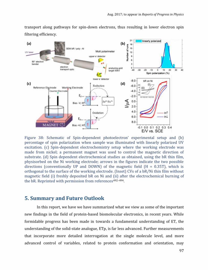
Aug. 2017; to appear in Reports of Progress in Physics
97
transport along pathways for spin-down electrons, thus resulting in lower electron spin
filtering efficiency.
Figure 38: Schematic of Spin-dependent photoelectron’ experimental setup and (b) percentage of spin polarization when sample was illuminated with linearly polarized UV excitation. (c) Spin-dependent electrochemistry setup where the working electrode was made from nickel; a permanent magnet was used to control the magnetic direction of substrate. (d) Spin dependent electrochemical studies as obtained, using the bR thin film, physisorbed on the Ni working electrode; arrows in the figures indicate the two possible directions (conventionally UP and DOWN) of the magnetic field (H = 0.35T), which is orthogonal to the surface of the working electrode. (Inset) CVs of a bR/Ni thin film without magnetic field (i) freshly deposited bR on Ni and (ii) after the electrochemical burning of the bR. Reprinted with permission from references492–494.
5. Summary and Future Outlook
In this report, we have we have summarized what we view as some of the important
new findings in the field of protein-based biomolecular electronics, in recent years. While
formidable progress has been made in towards a fundamental understanding of ET, the
understanding of the solid-state analogue, ETp, is far less advanced. Further measurements
that incorporate more detailed interrogation at the single molecule level, and more
advanced control of variables, related to protein conformation and orientation, may
(a) (b)
(c) (d)

Aug. 2017; to appear in Reports of Progress in Physics
98
provide parameters needed to optimize the use of proteins in bioelectronics. The
remarkable electronic transport efficiencies of proteins might be an unintended
consequence of properties that evolved, over billions of years of evolution, for other
purposes. The solid-state junction (ETp) approach to contacting proteins might be more
similar to what happens in nature (i.e., ET processes) than would seem to be the case at
first sight (cf. section 4.1., discussion of Fig. 21). If this is not accidental, then the reasons
for it are not (yet) clear. Understanding of ETp and any fundamental relationships with ET,
and finding conditions when these two can be combined, may well become possible in the
near future.
Proteins already represent refined structures capable of complex and specific
reactions. By exploiting the electronic properties of protein-based molecular junctions in
various environments, we foresee the creation of advanced solid-state biosensing devices
and bio-circuits that integrate such sensing devices. Implantable electronics for real-time
monitoring has been a reality for some time (e.g., heart pace makers), and, using organic
electronics, flexible electronic implants are rapidly developing towards widespread use.
The ability to use proteins as electronic components can avoid biocompatibility concerns
that are present with artificial electronic components, and may allow easier integration
into biological systems. However, before proteins can be used in these applications, it is
crucial to control how they interact with the solid-state platforms to which they will likely
be coupled to communicate with the outside world. Such connections should preserve their
functionality in downstream platforms, and this requires understanding the fundamental
electronic transport properties.
There are many aspects in the selection of the solid-state substrate and binding
methodologies that need to be considered for successful immobilization of active proteins.
An important conclusion is that protein conformation plays a key role in maintaining
protein stability and function after immobilization and can play a decisive role in the
measurement of reproducible electron transport characteristics. In nature, proteins have
evolved to function in very specific ways. To use these functionalities, protein function
must be preserved on solid supports, and specifically must preserve the integrity of what
are known or assumed to be important functional groups. Although maintaining protein

Aug. 2017; to appear in Reports of Progress in Physics
99
structural integrity is challenging in the experimental work discussed here, the results
reveal that immobilization methodology can be tailored to obtain useful results.
Our review of the literature shows that immobilization schemes that mimic the
protein’s biological attachment (e.g., Figures 6 and 10) and which allow for interrogation of
desired aspects is an effective way to retain protein function and to obtain reproducible
results. Different proteins assume a variety of conformations that are necessary for
biological functionality, and using this information to immobilize the proteins can yield
optimal protein function. For example, if the goal of an experiment is to measure transport
involving a particular site of the protein known to participate in ET with a partner (small
molecule, ion etc.), immobilization onto the electrode for in situ electrochemical ET
measurements or solid-state ETp measurements after ex situ binding should be optimized
to assure that the natural binding surface remains exposed for interaction. If the protein of
interest is membrane-bound in nature, it is likely to retain its function if covalently
tethered using a SAM, rather than directly bound to a solid surface. Taking these steps
allows for retention of protein activity, which can then be used in bioelectronic devices. By
determining protein orientation and conformation when immobilized, the ability to
correlate ETp with biological activity will be enhanced. For example, when optically active
cofactors of the membrane-bound proteins bacteriorhodopsin and halorhodopsin, were
modified through chemical reactions, the alteration in their optical absorptions was
directly correlated to their ETp efficiencies. Altering or removing metal-ion containing
cofactors was directly manifested in both their ET and ETp behavior and in their optical
absorption properties. Bacteriorhodopsin, immobilized on a ferromagnetic substrate,
serves as a spin-filter for electrons that are transmitted across it, which has been ascribed
to the 7 left-handed -helices that define its secondary conformation. Combining spin-
filtering with optoelectronic properties of proteins, which rely strongly on their
conformations, is a fascinating opportunity to understand the role of spin in biology and,
possibly, to future “biospintronic” devices.
Quite a few studies of protein ETp both at the nanoscale and macroscale have been
made, as discussed in this report. Large area monolayer structures can in principle be
integrated in future devices, in contrast to what seems (at least presently) possible for

Aug. 2017; to appear in Reports of Progress in Physics
100
single proteins. Therefore, the fabrication of macroscopic electrical junctions, described in
this report, which use protein monolayers of geometric areas ranging from 102 to 2×10⁵
m2 (as defined by the smallest electrode), constitute an important step towards using
proteins as electronic components. In the future, further miniaturization to a few m2 areas
may well become possible. At the same time working at the nanoscale and probing ETp of
single (or few) proteins can elucidate details about protein function that may be averaged
out in macroscopic measurements of the ensemble. In a biological system, proteins are
usually not in a monolayer structure, and are often isolated (non-aggregated), implying
that single molecule studies could offer more accurate information on actual protein
function if it is present in an environment that mimics their natural state (e.g., a membrane
protein in low concentrations in a monolayer of detergent). Thus, while unlikely to become
part of bioelectronic devices in the near future, studies at the single protein level are
important for obtaining fundamental understanding to make bioelectronic devices.
Presently most single molecule studies rely on nanoscale SPM techniques, apart of
some studies on break junctions described above. However, the conductance data for
proteins (and those for conjugated organic molecules) have a much larger degree of
variability for nanoscopic junctions than for macroscopic ones,47 even though both show
the same trends. ETp efficiency of proteins is remarkably high when compared to ET rates
in proteins and to the transport efficiency through conjugated molecules.
While the ETp studies reviewed here provide information that may well be
necessary for incorporating proteins into solid-state platforms, the question remains what
have we learned, and what can we learn about the transport mechanism across these
proteins from these measurements. Compared to organic molecules, what allows electron
transport across proteins to be more efficient than expected? How does electrode-protein
(and protein-protein) coupling affect solid-state electronic transport? How can electron
transport across relatively long distances (≥ 6 nm) observed in proteins be temperature-
independent? Although it is too ambitious to hope that solid state bioelectronics research
can answer all these questions in the short term, progress towards understanding the
connections between transport efficiency, protein-electrode coupling, and temperature-
independent transport are good places to start.

Aug. 2017; to appear in Reports of Progress in Physics
101
While there is a clear difference between the ionic transport-coupled electron
transfer in biology and all-electronic transport in the artificial solid-state - like systems for
future bioelectronics, an understanding of ETp may be relevant to ET because the presence
of efficient transport channels in a protein can be expected to affect both. This possibility is
intriguing within the context of the earlier mentioned (section 4.1), surprising long-
distance electron transport in pili of G. sulfurreducens and in nanowires of S. oneidensis.
Findings in various report on ETp in/through proteins can be linked back to the
proteins’ innate function, strengthening the notion that at least some of the biological
functionality can be studied with ETp. The importance of cofactors has been demonstrated
in redox-active and redox-inactive proteins, illustrating that cofactors are not simple
hopping sites. Possibly, the cofactors’ effects are related to the conformation and/or charge
envelope, as well as to the electronic energy levels of the protein, relevant to transport. The
interactions of cofactors with proteins that affect ETp may be relevant to protein function,
and understanding how these changes occur may shed light on transport mechanisms and
pathways. An important step to understand the effects of cofactors will be to develop the
ability to apply gate voltages to protein electronic junctions. While some reports of this
exist (and are discussed in this report), it has not yet been possible to achieve gating in a
manner that is clear enough so that it can be readily reproduced.
We have experimentally demonstrated that the interaction of proteins with
biological binding partners influences ETp characteristics. A possible next step is to
determine if ETp changes are related to the degree of conformational change caused by
binding, and to determine how ETp pathways are altered.
Although high-resolution SPM imaging allows observing single proteins, it does not
always guarantee isolation. This is important not only to prevent aggregation, but also to
enable future studies where the same protein can be studied under different conditions, as
is usually the case in biology. Nanolithography has helped create nanoarrays that allow
both isolation and indexing for enhanced studies, but control of protein conformation
remains a challenge. In-depth understanding of protein structure and conformation after
immobilization is indeed a great challenge. Protein conformation is highly dynamic around
room temperature, and movements of different structural elements have been
demonstrated to affect ETp. Currently, spectroscopic techniques can offer snapshots of

Aug. 2017; to appear in Reports of Progress in Physics
102
some general structural elements, but this is far from the ability to trace pathways of
electronic transfer or transport. To understand real-time conformational changes, and how
they relate to ETp, more quantitative molecular modeling of the protein conformation in
the monolayer environment will be required.
In closing, we note that, although the worlds of molecular life sciences and electronics
are often separated by a large divide, the growing mutual interest of these areas in each
other to address common challenges, is contributing to the creation of new scientific and
technological disciplines. Harnessing the unique chemical and physical properties of
proteins could allow devices that can be fully integrated with biological systems. We hope
that this report will boost interdisciplinary discussions and studies of topics that include
bio- and bio-inspired -electronics, with an eye also towards future implantable bio-
compatible integrated circuits and innovative healthcare applications.
Acknowledgements
DC, MS and IP thank the Minerva foundation (Munich), the Israel Science Foundation, the
Nancy and Stephen Grand Centre for Sensors and Security, the Kimmelman Center for
Biomolecular Structure and Assembly, the GMJ Schmidt Minerva Centre for Supramolecular
Architecture and the Benoziyo Endowment Fund for the Advancement of Science, for
partial support, the students and postdoctoral fellows that they had the pleasure to work
with over the past decade and a half and many colleagues, esp. H. B. Gray (Caltech), for
stimulating discussions. DC’s research is made possible in part by the historic generosity of
the Harold Perlman family. DL thanks the US National Institutes of Health, the West
Virginia Shared Research Facilities, and the US National Science Foundation for past
support, which allowed him to embark on research in bioelectronics and supported CB’s
research. The support of the Rosi and Max Varon Visiting Professorship at the Weizmann
Institute of Science, which allowed DL to spend time at the WIS exploring some of the ideas
discussed in this article, is also gratefully acknowledged. SM thanks the Council for Higher
Education (Israel) for a postdoctoral research. CB thanks the US National Institutes of
Health and National Science Foundation IGERT program, which supported his work on
protein electronics. M.S. holds the Katzir-Makineni Chair in Chemistry; D.C. holds the
Rowland and Sylvia Schaefer Chair in Energy Research.

Aug. 2017; to appear in Reports of Progress in Physics
103

Aug. 2017; to appear in Reports of Progress in Physics
104
References
1. Davis, J. J. et al. Molecular bioelectronics. J. Mater. Chem. 15, 2160–2174 (2005).
2. Finkelstein, J. Metalloproteins. Nature 460, 813 (2009).
3. Lu, Y., Yeung, N., Sieracki, N. & Marshall, N. M. Design of functional metalloproteins.
Nature 460, 855–862 (2009).
4. Pessarakli, M. Handbook of photosynthesis. (Boca Raton, FL : Taylor & Francis, 2005.,
2005).
5. Chakraborty, T. Charge Migration in DNA. (Springer-Verlag Berlin Heidelberg, 2007).
6. Clarke, M. J. et al. Long-Range Electron Transfer in Biology. (Berlin, Heidelberg :
Springer Berlin Heidelberg, 1991., 1991).
7. Jortner, J. & Bixon, M. Electron transfer - from isolated molecules to biomolecules, Part 1.
(New York : J. Wiley, c1999, 1999).
8. Rasor, J. P. & Voss, E. Enzyme-catalyzed processes in pharmaceutical industry. Appl.
Catal. Gen. 221, 145–158 (2001).
9. Van Beilen, J. B., Duetz, W. A., Schmid, A. & Witholt, B. Practical issues in the
application of oxygenases. Trends Biotechnol. 21, 170–177 (2003).
10. Vilker, V. L., Reipa, V., Mayhew, M. & Holden, M. J. Challenges in capturing oxygenase
activity in vitro. J. Am. Oil Chem. Soc. 76, 1283–1289 (1999).
11. Zaks, A. & Dodds, D. R. Application of biocatalysis and biotransformations to the
synthesis of pharmaceuticals. Drug Discov. Today 2, 513–531 (1997).
12. Schulze, B. & Wubbolts, M. G. Biocatalysis for industrial production of fine chemicals.
Curr. Opin. Biotechnol. 10, 609–615 (1999).

Aug. 2017; to appear in Reports of Progress in Physics
105
13. Tao, J. (Alex), Lin, G. Q. & Liese, A. Biocatalysis for the Pharmaceutical Industry:
Discovery, Development, and Manufacturing. (John Wiley & Sons, 2009).
14. Lamb, S. B., Lamb, D. C., Kelly, S. L. & Stuckey, D. C. Cytochrome P450 immobilisation as
a route to bioremediation/biocatalysis. FEBS Lett. 431, 343–346 (1998).
15. Leung, A., Shankar, P. M. & Mutharasan, R. A review of fiber-optic biosensors. Sens.
Actuators B Chem. 125, 688–703 (2007).
16. Wolfbeis, O. S. Fiber-Optic Chemical Sensors and Biosensors. Anal. Chem. 80, 4269–
4283 (2008).
17. Kimmel, D. W., LeBlanc, G., Meschievitz, M. E. & Cliffel, D. E. Electrochemical Sensors
and Biosensors. Anal. Chem. 84, 685–707 (2012).
18. Kuila, T. et al. Recent advances in graphene-based biosensors. Biosens. Bioelectron. 26,
4637–4648 (2011).
19. Wang, J. Carbon-Nanotube Based Electrochemical Biosensors: A Review.
Electroanalysis 17, 7–14 (2005).
20. Carrara, S., Baj-Rossi, C., Boero, C. & De Micheli, G. Do Carbon Nanotubes contribute to
Electrochemical Biosensing? Electrochimica Acta 128, 102–112 (2014).
21. Baj-Rossi, C., De, G. & Carrar, S. P450-Based Nano-Bio-Sensors for Personalized
Medicine. in Biosensors - Emerging Materials and Applications (ed. Serra, P. A.) (InTech,
2011).
22. Palchetti, I. & Mascini, M. Electroanalytical biosensors and their potential for food
pathogen and toxin detection. Anal. Bioanal. Chem. 391, 455–471 (2008).
23. Ligler, F. S. et al. Array biosensor for detection of toxins. Anal. Bioanal. Chem. 377,
469–477 (2003).

Aug. 2017; to appear in Reports of Progress in Physics
106
24. Carrara, S., Shumyantseva, V. V., Archakov, A. I. & Samorì, B. Screen-printed electrodes
based on carbon nanotubes and cytochrome P450scc for highly sensitive cholesterol
biosensors. Biosens. Bioelectron. 24, 148–150 (2008).
25. Baj-Rossi, C., Micheli, G. D. & Carrara, S. Electrochemical Detection of Anti-Breast-
Cancer Agents in Human Serum by Cytochrome P450-Coated Carbon Nanotubes.
Sensors 12, 6520–6537 (2012).
26. Aviram, A. & Ratner, M. A. Molecular rectifiers. Chem. Phys. Lett. 29, 277–283 (1974).
27. Salemme, F. R. Design principles for self-assembling devices from macromolecules. in
Bionanotechnology (eds. Renugopalakrishnan, V. & Lewis, R. V.) 1–8 (Springer
Netherlands, 2006).
28. Facci, P. Single metalloproteins at work: Towards a single-protein transistor. (Elsevier
Science Bv, 2002).
29. Li, D. Exploring electron transfer in myoglobin-based transistors. Ph. D. thesis West
Virginia Univ., 2009. (DAI-B 70/11, Dissertation Abstracts International, Ann Arbor
MI, USA, ISBN 9781109450248).
30. S. D’Amico et al. Ambipolar transistors based on azurin proteins. IEE Proc. -
Nanobiotechnology 151, 173–175 (2004).
31. Artés, J. M., Díez-Pérez, I. & Gorostiza, P. Transistor-like Behavior of Single
Metalloprotein Junctions. Nano Lett. 12, 2679–2684 (2012).
32. Li, D., Gannett, P. M. & Lederman, D. An investigation into the feasibility of myoglobin-
based single-electron transistors. Nanotechnology 23, 395705 (2012).
33. Zhang, T. et al. All-optical logic gates using bacteriorhodopsin films. Opt. Eng. 39, 527–
534 (2000).
Aug. 2017; to appear in Reports of Progress in Physics
107
34. Aguilella, V. M. & Alcaraz, A. Nanobiotechnology: A fluid approach to simple circuits.
Nat. Nanotechnol. 4, 403–404 (2009).
35. Ciesielski, P. N. et al. Photosystem I – Based biohybrid photoelectrochemical cells.
Bioresour. Technol. 101, 3047–3053 (2010).
36. Ditto, W. L. Chaos in neural systems: from epilepsy to neurocomputing. in Proceedings
of the 25th Annual International Conference of the IEEE Engineering in Medicine and
Biology Society, 2003 4, 3830–3833 Vol.4 (2003).
37. Ha, S., Wee, Y. & Kim, J. Nanobiocatalysis for Enzymatic Biofuel Cells. Top. Catal. 55,
1181–1200 (2012).
38. Reshetilov, A. N. Biosensors and biofuel cells: Research focused on practical
application (Review). Appl. Biochem. Microbiol. 51, 264–269 (2015).
39. Calabrese Barton, S., Gallaway, J. & Atanassov, P. Enzymatic Biofuel Cells for
Implantable and Microscale Devices. Chem. Rev. 104, 4867–4886 (2004).
40. Guiseppi-Elie, A., Brahim, S., Slaughter, G. & Ward, K. R. Design of a subcutaneous
implantable biochip for monitoring of glucose and lactate. IEEE Sens. J. 5, 345–355
(2005).
41. Farahi, R. H., Ferrell, T. L., Guiseppi-Elie, A. & Hansen, P. Integrated electronics
platforms for wireless implantable biosensors. IEEE NIH Life Sci. Syst. Appl. Workshop
LISA 27–30 (2007).
42. Carrara, S., Cavallini, A., Garg, A. & De Micheli, G. Dynamical spot queries to improve
specificity in P450s based multi-drugs monitoring. in ICME International Conference on
Complex Medical Engineering, 2009. CME 1–6 (2009).

Aug. 2017; to appear in Reports of Progress in Physics
108
43. Saga, Y., Watanabe, T., Koyama, K. & Miyasaka, T. Mechanism of Photocurrent
Generation from Bacteriorhodopsin on Gold Electrodes. J. Phys. Chem. B 103, 234–238
(1999).
44. Berna, A. Z., Anderson, A. R. & Trowell, S. C. Bio-Benchmarking of Electronic Nose
Sensors. PLoS ONE 4, e6406 (2009).
45. Winkler, J. R., Dunn, A. R., Hess, C. R. & Gray, H. B. Electron Tunneling Through Iron and
Copper Proteins. in Bioinorganic Electrochemistry (eds. Hammerich, O. & Ulstrup, J.) 1–
23 (Springer Netherlands, 2008).
46. Skourtis, S. S. Review: probing protein electron transfer mechanisms from the
molecular to the cellular length scales. Pept. Sci. 100, 82–92 (2013).
47. Amdursky, N. et al. Electronic Transport via Proteins. Adv. Mater. 26, 7142–7161
(2014).
48. Bianco, P. The Origin of Bioelectrochemistry: An Overview. in Encyclopedia of
Electrochemistry (Wiley-VCH Verlag GmbH & Co. KGaA, 2007).
49. Andolfi, L. & Cannistraro, S. Conductive atomic force microscopy study of plastocyanin
molecules adsorbed on gold electrode. Surf Sci 598, 68–77 (2005).
50. Chi, Q., Zhang, J., Friis, E. P., E.T. Andersen, J. & Ulstrup, J. Electrochemistry of self-
assembled monolayers of the blue copper protein Pseudomonas aeruginosa azurin on
Au(111). Electrochem. Commun. 1, 91–96 (1999).
51. Gray, H. B. & Winkler, J. R. Electron Transfer in Proteins. Annu. Rev. Biochem. 65, 537–
561 (1996).
52. Farver, O. & Pecht, I. Long range intramolecular electron transfer in azurins. J. Am.
Chem. Soc. 114, 5764–5767 (1992).

Aug. 2017; to appear in Reports of Progress in Physics
109
53. Li, W. et al. Temperature and Force Dependence of Nanoscale Electron Transport via
the Cu Protein Azurin. ACS Nano 6, 10816–10824 (2012).
54. Facci, P. Biomolecular Electronics: Bioelectronics and the Electrical Control of Biological
Systems and Reactions. (William Andrew, 2014).
55. Nicolini, C. Molecular Bioelectronics: The 19 Years of Progress. (World Scientific, 2016).
56. Cuevas, J. C. & Scheer, E. Molecular Electronics: An Introduction to Theory and
Experiment. (World Scientific, 2010).
57. Nitzan, A. Chemical dynamics in condensed phases : relaxation, transfer and reactions in
condensed molecular systems. (Oxford : Oxford University Press, 2006).
58. Xi, B., Wise, K. J., Stuart, J. A. & Birge, R. R. Bacteriorhodopsin-based 3D optical
memory. in Bionanotechnology: Proteins to Nanodevices (eds. Renugopalakrishnan, V.
& Lewis, R. V.) 39–59 (Springer Netherlands, 2006).
59. Artés, J., Hihath, J. & Díez-Pérez, I. Biomolecular Electronics. in Molecular Electronics:
An Experimental and Theoretical Approach (ed. Baldea, I.) 281–323 (Pan Stanford,
2015).
60. Alessandrini, A. & Facci, P. Electron transfer in nanobiodevices. Eur. Polym. J. 83, 450–
466 (2016).
61. Gray, H. B. & Winkler, J. R. Electron tunneling through proteins. Q. Rev. Biophys. 36,
341–372 (2003).
62. Waleed Shinwari, M., Jamal Deen, M., Starikov, E. B. & Cuniberti, G. Electrical
Conductance in Biological Molecules. Adv Funct Mater 20, 1865–1883 (2010).
63. Ron, I., Pecht, I., Sheves, M. & Cahen, D. Proteins as Solid-State Electronic Conductors.
Acc Chem Res 43, 945–953 (2010).

Aug. 2017; to appear in Reports of Progress in Physics
110
64. Sligar, S. G. & Salemme, F. R. Protein engineering for molecular electronics. Curr. Biol.
2, 494 (1992).
65. Winkler, J. R. & Gray, H. B. Electron Flow through Metalloproteins. Chem. Rev. 114,
3369–3380 (2014).
66. Cohen, S. R. Electron Flow Through Molecular Structures. in Scanning Probe
Microscopy: Electrical and Electromechanical Phenomena at the Nanoscale (eds.
Kalinin, S. & Gruverman, A.) 715–745 (Springer New York, 2007).
67. We note that systems can be thought of where the distinction does not hold, such as
solid-molecule-solid measurements in an electrolyte or use of an ionic liquid to gate a
solid state junction.
68. Niki, K., In:, Bard, A. J., Stratmann, M. & Wilson, G. Bioelectrochemistry in Encyclopedia
of Electrochemistry. 9, (Wiley-VCH, Weinheim, 2002).
69. Léger, C. & Bertrand, P. Direct Electrochemistry of Redox Enzymes as a Tool for
Mechanistic Studies. Chem. Rev. 108, 2379–2438 (2008).
70. Bockris, J. O., Reddy, A. K. N. & Gamboa-Aldeco, M. E. Modern Electrochemistry 2A:
Fundamentals of Electrodics. (Springer Science & Business Media, 2007).
71. Bockris, J. O., Reddy, A. K. N. & Gamboa-Aldeco, M. Modern Electrochemistry 2B:
Electrodics in Chemistry, Engineering, Biology and Environmental Science. (Springer
Science & Business Media, 2000).
72. Cordes, M. & Giese, B. Electron transfer in peptides and proteins. Chem. Soc. Rev. 38,
892–901 (2009).

Aug. 2017; to appear in Reports of Progress in Physics
111
73. Reece, S. Y., Hodgkiss, J. M., Stubbe, J. & Nocera, D. G. Proton-coupled electron transfer:
the mechanistic underpinning for radical transport and catalysis in biology. Philos.
Trans. R. Soc. B Biol. Sci. 361, 1351–1364 (2006).
74. We note that ET and ETp are generally described in terms of holes and electrons. We
define a hole as is commonly described now days as a missing electron, instead of the
traditional solid state physics definition of the behavior of electrons of a nearly full
band.
75. Wierzbinski, E. et al. The Single-Molecule Conductance and Electrochemical Electron-
Transfer Rate Are Related by a Power Law. ACS Nano 7, 5391–5401 (2013).
76. Venkatramani, R., Wierzbinski, E., Waldeck, D. H. & Beratan, D. N. Breaking the simple
proportionality between molecular conductances and charge transfer rates. Faraday
Discuss. 174, 57–78 (2014).
77. Vilan, A., Aswal, D. & Cahen, D. Large-Area, Ensemble Molecular Electronics:
Motivation and Challenges. Chem. Rev. 117, 4248–4286 (2017).
78. Riess, I. What does a voltmeter measure? Solid State Ion. 95, 327–328 (1997).
79. Gerischer, H. & Ekardt, W. Fermi levels in electrolytes and the absolute scale of redox
potentials. Appl. Phys. Lett. 43, 393–395 (1983).
80. Bockris, J. O. & Khan, S. U. M. Comment on ‘“Fermi levels in electrolytes and the
absolute scale of redox potentials”’ [Appl. Phys. Lett. 43, 393 (1983)]. Appl. Phys. Lett.
45, 913–913 (1984).
81. Gerischer, H. Response to ‘“Comment on ‘Fermi levels in electrolytes and the absolute
scale of redox potentials’ ”’ [Appl. Phys. Lett. 45, 913 (1984)]. Appl. Phys. Lett. 45, 913–
914 (1984).

Aug. 2017; to appear in Reports of Progress in Physics
112
82. Reiss, H. The Fermi level and the redox potential. J. Phys. Chem. 89, 3783–3791 (1985).
83. Cahen, D. & Kahn, A. Electron Energetics at Surfaces and Interfaces: Concepts and
Experiments. Adv. Mater. 15, 271–277 (2003).
84. Moser, C. C., Keske, J. M., Warncke, K., Farid, R. S. & Dutton, P. L. Nature of biological
electron transfer. Nature 355, 796–802 (1992).
85. Castañeda Ocampo, O. E. et al. Mechanism of Orientation-Dependent Asymmetric
Charge Transport in Tunneling Junctions Comprising Photosystem I. J Am Chem Soc
137, 8419–8427 (2015).
86. Amdursky, N., Pecht, I., Sheves, M. & Cahen, D. Electron Transport via Cytochrome C on
Si–H Surfaces: Roles of Fe and Heme. J Am Chem Soc 135, 6300–6306 (2013).
87. Raichlin, S., Pecht, I., Sheves, M. & Cahen, D. Protein Electronic Conductors: Hemin–
Substrate Bonding Dictates Transport Mechanism and Efficiency across Myoglobin.
Angew Chem Int Ed 54, 12379–12383 (2015).
88. Axford, D. N. & Davis, J. J. Electron flux through apo-and holoferritin. Nanotechnology
18, 145502 (2007).
89. Sepunaru, L., Friedman, N., Pecht, I., Sheves, M. & Cahen, D. Temperature-Dependent
Solid-State Electron Transport through Bacteriorhodopsin: Experimental Evidence for
Multiple Transport Paths through Proteins. J Am Chem Soc 134, 4169–4176 (2012).
90. Mukhopadhyay, S., Dutta, S., Pecht, I., Sheves, M. & Cahen, D. Conjugated Cofactor
Enables Efficient Temperature-Independent Electronic Transport Across ∼6 nm Long
Halorhodopsin. J. Am. Chem. Soc. 137, 11226–11229 (2015).
91. Hirst, J. Mitochondrial Complex I. Annu. Rev. Biochem. 82, 551–575 (2013).

Aug. 2017; to appear in Reports of Progress in Physics
113
92. Moser, C. C., Page, C. C. & Dutton, P. L. Darwin at the molecular scale: selection and
variance in electron tunnelling proteins including cytochrome c oxidase. Philos. Trans.
R. Soc. Lond. B Biol. Sci. 361, 1295–1305 (2006).
93. Fleming, G. R., Martin, J. L. & Breton, J. Rates of primary electron transfer in
photosynthetic reaction centres and their mechanistic implications. Nature 333, 190–
192 (1988).
94. Gunner, M. R., Robertson, D. E. & Dutton, P. L. Kinetic studies on the reaction center
protein from Rhodopseudomonas sphaeroides: the temperature and free energy
dependence of electron transfer between various quinones in the QA site and the
oxidized bacteriochlorophyll dimer. J. Phys. Chem. 90, 3783–3795 (1986).
95. Moser, C. C. et al. Length, time, and energy scales of photosystems. in Advances in
Protein Chemistry Volume 63, 71–109 (Academic Press, 2003).
96. De Vault, D. & Chance, B. Studies of Photosynthesis Using a Pulsed Laser. Biophys. J. 6,
825–847 (1966).
97. Gray, H. B. & Winkler, J. R. Long-range electron transfer. Proc. Natl. Acad. Sci. U. S. A.
102, 3534–3539 (2005).
98. McLendon, G. & Hake, R. Interprotein electron transfer. Chem. Rev. 92, 481–490
(1992).
99. Fontecilla-Camps, J. C., Volbeda, A., Cavazza, C. & Nicolet, Y. Structure/Function
Relationships of [NiFe]- and [FeFe]-Hydrogenases. Chem. Rev. 107, 4273–4303
(2007).

Aug. 2017; to appear in Reports of Progress in Physics
114
100. Dobbek, H., Gremer, L., Meyer, O. & Huber, R. Crystal structure and mechanism of CO
dehydrogenase, a molybdo iron-sulfur flavoprotein containing S-selanylcysteine. Proc.
Natl. Acad. Sci. U. S. A. 96, 8884–8889 (1999).
101. Doukov, T. I., Blasiak, L. C., Seravalli, J., Ragsdale, S. W. & Drennan, C. L. Xenon in and at
the End of the Tunnel of Bifunctional Carbon Monoxide Dehydrogenase/Acetyl-CoA
Synthase,. Biochemistry (Mosc.) 47, 3474–3483 (2008).
102. Moser, C. C., Anderson, J. L. R. & Dutton, P. L. Guidelines for tunneling in enzymes.
Biochim. Biophys. Acta BBA - Bioenerg. 1797, 1573–1586 (2010).
103. Marcus R.A. & Sutin, N. Electron Transfers in Chemistry and Biology. Biochim. Biophys.
Acta 811, 265–322 (1985).
104. Marcus, R. A. Electron Transfer Reactions in Chemistry: Theory and Experiment
(Nobel Lecture). Angew. Chem. Int. Ed. Engl. 32, 1111–1121 (1993).
105. Bendall, D. S. Protein Electron Transfer. (Garland Science, 1996).
106. Bill, N. L., Trukhina, O., Sessler, J. L. & Torres, T. Supramolecular electron transfer-
based switching involving pyrrolic macrocycles. A new approach to sensor
development? Chem. Commun. 51, 7781–7794 (2015).
107. Giese, B., Graber, M. & Cordes, M. Electron transfer in peptides and proteins. Curr. Opin.
Chem. Biol. 12, 755–759 (2008).
108. Hopfield, J. J. Electron Transfer Between Biological Molecules by Thermally Activated
Tunneling. Proc. Natl. Acad. Sci. 71, 3640–3644 (1974).
109. Hush, N. S. Adiabatic Rate Processes at Electrodes. I. Energy‐Charge Relationships. J.
Chem. Phys. 28, 962–972 (1958).

Aug. 2017; to appear in Reports of Progress in Physics
115
110. Blumberger, J. Recent Advances in the Theory and Molecular Simulation of Biological
Electron Transfer Reactions. Chem. Rev. 115, 11191–11238 (2015).
111. Halpern, J. & Orgel, L. E. The theory of electron transfer between metal ions in bridged
systems. Discuss. Faraday Soc. 29, 32–41 (1960).
112. Bortolotti, C. A. et al. Orientation-Dependent Kinetics of Heterogeneous Electron
Transfer for Cytochrome c Immobilized on Gold: Electrochemical Determination and
Theoretical Prediction. J Phys Chem C 111, 12100–12105 (2007).
113. Lvov, Y. M., Lu, Z., Schenkman, J. B., Zu, X. & Rusling, J. F. Direct Electrochemistry of
Myoglobin and Cytochrome P450cam in Alternate Layer-by-Layer Films with DNA and
Other Polyions. J. Am. Chem. Soc. 120, 4073–4080 (1998).
114. Zhan, T., Xi, M., Wang, Y., Sun, W. & Hou, W. Direct electrochemistry and
electrocatalysis of myoglobin immobilized on Fe2O3 nanoparticle–sodium alginate–
ionic liquid composite-modified electrode. J. Colloid Interface Sci. 346, 188–193
(2010).
115. Page, C. C., Moser, C. C., Chen, X. & Dutton, P. L. Natural engineering principles of
electron tunnelling in biological oxidation–reduction. Nature 402, 47–52 (1999).
116. Gupta, R. K. & Redfield, A. G. Double nuclear magnetic resonance observation of
electron exchange between ferri- and ferrocytochrome c. Science 169, 1204–1206
(1970).
117. Simonneaux, G. & Bondon, A. Mechanism of Electron Transfer in Heme Proteins and
Models: The NMR Approach. Chem. Rev. 105, 2627–2646 (2005).
118. Chi, Q. et al. Molecular Monolayers and Interfacial Electron Transfer of Pseudomonas
aeruginosa Azurin on Au(111). J. Am. Chem. Soc. 122, 4047–4055 (2000).

Aug. 2017; to appear in Reports of Progress in Physics
116
119. Farver, O., Zhang, J., Chi, Q., Pecht, I. & Ulstrup, J. Deuterium isotope effect on the
intramolecular electron transfer in Pseudomonas aeruginosa azurin. Proc. Natl. Acad.
Sci. 98, 4426–4430 (2001).
120. Nocera, D. G., Winkler, J. R., Yocom, K. M., Bordignon, E. & Gray, H. B. Kinetics of
intermolecular and intramolecular electron transfer from ruthenium(II) complexes to
ferricytochrome c. J. Am. Chem. Soc. 106, 5145–5150 (1984).
121. Winkler, J. R. & Gray, H. B. Long-Range Electron Tunneling. J. Am. Chem. Soc. 136,
2930–2939 (2014).
122. Farver, O. & Pecht, I. Elucidation of Electron- Transfer Pathways in Copper and Iron
Proteins by Pulse Radiolysis Experiments. in Progress in Inorganic Chemistry (ed.
Karlin, K. D.) 1–78 (John Wiley & Sons, Inc., 2007).
123. Winkler, J. R., Di, B. A. J., Farrow, N. A., Richards, J. H. & Gray, H. B. Electron tunneling in
biological molecules. Pure Appl. Chem. 71, 1753–1764 (1999).
124. Farver, O. et al. Long-Range Electron Transfer in Engineered Azurins Exhibits Marcus
Inverted Region Behavior. J. Phys. Chem. Lett. 6, 100–105 (2015).
125. Farver, O., Marshall, N. M., Wherland, S., Lu, Y. & Pecht, I. Designed azurins show lower
reorganization free energies for intraprotein electron transfer. Proc. Natl. Acad. Sci.
110, 10536–10540 (2013).
126. Lancaster, K. M. et al. Electron Transfer Reactivity of Type Zero Pseudomonas
aeruginosa Azurin. J. Am. Chem. Soc. 133, 4865–4873 (2011).
127. Farver, O., Hwang, H. J., Lu, Y. & Pecht, I. Reorganization Energy of the CuA Center in
Purple Azurin: Impact of the Mixed Valence-to-Trapped Valence State Transition. J.
Phys. Chem. B 111, 6690–6694 (2007).

Aug. 2017; to appear in Reports of Progress in Physics
117
128. Beratan, D. N., Onuchic, J. N. & Hopfield, J. J. Electron tunneling through covalent and
noncovalent pathways in proteins. J. Chem. Phys. 86, 4488–4498 (1987).
129. Beratan, D. N., Betts, J. N. & Onuchic, J. N. Protein electron transfer rates set by the
bridging secondary and tertiary structure. Science 252, 1285–1288 (1991).
130. Onuchic, J. N., Beratan, D. N., Winkler, J. R. & Gray, H. B. Pathway Analysis of Protein
Electron-Transfer Reactions. Annu. Rev. Biophys. Biomol. Struct. 21, 349–377 (1992).
131. Prytkova, T. R., Kurnikov, I. V. & Beratan, D. N. Coupling Coherence Distinguishes
Structure Sensitivity in Protein Electron Transfer. Science 315, 622–625 (2007).
132. Jones, M. L., Kurnikov, I. V. & Beratan, D. N. The Nature of Tunneling Pathway and
Average Packing Density Models for Protein-Mediated Electron Transfer. J. Phys. Chem.
A 106, 2002–2006 (2002).
133. Beratan, D. N. et al. Charge Transfer in Dynamical Biosystems, or The Treachery of
(Static) Images. Acc. Chem. Res. 48, 474–481 (2015).
134. Zhang, Y., Liu, C., Balaeff, A., Skourtis, S. S. & Beratan, D. N. Biological charge transfer
via flickering resonance. Proc. Natl. Acad. Sci. 111, 10049–10054 (2014).
135. Polizzi, N. F., Skourtis, S. S. & Beratan, D. N. Physical constraints on charge transport
through bacterial nanowires. Faraday Discuss. 155, 43–61 (2012).
136. n = 2 for for unbiased diffusive hopping, 1<n<2 – for for acceptor direction-biased
random walk, n>2 for donor direction biased random walk.
137. Stubbe, J., Nocera, D. G., Yee, C. S. & Chang, M. C. Y. Radical Initiation in the Class I
Ribonucleotide Reductase: Long-Range Proton-Coupled Electron Transfer? Chem Rev
2167–2202 (2003). doi:10.1002/chin.200332275
Aug. 2017; to appear in Reports of Progress in Physics
118
138. López-Pérez, D. E. et al. Intermolecular interactions in electron transfer through
stretched helical peptides. Phys. Chem. Chem. Phys. 14, 10332–10344 (2012).
139. Zohta, Y. On the Definition of Sequential Tunneling in a Double-Barrier Resonant
Tunneling Structure. Jpn. J. Appl. Phys. 32, L177–L179 (1993).
140. Büttiker, M. Coherent and sequential tunneling in series barriers. IBM J. Res. Dev. 32,
63–75 (1988).
141. Lambert, C. J. Basic concepts of quantum interference and electron transport in single-
molecule electronics. Chem. Soc. Rev. 44, 875–888 (2015).
142. Prabhulkar, S., Tian, H., Wang, X., Zhu, J.-J. & Li, C.-Z. Engineered Proteins: Redox
Properties and Their Applications. Antioxid. Redox Signal. 17, 1796–1822 (2012).
143. Eyer, C. S. & Backes, W. L. Relationship between the rate of reductase-cytochrome
P450 complex formation and the rate of first electron transfer. Arch. Biochem. Biophys.
293, 231–240 (1992).
144. Guengerich, F. P. & Johnson, W. W. Kinetics of Ferric Cytochrome P450 Reduction by
NADPH−Cytochrome P450 Reductase: Rapid Reduction in the Absence of Substrate
and Variations among Cytochrome P450 Systems†. Biochemistry (Mosc.) 36, 14741–
14750 (1997).
145. Jett, J. E. et al. Measurement of Electron Transfer through Cytochrome P450 Protein on
Nanopillars and the Effect of Bound Substrates. J. Am. Chem. Soc. 135, 3834–3840
(2013).
146. Bostick, C. D., Flora, D. R., Gannett, P. M., Tracy, T. S. & Lederman, D. Nanoscale electron
transport measurements of immobilized cytochrome P450 proteins. Nanotechnology
26, 155102 (2015).

Aug. 2017; to appear in Reports of Progress in Physics
119
147. Yang, M. et al. Electrocatalytic Drug Metabolism by CYP2C9 Bonded to A Self-
Assembled Monolayer-Modified Electrode. Drug Metab. Dispos. 37, 892–899 (2009).
148. Kumar, K. S., Pasula, R. R., Lim, S. & Nijhuis, C. A. Long-Range Tunneling Processes
across Ferritin-Based Junctions. Adv. Mater. 28, 1824–1830 (2016).
149. Rakshit, T. & Mukhopadhyay, R. Solid-state electron transport in Mn-, Co-, holo-, and
Cu-ferritins: Force-induced modulation is inversely linked to the protein conductivity.
J. Colloid Interface Sci. 388, 282–292 (2012).
150. Amdursky, N., Ferber, D., Pecht, I., Sheves, M. & Cahen, D. Redox activity distinguishes
solid-state electron transport from solution-based electron transfer in a natural and
artificial protein: cytochrome C and hemin-doped human serum albumin. Phys Chem
Chem Phys 15, 17142–17149 (2013).
151. Sepunaru, L., Pecht, I., Sheves, M. & Cahen, D. Solid-State Electron Transport across
Azurin: From a Temperature-Independent to a Temperature-Activated Mechanism. J
Am Chem Soc 133, 2421–2423 (2011).
152. Scheller, F. W. et al. Thirty years of haemoglobin electrochemistry. Adv. Colloid
Interface Sci. 116, 111–120 (2005).
153. Amdursky, N., Pecht, I., Sheves, M. & Cahen, D. Doping Human Serum Albumin with
Retinoate Markedly Enhances Electron Transport across the Protein. J Am Chem Soc
134, 18221–18224 (2012).
154. Amdursky, N. et al. Electron Transfer Proteins as Electronic Conductors: Significance
of the Metal and Its Binding Site in the Blue Cu Protein, Azurin. Adv. Sci. 2, 1400026
(2015).

Aug. 2017; to appear in Reports of Progress in Physics
120
155. Guo, C. et al. Tuning electronic transport via hepta-alanine peptides junction by
tryptophan doping. Proc. Natl. Acad. Sci. 113, 10785–10790 (2016).
156. Amdursky, N. et al. Solid-state electron transport via cytochrome c depends on
electronic coupling to electrodes and across the protein. Proc Natl Acad Sci U A 111,
5556–5561 (2014).
157. DeGrado, W. F., Summa, C. M., Pavone, V., Nastri, F. & Lombardi, A. De novo design and
structural characterization of proteins and metalloproteins. Annu. Rev. Biochem. 68,
779–819 (1999).
158. Case, M. A. & McLendon, G. L. Metal-Assembled Modular Proteins: Toward Functional
Protein Design. Acc. Chem. Res. 37, 754–762 (2004).
159. Sasaki, T. & Kaiser, E. T. Helichrome: synthesis and enzymic activity of a designed
hemeprotein. J. Am. Chem. Soc. 111, 380–381 (1989).
160. Das, A. & Hecht, M. H. Peroxidase Activity of De novo Heme Proteins Immobilized on
Electrodes. J. Inorg. Biochem. 101, 1820–1826 (2007).
161. Koder, R. L. et al. Design and engineering of an O(2) transport protein. Nature 458,
305–309 (2009).
162. Carter, P. Site-directed mutagenesis. Biochem. J. 237, 1–7 (1986).
163. Nobelprize.org. Nobel Media AB 2014. The Nobel Prize in Chemistry 1993. The Nobel
Prize in Chemistry 1993
164. Shortle, D., DiMaio, D. & Nathans, D. Directed Mutagenesis. Annu. Rev. Genet. 15, 265–
294 (1981).
165. Takafumi Ueno, T. O. Molecular engineering of cytochrome P450 and myoglobin for
selective oxygenations. J. Porphyr. Phthalocyanines 8, (2004).

Aug. 2017; to appear in Reports of Progress in Physics
121
166. Ozaki, S., Matsui, T. & Watanabe, Y. Conversion of Myoglobin into a Highly Stereo-
specific Peroxygenase by the L29H/H64L Mutation. J. Am. Chem. Soc. 118, 9784–9785
(1996).
167. Yeung, N. & Lu, Y. One Heme, Diverse Functions: using Biosynthetic Myoglobin Models
to Gain Insights into Heme Copper Oxidases and Nitric Oxide Reductases. Chem.
Biodivers. 5, 1437–1448 (2008).
168. Park, H.-S. et al. Design and Evolution of New Catalytic Activity with an Existing
Protein Scaffold. Science 311, 535–538 (2006).
169. Wegner, S. V., Boyaci, H., Chen, H., Jensen, M. P. & He, C. Engineering a uranyl-specific
binding protein from NikR. Angew. Chem. Int. Ed Engl. 48, 2339–2341 (2009).
170. Talbert, J. N. & Goddard, J. M. Enzymes on material surfaces. Colloids Surf. B
Biointerfaces 93, 8–19 (2012).
171. Secundo, F. Conformational changes of enzymes upon immobilisation. Chem. Soc. Rev.
42, 6250–6261 (2013).
172. Rabe, M., Verdes, D. & Seeger, S. Understanding protein adsorption phenomena at solid
surfaces. Adv. Colloid Interface Sci. 162, 87–106 (2011).
173. Alessandrini, A., Berti, L., Gazzadi, G. C. & Facci, P. Imparting chemical specificity to
nanometer-spaced electrodes. Nanotechnology 19, (2008).
174. Alessandrini, A., Salerno, M., Frabboni, S. & Facci, P. Single-metalloprotein wet
biotransistor. Appl. Phys. Lett. 86, (2005).
175. Carrara, S., Erokhin, V., Facci, P. & Nicolini, C. On the role of nanoparticle sizes in
monoelectron conductivity. 18, (Kluwer Academic Publ, 1996).

Aug. 2017; to appear in Reports of Progress in Physics
122
176. Facci, P., Erokhin, V., Carrara, S. & Nicolini, C. Room-temperature single-electron
junction. Proc. Natl. Acad. Sci. U. S. A. 93, 10556–10559 (1996).
177. Petrangolini, P., Alessandrini, A., Navacchia, M. L., Capobianco, M. L. & Facci, P. Electron
Transport Properties of Single-Molecule-Bearing Multiple Redox Levels Studied by EC-
STM/STS. J. Phys. Chem. C 115, 19971–19978 (2011).
178. Vergani, L. et al. FLUORESCENCE CYTOMETRY OF MICROTUBULES AND NUCLEAR-
DNA DURING CELL-CYCLE AND REVERSE-TRANSFORMATION. J. Cell. Biochem. 50,
201–209 (1992).
179. Alessandrini, A., Corni, S. & Facci, P. Unravelling single metalloprotein electron
transfer by scanning probe techniques. Phys. Chem. Chem. Phys. PCCP 8, 4383–4397
(2006).
180. Alessandrini, A. & Facci, P. Electrochemically assisted scanning probe microscopy: A
powerful tool in nano(bio)science. (Elsevier Science Bv, 2008).
181. Della Pia, E. A. et al. Fast electron transfer through a single molecule natively
structured redox protein. Nanoscale 4, 7106–7113 (2012).
182. Davis, J. J., Wang, N., Morgan, A., Zhang, T. & Zhao, J. Metalloprotein tunnel junctions:
compressional modulation of barrier height and transport mechanism. Faraday
Discuss 131, 167–179 (2006).
183. Davis, J. J., Wrathmell, C. L., Zhao, J. & Fletcher, J. The tunnelling conductance of
molecularly ordered metalloprotein arrays. J Mol Recognit 17, 167–173 (2004).
184. Raccosta, S., Baldacchini, C., Rita Bizzarri, A. & Cannistraro, S. Conductive atomic force
microscopy study of single molecule electron transport through the Azurin-gold
nanoparticle system. Appl Phys Lett 102, 203704 (2013).
Aug. 2017; to appear in Reports of Progress in Physics
123
185. Lewis, A. et al. Directly Probing Rapid Membrane Protein Dynamics with an Atomic
Force Microscope: A Study of Light-Induced Conformational Alterations in
Bacteriorhodopsin. Biophys. J 70, 2380–2384 (1996).
186. Zhao, J., Davis, J. J., Sansom, M. S. P. & Hung, A. Exploring the Electronic and Mechanical
Properties of Protein Using Conducting Atomic Force Microscopy. J Amer Chem Soc
126, 5601–5609 (2004).
187. Tao, N. J. Electron transport in molecular junctions. Nat. Nanotechnol. 1, 173–181
(2006).
188. Boussaad, S. & Tao, N. J. Electron Transfer and Adsorption of Myoglobin on Self-
Assembled Surfactant Films: An Electrochemical Tapping-Mode AFM Study. J. Am.
Chem. Soc. 121, 4510–4515 (1999).
189. Richter, C. A., Hacker, C. A. & Richter, L. J. Electrical and Spectroscopic Characterization
of Metal/Monolayer/Si Devices. J Phys Chem B 109, 21836–21841 (2005).
190. Moulton, S. E., Barisci, J. N., Bath, A., Stella, R. & Wallace, G. G. Investigation of protein
adsorption and electrochemical behavior at a gold electrode. J. Colloid Interface Sci.
261, 312–319 (2003).
191. Fleming, B. D., Praporski, S., Bond, A. M. & Martin, L. L. Electrochemical Quartz Crystal
Microbalance Study of Azurin Adsorption onto an Alkanethiol Self-Assembled
Monolayer on Gold. Langmuir 24, 323–327 (2008).
192. Bonanni, B., Alliata, D., Bizzarri, A. R. & Cannistraro, S. Topological and electron-
transfer properties of yeast cytochrome c adsorbed on bare gold electrodes.
Chemphyschem Eur. J. Chem. Phys. Phys. Chem. 4, 1183–1188 (2003).

Aug. 2017; to appear in Reports of Progress in Physics
124
193. Li, N., Xu, J.-Z., Yao, H., Zhu, J.-J. & Chen, H.-Y. The Direct Electron Transfer of Myoglobin
Based on the Electron Tunneling in Proteins. J. Phys. Chem. B 110, 11561–11565
(2006).
194. Della Pia, E. A., Macdonald, J. E., Elliott, M. & Jones, D. D. Direct Binding of a Redox
Protein for Single-Molecule Electron Transfer Measurements. Small 8, 2341–2344
(2012).
195. Schnyder, B., Kotz, R., Alliata, D. & Facci, P. Comparison of the self-chemisorption of
azurin on gold and on functionalized oxide surfaces. Surf. Interface Anal. 34, 40–44
(2002).
196. Rinaldi, R. et al. Electronic rectification in protein devices. Appl. Phys. Lett. 82, 472–
474 (2003).
197. Rinaldi, R. et al. Solid-State Molecular Rectifier Based on Self-Organized
Metalloproteins. Adv. Mater. 14, 1453–1457 (2002).
198. Alessandrini, A. & Facci, P. EC-STM/STS of Redox Metalloproteins and Co-Factors. in
Molecular Electronics: An Experimental and Theoretical Approach (ed. Baldea, I.) 325–
352 (Pan Stanford, 2015).
199. Norde, W. Adsorption of proteins from solution at the solid-liquid interface. Adv.
Colloid Interface Sci. 25, 267–340 (1986).
200. Haynes, C. A. & Norde, W. Globular proteins at solid/liquid interfaces. Colloids Surf. B
Biointerfaces 2, 517–566 (1994).
201. Malmsten, M. Biopolymers at Interfaces, Second Edition. (CRC Press, 2003).
202. Malmsten, M. & Lassen, B. Competitive protein adsorption at phospholipid surfaces.
Colloids Surf. B Biointerfaces 4, 173–184 (1995).
Aug. 2017; to appear in Reports of Progress in Physics
125
203. Gekko, K. & Hasegawa, Y. Compressibility-structure relationship of globular proteins.
Biochemistry (Mosc.) 25, 6563–6571 (1986).
204. Malmsten, M. Formation of Adsorbed Protein Layers. J. Colloid Interface Sci. 207, 186–
199 (1998).
205. Kondo, A., Murakami, F. & Higashitani, K. Circular dichroism studies on conformational
changes in protein molecules upon adsorption on ultrafine polystyrene particles.
Biotechnol. Bioeng. 40, 889–894 (1992).
206. Cruz, J. C., Pfromm, P. H., Tomich, J. M. & Rezac, M. E. Conformational changes and
catalytic competency of hydrolases adsorbing on fumed silica nanoparticles: I.
Tertiary structure. Colloids Surf. B Biointerfaces 79, 97–104 (2010).
207. Shah, S., Solanki, K. & Gupta, M. N. Enhancement of lipase activity in non-aqueous
media upon immobilization on multi-walled carbon nanotubes. Chem. Cent. J. 1, 1–6
(2007).
208. Petkar, M., Lali, A., Caimi, P. & Daminati, M. Immobilization of lipases for non-aqueous
synthesis. J. Mol. Catal. B Enzym. 39, 83–90 (2006).
209. Asuri, P. et al. Increasing Protein Stability through Control of the Nanoscale
Environment. Langmuir 22, 5833–5836 (2006).
210. Shang, W., Nuffer, J. H., Dordick, J. S. & Siegel, R. W. Unfolding of Ribonuclease A on
Silica Nanoparticle Surfaces. Nano Lett. 7, 1991–1995 (2007).
211. Shang, W. et al. Cytochrome C on silica nanoparticles: influence of nanoparticle size on
protein structure, stability, and activity. Small Weinh. Bergstr. Ger. 5, 470–476 (2009).

Aug. 2017; to appear in Reports of Progress in Physics
126
212. Vertegel, A. A., Siegel, R. W. & Dordick, J. S. Silica Nanoparticle Size Influences the
Structure and Enzymatic Activity of Adsorbed Lysozyme. Langmuir 20, 6800–6807
(2004).
213. Vilan, A. & Cahen, D. Chemical Modification of Semiconductor Surfaces for Molecular
Electronics. Chem. Rev. 117, 4624–4666 (2017).
214. Samanta, D. & Sarkar, A. Immobilization of bio-macromolecules on self-assembled
monolayers: methods and sensor applications. Chem. Soc. Rev. 40, 2567–2592 (2011).
215. Bonasio, R. et al. Specific and covalent labeling of a membrane protein with organic
fluorochromes and quantum dots. Proc. Natl. Acad. Sci. 104, 14753–14758 (2007).
216. Wammes, A. E. M. et al. Site-specific peptide and protein immobilization on surface
plasmon resonance chips via strain-promoted cycloaddition. Lab. Chip 13, 1863–1867
(2013).
217. Rusmini, F., Zhong, Z. & Feijen, J. Protein Immobilization Strategies for Protein
Biochips. Biomacromolecules 8, 1775–1789 (2007).
218. Murugan, P., Krishnamurthy, M., Jaisankar, S. N., Samanta, D. & Mandal, A. B. Controlled
decoration of the surface with macromolecules: polymerization on a self-assembled
monolayer (SAM). Chem. Soc. Rev. 44, 3212–3243 (2015).
219. Bera, S., Kolay, J., Banerjee, S. & Mukhopadhyay, R. Nanoscale On-Silico Electron
Transport via Ferritins. Langmuir 33, 1951–1958 (2017).
220. Armstrong, F. A. Recent developments in dynamic electrochemical studies of adsorbed
enzymes and their active sites. Curr. Opin. Chem. Biol. 9, 110–117 (2005).
221. Armstrong, F. A., Heering, H. A. & Hirst, J. Reaction of complex metalloproteins studied
by protein-film voltammetry. Chem. Soc. Rev. 26, 169–179 (1997).

Aug. 2017; to appear in Reports of Progress in Physics
127
222. Ferapontova, E. E. et al. Direct Electrochemistry of Proteins and Enzymes. in
Perspectives in Bioanalysis (ed. Emil Paleček, F. S. and J. W.) Volume 1, 517–598
(Elsevier, 2005).
223. Guo, L.-H., Allen, H. & Hill, O. Direct Electrochemistry of Proteins and Enzymes. in
Advances in Inorganic Chemistry (ed. A.G. Sykes) Volume 36, 341–375 (Academic
Press, 1991).
224. Hirst, J. & Armstrong, F. A. Fast-Scan Cyclic Voltammetry of Protein Films on Pyrolytic
Graphite Edge Electrodes: Characteristics of Electron Exchange. Anal. Chem. 70, 5062–
5071 (1998).
225. Yeh, P. & Kuwana, T. Reversible Electrode Reaction of Cytochrome c. Chem. Lett. 6,
1145–1148 (1977).
226. Eddowes, M. J. & Hill, H. A. O. Novel method for the investigation of the
electrochemistry of metalloproteins: cytochrome c. J. Chem. Soc. Chem. Commun. 771–
772 (1977).
227. Angove, H. A., Cole, J. A., Richardson, D. J. & Butt, J. N. Protein film voltammetry reveals
distinctive fingerprints of nitrite and hydroxylamine reduction by a cytochrome c
nitrite reductase. J. Biol. Chem. (2002).
228. Gwyer, J. D., Angove, H. C., Richardson, D. J. & Butt, J. N. Redox-triggered events in
cytochrome c nitrite reductase. Bioelectrochemistry Amst. Neth. 63, 43–47 (2004).
229. Léger, C. et al. Enzyme electrokinetics: energetics of succinate oxidation by fumarate
reductase and succinate dehydrogenase. Biochemistry (Mosc.) 40, 11234–11245
(2001).

Aug. 2017; to appear in Reports of Progress in Physics
128
230. Sucheta, A., Cammack, R., Weiner, J. & Armstrong, F. A. Reversible electrochemistry of
fumarate reductase immobilized on an electrode surface. Direct voltammetric
observations of redox centers and their participation in rapid catalytic electron
transport. Biochemistry (Mosc.) 32, 5455–5465 (1993).
231. Heering, H. A., Weiner, J. H. & Armstrong, F. A. Direct Detection and Measurement of
Electron Relays in a Multicentered Enzyme: Voltammetry of Electrode-Surface Films
of E. coli Fumarate Reductase, an Iron−Sulfur Flavoprotein. J. Am. Chem. Soc. 119,
11628–11638 (1997).
232. Turner, K. L. et al. Redox properties of flavocytochrome c3 from Shewanella
frigidimarina NCIMB400. Biochemistry (Mosc.) 38, 3302–3309 (1999).
233. Léger, C. et al. Enzyme Electrokinetics: Using Protein Film Voltammetry To Investigate
Redox Enzymes and Their Mechanisms†. Biochemistry (Mosc.) 42, 8653–8662 (2003).
234. Murgida, D. H. & Hildebrandt, P. Redox and redox-coupled processes of heme proteins
and enzymes at electrochemical interfaces. Phys. Chem. Chem. Phys. 7, 3773–3784
(2005).
235. Murgida, D. H. & Hildebrandt, P. Active-Site Structure and Dynamics of Cytochrome c
Immobilized on Self-Assembled Monolayers—A Time-Resolved Surface Enhanced
Resonance Raman Spectroscopic Study. Angew. Chem. Int. Ed. 40, 728–731 (2001).
236. Albrecht, T., Li, W., Ulstrup, J., Haehnel, W. & Hildebrandt, P. Electrochemical and
Spectroscopic Investigations of Immobilized De Novo Designed Heme Proteins on
Metal Electrodes. ChemPhysChem 6, 961–970 (2005).

Aug. 2017; to appear in Reports of Progress in Physics
129
237. Freire, R. S., Pessoa, C. A., Mello, L. D. & Kubota, L. T. Direct electron transfer: an
approach for electrochemical biosensors with higher selectivity and sensitivity. J. Braz.
Chem. Soc. 14, 230–243 (2003).
238. 1-Ethyl-3-(3-dimethylaminopropyl)carbodiimide. 1-Ethyl-3-(3-
dimethylaminopropyl)carbodiimide (https://en.wikipedia.org). Available at:
https://en.wikipedia.org/wiki/1-Ethyl-3-(3-dimethylaminopropyl)carbodiimide.
239. Fischer, M. J. E. Amine Coupling Through EDC/NHS: A Practical Approach. in Surface
Plasmon Resonance: Methods and Protocols (eds. Mol, N. J. & Fischer, M. J. E.) 55–73
(Humana Press, 2010).
240. Love, J. C., Estroff, L. A., Kriebel, J. K., Nuzzo, R. G. & Whitesides, G. M. Self-Assembled
Monolayers of Thiolates on Metals as a Form of Nanotechnology. Chem. Rev. 105,
1103–1170 (2005).
241. Wadu-Mesthrige, K., Amro, N. A. & Liu, G.-Y. Immobilization of proteins on self-
assembled monolayers. Scanning 22, 380–388 (2000).
242. Kim, D. & Herr, A. E. Protein immobilization techniques for microfluidic assays.
Biomicrofluidics 7, 41501 (2013).
243. Hodneland, C. D., Lee, Y.-S., Min, D.-H. & Mrksich, M. Selective immobilization of
proteins to self-assembled monolayers presenting active site-directed capture ligands.
Proc. Natl. Acad. Sci. 99, 5048–5052 (2002).
244. Liu, H., Yamamoto, H., Wei, J. & Waldeck, D. H. Control of the Electron Transfer Rate
between Cytochrome c and Gold Electrodes by the Manipulation of the Electrode’s
Hydrogen Bonding Character. Langmuir 19, 2378–2387 (2003).

Aug. 2017; to appear in Reports of Progress in Physics
130
245. van de Kamp, M. et al. Involvement of the hydrophobic patch of azurin in the electron-
transfer reactions with cytochrome c551 and nitrite reductase. Eur. J. Biochem. 194,
109–118 (1990).
246. van de Kamp, M., Floris, R., Hali, F. C. & Canters, G. W. Site-directed mutagenesis
reveals that the hydrophobic patch of azurin mediates electron transfer. J. Am. Chem.
Soc. 112, 907–908 (1990).
247. Kenaan, C., Zhang, H., Shea, E. V. & Hollenberg, P. F. Uncovering the role of hydrophobic
residues in Cytochrome P450-Cytochrome P450 Reductase interactions. Biochemistry
(Mosc.) 50, 3957–3967 (2011).
248. Wang, M. et al. Three-dimensional structure of NADPH–cytochrome P450 reductase:
Prototype for FMN- and FAD-containing enzymes. Proc. Natl. Acad. Sci. U. S. A. 94,
8411–8416 (1997).
249. Maneg, O., Malatesta, F., Ludwig, B. & Drosou, V. Interaction of cytochrome c with
cytochrome oxidase: two different docking scenarios. Biochim. Biophys. Acta BBA -
Bioenerg. 1655, 274–281 (2004).
250. Allen, P. M., Allen, H., Hill, O. & Walton, N. J. Surface modifiers for the promotion of
direct electrochemistry of cytochrome c. J. Electroanal. Chem. Interfacial Electrochem.
178, 69–86 (1984).
251. Taniguchi, I., Higo, N., Umekita, K. & Yasukouchi, K. Electrochemical behavior of horse
heart cytochrome c in acid solutions using a 6-mercaptopurine-modified gold
electrode. J. Electroanal. Chem. Interfacial Electrochem. 206, 341–348 (1986).

Aug. 2017; to appear in Reports of Progress in Physics
131
252. Ruzgas, T., Wong, L., Gaigalas, A. K. & Vilker, V. L. Electron Transfer between Surface-
Confined Cytochrome c and an N-Acetylcysteine-Modified Gold Electrode. Langmuir
14, 7298–7305 (1998).
253. Avila, A., Gregory, B. W., Niki, K. & Cotton, T. M. An Electrochemical Approach to
Investigate Gated Electron Transfer Using a Physiological Model System: Cytochrome
c Immobilized on Carboxylic Acid-Terminated Alkanethiol Self-Assembled Monolayers
on Gold Electrodes. J. Phys. Chem. B 104, 2759–2766 (2000).
254. Wu, Y. & Hu, S. Voltammetric investigation of cytochrome c on gold coated with a self-
assembled glutathione monolayer. Bioelectrochemistry 68, 105–112 (2006).
255. Liu, H.-H., Lu, J.-L., Zhang, M., Pang, D.-W. & Abruña, H. D. Direct electrochemistry of
cytochrome c surface-confined on DNA-modified gold electrodes. J. Electroanal. Chem.
544, 93–100 (2003).
256. Chen, S.-M. & Chen, S.-V. The bioelectrocatalytic properties of cytochrome C by direct
electrochemistry on DNA film modified electrode. Electrochimica Acta 48, 513–529
(2003).
257. Tarlov, M. J. & Bowden, E. F. Electron-transfer reaction of cytochrome c adsorbed on
carboxylic acid terminated alkanethiol monolayer electrodes. J. Am. Chem. Soc. 113,
1847–1849 (1991).
258. Nakamaru, S., Scholz, F., Ford, W. E., Goto, Y. & von Wrochem, F. Photoswitchable Sn-
Cyt c Solid-State Devices. Adv. Mater. 29, 1605924 (2017).
259. Bigelow, W. C., Pickett, D. L. & Zisman, W. A. Oleophobic monolayers. J. Colloid Sci. 1,
513–538 (1946).

Aug. 2017; to appear in Reports of Progress in Physics
132
260. Grumezescu, A. Fabrication and Self-Assembly of Nanobiomaterials: Applications of
Nanobiomaterials. (William Andrew, 2016).
261. Shafrin, E. G. & Zisman, W. . Hydrophobic monolayers adsorbed from aqueous
solutions. J. Colloid Sci. 4, 571–590 (1949).
262. Du, X. & Hlady, V. Monolayer Formation on Silicon and Mica Surfaces Rearranged from
N-Hexadecanoyl-l-alanine Supramolecular Structures. J. Phys. Chem. B 106, 7295–
7299 (2002).
263. Nuzzo, R. G. & Allara, D. L. Adsorption of bifunctional organic disulfides on gold
surfaces. J. Am. Chem. Soc. 105, 4481–4483 (1983).
264. Haimov, B. et al. Kinetics of Nanoscale Self-Assembly Measured on Liquid Drops by
Macroscopic Optical Tensiometry: From Mercury to Water and Fluorocarbons. J. Am.
Chem. Soc. 138, 2585–2591 (2016).
265. Andrews, A. M., Liao, W.-S. & Weiss, P. S. Double-Sided Opportunities Using Chemical
Lift-Off Lithography. Acc. Chem. Res. (2016). doi:10.1021/acs.accounts.6b00034
266. González-Granados, Z. et al. Formation of Mixed Monolayers from 11-
Mercaptoundecanoic Acid and Octanethiol on Au(111) Single Crystal Electrode under
Electrochemical Control. J. Phys. Chem. C 117, 24307–24316 (2013).
267. Yan, C., Zharnikov, M., Gölzhäuser, A. & Grunze, M. Preparation and Characterization of
Self-Assembled Monolayers on Indium Tin Oxide. Langmuir 16, 6208–6215 (2000).
268. Arya, S. K., Solanki, P. R., Datta, M. & Malhotra, B. D. Recent advances in self-assembled
monolayers based biomolecular electronic devices. Biosens. Bioelectron. 24, 2810–
2817 (2009).

Aug. 2017; to appear in Reports of Progress in Physics
133
269. Mak, L. H., Sadeghi, S. J., Fantuzzi, A. & Gilardi, G. Control of Human Cytochrome P450
2E1 Electrocatalytic Response as a Result of Unique Orientation on Gold Electrodes.
Anal. Chem. 82, 5357–5362 (2010).
270. Patel, N. et al. Immobilization of Protein Molecules onto Homogeneous and Mixed
Carboxylate-Terminated Self-Assembled Monolayers. Langmuir 13, 6485–6490
(1997).
271. Chapman, R. G., Ostuni, E., Yan, L. & Whitesides, G. M. Preparation of Mixed Self-
Assembled Monolayers (SAMs) That Resist Adsorption of Proteins Using the Reaction
of Amines with a SAM That Presents Interchain Carboxylic Anhydride Groups.
Langmuir 16, 6927–6936 (2000).
272. Cui, X. D. et al. Making electrical contacts to molecular monolayers. Nanotechnology
13, 5 (2002).
273. Bruening, M. & Dotzauer, D. Polymer films: Just spray it. Nat Mater 8, 449–450 (2009).
274. Ma, H., Hu, N. & Rusling, J. F. Electroactive Myoglobin Films Grown Layer-by-Layer
with Poly(styrenesulfonate) on Pyrolytic Graphite Electrodes. Langmuir 16, 4969–
4975 (2000).
275. Cosnier, S. Biomolecule immobilization on electrode surfaces by entrapment or
attachment to electrochemically polymerized films. A review. Biosens. Bioelectron. 14,
443–456 (1999).
276. Ho, D., Chu, B., Lee, H. & Montemagno, C. D. Protein-driven energy transduction across
polymeric biomembranes. Nanotechnology 15, 1084 (2004).

Aug. 2017; to appear in Reports of Progress in Physics
134
277. Nassar, A. E., Willis, W. S. & Rusling, J. F. Electron transfer from electrodes to
myoglobin: facilitated in surfactant films and blocked by adsorbed
biomacromolecules. Anal. Chem. 67, 2386–2392 (1995).
278. Zhang, Z., Nassar, A.-E. F., Lu, Z., Schenkman, J. B. & Rusling, J. F. Direct electron
injection from electrodes to CytochromeP450cam in biomembrane-like films. J. Chem.
Soc. Faraday Trans. 93, 1769–1774 (1997).
279. Shumyantseva, V. V. et al. Direct Electron Transfer of Cytochrome P450 2B4 at
Electrodes Modified with Nonionic Detergent and Colloidal Clay Nanoparticles. Anal.
Chem. 76, 6046–6052 (2004).
280. Kröning, S., Scheller, F. W., Wollenberger, U. & Lisdat, F. Myoglobin-Clay Electrode for
Nitric Oxide (NO) Detection in Solution. Electroanalysis 16, 253–259 (2004).
281. Lei, C., Lisdat, F., Wollenberger, U. & Scheller, F. W. Cytochrome c/Clay-Modified
Electrode. Electroanalysis 11, 274–276 (1999).
282. Zhou, Y., Hu, N., Zeng, Y. & Rusling, J. F. Heme Protein−Clay Films: Direct
Electrochemistry and Electrochemical Catalysis. Langmuir 18, 211–219 (2002).
283. Chen, X., Hu, N., Zeng, Y., Rusling, J. F. & Yang, J. Ordered Electrochemically Active Films
of Hemoglobin, Didodecyldimethylammonium Ions, and Clay. Langmuir 15, 7022–
7030 (1999).
284. Song, Y. et al. Direct Electrochemistry of Cytochrome c Based on
Poly(diallyldimethylammonium Chloride)- Graphene Nanosheets/Gold Nanoparticles
Hybrid Nanocomposites and Its Biosensing. Electroanalysis 25, 1400–1409 (2013).

Aug. 2017; to appear in Reports of Progress in Physics
135
285. Dinesh, B., Mani, V., Saraswathi, R. & Chen, S.-M. Direct electrochemistry of cytochrome
c immobilized on a graphene oxide–carbon nanotube composite for picomolar
detection of hydrogen peroxide. RSC Adv. 4, 28229–28237 (2014).
286. Tanne, J., Dietzel, B., Scheller, F. W. & Bier, F. Nanohybrid Materials Consisting of
Poly[(3-aminobenzoic acid)-co-(3-aminobenzenesulfonic acid)-co-aniline] and
Multiwalled Carbon Nanotubes for Immobilization of Redox Active Cytochrome c.
Electroanalysis 26, 732–738 (2014).
287. He, P. & Hu, N. Electrocatalytic Properties of Heme Proteins in Layer-by-Layer Films
Assembled with SiO2 Nanoparticles. Electroanalysis 16, 1122–1131 (2004).
288. Li, Z. & Hu, N. Direct electrochemistry of heme proteins in their layer-by-layer films
with clay nanoparticles. J. Electroanal. Chem. 558, 155–165 (2003).
289. Zhou, Y., Li, Z., Hu, N., Zeng, Y. & Rusling, J. F. Layer-by-Layer Assembly of Ultrathin
Films of Hemoglobin and Clay Nanoparticles with Electrochemical and Catalytic
Activity. Langmuir 18, 8573–8579 (2002).
290. Rusling, J. F. Enzyme Bioelectrochemistry in Cast Biomembrane-Like Films. Acc. Chem.
Res. 31, 363–369 (1998).
291. Hu, N. Direct electrochemistry of redox proteins or enzymes at various film electrodes
and their possible applications in monitoring some pollutants. Pure Appl. Chem. 73,
1979–1991 (2001).
292. Chapman, D. Biophysical chemistry of membrane function A. Kotyk, K. Janáček and J.
Koryta; John Wiley, Chichester, 1988; xvii 377 pages; £45.00. Febs Lett. - FEBS LETT
268, 435–436 (1990).
293. Rusling, J. F. Biomolecular Films: Design, Function, and Applications. (CRC Press, 2003).

Aug. 2017; to appear in Reports of Progress in Physics
136
294. Oliver, B. N., Egekeze, J. O. & Murray, R. W. Solid-state voltammetry of a protein in a
polymer solvent. J. Am. Chem. Soc. 110, 2321–2322 (1988).
295. Sun, H., Hu, N. & Ma, H. Direct Electrochemistry of Hemoglobin in Polyacrylamide
Hydrogel Films on Pyrolytic Graphite Electrodes. Electroanalysis 12, 1064–1070
(2000).
296. Hu, N. & Rusling, J. F. Electrochemistry and Catalysis with Myoglobin in Hydrated
Poly(ester sulfonic acid) Ionomer Films. Langmuir 13, 4119–4125 (1997).
297. Datta, S., Christena, L. R. & Rajaram, Y. R. S. Enzyme immobilization: an overview on
techniques and support materials. 3 Biotech 3, 1–9 (2013).
298. Sallez, Y., Bianco, P. & Lojou, E. Electrochemical behavior of c-type cytochromes at
clay-modified carbon electrodes: a model for the interaction between proteins and
soils. J. Electroanal. Chem. 493, 37–49 (2000).
299. Lei, C., Wollenberger, U., Jung, C. & Scheller, F. W. Clay-Bridged Electron Transfer
between Cytochrome P450cam and Electrode. Biochem. Biophys. Res. Commun. 268,
740–744 (2000).
300. Iler, R. K. Multilayers of colloidal particles. J. Colloid Interface Sci. 21, 569–594 (1966).
301. Lvov/Mohwald. Protein Architecture: Interfacing Molecular Assemblies and
Immobilization Biotechnology. (CRC Press, 1999).
302. Kimizuka, N., Tanaka, M. & Kunitake, T. Spatially Controlled Synthesis of
Protein/Inorganic Nano-assembly: Alternate Molecular Layers of Cyt c and TiO2
Nanoparticles. Chem. Lett. 28, 1333–1334 (1999).
303. Lvov, Y. & Caruso, F. Biocolloids with Ordered Urease Multilayer Shells as Enzymatic
Reactors. Anal. Chem. 73, 4212–4217 (2001).

Aug. 2017; to appear in Reports of Progress in Physics
137
304. McKenzie, K. J. & Marken, F. Accumulation and Reactivity of the Redox Protein
Cytochrome c in Mesoporous Films of TiO2 Phytate. Langmuir 19, 4327–4331 (2003).
305. Lvov, Y. et al. Films of Manganese Oxide Nanoparticles with Polycations or Myoglobin
from Alternate-Layer Adsorption. Langmuir 16, 8850–8857 (2000).
306. Gaigalas, A. K. & Niaura, G. Measurement of Electron Transfer Rates between Adsorbed
Azurin and a Gold Electrode Modified with a Hexanethiol Layer. J. Colloid Interface Sci.
193, 60–70 (1997).
307. Wong, L. S., Khan, F. & Micklefield, J. Selective Covalent Protein Immobilization:
Strategies and Applications. Chem. Rev. 109, 4025–4053 (2009).
308. Frey, B. L. & Corn, R. M. Covalent Attachment and Derivatization of Poly(l-lysine)
Monolayers on Gold Surfaces As Characterized by Polarization−Modulation FT-IR
Spectroscopy. Anal. Chem. 68, 3187–3193 (1996).
309. Chittur, K. K. FTIR/ATR for protein adsorption to biomaterial surfaces. Biomaterials
19, 357–369 (1998).
310. Gannett, P. M. et al. Preparation, Characterization, and Substrate Metabolism of Gold-
Immobilized Cytochrome P450 2C9. J Am Chem Soc 128, 8374–8375 (2006).
311. Zheng, Z. & Gunner, M. R. Analysis of the electrochemistry of hemes with Ems
spanning 800 mV. Proteins 75, 719–734 (2009).
312. Lang, D. & Böcker, R. Highly sensitive and specific high-performance liquid
chromatographic analysis of 7-hydroxywarfarin, a marker for human cytochrome P-
4502C9 activity. J. Chromatogr. B Biomed. Appl. 672, 305–309 (1995).
313. Fantuzzi, A. et al. A New Standardized Electrochemical Array for Drug Metabolic
Profiling with Human Cytochromes P450. Anal. Chem. 83, 3831–3839 (2011).

Aug. 2017; to appear in Reports of Progress in Physics
138
314. Panicco, P., Dodhia, V. R., Fantuzzi, A. & Gilardi, G. Enzyme-Based Amperometric
Platform to Determine the Polymorphic Response in Drug Metabolism by
Cytochromes P450. Anal. Chem. 83, 2179–2186 (2011).
315. Rzeźnicka, I. I. et al. Observation of photoactive yellow protein anchored to a modified
Au(1 1 1) surface by scanning tunneling microscopy. Chem. Phys. Lett. 472, 113–117
(2009).
316. Manickam, A., Johnson, C. A., Kavusi, S. & Hassibi, A. Interface Design for CMOS-
Integrated Electrochemical Impedance Spectroscopy (EIS) Biosensors. Sensors 12,
14467–14488 (2012).
317. Wollenberg, L. A. et al. Selective filling of nanowells in nanowell arrays fabricated
using polystyrene nanosphere lithography with cytochrome P450 enzymes.
Nanotechnology 23, 385101 (2012).
318. Smaali, K., Clément, N., Patriarche, G. & Vuillaume, D. Conductance Statistics from a
Large Array of Sub-10 nm Molecular Junctions. ACS Nano 6, 4639–4647 (2012).
319. Chi, Q., Farver, O. & Ulstrup, J. Long-range protein electron transfer observed at the
single-molecule level: In situ mapping of redox-gated tunneling resonance. Proc. Natl.
Acad. Sci. U. S. A. 102, 16203–16208 (2005).
320. Artés, J. M., Díez-Pérez, I., Sanz, F. & Gorostiza, P. Direct Measurement of Electron
Transfer Distance Decay Constants of Single Redox Proteins by Electrochemical
Tunneling Spectroscopy. ACS Nano 5, 2060–2066 (2011).
321. Muthusubramanian, N. et al. Insulator-protected mechanically controlled break
junctions for measuring single-molecule conductance in aqueous environments. Appl.
Phys. Lett. 109, 13102 (2016).

Aug. 2017; to appear in Reports of Progress in Physics
139
322. Schukfeh, M. I. et al. Towards nanometer-spaced silicon contacts to proteins.
Nanotechnology 27, 115302 (2016).
323. Reed, M. A., Zhou, C., Muller, C. J., Burgin, T. P. & Tour, J. M. Conductance of a Molecular
Junction. Science 278, 252–254 (1997).
324. Kergueris, C. et al. Electron transport through a metal-molecule-metal junction. Phys.
Rev. B 59, 12505–12513 (1999).
325. Park, J. et al. Coulomb blockade and the Kondo effect in single-atom transistors. Nature
417, 722–725 (2002).
326. Michael Galperin and Mark A Ratner and Abraham Nitzan. Molecular transport
junctions: vibrational effects. J. Phys. Condens. Matter 19, 103201 (2007).
327. Kubatkin, S. et al. Single-electron transistor of a single organic molecule with access to
several redox states. Nature 425, 698–701 (2003).
328. D. Boese and H. Schoeller. Influence of nanomechanical properties on single-electron
tunneling: A vibrating single-electron transistor. EPL Europhys. Lett. 54, 668 (2001).
329. Zant, H. S. J. van der et al. Molecular three-terminal devices: fabrication and
measurements. Faraday Discuss. 131, 347–356 (2006).
330. Ron, I. et al. Proteins as Electronic Materials: Electron Transport through Solid-State
Protein Monolayer Junctions. J Am Chem Soc 132, 4131–4140 (2010).
331. Haick, H. & Cahen, D. Making contact: Connecting molecules electrically to the
macroscopic world. Prog. Surf. Sci. 83, 217–261 (2008).
332. Jia, C., Ma, B., Xin, N. & Guo, X. Carbon Electrode–Molecule Junctions: A Reliable
Platform for Molecular Electronics. Acc. Chem. Res. 48, 2565–2575 (2015).

Aug. 2017; to appear in Reports of Progress in Physics
140
333. Lovrinčić, R. et al. A New Route to Nondestructive Top-Contacts for Molecular
Electronics on Si: Pb Evaporated on Organic Monolayers. J. Phys. Chem. Lett. 4, 426–
430 (2013).
334. Yu, X. et al. Fabrication of Reproducible, Integration-Compatible Hybrid Molecular/Si
Electronics. Small 10, 5151–5160 (2014).
335. Noy, G., Ophir, A. & Selzer, Y. Response of Molecular Junctions to Surface Plasmon
Polaritons. Angew. Chem. Int. Ed. 49, 5734–5736 (2010).
336. Shamai, T. & Selzer, Y. Spectroscopy of molecular junctions. Chem. Soc. Rev. 40, 2293–
2305 (2011).
337. Vadai, M. et al. Plasmon-Induced Conductance Enhancement in Single-Molecule
Junctions. J. Phys. Chem. Lett. 4, 2811–2816 (2013).
338. Zelinskyy, Y. & May, V. Laser pulse induced transient currents in a molecular junction:
Effects of plasmon excitations of the leads. J. Chem. Phys. 142, 224701 (2015).
339. Zhu, W. et al. Quantum mechanical effects in plasmonic structures with subnanometre
gaps. Nat. Commun. 7, 11495 (2016).
340. Wang, T. & Nijhuis, C. A. Molecular electronic plasmonics. Appl. Mater. Today 3, 73–86
(2016).
341. Yu, X. et al. Insights into Solid-State Electron Transport through Proteins from
Inelastic Tunneling Spectroscopy: The Case of Azurin. ACS Nano 9, 9955–9963 (2015).
342. Arielly, R., Vadai, M., Kardash, D., Noy, G. & Selzer, Y. Real-Time Detection of Redox
Events in Molecular Junctions. J. Am. Chem. Soc. 136, 2674–2680 (2014).
343. Maruccio, G. et al. Towards Protein Field-Effect Transistors: Report and Model of a
Prototype. Adv. Mater. 17, 816–822 (2005).

Aug. 2017; to appear in Reports of Progress in Physics
141
344. Léger, C., Jones, A. K., Albracht, S. P. J. & Armstrong, F. A. Effect of a Dispersion of
Interfacial Electron Transfer Rates on Steady State Catalytic Electron Transport in
[NiFe]-hydrogenase and Other Enzymes. J. Phys. Chem. B 106, 13058–13063 (2002).
345. Madden, C. et al. Catalytic Turnover of [FeFe]-Hydrogenase Based on Single-Molecule
Imaging. J. Am. Chem. Soc. 134, 1577–1582 (2012).
346. Parkin, A., Cavazza, C., Fontecilla-Camps, J. C. & Armstrong, F. A. Electrochemical
Investigations of the Interconversions between Catalytic and Inhibited States of the
[FeFe]-Hydrogenase from Desulfovibrio desulfuricans. J. Am. Chem. Soc. 128, 16808–
16815 (2006).
347. Jones, A., Sillery, E., Albracht, S. P. J. & Armstrong, F. A. Direct comparison of the
electrocatalytic oxidation of hydrogen by an enzyme and a platinum catalyst. Chem.
Commun. (2002).
348. Fleming, B. D., Johnson, D. L., Bond, A. M. & Martin, L. L. Recent progress in cytochrome
P450 enzyme electrochemistry. Expert Opin. Drug Metab. Toxicol. 2, 581–589 (2006).
349. Shukla, A., Gillam, E. M., Mitchell, D. J. & Bernhardt, P. V. Direct electrochemistry of
enzymes from the cytochrome P450 2C family. Electrochem. Commun. 7, 437–442
(2005).
350. A detailed review on CD approach, its application to the study of protein structure,
guidelines for obtaining reliable data and analysis method can be found at Biochimica
et Biophysica Acta 1751 (2005) 119 – 139, doi:10.1016/j.bbapap.2005.06.005.
351. Hong-Yu Hu & Lei-Lei Jiang and Jun-Ye Hong. Study of Protein Amyloid-Like
Aggregates by Solid-State Circular Dichroism Spectroscopy. Curr. Protein Pept. Sci. 17,
1–4 (2016).

Aug. 2017; to appear in Reports of Progress in Physics
142
352. Naaman, R., Beratan, D. N. & Waldeck, D. Electronic and Magnetic Properties of Chiral
Molecules and Supramolecular Architectures. (Springer, 2011).
353. Billsten, P., Wahlgren, M., Arnebrant, T., McGuire, J. & Elwing, H. Structural Changes of
T4 Lysozyme upon Adsorption to Silica Nanoparticles Measured by Circular
Dichroism. J. Colloid Interface Sci. 175, 77–82 (1995).
354. Karlsson, M. & Carlsson, U. Protein adsorption orientation in the light of fluorescent
probes: mapping of the interaction between site-directly labeled human carbonic
anhydrase II and silica nanoparticles. Biophys. J. 88, 3536–3544 (2005).
355. Locuson, C. W., Gannett, P. M. & Tracy, T. S. Heteroactivator effects on the coupling and
spin state equilibrium of CYP2C9. Arch. Biochem. Biophys. 449, 115–129 (2006).
356. Hutzler, J. M., Wienkers, L. C., Wahlstrom, J. L., Carlson, T. J. & Tracy, T. S. Activation of
cytochrome P450 2C9-mediated metabolism: mechanistic evidence in support of
kinetic observations. Arch. Biochem. Biophys. 410, 16–24 (2003).
357. Antonini, E. & Brunori, M. Hemoglobin and myoglobin in their reactions with ligands.
(North-Holland Pub. Co., 1971).
358. According to the Beer–Lambert law, the absorbance of a material A = εcl ; where ε is
the molar attenuation coefficient; c is the amount concentration; ℓ is the pathlength.
359. Facci, P., Alliata, D., Andolfi, L., Schnyder, B. & Kötz, R. Formation and characterization
of protein monolayers on oxygen-exposing surfaces by multiple-step self-
chemisorption. Surf. Sci. 504, 282–292 (2002).
360. Facci, P., Fontana, M. P., Dalcanale, E., Costa, M. & Sacchelli, T. Molecular
Reorganization in Langmuir−Blodgett Films of Mesogenic Zn−Porphyrin Octaesters.
Langmuir 16, 7726–7730 (2000).
Aug. 2017; to appear in Reports of Progress in Physics
143
361. Omura, T. & Sato, R. A New Cytochrome in Liver Microsomes. J. Biol. Chem. 237,
PC1375-PC1376 (1962).
362. Omura, T. & Sato, R. The Carbon Monoxide-binding Pigment of Liver Microsomes I.
Evidence for its hemoprotein nature. J. Biol. Chem. 239, 2370–2378 (1964).
363. Perlmann, G. E. & Diringer, R. The Structure of Proteins. Annu. Rev. Biochem. 29, 151–
182 (1960).
364. Reid, E., Cook, G. M. W. & Morré, D. J. Investigation of Membrane-Located Receptors.
(Springer Science & Business Media, 2013).
365. Mukhopadhyay, S. et al. Nanoscale Electron Transport and Photodynamics
Enhancement in Lipid-Depleted Bacteriorhodopsin Monomers. ACS Nano 8, 7714–
7722 (2014).
366. Mukhopadhyay, S., Gartner, W., Cahen, D., Pecht, I. & Sheves, M. Electron transport via
a soluble photochromic photoreceptor. Phys. Chem. Chem. Phys. (2016).
doi:10.1039/C6CP05011E
367. Nicolini, C. et al. P450scc Engineering and Nanostructuring for Cholesterol Sensing.
Langmuir 17, 3719–3726 (2001).
368. Homola, J., Yee, S. S. & Gauglitz, G. Surface plasmon resonance sensors: review. Sens.
Actuators B Chem. 54, 3–15 (1999).
369. Sexton, B. A., Feltis, B. N. & Davis, T. J. Characterisation of gold surface plasmon
resonance sensor substrates. Sens. Actuators Phys. 141, 471–475 (2008).
370. Millo, D., Ranieri, A., Koot, W., Gooijer, C. & van der Zwan, G. Towards Combined
Electrochemistry and Surface-Enhanced Resonance Raman of Heme Proteins:

Aug. 2017; to appear in Reports of Progress in Physics
144
Improvement of Diffusion Electrochemistry of Cytochrome c at Silver Electrodes
Chemically Modified with 4-Mercaptopyridine. Anal. Chem. 78, 5622–5625 (2006).
371. Parson, W. W. Modern Optical Spectroscopy: With Exercises and Examples from
Biophysics and Biochemistry. (Springer, 2015).
372. Kitagawa, T. & Hirota, S. Raman Spectroscopy of Proteins. in Handbook of Vibrational
Spectroscopy (John Wiley & Sons, Ltd, 2006).
373. Siebert, F. & Hildebrandt, P. Vibrational Spectroscopy in Life Science. (John Wiley &
Sons, 2008).
374. Andrew, C. R. & Sanders-Loehr, J. Copper−Sulfur Proteins: Using Raman Spectroscopy
To Predict Coordination Geometry. Acc. Chem. Res. 29, 365–372 (1996).
375. Schulte, A., Buchter, S., Galkin, O. & Williams, C. Raman Spectroscopic Observation of a
Conformational Change at the Heme-Protein Linkage in Myoglobin at High Pressure. J.
Am. Chem. Soc. 117, 10149–10150 (1995).
376. Delfino, I., Bizzarri, A. R. & Cannistraro, S. Single-molecule detection of yeast
cytochrome c by Surface-Enhanced Raman Spectroscopy. Biophys. Chem. 113, 41–51
(2005).
377. Nack, M., Radu, I., Bamann, C., Bamberg, E. & Heberle, J. The retinal structure of
channelrhodopsin-2 assessed by resonance Raman spectroscopy. FEBS Lett. 583,
3676–3680 (2009).
378. Unno, M., Kumauchi, M., Sasaki, J., Tokunaga, F. & Yamauchi, S. Resonance Raman
Spectroscopy and Quantum Chemical Calculations Reveal Structural Changes in the
Active Site of Photoactive Yellow Protein. Biochemistry (Mosc.) 41, 5668–5674 (2002).

Aug. 2017; to appear in Reports of Progress in Physics
145
379. Gerscher, S. et al. Chromophore−Anion Interactions in Halorhodopsin from
Natronobacterium pharaonis Probed by Time-Resolved Resonance Raman
Spectroscopy. Biochemistry (Mosc.) 36, 11012–11020 (1997).
380. Todorovic, S., Jung, C., Hildebrandt, P. & Murgida, D. H. Conformational transitions and
redox potential shifts of cytochrome P450 induced by immobilization. J. Biol. Inorg.
Chem. 11, 119–127 (2006).
381. Bonifacio, A. et al. Active-site structure, binding and redox activity of the heme–
thiolate enzyme CYP2D6 immobilized on coated Ag electrodes: a surface-enhanced
resonance Raman scattering study. J. Biol. Inorg. Chem. 13, 85–96 (2008).
382. Wollenberg, L. A. Immobilized Cytochrome P450 2C9 (CYP2C9): Applications for
Metabolite Generation, Monitoring Protein-Protein Interactions, and Improving In-
vivo Predictions Using Enhanced In-vitro Models. Ph.D. Thesis West Virginia Univ.,
2011. (DAI-B 73/03, Dissertation Abstracts International).
383. Peterlinz, K. A. & Georgiadis, R. In Situ Kinetics of Self-Assembly by Surface Plasmon
Resonance Spectroscopy. Langmuir 12, 4731–4740 (1996).
384. Drake, P. A. & Bohn, P. W. Anomalous Diffusion. Surface Plasmon Resonance
Measurements as Probes of Nanometer-Scale Film-Swelling Dynamics for CH3OH in
Poly(methyl methacrylate). Anal. Chem. 67, 1766–1771 (1995).
385. Jordan, C. E., Frutos, A. G., Thiel, A. J. & Corn, R. M. Surface Plasmon Resonance Imaging
Measurements of DNA Hybridization Adsorption and Streptavidin/DNA Multilayer
Formation at Chemically Modified Gold Surfaces. Anal. Chem. 69, 4939–4947 (1997).

Aug. 2017; to appear in Reports of Progress in Physics
146
386. Jin, Y., Shao, Y. & Dong, S. Direct Electrochemistry and Surface Plasmon Resonance
Characterization of Alternate Layer-by-Layer Self-Assembled DNA−Myoglobin Thin
Films on Chemically Modified Gold Surfaces. Langmuir 19, 4771–4777 (2003).
387. Saban, S. Development of an in vitro model system for studying the interaction of
Equus caballus lgE with its high-affinity Fc receptor. (The University of Sheffield,
2011).
388. Baba, A., Knoll, W. & Advincula, R. Simultaneous in situ electrochemical, surface
plasmon optical, and atomic force microscopy measurements: Investigation of
conjugated polymer electropolymerization. Rev. Sci. Instrum. 77, 64101 (2006).
389. Baba, A., Taranekar, P., Ponnapati, R. R., Knoll, W. & Advincula, R. C. Electrochemical
Surface Plasmon Resonance (EC-SPR) and Waveguide Enhanced Glucose Biosensing
with N-Alkylaminated Polypyrrole/Glucose Oxidase Multilayers. ACS Appl. Mater.
Interfaces 2, 2347–2354 (2010).
390. Alessandrini, A., Salerno, M., Frabboni, S. & Facci, P. Single-metalloprotein wet
biotransistor. Appl. Phys. Lett. 86, 133902-133902–3 (2005).
391. Tao, N. J. Probing Potential-Tuned Resonant Tunneling through Redox Molecules with
Scanning Tunneling Microscopy. Phys. Rev. Lett. 76, 4066–4069 (1996).
392. Chi, Q. et al. Approach to interfacial and intramolecular electron transfer of the diheme
protein cytochrome c4 assembled on Au(111) surfaces. J. Phys. Chem. B 114, 5617–
5624 (2010).
393. Karlsson, J.-J., Nielsen, M. F., Thuesen, M. H. & Ulstrup, J. Electrochemistry of
Cytochrome c4 from Pseudomonas stutzeri. J. Phys. Chem. B 101, 2430–2436 (1997).
Aug. 2017; to appear in Reports of Progress in Physics
147
394. Wang, M., Bugarski, S. & Stimming, U. Topological and Electron-Transfer Properties of
Glucose Oxidase Adsorbed on Highly Oriented Pyrolytic Graphite Electrodes. J. Phys.
Chem. C 112, 5165–5173 (2008).
395. Vaz-Domínguez, C., Pita, M., de Lacey, A. L., Shleev, S. & Cuesta, A. Combined ATR-
SEIRAS and EC-STM Study of the Immobilization of Laccase on Chemically Modified Au
Electrodes. J. Phys. Chem. C 116, 16532–16540 (2012).
396. Möller, C., Allen, M., Elings, V., Engel, A. & Müller, D. J. Tapping-Mode Atomic Force
Microscopy Produces Faithful High-Resolution Images of Protein Surfaces. Biophys. J.
77, 1150–1158 (1999).
397. Nussio, M. R. et al. AFM study of the interaction of cytochrome P450 2C9 with
phospholipid bilayers. Chem. Phys. Lipids 163, 182–189 (2010).
398. Alessandrini, A. & Facci, P. Unraveling lipid/protein interaction in model lipid bilayers
by Atomic Force Microscopy. J. Mol. Recognit. 24, 387–396 (2011).
399. Tran, T. D. et al. Electrochemical analysis of azurin thermodynamic and adsorption
properties at monolayer-protected cluster film assemblies - evidence for a more
homogeneous adsorption interface. J. Colloid Interface Sci. 352, 50–58 (2010).
400. Bayburt, T. H. & Sligar, S. G. Single-molecule height measurements on microsomal
cytochrome P450 in nanometer-scale phospholipid bilayer disks. Proc. Natl. Acad. Sci.
U. S. A. 99, 6725–6730 (2002).
401. Archakov, A. I. & Ivanov, Y. D. Application of AFM and optical biosensor for
investigation of complexes formed in P450-containing monooxygenase systems.
Biochim. Biophys. Acta BBA - Proteins Proteomics 1814, 102–110 (2011).

Aug. 2017; to appear in Reports of Progress in Physics
148
402. Wang, H., Yang, Y. & Erie, D. A. Characterization of Protein–Protein Interactions Using
Atomic Force Microscopy. in Protein Interactions (ed. Schuck, P.) 39–77 (Springer US,
2007).
403. Zhang, X. et al. The dynamic conductance response and mechanics-modulated
memristive behavior of the Azurin monolayer under cyclic loads. Phys. Chem. Chem.
Phys. 19, 6757–6767 (2017).
404. Lyshevski, S. E. Nano and molecular electronics handbook. (Boca Raton : CRC Press,
c2007., 2007).
405. Albrecht, T., Li, W.-W., Haehnel, W., Hildebrandt, P. & Ulstrup, J. Voltammetry and in
situ scanning tunnelling microscopy of de novo designed heme protein monolayers on
Au(111)-electrode surfaces. Bioelectrochemistry 69, 193–200 (2006).
406. Leopold, M. C. & Bowden, E. F. Influence of Gold Substrate Topography on the
Voltammetry of Cytochrome c Adsorbed on Carboxylic Acid Terminated Self-
Assembled Monolayers. Langmuir 18, 2239–2245 (2002).
407. Zhang, J. et al. Interfacial electrochemical electron transfer in biology – Towards the
level of the single molecule. Electronproton Coupling Biol. Energy Transduct. 586, 526–
535 (2012).
408. Beratan, D. N. & Balabin, I. A. Heme–copper oxidases use tunneling pathways. Proc.
Natl. Acad. Sci. 105, 403–404 (2008).
409. Horsley, J. R., Yu, J., Moore, K. E., Shapter, J. G. & Abell, A. D. Unraveling the Interplay of
Backbone Rigidity and Electron Rich Side-Chains on Electron Transfer in Peptides:
The Realization of Tunable Molecular Wires. J. Am. Chem. Soc. 136, 12479–12488
(2014).

Aug. 2017; to appear in Reports of Progress in Physics
149
410. Sepunaru, L. et al. Electronic Transport via Homopeptides: The Role of Side Chains and
Secondary Structure. J Am Chem Soc 137, 9617–9626 (2015).
411. Yu, J., Huang, D. M., Shapter, J. G. & Abell, A. D. Electrochemical and Computational
Studies on Intramolecular Dissociative Electron Transfer in β-Peptides. J. Phys. Chem. C
116, 26608–26617 (2012).
412. Beratan, D. N. & Hopfield, J. J. Calculation of tunneling matrix elements in rigid
systems: mixed-valence dithiaspirocyclobutane molecules. J. Am. Chem. Soc. 106,
1584–1594 (1984).
413. Pirbadian, S. et al. Shewanella oneidensis MR-1 nanowires are outer membrane and
periplasmic extensions of the extracellular electron transport components. Proc Natl
Acad Sci U A 111, 12883–12888 (2014).
414. Jiang, N. et al. Distance-Independent Charge Recombination Kinetics in Cytochrome c–
Cytochrome c Peroxidase Complexes: Compensating Changes in the Electronic
Coupling and Reorganization Energies. J. Phys. Chem. B 117, 9129–9141 (2013).
415. Payne, T. M., Yee, E. F., Dzikovski, B. & Crane, B. R. Constraints on the Radical Cation
Center of Cytochrome c Peroxidase for Electron Transfer from Cytochrome c.
Biochemistry (Mosc.) 55, 4807–4822 (2016).
416. El-Naggar, M. Y. et al. Electrical transport along bacterial nanowires from Shewanella
oneidensis MR-1. Proc. Natl. Acad. Sci. 107, 18127–18131 (2010).
417. J E Enderby and A C Barnes. Liquid semiconductors. Rep. Prog. Phys. 53, 85 (1990).
418. Ding, M. et al. Nanoelectronic Investigation Reveals the Electrochemical Basis of
Electrical Conductivity in Shewanella and Geobacter. ACS Nano 10, 9919–9926 (2016).

Aug. 2017; to appear in Reports of Progress in Physics
150
419. Malvankar, N. S. et al. Tunable metallic-like conductivity in microbial nanowire
networks. Nat Nano 6, 573–579 (2011).
420. Clarke, T. A. et al. Structure of a bacterial cell surface decaheme electron conduit. Proc.
Natl. Acad. Sci. 108, 9384–9389 (2011).
421. Malvankar, N. S. et al. Structural Basis for Metallic-Like Conductivity in Microbial
Nanowires. mBio 6, e00084-15 (2015).
422. Xiao, K. et al. Low Energy Atomic Models Suggesting a Pilus Structure that could
Account for Electrical Conductivity of Geobacter sulfurreducens Pili. Sci. Rep. 6, 23385
(2016).
423. Lee, C. & Mertz, B. Theoretical Evidence for Multiple Charge Transfer Pathways in
Bacteriorhodopsin. J. Chem. Theory Comput. 12, 1639–1646 (2016).
424. Li, R. et al. Bidirectional mediation of TiO2 nanowires field effect transistor by dipole
moment from purple membrane. Nanoscale 2, 1474–1479 (2010).
425. Hansen, A. G. et al. Adsorption and Interfacial Electron Transfer of Saccharomyces
Cerevisiae Yeast Cytochrome c Monolayers on Au(111) Electrodes. Langmuir 19,
3419–3427 (2003).
426. Heering, H. A., Wiertz, F. G. M., Dekker, C. & de Vries, S. Direct Immobilization of Native
Yeast Iso-1 Cytochrome c on Bare Gold: Fast Electron Relay to Redox Enzymes and
Zeptomole Protein-Film Voltammetry. J. Am. Chem. Soc. 126, 11103–11112 (2004).
427. Baymann, F. et al. Voltammetry of a ‘protein on a rope’. FEBS Lett. 539, 91–94 (2003).
428. Davis, J. J., Bruce, D., Canters, G. W., Crozier, J. & Hill, H. A. O. Genetic modulation of
metalloprotein electron transfer at bare gold. Chem. Commun. 576–577 (2003).
doi:10.1039/B211246A

Aug. 2017; to appear in Reports of Progress in Physics
151
429. Davis, J. J. et al. Protein adsorption at a gold electrode studied by insitu scanning
tunnelling microscopy. New J. Chem. 22, 1119–1123 (1998).
430. Kazlauskaite, J., Westlake, A. C. G., Wong, L.-L. & Hill, H. A. O. Direct electrochemistry of
cytochrome P450cam. Chem. Commun. 2189–2190 (1996).
doi:10.1039/CC9960002189
431. Fantuzzi, A., Fairhead, M. & Gilardi, G. Direct Electrochemistry of Immobilized Human
Cytochrome P450 2E1. J. Am. Chem. Soc. 126, 5040–5041 (2004).
432. Lo, K. K.-W., Wong, L.-L. & Hill, H. A. O. Surface-modified mutants of cytochrome
P450cam: enzymatic properties and electrochemistry. FEBS Lett. 451, 342–346
(1999).
433. Armstrong, F. A., Hill, H. A. O. & Walton, N. J. Direct electrochemistry of redox proteins.
Acc. Chem. Res. 21, 407–413 (1988).
434. Dolidze, T. D., Rondinini, S., Vertova, A., Waldeck, D. H. & Khoshtariya, D. E. Impact of
self-assembly composition on the alternate interfacial electron transfer for
electrostatically immobilized cytochrome c. Biopolymers 87, 68–73 (2007).
435. Chi, Q., Zhang, J., Andersen, J. E. T. & Ulstrup, J. Ordered Assembly and Controlled
Electron Transfer of the Blue Copper Protein Azurin at Gold (111) Single-Crystal
Substrates. J. Phys. Chem. B 105, 4669–4679 (2001).
436. Ulman, A. Characterization of organic thin films. (Butterworth-Heinemann ; Manning,
1995).
437. Sagara, T., Niwa, K., Sone, A., Hinnen, C. & Niki, K. Redox reaction mechanism of
cytochrome c at modified gold electrodes. Langmuir 6, 254–262 (1990).

Aug. 2017; to appear in Reports of Progress in Physics
152
438. Rusling, J. F. & Nassar, A. E. F. Enhanced electron transfer for myoglobin in surfactant
films on electrodes. J. Am. Chem. Soc. 115, 11891–11897 (1993).
439. Hahn, C. E. W., Hill, H. A. O., Ritchie, M. D. & Sear, J. W. The electrochemistry of proteins
entrapped in nafion. J. Chem. Soc. Chem. Commun. 125–126 (1990).
doi:10.1039/C39900000125
440. Munge, B., Estavillo, C., Schenkman, J. B. & Rusling, J. F. Optimization of
Electrochemical and Peroxide-Driven Oxidation of Styrene with Ultrathin Polyion
Films Containing Cytochrome P450cam and Myoglobin. ChemBioChem 4, 82–89
(2003).
441. Lee, I., Lee, J. W., Warmack, R. J., Allison, D. P. & Greenbaum, E. Molecular electronics of
a single photosystem I reaction center: studies with scanning tunneling microscopy
and spectroscopy. Proc. Natl. Acad. Sci. 92, 1965–1969 (1995).
442. Mikayama, T. et al. The Electronic Behavior of a Photosynthetic Reaction Center
Monitored by Conductive Atomic Force Microscopy. J. Nanosci. Nanotechnol. 9, 97–107
(2009).
443. Lee, I., Lee, J. W. & Greenbaum, E. Biomolecular Electronics: Vectorial Arrays of
Photosynthetic Reaction Centers. Phys Rev Lett 79, 3294–3297 (1997).
444. Gerster, D. et al. Photocurrent of a single photosynthetic protein. Nat Nano 7, 673–676
(2012).
445. Carmeli, I., Frolov, L., Carmeli, C. & Richter, S. Photovoltaic Activity of Photosystem I-
Based Self-Assembled Monolayer. J. Am. Chem. Soc. 129, 12352–12353 (2007).

Aug. 2017; to appear in Reports of Progress in Physics
153
446. Segal, D., Nitzan, A., Davis, W. B., Wasielewski, M. R. & Ratner, M. A. Electron Transfer
Rates in Bridged Molecular Systems 2. A Steady-State Analysis of Coherent Tunneling
and Thermal Transitions. J. Phys. Chem. B 104, 3817–3829 (2000).
447. Albrecht, T., Guckian, A., Kuznetsov, A. M., Vos, J. G. & Ulstrup, J. Mechanism of
Electrochemical Charge Transport in Individual Transition Metal Complexes. J. Am.
Chem. Soc. 128, 17132–17138 (2006).
448. Skov, L. K., Pascher, T., Winkler, J. R. & Gray, H. B. Rates of Intramolecular Electron
Transfer in Ru(bpy)2(im)(His83)-Modified Azurin Increase below 220 K. J Am Chem
Soc 120, 1102–1103 (1998).
449. Wang, H. et al. Unusual Temperature Dependence of Photosynthetic Electron Transfer
due to Protein Dynamics. J. Phys. Chem. B 113, 818–824 (2009).
450. Beebe, J. M., Kim, B., Gadzuk, J. W., Daniel Frisbie, C. & Kushmerick, J. G. Transition from
Direct Tunneling to Field Emission in Metal-Molecule-Metal Junctions. Phys Rev Lett
97, 26801 (2006).
451. Xie, Z., Bâldea, I., Smith, C. E., Wu, Y. & Frisbie, C. D. Experimental and Theoretical
Analysis of Nanotransport in Oligophenylene Dithiol Junctions as a Function of
Molecular Length and Contact Work Function. ACS Nano 9, 8022–8036 (2015).
452. Raichlin, S. Solid State Electron Transport via Myoglobin Protein and the role of
substrate-protein coupling in such systems Ph.D. Thesis Weizmann Inst. of Science.,
2017.
453. Sedghi, G. et al. Long-range electron tunnelling in oligo-porphyrin molecular wires.
Nat Nano 6, 517–523 (2011).

Aug. 2017; to appear in Reports of Progress in Physics
154
454. Bostick, C. D. et al. Immobilized Cytochrome P450 for Monitoring of P450-P450
Interactions and Metabolism. Drug Metab. Dispos. Biol. Fate Chem. 44, 741–749
(2016).
455. Frenkel, J. On Pre-Breakdown Phenomena in Insulators and Electronic Semi-
Conductors. Phys. Rev. 54, 647–648 (1938).
456. Hummel, M. A., Gannett, P. M., Aguilar, J. & Tracy, T. S. Substrate proton to heme
distances in CYP2C9 allelic variants and alterations by the heterotropic activator,
dapsone. Arch. Biochem. Biophys. 475, 175–183 (2008).
457. Wang, W., Lee, T., Kretzschmar, I. & Reed, M. A. Inelastic Electron Tunneling
Spectroscopy of an Alkanedithiol Self-Assembled Monolayer. Nano Lett. 4, 643–646
(2004).
458. Binnig, G., Garcia, N. & Rohrer, H. Conductivity sensitivity of inelastic scanning
tunneling microscopy. Phys. Rev. B 32, 1336–1338 (1985).
459. Ho, W. Single-molecule chemistry. J. Chem. Phys. 117, 11033–11061 (2002).
460. Hansma, P. K. & Coleman, R. V. Spectroscopy of Biological Compounds with Inelastic
Electron Tunneling. Science 184, 1369 (1974).
461. Hillebrecht, J. R. et al. Optimization of protein-based volumetric optical memories and
associative processors by using directed evolution. NanoBiotechnology 1, 141–151
(2005).
462. Hayashi, S., Tajkhorshid, E. & Schulten, K. Molecular Dynamics Simulation of
Bacteriorhodopsin’s Photoisomerization Using Ab Initio Forces for the Excited
Chromophore. Biophys. J. 85, 1440–1449

Aug. 2017; to appear in Reports of Progress in Physics
155
463. J. Xu, P. Bhattacharya, D. L. Marcy, J. A. Stuart & R. R. Birge. Photoconduction in
bacteriorhodopsin/GaAs heterostructures. Electron. Lett. 37, 648–649 (2001).
464. Gillespie, N. B. et al. Characterization and Photochemistry of 13-Desmethyl
Bacteriorhodopsin. J. Phys. Chem. B 109, 16142–16152 (2005).
465. He, T., Friedman, N., Cahen, D. & Sheves, M. Bacteriorhodopsin Monolayers for
Optoelectronics: Orientation and Photoelectric Response on Solid Supports. Adv Mater
17, 1023–1027 (2005).
466. Lee, I. et al. Photoinduced Surface Potential Change of Bacteriorhodopsin Mutant
D96N Measured by Scanning Surface Potential Microscopy. J. Phys. Chem. B 110,
10982–10990 (2006).
467. Shin, J., Bhattacharya, P., Yuan, H.-C., Ma, Z. & Váró, G. Low-power bacteriorhodopsin-
silicon n-channel metal-oxide field-effect transistor photoreceiver. Opt. Lett. 32, 500–
502 (2007).
468. Xu, J. et al. Direct Measurement of the Photoelectric Response Time of
Bacteriorhodopsin via Electro-Optic Sampling. Biophys. J. 85, 1128–1134 (2003).
469. Koyama, K., Yamaguchi, N. & Miyasaka, T. Antibody-Mediated Bacteriorhodopsin
Orientation for Molecular Device Architectures. Science 265, 762–765 (1994).
470. Koyama, K., Yamaguchi, N. & Miyasaka, T. Molecular organization of bacterio-
rhodopsin films in optoelectronic devices. Adv. Mater. 7, 590–594 (1995).
471. Jin, Y. D., Friedman, N., Sheves, M. & Cahen, D. Bacteriorhodopsin-Monolayer-Based
Planar Metal–Insulator–Metal Junctions via Biomimetic Vesicle Fusion: Preparation,
Characterization, and Bio-optoelectronic Characteristics. Adv Funct Mater 17, 1417–
1428 (2007).

Aug. 2017; to appear in Reports of Progress in Physics
156
472. Jin, Y. D. et al. Bacteriorhodopsin as an Electronic Conduction Medium for
Biomolecular Electronics. Chem Soc Rev 37, 2422–2432 (2008).
473. Berthoumieu, O. et al. Molecular Scale Conductance Photoswitching in Engineered
Bacteriorhodopsin. Nano Lett 12, 899–903 (2012).
474. Patil, A. V., Premaraban, T., Berthoumieu, O., Watts, A. & Davis, J. J. Engineered
Bacteriorhodopsin: A Molecular Scale Potential Switch. Chem Eur J 18, 5632–5636
(2012).
475. Patil, A. V., Premaruban, T., Berthoumieu, O., Watts, A. & Davis, J. J. Enhanced
Photocurrent in Engineered Bacteriorhodopsin Monolayer. J Phy Chem B 116, 683–
689 (2012).
476. Salomon, A. et al. What is the Barrier for Tunneling Through Alkyl Monolayers?
Results from n- and p-Si–Alkyl/Hg Junctions. Adv. Mater. 19, 445–450 (2007).
477. Lee, I., Lee, J. W., Stubna, A. & Greenbaum, E. Measurement of Electrostatic Potentials
above Oriented Single Photosynthetic Reaction Centers. J. Phys. Chem. B 104, 2439–
2443 (2000).
478. Feifel, S. C., Lokstein, H., Hejazi, M., Zouni, A. & Lisdat, F. Unidirectional Photocurrent of
Photosystem I on π-System-Modified Graphene Electrodes: Nanobionic Approaches
for the Construction of Photobiohybrid Systems. Langmuir 31, 10590–10598 (2015).
479. Korpany, K. V. et al. Conductance Switching in the Photoswitchable Protein Dronpa. J
Am Chem Soc 134, 16119–16122 (2012).
480. Lee, T., Kim, S.-U., Min, J. & Choi, J.-W. Multilevel Biomemory Device Consisting of
Recombinant Azurin/Cytochrome c. Adv. Mater. 22, 510–514 (2010).

Aug. 2017; to appear in Reports of Progress in Physics
157
481. Gould, J. L. Magnetic Field Sensitivity in Animals. Annu. Rev. Physiol. 46, 585–598
(1984).
482. Ritz, T., Adem, S. & Schulten, K. A Model for Photoreceptor-Based Magnetoreception in
Birds. Biophys. J. 78, 707–718
483. Xie, Z. et al. Spin Specific Electron Conduction through DNA Oligomers. Nano Lett. 11,
4652–4655 (2011).
484. Naaman, R. & Waldeck, D. H. Chiral-Induced Spin Selectivity Effect. J. Phys. Chem. Lett.
3, 2178–2187 (2012).
485. Ernesto Medina and Floralba López and Mark A. Ratner and Vladimiro Mujica. Chiral
molecular films as electron polarizers and polarization modulators. EPL Europhys.
Lett. 99, 17006 (2012).
486. Mondal, P. C., Fontanesi, C., Waldeck, D. H. & Naaman, R. Spin-Dependent Transport
through Chiral Molecules Studied by Spin-Dependent Electrochemistry. Acc. Chem. Res.
49, 2560–2568 (2016).
487. Göhler, B. et al. Spin Selectivity in Electron Transmission Through Self-Assembled
Monolayers of Double-Stranded DNA. Science 331, 894 (2011).
488. Gutierrez, R., Díaz, E., Naaman, R. & Cuniberti, G. Spin-selective transport through
helical molecular systems. Phys Rev B 85, 81404 (2012).
489. Kumar, A. et al. Chirality-induced spin polarization places symmetry constraints on
biomolecular interactions. Proc. Natl. Acad. Sci. 114, 2474–2478 (2017).
490. Naaman, R. & Waldeck, D. H. Spintronics and Chirality: Spin Selectivity in Electron
Transport Through Chiral Molecules. Annu. Rev. Phys. Chem. 66, 263–281 (2015).

Aug. 2017; to appear in Reports of Progress in Physics
158
491. Carmeli, I., Kumar, K. S., Heifler, O., Carmeli, C. & Naaman, R. Spin Selectivity in
Electron Transfer in Photosystem I. Angew. Chem. Int. Ed. 53, 8953–8958 (2014).
492. Mishra, D. et al. Spin-dependent electron transmission through bacteriorhodopsin
embedded in purple membrane. Proc. Natl. Acad. Sci. 110, 14872–14876 (2013).
493. Einati, H., Mishra, D., Friedman, N., Sheves, M. & Naaman, R. Light-Controlled Spin
Filtering in Bacteriorhodopsin. Nano Lett. 15, 1052–1056 (2015).
494. Roy, P. et al. Spin-Controlled Photoluminescence in Hybrid Nanoparticles Purple
Membrane System. ACS Nano 10, 4525–4531 (2016).