Protein Based Biopolymers. - Politecnico di Milano · Alpha Helix The sequence of amino acids...
68
Protein Based Biopolymers. Prof. Attilio Citterio Dipartimento CMIC “Giulio Natta” https://iscamapweb.chem.polimi.it/citterio/it/education/course-topics/ School of Industrial and Information Engineering Course 096125 (095857) Introduction to Green and Sustainable Chemistry
Transcript of Protein Based Biopolymers. - Politecnico di Milano · Alpha Helix The sequence of amino acids...

Protein Based Biopolymers.
Prof. Attilio Citterio
Dipartimento CMIC “Giulio Natta”
https://iscamapweb.chem.polimi.it/citterio/it/education/course-topics/
School of Industrial and Information Engineering
Course 096125 (095857)
Introduction to Green and Sustainable Chemistry

Attilio Citterio
Protein Building Blocks.
The monomers of proteins are the amino acids
Amino group (ammonium
group if protonated)
Carboxylic group(carboxyl,
carboxylate if deprotonated)
R = AN ORGANIC CHEMICAL GROUP (SIDE CHAIN)
At pH 7, Most Amino Acids are Zwitterions (charged but electrically neutral)
Ka1Ka2
H+ H+

Attilio Citterio
Numbering (lettering) Amino Acids.
Alpha-carbon
Alpha-carboxyl(attached to the α-carbon)
Alpha-aminoβ
γ
δ
ε
ε-amino group
lysineL- configuration of a carbon
• Proteins are built up by amino acids that are linked by peptide bonds to form a polypeptide chain.
• An amino acid has several structural components:– A central carbon atom (Ca) is attached to
– an amino group (NH2), – a carboxyl group (COOH), – a hydrogen atom (H),– a side chain (R).

Attilio Citterio
Amino Acids in 3 Dimensions.
• Asymmetric carbon (4 different
groups attached)
• Stereoisomers
• Rotate polarized light
• Optical isomers
• Non-superimposable
• Mirror images
• L and D forms
• Natural: only L configuration
Asymmetric
carbon atom

Attilio Citterio
The “Handedness" of Amino Acids.
• Looking down the H-Ca bond from the hydrogen atom, the L-form has
CO, R, and N substituents from Ca going in a clockwise direction. For
the L-form the groups read CORN in the clockwise direction.
• All a.a. except Gly (R = H) have a chiral center
• All a.a. incorporated into proteins by organisms are in the L-form.
Ca
R
CON
D-formL-form
HCa
R
NOC H

Attilio Citterio
Configuration vs. Conformation.
Configuration = different geometries
due to orientation in space
cis vs. trans (planar peptide bond)
D vs. L; R vs. S (chiral amino
acids)
You can not move from one
configuration to another without
breaking bonds.
Conformation = alternating atom
arrangement derived from molecular
motion around a single bond
Chair conformation vs. boat
conformation in cyclohexane
You can convert one conformation
to another.

Attilio Citterio
Natural Amino Acids (20).
Gly Ala Val Ile Leu Pro His
Cys Met Ser Thr Sec Asp Asn
Phe Tyr Trp Lys Arg Glu Gln

Attilio Citterio
The Twenty Amino Acids Found in Proteins (1).
1) Amino acids with positive/negative charged hydrophilic side chains
Positive Negative
Arginine
(Arg) (R)
Histidine
(His) (H)
Lysine
(Lys) (K)
Aspartic acid
(Asp) (D)
Glutamic acid
(Glu) (E)
+ -
C C C C C

Attilio Citterio
The Twenty Amino Acids Found in Proteins (2).
2) Amino acids with polar (hydrophilic) uncharged side chains
Serine
(Ser) (S)
Threonine
(Thr) (T)
Asparagine
(Asn) (N)
Glutamine
(Gln) (Q)
Tyrosine
(Tyr) (Y)
Cysteine
(Cys) (C)
Glycine
(Gly) (G)
Proline
(Pro) (P)
2') Special cases
CCC C C
C C C
Attilio Citterio
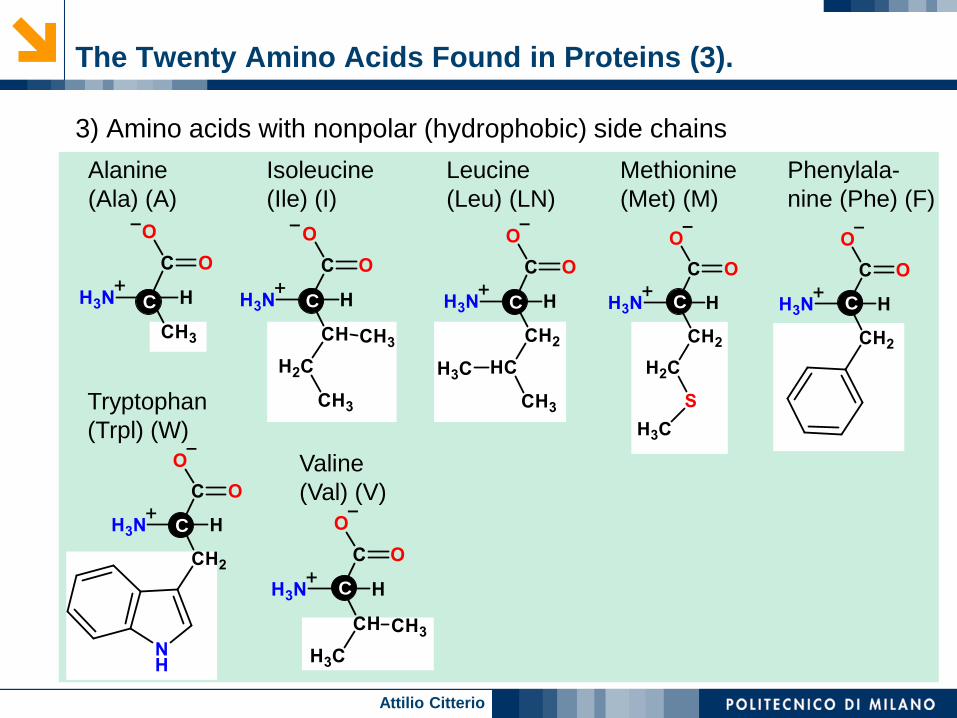
The Twenty Amino Acids Found in Proteins (3).
3) Amino acids with nonpolar (hydrophobic) side chains
Alanine
(Ala) (A)
Isoleucine
(Ile) (I)
Leucine
(Leu) (LN)
Methionine
(Met) (M)
Phenylala-
nine (Phe) (F)
Valine
(Val) (V)
Tryptophan
(Trpl) (W)
CC C C C
C
C

Attilio Citterio
Classes of Amino Acids.
Nonpolar (NP), non-interactive
ala, val, leu, ile, pro, trp, phe, met
Nonpolar (overall), interactive (I) group
cys, tyr
Polar (P)
gly, ser, thr, asn, gln (gly is non-interactive)
Acidic (A)
asp, glu
Basic (B)
lys, arg, his

Attilio Citterio
pKa Values of Natural Amino Acids.
AMINO ACID a- Carboxy a- Amino Side Chain Unit
Gly 2.34 9.60
Ala 2.34 9.69
Val 2.32 9.62
Leu 2.36 9.68
Ile 2.36 9.68
Ser 2.21 9.15
Thr 2.63 10.43
Met 2.28 9.21
Phe 1.83 9.13
Trp 2.38 9.39
Asn 2.02 8.80
Gln 2.17 9.13
Pro 1.99 10.6
Asp 2.09 9.82 3.86
Glu 2.19 9.67 4.25
His 1.82 9.17 6.00
Lys 2.18 8.95 10.53
Arg 2.17 9.04 12.48
Cys 1.71 10.78 8.33
Tyr 2.20 9.11 10.07

Attilio Citterio
Isoelectric Point.
The net charge on an amino acid or peptide changes as the pH is
changed.
Isoelectric Point (pI) - The pH at which the net charge on an amino
acid or peptide chain is zero.
Electrophoresis - A method of separating charged species by causing
them to migrate toward a positive or negative electrode.
Positive ions move toward the negative electrode
Negative ions move toward the positive electrode.
ep ep
V
Lu
V : applied voltage / V
ep : electrophoretic
mobility / m2/(s V)length L
Charged particle
uep
uep
voltage V
+ -
-
+
++++
+
--
----

Attilio Citterio
Peptides, Polypeptides and Proteins.
peptide = a condensation
product of amino acids
dipeptide = 2 aa, ….. etc
oligopeptide = up to 20 aa
polypeptide = > 20 aa
protein = a functional
polypeptide with a
biological role
sometimes contains non-
polypeptide portions as well.
very small “proteins” are
often hormones

Attilio Citterio
The Four Levels of Protein Structure.

Attilio Citterio
Levels of Protein Structure – I.
primary structure (1o)
the sequence of amino acids with modifications, including
additions of other units covalently
secondary structure (2o)
patterned large scale H-bonding involving the backbone
components of the chain
domains and motifs
an intermediate type of terms meant to describe a certain region of
a protein having certain structural features

Attilio Citterio
Tridimensional Structure of Proteins.
Peptide chain
= primary structure
Secondary structure
Alfa helix Antiparallel beta sheet
Side chain
Carbonalfa
Lateral Chain
Carbon
alpha Carbonalfa
Carbon
alpha
Main polypeptide Chain
Lateral Chain
Lateral Chain

Attilio Citterio
Denaturation of Proteins.
Cooking denatures
proteins
Changes that occur to
an egg white when is
cooked
Protein modify
irreversibly their native
tridimensional structure
and can liberate the
species they bind (i.e.
biotin and iron) for
digestion.
Other conditions (acidity, radiation, radicals, some chemicals, ….) produce similar irreversible denaturation processes.

Attilio Citterio
Secondary Structure of a Protein: Collagen.
Triple Helix - Three coiled polypeptide
chains intertwined to form a rope
Structural function (connective tissue)
H-bond is perpendicular to chains
H-bonds are inter-molecular
H-bond is between glycine donor N-H and
proline (hydroxyproline) C=O acceptor
alternating H-bonds between chains
one H-bond per one unit of triple helix
It represent 25-30% of all human proteins.

Attilio Citterio
Amino Acid Composition of Fibrous Proteins.
a-Keratin (Wool) Fibroin (Silk) Collagen (Tendon) Elastin (Aorta)
Gly 8.1 44.6 32.7 32.3
Ala 5.0 29.4 12.0 23.0
Ser 10.2 12.2 3.4 1.3
Glu + Gln 12.1 1.0 7.7 2.1
Cys 11.2 0 0 tr.
Pro 7.5 0.3 22.1 10.7
Arg 7.2 0.5 5.0 0.6
Leu 6.9 0.5 2.1 5.1
Thr 6.5 0.9 1.6 1.6
Asp + Asn 6.0 1.3 4.5 0.9
Val 5.1 2.2 1.8 12.1
Tyr 4.2 5.2 0.4 1.7
Ile 2.8 0.7 0.9 1.9
Phe 2.5 0.5 1.2 3.2
His 0.7 0.2 0.3 tr.
Met 0.5 0 0.7 tr.
Trp 1.2 0.2 0 tr.

Attilio Citterio
Levels of Protein Structure (2).
tertiary structure (3o)
other attraction and reactions within a single
polypeptide chain
quaternary structure (4o)
associations of polypeptide chains with:
a) other polypeptides
b) other biopolymers
c) with small (organic) molecules
d) with small (inorganic) usually metal ions

Attilio Citterio
Tertiary Structure of a Protein.
Five classes of tertiary structure:
Salt linkages (a)
Side chain H-bonding (b)
Hydrophobic force (attraction) (c)
Dipole-dipole interaction(d)
Disulfide linkages (e)
All involve interactions between
side chains of amino acids in
chain.
Disulfide linkages are covalent
bonds; others are weak
attractions (exception are ionic
bonds).
H3CCH3
b
c
SS
e
+ ─a
H2C
H-O
CH2
O-H
+ ─a
c
c
d

Attilio Citterio
Tertiary Structure of Proteins (2).
Hydrophobic
interaction

Attilio Citterio
Alternative Visualization of Proteins.
Ribbons Lines
α helix
β sheets
and hair
connectors

Attilio Citterio
Quaternary Structure in Proteins.
■ The quaternary structure
contains two or more tertiary
subunits (protein chains)
■ Held together by same
interactions as tertiary
structure
■ Hemoglobin contains four
chains
■ The heme group in each
subunit picks up oxygen for
transport in the blood to the
tissues

Attilio Citterio
Variability in Protein Organization.

Attilio Citterio
Summary of Structural Levels in Proteins.
PRIMARY
STRUCTURE
SECONDARY
STRUCTURE
TERTIARY
STRUCTURE
QUATERNARY
STRUCTURE
Alpha Helix
The sequence of amino acids
present in a protein’s peptide
chain or chains
The regularly repeating
ordered spatial
arrangements of amino
acids near each other in the
protein chain, which result
from hydrogen bonds
between carbonyl oxygen
atoms and amino hydrogen
atoms.
The overall three-
dimensional shape that
results from the attractive
forces between amino acid
side chains (R groups) that
are not near each other in
the protein chain
The three-dimensional shape
of a protein consisting of
two or more independent
peptide chains, which
results from noncovalent
interactions between R
groups
Hydrogen bonds between
every fourth amino acids
Disulfide bonds
Electrostatic interaction
Hydrogen bonds
Hydrophobic Interactions
Electrostatic interaction
Hydrogen bonds
Hydrophobic Interactions
Hydrogen bonds between two
side-by-side chains, or a single
chain that is folded back on
itself.
Beta Pleated Sheet

Attilio Citterio
Protein Classification.
Classification by shape
globular
fibrous
Classification by function
Structural
Signaling pathways
Metabolic
Transportation
Classification by what else is present
• derivative proteins
trypsin
collagen

Attilio Citterio
Protein Classification by Function.
Classification:
Enzymes
Structural proteins
Defense proteins
Transport proteins
Storage proteins
Effector proteins
hormones, reg. proteins, toxins
Functions of protein:
• Provide structural and
mechanical support
• Maintain body tissues
• Functions as enzymes and
hormones
• Help maintain acid-base
balance
• Transport nutrients
• Assist the immune system
• Serve as a source of energy
when necessary
• Serve for cell movement and
transport.
Enzymes45%
Heat Shock
4%
Other30%
Structural9%
Ribosomal4%
Hypothetical
3%
Channels1%
Factors4%

Attilio Citterio
Classification of Proteins.
Proteins
Simple Conjugated Derived
Globular Sclero Primary Secondary
Albumins
Globulins
Gluteins
Prolamins
Histones
Globins
Prolamins
Collagen
Elastins
Keratins
Nucleoproteins
Glycoproteins
Lipoproteins
Phosphoproteins
Chromoproteins
Metalloproteins
Coagulated
proteins
Proteans
Metaproteins
Proteases
Peptones
Polypeptides
Peptides

Attilio Citterio
.
Derivative Proteins.
Glycoproteins
Lipoproteins
Nucleoproteins
Conjugated proteins
(holoproteins)
protein portion =
apoprotein
attached small groups =
organic/inorganic pieces
heme, flavin, metals,
phosphate

Attilio Citterio
Protein Synthesis in Cell.

Attilio Citterio
PROTEINS – Definitions.
Apoprotein – amino acids only.
Cofactors – small organic (e.g., vitamins, ATP, NAD, FAD) or inorganic
molecules (particularly metal ions) that are required for activity; can be
loosely bound (coenzymes) or tightly bound (prosthetic groups).
Prosthetic group – tightly bound group (e.g., heme) to apoprotein.
Holoprotein – active protein with cofactors and prosthetic groups
attached.

Attilio Citterio
COFACTORS.
They may participate directly in catalytic processes or carry other
small molecules; binding to proteins may be weak or strong
are required in small quantities, may have to be supplied in diet and
are either water or fat soluble
Functions (see slides on secondary metabolites)
metal ions maintain protein conformation through electrostatic
interactions (METAL LIGATION)
prosthetic groups like heme may bind to active site and change
the conformation to control bonding
may accept a substrate during reaction• common bridging ligands
O2-, OH-, -CH2S-, S2-, -CH2CO2
-, imidazole
• exogenous terminal ligands are also often bound to metals
• H2O, OH-, O2-, HS-, S2-

Attilio Citterio
Enzymes.
Produced by living organisms, are compounds of proteic nature with
catalytic properties. These catalysts are both efficient and highly
specific for an individual chemical reaction which involves the synthesis,
degradation or alteration of a compound. In these reactions, where
molecules are reduced, oxidized, transposed, or assembled, cofactors
are frequently involved. Some enzymes are modified covalently by
phosphorylation, glycosylation, and other processes.
CPOsubtilisin phytase
Promotes the proteolysis of
a peptide bond. .
Chloroperoxidase catalyzes several
oxidations of organic substrates.
Catalyzes the hydrolysis of
phytic acid.

Attilio Citterio
Proteins and Bio-transport:
Types of Movement in Living Organisms.
1. Continuous movement: occurs inside each cell of the living
organism cells for the continuity of its vital activities, such as
cytoplasmic streaming.
2. Positional movement: occurs in some organs of the living
organism, peristalsis movement in the intestines of vertebrates.
3. Total movement: By which the living organism can move from a
place to another in order to search for food or a mate or to avoid
some dangers, It leads to the spread of the animal in nature and as
the means of movement was strong and fast, the circle of animal
spread increases.
Motion has evolved in a variety of structured
systems (or organs) by using very similar
molecules, i.e. special proteins
generally named molecular motors

Attilio Citterio
Molecular Motors.
Most forms of movement in living world are powered by tiny protein machines.
Among the best known are motors that use sophisticated intramolecular
amplification mechanisms to take nanometer steps along protein tracks in the
cytoplasm.
Kinesin and Dynein.
Kinesin and dynein are the molecular
motors responsible for transport along
microtubules.
Actin.
Actin is a family of globular multi-
functional proteins that form micro-
filaments in all eukaryotic cells.
Myosins.
Myosins are ATPases that
generate force for movement
of actin filaments.

Attilio Citterio
Proteins in Locomotion in Living Organisms.
Locomotion in the higher unicellular organism is carried out by:
1. Amoeboid (= Pseudopodial) Movement:
2. Flagellar Movement: long, sheathed cylinder containing microtubules
in a 9+2 arrangement:• covered by an extension of the cell membrane
• 10X thicker than prokaryotic flagella
• function in motility
3. Ciliary Movement: similar in overall structure to flagella,
but shorter and more numerous found only on a
single group of protozoa and certain animal cells
The multicellular organism like humans and other members of Animal
kingdom uses locomotory organs such as legs, hands, fins, etc. to move
in the surrounding
4. Muscular Movement:

Attilio Citterio
Amoeboid Movement.
• Cytoplasmic extension• Actin (45 kDalton)
polymerization thrusts
leading edge of cell
cytoplasm forward
• Adhesion• Leading edge adheres to
surface
• Retraction• Interaction between actin and
myosin
• ATP hydrolysis
• Pulls cell forward

Attilio Citterio
Muscular Movement.
Myosin is a motor protein that
generates the force in a muscle
contraction. It consists of a head and a
tail region. Together, the tails of
approximately three hundred myosin
molecules form the shaft of the thick
filament. The myosin heads of these
molecules project outward toward the
thin filaments like the oars of a rowboat.
Actin Molecules and Thin Filaments
Actin is a spherical protein that forms,
among other things, the thin filament in
muscle cells. Thin filaments are
composed of two long chains of these
actin molecules that are twisted around
one another. Each actin molecule has a
myosin-binding site where a myosin
head can bind. ATP
Actin-Myosin interaction.

Attilio Citterio
The Cross-bridge Muscle Contraction Cycle,
which is Triggered by Ca2+ and ATP
The motion of muscle shortening
occurs as myosin heads bind to
actin and pull the actin inwards.
(net energy balance 15-35%).
This action requires energy,
which is provided by ATP.
Myosin binds to actin at a
binding site on the globular
actin protein. Myosin has
another binding site for ATP at
which enzymatic activity
hydrolyzes ATP to ADP,
releasing an inorganic
phosphate molecule and energy.
Tropomyosin and Troposin are
regulatory proteins: the first
blocks myosin, the second
activate via Ca2+ the contraction.

Attilio Citterio
Proteins as Binder in Composites with Inorganics.
Plot of stiffness against
toughness
■ Direct line:
Expected area of the
composite
■ Circle:
Actual position of the
composite
Synergistic Effect: 1+1>2
P. Fratzl, H.S. Gupta, E.P. Paschalis, P. Roschger, J. Mater. Chem. 2004, 14 ,2115 – 2123
100
10
1
0,1
0,01
0,001
0,01 0,1 1 10 100 1000
toughness (
kJ·m
-2)
stiffness (GPa)
proteinantler
bone
dentin
Mollusc
shell
enamel
calcium phosphate
calcite

Attilio Citterio
Structure and Properties of Nacre Composite.
Nacre Steel
E-Module 80 GPa/m2
210 MPa/m2
Tensile strength 800 MPa/m2
150 MPa/m2
Compression strength 450 MPa/m2
500 MPa/m2
Hexagonal aragonite platelets
Matrix of b-chitin - proteins

Attilio Citterio
Main Use of Proteins as Food.
Best Sources of Proteins for Human Diet.

Attilio Citterio
Digestibility of Proteins by Humans.
Digestibility of a protein varies from food to food.
The amino acids from animal sources are more easily digested:
Animal sources: 90+% digested and absorbed
Legumes: ≈ 80%-90% digested and absorbed
Grains and other plant foods: ≈ 70%-90% digested
High-quality proteins
Dietary proteins containing all of the essential amino acids in relatively
the same amounts that human beings require (may also contain
nonessential amino acids).
Limiting Amino Acids
Is an essential amino acid present in dietary protein in a small amount
Thereby limiting the body’s ability to build protein
Lack of availability will slow protein synthesis
When the limiting amino acids are available again cells resume their
normal protein synthesis.

Attilio Citterio
Protein Based Biopolymers (Natural and Bio-derived).
Soy Protein P-g-GA Synthetic Polypeptides
Soybeans Bacteria Tailor made synth.
agricultural surplus homopolymers peptides
40% of protein: chemical synthesis:
mainly globulins 1. Merrifield synth.
2. N-carboxyanhydrides
biological synthesis:
Gene expression

Attilio Citterio
Soy Proteins.
Composition: (main constituents)
aspartic acid + asparagine
11.3 %
glutamic acid + glutamine
17.2 %

Attilio Citterio
1. Oil extraction margarine production
2. Alkali extraction 90%
3. Aqueous alcohol leaching > 65%
4. Acid leaching > 65%
5. Moist heat denaturing > 65%
Soy protein concentrate: 1.1 – 1.5 $/kg
Recovered soy protein concentrate by:
Soy Protein Isolation.
Soy is increasing in popularity because:
High-quality protein source
Low in saturated fat
Contains isoflavones
Phytoestrogens
Recovery of soy protein concentrate occurs, after oil extraction, by:

Attilio Citterio
Soy Protein for Plastics.
The main proteins in soybeans are two storage proteins:
1. β-Conglycinin (7s) and
2. Glycinin globulins (11s)
Trimeric structure of
β-Conglycinin (7s) (180 kDa)
Structure of Glycinin hexamer (11s) (320
kDa) orange (A1), pink (A2), red (A3), green (B1), cyan (B2), and
dark blue (B3)

Attilio Citterio
Soy Protein for Plastics (2).
Filler in petroleum-based plastics
enhances biodegradability
Processing from melt (extrusion, injection moulding, blow moulding
compression moulding)
• compression-moulded soy proteins: brittle
• use of plasticizers (water, PVA, EG, PG, glycerol)
• potential addition of fillers (cellulose from ramie or hemp)
Cross linking with formaldehyde or glutaraldehyde
• The main investigated materials (with reinforcing fibres) used for
internal parts of cars.
Composites with inorganic materials and nanoparticles.

Attilio Citterio
1940s: strong interest in use of soy-based plastics
plastics: car-parts
fibres: textile application
Historically:
After WWII: Trend reversed by more cheaply produced
petroleum based polymers
Only 0.5 % of soy protein used in industrial products
• Coating of paperCurrently
soy mixed with starch mouldable plastics
films O2/UV blocker packaging
agricultural mulch films
foams insulation
replacement of styrofoam
Future
Application of Soy Proteins.
Rakesh Kumar et al. Industrial Crops and Products 16, 155–172, 2002.

Attilio Citterio
52Proteins from Rapeseed Meal (10% moisture
basis) - Typical Chemical Composition
Component Average
Moisture (%) 1 0.0
Crude protein (N x 6.25;%) 35.0
Rumen bypass protein (%) 35.0
Oil (%) 3.5
Linoleic acid (%) 0.6
Ash 6.1
Crude fibre (%) 12.0
Tannins (%) 1.5
Sinapine (%) 1.0
Phytic acid (%) 4.0
Glucosinolates (μmoles/g) 16
Amino acid Average %
Alanine 1.53
Arginine 2.12
Aspartate 2.55
Cysteine 0.94
Glutamate 6.43
Glycine 1.75
Histidine 1.13
Isoleucine 1.41
Leucine 2.39
Lysine 2.02
Methionine 0.77
Methionine + cysteine 1.71
Phenylalanine 1.54
Proline 2.23
Serine 1.64
Threonine 1.50
Tryptophan 0.46
Tyrosine 1.05
Valine 1.71

Attilio Citterio
53
Process to Recover Proteins from Rapeseed Meal.
Slurry
tank
Rapeseed meal (T’ < 80°C)
Fiber filter
Pump
Source
Extract
water
Dephytinization, Curding,
Dewatering
Pump
Fiber filter
Pump
F-Proteins
(40-50%)
I-Proteins
(40% recovery)
(65% protein content)
88°C
1h
80°C

Attilio Citterio
Natural Homopolymer Protein:
Poly-g-Glutamic Acid.
Extracellular polymer excreted
by Bacillus bacteria
Acid extraction
polymer precipitation in alcohol
MW = 300,000 – 500,000 Dalton
} All aqueous
processes

Attilio Citterio
Esterification reduce water-solubility
induce meltability
R = (CH2)4 – CH3 DS> 95%
(CH2)9 – CH3 Tm = 152 °C
Tg = - 49 °C
Shah et al. Polym. Preprints 1992, 33(2) 488; US Patent 5,378,807
Ogunleye A, Bhat A, Irorere VU, Hill D, Williams C, Radecka I. Microbiology. 2015 Jan;161(Pt 1):1-17
Poly-g-Glutamic Acid – Chemistry.
Owing to its biodegradable, non-toxic and non-immunogenic properties, it has
been used successfully in the food, medical and wastewater industries

Attilio Citterio
Water solubility rheological modifiers
water treatment
super absorbents
polymeric emulsifiers
detergents
}Replace p-(acrylic
acid, p-(acrylamide)
PVA, PEG
Biomedical application: blood plasma extenders
stimuli responsive “smart” polymers
hydrogels
Currently: limited commercial supplier
Poly-g-Glutamic acid – Application.

Attilio Citterio
Synthesis of Polypeptides and
Biotechnological Proteins.
Proteins = biological macromolecules
Conventional materials: silk, wool
Chemically-derived polypeptides:
• polyamides with a large number of repeat units and a
multitude of sequences
Biotechnological Proteins: natural but produced via
engineered organism
Necessitates sequence
controlTailoring

Attilio Citterio
Synthetic Polypeptides Classification
Fiber-forming Elastomers Adhesives
Spider silk: arterial wall material: glue:
High strength and > one billion cycles of from animal bone
elongation extension/relaxation in = collagen
human lifetime without mussels attached
evidence of fatigue or to (submarine)
hysteresis surface via
3-protein cement
Structure of prim.
repeat unit
(Val-Pro-Gly-Val-Gly)n
n = 11

Attilio Citterio
Fiber Forming Polypeptides.
Elongation Energy to break
[%] [× 103 J/kg]
Spider silk * 10 – 39 120
Bx 15 – 55 70
Steel 8 2
Kevlar 4 30
* Nephila clavipes, dragline

Attilio Citterio
Commercial Application of Synthetic
Polypeptides.
Fiber-forming Elastomers Adhesives
Biomedical Biomedical Metal recovery
Bioerodible sutures Artificial elastin from waste stream
proven to be fully (Fe(II), Fe(III)
Tissue regeneration biocompatible dissociation const.
for complex
High strength with mussel protein
engineering application 1039 M-1
under-water
application

Attilio Citterio
Synthesis of Polypeptides.
1. Chemistry
1.1 Polycondensation: Merrifield Synthesis
Protecting group
N-terminus blockedPS support
if R = COOH, needs protecting group
e.g. –CH2-
DeprotectionH+
- CO2
- (CH3)3-C=CH2
CH2Cl + HOOC CH
R
NH COO C (CH3)3
CH2OOC-CH-NH2
R

Attilio Citterio
Synthesis of Polypeptides (2).
CDC
-H2O
2nd amino acid

Attilio Citterio
Synthesis of Polypeptides (3).
1.2. Polymerization of N-carboxyanhydrides
Example: poly(glutamic acid) synthesis
Phosgenation reaction
Reaction product:N-carboxyanhydride - NCA

Attilio Citterio
Synthesis of Polypeptides (4).
hydrolysis
Attacks next NCA

Attilio Citterio
Synthesis of Polypeptides (5).
Innumerable Variation in aa Sequence
2. Biosynthetic Production
Complete tailoring of sequences
Rather fault-free
Genetic Design: determine aa sequence
each natural aa encoded by 3 consecutive nucleotides
most aa specified by more than one codon
Ascertain materials
properties
Design aa sequence
Synthesize DNA
Express protein
Isolate protein

Attilio Citterio
Biosynthetic Production of Polylysine
ε-Polylysine is a homopolymer of L-lysine, an essential
amino acid. ε-Polylysine differs from usual proteins in
that the amide linkage is not between the alpha-amino
and carboxyl groups as typical of peptide bonds, but is
between the ε-amino and carboxyl group. ε-Polylysine
is produced from aerobic bacterial fermentation by
Streptomyces albulusi into a medium at concentrations
of up to 4–5 g/L. The bacterium strain 346 was first
isolated from Japanese soil. A more active mutant (4
times more active) was later isolated).
ε-Polylysine molecules are cationic, surface active
agents due to their positively charged amino groups in
water. They have hydrophobic methylene groups on
the inside and hydrophilic carboxyl and amino groups
on the outside of the molecule in polar solutions.
Cationic surface-active compounds generally inhibit
the proliferation of microorganisms and ε-Polylysine is
an effective antimicrobial by growth inhibition studies
with yeast, fungi and bacteria.
ε-Polylysine
Poly[imino[(2S)-2-amino-
1-oxo-1,6-hexanediyl
α-Polylysine is synthetically
produced by a basic
polycondensation reaction.
a-Polylysine

Attilio Citterio
Biosynthetic Production of Proteins:
Recombinant DNA Technology.
Insulin polypeptide (chemically identical to
its naturally produced counterpart) was
produced by inserting the insulin gene
into a suitable vector, the E. coli bacterial
cell via the Recombinant DNA technology.
This method (summarized on the right)is a
more reliable and sustainable than
extracting and purifying the abattoir by-
product or recovering insulin from other
animals.
The biosynthesis of polypeptides/proteins
with controlled presentation of functional
groups in multiple positions, coupled with
their subsequent chemical modification
with biologically relevant ligands, permit
the production of well-defined, bioactive
macromolecules useful as pharma drugs
or enzymes.
An overview of the recombination process.
Source: Novo - Nordisk promotional brochure, p 6.

Attilio Citterio
Expanded Genetic Code
Some natural aminoacyl-tRNAs are tolerated by the ribosome, others are
not. In addition, the unnatural aminoacyl-tRNA must be efficiently
transported into the cytoplasm when it is added to the growth medium or
biosynthesized by the host, and it must be stable in the presence of
endogenous metabolic enzymes:
Peter Schultz’s Laboratory has
used genetic manipulation to
engineer a bacteria able to
synthesize p-aminophenylalanine
(35 previous slide) and
incorporate it into proteins. To do
this, a mutant aminoacyl-tRNA
synthetase capable of loading this
amino acid onto a mutant
suppressor tRNA was evolved.
J. Am. Chem. Soc. 2003 Jan 29;125(4):935-9.