Protective effect of vasoactive intestinal peptide on bone destruction in the collagen-induced...
12
Open Access Available online http://arthritis-research.com/content/7/5/R1034 R1034 Vol 7 No 5 Research article Protective effect of vasoactive intestinal peptide on bone destruction in the collagen-induced arthritis model of rheumatoid arthritis Yasmina Juarranz 1 , Catalina Abad 1 , Carmen Martinez 2 , Alicia Arranz 1 , Irene Gutierrez-Cañas 3 , Florencia Rosignoli 1 , Rosa P Gomariz 1 and Javier Leceta 1 1 Departamento Biología Celular, Facultad de Biología, Universidad Complutense de Madrid, Madrid, Spain 2 Departamento Biología Celular, Facultad de Medicina, Universidad Complutense de Madrid, Madrid, Spain 3 Servicio de Reumatología y Unidad de Investigación, Hospital 12 de Octubre, Madrid, Spain Corresponding author: Yasmina Juarranz, [email protected] Received: 6 Apr 2005 Revisions requested: 6 May 2005 Revisions received: 17 May 2005 Accepted: 2 Jun 2005 Published: 23 Jun 2005 Arthritis Research & Therapy 2005, 7:R1034-R1045 (DOI 10.1186/ar1779) This article is online at: http://arthritis-research.com/content/7/5/R1034 © 2005 Juarranz et al.; licensee BioMed Central Ltd This is an Open Access article distributed under the terms of the Creative Commons Attribution License (http://creativecommons.org/licenses/by/ 2.0 ), which permits unrestricted use, distribution, and reproduction in any medium, provided the original work is properly cited. Abstract Rheumatoid arthritis (RA) is an autoimmune disease of unknown etiology, characterized by the presence of inflammatory synovitis accompanied by destruction of joint cartilage and bone. Treatment with vasoactive intestinal peptide (VIP) prevents experimental arthritis in animal models by downregulation of both autoimmune and inflammatory components of the disease. The aim of this study was to characterize the protective effect of VIP on bone erosion in collagen-induced arthritis (CIA) in mice. We have studied the expression of different mediators implicated in bone homeostasis, such as inducible nitric oxide synthase (iNOS), cyclooxygenase-2 (COX-2), receptor activator of nuclear factor-κB (RANK), receptor activator of nuclear factor-κB ligand (RANKL), osteoprotegerin (OPG), IL-1, IL-4, IL- 6, IL-10, IL-11 and IL-17. Circulating cytokine levels were assessed by ELISA and the local expression of mediators were determined by RT-PCR in mRNA extracts from joints. VIP treatment resulted in decreased levels of circulating IL-6, IL-1β and TNFα, and increased levels of IL-4 and IL-10. CIA-mice treated with VIP presented a decrease in mRNA expression of IL-17, IL-11 in the joints. The ratio of RANKL to OPG decreased drastically in the joint after VIP treatment, which correlated with an increase in levels of circulating OPG in CIA mice treated with VIP. In addition, VIP treatment decreased the expression of mRNA for RANK, iNOS and COX-2. To investigate the molecular mechanisms involved, we tested the activity of NFκB and AP-1, two transcriptional factors closely related to joint erosion, by EMSA in synovial cells from CIA mice. VIP treatment in vivo was able to affect the transcriptional activity of both factors. Our data indicate that VIP is a viable candidate for the development of treatments for RA. Introduction Rheumatoid arthritis (RA) is an autoimmune disease charac- terized by synovial inflammation, erosion of bone and cartilage, and severe joint pain [1-5]. Immunization of DBA-1 mice with type II collagen in complete Freund adjuvant induces the development of an inflammatory, erosive arthritis (collagen- induced arthritis (CIA) [6] accompanied by infiltration of the synovial membrane and synovial cavity as well as by extensive local bone and cartilage destruction and loss of bone mineral density [7]. This condition in mice mimics many of the clinical and pathological features of human RA. A link between the immune system and bone resorption is supported by the find- ing that several cytokines, such as tumor necrosis factor (TNF)α, IL-1β, IFNγ, IL-6, IL-11, and IL-17 with regulatory effects on immune function also contribute to bone homeosta- sis by enhancing bone resorption [8]. These cytokines have CIA = collagen-induced arthritis; COX-2 = cyclooxygenase-2; DTT = dithiothreitol; ELISA = enzyme-linked immunosorbent assay; EMSA = electro- phoretic mobility shift assay; IFN = interferon; IL = interleukin; iNOS = inducible nitric oxide synthase; JNK = c-Jun N-terminal kinase; NO = nitric oxide; OPG = osteoprotegerin; PAC 1 = PACAP receptor; PACAP = pituitary adenylate cyclase-activating polypeptide; PBS = phosphate-buffered saline; PGE-2 = prostaglandin E-2; PMSF = phenylmethylsulphonylfluoride; RA = rheumatoid arthritis; RANK = receptor activator of nuclear factor- κB; RANKL = receptor activator of nuclear factor-κB ligand; TNF = tumor necrosis factor; VIP = vasoactive intestinal peptide; VPAC 1 = type 1 VIP receptor; VPAC 2 = type 2 VIP receptor.
-
Upload
carmen-martinez -
Category
Documents
-
view
214 -
download
0
Transcript of Protective effect of vasoactive intestinal peptide on bone destruction in the collagen-induced...
Available online http://arthritis-research.com/content/7/5/R1034
Open AccessVol 7 No 5Research articleProtective effect of vasoactive intestinal peptide on bone destruction in the collagen-induced arthritis model of rheumatoid arthritisYasmina Juarranz1, Catalina Abad1, Carmen Martinez2, Alicia Arranz1, Irene Gutierrez-Cañas3, Florencia Rosignoli1, Rosa P Gomariz1 and Javier Leceta1
1Departamento Biología Celular, Facultad de Biología, Universidad Complutense de Madrid, Madrid, Spain2Departamento Biología Celular, Facultad de Medicina, Universidad Complutense de Madrid, Madrid, Spain3Servicio de Reumatología y Unidad de Investigación, Hospital 12 de Octubre, Madrid, Spain
Corresponding author: Yasmina Juarranz, [email protected]
Received: 6 Apr 2005 Revisions requested: 6 May 2005 Revisions received: 17 May 2005 Accepted: 2 Jun 2005 Published: 23 Jun 2005
Arthritis Research & Therapy 2005, 7:R1034-R1045 (DOI 10.1186/ar1779)This article is online at: http://arthritis-research.com/content/7/5/R1034© 2005 Juarranz et al.; licensee BioMed Central Ltd This is an Open Access article distributed under the terms of the Creative Commons Attribution License (http://creativecommons.org/licenses/by/2.0), which permits unrestricted use, distribution, and reproduction in any medium, provided the original work is properly cited.
Abstract
Rheumatoid arthritis (RA) is an autoimmune disease of unknownetiology, characterized by the presence of inflammatory synovitisaccompanied by destruction of joint cartilage and bone.Treatment with vasoactive intestinal peptide (VIP) preventsexperimental arthritis in animal models by downregulation ofboth autoimmune and inflammatory components of the disease.The aim of this study was to characterize the protective effect ofVIP on bone erosion in collagen-induced arthritis (CIA) in mice.We have studied the expression of different mediatorsimplicated in bone homeostasis, such as inducible nitric oxidesynthase (iNOS), cyclooxygenase-2 (COX-2), receptor activatorof nuclear factor-κB (RANK), receptor activator of nuclearfactor-κB ligand (RANKL), osteoprotegerin (OPG), IL-1, IL-4, IL-6, IL-10, IL-11 and IL-17. Circulating cytokine levels wereassessed by ELISA and the local expression of mediators weredetermined by RT-PCR in mRNA extracts from joints. VIP
treatment resulted in decreased levels of circulating IL-6, IL-1βand TNFα, and increased levels of IL-4 and IL-10. CIA-micetreated with VIP presented a decrease in mRNA expression ofIL-17, IL-11 in the joints. The ratio of RANKL to OPG decreaseddrastically in the joint after VIP treatment, which correlated withan increase in levels of circulating OPG in CIA mice treated withVIP. In addition, VIP treatment decreased the expression ofmRNA for RANK, iNOS and COX-2. To investigate themolecular mechanisms involved, we tested the activity of NFκBand AP-1, two transcriptional factors closely related to jointerosion, by EMSA in synovial cells from CIA mice. VIP treatmentin vivo was able to affect the transcriptional activity of bothfactors. Our data indicate that VIP is a viable candidate for thedevelopment of treatments for RA.
IntroductionRheumatoid arthritis (RA) is an autoimmune disease charac-terized by synovial inflammation, erosion of bone and cartilage,and severe joint pain [1-5]. Immunization of DBA-1 mice withtype II collagen in complete Freund adjuvant induces thedevelopment of an inflammatory, erosive arthritis (collagen-induced arthritis (CIA) [6] accompanied by infiltration of thesynovial membrane and synovial cavity as well as by extensive
local bone and cartilage destruction and loss of bone mineraldensity [7]. This condition in mice mimics many of the clinicaland pathological features of human RA. A link between theimmune system and bone resorption is supported by the find-ing that several cytokines, such as tumor necrosis factor(TNF)α, IL-1β, IFNγ, IL-6, IL-11, and IL-17 with regulatoryeffects on immune function also contribute to bone homeosta-sis by enhancing bone resorption [8]. These cytokines have
R1034
CIA = collagen-induced arthritis; COX-2 = cyclooxygenase-2; DTT = dithiothreitol; ELISA = enzyme-linked immunosorbent assay; EMSA = electro-phoretic mobility shift assay; IFN = interferon; IL = interleukin; iNOS = inducible nitric oxide synthase; JNK = c-Jun N-terminal kinase; NO = nitric oxide; OPG = osteoprotegerin; PAC1 = PACAP receptor; PACAP = pituitary adenylate cyclase-activating polypeptide; PBS = phosphate-buffered saline; PGE-2 = prostaglandin E-2; PMSF = phenylmethylsulphonylfluoride; RA = rheumatoid arthritis; RANK = receptor activator of nuclear factor-κB; RANKL = receptor activator of nuclear factor-κB ligand; TNF = tumor necrosis factor; VIP = vasoactive intestinal peptide; VPAC1 = type 1 VIP receptor; VPAC2 = type 2 VIP receptor.

Arthritis Research & Therapy Vol 7 No 5 Juarranz et al.
R1035
been identified in the rheumatoid synovium and could promotesynovial membrane inflammation and osteocartilaginousresorption via stimulation of osteoclastic mediators [4,5,9,10].
A better understanding of the pathogenesis of bone erosion inRA relates to the discovery of osteoclast-mediated boneresorption that is regulated by the receptor activator of nuclearfactor-κB (RANK) ligand (RANKL) [2-5,11,12]. RANKL isexpressed by a variety of cell types involved in RA, includingactivated T cells and synoviocytes [8]. These cells, in the pres-ence of cytokines like TNFα and macrophage colony stimulat-ing factor, contribute to osteoclast differentiation andactivation [8]. On the other hand, osteoprotegerin (OPG),which is a member of the TNF-receptor family expressed byosteoblasts, is a decoy receptor for RANKL [11,13]. OPGinhibits bone resorption and binds with strong affinity to its lig-and, RANKL, thereby preventing RANKL binding to its recep-tor, RANK [11,13,14].
Vasoactive intestinal peptide (VIP) is a 28 amino acid peptideof the secretin/glucagon family present in the central andperipheral nervous system. It is also produced by endocrineand immune cells [15,16]. This peptide elicits a broad spec-trum of biological functions, including anti-inflammatory andimmunoregulatory properties, that lead to the amelioration orprevention of several inflammatory and autoimmune disordersin animal models and in human RA [17-23]. VIP has also beenimplicated in the neuro-osteogenic interactions in the skele-ton. This function is supported by its presence in nerve fibersin the periosteum, the epiphyseal growth plate and the bonemarrow [24]. The biological effects of VIP are mediated by Gprotein-coupled receptors (VPAC1 and VPAC2) that bind VIPand pituitary adenylate cyclase-activating polypeptide(PACAP) with equal affinity, and a PACAP selective receptor(PAC1) [25]. We have extensively studied the expression anddistribution of these receptors in the immune system in cells ofcentral and peripheral lymphoid organs [16-19]. Osteoclastsand osteoblasts have been shown to express different sub-types of VIP receptors [26,27]. The hypothesis that VIP maycontribute to the regulation of osteoclast formation and activa-tion has been investigated in different in vitro systems [28].This study has shown a dual and opposite effect of VIP onosteoclast differentiation and activation [28]. Because boneresorption is a major pathological factor in arthritis and treat-ment with VIP significantly reduced the incidence and severityof arthritis in the CIA model [22], the aim of this study was toanalyze the effects of VIP treatment in vivo on different media-tors that interfere with bone homeostasis in this animal model.
Materials and methodsAnimalsMale DBA/1J mice 6–10 weeks of age were purchased fromThe Jackson Laboratory (Bar Harbor, ME, USA). Water andfood were provided ad libitum and all experiments were
approved by the Institutional Animal Care and Use Committeeof Complutense University in the Faculty of Biology.
Induction, assessment and treatment of collagen-induced arthritisNative bovine type II collagen (Sigma, St. Louis, MO, USA)was dissolved in 0.05 M acetic acid at 4°C overnight thenemulsified with an equal volume of complete Freund adjuvant(DIFCO, Detroit, Michigan, USA). Mice were injected intrader-mally at the base of the tail with 0.15 ml of the emulsion con-taining 200 µg of type II collagen. At 21 days after primaryimmunization, mice were boosted intraperitoneally with 200µg type II collagen in PBS. The analysis of mice was con-ducted every other day, with signs of arthritis onset monitoredusing paw swelling and clinical score as representative param-eters. The study was conducted in a blinded manner by twoindependent examiners who determined the level of pawswelling by measuring the thickness of the affected hind pawswith 0–10 mm callipers. Arthritis symptoms were assessed byusing a scoring system (grade 0, no swelling; grade 1, slightswelling and erythema; grade 2, pronounced edema; grade 3,joint rigidity and ankylosis). Each limb was observed andgraded with a maximum possible score of 12 per animal.
Three groups of animals were used in each experiment: con-trol animals (no arthritic mice); a group of arthritic animalsinjected intraperitoneally with 1 nmol of VIP (Neosystem,Strasbourg, France) every other day between days 25 and 35after primary immunization; and a group of arthritic miceinjected with PBS instead of the VIP treatment.
HistopathologyThirty-five days after the first immunization, paws were fixedwith 10% (w/v) paraformaldehyde, decalcified in 5% (v/v) for-mic acid, and embedded in paraffin. Sections (5 µm) werestained with hematoxylin-eosin-safranin O. Histopathologicalchanges were scored in a blinded manner, using the followingparameters. Cartilage destruction was graded on a scale of 0to 3, from the appearance of dead chondrocytes (empty lacu-nae) to the complete loss of joint cartilage. Bone erosion wasgraded on a scale of 0 to 3, from normal appearance to com-pletely eroded cortical bone structure.
RNA extractionMice were sacrificed on day 35 after the first immunization andhind paws were homogenized using a tissue tearer. RNA wasextracted using the Ultraspec phenol kit (Biotecx, Houston,TX, USA) as recommended by the manufacturer, resuspendedin DEPC water and quantified by measuring the A260/280nm.
Quantitative real-time RT-PCRQuantitative RT-PCR analysis was performed using theSYBR® Green PCR Master Mix and RT-PCR kit (Applied Bio-systems, Foster City, CA, USA) as suggested by the
Available online http://arthritis-research.com/content/7/5/R1034
R1036
manufacturer. Briefly, reactions were performed in 20 µl with20 ng RNA, 10 µl 2× SYBR Green PCR Master Mix, 6.25 UMultiScribe reverse transcriptase, 10 U RNase inhibitor and0.1 µM primers. The sequences of primers used and acces-sion numbers of the genes analyzed are summarized in Table1. Amplification conditions were 30 minutes at 48°C, 10 min-utes at 95°C, 40 cycles of denaturation at 95°C for 15 s, andannealing/extension at 60°C for 1 minute.
For relative quantification we used a method that comparedthe amount of target normalized to an endogenous reference.The formula used was 2-∆∆Ct, representing the n-fold differen-tial expression of a specific gene in a treated sample com-pared with the control sample, where Ct is the mean ofthreshold cycle (at which the amplification of the PCR productis initially detected). ∆Ct was the difference in the Ct values forthe target gene and the reference gene, β-actin (in eachsample assayed), and ∆∆Ct represents the differencebetween the Ct from the control and each datum. Before usingthis method, we performed a validation experiment comparingthe standard curve of the reference and the target to demon-strate that efficiencies were approximately equal [29]. The cor-rect size of the amplified products was checked byelectrophoresis.
Cytokine determination in serum samples: ELISA assayThe amounts of IL-6, TNFα and IL-10 in serum were deter-mined with a mouse capture ELISA assay. Briefly, a capturemonoclonal anti-mouse IL-6, TNFα or IL-10 antibody (Pharmin-gen, Becton Dickinson Co, San Diego, USA) was used to coatmicro titre plates (ELISA plates; Corning, NY, USA) at 2 µg/ml
at 4°C for 16 h. After washing and blocking with PBS contain-ing 3%(w/v) bovine serum albumin, serums were added toeach well for 12 h at 4°C. Unbound material was washed offand a biotinylated monoclonal anti-human IL-6, TNFα or IL-10antibody (Pharmingen, Becton Dickinson Co, San Diego,USA) was used at 2 µg/ml for 45 minutes. Bound antibodywas detected by addition of avidin-peroxidase for 30 minutesfollowed by incubation of the ABTS substrate solution.Absorbance at 405 nm was measured 20 minutes after addi-tion of substrate. A standard curve was constructed using var-ious dilutions of mouse rIL-6, rTNFα or rIL-10 in PBScontaining 10% (v/v) fetal bovine serum. The amounts ofcytokine in the serum were determined by extrapolation ofabsorbance to the standard curve. The intra-assay and inter-assay variability for the determination was <5%. For IL-1βdetermination, murine IL-1β Quantikine® M (R&D Systems,Minneapolis, USA) was employed according to the manufac-turer's recommendations and absorbance was measured at450 nm. For IL-4 determination, murine IL4 Eli-pair kit (Dia-clone Research, Besancon, France) were used according tothe manufacturer's recommendations and absorbance wasmeasured at 450 nm.
Determination of osteoprotegerin in serumMouse OPG in serum was assayed using a commercial murineOPG ELISA kit (mouse OPG/TNFSRSF11B immunoassay,R&D Systems). The standard curve was generated by serialdilution of a 2000 pg/ml stock provided by the manufacturer.Serum samples were diluted 1:5 with provided buffer and theassay was performed following the manufacturer's directions.Optical density was read at 450 nm with a reference filter set
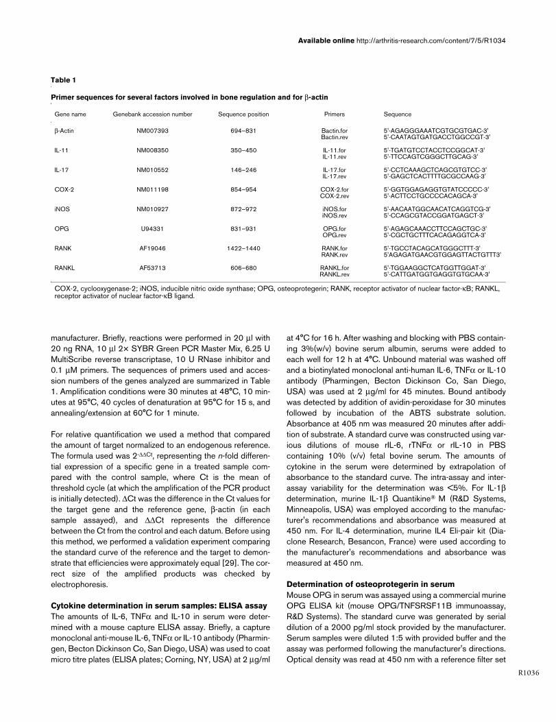
Table 1
Primer sequences for several factors involved in bone regulation and for β-actin
Gene name Genebank accession number Sequence position Primers Sequence
β-Actin NM007393 694–831 Bactin.forBactin.rev
5'-AGAGGGAAATCGTGCGTGAC-3'5'-CAATAGTGATGACCTGGCCGT-3'
IL-11 NM008350 350–450 IL-11.forIL-11.rev
5'-TGATGTCCTACCTCCGGCAT-3'5'-TTCCAGTCGGGCTTGCAG-3'
IL-17 NM010552 146–246 IL-17.forIL-17.rev
5'-CCTCAAAGCTCAGCGTGTCC-3'5'-GAGCTCACTTTTGCGCCAAG-3'
COX-2 NM011198 854–954 COX-2.forCOX-2.rev
5'-GGTGGAGAGGTGTATCCCCC-3'5'-ACTTCCTGCCCCACAGCA-3'
iNOS NM010927 872–972 iNOS.foriNOS.rev
5'-AACAATGGCAACATCAGGTCG-3'5'-CCAGCGTACCGGATGAGCT-3'
OPG U94331 831–931 OPG.forOPG.rev
5'-AGAGCAAACCTTCCAGCTGC-3'5'-CGCTGCTTTCACAGAGGTCA-3'
RANK AF19046 1422–1440 RANK.forRANK.rev
5'-TGCCTACAGCATGGGCTTT-3'5'AGAGATGAACGTGGAGTTACTGTTT3'
RANKL AF53713 606–680 RANKL.forRANKL.rev
5'-TGGAAGGCTCATGGTTGGAT-3'5'-CATTGATGGTGAGGTGTGCAA-3'
COX-2, cyclooxygenase-2; iNOS, inducible nitric oxide synthase; OPG, osteoprotegerin; RANK, receptor activator of nuclear factor-κB; RANKL, receptor activator of nuclear factor-κB ligand.

Arthritis Research & Therapy Vol 7 No 5 Juarranz et al.
R1037
to 540 nm. The intra-assay variability was <5.5% and the limitof detection was 4.5 pg/ml.
Electrophoretic mobility shift assaysMice were sacrificed at day 35 after primary immunization, therear limbs were removed, and the synovial membrane of theknee joints was carefully separated from the bone and carti-lage by microscopic dissection. Cell suspensions were pre-pared by digestion of the synovial tissue in the presence ofRPMI 1640, 250 mg/ml Colagenase D (Roche, Indianapolis,USA) and 0.1 mg/ml DNase I (Roche) for 2 h at 37°C, thensamples were tapped through a 60 µm wire mesh. Nuclearextracts were prepared by the mini-extraction procedure ofSchreiber et al. [30] with slight modifications. Briefly, 107 syn-ovial cells centrifuged at 1,800 × g for 10 minutes. The cellpellets were homogenized with 0.4 ml of buffer A (10 mMHEPES pH 7.9, 10 mM KCl, 0.1 mM EDTA, 0.1 mM EGTA, 1mM dithiothreitol (DTT), 0.5 mM phenylmethylsulphonylfluo-ride (PMSF), 10 µg/ml aprotinin, 10 µg/ml leupeptin, 10 µg/mlpepstatin, 1 mM NaN3, 5 mM NaF and 1 mM Na3VO3). After15 minutes on ice, Nonidet P-40 was added to a final 0.5%concentration, the tubes were gently vortexed for 15 s andnuclei were sedimented and separated from cytosol by centrif-ugation at 12,000 × g for 40 s. Pelleted nuclei were washedonce with 0.2 ml of ice-cold buffer A, and the soluble nuclearproteins were released by adding 0.1 ml of buffer C (20 mMHEPES pH 7.9, 0.4 M NaCl, 1 mM EDTA, 1 mM EGTA, 25%(w/v) glycerol, 1 mM DTT, 0.5 mM PMSF, 10 µg/ml aprotinin,10 µg/ml leupeptin, 10 µg/ml pepstatin and 1 mM NaN3).After incubation for 30 minutes on ice, followed by centrifuga-tion for 10 min at 12,000 × g at 4°C, the supernatants contain-ing the nuclear proteins were harvested, the proteinconcentration was determined by the Bradford method, andaliquots were stored at -80°C for later use in EMSAs.
Double-stranded oligonucleotides (50 ng) corresponding tothe NFκB and AP-1 sites (5'-AGTTGAGGGGACTTTC-CCAGGC-3' and 5'-CGCTTGATGACTCAGCCGGAA-3',respectively), were end-labeled with γ32P-ATP (AmershamPharmacia Biotech, NJ, USA) by using T4 polynucleotidekinase (Invitrogen, Carlsbad, CA, USA). For EMSAs with syn-ovial cell nuclear extracts, 20,000 to 50,000 cpm of double-stranded oligonucleotides, corresponding to approximately0.5 ng, were used for each reaction. The binding reaction mix-tures (15 µl) were set up containing: 0.5 ng DNA probe, 8 µgnuclear extract, 2 µg poly(dI-dC)•poly(dI-dC) and bindingbuffer (50 mM NaCl, 0.2 mM EDTA, 0.5 mM DTT, 5% (w/v)glycerol and 10 mM Tris-HCl pH 7.5). The mixtures were incu-bated on ice for 15 minutes before adding the probe followedby another 20 minutes at room temperature, electrophoresedon a vertical 4% non-denaturing polyacrylamide gel using TGEbuffer (50 mM Tris-HCl pH 7.5, 0.38 M glycine and 2 mMEDTA) and autoradiographed. For supershift assays, nuclearextracts were incubated for 15 minutes at room temperaturewith the specific antibody (1 µg of anti-p65, anti-p50, anti-
cRel, anti-cFos, anti-cJun or anti-JunB) (Santa Cruz Biotech-nology, Santa Cruz, CA, USA,) before the addition of the radi-olabeled probe.
Western blot analysis of IκB-α and phosphorylated cJun in cytoplasm extracts from synovial cellsFor western blotting, the cytoplasm fraction (see above) con-taining 60 µg of protein were subjected to reducing SDS-PAGE (12.5%). After electrophoresis, the gel was electroblot-ted in Tris-glycine buffer containing 40% methanol onto areinforced nitrocellulose membrane (Amersham). The mem-brane was blocked with TBS-T buffer (10 mM Tris, pH 8.0,150 mM NaCl, 0.05% (w/v) Tween 20) containing 5% (v/v)milk powder for 1 h at room temperature, then incubated withprimary antibodies at 1:500 dilutions, rabbit anti-mouse IgGagainst IκB-α (Santa Cruz) or with mouse IgG against phos-phorylated-cJun (Santa Cruz), in TBS-T containing 1% (w/v)milk powder for 2 h at room temperature. The membrane waswashed with TBS-T and incubated with secondary antibody:peroxidase-conjugated goat anti-rabbit IgG (Santa Cruz) or ratanti-mouse IgG (Santa Cruz) at 1:5000 dilutions for 1 h atroom temperature. After washing three times in TBS-T for 5minutes each, and once in TBS for 5 minutes, the membranewas drained quickly and subjected to the enhanced chemilu-miniscence detection system (PIERCE). The X-ray films wereexposed for 5 to 20 minutes.
Statistical analysisAll data were expressed as mean ± SEM. Multiple-samplecomparison (analysis of variance) was used to test differencesbetween groups for significance. A value of p < 0.05 was con-sidered to be significant. The program Statgraphics plus 5.0(Statpoint Inc, Virginia, USA) was used for all statisticalcalculations.
ResultsVIP modulates serum levels of cytokines implicated in bone homeostasisWe have previously reported the beneficial effects of VIP in aCIA model [22]. VIP improves clinical symptoms, decreasingthe incidence and severity of CIA in mice. Notably, histopatho-logical analysis of joints showed that inflammation, cartilagedestruction and bone erosion were abrogated. A link betweeninflammation and bone homeostasis has been attributed to theeffects of cytokines such as IL-1, TNFα, and IL-6 on boneresorption. Other cytokines, such as IL-4 and IL-10 have beenshown to have protective effects if they are administered sys-temically [31]. We have previously reported that VIP treatmentmodulates the expression of different cytokines in the joints ofCIA mice [22].
Treatment of established CIA with VIP (1 nmol every other dayper animal) resulted in suppression of disease activity (Table2). Both cartilage pathology and bone destruction werereduced in VIP treated animals by the end of the experiment as
Available online http://arthritis-research.com/content/7/5/R1034
R1038
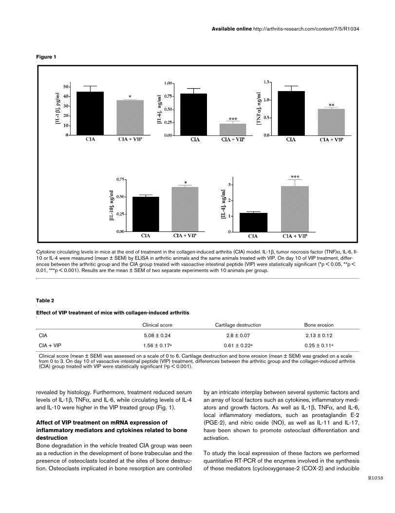
revealed by histology. Furthermore, treatment reduced serumlevels of IL-1β, TNFα, and IL-6, while circulating levels of IL-4and IL-10 were higher in the VIP treated group (Fig. 1).
Affect of VIP treatment on mRNA expression of inflammatory mediators and cytokines related to bone destructionBone degradation in the vehicle treated CIA group was seenas a reduction in the development of bone trabeculae and thepresence of osteoclasts located at the sites of bone destruc-tion. Osteoclasts implicated in bone resorption are controlled
by an intricate interplay between several systemic factors andan array of local factors such as cytokines, inflammatory medi-ators and growth factors. As well as IL-1β, TNFα, and IL-6,local inflammatory mediators, such as prostaglandin E-2(PGE-2), and nitric oxide (NO), as well as IL-11 and IL-17,have been shown to promote osteoclast differentiation andactivation.
To study the local expression of these factors we performedquantitative RT-PCR of the enzymes involved in the synthesisof these mediators (cyclooxygenase-2 (COX-2) and inducible
Figure 1
Cytokine circulating levels in mice at the end of treatment in the collagen-induced arthritis (CIA) modelCytokine circulating levels in mice at the end of treatment in the collagen-induced arthritis (CIA) model. IL-1β, tumor necrosis factor (TNF)α, IL-6, Il-10 or IL-4 were measured (mean ± SEM) by ELISA in arthritic animals and the same animals treated with VIP. On day 10 of VIP treatment, differ-ences between the arthritic group and the CIA group treated with vasoactive intestinal peptide (VIP) were statistically significant (*p < 0.05, **p < 0.01, ***p < 0.001). Results are the mean ± SEM of two separate experiments with 10 animals per group.
Table 2
Effect of VIP treatment of mice with collagen-induced arthritis
Clinical score Cartilage destruction Bone erosion
CIA 5.08 ± 0.24 2.8 ± 0.07 2.13 ± 0.12
CIA + VIP 1.56 ± 0.17a 0.61 ± 0.22a 0.25 ± 0.11a
Clinical score (mean ± SEM) was assessed on a scale of 0 to 6. Cartilage destruction and bone erosion (mean ± SEM) was graded on a scale from 0 to 3. On day 10 of vasoactive intestinal peptide (VIP) treatment, differences between the arthritic group and the collagen-induced arthritis (CIA) group treated with VIP were statistically significant (ap < 0.001).

Arthritis Research & Therapy Vol 7 No 5 Juarranz et al.
R1039
nitric oxide synthase (iNOS)) as well as IL-11 and IL-17 inmRNA extracted from the joints. COX-2 and iNOS expressionincreased 25-fold and almost 2-fold, respectively, in the jointsof CIA mice compared with the joints of control (non-CIA)mice (Fig. 2a). Also, IL-11 and IL-17 mRNA expressionshowed a four-fold increase in CIA mice (Fig. 2b). In CIA micetreated with VIP, the mRNA levels of COX-2, IL-11, and IL-17in the joints were reduced compared with vehicle treated CIAmice, being similar to those of control (non-CIA) mice. The inhi-bition of iNOS expression was even higher.
VIP modulates the RANK/RANKL/OPG system in the arthritic jointAs noted above, a link between the activation of the immunesystem and bone destruction is consistent with the finding thatseveral cytokines contribute to bone resorption via stimulationof osteoclastic mediators. Mechanisms involved in this proc-ess operate by modulating the expression of RANK, RANKLand OPG. To study the modulation of the RANK/RANKL sys-tem and the ratio of RANKL to OPG by VIP during CIA devel-opment we performed quantitative RT-PCR in mRNA extractsfrom the joints of the different groups of animals. We alsodetected circulating OPG levels by ELISA in serum samples.The mRNA expression of RANK and RANKL was heavily stim-ulated in joints after CIA induction (Fig. 3a). In particular, CIA
Figure 2
mRNA expression of inflammatory mediators and cytokines related to bone destructionmRNA expression of inflammatory mediators and cytokines related to bone destruction. (a) Expression of mRNA for cyclooxygenase-2 (COX-2) and inducible nitric oxide synthase (iNOS) in the hind paws was measured by quantitative real-time PCR and corrected by mRNA expression for β-actin in each sample (see Materials and methods). (b) Expression of mRNA for IL-11 and IL-17 in the hind paws was measured by quantitative real-time PCR and corrected by mRNA expression for β-actin in each sample (see Materials and methods). On day 10 of vasoactive intestinal peptide (VIP) treatment, differences between the arthritic group and the CIA group treated with VIP were statistically significant (*p < 0.05, **p < 0.01, ***p < 0.001). Results are the mean ± SEM of two separate experiments with 10 animals per group.

Available online http://arthritis-research.com/content/7/5/R1034
R1040
induction was accompanied by a 50-fold increase in RANKLexpression in the affected joints. Though we also found a smallincrease in OPG mRNA in the same animals, no significant dif-ferences in OPG expression levels were detected after CIAinduction. In spite of this small difference in its expression atthe local level, however, the OPG circulating levels were sig-nificantly higher after CIA induction (Fig. 3b). On the other
hand, the RANKL/OPG ratio was strongly enhanced in CIAmice (Table 3). VIP treatment of CIA mice resulted in a signif-icant reduction in the expression of both RANK and RANKL,the mRNA levels of which in joints fell to near control values(non-CIA mice). Although in VIP treated mice OPG mRNA lev-els were slightly increased, a seven-fold drop in the RANKL/OPG ratio was observed (Table 3). The circulating levels ofOPG were also significantly higher in VIP treated mice com-pared with CIA mice (Fig. 3b).
VIP prevents in vivo NFκB translocation and inhibits c-Jun N-terminal kinaseCrucial events in signalling by RANKL and other osteoclasticcytokines are the translocation of NFκB to the nucleus and theactivation of c-Jun N-terminal kinase (JNK), which leads to theactivation of AP-1 [32,33]. A central role for these transcrip-tion factors is supported by the fact that both are activated bythe tumor necrosis factor receptor-associated factor (TRAF)family of signal transducers and selective inhibition of NFκBblocks osteoclastogenesis and prevents inflammatory bonedestruction in vivo [32,34]. Previous studies have shown thatVIP induces a downregulation of NFκB transcriptional activityin human monocytes in culture [35,36], as well as an AP-1
Figure 3
Vasoactive intestinal peptide (VIP) modulates the pattern of expression of the RANK/RANKL/OPG system in joints from mice with collagen-induced arthritis (CIA)Vasoactive intestinal peptide (VIP) modulates the pattern of expression of the RANK/RANKL/OPG system in joints from mice with collagen-induced arthritis (CIA). (a) Expression of mRNA for receptor activator of nuclear factor-κB (RANK), receptor activator of nuclear factor-κB ligand (RANKL) or osteoprotegerin (OPG) in the hind paws was measured by quantitative real time PCR and corrected by mRNA expression for β-actin in each sample (see Materials and methods). (b) Serum levels of OPG in control, CIA or VIP-treated CIA mice were determined by ELISA. On day 10 of VIP treat-ment, differences between the arthritic group and the CIA group treated with VIP were statistically significant (**p < 0.01, ***p < 0.001). Results are the mean ± SEM of two independent experiments with 10 animals per group
Table 3
Ratio of RANKL to OPG in mice with collagen-induced arthritis
CIA CIA + VIP
RANKL 51.20 ± 3.57 13.08 ± 2.45a
OPG 1.46 ± 0.27 2.46 ± 0.92
RANKL/OPG 35.09 ± 3.18 5.30 ± 0.95a
The mRNA expression for RANKL and OPG in hind paws of mice with collagen-induced arthritis (CIA) was measured by quantitative real time PCR and corrected by mRNA expression for β-actin in each sample. On day 10 of vasoactive intestinal peptide (VIP) treatment, differences between the arthritic group and the CIA group treated with VIP were statistically significant (ap < 0.001). Results are the mean ± SEM of two independent experiments with 10 animals per group. OPG, osteoprotegerin; RANKL, receptor activator of nuclear factor-κB ligand.
Arthritis Research & Therapy Vol 7 No 5 Juarranz et al.
R1041
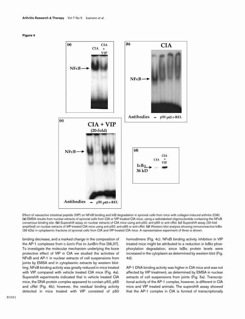
binding decrease, and a marked change in the composition ofthe AP-1 complexes from c-Jun/c-Fos to JunB/c-Fos [36,37].To investigate the molecular mechanism underlying the boneprotective effect of VIP in CIA we studied the activities ofNFκB and AP-1 in nuclear extracts of cell suspensions fromjoints by EMSA and in cytoplasmic extracts by western blot-ting. NFκB binding activity was greatly reduced in mice treatedwith VIP compared with vehicle treated CIA mice (Fig. 4a).Supershift experiments indicated that in vehicle treated CIAmice, the DNA protein complex appeared to contain p50, p65and cRel (Fig. 4b); however, the residual binding activitydetected in mice treated with VIP consisted of p50
homodimers (Fig. 4c). NFκB binding activity inhibition in VIPtreated mice might be attributed to a reduction in IκBα phos-phorylation degradation, since IκBα protein levels wereincreased in the cytoplasm as determined by western blot (Fig.4d).
AP-1 DNA binding activity was higher in CIA mice and was notaffected by VIP treatment, as determined by EMSA in nuclearextracts of cell suspensions from joints (Fig. 5a). Transcrip-tional activity of the AP-1 complex, however, is different in CIAmice and VIP treated animals. The supershift assay showedthat the AP-1 complex in CIA is formed of transcriptionally
Figure 4
Effect of vasoactive intestinal peptide (VIP) on NFκB binding and IκB degradation in synovial cells from mice with collagen-induced arthritis (CIA)Effect of vasoactive intestinal peptide (VIP) on NFκB binding and IκB degradation in synovial cells from mice with collagen-induced arthritis (CIA). (a) EMSA results from nuclear extracts of synovial cells from CIA or VIP-treated CIA mice, using a radiolabeled oligonucleotide containing the NFκB consensus binding site. (b) Supershift assay on nuclear extracts of CIA mice using anti-p50, anti-p65 or anti-cRel. (c) Supershift assay (20-fold amplified) on nuclear extracts of VIP-treated CIA mice using anti-p50, anti-p65 or anti-cRel. (d) Western blot analysis showing immunoreactive IκBα (36 kDa) in cytoplasmic fractions of synovial cells from CIA and VIP-treated CIA mice. A representative experiment of three is shown.

Available online http://arthritis-research.com/content/7/5/R1034
R1042
active c-Jun/c-Fos heterodimers (Fig. 5b), while in VIP treatedanimals the AP-1 complex is formed by the transcriptionallyinactive heterodimer c-Fos/Jun-B (Fig. 5c). The shift in thecomposition of the AP-1 complex may be mediated byinhibition of JNK activity because the western blot analysisindicated that phospho-c-Jun decreases in the cytoplasm afterVIP treatment (Fig. 5d).
DiscussionData presented in this report indicate that VIP treatment pre-vents bone erosion in the CIA model of RA. Several mecha-nisms may account for this effect. VIP inhibits local andsystemic levels of pro-inflammatory mediators implicated inbone resorption, such as IL-1β, IL-6, IL-11, IL-17, TNFα, PGEand NO, while the circulating levels of cytokines with boneprotective effects, such as IL-4 and IL-10, are increased. Onthe other hand, VIP modulates the RANK/RANKL/OPG sys-tem, which is biased toward bone formation. Finally, osteoclastfunction may be inhibited as it depends on NFkB and AP-1transcription factor activity, which is impaired in VIP treatedmice.
VIP has been shown to regulate several bone cell functions; itaffects bone resorbing activity of isolated osteoclasts andosteoclast formation [28] as well as osteoblast anabolicprocesses [24]. These effects are mediated by the presenceof different VIP receptors in both types of bone cells: VPAC1and PAC1 have been detected in osteoclasts [26] whileVPAC2 is expressed in osteoblasts and VPAC1 is induced inadvanced cultures of this cell type [27]. In vitro studies withisolated cells have shown contradictory results; while VIP hasbeen shown to promote the formation of mineralised nodules
in cultures of osteoblasts [24], it induces a transient inhibitionand a delayed stimulation of osteoclast activity [38]. Ourresults show that VIP treatment in vivo in pathological condi-tions such as RA results in the prevention of bone destruction.
Cytokine balance contributes to the onset and progression ofinflammation and skeletal destruction during RA. In thisrespect, TNFα, IL-1β and IL-6 have been shown to bedominant in the induction of inflammation and bone erosion[39-41], while IL-4 and IL-10 have potent anti-inflammatoryeffects and suppress cartilage and bone pathology in RA [31].Both a systemic and a paracrine mode of action can be postu-lated for these agents. Alteration of the systemic balance ofcytokines has been studied by blocking TNFα and IL-1β usingbiological agents such as anti-TNFα or IL-1 inhibitors [39].Therefore, a combined cytokine and anti-cytokine therapy hasbeen proposed as being the more effective for achieving ananti-inflammatory and anti-destructive therapy for RA. VIP thusemerges as a new, promising biological agent in this sense, astreatment of CIA mice with this peptide shifts the systemic bal-ance of cytokines toward a bone protecting pattern that actsto both lower serum levels of TNFα, IL-1β and IL-6 and raisethe levels of IL-4 and IL-10, as described in this report.
Bone loss in RA is indirectly mediated mainly by cytokines pro-duced by macrophages, fibroblasts and T cells of the synovialtissue. These cytokines lead to the differentiation of osteoclastprecursors and activate osteoclasts. Macrophage and fibrob-last derived inflammatory cytokines such as IL-1β and TNFαperpetuate inflammation in a paracrine manner. In a previousreport, we have shown that VIP reduces the expression ofsuch mediators in the joint microenvironment of arthritic mice
Figure 5
AP-1 binding and c-Jun activation in synovial cells from mice with collagen-induced arthritis (CIA) after vasoactive intestinal peptide (VIP) treatmentAP-1 binding and c-Jun activation in synovial cells from mice with collagen-induced arthritis (CIA) after vasoactive intestinal peptide (VIP) treatment. (a) EMSA results from nuclear extracts of synovial cells from CIA or VIP-treated CIA mice, using a radiolabeled oligonucleotide containing the AP-1 consensus binding site. (b) Supershift assay on nuclear extracts of CIA mice using anti-c-Jun, anti-c-Fos or anti-Jun B. (c) Supershift assay on nuclear extracts of VIP-treated CIA mice using anti-c-Jun, anti-c-Fos or anti-Jun B. (d) Western blot analysis showing immunoreactive phosphor-ylated c-Jun (39 kDa) in cytoplasmic fractions of synovial cells from CIA and VIP-treated CIA mice. A representative experiment of three is shown.

Arthritis Research & Therapy Vol 7 No 5 Juarranz et al.
R1043
[22]. At the same time, VIP augments the local production ofthe anti-inflammatory cytokine IL-10 and the IL-1 inhibitor IL-1Ra [22]. PGE [42] and NO [43] are two potent mediatorsinduced by inflammatory cytokines that stimulate their osteo-clastogic activities. They are also inhibited in the joints of VIPtreated mice, as can be deduced from the lower expression ofiNOS and COX-2.
VIP can also impair osteoclast differentiation in RA through itseffect on T cell differentiation and activation. T cells present inthe synovial tissue in RA express a Th1/Th0 pattern of cytokinesecretion [44]. Activated T cells and T cells from RA synovialtissue express both the membrane-bound and soluble forms ofRANKL, which induce the differentiation of osteoclast precur-sors [45]. Cytokines also participate in this process. IL-17 is acytokine produced by a subset of activated memory Th1/Th0cells [46] that has been shown to be an important osteoclastdifferentiation factor, inducing RANKL expression leading tobone erosion in arthritis [10]. IL-11 also supports osteoclastformation by increasing RANKL expression in a STAT (Signaltransducers and activators of transcription) activation depend-ent mechanism [47]. As we have described in this report, VIPtreatment greatly reduces the local expression of both thesecytokines in the joints of arthritic mice, which may account forthe block in joint erosion induced in the CIA model. Addition-ally, VIP shifts the immune response towards a Th2 pattern ofcytokine secretion [17], which inhibits the production ofinflammatory and Th1 cytokines [48].
Most of the osteoclastogenic factors present in RA joints arethought to act indirectly, enhancing RANKL expression andthereby altering the RANK/RANKL/OPG system, which is thefinal regulator of bone resorption [2,3,49]. RANK is expressedon the surface of haematopoietic osteoclast progenitors thatbelong to the monocyte/macrophage lineage, and also onmature osteoclasts, as well as on T cells and dendritic cells. Inarthritis, osteoclast precursors that express RANK recognizeRANKL through cell-to-cell interaction with osteoblasts/stro-mal cells, and differentiate into osteoclasts [50]. In the presentstudy, we report a high level of RANK expression in the jointsof arthritic mice, probably induced by the recruitment of oste-oclast precursors induced by the local production ofchemokines chemotactic for monocytes [51]. We alsodescribe how VIP lowers the expression of RANK in the jointsof CIA mice to the levels detected in non-arthritic control mice.This effect may be due to the inhibition of RANK synthesis or,alternatively, to the inhibition of monocyte recruitment; wehave reported previously that VIP inhibits the local expressionof the monocyte chemoatractant chemokines CCL3 (MIP1α)and CCL2 (MCP-1) [22,23]. RANKL expression can beupregulated by bone resorbing factors such as glucocorti-coids, vitamin D, IL-1β, IL-6, IL-11, IL-17, TNFα, PGE2, or par-athyroid hormone in osteoblasts. RANKL is expressed on thecell surface of activated T cells and can be detected in bothsynovial cells and infiltrating cells by in situ hybridization at the
onset of clinical signs of arthritis in animal models [52]. T-cellactivation in RA patients may lead to osteoclastogenesiswithin the synovium, probably via RANKL secretion byactivated T cells in an environment conducive to osteoclastdifferentiation from synovial macrophages. This mechanismmay contribute to the bone destruction seen in RA [14].
VIP has been reported to inhibit the expression of RANKL andRANK induced by vitamin D in mouse bone marrow cultures[28]. Results shown in this report indicate that VIP reduces theexpression of RANK and RANKL in the joints of arthritic mice,and may account for the bone protective properties of VIP inRA. On the other hand, its effects on the expression of OPGfurther support the postulated bone protective property of VIP.This molecule is secreted by stromal cells and osteoblasts andcompetitively inhibits RANKL binding to RANK on the cell sur-face of osteoclast precursor cells and mature osteoclasts,thus inhibiting the osteoclastogenic actions of RANKL. Exces-sive production of RANKL and/or a deficiency of OPG could,therefore, contribute to the increased bone resorption typifiedby the focal bone erosion and bone loss in RA. Our data indi-cate that OPG circulating levels rise in CIA, as has beenreported during inflammation [14]. These levels were evenhigher in VIP treated mice. In this way, the ratio of RANKL-RANK to OPG that determines the erosive nature of RA isgreatly reduced by VIP, accounting for the bone protectionachieved by the treatment.
The molecular mechanisms underlying the discussed effectsof VIP in bone protection during RA (mainly cytokine secretion,RANKL expression, and osteoclast differentiation) may involvethe transcription factors NFκB and AP-1. Several cell typesshare these signalling pathways to express mediators impli-cated in tissue damage and destruction. After exposure to pro-inflammatory cytokines, the IκB kinase (IKK) signal complex isactivated in synoviocytes, leading to phosphorylation of IκB.We describe in this report that IκB phosphorylation is inhibitedin the arthritic joints of mice treated with VIP. NFκB is activatedin this manner in the synovium of patients with RA and regu-lates genes encoding proteins that contribute to inflammation,including inflammatory cytokines such as TNFα, IL-1β, IL-6and chemokines as well as enzymes such as iNOS and COX-2. NFκB is also crucial for the differentiation of osteoclasts andits selective inhibition blocks RANKL induced osteoclastogen-esis both in vitro and in vivo [32]. The MAPK (Mitogen-acti-vated protein kinases) pathway is also involved and particularlythe JNK pathway, which has been implicated in the regulationof matrix metalloproteinases. As reported here, JNK activity inthe joints of arthritic mice is affected by VIP treatment. Ourunderstanding of the signal transduction pathways implicatedin RA has led to drug development programmes targetingMAPK and NFκB inhibitors [53]. Several of these compounds,however, have been shown to be toxic. VIP on the other handhas been shown to target these signalling pathways and notoxicity has been cited for this peptide. Ourselves and others

Available online http://arthritis-research.com/content/7/5/R1034
R1044
have previously reported that VIP inhibits the nuclear translo-cation of NFκB and also the JNK signalling pathway in LPS(lipopolysaccharide) stimulated macrophage and monocyticcell lines [35-37] In the present report, we describe that thismechanism also operates in vivo and may involve other celltypes involved in the pathogenesis of RA.
In summary, the protective effect of VIP in bone destructionduring CIA could be due to different mechanisms that are notmutually exclusive. One would be an indirect mechanism thatworks via decreasing proinflammatory cytokines and othermediators involved in the differentiation and activation of oste-oclast-precursor cells, and increasing anti-inflammatorycytokines. A second would be the VIP-induced modification ofthe cell types present in the joint, which would decrease theamount of Th1-lymphocytes that express RANKL. And a thirdwould be a VIP-induced direct effect on OPG, RANK orRANKL expression on skeletal tissue, fibroblast or immunecells present in the inflamed joint.
ConclusionWe have shown that VIP treatment in CIA mice reduces thelocal and systemic levels of osteoclastogenic mediators, suchas TNFα, IL-1β, IL-6, IL-11, IL-17, PGE and NO. This reductionis accompanied by a large decrease in the RANK-RANKL/OPG ratio. Molecular mechanisms associated with theseevents include a reduction in the activity of the transcriptionfactor NFκB and a change in the activity of AP-1. Our resultshighlight the possibility of the therapeutic application of VIP inthe treatment of human RA.
Competing interestsWe have signed a research agreement with a company ((Gen-etrix S.L., Spain)) interested in the development of new thera-peutic approaches to treat RA, although this company did notfinance this manuscript. We have no stocks or shares with anyorganization. We do not have any patent application related tothe content of this manuscript. We do not have any financial ornon-financial competing interest.
Authors' contributionsYJ made substantial contributions to the conception anddesign of this study and the acquisition, analysis and interpre-tation of data. CA carried out the histopathological studies.CM prepared the samples and gave final approval of the man-uscript for publication. AA was involved in the real-time analy-sis. IGC prepared the samples and performed the statisticalanalysis. FR was involved in the design of the figures. RPGmade substantial contributions to the conception and designof the study, gave final approval of the manuscript forpublication. JL made substantial contributions to the concep-tion and design of the study and was involved in revising thearticle critically for important intellectual content. All authorsread and approved the final manuscript.
AcknowledgementsThis work was supported by grants BFI 2002-03489 from Ministerio de Ciencia y Tecnología (Spain), G03/152 from Fondo de Investigación Sanitaria (Spain), a predoctoral fellowship from Ministerio de Ciencia y Tecnología (to AA), and a postdoctoral contract from Madrid Commu-nity (to YJ).
References1. Gravallese EM: Bone destruction in arthritis. Ann Rheum Dis
2002, 61:ii84-ii86.2. Takayanagi H, Iizuka H, Juji T, Nakagawa T, Yamamoto A, Myazaki
T, Koshihara Y, Oda H, Nakamura K, Tanaka S: Involvement ofreceptor activator of nuclear factor kB ligand/osteoclast dif-ferentiation factor in osteoclastogenesis from synoviocytes inrheumatoid arthritis. Arthritis Rheum 2000, 43:259-269.
3. Hofbauer LC, Heulfelder AE: The role of osteoprotegerin andreceptor activator of nuclear factor kappaB ligand in thepathogenesis and treatment of rheumatoid arthritis. ArthritisRheum 2001, 44:253-259.
4. Nakashima T, Wada T, Penninger JM: RANKL and RANK as noveltherapeutic targets for arthritis. Curr Opin Rheumatol 2003,15:280-287.
5. O'Gradaigh D, Ireland D, Bord S, Compston JE: Joint erosion inrheumatoid arthritis: interactions between tumor necrosis fac-tor α, interleukin 1, and receptor activator of nuclear factor kBligand (RANKL) regulate osteoclasts. Ann Rheum Dis 2004,63:354-359.
6. Myers LK, Rosloniec EF, Cremer MA, Kang AH: Collagen-induced arthritis, an animal model of autoimmunity. Life Sci1878, 61:1861-1872.
7. Lubberts E, Oppers-Walgreen B, Pettit AR, van den Bersselaar L,Joosten LAB, Goldring SR, Gravallese EM, van den Berg WB:Increase in expression of receptor activator of nuclear factorκB at sites of bone erosion correlates with progression ofinflammation in evolving collagen-induced arthritis. ArthritisRheum 2002, 46:3055-3064.
8. Blair HC, Athanasou NA: Recent advances in osteoclast biologyand pathological bone resorption. Histol Histopathol 2004,19:189-199.
9. Ahlen J, Andersson S, Mukohyama H, Roth C, Bäckman A, Cona-way HH, Lerner UH: Characterization of the bone-resorptiveeffect of interleukin-11 in cultured mouse calvarial bones.Bone 2002, 31:242-251.
10. Lubberts E, van den Bersselaar L, Oppers-Walgreen B,Schwarzenberger P, Coenen-de Roo CJJ, Kolls JK, Joosten LAB,van den Berg WB: IL-17 promotes bone erosion in murine col-lagen-induced arthritis through loss of the receptor activatorof NF-kB ligand/osteoprotegerin balance. J Immunol 2003,170:2655-2662.
11. Nakagawa N, Kinosaki M, Yamaguchi K, Shima N, Yasuda H, YanoK, Morinaga T, Higashio K: RANK is the essential signalingreceptor for osteoclast differentiation factor inostoclastogenesis. Biochem Biophys Res Commun 1998,253:395-400.
12. Boyle WJ, Simonet WS, Lacey DL: Osteoclast differentiationand activation. Nature 2003, 423:337-342.
13. Teitelbaum SL: Bone resorption by osteoclast. Science 2000,289:1504-1508.
14. Saidenberg-Kermanac'h N, Cohen-Solal M, Bessis N, De Ver-nejoul MC, Boissier MC: Role of osteoprotegerin in rheumatoidinflammation. Joint Bone Spine 2004, 71:9-13.
15. Gomariz RP, Lorenzo MJ, Cacicedo L, Vicente A, Zapata AG:Demonstration of immunoreactive vasoactive intestinal pep-tide (IR-VIP) and somatostatin (IR-SOM) in rat thymus. BrainBehav Immun 1990, 4:151-161.
16. Gomariz RP, Martinez C, Abad C, Leceta J, Delgado M: Immuno-biology of VIP: a review and therapeutical perspectives. CurrPharm Des 2001, 7:89-111.
17. Delgado M, Abad C, Martinez C, Juarranz MG, Arranz A, GomartizRP, Leceta J: Vasoactive intestinal peptide in the immune sys-tem: potential therapeutic role in inflammatory and autoim-mune disease. J Mol Med 2002, 80:16-24.
18. Gomariz RP, Abad C, Martinez C, Juarranz MG, da Costa S, ArranzA, Delgado M, Leceta J: Vasoactive intestinal peptide, pituitary
http://www.ncbi.nlm.nih.gov/entrez/query.fcgi?cmd=Retrieve&db=PubMed&dopt=Abstract&list_uids=9364191
http://www.ncbi.nlm.nih.gov/entrez/query.fcgi?cmd=Retrieve&db=PubMed&dopt=Abstract&list_uids=9364191
http://www.ncbi.nlm.nih.gov/entrez/query.fcgi?cmd=Retrieve&db=PubMed&dopt=Abstract&list_uids=9878548
http://www.ncbi.nlm.nih.gov/entrez/query.fcgi?cmd=Retrieve&db=PubMed&dopt=Abstract&list_uids=9878548
http://www.ncbi.nlm.nih.gov/entrez/query.fcgi?cmd=Retrieve&db=PubMed&dopt=Abstract&list_uids=9878548
http://www.ncbi.nlm.nih.gov/entrez/query.fcgi?cmd=Retrieve&db=PubMed&dopt=Abstract&list_uids=1975506
http://www.ncbi.nlm.nih.gov/entrez/query.fcgi?cmd=Retrieve&db=PubMed&dopt=Abstract&list_uids=1975506

Arthritis Research & Therapy Vol 7 No 5 Juarranz et al.
R1045
adenylate cyclase-activating polypeptide and immune system:from basic research to potential clinical application. Biomedi-cal Rev 2001, 12:1-9.
19. Delgado M, Pozo D, Ganea D: The significance of vasoactiveintestinal peptide in immunomodulation. Pharmacol Rev 2004,56:249-290.
20. Delgado M, Martinez C, Pozo D, Calvo JR, Leceta J, Ganea D,Gomariz RP: Vasoactive intestinal peptide (VIP) and pituitaryadenylate cyclase-activation polypeptide (PACAP) protectmice from lethal endotoxemia through the inhibition of TNF-alpha and IL-6. J Immunol 1999, 162:1200-1205.
21. Abad C, Martinez C, Juarranz MG, Arranz A, Leceta J, Delgado M,Gomariz RP: Therapeutic effects of vasoactive intestinal pep-tide in the trinitrobenzene sulfonic acid mice model of Crohn'sdisease. Gastroenterology 2003, 124:961-971.
22. Delgado M, Abad C, Martinez C, Leceta J, Gomariz RP: Vasoac-tive intestinal peptide prevents experimental arthritis by down-regulating both autoimmune and inflammatory components ofthe disease. Nat Med 2001, 7:563-568.
23. Juarranz MG, Santiago B, Torroba M, Gutierrez-Cañas I, Palao G,Galindo M, Abad C, Martinez C, Leceta J, Pablos JL, Gomariz RP:Vasoactive intestinal peptide modulates proinflammatorymediator synthesis in osteoartritic and rheumatoid synovialcells. Rheumatology (Oxford) 2004, 43:416-422.
24. Lundberg P, Boström I, Mukohyama H, Bjurholm A, Smans K,Lerner UH: Neuro-hormonal control of bone metabolism:vasoactive intestinal peptide stimulates alkaline phosphataseactivity and mRNA expression in mouse calvarial osteoblastsas well as calcium accumulation mineralized bone nodules.Regul Pept 1999, 85:47-58.
25. Harmar AJ, Arimura A, Gozes I, Journot L, Laburthe M, Pisegna JR,Rawlings SR, Robberecht P, Said SI, Sreedharan SP, et al.: Inter-national Union of Pharmacology. XVIII. Nomenclature ofreceptors for vasoactive intestinal peptide and pituitary ade-nylate cyclase-activating polypeptide. Pharmacol Rev 1998,50:265-270.
26. Ransjö M, Lie A, Mukohyama H, Lundberg P, Lerner UH: Microiso-lated mouse osteoclasts express VIP-1 and PACAP receptors.Biochem Biophys Res Commun 2000, 274:400-404.
27. Lundberg P, Lundgren I, Mukohyama H, Lehenkari PP, Horton MA,Lerner UH: Vasoactive intestinal peptide (VIP)/pituitary ade-nylate cyclase-activating peptide receptor subtypes in mousecalvarian osteoblasts: presence of VIP-2 receptors and differ-entiation-induced expression of VIP-1 receptors. Endocrinol-ogy 2001, 142:339-347.
28. Mukohyama H, Ransjö M, Taniguchi H, Ohyama T, Lerner UH: Theinhibitory effects of vasoactive intestinal peptide and pituitaryadenylate cyclase-activating polypeptide on osteoclast forma-tion are associated with upregulation of osteoprotegerin anddownregulation of RANKL and RANK. Biochem Biophys ResCommun 2000, 271:158-163.
29. Bustin SA: Absolute quantification of mRNA using real-timereverse transcription polymerase chain reaction assays. J MolEndocrinol 2000, 25:169-193.
30. Schreiber E, Metthias P, Muller W, Shaffner W: Rapid detectionof octamer binding proteins with "mini-extracts" preparedfrom a small number of cells. Nucleic Acids Res 1989, 17:6419.
31. Joosten LAB, Lubberts E, Helsen MMA, Saxne T, Coenen-de RooCJJ, Heinegard D, van den Berg W: Protection against cartilageand bone destruction by systemic interleukin-4 treatment inestablished murine type II collagen-induced arthritis. ArthritisRes 1999, 1:81-91.
32. Jimi E, Aoki K, Saito H, DÀcquisto F, May MJ, Nakamura I, Sudo T,Kojima T, Okamoto F, Fukushima H, et al.: Selective inhibition ofNF-kB blocks osteoclastogenesis and prevents inflammatorybone destrction in vivo. Nature Medicine 2004, 10:617-624.
33. Ikeda F, Nishimura R, Matsubara T, Tanaka S, Inoue JI, Reddy S,Hata K, Yamashita K, Hiraga T, Watanabe T, et al.: Critical rolesof c-Jun signalling in regulation of NFAT family and RANKL-regulated osteoclast differentiation. J Clin Invest 2004,114:475-484.
34. Wong BR, Josien R, Lee Sy, Vologodskaia M, Steinman RM, ChoinY: The TRAF family of signal transducers mediates NF-kappaB activation by the TRANCE receptor. J Biol Chem 1998,273:28355-28359.
35. Delgado M, Ganea D: Vasoactive intestinal peptide and pitui-tary adenylate cyclase-activating polypeptide inhibit nuclear
factor-kB-dependent gene activation at multiple levels in thehuman monocytic cell line THP-1. J Biol Chem 2001,276:369-380.
36. Leceta J, Gomariz RP, Martinez C, Abad C, Ganea D, Delgado M:Receptors and transcriptional factors involved in the anti-inflammatory activity of VIP and PACAP. Ann NY Acad Sci2000, 921:92-102.
37. Delgado M, Ganea D: Vasoactive intestinal peptide and pitui-tary adenylate cyclase activating polypeptide inhibit theMEKK1/MEK4/JNK signaling pathway in LPS-stimulatedmacrophages. J Neuroimmunol 2000, 110:97-105.
38. Lundberg P, Lie A, Bjurholm A, Lehenkari PP, Horton MA, LernerUH, Ransjö M: Vasoactive intestinal peptide regulates osteo-clast activity via specific binding sites on both osteoclasts andosteoblasts. Bone 2000, 27:803-810.
39. Van den Berg WB: Anti-cytokine therapy in chronic destructivearthritis. Arthritis Res 2001, 3:18-26.
40. Joosten LAB, Helsen MMA, Saxne T, van de Loo FAJ, HeinegardD, van den Berg WB: IL-1αβ blockade prevents cartilage andbone destruction in murine type II collagen-induced arthritis,whereas TNF-a blockade only ameliorates joint inflammation.J Immunol 1999, 163:5049-5055.
41. Bonecchi R, Bianchi G, Bordignon PP, D'Ambrosio D, Lang R,Borsatti A, Sozzani S, Allavena P, Gray PA, Mantovani A, SinigagliaF: Differential expression of chemokine receptors and chemo-tactic responsiveness of type 1 T helper cells (Th1s) and Th2s.J Exp Med 1998, 187:129-134.
42. Miyaura C, Inada M, Matsumoto C, Ohshiba T, Uozumi N, ShimuzuT, Ito A: An essential role of cytosolic phospholipase A2α inprostaglandin E2-mediated bone resorption associated withinflammation. J Exp Med 2003, 197:1303-1310.
43. Van't Hof RJ, Armour KJ, Smith LM, Armour KE, Wei XQ, Liew FY,Ralston SH: Requirement of the inducible nitric oxide synthasepathway for IL-1-induced osteoclastic bone resorption. ProcNatl Acad Sci USA 2000, 97:7993-7998.
44. Gerli R, Bistoni O, Russano A, Fiorucci S, Borgato L, CesarottiMEF, Lunardi C: In vivo activated T cells in rheumatoid synovi-tis. Analysis of Th1-and Th2-type cytokine production at clonallevel in different stages of disease. Clin Exp Immunol 2002,129:549-555.
45. Horwood NJ, Kartsogiannis V, Quinn JMW, Romas E, Martin TJ,Gillespie MT: Activated T lymphocytes support osteoclast for-mation in vitro. Biochim Biophys Res Commun 1999,265:144-150.
46. Aarvak T, Chabaud M, Miossec P, Natvig JB: IL-17 is produced bysome proinflammatory Th1/Th0 cells but not by Th2 cells. JImmunol 1999, 162:1246-1251.
47. Walton JK, Duncan JM, Deschamps P, Shaughnessy SG: Heparinacts synergistically with interleukin-11 to induce STAT3 activa-tion and in vitro osteoclast formation. Blood 2002,100:2530-2536.
48. Schulze-Koops H, Kalden JR: The balance of Th1/Th2 cytokinesin rheumatoid arthritis. Best Pract Res Clin Rheumatol 2001,15:677-691.
49. Romas E, Gillespie MT, Martin TJ: Involvement of receptor acti-vator of NFkB ligand and tumor necrosis factor-α in bonedestruction in rheumatoid arthritis. Bone 2002, 30:340-346.
50. Jones DH, Kong Y-Y, Penninger JM: Role of RANKL and RANK inbone loss and arthritis. Ann Rheum Dis 2002, 61Suppl:ii32-ii39.
51. Szekanecz Z, Kim J, Koch AE: Chemokines and chemokinereceptors in rheumatoid arthritis. Semin Immunol 2003,15:15-21.
52. Kong Y-Y, Feige U, Sarosi I, Bolon B, Tarufi A, Morony S, Cap-parelli C, Li J, Elliot R, McCabe S, et al.: Activated T cells regulatebone loss and joint destruction in adjuvant arthritis throughosteoprotegerin ligand. Nature 1999, 402:304-309.
53. Smolen JS, Steiner G: Therapeutic strategies for rheumatoidarthritis. Nat Rev Drug Discov 2003, 2:473-488.
http://www.ncbi.nlm.nih.gov/entrez/query.fcgi?cmd=Retrieve&db=PubMed&dopt=Abstract&list_uids=9916753
http://www.ncbi.nlm.nih.gov/entrez/query.fcgi?cmd=Retrieve&db=PubMed&dopt=Abstract&list_uids=9916753
http://www.ncbi.nlm.nih.gov/entrez/query.fcgi?cmd=Retrieve&db=PubMed&dopt=Abstract&list_uids=9916753
http://www.ncbi.nlm.nih.gov/entrez/query.fcgi?cmd=Retrieve&db=PubMed&dopt=Abstract&list_uids=9647867
http://www.ncbi.nlm.nih.gov/entrez/query.fcgi?cmd=Retrieve&db=PubMed&dopt=Abstract&list_uids=9647867
http://www.ncbi.nlm.nih.gov/entrez/query.fcgi?cmd=Retrieve&db=PubMed&dopt=Abstract&list_uids=9647867
http://www.ncbi.nlm.nih.gov/entrez/query.fcgi?cmd=Retrieve&db=PubMed&dopt=Abstract&list_uids=2771660
http://www.ncbi.nlm.nih.gov/entrez/query.fcgi?cmd=Retrieve&db=PubMed&dopt=Abstract&list_uids=2771660
http://www.ncbi.nlm.nih.gov/entrez/query.fcgi?cmd=Retrieve&db=PubMed&dopt=Abstract&list_uids=2771660
http://www.ncbi.nlm.nih.gov/entrez/query.fcgi?cmd=Retrieve&db=PubMed&dopt=Abstract&list_uids=9774460
http://www.ncbi.nlm.nih.gov/entrez/query.fcgi?cmd=Retrieve&db=PubMed&dopt=Abstract&list_uids=9774460
http://www.ncbi.nlm.nih.gov/entrez/query.fcgi?cmd=Retrieve&db=PubMed&dopt=Abstract&list_uids=9419219
http://www.ncbi.nlm.nih.gov/entrez/query.fcgi?cmd=Retrieve&db=PubMed&dopt=Abstract&list_uids=9419219
http://www.ncbi.nlm.nih.gov/entrez/query.fcgi?cmd=Retrieve&db=PubMed&dopt=Abstract&list_uids=9973376