Genome-wide association study of serum prostate-specific ...

www.practicalradonc.org
Practical Radiation Oncology (2018) 8, 28-39
Basic Original Report
Prostate cancer–specific PET radiotracers:A review on the clinical utility inrecurrent disease
Jaden D. Evans MD a, 1, Krishan R. Jethwa MD a, 1, Piet Ost MD, PhD b,Scott Williams MD c, Eugene D. Kwon MD d, Val J. Lowe MD e,Brian J. Davis MD, PhD, FABS a,⁎aDepartment of Radiation Oncology, Mayo Clinic, Rochester, MinnesotabDepartment of Radiation Oncology and Experimental Cancer Research, Ghent University Hospital, Ghent, BelgiumcDepartment of Radiation Oncology, Peter MacCallum Cancer Centre, Melbourne, AustraliadDepartment of Urology, Mayo Clinic, Rochester, MinnesotaeDepartment of Radiology, Mayo Clinic, Rochester, Minnesota
Received 9 June 2017; revised 12 July 2017; accepted 18 July 2017
Abstract Prostate cancer–specific positron emission tomography (pcPET) has been shown to detectsites of disease recurrence at serum prostate-specific antigen (PSA) levels that are lower than those levelsdetected by conventional imaging. Commonly used pcPET radiotracers in the setting of biochemicalrecurrence are reviewed including carbon 11/fludeoxyglucose 18 (F-18) choline, gallium 68/F-18 prostate-specificmembrane antigen (PSMA), and F-18 fluciclovine. Review of the literature generally favors PSMA-based agents for the detection of recurrence as a function of lowPSA levels. Positive gallium68/F-18PSMApositron emission tomography/computed tomography scans detectedpotential sites of recurrence in amedian51.5% of patients when PSA level is b1.0 ng/mL, 74% of patients when PSA level is 1.0 to 2.0 ng/mL, and90.5% of patients when PSA level is N2.0 ng/mL. Review of carbon 11/fludeoxyglucose 18 (F-18) cholineand F-18 fluciclovine data commonly demonstrated lower detection rates for each respective PSA cohort,although with some important caveats, despite having similar operational characteristics to PSMA-basedimaging. Sensitive pcPET imaging has provided new insight into the early patterns of disease spread, whichhas prompted judicious reconsideration of additional local therapy after either prostatectomy, definitiveradiation therapy, or postprostatectomy radiation therapy. This review discusses the literature, clinical utility,availability, and fundamental understanding of pcPET imaging needed to improve clinical practice.©2017TheAuthors. PublishedbyElsevier Inc. on behalf ofAmericanSociety forRadiationOncology.This isan open access article under the CC BY-NC-ND license (http://creativecommons.org/licenses/by-nc-nd/4.0/).
Supplementary material for this article (https://doi.org/10.1016/j.prro.2017.07.012) can be found at www.practicalradonc.org.Conflicts of interest: None.
⁎ Corresponding author. Department of Radiation Oncology, Mayo Clinic, 200 1st Street SW, Rochester, MN 55905.E-mail address: [email protected] (B.J. Davis).
1 J.D.E. and K.R.J. contributed equally to this work.
https://doi.org/10.1016/j.prro.2017.07.0111879-8500/© 2017TheAuthors. Published byElsevier Inc. on behalf of American Society for RadiationOncology. This is an open access article under the CCBY-NC-ND license (http://creativecommons.org/licenses/by-nc-nd/4.0/).

Prostate cancer–specific PET radiotracers 29Practical Radiation Oncology: January-February 2018
Introduction
Prostate cancer remains 1 of the most commonmalignancies affecting men worldwide.1,2 Prostate cancerrecurrence following primary treatment is usually signaledby a rising serum prostate-specific antigen (PSA) level,which can be quite anxiety-provoking for patients andclinicians.3-5 Fortunately, advances in prostate cancer–specific positron emission tomography (pcPET) havedemonstrated new insights into patterns of diseaserecurrence.6-8 Emerging pcPET radiotracers includingcarbon 11 (C-11) choline, gallium 68 (Ga-68) prostatespecific membrane antigen (PSMA), C-11 acetate, and18F-fluorocyclobutane-1-carboxylic acid fluciclovine(FACBC) provide opportunities to localize prostate cancerrecurrence at an earlier state in the disease course when thePSA level is low, to inform medical decision-making, andto study PET-directed local therapy.9-13
In anticipation of increased use and availability ofpcPET radiotracers, a critical review of the following is ofinterest: (1) fundamentals of PET; (2) current systematicreviews and meta-analyses of commonly used pcPETradiotracers; (3) comparative studies evaluating pcPETradiotracers; (4) US Food and Drug Administration (FDA)approval and availability; and (5) future directions ofpcPET technology in the management of prostate cancer.We limit the scope of our discussion to pcPET radiotracersthat image both soft tissue and bone and do not addressother novel methods such as F-18 sodium fluoride PET orthe use of whole body magnetic resonance imaging (MRI).
Methods and materials
A comprehensive literature search was performed usingelectronic databases, including: MEDLINE, EMBASE,PubMed, ScienceDirect, Web of Science, CochraneLibrary, and Google Scholar. Search keywords included,but were not limited to: prostate, prostate cancer, prostatemalignancy, prostate recurrence, recurrent prostatecancer, biochemical recurrence, positron emissiontomography, PET, prostate specific membrane antigen,PSMA, choline, C-11 or F-18 choline PET, C-11 acetatePET, fluciclovine, FACBC, and Axumin. Additional
Table 1 Properties of important prostate cancer–specific positron
Isotope Half-life (m
Carbon 11 (11C) 20.3Gallium 68 (68Ga) 67.7Fluorine 18 (18F) 109.8Copper 64 (64Cu) 762.1
articles were identified by searching bibliographies ofrelevant literature.
Discussion
Fundamentals of PET
PET is a type of functional imaging technique used tolocalize metabolic processes. A radionuclide producedfrom either a cyclotron or a generator is attached to abiologically active molecule forming a PET radiotracer.The PET radiotracer is then introduced into the patient byinjection, ingestion, or inhalation. In modern practice, thefunctional information from PET is almost alwaysacquired simultaneously with anatomic informationprovided via computed tomography (CT) scanning or MRI.Once the PET radiotracer is administered, the patient ispositioned so that detectors can register incident gamma rays,2 511 keV photons traveling in opposite directions, producedas the radionuclide decays resulting in an annihilation eventfrom a positron combining with an electron after traversing ashort distance. The detector’s electronics are synced in such away that the 2 photons emitted are detected on opposite sidesand are called coincident and therefore must have originatedfrom the same annihilation event. These coincidentprojections are assigned to a line of response and are thenreconstructed using standard tomographic techniques toidentify the location of the annihilation event. By usingmodern “time of flight” information in PET imagereconstruction with very fast scintillators, the origin of theannihilation event along the line of response is detected withimproved accuracy.14 More recent advancements in PETimaging and spatial resolution have been further improved bythe use of iterative reconstruction algorithms such as theOrdered Subsets Expectation Maximization and Bayesianpenalized-likelihood reconstruction algorithms.15 Newerreconstruction algorithms have mean standardized uptakevalue levels 2 to 3 times higher than conventional OrderedSubsets ExpectationMaximization technology, which shouldbe considered when comparing studies of intergenerationalscanners.16 Properties of important pcPET radiotracers areshown in Table 1.17
emission tomography radiotracers
in) Production method
CyclotronGenerator/cyclotronCyclotronCyclotron
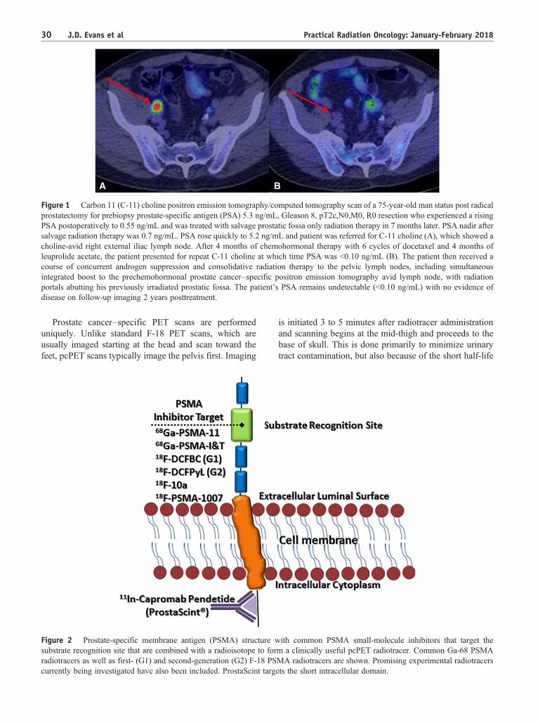
Figure 1 Carbon 11 (C-11) choline positron emission tomography/computed tomography scan of a 75-year-old man status post radicalprostatectomy for prebiopsy prostate-specific antigen (PSA) 5.3 ng/mL, Gleason 8, pT2c,N0,M0, R0 resection who experienced a risingPSA postoperatively to 0.55 ng/mL and was treated with salvage prostatic fossa only radiation therapy in 7 months later. PSA nadir aftersalvage radiation therapy was 0.7 ng/mL. PSA rose quickly to 5.2 ng/mL and patient was referred for C-11 choline (A), which showed acholine-avid right external iliac lymph node. After 4 months of chemohormonal therapy with 6 cycles of docetaxel and 4 months ofleuprolide acetate, the patient presented for repeat C-11 choline at which time PSA was b0.10 ng/mL (B). The patient then received acourse of concurrent androgen suppression and consolidative radiation therapy to the pelvic lymph nodes, including simultaneousintegrated boost to the prechemohormonal prostate cancer–specific positron emission tomography avid lymph node, with radiationportals abutting his previously irradiated prostatic fossa. The patient’s PSA remains undetectable (b0.10 ng/mL) with no evidence ofdisease on follow-up imaging 2 years posttreatment.
30 J.D. Evans et al Practical Radiation Oncology: January-February 2018
Prostate cancer–specific PET scans are performeduniquely. Unlike standard F-18 PET scans, which areusually imaged starting at the head and scan toward thefeet, pcPET scans typically image the pelvis first. Imaging
Figure 2 Prostate-specific membrane antigen (PSMA) structure wsubstrate recognition site that are combined with a radioisotope to forradiotracers as well as first- (G1) and second-generation (G2) F-18 PScurrently being investigated have also been included. ProstaScint targ
is initiated 3 to 5 minutes after radiotracer administrationand scanning begins at the mid-thigh and proceeds to thebase of skull. This is done primarily to minimize urinarytract contamination, but also because of the short half-life
ith common PSMA small-molecule inhibitors that target them a clinically useful pcPET radiotracer. Common Ga-68 PSMAMA radiotracers are shown. Promising experimental radiotracersets the short intracellular domain.

Prostate cancer–specific PET radiotracers 31Practical Radiation Oncology: January-February 2018
of isotopes such as C-11. Urinary tract contamination isthe primary reason pcPET protocols are performed in thismanner, including those involving radiotracers with longerhalf-lives such as F-18 fluciclovine.
Background for choline, PSMA, and fluciclovine
This review will focus on 3 PET radiotracers of interest:C-11/F-18 choline, Ga-68/F-18 PSMA, and F-18 fluciclovine.
Choline metabolism has been shown to be altered inprostate cancer cells. Increased levels of choline com-pounds concentrate preferentially in human prostatecancer cells derived from metastases.18 Alteration ofcholine metabolites within the cancer cell relates to cholinetransport, incorporation, and utilization within thecell.19-21 Preclinical data conflict on the theory ofaugmented choline use by the cell because of increasedcell membrane synthesis and proliferation.18,22,23 Multiplemetabolomic studies on prostate cancer have shownpermutations in choline metabolism not related to cellmembranogenesis23; however, it is well accepted thatcholine is used via a 3-step process known as the Kennedypathway for the de novo synthesis of phosphatidylcholine,which is an essential component of the cell membrane.24
Preclinical data have shown that there is an increase inthe expression of choline transporters and an increasein the choline transport rate in malignant prostate cellswhen compared with normal prostate tissues. 25
Interestingly, preclinical data have also shown thattreatment of prostate cancer cells leads to changesin energetic metabolism and choline metabolism.26
This notion is consistent with what experienced
Table 2 Summary of sensitivity and specificity of meta-analyses e
Systematic review and meta-analysis No. of studies No. of pati
PSMAPerera10 N = 16 N = 1309
CholineFanti54 N = 12 N = 1270
Evangelista55 N = 19 N = 1555
Umbehr56 N = 12 N = 1055
Shen57
(bone metastases)N = 9 N = 423
FluciclovineRen59 N = 6 N = 251
CI, confidence interval; CT, computed tomography; PET, positron emissiona Meta-analysis did not include 2 recent studies evaluating operational ch
than reported.
centers have observed after administration of systemictherapy to patients with C-11/F-18 choline PET-positivelymph node(s), wherein the nodes are no longercholine-avid (Fig 1).
PSMA is highly overexpressed in prostate cancer cellsas a transmembrane protein.27 PSMA is a folate hydrolasecell surface glycoprotein expressed in a number ofdifferent tissue types, including other cancers, but benignprocesses as well. Before malignant transformation hasoccurred, PSMA is localized to the cytoplasm andapical side of the prostate epithelium that lines prostaticducts.28 The function of cytoplasmic PSMA is not fullyunderstood; however, as malignant transformation occurs,PSMA is transferred to the luminal surface of the prostaticducts. 28 PSMA expression has been shown to bewidespread in most prostate tumors even when PSAstaining is negative or weak. 29 Increased PSMAexpression has also been observed when the cell becomescastrate-resistant.30 As a result, PSMA has emerged as 1 ofthe most favorable targets for PET imaging. Prostatecancer PSMA overexpression has been shown to be 100-to 1000-fold that of normal tissue expression; furthermore,PSMA expression may increase as tumor grade andcastrate resistance increases.31,32 PSMA is hypothesizedto have a transport function because it internalizes ligandssimilar to J591, a monoclonal antibody that targets theextracellular domain. In general, targeted antibodieshave presented challenges as diagnostic radiopharmaceu-ticals with their long circulating half-life and resultanthigh nonspecific background-to-tumor noise. Consequent-ly, the more recent focus of PSMA radiopharmaceuticaldevelopment has focused on small-molecule inhibitors
valuating PSMA, choline, and fluciclovine PET/CT
ents Sensitivity(per lesion)(95% CI)
Specificity(per lesion)(95% CI)
Sensitivity(per patient)(95% CI)
Specificity(per patient)(95% CI)
80%(66-89)
97%(92-99)
86%(37-98)
86%(3-100)
89%(83-93)
89%(73-96)
86%(83-88)
93%(90-95)
90%(74-97)
95%(92-97)
85%(79-89)
88%(73-95)
83%(81-85)
95%(94-97)
87%(79-93)
97%(93-99)
87%(80-92)
66%(56-75) a
tomography; PSMA, prostate-specific membrane antigen.aracteristics for F-18 fluciclovine specificity. Specificity may be higher

Figure 3 Summary of data evaluating pcPET detection rates as a function of PSA. All patients included in the analysis had biochemicalrecurrence. General trend favors PSMA across all PSA levels. FACBC, 18F-fluorocyclobutane-1-carboxylic acid fluciclovine. Otherabbreviations as in Fig 1.
Study PETRadiotracer
% ofPatientswith BCR
% of Patients with Positive PET/CT
PSA b1.0 PSA 1.0-2.0 PSA N2.0
CholineMitchell65 C-11 Choline 100% (176/176) 44% (15/34) 67% (21/31) 86% (96/111)Giovacchini81 C-11 Choline 100% (358/358) 19% (27/141) 46% (39/85) 72% (95/132)Richter82 C-11 Choline 100% (73/73) 7% (1/15) 46% (6/13) 80% (36/45)Krause83 C-11 Choline 100% (63/63) 36% (8/22) 43% (3/7) 71% (24/34)Castellucci84 C-11 Choline 100% (190/190) 19% (10/51) 25% (10/39) 54% (54/100)Nanni66 C-11 Choline 100% (89/89) 14% (4/28) 29% (8/28) 55% (18/33)Schwenck69 C-11 Choline 100% (101/101) 44% (8/18) 81% (21/26) 89% (51/57)Cimitan85 F-18 Choline 100% (1000/1000) 31% (66/211) 43% (66/153) 81% (513/636)Schillaci86 F-18 Choline 100% (49/49) 20% (2/10) 56% (5/9) 83% (25/30)Morigi67 F-18Methchol 100% (38/38) 13% (2/16) 36% (5/14) 63% (5/8)
PSMASchwenck69 Ga-68 PSMA 100% (101/101) 61% (11/18) 76% (20/26) 93% (53/57)Morigi67 Ga-68 PSMA 100% (38/38) 50% (8/16) 71% (10/14) 88% (7/8)Afshar-Oromieh87 Ga-68 PSMA 100% (319/319) 53% (27/51) 72% (28/39) 92% (204/221)Eiber88 Ga-68 PSMA 100% (248/248) 67% (35/52) 93% (67/72) 97% (120/124)Bluemel89 Ga-68 PSMA 100% (32/32) 29% (4/14) 46% (5/11) 71% (5/7)Verburg90 Ga-68 PSMA 100% (155/155) 44% (12/27) 79% (15/19) 89% (97/109)
FluciclovineNanni66 F-18 FACBC 100% (89/89) 21% (6/28) 46% (13/28) 55% (18/33)Odewole63 F-18 FACBC 100% (53/53) 38% (3/8) 78% (7/9) 86% (31/36)Bach-Gansmo91 F-18 FACBC 100% (596/596) 41% (53/128) 58% (N?) 75-85% (N?)Schuster62 F-18 FACBC 100% (93/93) 72% (N?)
32 J.D. Evans et al Practical Radiation Oncology: January-February 2018
that target the active substrate recognition site (Fig 2).Eder et al first described the most commonly used PSMAinhibitor in PET imaging, Ga-68 PSMA-HBED-CC,also known as Ga-68 PSMA-11, which also isinternalized and accumulates in high levels even in smallmetastases.28,33
There appears to be growing interest in developing an18-F–labeled PSMA agent. Some experts argue that itwould offer advantages with respect to availability,production amount, and image resolution. This approachwas first explored at Johns Hopkins University where F-18DCFBC, the first-generation F-18 PSMA radiotracer, was

Prostate cancer–specific PET radiotracers 33Practical Radiation Oncology: January-February 2018
developed and is currently licensed to Cyclotek for clinicaluse in Australia and New Zealand.34,35 Since thedevelopment of F-18 DCFBC, second-generation tracerssuch as F-18 DCFPyL have been developed.36 Currently,there are a number of groups working to develop the mostclinically useful next-generation F-18–labeled PSMAradiotracer.37-42 PSMA’s unique expression differentialbetween cancer and normal cells coupled with its largeextracellular domain provides an excellent target forimaging, but also for therapeutics such as theranosticapplications with lutetium 177 PSMA. Less than 10% ofprostate cancers have no uptake on PSMA PET.43
Additionally, the short half-life of Ga-68 (68 minutes)results in low radiation exposure to patients. Furthermore,the agent is rapidly cleared from nontarget tissue.On average, patients receive 3.0 mSv from the PETcomponent of 150 MBq of Ga-68-PSMA-11, which islower than most other pcPET agents such as C-11 and 18-Fcholine scans.44,45
Fluciclovine is a synthetic amino acid, and an analog ofL-leucine, which is preferentially taken up by prostatecancer cells and gliomas via specialized amino acidtransporters, namely alanine-serine-cysteine transporter 2(ASCT2) and LAT-1. 46-50 Its chemical name isanti-1-amino-3-FACBC, and is commonly known by itstrade name Axumin. Amino acid transporters such asASCT2 play a critical role in amino acid metabolism inprostate cancer cells. ASCT2 is an important transporter ofglutamine, which is known to be an essential tumornutrient and has been implicated in cancer signalingpathways.51,52 Fluciclovine is predominantly transportedby ASCT2 and transports in a manner similar toglutamine.53 Unlike glutamine, however, 18-F fluciclovinedoes not undergo additional metabolism in the cell, whichlends to its intracellular accumulation particularly in prostatecancer cells and at major sites of amino acid metabolismsuch as the liver and pancreas.54
Additional pcPET radiotracers used in prostate cancerimaging have been developed as previously noted. Theseinclude C-11 acetate and F-18 sodium fluoride. Inaddition, F-18 PET may be useful in imaging prostatecancer patients who have developed dedifferentiatedneuroendocrine tumors of the prostate,55 which converselymay not image well using these pcPET agents.
Operational characteristics of PET radiotracers
In recent years, numerous systematic reviews andmeta-analyses have been published evaluating the pooledoperational characteristics of various pcPET radiotracersin the setting of prostate cancer recurrence (Table 2).These reports are often analyzed on a per-patient orper-lesion basis. Caution should be exercised in interpretingsensitivities and specificities because a comparative goldstandard such as histologic confirmation is not alwaysavailable. The focus of this review is recurrent disease;
consequently, operational characteristics are emphasized inthe setting of biochemical recurrence after definitivetreatment. The use of these pcPET agents in initial staging,response to therapy, and radiation therapy planning are ofgreat interest but beyond the scope of this review. Ga-68PSMAwas recently evaluated by Perera et al, in which 16articles including 1309 patients were evaluated.10 Whenevaluating on a per-patient basis, the summary sensitivityand specificity were identical at 86%. When analyzed ona per-lesion basis, summary sensitivity was 80% andspecificity was 97%. Additionally, it was noted thatpatients with biochemical recurrence had increasinglypositive Ga-68 PSMA PET scans as the pre-PET PSAincreased. They found that 58% were positive at apre-PET PSA of 0.2 to 1 ng/mL, which increased to 76%with a PSA of 1 to 2 ng/mL and further increased to 95% forPSA N2 ng/mL.
C-11 choline was also recently evaluated by Fanti et al,specifically looking at its ability to detect sites ofrecurrence in the setting of biochemical recurrence afterdefinitive treatment.56 There were 12 studies including1270 patients to derive a pooled sensitivity and specificityof 89%. This was similar to previously publishedmeta-analyses by Evangelista et al, Umbehr et al, andShen et al,57-59 although these reports included both C-11and F-18 choline studies. Fanti et al highlight the accuracyof C-11 choline PET at different sites of recurrence,reporting a decreased pooled sensitivity of 61% fordetection of local recurrence. This result is consistentwith comparative studies that have shown multiparametricMRI with endorectal coil to be superior to C-11 choline forthe detection of local recurrence, whereas C-11 cholinePET/CT was shown to be superior to MRI for pelviclymph node metastases and equal with respect to bonemetastases.60
F-18 fluciclovine was evaluated by Ren et al andincluded 6 studies including 251 patients with biochemicalrecurrence.61 The pooled sensitivity and specificity on aper-patient analysis was 87% and 66%, respectively;however, caution should be exercised when interpretingthe specificity in this meta-analysis. Two recent importantpapers evaluating the operational characteristics of F-18fluciclovine were not included in this analysis. Schuster etal reported specificities of 40% and 97% for prostate bedand extraprostatic lesions, respectively.62 Odewole et alsimilarly demonstrated specificities of 56% and 100% forprostate bed and extraprostatic lesions, respectively.63
These data indicate that the specificities may be higherthan the meta-analysis suggests, particularly for extrapro-static disease.
Important factors to consider when interpretingoperational characteristics of various pcPET radiotracersinclude the reference standard used to establish positiveand negative proof, particularly with respect to extrapro-static disease, because these sites can be challenging toobtain histologic confirmation. Furthermore, whether the

Figure 4 These are the locations of prostate cancer-specific PET scanners across the United States as of 2017. Abbreviations as in Fig 3.
34 J.D. Evans et al Practical Radiation Oncology: January-February 2018
analysis was performed on a per-patient or per-lesion basisprovides additional insight into the interpretation of data.For example, many studies relied on histologic confirma-tion per-patient for positivity of extraprostatic disease,given it would be impractical to sample every PET avidsite, whereas studies that used a per-lesion analysis oftenused a nonhistologic method of disease confirmation,which is subject to study examination bias.64
Detection rates as a function of PSA
PSA is routinely followed in prostate cancer patientsafter definitive treatment; however, the optimal timing ofpcPET imaging is often debated amongst providers in thesetting of a rising PSA after definitive treatment. Dataregarding detection rates as a function of PSA aresummarized in Fig 3. Reviews of 10 choline, 6 PSMA,and 4 fluciclovine studies evaluating detection rates as a
function of PSA are shown. The median percentage ofpatients with positive pcPET scans is shown as the boldednumber over each histogram cluster. The general trendsuggests Ga-68 PSMA is superior to both C-11/F-18choline and F-18 fluciclovine in detecting recurrence atPSA levels b2.0 ng/mL.
There are important caveats to this comparative review,however. First, the fluciclovine data are limited by fewdata points. Second, the dose of radiotracer varies greatlybetween studies and consequently effects sensitivity,specificity, and detection rates. For example, in thestudy by Mitchell et al, in which detection rates wererelatively high, the C-11 choline dose ranged from 555 to740 MBq.65 This dose was significantly greater than thecholine dose given in prospective comparative studies thatshowed lower choline PET detection rates, which oftenused 3.4 to 3.5 MBq/kg.66,67 For an 80-kg patient, thiscomputes to 280 MBq, essentially half the dose used in the

Prostate cancer–specific PET radiotracers 35Practical Radiation Oncology: January-February 2018
Mitchell et al study. Finally, the selection of radioisotope,such as Ga-68 versus F-18 PSMA, and the use of advancediterative reconstruction algorithms will inevitably influencedetection rates in the future, which are important details notalways addressed in related studies. Nevertheless, thegeneral trend of these data presented in Fig 3 suggestssuperiority with PSMAparticularly at PSA levelsb1 ng/mL.Prospective studies comparing PSMA to choline and/orfluciclovine PET/CT are currently under way.
Comparative investigations of PET radiotracers
A prospective study by Morigi et al compared Ga-68PSMA with F-18 fluoromethylcholine.67 The findingsfrom this study were that Ga-68 PSMA was better thanF-18 fluoromethylcholine in patients with biochemicalfailure. It should be noted, however, that this study usedlow administered choline doses (3.5 MBq/kg) and aslightly different radioisotope, F-18 fluoromethylcholine,as opposed to C-11 choline. Additional data comparingPSMA to choline come from retrospective series.Afshar-Oromieh et al evaluated 37 patients with biochem-ical recurrence that underwent scans with both F-18fluoromethylcholine and Ga-68 PSMA PET/CT within30 days of 1 another.68 The authors concluded thatPSMA offered a higher detection rate, higher maximumstandardized uptake value, and higher tumor-to-back-ground ratio when compared with the F-18 fluoromethyl-choline scan. Schwenck et al retrospectively comparedGa-68 PSMA-11 with C-11 choline69 and demonstrated ahigher detection rate with PSMA. Interestingly, however,of the 67 patients with biochemical recurrence, 458 lymphnode metastases were detected. Although 39% wereexclusively identified with Ga-68 PSMA, there were 6%identified with C-11 choline only, and the majority (55%)were identified by both. The advantage of PSMA, and theclinical situation in which the majority of PSMA-onlydetection took place, was in patients presenting with PSAlevels b1 ng/mL.
Comparisons between F-18 FACBC and C-11 cholinehave largely been undertaken by Nanni et al. Before 2016,3 preliminary studies comparing these 2 imaging modal-ities in patients with biochemical recurrence werepublished.70-72 These studies reported favorable detectionrates for fluciclovine compared with choline and providedbackground for the publication of their prospective trial.66
The authors showed that, in patients with biochemicalrelapse after prostatectomy, F-18 FACBC had highersensitivity and specificity compared with C-11 choline(37% and 67% vs 32% and 40%). They emphasized thatF-18 FACBC had better true-positive findings at lowerPSA levels (b1 ng/mL) with 6/28 (21%) patients with F-18FACBC versus 4/28 (14%) patients with C-11 choline. Amajor limitation of this trial, and a limitation of manyimaging studies evaluating operational characteristics, isthe use of a suboptimal reference standard. The standard of
reference in this particular study was reevaluation of theclinical and imaging history after following patients for anaverage of 1 year. In some cases, this meant histologicconfirmation including 31% (4/13) of patients withpositive local relapse, 15% (4/26) of patients with positivelymph nodes, and 0% (0/7) of patients with positive bonelesions. For most cases, however, the standard of referencewas by repeat imaging or PSA trend after therapy.Furthermore, a low choline dose (3.4 MBq/kg) wasadministered, which may limit the study’s generalizabilityparticularly for centers that use higher choline doses.
There have been no direct comparisons betweenfluciclovine and Ga-68/F-18 PSMA to date. Schusteret al prospectively evaluated patients with biochemicalrecurrence comparing F-18 FACBC against indium 111capromab pendetide (ProstaScint), a radiolabeled mono-clonal antibody that binds to PSMA.62 This study showedFACBC performed better than ProstaScint, demonstratingFACBC’s superiority in detecting more prostatic andextraprostatic disease and effectively upstaging 25% ofpatients. A major strength of this study was the highincidence of pathologic confirmation of true positives, with96% (74/77) of index lesions histologically confirmedincluding 55 prostate bed and 22 extraprostatic lesions.
Availability and FDA approval
Currently there are 2 pcPET radiotracers that havegained FDA approval in the United States for theindication of identifying recurrent prostate cancer. C-11choline received FDA approval on September 12, 2012,for the indication of PET imaging of patients withsuspected prostate cancer recurrence.73 F-18 FACBCreceived FDA approval on May 27, 2016, for prostatecancer patients with suspected prostate cancer recurrencebased on elevated PSA levels following prior treatment.74
Ga-68 PSMA has not yet received FDA approval.ProstaScint, indium-111 capromab pendetide, has gainedFDA approval for its use in the evaluation of patients withnewly diagnosed, biopsy-proven prostate cancer thoughtto be clinically localized but high risk for pelvic lymphnode metastasis. Given the growing body of literatureregarding the clinical utility of choline, fluciclovine, andPSMA, availability around the United States is rapidlyexpanding (Fig 4). Sites that provide choline scans aremostly C-11. Exceptions include the University ofMichigan and The Queen’s Medical Center in Honolulu,Hawaii, both of which offer F-18 choline scans. Sites thatprovide PSMA scans are mostly Ga-68 except for JohnsHopkins University and University of Wisconsin, both ofwhich are evaluating F-18 PSMA scans.
In many parts of Europe, Asia, and Australia, pcPETradiotracers have made their way into widespread clinicalpractice. Some have argued that the current FDAregulation of PET radiotracers has been too prohibitiveand stifling to the innovative process. Such limitations are

36 J.D. Evans et al Practical Radiation Oncology: January-February 2018
not as common in other parts of the world, particularly inEurope and Australia, where much of the innovation anddata in pcPET radiotracers have been generated.Widespreadinternational availability has led to multi-institutionalprospectively controlled trials that accrue quickly and, inturn, are rapidly advancing the science of functionalimaging. As a result, robust data are forthcoming andessential to determine how best to use this technology.
Clinical application of prostate cancer–specificPET imaging
Patients with a rising PSA after definitive therapy oftenask the clinically relevant question, “Where is the origin ofmy rising PSA?” Before the advent of widespread clinicaluse of pcPET radiotracers and multiparametric MRI,clinicians relied on suboptimal tools, primarily bone scansand CT scans, to explore the answer to this question.As shown in Fig 3, pcPET radiotracers are able to detectsites of recurrence when the PSA level is low, even PSAlevels b1.0 ng/mL. By comparison, bone scans detectosseous metastases at a median PSA level of 40 ng/mL.75
Abuzallouf et al reviewed 23 studies evaluating bone scansin newly diagnosed cases of prostate cancer and showedosseous detection rates of 2.3% for PSA b10 ng/mL, 5.3%for PSA 10.1 to 19.9 ng/mL, and 16.4% for PSA 20.0 to49.9 ng/mL.76 In the same review, 25 studies evaluatingCT scans found lymph node metastases in 0% of patientswith PSA b20 ng/mL and 1.1% of patients withPSA N20 ng/mL. Additional evidence from a prospectivepopulation-based analysis of newly diagnosed prostate cancershowed CT scan detection rates were b15% for patients withPSA levels between 4 and 20 ng/mL.77 Based on these poorpositive yields, the overall use of bone scan and CT imaginghas declined in pretreatment evaluation, which has alsotranslated to limited use in the recurrent setting.78
As pcPET radiotracers improve, identification of theorigin of PSA relapse is occurring at lower PSA levels thanever before demonstrated. Figure 3 demonstrates that amedian of 51.5% of patients have potential sites ofrecurrence detected when the PSA level is b1.0 ng/mLusing Ga-68/F-18 PSMA. The detection rate increases to74% when PSA rises above 1.0 ng/mL, and surpasses 90%once the PSA level is N2.0 ng/mL. Similar, albeit lower,trends are observed with choline- and fluciclovine-basedradiotracers. Sensitive functional imaging has led topatterns of recurrence studies that provide insight intohow prostate cancer spreads early on in the process ofmetastasis in a variety of clinical scenarios includingpostprostatectomy, post-definitive radiation therapy,and postprostatectomy radiation therapy.6-8 Patterns ofrecurrence studies, such as these and others, haveprompted further discussion regarding additional localtherapy directed to the at-risk nodal basins or aggressivemetastasis-directed therapy.
The era of functional imaging has arrived, andclinicians around the globe are using this technology todevelop customized radiation therapy plans. In a recentmeta-analysis by Ost et al, metastasis-directed therapy toregional and distant recurrences included 66% of patientsreceiving radiation therapy.11 The authors found that 51%of patients were progression free 1 to 3 years after salvagemetastasis-directed therapy. Toxicity evaluation revealedmetastasis-directed radiation therapy was well tolerated,with 8.5% of patients experiencing grade 2 toxicities and 1case of grade 3 toxicity. Retrospective data coupled withgrowing experience using pcPET-directed therapy haveprompted the development of prospective studies (TableSupplementary Material (PDF); available as supplemen-tary material online only at www.practicalradonc.org). Inaddition to pcPET-directed external beam radiationtherapy, there is also growing experience regardingtheranostic applications; the most commonly discussedbeing lutetium 177 PSMA, which is beyond the scope ofthis review.
Conclusions
Biochemical recurrence in the prostate cancer patientoften presents a therapeutic challenge to the treatingoncologist. Data support early intervention with salvageradiation therapy after prostatectomy and argues againstprolonged monitoring of detectable postprostatectomyPSA levels.79,80 Patients in this clinical situation maystill benefit from pcPET imaging to identify the area ofrecurrence, even at very low PSA levels. Furthermore,imaging with both a pcPET scan and a multiparametricMRI scan can provide complementary insight as to thelocation of recurrence. Not all patients presenting to thetreating oncologist fall into this relatively common clinicalscenario of a rising PSA early after prostatectomy,however. Indeed, some patients present with rising PSAafter definitive radiation therapy, whereas others presentafter they have received postprostatectomy radiationtherapy, and others still after a late PSA rise years afterinitial surgery. It is within these challenging cases thatpcPET imaging has important clinical utility. Review ofthe current literature generally favors PSMA-basedimaging in the setting of biochemical recurrence;nevertheless, more comparative studies are needed tofurther clarify which pcPET radiotracer is most appropri-ate in each of a variety of clinical presentations. Functionalimaging studies that incorporate genomic profiling mayprovide additional insight as to which patients will derivethe greatest benefit from pcPET imaging and whichpatients have the most to gain from additional localtherapy. Prospective studies are ongoing to assess theefficacy of pcPET-directed local therapy in patients withbiochemical failure.
Prostate cancer–specific PET radiotracers 37Practical Radiation Oncology: January-February 2018
References
1. Siegel RL, Miller KD, Jemal A. Cancer statistics, 2017. CA Cancer JClin. 2017;67:7-30.
2. Cooperberg MR, Carroll PR. Trends in management forpatients with localized prostate cancer, 1990-2013. JAMA.2015;314:80-82.
3. Dale W, Bilir P, Han M, Meltzer D. The role of anxiety in prostatecarcinoma: A structured review of the literature. Cancer. 2005;104:467-478.
4. Lofters A, Juffs HG, Pond GR, Tannock IF. “PSA-itis:"Knowledge of serum prostate specific antigen and other causes ofanxiety in men with metastatic prostate cancer. J Urol. 2002;168:2516-2520.
5. Clark JA, Talcott JA. Confidence and uncertainty long after initialtreatment for early prostate cancer: Survivors' views of cancer control andthe treatment decisions they made. J Clin Oncol. 2006;24:4457-4463.
6. Sobol I, Zaid HB, Haloi R, et al. Contemporary mapping ofpost-prostatectomy prostate cancer relapse with 11C-choline posi-tron emission tomography and multiparametric magnetic resonanceimaging. J Urol. 2017;197:129-134.
7. Parker WP, Davis BJ, Park SS, et al. Identification of site-specificrecurrence following primary radiation therapy for prostate cancerusing C-11 choline positron emission tomography/computedtomography: A nomogram for predicting extrapelvic disease. EurUrol. 2017;71:340-348.
8. Parker WP, Evans JD, Stish BJ, et al. Patterns of recurrence afterpostprostatectomy fossa radiation therapy identified by c-11 cholinepositron emission tomography/computed tomography. Int J RadiatOncol Biol Phys. 2017;97(3):526-535.
9. Evangelista L, Briganti A, Fanti S, et al. New clinical indications for(18)F/(11)C-choline, new tracers for positron emission tomographyand a promising hybrid device for prostate cancer staging: Asystematic review of the literature. Eur Urol. 2016;70:161-175.
10. Perera M, Papa N, Christidis D, et al. Sensitivity, specificity, andpredictors of positive 68Ga-prostate-specific membrane antigenpositron emission tomography in advanced prostate cancer: Asystematic review and meta-analysis. Eur Urol. 2016;70(6):926-937.
11. Ost P, Bossi A, Decaestecker K, et al. Metastasis-directed therapy ofregional and distant recurrences after curative treatment of prostatecancer: A systematic review of the literature.EurUrol. 2015;67:852-863.
12. Supiot S, Rio E, Pacteau V, Mauboussin MH, Campion L, Pein F.OLIGOPELVIS - GETUG P07: a multicentre phase II trial ofcombined salvage radiotherapy and hormone therapy in oligometastaticpelvic node relapses of prostate cancer. BMC Cancer. 2015;15:646.
13. Mohsen B, Rio E, Pacteau V, Mauboussin MH, Campion L, Pein F.Application of C-11-acetate positron-emission tomography (PET)imaging in prostate cancer: Systematic review and meta-analysis ofthe literature. BJU Int. 2013;112(8):1062-1072.
14. Surti S. Update on time-of-flight PET imaging. J Nucl Med.2015;56:98-105.
15. O'Doherty J, McGowan DR, Abreu C, Barrington S. Effect ofBayesian-penalized likelihood reconstruction on [13N]-NH3 restperfusion quantification. J Nucl Cardiol. 2017;24(1):282-290.
16. Ross SQ. Clear White Paper. 2014; Available from: http://www3.gehealthcare.com/sitecore%20modules/web/~/media/downloads/italy/ge-healthcare-white-paper_qclear.pdf.
17. Cherry SR. Fundamentals of positron emission tomography andapplications in preclinical drug development. J Clin Pharmacol.2001;41:482-491.
18. Ackerstaff E, Pflug BR, Nelson JB, Bhujwalla ZM.Detection of increased choline compounds with proton nuclearmagnetic resonance spectroscopy subsequent to malignanttransformation of human prostatic epithelial cells. Cancer Res.2001;61:3599-3603.
19. Hernandez-Alcoceba R, Saniger L, Campos J, et al. Choline kinaseinhibitors as a novel approach for antiproliferative drug design.Oncogene. 1997;15:2289-2301.
20. Katz-Brull R, Degani H. Kinetics of choline transport andphosphorylation in human breast cancer cells; NMR application ofthe zero trans method. Anticancer Res. 1996;16:1375-1380.
21. Janardhan S, Srivani P, Sastry GN. Choline kinase: An importanttarget for cancer. Curr Med Chem. 2006;13:1169-1186.
22. Robert MJ, Schirra HJ, Lavin MF, Gardiner RA. Metabolomics: Anovel approach to early and noninvasive prostate cancer detection.Korean J Urol. 2011;52:79-89.
23. Lima AR, Bastos Mde L, Carvalho M, Guedes de Pinho P.Biomarker discovery in human prostate cancer: An update inmetabolomics studies. Transl Oncol. 2016;9:357-370.
24. Awwad HM, Geisel J, Obeid R. The role of choline in prostatecancer. Clin Biochem. 2012;45:1548-1553.
25. Muller SA, Holzapfel K, Seidl C, Treiber U, Krause BJ,Senekowitsch-Schmidtke R. Characterization of choline uptake inprostate cancer cells following bicalutamide and docetaxel treat-ment. Eur J Nucl Med Mol Imaging. 2009;36:1434-1442.
26. Lodi A, Ronen SM. Magnetic resonance spectroscopy detectablemetabolomic fingerprint of response to antineoplastic treatment.PLoS One. 2011;6:e26155.
27. Leek J, Lench N, Maraj B, et al. Prostate-specific membrane antigen:Evidence for the existence of a second related human gene. Br JCancer. 1995;72:583-588.
28. Maurer T, Eiber M, Schwaiger M, Gschwend JE. Current use ofPSMA-PET in prostate cancer management. Nat Rev Urol. 2016;13:226-235.
29. Birtle AJ, Freeman A, Masters JR, et al. Tumourmarkers formanagingmen who present with metastatic prostate cancer and serumprostate-specific antigen levels ofb10 ng/mL.BJU Int. 2005;96:303-307.
30. Evans MJ, Smith-Jones PM, Wongvipat J, et al. Noninvasivemeasurement of androgen receptor signaling with a positron-emittingradiopharmaceutical that targets prostate-specific membrane antigen.Proc Natl Acad Sci U S A. 2011;108:9578-9582.
31. Silver DA, Pellicer I, Fair WR, Heston WD, Cordon-Cardo C.Prostate-specific membrane antigen expression in normal andmalignant human tissues. Clin Cancer Res. 1997;3:81-85.
32. Bostwick DG, Pacelli A, Blute M, Roche P, Murphy GP. Prostatespecific membrane antigen expression in prostatic intraepithelialneoplasia and adenocarcinoma: A study of 184 cases. Cancer.1998;82:2256-2261.
33. Eder M, Schager M, Bauder Wust U, et al. 68Ga-complexlipophilicity and the targeting property of a urea-based PSMAinhibitor for PET imaging. Bioconjug Chem. 2012;23:688-697.
34. Foss CA, Mease RC, Fan H, et al. Radiolabeled small-moleculeligands for prostate-specific membrane antigen: In vivo imaging inexperimental models of prostate cancer. Clin Cancer Res. 2005;11:4022-4028.
35. Cho SY, Gage KL, Mease RC, et al. Biodistribution, tumor detection,and radiation dosimetry of 18F-DCFBC, a low-molecular-weightinhibitor of prostate-specific membrane antigen, in patients withmetastatic prostate cancer. J Nucl Med. 2012;53:1883-1891.
36. Szabo Z, Mena E, Rowe SP, et al. Initial evaluationof [(18)F]DCFPyL for prostate-specific membrane antigen(PSMA)-targeted PET imaging of prostate cancer. Mol ImagingBiol. 2015;17:565-574.
37. Rowe SP, Macura KJ, Mena E, et al. PSMA-based [(18)F]DCFPyLPET/CT is superior to conventional imaging for lesion detection inpatients with metastatic prostate cancer. Mol Imaging Biol. 2016;18:411-419.
38. Kelly J, Amora-Coarasa A, Nikolopoulou A, et al. Synthesis andpre-clinical evaluation of a new class of high-affinity 18F-labeledPSMA ligands for detection of prostate cancer by PET imaging. EurJ Nucl Med Mol Imaging. 2017;44:647-661.
38 J.D. Evans et al Practical Radiation Oncology: January-February 2018
39. Dietlein M, Kobe C, Kuhnert G, et al. Comparison of[(18)F]DCFPyL and [(68)Ga]Ga-PSMA-HBED-CC forPSMA-PET Imaging in patients with relapsed prostate cancer. MolImaging Biol. 2015;17:575-584.
40. Cardinale J, Schafer M, Benesova M, et al. Preclinical evaluation of18F-PSMA-1007, a new prostate-specific membrane antigen ligandfor prostate cancer imaging. J Nucl Med. 2017;58:425-431.
41. Harada N, Kimura H, Onoe S, et al. Synthesis and biologicevaluation of novel 18f-labeled probes targeting prostate-specificmembrane antigen for PET of prostate cancer. J Nucl Med. 2016;57:1978-1984.
42. Bouvet V, Wuest M, Jans HS, et al. Automated synthesis of[(18)F]DCFPyL via direct radiofluorination and validation inpreclinical prostate cancer models. EJNMMI Res. 2016;6:40.
43. Budaus L, Leyh-Bannurah SR, Salomon G, et al. Initial experienceof (68)Ga-PSMA PET/CT imaging in high-risk prostate cancerpatients prior to radical prostatectomy. Eur Urol. 2016;69:393-396.
44. Pfob CH, Ziegler S, Graner FP, et al. Biodistribution and radiationdosimetry of (68)Ga-PSMA HBED CC-a PSMA specific probe forPET imaging of prostate cancer. Eur J Nucl Med Mol Imaging.2016;43:1962-1970.
45. Afshar-Oromieh A, Hetzheim H, Kubler W, et al. Radiationdosimetry of (68)Ga-PSMA-11 (HBED-CC) and preliminaryevaluation of optimal imaging timing. Eur J Nucl Med Mol Imaging.2016;43:1611-1620.
46. Oka S, Hattori R, Kurosaki F, et al. A preliminary study ofanti-1-amino-3-18F-fluorocyclobutyl-1-carboxylic acid for the de-tection of prostate cancer. J Nucl Med. 2007;48:46-55.
47. Sasajima T, Ono T, Shimada N, et al. Trans-1-amino-3-18F-fluor-ocyclobutanecarboxylic acid (anti-18F-FACBC) is a feasiblealternative to 11C-methyl-L-methionine and magnetic resonanceimaging for monitoring treatment response in gliomas. Nucl MedBiol. 2013;40:808-815.
48. Oka S, Okudaira H, Yoshida Y, et al. Transport mechanisms oftrans-1-amino-3-fluoro[1-(14)C]cyclobutanecarboxylic acid in pros-tate cancer cells. Nucl Med Biol. 2012;39:109-119.
49. Schuster DM, Nanni C, Fanti S. PET tracers beyond FDG in prostatecancer. Semin Nucl Med. 2016;46:507-521.
50. Savir-Baruch B, Zanoni L, Schuster DM. Imaging of prostate cancerusing fluciclovine. PET Clin. 2017;12:145-157.
51. Ganapathy V, Thangaraju M, Prasad PD. Nutrient transporters incancer: Relevance to Warburg hypothesis and beyond. PharmacolTher. 2009;121:29-40.
52. Nakanishi T, Tamai I. Solute carrier transporters as targets for drugdelivery and pharmacological intervention for chemotherapy. JPharm Sci. 2011;100:3731-3750.
53. Oka S, Okudaira H, Ono M, et al. Differences in transportmechanisms of trans-1-amino-3-[18F]fluorocyclobutanecarboxylicacid in inflammation, prostate cancer, and glioma cells: ComparisonwithL-[methyl-11C]methionine and 2-deoxy-2-[18F]fluoro-D-glucose. MolImaging Biol. 2014;16:322-329.
54. Asano Y, Inoue Y, Ikeda Y, et al. Phase I clinical study of NMK36:A new PET tracer with the synthetic amino acid analogueanti-[18F]FACBC. Ann Nucl Med. 2011;25:414-418.
55. Spratt DE, Gavane S, Tarlinton L, et al. Utility of FDG-PETin clinical neuroendocrine prostate cancer.Prostate. 2014;74:1153-1159.
56. Fanti S, Minozzi S, Castellucci P, et al. PET/CT with (11)C-cholinefor evaluation of prostate cancer patients with biochemicalrecurrence: Meta-analysis and critical review of available data. EurJ Nucl Med Mol Imaging. 2016;43:55-69.
57. Evangelista L, Zattoni F, Guttila A, et al. Choline PET or PET/CTand biochemical relapse of prostate cancer: A systematic review andmeta-analysis. Clin Nucl Med. 2013;38:305-314.
58. Umbehr MH, Müntener M, Hany T, Sulser T, Bachmann LM. Therole of 11C-choline and 18F-fluorocholine positron emissiontomography (PET) and PET/CT in prostate cancer: A systematicreview and meta-analysis. Eur Urol. 2013;64:106-117.
59. Shen G, Deng H, Hus S, Jia Z. Comparison of choline-PET/CT,MRI, SPECT, and bone scintigraphy in the diagnosis of bonemetastases in patients with prostate cancer: A meta-analysis. SkeletRadiol. 2014;43(11):1503-1513.
60. Kitajima K, Murphy RC, Nathan MA, et al. Detection of recurrentprostate cancer after radical prostatectomy: Comparison of11C-choline PET/CT with pelvic multiparametric MR imagingwith endorectal coil. J Nucl Med. 2014;55:223-232.
61. Ren J, Yuan L, Wen G, Yang J. The value of anti-1-amino-3-18F-fluorocyclobutane-1-carboxylic acid PET/CT in the diagnosis ofrecurrent prostate carcinoma: A meta-analysis. Acta Radiol. 2016;57:487-493.
62. Schuster DM, Nieh PT, Jani AB, et al. Anti-3-[(18)F]FACBCpositron emission tomography-computerized tomography and(111)In-capromab pendetide single photon emission computerizedtomography-computerized tomography for recurrent prostate carci-noma: Results of a prospective clinical trial. J Urol. 2014;191:1446-1453.
63. Odewole OA, Tade FI, Nieh PT, et al. Recurrent prostate cancerdetection with anti-3-[(18)F]FACBC PET/CT: Comparison with CT.Eur J Nucl Med Mol Imaging. 2016;43:1773-1783.
64. Sica GT. Bias in research studies. Radiology. 2006;238:780-789.65. Mitchell CR, Lowe VJ, Rangel LJ, Hung JC, Kwon ED, Karnes RJ.
Operational characteristics of (11)c-choline positron emissiontomography/computerized tomography for prostate cancer withbiochemical recurrence after initial treatment. J Urol. 2013;189:1308-1313.
66. Nanni C, Zanoni L, Pultrone C, et al. (18)F-FACBC(anti1-amino-3-(18)F-fluorocyclobutane-1-carboxylic acid) versus(11)C-choline PET/CT in prostate cancer relapse: Results of aprospective trial. Eur J Nucl Med Mol Imaging. 2016;43:1601-1610.
67. Morigi JJ, Stricker PD, van Leeuwen PJ, et al. Prospectivecomparison of 18F-fluoromethylcholine versus 68Ga-PSMAPET/CT in prostate cancer patients who have rising PSA aftercurative treatment and are being considered for targeted therapy. JNucl Med. 2015:561185-561190.
68. Afshar-Oromieh A, Zechmann CM, Malcher A, et al. Comparison ofPET imaging with a (68)Ga-labelled PSMA ligand and (18)F-choline-based PET/CT for the diagnosis of recurrent prostate cancer.Eur J Nucl Med Mol Imaging. 2014;41:11-20.
69. Schwenck J, Rempp H, Reischl G, et al. Comparison of 68Ga-labelledPSMA-11 and 11C-choline in the detection of prostate cancermetastases by PET/CT.Eur J NuclMedMol Imaging. 2017;44:92-101.
70. Nanni C, Schiavnia R, Brunocilla E, et al. 18F-fluciclovine PET/CTfor the detection of prostate cancer relapse: A comparison to11C-choline PET/CT. Clin Nucl Med. 2015;40:e386-e391.
71. Nanni C, Zanoni L, Pultrone C, et al. 18F-FACBC compared with11C-choline PET/CT in patients with biochemical relapse afterradical prostatectomy: A prospective study in 28 patients. ClinGenitourin Cancer. 2014;12:106-110.
72. Nanni C, Schiavnia R, Boschi S, et al. Comparison of 18F-FACBCand 11C-choline PET/CT in patients with radically treated prostatecancer and biochemical relapse: preliminary results. Eur J Nucl MedMol Imaging. 2013;40(suppl 1):S11-S17.
73. FDA approves 11C-choline for PET in prostate cancer.J Nucl Med.2012;53:11N.
74. FDA approves new diagnostic imaging agent to detect recurrentprostate cancer. FDA news release. Available at: https://www.fda.gov/NewsEvents/Newsroom/PressAnnouncements/ucm503920.htm, Accessed date: March 2017.
75. Taneja SS. Imaging in the diagnosis and management of prostatecancer. Rev Urol. 2004;6:101-113.
76. Abuzallouf S, Dayes I, Lukka H. Baseline staging of newlydiagnosed prostate cancer: A summary of the literature. J Urol.2004;171:2122-2127.
77. Albertsen PC, Hanley JA, Harlan LC, et al. The positive yield ofimaging studies in the evaluation of men with newly diagnosed
Prostate cancer–specific PET radiotracers 39Practical Radiation Oncology: January-February 2018
prostate cancer: A population based analysis. J Urol. 2000;163:1138-1143.
78. Cooperberg MR, Lubeck DP, Grossfeld GD, Mehta SS, Carroll PR.Contemporary trends in imaging test utilization for prostate cancerstaging: Data from the cancer of the prostate strategic urologicresearch endeavor. J Urol. 2002;168:491-495.
79. Tendulkar RD, Agrawal S, Gao T, et al. Contemporary update of amulti-institutional predictive nomogram for salvage radiotherapyafter radical prostatectomy. J Clin Oncol. 2016. [e-pub ahead ofprint, pii: JCO679647].
80. Stish BJ, Pisansky TM, HarmsenWS, et al. Improved metastasis-freeand survival outcomes with early salvage radiotherapy inmen with detectable prostate-specific antigen after prostatectomy forprostate cancer.JClinOncol.2016. [e-pub ahead of print, pii: JCO683425].
81. Giovacchini G, Picchio M, Coradeschi, et al. Predictive factors of[(11)C]choline PET/CT in patients with biochemical failure afterradical prostatectomy. Eur J Nucl Med Mol Imaging. 2010;37:301-309.
82. Richter JA, Rodriguez M, Rioja J, et al. Dual tracer 11C-cholineand FDG-PET in the diagnosis of biochemical prostate cancer relapseafter radical treatment. Mol Imaging Biol. 2010;12(2):210-217.
83. Krause BJ, Souvatzoglou M, Tuncel M, et al. The detection rate of[11C]choline-PET/CT depends on the serum PSA-value in patientswith biochemical recurrence of prostate cancer. Eur J Nucl Med MolImaging. 2008;35:18-23.
84. Castellucci P, Fuccio C, Nanni C, et al. Influence of trigger PSA andPSA kinetics on 11C-Choline PET/CT detection rate in patients withbiochemical relapse after radical prostatectomy. J Nucl Med.2009;50:1394-1400.
85. Cimitan M, Evangelista L, Hodolic M, et al. Gleason score atdiagnosis predicts the rate of detection of 18F-choline PET/CTperformed when biochemical evidence indicates recurrence ofprostate cancer: Experience with 1,000 patients. J Nucl Med.2015;56:209-215.
86. Schillaci O, Calabria F, Tabolozza M, et al. Influence of PSA, PSAvelocity and PSA doubling time on contrast-enhanced 18F-cholinePET/CT detection rate in patients with rising PSA after radicalprostatectomy. Eur J Nucl Med Mol Imaging. 2012;39:589-596.
87. Afshar-Oromieh A, Avtzi E, Giesel FL, et al. The diagnostic value ofPET/CT imaging with the (68)Ga-labelled PSMA ligand HBED-CCin the diagnosis of recurrent prostate cancer. Eur J Nucl Med MolImaging. 2015;42:197-209.
88. Eiber M, Maurer T, Souvatzoglou M, et al. Evaluation of hybrid(6)(8)Ga-PSMA ligand PET/CT in 248 patients with biochemicalrecurrence after radical prostatectomy. J Nucl Med. 2015;56:668-674.
89. Bluemel C, Krebs M, Polat B, et al. 68Ga-PSMA-PET/CT in patientswith biochemical prostate cancer recurrence and negative18F-choline-PET/CT. Clin Nucl Med. 2016;41:515-521.
90. Verburg FA, Pfister D, Heidenreich A, et al. Extent of disease inrecurrent prostate cancer determined by [(68)Ga]PSMA-HBED-CCPET/CT in relation to PSA levels, PSA doubling time and Gleasonscore. Eur J Nucl Med Mol Imaging. 2016;43:397-403.
91. Bach-Gansmo T, Nanni C, Nieh PT, et al. Multisite experience of thesafety, detection rate and diagnostic performance of fluciclovine(18F) positron emission tomography/computerized tomographyimaging in the staging of biochemically recurrent prostate cancer.J Urol. 2017;197(3 Pt 1):676-683.