
Prostaglandins and Kidney Function: A Comparison Between ...
59
University of Nebraska Medical Center University of Nebraska Medical Center DigitalCommons@UNMC DigitalCommons@UNMC Theses & Dissertations Graduate Studies Spring 5-4-2019 Prostaglandins and Kidney Function: A Comparison Between the Prostaglandins and Kidney Function: A Comparison Between the Cortical and Inner Medullary Collecting Duct on Prostaglandin Cortical and Inner Medullary Collecting Duct on Prostaglandin Production Production Dane N. Wilson University of Nebraska Medical Center Follow this and additional works at: https://digitalcommons.unmc.edu/etd Part of the Anatomy Commons Recommended Citation Recommended Citation Wilson, Dane N., "Prostaglandins and Kidney Function: A Comparison Between the Cortical and Inner Medullary Collecting Duct on Prostaglandin Production" (2019). Theses & Dissertations. 370. https://digitalcommons.unmc.edu/etd/370 This Thesis is brought to you for free and open access by the Graduate Studies at DigitalCommons@UNMC. It has been accepted for inclusion in Theses & Dissertations by an authorized administrator of DigitalCommons@UNMC. For more information, please contact [email protected].
Transcript of Prostaglandins and Kidney Function: A Comparison Between ...

University of Nebraska Medical Center University of Nebraska Medical Center
DigitalCommons@UNMC DigitalCommons@UNMC
Theses & Dissertations Graduate Studies
Spring 5-4-2019
Prostaglandins and Kidney Function: A Comparison Between the Prostaglandins and Kidney Function: A Comparison Between the
Cortical and Inner Medullary Collecting Duct on Prostaglandin Cortical and Inner Medullary Collecting Duct on Prostaglandin
Production Production
Dane N. Wilson University of Nebraska Medical Center
Follow this and additional works at: https://digitalcommons.unmc.edu/etd
Part of the Anatomy Commons
Recommended Citation Recommended Citation Wilson, Dane N., "Prostaglandins and Kidney Function: A Comparison Between the Cortical and Inner Medullary Collecting Duct on Prostaglandin Production" (2019). Theses & Dissertations. 370. https://digitalcommons.unmc.edu/etd/370
This Thesis is brought to you for free and open access by the Graduate Studies at DigitalCommons@UNMC. It has been accepted for inclusion in Theses & Dissertations by an authorized administrator of DigitalCommons@UNMC. For more information, please contact [email protected].
PROSTAGLANDINS AND KIDNEY FUNCTION: A
COMPARISON BETWEEN THE CORTICAL AND INNER
MEDULLARY COLLECTING DUCT ON
PROSTAGLANDIN PRODUCTION
by
Dane Norris Wilson
A THESIS
Presented to the Faculty of
the University of Nebraska Graduate College
in Partial Fulfillment of the Requirements
for the Degree of Master of Science
Genetics, Cell Biology, and Anatomy
Graduate Program
Under the Supervision of Dr. Erika Boesen
University of Nebraska Medical Center
Omaha, Nebraska
May, 2019
Advisory Committee:
Erika Boesen, PhD.
Samantha Simet, PhD.
Karen Gould, PhD.
Pamela Carmines, PhD.

i
ACKNOWLEDGMENTS
I would like to thank my advisor, Dr. Erika Boesen, for her guidance, patience, and
supervision throughout this project as well as for full use of her laboratory and never giving up on
me. I would also like to thank Dr. Karen Gould, Dr. Samantha Simet, and Dr. Pamela Carmines
for being members of my thesis committee. A special thanks goes out to Jun and Ryan for their
added help during my time in lab. Finally, I am very grateful for Edson, a fellow Masters of
Anatomy student, and friend, who constantly reminded me to keep “grinding,” and pushing
forward. I look forward to hearing about all of your medical school experiences.

ii
PROSTAGLANDINS AND KIDNEY FUNCTION: A
COMPARISON BETWEEN THE CORTICAL AND INNER
MEDULLARY COLLECTING DUCT ON
PROSTAGLANDIN PRODUCTION
Dane Wilson, M.S.
University of Nebraska Medical Center, 2019
Advisor: Erika Boesen, PhD
Prostaglandins are paracrine and autocrine signaling molecules that play important roles through
various physiological and pathophysiological functions in the human body including the
inflammatory response, control of blood pressure, and water and salt homeostasis in the kidney.
Prostaglandins have been shown to have great influence on kidney function. In particular,
prostaglandin E2 influences the function of the collecting duct, primarily decreasing vasopressin-
stimulated collecting duct water permeability. The aim of this study was to see how various
treatments to cells, as well as different growth times, impacted prostaglandin production between
two different collecting duct cell lines, one arising from the cortical collecting duct and the other
arising from the inner medullary collecting duct. This was accomplished by growing and
culturing the cells on 6-well plates, collecting the cell media and measuring prostaglandin
accumulation via Enzyme-linked immunosorbent assay (ELISA) kits. Results showed that longer
growth times decreased prostaglandin E2 and F2α accumulation in both cell lines. The effect of
dDAVP, a vasopressin analog, increased prostaglandin accumulation in the inner medullary
collecting duct cells, and increased PGF2α while having virtually no effect on PGE2 in the cortical
collecting duct cells. The addition of a cyclooxygenase-1 and cyclooxygenase-2 inhibitor
significantly decreased prostaglandin accumulation in both cell lines. Finally, changing the
osmolarity of the media by adding sodium chloride (NaCl) increased prostaglandin accumulation

iii
in both cell lines. Prostaglandin synthesis has relevant clinical implications, as non-steroidal anti-
inflammatory drugs, a common over-the-counter group of pain medications, inhibit
cyclooxygenase, an enzyme involved in the biosynthesis of prostaglandins.

iv
TABLE OF CONTENTS
ACKNOWLEDGMENTS………………………………………………………………………….i
ABSTRACT……………………………………………………………………………………….ii
TABLE OF CONTENTS………………………………………………………………………….iv
LIST OF FIGURES……………………………………………………………………………….vi
LIST OF ABBREVIATIONS…………………………………………………………………….vii
CHAPTER 1: INTRODUCTION………………………………………………………………….1
Overview of prostaglandins……………………………………………………………….1
Kidney basics – the anatomy and physiology of the kidneys……………………………..8
Renal blood flow…………………………………………………………………………..8
Renal corpuscle anatomy and function……………………………………………………9
Prostaglandins and their effects on renal blood flow and ultrafiltration of blood………...9
Prostaglandins and renin release…………………………………………………………10
Nephron tubule anatomy and function…………………………………………………...10
Prostaglandins and their effects on the nephron tubule………………………………….13
Collecting duct anatomy and function…………………………………………………...13
Prostaglandins and their effects on the collecting duct………………………………….15
Differences between the cortical and inner medullary collecting duct…………………..17
Cell lines…………………………………………………………………………………17
Aim of the study and hypothesis…………………………………………………………18
CHAPTER 2: MATERIALS AND METHODS…………………………………………………20
Cell Culture………………………………………………………………………………20
Time course experiments………………………………………………………………...20

v
Treatment experiments…………………………………………………………………...20
Sample Collection………………………………………………………………………..22
Measuring prostaglandin synthesis and prostaglandin E metabolite production utilizing
Enzyme-linked immunosorbent assay (ELISA) kits……………………………………..22
Statistical Analysis……………………………………………………………………….23
CHAPTER 3: RESULTS…………………………………………………………………………24
Effect of cell growth on prostaglandin production………………………………………24
Effect of dDAVP on prostaglandin production………………………………………….28
Effect of COX-1 (SC-560) and COX-2 (NS-398) inhibition on prostaglandin
production………………………………………………………………………………..30
Effect of extracellular osmolality changes on prostaglandin production………………...33
CHAPTER 4: DISCUSSION……………………………………………………………………..35
BIBLIOGRAPHY………………………………………………………………………………...44

vi
LIST OF FIGURES
Figure 1: Biosynthetic pathway of prostanoids (Ricciotti, 2011)………………………………….2
Figure 2: Localization and physiological function of EP receptors and cyclooxygenases (COX-
1/COX-2) throughout the nephron (Modified from Nasrallah, 2014 as source material)…………6
Figure 3: Renal functions, possible signaling pathways, and renal distributions of prostaglandin
receptors (Modified from Li, 2018)………………………………………………………………..7
Figure 4: Aquaporin distribution within the nephron (Tamma, 2011)…………………………...11
Figure 5: Proposed diuretic effects of PGE2 along the kidney nephron and interstitium (Olesen,
2013)……………………………………………………………………………………………...16
Figure 6: Photographed images of cultured mIMCD3 and mpkCCD cells at a subconfluent (24
hours) and confluent (72 hours) growth…………………………………………………………..25
Figure 7: Effect of growth time on PGE2 (a), PGEM (b), and PGF2α (c) production……………27
Figure 8: Effect of dDAVP on PGE2 (a), PGEM (b), and PGF2α (c) production………………...29
Figure 9: Effect of COX-1 (a-c) and COX-2 (d-f) inhibition on PGE2 (a,d), PGEM (b,e), and
PGF2α (c,f) production……………………………………………………………………………32
Figure 10: Effect of NaCl on PGE2 (a) and PGEM (b) production………………………………34
vii
LIST OF ABBREVIATIONS
AVP Arginine vasopressin
COX-1 Cyclooxygenase-1
COX-2 Cyclooxygenase-2
dDAVP 1-deamino-8-D-arginine vasopressin
EP1 Prostaglandin E2 receptor 1
EP2 Prostaglandin E2 receptor 2
EP3 Prostaglandin E2 receptor 3
EP4 Prostaglandin E2 receptor 4
FP Prostaglandin F2α receptor
IP Prostaglandin I2 receptor
mIMCD3 mouse Inner Medullary Collecting Duct 3 cell
mpkCCD murine immortalized cortical collecting duct cell
PG Prostaglandin
PGE2 Prostaglandin E2
PGF2α Prostaglandin F2α
PGI2 Prostaglandin I2
PLA Phospholipase A
1
CHAPTER 1: INTRODUCTION
Overview of prostaglandins
Biosynthesis
Prostaglandins are lipid structures derived from arachidonic acid that are formed when
arachidonic acid is released from the plasma membrane by phospholipases with subsequent
peroxidase activity by a class of cyclooxygenase (COX-1 and COX-2) enzymes forming
prostaglandin H2 (Ricciotti, 2011). Tissue specific synthase enzymes subsequently form the more
stable and biologically active prostaglandins such as prostaglandin E2 (Li, 2018). There are four
biologically active prostaglandins generated in vivo: prostaglandins: D2 (PGD2), E2 (PGE2), I2
(PGI2), and F2α (PGF2α). Thromboxane A2 (TxA2), a prostanoid, is also produced via a similar
mechanism, as shown below in Figure 1 (Ricciotti, 2011). Upon being synthesized,
prostaglandins then bind to a specific G-protein coupled receptor to exert its particular effect. As
seen below in Figure 1, prostaglandins have effects on several tissues throughout the body.

2
Figure 1: Biosynthetic pathway of prostanoids (Ricciotti, 2011)
The role of Phospholipase A in the biosynthesis of prostaglandins
Phospholipase A (PLA) comprises a family of enzymes which play a key role in the
production of prostaglandins. One of the isoforms of PLA is cytosolic PLA (cPLA). The primary
function of cPLA is the liberation of arachidonic acid from membrane phospholipids. cPLA
activity depends on an increase in intracellular calcium as calcium binds to the enzyme at its C2
domain, facilitating the translocation of the enzyme to intracellular phospholipid membranes.
Once bound to the phospholipid membrane, many other enzymes can help to regulate the activity
of cPLA through phosphorylation (Casale, 2018).

3
Enzymatic Regulation
The biological effects of prostaglandins can be regulated at the enzymatic reaction of
cPLA. One isoform, cPLA2, is regulated by intracellular calcium levels. Calcium binding to the
C2 domain activates the enzyme, creating a conformational shift in the C2 domain, which is
critical for proper orientation to bind to the phospholipid membrane. Once localized to the
membrane, a catalytic domain catalyzes the hydrolysis, freeing arachidonic acid. The enzyme can
be further activated by lipid second messengers such as phosphatidylinositol 4,5-bisphosphate
(PIP2) or by enzymes such as mitogen-activated protein kinase (MAPK) through phosphorylation
(Dennis, 2011).
The role of cyclooxygenase in the biosynthesis of prostaglandins
Cyclooxygenase (COX) catalyzes the conversion of arachidonic acid to prostaglandin H2,
a precursor to the biologically active prostaglandins. This is the rate-limiting step in the formation
of prostaglandins. The conversion from arachidonic acid to PGH2 occurs in two steps; the
cyclooxygenase reaction forms the unstable PGG2; PGG2 is immediately converted into PGH2 in
a peroxidase reaction (Zarghi, 2011).
There are two main isoforms of COX: COX-1 and COX-2. COX-1 is expressed
constitutively in many tissues (Zarghi, 2011). In the kidney, it is expressed in the afferent and
efferent arterioles, medullary interstitial cells, and the cortical and medullary collecting ducts
(Komhoff, 1997). Prostaglandins produced by COX-1 mediate more regulatory functions, such as
regulation of renal blood flow (Zarghi, 2011). In contrast, COX-2 is not detected in most tissues,
but has been detected predominantly in medullary interstitial cells and cells associated with the
macula densa, with less distribution in the collecting duct (Hao, 2008). Its expression is rapidly
induced by various stimuli such as proinflammatory cytokines and disorders of water-electrolyte
homeostasis (Zarghi, 2011).

4
Various studies have been conducted to elucidate similarities and differences in function
between the two isoforms. Contributing to these differences is that COX-1 and COX-2 have been
demonstrated to utilize arachidonic acid generated by different phospholipases (Reddy, 1997;
Langenbach, 1999). In addition, studies have indicated that the two isoforms may have different
cellular localizations (Morita, 1995). COX-1 is commonly referred to as the “housekeeping”
isoform, where COX-2 is commonly referred to as the “inducible” isoform, however COX-1 has
been shown to be inducible under certain conditions, and COX-2 is constitutively expressed in
certain tissues within the brain and kidney (Langenbach, 1999). Harris, et al. (1994) utilized
various techniques to determine that COX-2 expression was localized to the macula densa, and
that the level of COX-2 expression increased threefold when the rats were chronically sodium
restricted. In addition, intrarenal COX-2 was shown to be upregulated during chronic angiotensin
II infusion in cultured inner medullary collecting duct cells (Gonzalez, 2014). The authors also
observed that COX-2 inhibition additionally decreased glomerular filtration and renal blood flow
with angiotensin II infusion in anesthetized rats (Gonzalez, 2014). Within the renal medulla,
COX-2 expression is regulated by dietary salt intake, and inhibition of COX-2 within the medulla
has been shown to lead to sodium-sensitive hypertension in rats (Zewde, 2004). These findings
note its importance in the regulation of blood volume and pressure.
Langenbach et al. (1999) found COX-1 deficient mice to be surprisingly healthy, with no
gastric or kidney pathologies, however these mice did show impairment in platelet aggregation.
COX-2 deficient mice had several noted pathologies; with regard to the kidney, COX-2 null mice
had smaller kidneys, reduced glomeruli which were also poorly developed, and atrophied renal
tubules.
Prostaglandin production and receptor distribution
As previously mentioned, prostaglandins primarily work in an autocrine or paracrine
manner. In vivo, prostaglandins are rapidly degraded, limiting their effect to the immediate

5
vicinity of their site of synthesis (Hao, 2008). Various kidney cells produce prostaglandins:
tubular epithelial cells, renal medullary interstitial cells, and cells of the glomeruli (Dunn, 1977).
The main sites of prostaglandin synthesis occurs in the medullary tissue of the kidney, established
by medullary slices or isolated cell preps (Bonvalet, 1987). In order for prostaglandins to exert
local effects, they must bind to their receptor locally. Prostaglandin receptors are expressed
throughout various parts of the kidney. PGE2 and PGF2α have long been recognized as major
renal prostaglandins (Dunn, 1977). More so recently, the emergence of the importance of
prostacyclin, PGI2 has been noted (Kim, 2008). All renal cell types can synthesize PGE2, but the
highest production is seen in the glomeruli and collecting ducts (Nasrallah, 2014); it is also the
major prostaglandin synthesized in the renal medulla (Kim, 2008). PGI2 is predominantly
synthesized in the endothelial and epithelial cells of the glomerulus. Although PGF2α can be
detected in the kidney, the enzymes catalyzing the biosynthesis of PGF2α remain poorly defined
(Hao, 2008).
The most abundant prostaglandin receptors in the kidney are those for PGE2 (Kim, 2008).
Four prostaglandin E (EP) receptor subtypes have been identified, and are highly expressed
throughout the kidney (Breyer, 1996). Collecting ducts express all EP receptors, however, EP2 is
only expressed in the cortical segment of the collecting duct (Nasrallah, 2014; Katsuyama, 1995).
The medullary thick ascending limb (mTAL) expresses high levels of EP3 receptor mRNA. In
addition, the glomeruli express the EP2 receptor and high levels of EP4 receptor mRNA (Kim,
2008; Nasrallah, 2014). Other expression sites of the EP receptors as well as COX-1 and COX-2
are shown in Figure 2 below.
Nephron Segment/Region Prostaglandin E2 receptor or COX
localization
Afferent Arteriole EP2/EP4 – vasodilators
EP1/EP3 - vasoconstrictors

6
Glomerulus COX-1, COX-2, EP1, EP3, EP4
Proximal Tubule COX-1, COX-2, EP1, EP4 - transport
Thick Ascending Limb COX-2, EP3, EP4 - transport
Macula Densa COX-2, EP3, EP4 – renin, transport
Distal Tubule EP3, EP4 - transport
Collecting Duct COX-1, COX-2, EP1, EP3, EP4 - transport
Medullary Interstitial Cells COX-1, COX-2
Vasa Recta EP2, EP4 - vasodilators
Figure 2: Localization and physiological function of EP receptors and cyclooxygenases (COX-
1/COX-2) throughout the nephron (Modified from Nasrallah, 2014 as source material)
The receptors are cell surface G-protein coupled receptors, thereby primarily influencing
the action of adenylate cyclase which subsequently affects intracellular cyclic adenosine 3'5'-
monophosphate (cAMP) levels. Each EP receptor not only selectively binds PGE2, but also
preferentially couples to different signal transduction pathways as shown in Figure 3 below
(Modified from Li, 2018).
EP2 and EP4 activate Gs, which stimulates intracellular cAMP levels (Nasrallah, 2014).
EP1 is coupled to Gq, which mobilizes intracellular calcium via phospholipase C and activates
Protein Kinase C. EP3 activates Gi, which inhibits cAMP formation but increases intracellular
calcium levels via phospholipase C (Breyer, 1996; Nasrallah, 2014).
As evident, PGE2 exerts its effects through various signaling pathways, and the effect of
PGE2 can have opposite effects, depending on which receptor it binds to. For example, PGE2 is a
vasodilator in some vascular beds, however, it can also result in vasoconstriction (Breyer, 1996).
This is also evident as seen from Figure 2 above: PGE2 binding to its EP2 or EP4 receptor
vasodilates the afferent arteriole whereas binding to EP1 or EP3 receptor results in
vasoconstriction of the afferent arteriole (Nasrallah, 2014). The IP receptor, distributed
throughout the renal cortex and medulla, binds PGI2, and increases intracellular cAMP levels

7
(Kim, 2008). PGF2α binds to its FP receptor which is coupled to Gq, mobilizing intracellular
calcium and activating Protein Kinase C. At present, there is little evidence for DP receptor
(PGD2) expression in the kidney (Hao, 2008). Below is a summary of renal functions, possible
signaling pathways, and renal distributions of prostaglandin receptors (Modified from Li, 2018).
Because multiple prostaglandins can be synthesized from COX, and because these prostaglandins
can interact with different receptors that can vary in their signaling pathways, the biological
effects of COX-derived prostaglandins are diverse and complex (Hao, 2008).
Prostaglandin
receptors
Renal function Signaling pathways Expression
sites
EP1 Hemodynamics,
transport, renin release
Gq-Ca2+ CD, MG, P,
PT, V
EP2 Hemodynamics,
transport, renin release
Gs-cAMP IC, MD, P, V,
CD
EP3 Transport,
vasoconstriction
Gi-cAMP DT, CD, MD,
V
EP4 Hemodynamics,
transport, renin release,
vasodilation
Gs-cAMP P, CD, MG,
MD, DT, PT,
V
IP Hemodynamics,
vasodilation, transport,
renin release
Gs-cAMP MG, MD, DT,
CD, PT, P, V
FP Transport,
hemodynamics
Gq-Ca2+ CD, DT, P
Figure 3: Renal functions, possible signaling pathways, and renal distributions of prostaglandin
receptors (Modified from Li, 2018)
Abbreviations: CD: Collecting duct, MG: Mesangial Cells, P: Podocytes, PT: Proximal
Tubule, DT: Distal Tubule, V: Vasculature, IC: Interstitial Cells, MD: Macula Densa

8
Kidney basics - the anatomy and physiology of the kidneys
The kidneys, located on either side of the lower vertebral column near the posterior
abdominal wall, are traditionally known for their role in excreting wastes from the body. While
this is a vital function that they carry out, the kidneys also perform a wide array of other functions
that are essential to our overall health. Some of those functions are, but not limited to, hormone
secretion, blood pressure maintenance, and regulation of: water and electrolyte balance, plasma
osmolality, acid-base balance, and red blood cell production (Eaton, 2013). These functions allow
the kidney to produce diluted or concentrated urine, depending on the body’s water needs. The
functional units of the kidney, the nephron, consists of two parts: the renal corpuscle, which is
involved in the ultrafiltration of the blood across the walls of the glomerular capillaries within,
and the tubular system, which is lined by epithelial cells involved in the reabsorption and
secretion process (Eaton, 2013).
Prostaglandins have been shown to impact several of these functions and are of high
relevance to overall renal function. Prostaglandins have important physiological and
pathophysiological effects on salt and water handling, intrarenal hemodynamics, renin release,
inflammation and injury (Hao, 2008; Imig, 2015; Nasrallah, 2014).
Renal blood flow
Renal blood flow is about 1L/min, constituting roughly 20% of the resting cardiac output
(Eaton, 2013). Blood enters the kidney through the renal artery. After several divisions into
smaller arteries, a series of afferent arterioles are formed, leading to the glomerulus. Leaving the
glomerulus is the efferent arteriole. The efferent arterioles branch several times, forming the
peritubular capillaries. These capillaries intermingle with the tubular segments of the nephron,
where reabsorption and secretion occurs. These capillaries feed into venules, which ultimately
lead to the formation of the renal vein, which leaves the kidney. Not all efferent arterioles merge

9
to form peritubular capillaries; some form parallel arrangement vessels, called vasa recta, which
carry out similar functions to peritubular capillaries. Various substances can leave the capillaries
to be secreted into the tubular system or be reabsorbed from the tubular system and enter the
capillaries. Renal blood flow follows the same hemodynamic principles as blood flow throughout
the body. Renal blood flow is a ratio of the change in pressure between the renal artery and renal
vein over the total vascular resistance (Eaton, 2013).
Renal corpuscle anatomy and function
The ultrafiltration of blood occurs across the walls of the glomerulus, a tuft of
interconnected capillaries. The glomerulus is surrounded by a hollow sphere, the Bowman’s
capsule. These two structures collectively comprise the renal corpuscle. Two arterioles penetrate
the Bowman’s capsule at the vascular pole. The afferent arteriole brings blood to the glomerular
capillaries. The efferent arteriole drains blood away from the glomerulus. As the blood passes
through the glomerular capillaries, it undergoes filtration, wherein large proteins and cells are
retained in the blood, whereas water and solutes freely pass into the Bowman’s space, the space
within the Bowman’s capsule. Whatever contents enter the Bowman’s space will eventually pass
into the tubular system of the nephron (Eaton, 2013). The kidneys filter roughly 180 liters of
blood each day in an average 70 kg human and the rate at which the kidney filters the blood is
known as the glomerular filtration rate (Pitts, 1968).
Prostaglandins and their effects on renal blood flow and the ultrafiltration of blood
COX-derived prostaglandins play a critical role in modulating renal blood flow and
glomerular filtration (Hao, 2008). PGI2 and PGE2 are primarily vasodilatory, preferentially
vasodilating the afferent arteriole (Kim, 2008; Edwards, 1985). Vasodilation relaxes vascular
smooth muscle cells, decreasing resistance to blood flow in the afferent arteriole, thus increasing
renal blood flow and glomerular filtration rate under conditions associated with decreased

10
effective circulating volume (Kim, 2008). Examples of states associated with decreased
circulating blood volume include congestive heart failure and cirrhosis (Hao, 2008). Afferent
arteriole vasodilation is cAMP-dependent: both PGI2 and PGE2 bind to their receptors, IP2 and
EP2/EP4 respectively, which stimulates intracellular cAMP accumulation (Kim, 2008; Nasrallah,
2014). cAMP inhibits myosin light chain kinase, resulting in vasodilation (Klabunde, 2012).
The vasodilatory role of these prostaglandins in healthy individuals has little importance
in renal hemodynamics (Dibona, 1986). Thus, under conditions of decreased renal perfusion, the
production of renal prostaglandins serves as an important compensatory mechanism (Kim, 2008).
Prostaglandins and renin release
Renal prostaglandins can stimulate or inhibit the release of renin. PGI2 and PGE2
stimulate the secretion of renin, while PGF2α inhibits its release (Kim, 2008; Dunn, 1977). PGE2
and PGI2 increases in renin secretion is cAMP-mediated; cAMP activates Protein Kinase A,
which phosphorylates renin vesicles, leading to the release of renin into the circulation. Renin
release activates the renin-angiotensin-aldosterone system (RAAS). Through RAAS signaling, a
potent systemic vasoconstrictor, angiotensin II, is formed. Angiotensin II constricts the
glomerular efferent arteriole, increasing intraglomerular pressure, impacting renal blood flow and
maintaining the glomerular filtration rate (Hao, 2008). In addition, through renin regulation,
prostaglandins influence sodium and water reabsorption primarily in the distal segments of the
nephron. Angiotensin II stimulates the release of aldosterone from the adrenal cortex which acts
to increase sodium and water reabsorption and potassium secretion in the distal tubule.
Nephron tubule anatomy and function
As the blood is filtered at the glomerulus, water, solutes and other various substances
pass the glomerular filtration barrier, and enter into Bowman’s space. The now called filtrate then
passes into the nephron tubule. Throughout different parts of the tubule, the filtrate will be

11
modified via two primary processes: reabsorption and secretion. Reabsorption is the process
where water and various solutes are removed from the tubular fluid and are reabsorbed back into
the bloodstream. In contrast, secretion is the movement of substances back into the tubular fluid
from the epithelial cells of the tubule or from the blood.
The first part of the tubule is the proximal convoluted tubule. This is the site of the
majority of water and solute reabsorption. As shown in Figure 4 below, aquaporin 1 is
constitutively expressed in the epithelium of the proximal tubule as well as the thin descending
limb of the loop of Henle which allows for water reabsorption in these segments (Tamma, 2011;
Kwon, 2001).
Figure 4: Aquaporin distribution within the nephron (Tamma, 2011)
After entering the proximal tubule, the filtrate enters the loop of Henle. The loop of
Henle begins as a descending component, diving down into the medullary tissue of the kidney; it
then makes a hairpin turn as the ascending limb and ascends back towards the cortex. The
ascending limb typically has a thin and thick component. The descending limb is highly
permeable to water due to the abundance of aquaporin 1 water channels (Kwon, 2001; Tamma,

12
2011), but impermeable to sodium due to a lack of sodium channels (Kokko, 1970). In contrast,
the ascending limb is permeable to sodium, due to the presence of various sodium transporters
such as the sodium-potassium-2 chloride cotransporter 2 (NKCC2) and the sodium-hydrogen
exchanger (Greger, 1985). The primary function of the thick ascending limb is to reabsorb large
amounts of remaining sodium chloride therefore diluting the tubular fluid just prior to entering
the next segment, the distal convoluted tubule (Ares, 2011). The ascending limb is impermeable
to water due to the absence of aquaporin water channels. The NKCC2 in the medullary thick
ascending limb of the loop of Henle plays a role in countercurrent multiplication in the outer
medulla, which is one of the two important factors in establishing the medullary osmotic gradient.
The thick ascending limb reabsorbs 25-30% of the sodium chloride filtered by the glomeruli in a
process mediated by NKCC2 (Ares, 2011). As a result, the medullary interstitium becomes
hypertonic, whereas the tubular fluid becomes hypotonic. The hypertonic interstitium will then
attract water, which the descending limb of the loop of Henle is permeable to (Chou, 1999). This
is the basis of countercurrent multiplication within the loop of Henle.
Following the loop of Henle is the distal convoluted tubule, which is involved in sodium
and chloride reabsorption via sodium-chloride cotransporters (McCormick, 2015). The final
segment of the nephron tubule is the collecting duct. The collecting duct collects filtrate from
multiple nearby nephron tubules and is the final site of regulation of the volume and composition
of urine. The collecting duct will be further discussed in more detail later.
As indicated, substances such as ions, glucose, amino acids, water and urea can be
reabsorbed or secreted along the various components of the nephron depending on the body’s
needs. The substances that are filtered and not reabsorbed through any segment of the nephron
tubule, or that which is secreted and not later reabsorbed, will become a part of the final
composition of urine which will be excreted from the body. Various hormones also play a role in
the urinary concentrating mechanism and ultimately are critical to body fluid and electrolyte
13
homeostasis. The kidney’s ability to reabsorb or excrete water to maintain body fluid osmolality
is critical to the health of every cell in the body.
Prostaglandins and their effects on the nephron tubule
PGE2 inhibits salt and water reabsorption through a variety of methods. One of those
occurs at the thick ascending limb of the loop of Henle; PGE2 decreases sodium reabsorption,
thereby promoting salt excretion, via inhibition of the NKCC2 (Ares, 2011). EP3 receptor mRNA
is expressed highly in the medullary thick ascending limb (Breyer, 1998). PGE2 binds to the EP3
receptor, causing decreases in intracellular cAMP via Gi; through the cAMP regulatory element,
expression of the transporter decreases (Shankar, 2003). 20-Hydroxyeicosatetraenoic acid (20-
HETE), a cytochrome P450 derived metabolite of arachidonic acid, inhibits sodium reabsorption
by the thick ascending limb. However, it is not known whether NKCC2 is the primary target
since these lipid mediators may also inhibit Na+-K+-ATPase activity (Ares, 2011).
Collecting duct anatomy and function
The final segment of the nephron tubule is the collecting duct. The collecting duct
receives filtrate from many surrounding nephrons. Any substance within the tubular fluid not
reabsorbed by the collecting duct will be a part of the final urine composition. There are two main
cell types present in the collecting duct: intercalated and principal cells. Intercalated cells help
with acid-base regulation whereas the principal cells are involved in the final makeup of the urine
volume and composition, and thus are important in water and salt regulation within the collecting
duct (Eaton, 2013). The collecting duct mediates the final stages of regulation of urine volume
and composition, and carries out the important role of maintaining a constant water balance for
the body. Around 5% of the sodium filtered load is reabsorbed in the collecting duct (Garty,
1997). Water imbalances or urinary concentrating defects can have adverse effects on the body.
Diabetes insipidus is a disorder that arises from a failure to maintain water balance, and the

14
patient produces large volumes of urine. These increases in dilute urine production can increase
plasma osmolality, leading to a potentially fatal condition, hypernatremia (Moeller, 2012).
The renal cortical-medullary interstitial osmotic gradient and the regulation of water
permeability of the collecting duct are key to forming a more concentrated or dilute urine as
needed to control water balance. Water permeability of the collecting duct is regulated by
vasopressin, secreted by the posterior pituitary gland, leading to increased water reabsorption.
Principal cells contain multiple different isoforms of aquaporin water channels. Aquaporin 2 is
the only aquaporin sensitive to vasopressin (Verkman, 2008). When released, vasopressin binds
to its vasopressin-2 receptor within the collecting duct, promoting the shuttling of aquaporin 2
water channels from subapical vesicles into the apical membrane. The vasopressin 2 receptor is a
G-protein coupled receptor and stimulates the increase of intracellular cAMP levels. Once
phosphorylated by protein kinase A, the sub-apically aquaporin 2 vesicles are shuttled to the
apical membrane, allowing for passive reabsorption of water. Water will pass transcellularly
through the principal cell and exit the basolateral membrane via aquaporin 3 and 4 water
channels. Water molecules are then able to move into the hyperosmotic interstitium, established
by the medullary osmotic gradient, and enter into the bloodstream through nearby capillaries.
Defective or dysregulated aquaporin 2 targeting and synthesis underlies a variety of clinical
conditions such as nephrogenic diabetes insipidus, resulting in loss of body water, or the serious
complication of water retention that can occur in heart disease (Moeller, 2012).
The formation of a concentrated urine requires the presence of the medullary interstitial
osmotic gradient, and water reabsorption via aquaporin 2 in the collecting duct. Earlier, it was
mentioned that there are two important factors in establishing the medullary osmotic gradient.
Countercurrent multiplication in the outer medulla was previously discussed. The other factor is
the accumulation of urea in the medullary interstitium by way of the collecting duct. Urea is
passively reabsorbed via urea transporters within the inner medullary collecting duct and will

15
accumulate within the medullary interstitium. Urea reabsorption primarily occurs through the
urea transporter A1 (UT-A1), which is expressed in inner medullary collecting duct epithelial
cells (Chen, 2013).
Prostaglandins and their effects on the collecting duct
PGE2 is known to influence the trafficking of aquaporin 2 in the principal cells of the
collecting duct. It can do so by binding to one of its four receptors, EP 1-4, all of which are
expressed in the collecting duct. EP2 is only expressed in the cortical segment of the collecting
duct (Katsuyama, 1995), whereas EP1, 3, and 4 are expressed along the entire collecting duct
(Nasrallah, 2014). As the EP receptors are coupled to different signal transduction pathways, they
can evoke opposite effects on aquaporin 2 trafficking, depending on which receptor PGE2 binds
to.
The EP3 receptor is most recognized for its diuretic role opposing vasopressin (Nasrallah,
2014; Olesen, 2013); it mediates the inhibition of vasopressin-stimulated water permeability by
PGE2 in the collecting duct (Breyer, 1998). As shown below in Figure 7, within the cortical
collecting duct, EP3 reduces intracellular cAMP levels, which reduces aquaporin 2 water channel
vesicle transport to the apical membrane. EP1 binds Gq, and through the action of protein kinase
C, increases the endocytosis of aquaporin 2. Both of these reduce water reabsorption, although
the mechanisms are different. Within the inner medullary collecting duct, EP3 similarly reduces
aquaporin 2 transport to the apical membrane (Olesen, 2013). PGE2 may also exert a diuretic
effect by binding to EP2 within the papillary interstitial cells. Binding to EP2 increases
hyaluronan synthesis in the interstitial cells, which has a high capacity to bind water, thereby
inhibiting water flow (Olesen, 2013; Rugheimer, 2009).
Nadler et al. (1992) also found that PGE2 and agonists of EP1 and EP3 inhibit
vasopressin-mediated water and urea permeability of the collecting duct in rat terminal inner

16
medullary collecting ducts. Rouch et al. (2000) discovered similar findings, in that PGE2 inhibits
water and urea permeability in the rat inner medullary collecting duct via post-cAMP dependent
events.
Earlier it was discussed that PGE2, through its EP3 receptor, inhibited NKCC2 in the
medullary thick ascending limb of the loop of Henle. This is also shown in Figure 7 below.
Sodium reabsorption in this part of the nephron is important in countercurrent multiplication,
which helps establish the medullary interstitial osmotic gradient. With less sodium reabsorption,
the medullary osmotic gradient is diminished, resulting in less drive for water reabsorption to
occur in the collecting duct.
Figure 5: Proposed diuretic effects of PGE2 along the kidney nephron and interstitium (Olesen,
2013)
In contrast, PGE2 binding to EP2 or EP4 increases aquaporin 2 phosphorylation and
transport to the apical membrane. The effects of EP2 are mediated through an increase in cAMP,
however, the mechanism of aquaporin 2 transport to the apical membrane via the effects of EP4
are unknown (Olesen, 2013).

17
Differences between the cortical and inner medullary collecting duct
The collecting duct extends from the outermost parts of the renal cortex, down to the
deepest parts of the renal medulla. Along the length of the collecting duct, there are differences in
tubular fluid and surrounding interstitial tissue osmolarities. When forming a hypo-osmotic or
more dilute urine, tubular fluid osmolarity can decrease to as low as 50 mOsm/L. Under the
influence of vasopressin, in the formation of a hyper-osmotic or more concentrated urine, tubular
fluid osmolarity can increase to as high as 1200 mOsm/L. Vasopressin increases the permeability
of the collecting duct to water along the entire length of the collecting duct; it also increases the
permeability to urea within the inner medullary collecting duct. Urea reabsorption contributes
greatly to the high osmolarity within the inner medulla. As the collecting duct descends further
down into the medulla, the magnitude of the medullary interstitial osmotic gradient increases,
favoring even more water reabsorption. The medullary osmotic gradient is established by
countercurrent multiplication in the loop of Henle and urea deposition within the medulla, and
maintained by countercurrent exchange in the vasa recta. As evident, vasopressin regulation of
water permeability in the collecting duct dictates the formation of a more dilute or heavily
concentrated urine. Another important difference is vasopressin 2 receptor mRNA distribution;
the distribution is of greater intensity in the renal medullary collecting duct compared to the
cortical collecting duct (Mutig, 2007).
Cell lines
The cell lines, mpkCCD and mIMCD3 cells, utilized in this study arise from different
parts of the collecting duct. The mpkCCD cells are mouse cortical collecting duct cells whereas
the mIMCD3 cells are mouse inner medullary collecting duct cells, specifically being derived
from the terminal one third of the IMCD (Valkova, 2006). Therefore, they experience different
environments of tubular fluid and surrounding interstitial osmolarity. mpkCCD cells
endogenously express aquaporin 2 water channels, and expression is upregulated in the presence
18
of vasopressin. mIMCD3 cells do not express aquaporin 2. The endogenous aquaporin 2
expression in mpkCCD cells shows vasopressin-regulated phosphorylation of aquaporin 2
vesicles, increasing intracellular trafficking to the apical membrane, hence, a widely used
collecting duct cell model. mIMCD3 cells have been shown to be an effective cell line for
studying cellular adaptation to osmotic stress. They are readily able to adapt to grow in
hypertonic mediums up to 910 mOsmol/kg H20, an environment that is lethal to most other cells
(Rauchman, 1993). Hypertonic stress results in water escaping from the cell, and to compensate
for the volume loss, they take up inorganic ions. In addition, they adapt to increases in osmotic
pressure by accumulating organic osmolytes such as sorbitol and betaine (Valkova, 2006).
mpkCCD cells have been shown to produce prostaglandins with PGE2 and PGF2α being
the major prostaglandins produced (Kortenoeven, 2011). EP1, EP4 and FP receptors could be
detected by RT-PCR in the same study. In mIMCD3 cells, mRNA expression of each of the 4 EP
receptors was determined using RT-PCR. EP1 was detected at relatively high levels, with lower
levels of EP4 and EP3, and no detection of EP2 (Elberg, 2012). This is consistent with reports of
EP2 only being present in the cortical collecting duct (Katsuyama, 1995). There are currently no
reported studies comparing prostaglandin production in mIMCD3 cells.
Aim of the study and hypothesis
A comparison of PGE2 and PGF2α production between the two cell lines will be carried
out, while studying different variables, such as cell growth and treatments with various
substances, such as COX inhibitors or changes in osmolarity. Due to the differences in the
location of these cells within the collecting duct, expression profile differences of vasopressin 2
receptor mRNA along the collecting duct, varying degrees of prostaglandin production and
receptor expression, as well as extracellular and tubular fluid osmolarity differences that these
cells are exposed to, I hypothesize that these two cell lines will respond differently in terms of

19
prostaglandin production in the presence of the different, and that cell growth time will be
directly related to prostaglandin production.

20
CHAPTER 2: MATERIALS AND METHODS
Cell Culture
The mpkCCD (murine immortalized cortical collecting duct) cells, kindly provided by
Dr. Knepper, NIH, and the mIMCD3 (mouse inner medullary collecting duct 3) cells, purchased
from ATCC, were utilized in this study. The mpkCCD cells were cultured as previously described
(Hasler, 2002) in modified DMEM/F-12 medium with the addition of the following supplements:
2 mM L-glutamine, 20 mM HEPES (4-(2-hydroxyethyl)-1-piperazineethanesulfonic acid), 50 nM
dexamethasone, 10 ng/mL epidermal growth factor, 1 nM tri-iodothyronine, and 2% fetal bovine
serum (FBS). mIMCD3 cells were cultured in DMEM/F-12 with 10% FBS. T-75 flasks and 6-
well plates were seeded to grow the cells with each subsequent passage of cell collection. Cells
were cultured at 37 °C, with 5% CO2.
Time Course Experiments
A time course experiment was carried out to see how different growth times affected
prostaglandin production. mIMCD3 and mpkCCD cells were seeded at 50,000 cells/well in 6-
well plates and grew in their respective complete media. One plate from each cell line grew in
complete medium for 24 hours that, at this point, was switched over to serum and hormone (SH) -
free media (complete media minus dexamethasone, epidermal growth factor, tri-iodothyronine,
and FBS for mpkCCD cells and DMEM/F12 without FBS for mIMCD3 cells). Another plate of
cells grew in the medium for 72 hours, prior to switching to SH-free media. Following the time
interval, the cells were washed with 2 mL DPBS, and then exactly 2 mL SH-free medium was
added to each well. Cells were then incubated for a further 48 hours at 37 °C and then the media
and cell lysates were collected, as described below.
Treatment Experiments

21
Various treatments to the cells were carried out to compare their effects on prostaglandin
production by the two cell lines. Five 6-well plates of each cell line were seeded at 50,000
cells/well. Cells crew in cultured complete medium for 72 hours, at which point, the medium was
exchanged for 2 mL SH-free medium, and the treatment was added. Cells remained in SH-free
medium plus treatment or vehicle for 48 hours (except where indicated) and then media and cell
lysates collected as described below. Treatments included: 1-deamino-8-D-arginine vasopressin
(dDAVP, a vasopressin analog), SC-560 (a cell-permeable selective inhibitor of COX-1), NS-398
(a cell-permeable selective inhibitor of COX-2), and sodium chloride (NaCl). In each case, three
wells received the experimental treatment while the other three wells received the corresponding
vehicle. The final concentrations of treatments were determined through prior literature utilizing
these treatments.
dDAVP: 2 μL of stock solution, dissolved in 0.9% sterile saline (Sigma-Aldrich, St.
Louis, MO) was added to each 2 mL of treated cell medium for a final concentration of 1
nM. 2 μL was re-added at 24 hours to replenish dDAVP (the media was not removed)
SC-560 (a selective COX-1 inhibitor): SC-560 (Sigma-Aldrich, St. Louis, MO) was
added to treated cells to give a final concentration of 100 nM (Flores, 2012). For the
control wells, an equivalent volume of vehicle (5 μL DMSO) was added. SC-560
displays 700-fold selectivity for COX-1 over COX-2, and the IC50 for COX-1 is 9 nM,
and 6.3 μM for COX-2.
NS-398 (a selective COX-2 inhibitor): NS-398 (Sigma-Aldrich) was added to treated
cells for a final concentration of 5 μM. An equivalent volume of vehicle was added (5 μL
DMSO). The dose chosen was based on a study of mIMCD3 cells by Rocha, et al.
(2001). The IC50 for COX-2 is 3.8 μM and COX-1 activity is unaffected up to
concentrations of 100 μM.
22
NaCl/Osmolarity: One plate of each cell line was seeded and collected at 48 hours of
treatment. 4.5 mg of NaCl was added per 1 mL of SH-free media to increase media
osmolality. A 0.22 μM syringe filter was used to sterilize the media. Exactly 2 mL of the
sterilized SH-free media with NaCl was added to the treated wells. Exactly 2 mL of SH-
free media was added to the control wells. The remainder of the sterilized media with
NaCl and a 1 mL aliquot of SH free media was saved and the osmolality was determined
using a vapor pressure osmometer (Wescor, Logan, UT).
Sample Collection
The media in each well of the 6-well plates was collected, centrifuged at 10,000 g for 5
minutes at 4 °C to remove debris and the supernatant was stored at -80 °C for later analysis. Cells
were then washed briefly in chilled DPBS, then with protease and phosphatase inhibitors added
(1 mM phenylmethylsulfonyl fluoride, 2 μM leupeptin, 2 μM pepstatin, 0.1% aprotinin, 5 μL 2-
mercaptoethanol (BME), and 50 μL phosphatase inhibitors) were subjected to lysis in 200 μL M-
PER mammalian protein extraction reagent (Thermo Scientific, Rockford, IL). The cells were
detached with a cell scraper and placed into Eppendorf tubes. The lysates were centrifuged at
10,000 g for 5 minutes at 4 °C and the supernatant was collected. A protein assay to measure
protein concentration of the lysate was then done (Bradford method, Bio-Rad protein assay kit,
Bio-Rad Laboratories, Hercules, CA). Lysates were stored at -80 °C for later analysis.
Measuring Prostaglandin E2 and F2α, and Prostaglandin E Metabolite Production Utilizing
Enzyme-linked Immunosorbent Assay (ELISA) kits
Mouse-specific ELISA kits (Cayman Chemicals, Ann Arbor, MI) were used to determine
prostaglandin concentrations in cell culture media. Culture media samples were assayed for PGE2
and PGF2α without further purification according to the manufacturers’ instructions. For PGEM
determination, all samples and standards underwent derivatization for 20 hours at 37 °C

23
according to the manufacturers’ instructions prior to assay. Results are calculated as total pg per 2
mL culture media, normalized to total cell protein per well, determined from all lysates via
Bradford assay as described above. The average percentage changes in treatment vs. control
groups were also calculated.
Statistical Analysis
All data are represented as mean ± SEM for n (indicating number of wells) as indicated.
Statistical analysis was performed using Graph Prism (v.6). Results of the time course
experiments were compared between the two cell lines by two-way analysis of variance
(ANOVA), testing for main effects of growth time (PGrowth), cell line (PLine), and the interaction
between the two (PGrowth*Line). Results of the treatment experiments were compared between the
two cell lines by two-way analysis of variance (ANOVA), testing for main effects of treatment
(PTreatment), cell line (PLine), and the interaction between the two (PTreatment*Line). The percentage
changes in prostaglandin production with different treatments were compared between cell lines
by Students’ unpaired t-test. P < 0.05 was considered statistically significant, and was determined
by post-hoc Bonferroni test

24
CHAPTER 3: RESULTS
Effect of cell growth on prostaglandin production
It has been previously reported that prostaglandin production is affected by cell growth
time in culture. Hammarström (1977) determined maximal prostaglandin E2 concentrations in
fibroblast cells were obtained between 6 and 18 hours following a media change, and that
prostaglandin concentrations decreased with later times. In addition, Jiang et al. (2004) reported
that mechanically wounding endothelial cell monolayers stimulated rapid COX-2 activity, leading
to increased prostaglandin production, and that COX-2 activity decreased in more confluent cells.
Prostaglandin accumulation in SH-free media was determined over a 48 h period following
growth in complete media of 24 h to reflect an early, sub-confluent time point and 72 h
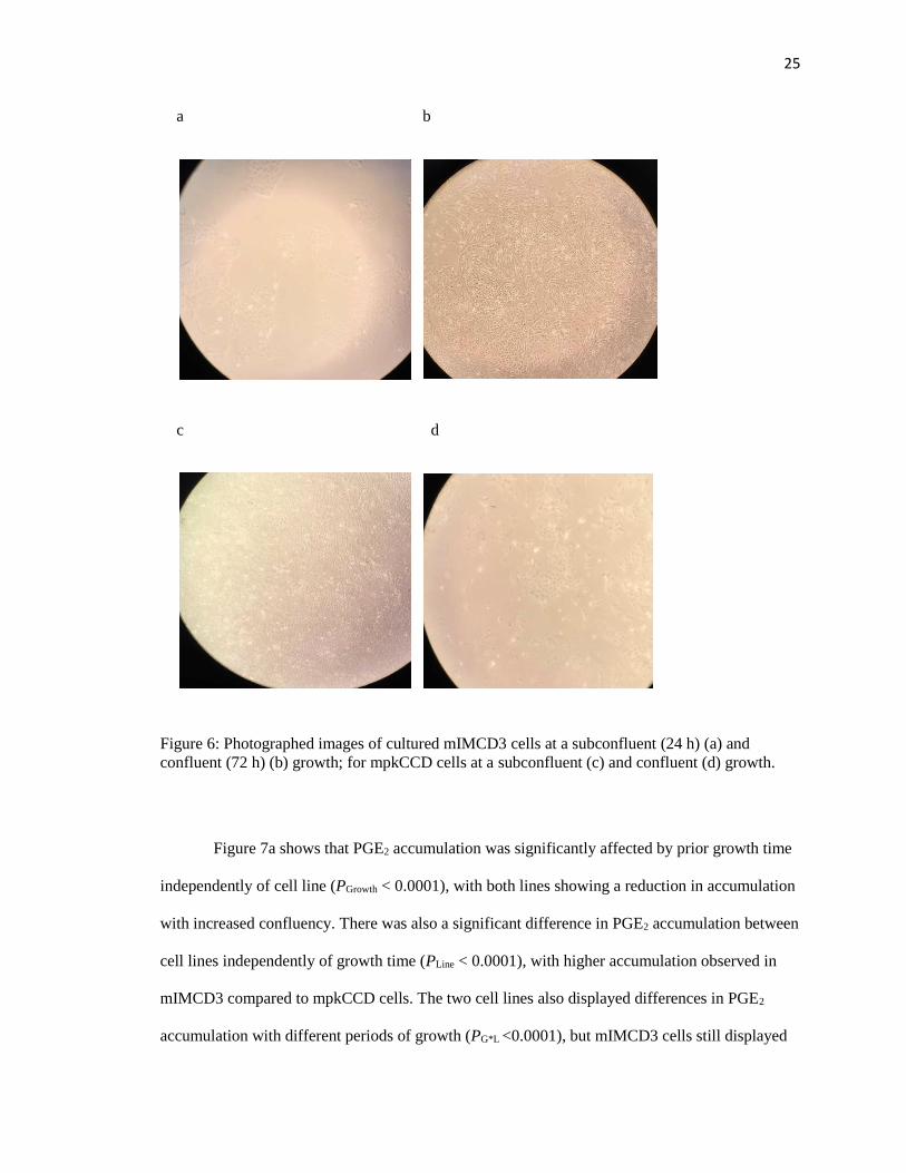
representing 80-90% confluency. Photographed images of cultured mIMCD3 and mpkCCD cells
at a subconfluent and confluent growth were taken (Figure 6).
25
a b
c d
Figure 6: Photographed images of cultured mIMCD3 cells at a subconfluent (24 h) (a) and
confluent (72 h) (b) growth; for mpkCCD cells at a subconfluent (c) and confluent (d) growth.
Figure 7a shows that PGE2 accumulation was significantly affected by prior growth time
independently of cell line (PGrowth < 0.0001), with both lines showing a reduction in accumulation
with increased confluency. There was also a significant difference in PGE2 accumulation between
cell lines independently of growth time (PLine < 0.0001), with higher accumulation observed in
mIMCD3 compared to mpkCCD cells. The two cell lines also displayed differences in PGE2
accumulation with different periods of growth (PG*L <0.0001), but mIMCD3 cells still displayed
26
increased PGE2 accumulation compared to mpkCCD cells at both 24 and 72 h (P < 0.05 by
Bonferroni post hoc test).
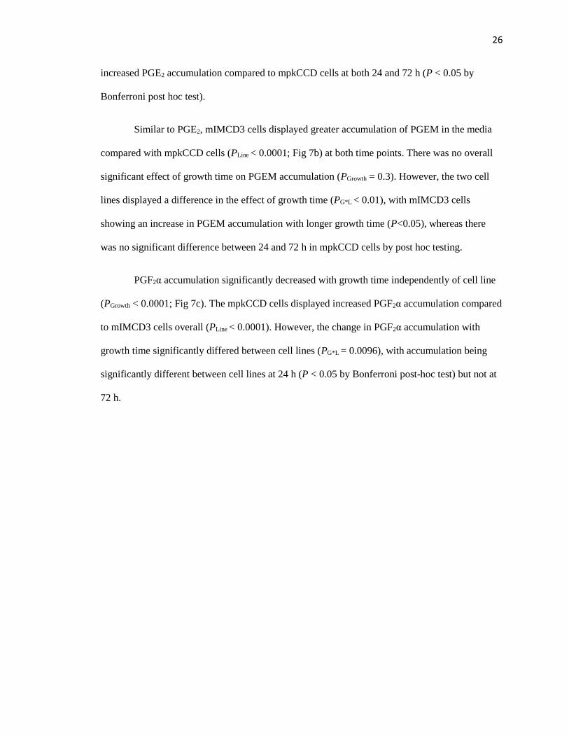
Similar to PGE2, mIMCD3 cells displayed greater accumulation of PGEM in the media
compared with mpkCCD cells (PLine < 0.0001; Fig 7b) at both time points. There was no overall
significant effect of growth time on PGEM accumulation (PGrowth = 0.3). However, the two cell
lines displayed a difference in the effect of growth time (PG*L < 0.01), with mIMCD3 cells
showing an increase in PGEM accumulation with longer growth time (P<0.05), whereas there
was no significant difference between 24 and 72 h in mpkCCD cells by post hoc testing.
PGF2α accumulation significantly decreased with growth time independently of cell line
(PGrowth < 0.0001; Fig 7c). The mpkCCD cells displayed increased PGF2α accumulation compared
to mIMCD3 cells overall (PLine < 0.0001). However, the change in PGF2α accumulation with
growth time significantly differed between cell lines (PG*L = 0.0096), with accumulation being
significantly different between cell lines at 24 h (P < 0.05 by Bonferroni post-hoc test) but not at
72 h.

27
m p k C C D m IM C D 3
0
5 0 0
1 0 0 0
1 5 0 0
C e ll L in e
PG
E2
(p
g/m
g)
P G ro w th < 0 .0 0 0 1
P L in e < 0 .0 0 0 1
P G * L < 0 .0 0 0 1
*P < 0 .0 5
2 4 h
7 2 h
*
*
m p k C C D m IM C D 3
0
1 0 0
2 0 0
3 0 0
4 0 0
C e ll L in e
PG
EM
(p
g/m
g)
P G ro w th = 0 .3
P L in e < 0 .0 0 0 1
P G * L = 0 .0 0 3 1
*P < 0 .0 5
* 2 4 h
7 2 h
m p k C C D m IM C D 3
0
1 0 0 0
2 0 0 0
3 0 0 0
C e ll L in e
PG
F2
(p
g/m
g)
P G ro w th < 0 .0 0 0 1
P L in e < 0 .0 0 0 1
P G *L = 0 .0 0 9 6
*P < 0 .0 5
*
2 4 h
7 2 h*
F ig u re 1 0 : E ffe c t o f g ro w th t im e o n P G E 2 (a ) , P G E M (b ), a n d P G F 2 (c ) p ro d u c t io n . P v a lu e s w e re
d e te rm in e d b y tw o -w a y A N O V A , te s tin g fo r e ffe c ts o f c e ll l in e (P L in e ) , g ro w th t im e (P G ro w th ) a n d th e
in te ra c tio n b e tw e e n th e tw o (P G * L ) . *P < 0 .0 5 d e te rm in e d b y p o s t-h o c B o n fe rro n i te s t. D a ta p re s e n te d
a re m e a n ± S E M fo r n = 6 p e r c e ll l in e , p e r t im e .
a
b
c
Figure 7: Effect of growth time on PGE2 (a), PGEM (b), and PGF2α (c) production. P values were
determined by two-way ANOVA, testing for effects of cell line (PLine), growth time (PGrowth), and
the interaction between the two (PG*L). *P < 0.05 determined by post-hoc Bonferroni test. Data
presented are mean ± SEM for n=6 per cell line, per time.

28
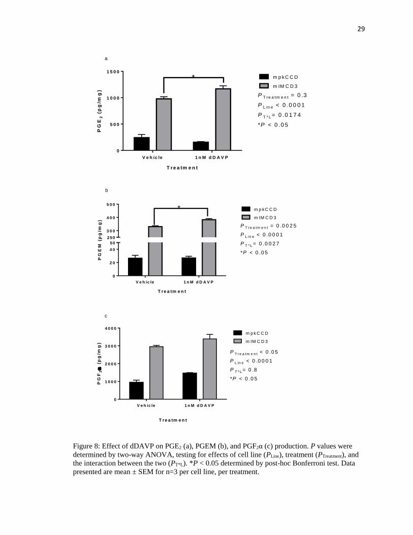
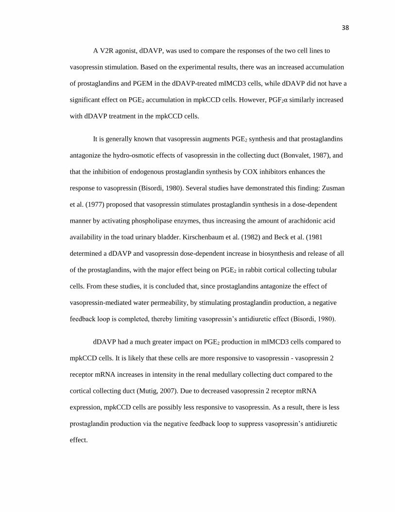
Effect of dDAVP on prostaglandin production
It has been previously reported that vasopressin augments PGE2 synthesis, resulting in a
negative feedback loop to limit vasopressin’s antidiuretic effect (Bisordi, 1980). Zusman et al.
(1977) determined that vasopressin stimulates prostaglandin synthesis in a dose-dependent
manner, while Kirschenbaum et al. (1982) and Beck et al. (1981) determined both a dDAVP and
vasopressin dose-dependent increase in biosynthesis and release of all of the prostaglandins, with
the major effect being on PGE2. The effect of dDAVP, a vasopressin analog, on prostaglandin
production was thus assessed.
mIMCD3 cells showed significantly greater PGE2 accumulation compared to mpkCCD
cells, independently of treatment (PLine < 0.001; Fig 8a). While there was no significant effect of
dDAVP treatment when taking both cell lines into account (PTreatment = 0.3), the two lines
responded differently (PT*L< 0.05), with mIMCD3 cells showing an approximately 20% increase
in PGE2 accumulation in response to dDAVP (P < 0.05), whereas mpkCCD cells did not.
Likewise, mIMCD3 cells showed significantly greater PGEM accumulation compared to
mpkCCD cells, independently of treatment (PLine < 0.0001; Fig 8b). There was a significant effect
of dDAVP treatment when taking both cell lines into account (PTreatment = 0.0025); the two cell
lines did respond differently (PT*L = 0.0027), with mIMCD3 cells showing a statistically
significant 15% increase in PGEM accumulation in response to dDAVP (P <0.05), whereas
mpkCCD cells did not.
dDAVP-treated cells showed increased PGF2α compared to control cells in each cell line
(Fig 8c). mIMCD3 cells showed significantly greater PGE2 accumulation compared to mpkCCD
cells, independently of treatment (PLine < 0.0001). There was a significant effect of dDAVP
treatment when taking both cell lines into account (PTreatment < 0.05). The effect of dDAVP to
increase PGF2α production did not differ between cell lines (PT*L = 0.8).
29
V e h ic le 1 n M d D A V P
0
5 0 0
1 0 0 0
1 5 0 0
T re a tm e n t
PG
E2
(p
g/m
g)
m IM C D 3
m p k C C D
P T re a tm e n t = 0 .3
P L in e < 0 .0 0 0 1
P T * L = 0 .0 1 7 4
*P < 0 .0 5
*
F ig u re 1 1 : E ffe c t o f d D A V P o n P G E 2 (a ) , P G E M (b ), a n d P G F 2 (c ) p ro d u c t io n . P v a lu e s w e re
d e te rm in e d b y tw o -w a y A N O V A , te s tin g fo r e ffe c ts o f c e ll l in e (P L in e ) , tre a tm e n t (P T re a tm e n t) a n d
th e in te ra c tio n b e tw e e n th e tw o (P T *L ) . A n u n p a ire d T te s t w a s u s e d to c o m p a re tre a tm e n t P
v a lu e fo r (c ) . *P < 0 .0 5 d e te rm in e d b y p o s t-h o c B o n fe rro n i te s t. D a ta p re s e n te d a re m e a n ±
S E M fo r n = 3 p e r c e ll l in e , p e r tre a tm e n t.
V e h ic le 1 n M d D A V P
0
2 0
4 0
3 0 0
4 0 0
5 0 0
T re a tm e n t
PG
EM
(p
g/m
g)
P T re a tm e n t = 0 .0 0 2 5
P L in e < 0 .0 0 0 1
P T * L = 0 .0 0 2 7
*P < 0 .0 5
m p k C C D
m IM C D 3
*
50
250
a
c
b
V e h ic le 1 n M d D A V P
0
1 0 0 0
2 0 0 0
3 0 0 0
4 0 0 0
T re a tm e n t
PG
F2
(p
g/m
g)
P T re a tm e n t < 0 .0 5
P L in e < 0 .0 0 0 1
P T *L = 0 .8
*P < 0 .0 5
m p k C C D
m IM C D 3
Figure 8: Effect of dDAVP on PGE2 (a), PGEM (b), and PGF2α (c) production. P values were
determined by two-way ANOVA, testing for effects of cell line (PLine), treatment (PTreatment), and
the interaction between the two (PT*L). *P < 0.05 determined by post-hoc Bonferroni test. Data
presented are mean ± SEM for n=3 per cell line, per treatment.

30
Effect of COX-1 (SC-560) and COX-2 (NS-398) inhibition on prostaglandin production
Ferguson et al. (1999) reports that COX-2 is the major contributor to the pool of PGE2
synthesized by the cortical collecting duct. From the study, when COX enzyme activity was
measured in murine M-1 cortical collecting duct cells, both indomethacin (a COX-1 and -2
inhibitor) and the specific COX-2 inhibitor NS-398 effectively blocked PGE2 synthesis,
demonstrating that COX-2 is the major contributor to PGE2 synthesis. Other studies have
concluded that hyperosmolar conditions, conditions such as what inner medullary collecting duct
cells are exposed to, have led to increased COX-2 expression, while inhibiting COX abolished
PGE2 production (Zhang, 1995).
The individual effects of COX-1 and COX-2 inhibition on mpkCCD and mIMCD3 cell
prostaglandin production was tested. The addition of a COX-1 or COX-2 inhibitor to both cell
lines showed a statistically significant decrease of both PGE2 and PGF2α accumulation in the
media (P <0.05) compared to the control groups (Fig 9 a, c, d, f).
PGE2 levels were higher in mIMCD3 cells compared to mpkCCD cells with a COX-1
inhibitor (Fig 9a). PGE2 accumulation by SC-560-treated mpkCCD cells is suppressed to
approximately 22% of that seen in vehicle control cells whereas mIMCD3 cells treated with SC-
560 is suppressed to approximately 28% of that seen in vehicle control cells (PTreatment and PLine <
0.0001). Similarly, PGE2 accumulation was greater in both the treated and control groups of
mIMCD3 cells with a COX-2 inhibitor (Fig. 9d). There was similar inhibition of PGE2
accumulation in the media; NS-398-treated mpkCCD cells was suppressed to approximately 25%
of that seen in vehicle control cells whereas mIMCD3 cells treated with NS-398 is suppressed to
approximately 20% of that seen in vehicle control cells (PTreatment and PLine < 0.0001).
PGEM accumulation was significantly greater in the mIMCD3 cells compared to
mpkCCD cells (PLine < 0.0001), in both the treated and control cells, and in the presence of both a

31
COX-1 and COX-2 inhibitor (Fig. 9 b, e). Treated mIMCD3 cells paradoxically showed an
increase in PGEM accumulation compared to untreated cells; there was a 28% increase in PGEM
accumulation with a COX-1 inhibitor present and a 16% increase with a COX-2 inhibitor, both of
which were statistically significant (P < 0.05).
As stated above, PGF2α levels were significantly decreased with the addition of a COX-1
or COX-2 inhibitor in both cell lines (Fig. 9 c, f). PGF2α levels in SC-560-treated mpkCCD cells
is suppressed to approximately 10% of that seen in vehicle control cells whereas mIMCD3 cells
treated with SC-560 is suppressed to approximately 34% of that seen in vehicle control cells
(PTreatment < 0.0001). There was not a statistically significant difference in PGF2α production
between the two cell lines (PLine = 0.1068). Just as with a COX-1 inhibitor, mpkCCD cells treated
with a COX-2 inhibitor showed a greater reduction in PGF2α accumulation compared to
mIMCD3 treated cells; NS-398-treated mpkCCD cells was suppressed to approximately 10% of
that seen in vehicle control cells whereas mIMCD3 cells treated with NS-398 is suppressed to
approximately 27% of that seen in vehicle control cells (PTreatment < 0.0001; PLine = 0.0192).

32
V e h ic le S C 5 6 0
0
2 0 0
4 0 0
6 0 0
8 0 0
1 0 0 0
T re a tm e n t
PG
E2
(p
g/m
g)
P T re a tm e n t < 0 .0 0 0 1
P L in e < 0 .0 0 0 1
P T * L < 0 .0 0 0 1
*P < 0 .0 5
*
m p k C C D
m IM C D 3
*
V e h ic le N S 3 9 8
0
2 0 0
4 0 0
6 0 0
8 0 0
1 0 0 0
T re a tm e n t
PG
E2
(p
g/m
g)
P T re a tm e n t < 0 .0 0 0 1
P L in e < 0 .0 0 0 1
P T * L < 0 .0 0 0 1
*P < 0 .0 5*
*
m p k C C D
m IM C D 3
V e h ic le S C 5 6 0
0
2 0
4 0
3 0 0
4 0 0
5 0 0
6 0 0
T re a tm e n t
PG
EM
(p
g/m
g)
P T re a tm e n t = 0 .0 4 7 0
P L in e < 0 .0 0 0 1
P T * L = 0 .0 5 2 8
*P < 0 .0 5
*m p k C C D
m IM C D 3
50
V e h ic le N S 3 9 8
0
2 0
4 0
3 0 0
4 0 0
5 0 0
6 0 0
T re a tm e n t
PG
EM
(p
g/m
g)
P T re a tm e n t = 0 .0 5 2 4
P L in e < 0 .0 0 0 1
P T * L = 0 .0 0 6 5
*P < 0 .0 5
*
m p k C C D
m IM C D 3
50
V e h ic le S C 5 6 0
0
5 0 0
1 0 0 0
1 5 0 0
2 0 0 0
2 5 0 0
T re a tm e n t
PG
F2
(p
g/m
g)
P T re a tm e n t < 0 .0 0 0 1
P L in e = 0 .1 0 6 8
P T * L = 0 .0 2 2 3
*P < 0 .0 5*
m p k C C D
m IM C D 3
*
V e h ic le N S 3 9 8
0
5 0 0
1 0 0 0
1 5 0 0
2 0 0 0
2 5 0 0
T re a tm e n t
PG
F2
(p
g/m
g)
P T re a tm e n t < 0 .0 0 0 1
P L in e = 0 .0 1 9 2
P T * L = 0 .0 2 7 3
*P < 0 .0 5*
m p k C C D
m IM C D 3
*
F ig u re 1 2 : E ffe c t o f 0 .1 M S C -5 6 0 (C O X -1 ) (a -c ) a n d 5 M N S -3 9 8 (C O X -2 ) (d - f) in h ib it io n o n P G E 2 ( a ,d ) ,
P G E M (b ,e ) , a n d P G F 2 (c , f ) p ro d u c t io n . P v a lu e s w e re d e te rm in e d b y tw o -w a y A N O V A , te s tin g fo r e ffe c ts o f
c e ll l in e (P L in e ) , tre a tm e n t (P T re a tm e n t) a n d th e in te ra c tio n b e tw e e n th e tw o (P T * L ) . *P < 0 .0 5 d e te rm in e d b y
p o s t-h o c B o n fe rro n i te s t c o m p a re d to th e c o rre s p o n d in g v e h ic le g ro u p . D a ta p re s e n te d a re m e a n ± S E M fo r n =
3 p e r c e ll l in e , p e r tre a tm e n t.
b
a
c
d
e
f
Figure 9: Effect of 0.1 µM SC-560 (COX-1) (a-c) and 5 µM NS-398 (COX-2) (d-f) inhibition on
PGE2 (a, d), PGEM (b, e), and PGF2α (c, f) production. P values were determined by two-way
ANOVA, testing for effects of cell line (PLine), treatment (PTreatment), and the interaction between
the two (PT*L). *P < 0.05 determined by post-hoc Bonferroni test, and indicates statistical
significance compared to the vehicle treatment in the same cell line. Data presented are mean ±
SEM for n=3 per cell line, per treatment.
33
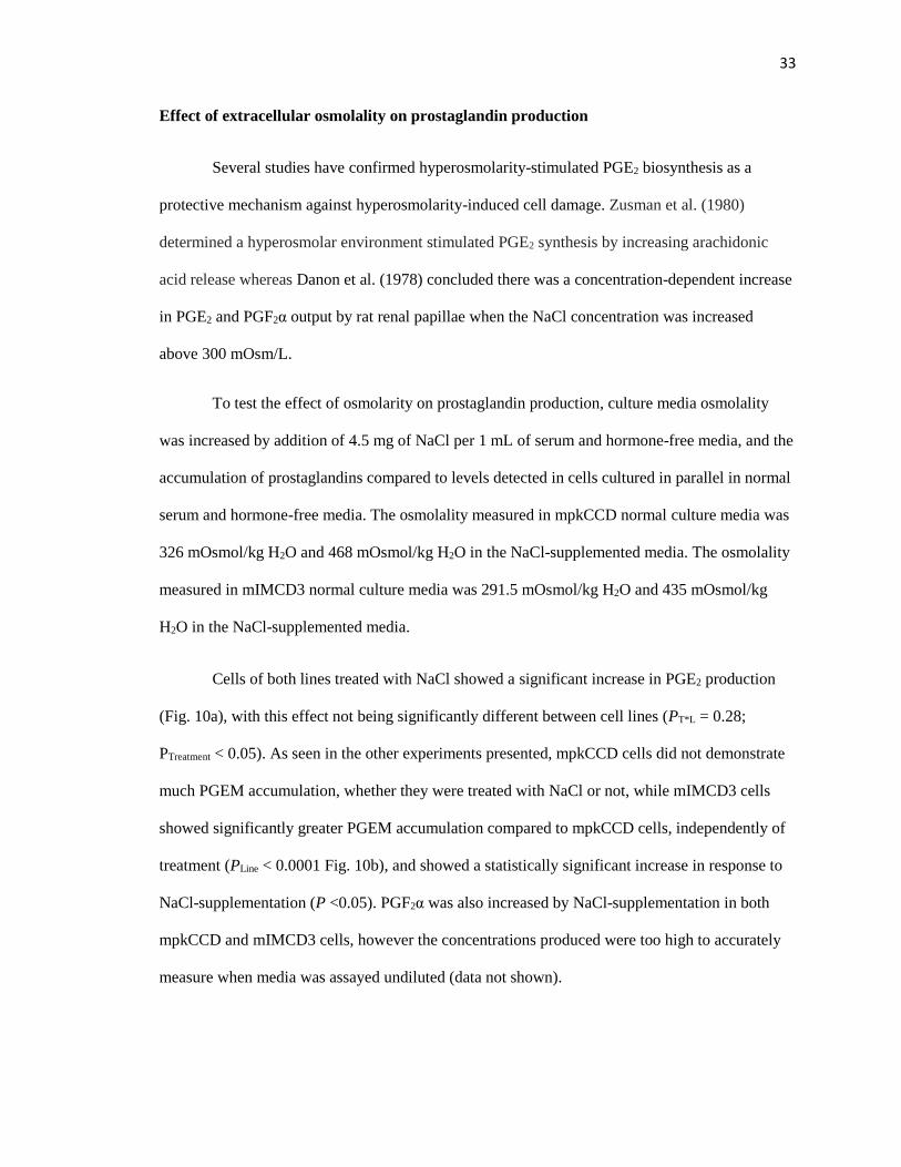
Effect of extracellular osmolality on prostaglandin production
Several studies have confirmed hyperosmolarity-stimulated PGE2 biosynthesis as a
protective mechanism against hyperosmolarity-induced cell damage. Zusman et al. (1980)
determined a hyperosmolar environment stimulated PGE2 synthesis by increasing arachidonic
acid release whereas Danon et al. (1978) concluded there was a concentration-dependent increase
in PGE2 and PGF2α output by rat renal papillae when the NaCl concentration was increased
above 300 mOsm/L.
To test the effect of osmolarity on prostaglandin production, culture media osmolality
was increased by addition of 4.5 mg of NaCl per 1 mL of serum and hormone-free media, and the
accumulation of prostaglandins compared to levels detected in cells cultured in parallel in normal
serum and hormone-free media. The osmolality measured in mpkCCD normal culture media was
326 mOsmol/kg H2O and 468 mOsmol/kg H2O in the NaCl-supplemented media. The osmolality
measured in mIMCD3 normal culture media was 291.5 mOsmol/kg H2O and 435 mOsmol/kg
H2O in the NaCl-supplemented media.
Cells of both lines treated with NaCl showed a significant increase in PGE2 production
(Fig. 10a), with this effect not being significantly different between cell lines (PT*L = 0.28;
PTreatment < 0.05). As seen in the other experiments presented, mpkCCD cells did not demonstrate
much PGEM accumulation, whether they were treated with NaCl or not, while mIMCD3 cells
showed significantly greater PGEM accumulation compared to mpkCCD cells, independently of
treatment (PLine < 0.0001 Fig. 10b), and showed a statistically significant increase in response to
NaCl-supplementation (P <0.05). PGF2α was also increased by NaCl-supplementation in both
mpkCCD and mIMCD3 cells, however the concentrations produced were too high to accurately
measure when media was assayed undiluted (data not shown).

34
V e h ic le N a C l
0
2 0 0 0
4 0 0 0
6 0 0 0
8 0 0 0
T re a tm e n t
PG
E2
(p
g/m
g)
P T re a tm e n t = 0 .0 0 1 6
P L in e = 0 .8 7 2 9
P T * L = 0 .2 8
*P < 0 .0 5
*m p k C C D
m IM C D 3
V e h ic le N a C l
0
2 0
4 0
2 0 0
4 0 0
6 0 0
8 0 0
1 0 0 0
T re a tm e n t
PG
EM
(p
g/m
g)
P T re a tm e n t < 0 .0 0 0 1
P L in e < 0 .0 0 0 1
P T * L < 0 .0 0 0 1
*P < 0 .0 5
* m p k C C D
m IM C D 3
F ig u re 1 3 : E ffe c t o f N a C l o n P G E 2 (a ) a n d P G E M (b ) p ro d u c t io n . P v a lu e s w e re d e te rm in e d b y
tw o -w a y A N O V A , te s t in g fo r e ffe c ts o f c e ll l in e (P L in e ) , tre a tm e n t (P T re a tm e n t) a n d th e in te ra c t io n
b e tw e e n th e tw o (P T *L ) . *P < 0 .0 5 d e te rm in e d b y p o s t-h o c B o n fe rro n i te s t. D a ta p re s e n te d a re
m e a n ± S E M fo r n = 3 p e r c e ll l in e , p e r tre a tm e n t.
b
a
Figure 10: Effect of NaCl on PGE2 (a) and PGEM (b) production. P values were determined by
two-way ANOVA, testing for effects of cell line (PLine), treatment (PTreatment), and the interaction
between the two (PT*L). *P < 0.05 determined by post-hoc Bonferroni test. Data presented are
mean ± SEM for n=3 per cell line, per treatment.

35
CHAPTER 4: DISCUSSION
The collecting duct is the terminal segment of the nephron tubule that is involved in the
fine-tuning of urine volume and composition, creating a more dilute or more concentrated urine,
based on the body’s water needs. The collecting duct extends from the outer renal cortex, down to
the deepest parts of the renal medulla. The water permeability of the collecting duct is regulated
by the hormone vasopressin. In the short term, vasopressin stimulates the shuttling of aquaporin 2
water channels to the apical membrane, allowing passive water movement to move from the
nephron tubular lumen and into the surrounding interstitium; long term, vasopressin enhances
aquaporin 2 gene expression (Kuo, 2018). Prostaglandins, primarily PGE2, is a known negative
regulator of collecting duct water and urea permeability. Through various signaling pathways,
depending on which particular receptor PGE2 binds to, collecting duct water permeability can be
decreased either by increased endocytosis of aquaporin 2 vesicles, or a decrease in aquaporin 2
trafficking to the apical membrane as a result of decreased intracellular cAMP levels.
The cell lines utilized come from different areas of the collecting duct. mpkCCD cells are
cortical collecting duct cells whereas mIMCD3 cells are inner medullary collecting duct cells.
These cells are exposed to different extracellular osmolarities. As demonstrated, they also differ
in prostaglandin production, both as a result of different cell growth confluency and various
treatment agents. There were general findings of the study. A general finding was that mIMCD3
cells made more PGE2 than mpkCCD cells, but the amount of PGF2α was more similar between
the two cell lines. Additionally, as a general trend, PGEM levels did not change all that much
between vehicle and treated cells of each cell line and between subconfluent and confluent cells
for each cell line. Finally, subconfluent cells showed more prostaglandin production compared to
confluent cells in both cell lines.
PGE2 and PGF2α accumulation decreased as cells approached confluency
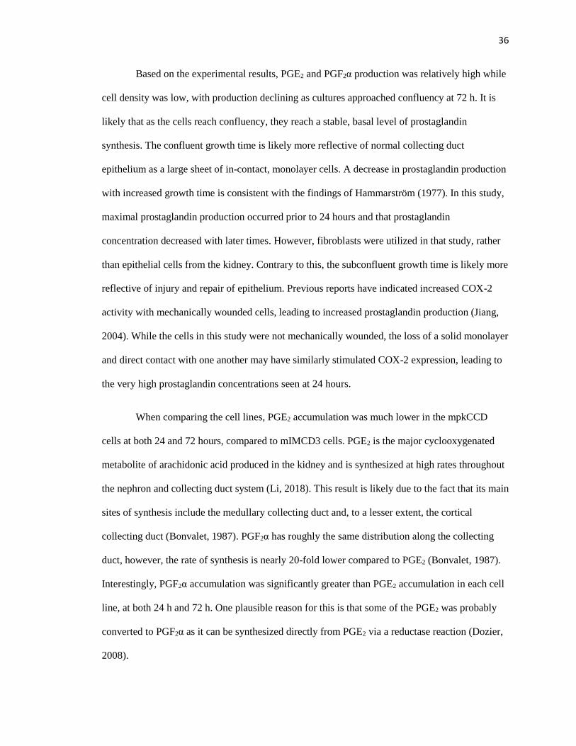
36
Based on the experimental results, PGE2 and PGF2α production was relatively high while
cell density was low, with production declining as cultures approached confluency at 72 h. It is
likely that as the cells reach confluency, they reach a stable, basal level of prostaglandin
synthesis. The confluent growth time is likely more reflective of normal collecting duct
epithelium as a large sheet of in-contact, monolayer cells. A decrease in prostaglandin production
with increased growth time is consistent with the findings of Hammarström (1977). In this study,
maximal prostaglandin production occurred prior to 24 hours and that prostaglandin
concentration decreased with later times. However, fibroblasts were utilized in that study, rather
than epithelial cells from the kidney. Contrary to this, the subconfluent growth time is likely more
reflective of injury and repair of epithelium. Previous reports have indicated increased COX-2
activity with mechanically wounded cells, leading to increased prostaglandin production (Jiang,
2004). While the cells in this study were not mechanically wounded, the loss of a solid monolayer
and direct contact with one another may have similarly stimulated COX-2 expression, leading to
the very high prostaglandin concentrations seen at 24 hours.
When comparing the cell lines, PGE2 accumulation was much lower in the mpkCCD
cells at both 24 and 72 hours, compared to mIMCD3 cells. PGE2 is the major cyclooxygenated
metabolite of arachidonic acid produced in the kidney and is synthesized at high rates throughout
the nephron and collecting duct system (Li, 2018). This result is likely due to the fact that its main
sites of synthesis include the medullary collecting duct and, to a lesser extent, the cortical
collecting duct (Bonvalet, 1987). PGF2α has roughly the same distribution along the collecting
duct, however, the rate of synthesis is nearly 20-fold lower compared to PGE2 (Bonvalet, 1987).
Interestingly, PGF2α accumulation was significantly greater than PGE2 accumulation in each cell
line, at both 24 h and 72 h. One plausible reason for this is that some of the PGE2 was probably
converted to PGF2α as it can be synthesized directly from PGE2 via a reductase reaction (Dozier,
2008).

37
PGEM was measured to see if the observed decrease in prostaglandin production at a
confluent time growth was a result of metabolism. While there was an increase in PGEM from 24
to 72 hours in mIMCD3 cells, the increase does not account for the huge reduction seen in PGE2
seen at 72 hours compared to 24 hours (approximately 25% of what was produced by cells
allowed to grow for 24 hours; Fig. 7a). Most cultured cells that produce prostaglandin E2
generally do not contain the enzymes required for PGE2 metabolism, likely explaining this result
in that most PGE2 was not metabolized into its metabolites. There wasn’t low PGEM observed at
24h growth and high PGEM at 72h growth, suggesting that production of PGE2 is truly lower in
confluent cells rather than the decrease reflecting increased metabolism. An assay to measure
PGF2αM should be included as a future step to observe the effects that cell confluency has on
PGF2αM, and see how that compares in the two cell lines.
PGE2 accumulation increased with dDAVP treatment in mIMCD3 cells while not having a
significant effect in mpkCCD cells. PGF2α accumulation increased with dDAVP treatment
in both cell lines
Vasopressin has important physiological roles. In vivo, vasopressin is released in
response to an increase in blood osmolarity or a decrease in blood volume. In the collecting duct,
vasopressin binds to its vasopressin 2 receptor (V2R), which initiates a Gs-cAMP-Protein Kinase
A mediated trafficking of aquaporin 2 water channels to insert into the apical membrane. As a
result, water passively flows through the water channels, and enters the surrounding interstitium
via aquaporin 3 and 4, where it is returned to the blood. PGE2 is known for its diuretic role,
opposing vasopressin-mediated water permeability of the collecting duct. This occurs through
PGE2 binding to its EP3 receptor, which decreases intracellular cAMP levels, or binding to its
EP1 receptor, which activates Gq and Protein Kinase C, which increases the endocytosis of
aquaporin 2 vesicles. PGF2α primarily signals through Gq-Protein Kinase C, and therefore likely
impacts aquaporin 2 trafficking in a similar manner as PGE2 and EP1.
38
A V2R agonist, dDAVP, was used to compare the responses of the two cell lines to
vasopressin stimulation. Based on the experimental results, there was an increased accumulation
of prostaglandins and PGEM in the dDAVP-treated mIMCD3 cells, while dDAVP did not have a
significant effect on PGE2 accumulation in mpkCCD cells. However, PGF2α similarly increased
with dDAVP treatment in the mpkCCD cells.
It is generally known that vasopressin augments PGE2 synthesis and that prostaglandins
antagonize the hydro-osmotic effects of vasopressin in the collecting duct (Bonvalet, 1987), and
that the inhibition of endogenous prostaglandin synthesis by COX inhibitors enhances the
response to vasopressin (Bisordi, 1980). Several studies have demonstrated this finding: Zusman
et al. (1977) proposed that vasopressin stimulates prostaglandin synthesis in a dose-dependent
manner by activating phospholipase enzymes, thus increasing the amount of arachidonic acid
availability in the toad urinary bladder. Kirschenbaum et al. (1982) and Beck et al. (1981
determined a dDAVP and vasopressin dose-dependent increase in biosynthesis and release of all
of the prostaglandins, with the major effect being on PGE2 in rabbit cortical collecting tubular
cells. From these studies, it is concluded that, since prostaglandins antagonize the effect of
vasopressin-mediated water permeability, by stimulating prostaglandin production, a negative
feedback loop is completed, thereby limiting vasopressin’s antidiuretic effect (Bisordi, 1980).
dDAVP had a much greater impact on PGE2 production in mIMCD3 cells compared to
mpkCCD cells. It is likely that these cells are more responsive to vasopressin - vasopressin 2
receptor mRNA increases in intensity in the renal medullary collecting duct compared to the
cortical collecting duct (Mutig, 2007). Due to decreased vasopressin 2 receptor mRNA
expression, mpkCCD cells are possibly less responsive to vasopressin. As a result, there is less
prostaglandin production via the negative feedback loop to suppress vasopressin’s antidiuretic
effect.

39
Another possible explanation that dDAVP had little effect on PGE2 production was that
the concentration of dDAVP was not high enough. However Kortenoeven et al. (2011) utilized
the same concentration of 1 nM dDAVP to maximally induce aquaporin 2 expression in their
study. They determined that dDAVP-treated mpkCCD cells did stimulate both sufficient levels of
PGE2 and PGF2α production compared to control mpkCCD cells. However, they utilized different
culturing and incubation times, culturing the cells for 8 days and treating the cells with 1nM
dDAVP for 96 hours to maximally induce aquaporin 2 expression. Their model also more
accurately reflects in vivo conditions, using a semipermeable filter to create an apical and
basolateral surface. Repeating this experiment with higher concentrations of dDAVP to see if this
would result in increased PGE2 accumulation in the mpkCCD cells or not is a potential next step,
as well as utilizing their model of mpkCCD cell incubation with dDAVP.
PGE2 and PGF2a accumulation significantly decreased with COX-1 and COX-2 inhibitor
treatment in both mIMCD3 and mpkCCD cells.
The biosynthetic pathway of prostaglandins involves the conversion of arachidonic acid
to prostaglandin H2, catalyzed by cyclooxygenase-1 and -2 (COX-1 and COX-2) enzymes. This is
the rate-limiting step in prostaglandin synthesis. Therefore, the presence of COX-1 and COX-2
inhibitors would expect to greatly reduce prostaglandin production. In the kidney, COX-1 is
constitutively expressed in the collecting duct, localized to both the cortical and medullary
segments (Hao, 2008). COX-1 has also been determined to be present in the medullary interstitial
cells (Nasrallah, 2014). The expression of COX-2 is much more unclear and conflicting in most
studies. It has been reported to have varying amounts of expression within the collecting duct.
Kortenoeven et al. (2011) report strong COX-2 expression in the collecting duct in three studies,
and absence of it was suggested in three other studies. They also report that increased COX-2
levels have been detected in collecting ducts of animals that were dehydrated or subjected to a
40
chronic NaCl load, noting its inducible expression under stress conditions. It is, however, highly
expressed in renal medullary interstitial cells (Hao, 2008; Kortenoeven, 2011).
Based on the experimental results, as expected, mpkCCD and mIMCD3 cells treated with
either a COX-1 or COX-2 inhibitor greatly reduced prostaglandin accumulation. Both COX-1
(SC-560) and COX-2 (NS-398) inhibition resulted in similar decreases of PGE2 accumulation in
both cell lines. In mIMCD3 cells, PGE2 accumulation was more reduced in the presence of a
COX-2 inhibitor compared to a COX-1 inhibitor (28% vs. 20%). In mpkCCD cells, the presence
of a COX-1 inhibitor reduced PGE2 accumulation to 22% of that compared to vehicle control
cells, and 25% with a COX-2 inhibitor. Due to the similarity in reduction of PGE2 accumulation
in the media with a COX-1 and COX-2 inhibitor in both cells, it is difficult to report what enzyme
contributes more to PGE2 synthesis in these cells. Ferguson et al. (1999) reports that COX-2 is the
major contributor to the pool of PGE2 synthesized by the cortical collecting duct. From the study,
when COX enzyme activity was measured in their murine M-1 cortical collecting duct cells, both
indomethacin (COX-1 and -2 inhibitor) and the specific COX-2 inhibitor NS-398 effectively
blocked PGE2 synthesis, demonstrating that COX-2 is the major contributor to the pool of PGE2
synthesized by the cortical collecting duct. However, they did utilize a different cortical cell line,
which may contribute to their results.
For PGF2α, while both COX-1 and COX-2 inhibitors reduced accumulation in the media
in both cell lines, the reduction was greater in the mpkCCD cells, for both COX-1 and COX-2
inhibition. SC-560 and NS-398 treated mpkCCD cells suppressed PGF2α accumulation to 10% of
that seen in the vehicle control cells, whereas treated mIMCD3 cells with SC-560 and NS-398
suppressed PGF2α accumulation to 34% and 27% of that seen in vehicle treated cells,
respectively. Again, these percentages are too close to confidently say which enzyme contributes
more to PGF2α production, and this would need to be run again to determine which enzyme has a
greater effect on prostaglandin production. Interestingly, there was a paradoxical increase in

41
PGEM accumulation in the mIMCD3 cells, in both the COX-1 and COX-2 inhibitor treated cells;
however, there was very little PGEM accumulation in the mpkCCD cells, with both COX-1 and
COX-2 inhibition as well as in the control cells.
PGE2 and PGF2α accumulation significantly increased with NaCl treatment in both
mIMCD3 and mpkCCD cells.
The osmolarity of the tubular fluid as well as the surrounding interstitium varies along the
length of the collecting duct. These cell lines are located in different regions of the collecting
duct, and are thus exposed to the different osmolarities. In the formation of a concentrated urine,
under the influence of vasopressin, the deepest parts of the interstitial medullary region as well as
the tubular fluid within the nephron, can be exposed to osmolarities up to 1200 mOsm/L. Thus,
the cells in this area must be able to function with significant increases in osmolarity. In a state of
dehydration, the interstitial osmolality can increase several fold, and if prolonged, lead to cellular
distress (Price, 2011). In the formation of a dilute urine, osmolarities of the tubular fluid can be as
low as 50 mOsm/L, with a medullary interstitium osmolarity of 600 mOsm/L. The cortical
collecting duct tubular fluid and surrounding interstitium usually has an osmolarity around 300
mOsm/L.
Based on the experimental results, PGE2 and PGF2α levels were significantly increased in
NaCl-treated cells of both lines. PGF2α concentration levels produced were too high to accurately
measure when media was assayed undiluted in both cell lines. The result of a surge in
prostaglandin production in the presence of NaCl is most likely a stress-protective mechanism,
which is explained below. When comparing the two cell lines, PGE2 was much greater in the
mpkCCD-treated cells. This could be due to the idea that these cells are not as adaptable to such
increases in osmolarity as the mIMCD3 cells are, and this protective mechanism was further
enhanced. Levels of PGF2α as a result of the addition of NaCl is something that needs to be
determined.

42
Several studies have confirmed hyperosmolarity stimulated PGE2 biosynthesis as a
protective mechanism against hyperosmolarity-induced cell damage. In align with this, a well-
noted phenomenon in clinical medicine is that certain COX-inhibitory drugs can cause necrosis of
the renal papilla (Delmas, 1995; Eknoyan, 1994). Zusman et al. (1980) determined a
hyperosmolar environment stimulated PGE2 biosynthesis by increasing arachidonic acid release
in rabbit medullary interstitial cells. Via a similar mechanism of increasing arachidonic acid
mobilization from tissue lipids, Danon et al. (1978) concluded that there was a concentration-
dependent increase in PGE2 and PGF2α output by rat renal papillae when the NaCl concentration
was increased above 300 mOsm/L. Outside of the kidney, PGE2 production by
lipopolysaccharide-stimulated rat Kupffer cells was increased if exposed to a medium osmolarity
above 305 mOsm/L, whereas a hypo-osmotic medium (255 mOsm/L) diminished PGE2
formation. In this study involving the rat liver macrophages, the hyperosmolarity lead to COX-2
induction and inhibiting COX with indomethacin and specifically COX-2 with NS-398, abolished
PGE2 production (Zhang, 1995). In another relevant study, it was determined that hypertonicity
increased COX-2 activity, leading to PGE2-mediated inactivation of the pro-apoptotic protein,
BAD, conferring a hyperosmolarity-induced prostaglandin production protective mechanism
(Price, 2011). Several other studies have conferred a cytoprotective role of COX-2 in the osmotic
response (Yang, 2000).
There are obvious limitations to this study. First and foremost, due to the lack of time,
these experiments were only completed once. To verify these results and to ensure the results
were not by random chance, these exact experiments should be run again, possibly three times, to
gather stronger and more firm conclusions. In addition, the sample size utilized was low, which
reduces the power of a study and increases the margin of error. With such a low sample size, it is
difficult to discern whether these are the expected results in all of the experiments, or if the results
would differ with a larger sample size. In alignment with the lack of available time, some of the
43
prostaglandins accumulation data needs to be repeated with diluted samples, as the concentration
levels produced were too high to be accurately measured when media was assayed undiluted.
With that said, this leads us to some potential future directions. Re-running these experiments is
also a future direction, as well as trying different treatment concentrations, to see if there is a
dose-dependent response. Another future direction would be to look at other prostaglandins, as
the kidneys synthesize more than just PGE2 and PGF2α. In addition, comparing expression levels
of various enzymes, such as COX, between the two cell lines, by western blot is warranted.
Overall, the models used to study the collecting duct were appropriate for this study.
There are many differences between the two cell lines that have been mentioned. These
differences are primarily due to their differences in their location within the collecting duct. As a
result, the two cell lines responded differently in prostaglandin production in response to various
treatments.

44
BIBLIOGRAPHY
1. Ares, G. R., Caceres, P. S., & Ortiz, P. A. (2011). Molecular regulation of NKCC2 in the
thick ascending limb. American Journal of Physiology. Renal Physiology, 301(6), 1143.
2. Beck, T. & Dunn, M. (1981). The relationship of anti-diuretic hormone and renal
prostaglandins. Mineral Electrolyte Metabolism, 6, 46.
3. Bisordi, J. E., Schlondorff, D., & Hays, R. M. (1980). Interaction of vasopressin and
prostaglandins in the toad urinary bladder. The Journal of clinical investigation, 66(6),
1200–1210.
4. Bonvalet, J., Pradelles, P., & Farman, N. (1987). Segmental synthesis and actions of
prostaglandins along the nephron. Am J Physiol, 253(3), F377-87.
5. Breyer, M. D., Jacobson, H. R., & Breyer, R. M. (1996). Functional and molecular
aspects of renal prostaglandin receptors. Journal of the American Society of Nephrology,
7(1), 8-17.
6. Breyer, M. D., Zhang, Y., Guan, Y. F., Hao, C. M., Hebert, R. L., & Breyer, R. M.
(1998). Regulation of renal function by prostaglandin E receptors. Kidney International.
Supplement, 67, 88.
7. Casale, J., & Varacallo, M. (2018). Biochemistry, phospholipase A2. StatPearls.
Treasure Island (FL): StatPearls Publishing.
8. Chen, G. (2013). New advances in urea transporter UT-A1 membrane trafficking.
International Journal of Molecular Sciences, 14(5), 10674-10682.
9. Chou, C. L., Knepper, M. A., Hoek, A. N., Brown, D., Yang, B., Ma, T., & Verkman, A.
S. (1999). Reduced water permeability and altered ultrastructure in thin descending limb
of henle in aquaporin-1 null mice. The Journal of Clinical Investigation, 103(4), 491-496.
10. Danon, A., Knapp, H., Oelz, O., & Oates, J. (1978) Stimulation of prostaglandin
biosynthesis in the renal papilla by hypertonic mediums. Am. J. Physiol., 234(1), F64-7.

45
11. Delmas, P. (1995). Non-steroidal anti-inflammatory drugs and renal function.
Rheumatology, Volume XXXIV, 25–28.
12. Dennis, E. A., Cao, J., Hsu, Y., Magrioti, V., & Kokotos, G. (2011). Phospholipase A2
enzymes: Physical structure, biological function, disease implication, chemical inhibition,
and therapeutic intervention. Chemical Reviews, 111(10), 6130-6185.
13. Dibona, G. F. (1986). Prostaglandins and nonsteroidal anti-inflammatory drugs: Effects
on renal hemodynamics. The American Journal of Medicine, 80(1), 12-21.
14. Dozier, B. L., Watanabe, K., & Duffy, D. M. (2008). Two pathways for prostaglandin F2
alpha synthesis by the primate periovulatory follicle. Reproduction (Cambridge,
England), 136(1), 53–63.
15. Dunn, M. J., & Hood, V. L. (1977). Prostaglandins and the kidney. Am. J. Physiol,
233(3), F169-F184.
16. Eaton, D.C., & Pooler, J. P. (2013). Vander’s Renal Physiology, 8th edition.
17. Edwards, R. M. (1985). Effects of prostaglandins on vasoconstrictor action in isolated
renal arterioles. American Journal of Physiology-Renal Physiology, 248(6), F784.
18. Eknoyan, G. (1994). Current status of chronic analgesic and nonsteroidal anti-
inflammatory drug nephropathy. Curr Opin Nephrol Hypertens, 3(2), 182-8.
19. Elberg, D., Turman, M., Pullen, N., & Elberg, G. (2012). Prostaglandin E2 stimulates
cystogenesis through EP4 receptor in IMCD-3 cells. Prostaglandins and Other Lipid
Mediators, 98(1-2), 11-16.
20. Ferguson, S., Hebert, R., & Laneuville, O. (1999). NS-398 upregulates constitutive
cyclooxygenase-2 expression in the M-1 cortical collecting duct cell line. J Am Soc
Nephrol, 10(11), 2261-71.
21. Flores, D., Liu, Y., Liu, W., Satlin, L., & Rohatgi, R. (2012). Flow-induced prostaglandin
E2 release regulates Na and K transport in the collecting duct. Am. J. Physiol. Renal
Physiol, 303, F632-8.
46
22. Garty, H., & Palmer, L. G. (1997). Epithelial sodium channels: Function, structure, and
regulation. Physiological Reviews, 77(2), 359-396.
23. Gonzalez, A. A., Green, T., Luffman, C., Bourgeois, C. R., Gabriel Navar, L., & Prieto,
M. C. (2014). Renal medullary cyclooxygenase-2 and (pro)renin receptor expression
during angiotensin II-dependent hypertension. American journal of physiology. Renal
physiology, 307(8), F962–F970.
24. Greger, R. (1985). Ion transport mechanisms in thick ascending limb of henle's loop of
mammalian nephron. Physiological Reviews, 65(3), 760-797.
25. Hammarström, S. (1977). Prostaglandin Production by Normal and Transformed 3T3
Fibroblasts in Cell Culture. European Journal of Biochemistry, 74, 7-12.
26. Hao, C., & Breyer, M. D. (2008). Physiological regulation of prostaglandins in the
kidney. Annual Review of Physiology, 70(1), 357-377.
27. Harris, R., McKanna, J., Akai, Y., Jacobson, H., Dubois, R., & Breyer, M. (1994).
Cyclooxygenase-2 is associated with the macula densa of rat kidney and increases with
salt restriction. The Journal of Clinical Investigation, 94(6), 2504-2510.
28. Hasler, U., Mordasini, D., Bens, M., Bianchi, M., Cluzeaud, F., Rousselot, M.,
Vandewalle, A., Feraille, E., & Martin, P-Y. (2002) Long Term Regulation of
Aquaporin-2 Expression in Vasopressin-responsive Renal Collecting Duct Principal
Cells. The Journal of Biological Chemistry, 277, 10379-10386.
29. Imig, J.D. (2015). Epoxyeicosatrienoic acids, hypertension, and kidney injury.
Hypertension, 65(3), 476-482.
30. Katsuyama, M., Nishigaki, N., Sugimoto, Y., Morimoto, K., Negishi, M., Narumiya, S.,
& Ichikawa, A. (1995). The mouse prostaglandin E receptor EP2 subtype: Cloning,
expression, and northern blot analysis. FEBS Letters, 372(2-3), 151-156.
31. Kim, G. (2008). Renal effects of prostaglandins and cyclooxygenase-2 inhibitors.
Electrolyte & Blood Pressure, 6(1), 35-41.

47
32. Kirschenbaum, M. A., Lowe, A. G., Trizna, W., & Fine, L. G. (1982). Regulation of
vasopressin action by prostaglandins. Evidence for prostaglandin synthesis in the rabbit
cortical collecting tubule. The Journal of clinical investigation, 70(6), 1193–1204.
33. Klabunde, R. E. (2012). Cardiovascular Physiology Concepts, 2nd edition.
34. Kokko J. P. (1970). Sodium chloride and water transport in the descending limb of Henle.
The Journal of clinical investigation, 49(10), 1838-46.
35. Komhoff, M., Grone, H. J., Klein, T., Seyberth, H. W., & Nusing, R. M. (1997).
Localization of cyclooxygenase-1 and -2 in adult and fetal human kidney: Implication for
renal function. American Journal of Physiology-Renal Physiology, 272(4), F468.
36. Kortenoeven, M., Schweer, H., Cox, R., Wetzels, J., & Deen, P. (2011). Lithium reduces
aquaporin-2 transcription independent of prostaglandins. Am. J. Physiol Cell Physiol.
302, C131-C140.
37. Kwon, T. H., Hager, H., Nejsum, L. N., Andersen, M. L., Frøkiaer, J., & Nielsen, S.
(2001). Physiology and pathophysiology of renal aquaporins. Seminars in Nephrology,
21(3), 231-238.
38. Langenbach, R., Loftin, C., Lee, C., & Tiano, H. (1999). Cyclooxygenase knockout mice:
Models for elucidating isoform-specific functions. Biochemical Pharmacology, 58(8),
1237-1246.
39. Li, Y., Xia, W., Zhao, F., Wen, Z., Zhang, A., Huang, S., Jia, Z., & Zhang, Y. (2018).
Prostaglandins in the pathogenesis of kidney diseases. Oncotarget, 9(41), 26586-26602.
40. McCormick, J. A., & Ellison, D. H. (2015). Distal convoluted tubule. Comprehensive
Physiology, 5(1), 45-98.
41. Moeller, H.B., & Fenton, R.A. (2012). Cell biology of vasopressin-regulated aquaporin-2
trafficking. European Journal of Physiology, 464(2), 133-144.

48
42. Morita, I., Schindler, M., Regier, M., Otto, J., Hori, T., DeWitt, D., & Smith, W. (1995).
Different intracellular locations for prostaglandin endoperoxide H synthase-1 and -2. J.
Biol. Chem, 270, 10902-10908.
43. Mutig, K., Paliege, A., Kahl, T., Jons, T., Muller-Esterl, W., & Bachmann, S. (2007).
Vasopressin V2 receptor expression along rat, mouse, and human renal epithelia with
focus on TAL. Am J Renal Physiol, 293, F1166-F1177.
44. Nadler, S. P., Zimpelmann, J.A., & Hébert, R.L. (1992). PGE2 inhibits water permeability
at a post-cAMP site in rat terminal inner medullary collecting duct. Am. J. Physiol Renal
Physiol 262, F229-F235.
45. Nasrallah, R., Hassouneh, R., & Richard L. Hébert. (2014). Chronic kidney disease:
Targeting prostaglandin E2 receptors. American Journal of Physiology-Renal Physiology,
307(3), F250.
46. Olesen, E., Fenton, R. A. (2013). Is there a role for PGE2 in urinary concentration? JASN,
24(2), 169-178.
47. Pitts, R.F. (1968). Physiology of the kidney and body fluids: an introductory text.
48. Price, S., & Klein, J. D. (2011). Cyclooxygenase-2 in the kidney: good, BAD, or both?
Kidney international, 80(9), 905–907.
49. Rauchman, M., Nigam, S., Delpire, E., & Gullans, S. (1993). An osmotically tolerant
inner medullary collecting duct cell line from an SV40 transgenic mouse. Am J Physiol,
265(3 Pt 2), F416-24.
50. Reddy, S. & Herschman, H. (1997). Prostaglandin synthase-1 and prostaglandin-2 are
coupled to distinct phospholipases for the generation of prostaglandin D2 in activated
mast cells. J Biol Chem, 272, 3231-3237.
51. Ricciotti, E., & FitzGerald, G. (2011). Prostaglandins and inflammation. Arteriosclerosis,
Thrombosis, and Vascular Biology, 31(5), 986-1000.

49
52. Rocha, G., Michea, L., Peters, E., & Kirby, M. (2001). Direct toxicity of nonsteroidal
anti-inflammatory drugs for renal medullary cells. PNAS, 98, 5317-22.
53. Rouch, A. J., & Kudo, L.H. (2000). Role of PGE2 in α2-induced inhibition of AVP- and
cAMP-stimulated H2O, Na+, and urea transport in rat IMCD. American Journal of
Physiology-Renal Physiology, 279(2), F301.
54. Rugheimer, L., Olerud, J., Johnnson, C., Takahashi, T., Shimizu, K., & Hansell, P.
(2009). Hyaluronan synthases and hyaluronidases in the kidney during changes in
hydration status. Matrix Biol, 28, 390-395.
55. Shankar, S. S., & Craig Brater, D. (2003). Loop diuretics: From the Na-K-2Cl transporter
to clinical use. American Journal of Physiology-Renal Physiology, 284(1), F21.
56. Tamma, G., Procino, G., Svelto, M., Valenti, G. (2011). Cell culture models and animal
models for studying the pathophysiological role of renal aquaporins. Cellular and
Molecular Life Sciences, 69(12), 1931-1946.
57. Valkova, N. & Kultz, D. (2006). Constitutive and inducible stress proteins dominate the
proteome of the murine inner medullary collecting duct-3 (mIMCD3) cell line.
Biochimica et Biophysica Acta (BBA) – Proteins and Proteomics, 1764(6), 1007-1020.
58. Verkman, A.S. (2008). Dissecting the roles of aquaporins in renal pathophysiology using
transgenic mice. Seminars in Nephrology, 28(3), 217-226.
59. Yang, T., Huang, Y., Heasley, L., Berl, T., Schnermann, J., & Briggs, J. (2000). MAPK
Mediation of Hypertonicity-stimulated Cyclooxygenase-2 Expression in Renal Medullary
Collecting Duct Cells. The Journal of Biological Chemistry, 275, 23281-23286.
60. Zarghi, A., & Arfaei, S. (2011). Selective COX-2 inhibitors: A review of their structure-
activity relationships. Iranian Journal of Pharmaceutical Research, 10(4), 655-683.
61. Zewde, T. & Mattson, D. (2004). Inhibition of cyclooxygenase-2 in the rat renal medulla
leads to sodium sensitive hypertension. Hypertension, 44(4), 424-428.

50
62. Zhang, F., Warskulat, U., Wettstein, M., Schreiber, R., Henninger, H. P., Decker, K., &
Häussinger, D. (1995). Hyperosmolarity stimulates prostaglandin synthesis and
cyclooxygenase-2 expression in activated rat liver macrophages. The Biochemical
journal, 312 ( Pt 1), 135–143.
63. Zusman, R. & Keiser, H. (1980). Regulation of prostaglandin E2 synthesis by angiotensin
II, potassium, osmolality, and dexamethasone. Kidney Int., 17(3), 277-83.
64. Zusman, R., Keiser, H. & Handler, J. S. (1977). Vasopressin-stimulated prostaglandin E
biosynthesis in the toad urinary bladder. Effect of water flow. The Journal of clinical
investigation, 60(6), 1339–1347.


![PROSTAGLANDINS, PEPTIDOMIMETIC COMPOUNDS, AND …2 PROSTAGLANDINS, PEPTIDOMIMETIC COMPOUNDS, AND RETINOIDS. cascade [4] (Scheme 1.1). The first pathway to be identified starts with](https://static.fdocuments.in/doc/165x107/5fa972d89278580d002e033c/prostaglandins-peptidomimetic-compounds-and-2-prostaglandins-peptidomimetic-compounds.jpg)
