Prostaglandin E receptor EP4 is a therapeutic … E receptor EP4 is a therapeutic target in breast...
13
PRECLINICAL STUDY Prostaglandin E receptor EP4 is a therapeutic target in breast cancer cells with stem-like properties Namita Kundu • Xinrong Ma • Tyler Kochel • Olga Goloubeva • Paul Staats • Keyata Thompson • Stuart Martin • Jocelyn Reader • Yukinori Take • Peter Collin • Amy Fulton Received: 12 July 2013 / Accepted: 15 November 2013 / Published online: 27 November 2013 Ó The Author(s) 2013. This article is published with open access at Springerlink.com Abstract The cyclooxygenase pathway is strongly implicated in breast cancer progression but the role of this pathway in the biology of breast cancer stem/progenitor cells has not been defined. Recent attention has focused on targeting the cyclooxygenase 2 (COX-2) pathway down- stream of the COX-2 enzyme by blocking the activities of individual prostaglandin E (EP) receptors. Prostaglandin E receptor 4 (EP4) is widely expressed in primary invasive ductal carcinomas of the breast and antagonizing this receptor with small molecule inhibitors or shRNA directed to EP4 inhibits metastatic potential in both syngeneic and xenograft models. Breast cancer stem/progenitor cells are defined as a subpopulation of cells that drive tumor growth, metastasis, treatment resistance, and relapse. Mammo- sphere-forming breast cancer cells of human (MDA-MB- 231, SKBR3) or murine (66.1, 410.4) origin of basal-type, Her-2 phenotype and/or with heightened metastatic capacity upregulate expression of both EP4 and COX-2 and are more tumorigenic compared to the bulk population. In contrast, luminal-type or non-metastatic counterparts (MCF7, 410, 67) do not increase COX-2 and EP4 expres- sion in mammosphere culture. Treatment of mammo- sphere-forming cells with EP4 inhibitors (RQ-15986, AH23848, Frondoside A) or EP4 gene silencing, but not with a COX inhibitor (Indomethacin) reduces both mam- mosphere-forming capacity and the expression of pheno- typic markers (CD44 hi /CD24 low , aldehyde dehydrogenase) of breast cancer stem cells. Finally, an orally delivered EP4 antagonist (RQ-08) reduces the tumor-initiating capacity and markedly inhibits both the size of tumors arising from transplantation of mammosphere-forming cells and phe- notypic markers of stem cells in vivo. These studies sup- port the continued investigation of EP4 as a potential therapeutic target and provide new insight regarding the N. Kundu Á P. Staats Á A. Fulton Department of Pathology, University of Maryland, Baltimore, MD, USA e-mail: [email protected] P. Staats e-mail: [email protected] N. Kundu Á X. Ma Á T. Kochel Á O. Goloubeva Á P. Staats Á K. Thompson Á S. Martin Á J. Reader Á A. Fulton (&) Greenebaum Cancer Center, University of Maryland, 655 W. Baltimore St., Baltimore, MD 21201, USA e-mail: [email protected] X. Ma e-mail: [email protected] T. Kochel e-mail: [email protected] O. Goloubeva e-mail: [email protected] K. Thompson e-mail: [email protected] S. Martin e-mail: [email protected] J. Reader e-mail: [email protected] Y. Take AskAt Inc., RaQualia Pharma Inc., Aichi, Japan e-mail: [email protected] P. Collin Coastside Bio Resources, Stonington, ME, USA e-mail: [email protected] A. Fulton Baltimore Veterans Administration, Baltimore, MD, USA 123 Breast Cancer Res Treat (2014) 143:19–31 DOI 10.1007/s10549-013-2779-4
Transcript of Prostaglandin E receptor EP4 is a therapeutic … E receptor EP4 is a therapeutic target in breast...

PRECLINICAL STUDY
Prostaglandin E receptor EP4 is a therapeutic target in breastcancer cells with stem-like properties
Namita Kundu • Xinrong Ma • Tyler Kochel • Olga Goloubeva • Paul Staats •
Keyata Thompson • Stuart Martin • Jocelyn Reader • Yukinori Take •
Peter Collin • Amy Fulton
Received: 12 July 2013 / Accepted: 15 November 2013 / Published online: 27 November 2013
� The Author(s) 2013. This article is published with open access at Springerlink.com
Abstract The cyclooxygenase pathway is strongly
implicated in breast cancer progression but the role of this
pathway in the biology of breast cancer stem/progenitor
cells has not been defined. Recent attention has focused on
targeting the cyclooxygenase 2 (COX-2) pathway down-
stream of the COX-2 enzyme by blocking the activities of
individual prostaglandin E (EP) receptors. Prostaglandin E
receptor 4 (EP4) is widely expressed in primary invasive
ductal carcinomas of the breast and antagonizing this
receptor with small molecule inhibitors or shRNA directed
to EP4 inhibits metastatic potential in both syngeneic and
xenograft models. Breast cancer stem/progenitor cells are
defined as a subpopulation of cells that drive tumor growth,
metastasis, treatment resistance, and relapse. Mammo-
sphere-forming breast cancer cells of human (MDA-MB-
231, SKBR3) or murine (66.1, 410.4) origin of basal-type,
Her-2 phenotype and/or with heightened metastatic
capacity upregulate expression of both EP4 and COX-2 and
are more tumorigenic compared to the bulk population. In
contrast, luminal-type or non-metastatic counterparts
(MCF7, 410, 67) do not increase COX-2 and EP4 expres-
sion in mammosphere culture. Treatment of mammo-
sphere-forming cells with EP4 inhibitors (RQ-15986,
AH23848, Frondoside A) or EP4 gene silencing, but not
with a COX inhibitor (Indomethacin) reduces both mam-
mosphere-forming capacity and the expression of pheno-
typic markers (CD44hi/CD24low, aldehyde dehydrogenase)
of breast cancer stem cells. Finally, an orally delivered EP4
antagonist (RQ-08) reduces the tumor-initiating capacity
and markedly inhibits both the size of tumors arising from
transplantation of mammosphere-forming cells and phe-
notypic markers of stem cells in vivo. These studies sup-
port the continued investigation of EP4 as a potential
therapeutic target and provide new insight regarding the
N. Kundu � P. Staats � A. Fulton
Department of Pathology, University of Maryland, Baltimore,
MD, USA
e-mail: [email protected]
P. Staats
e-mail: [email protected]
N. Kundu � X. Ma � T. Kochel � O. Goloubeva � P. Staats �K. Thompson � S. Martin � J. Reader � A. Fulton (&)
Greenebaum Cancer Center, University of Maryland,
655 W. Baltimore St., Baltimore, MD 21201, USA
e-mail: [email protected]
X. Ma
e-mail: [email protected]
T. Kochel
e-mail: [email protected]
O. Goloubeva
e-mail: [email protected]
K. Thompson
e-mail: [email protected]
S. Martin
e-mail: [email protected]
J. Reader
e-mail: [email protected]
Y. Take
AskAt Inc., RaQualia Pharma Inc., Aichi, Japan
e-mail: [email protected]
P. Collin
Coastside Bio Resources, Stonington, ME, USA
e-mail: [email protected]
A. Fulton
Baltimore Veterans Administration, Baltimore, MD, USA
123
Breast Cancer Res Treat (2014) 143:19–31
DOI 10.1007/s10549-013-2779-4
role of EP4 in supporting a breast cancer stem cell/tumor-
initiating phenotype.
Keywords COX-2 � Prostaglandin E �Prostaglandin E receptor EP4 � Breast cancer � Stem
cells � Tumor-initiating cells
Abbreviations
ALDH Aldehydedehydrogenase
COX-2 Cyclooxygenase 2
EP4 Prostaglandin E receptor 4
PGE2 Prostaglandin E2
MS Mammosphere
Introduction
Elevated cyclooxygenase 2 (COX-2) expression is com-
mon in breast cancer and is associated with a worse
prognosis [1, 2] but the role of COX-2 pathway members in
the behavior of breast cancer stem cells has yet to be
defined. The principle COX-2 product in tumors is pros-
taglandin E2 (PGE2) which mediates cellular responses by
acting on a family of four G protein-coupled receptors
(EP1–EP4). Prostaglandin E receptor 4 (EP4) is expressed
in a wide range of epithelial malignancies [3–12] and
pharmacologic blockade or genetic silencing of the EP4
receptor inhibits proliferation and migration in vitro and
growth and metastasis in vivo [13–17]. These data provide
evidence that EP4 and COX-2 are important to the
behavior of the general population of tumor cells. Recently,
mesenchymal stem cells were shown to create a supportive
microenvironment for cancer stem cells by a PGE2-
dependent mechanism [18]. We asked if the COX-2 path-
way is supportive of breast cancer stem cell survival by
examining the expression and function of COX-2 and EP4
in cells with a stem cell phenotype. We now show that both
EP4 and COX-2 are highly induced on candidate tumor-
initiating/stem cell populations and that EP4 antagonists
reduce cancer stem cell properties in vitro and in vivo
supporting the hypothesis that EP4 and/or COX-2 may
represent novel targets expressed by the most high risk and
resistant subpopulations.
Methods
Cell lines and mice
Murine mammary tumor lines 66.1 and 410.4 are highly
tumorigenic and metastatic in syngeneic Balb/cByJ mice;
lines 67 and 410 are poorly tumorigenic and non-metastatic
in the same hosts. Human breast cancer cell lines MDA-
MB-231, SKBR3, and MCF7 were obtained from ATCC.
The EP4 antagonists AH23848 (Sigma Chem. Co, St.
Louis, MO) and RQ-00015986, hereafter abbreviated RQ-
15986 and RQ-08 (gifts of RaQualia Pharma, Inc., Ref. 19
as CJ-042794), Frondoside A (a gift of Coastside Bio
Resources, Ref. 20), indomethacin (Sigma) were added to
cell cultures to achieve final concentrations as indicated.
Line 410.4 and 66.1 tumor cells were transfected with a
plasmid expressing shRNA targeting the murine EP4 gene
or control vector. For some studies, the targeting vector
was from OpenBiosystems, Huntsville, AL [21]; for other
studies an EP4shRNA was obtained from OriGene, Rock-
ville, MD. MDA-MB-231-luc cells were a generous gift of
Dr. Stuart Martin, UMB. Balb/cByJ female mice were
purchased from the Jackson Laboratory (Bar Harbor, ME);
Balb/c/SCID mice were purchased from Charles Rivers
Laboratory (Wilmington, MA). Limiting dilution assays
were carried out by injecting the indicated number of cells
proximal to the inguinal mammary fat pad. Mice were
euthanized on an individual basis when tumors measured
18 mm in largest diameter and lung surface tumor colonies
were counted under a dissecting microscope. Tumor vol-
ume was calculated by the formula: (a 9 b2) 9 0.5236,
where a = longest diameter and b = perpendicular diam-
eter. Lung colonization was evaluated by injecting
1–2 9 105 viable tumor cells i.v. into the lateral tail vein of
Balb/cByJ female mice. All mice were euthanized on day
18–22 post-transplantation or earlier if moribund. Lungs
were examined for tumor colonies.
Live animal imaging
Balb/c/SCID mice were injected i.v. with 1 9 105 MDA-
MB-231-luc cells treated with 3 lM RQ-15986 or DMSO.
On the days indicated, sedated mice were injected with D-
Luciferin (Caliper Life Sci., Hopkinton, MA) and whole-
body bioluminescence determined by Xenogen system.
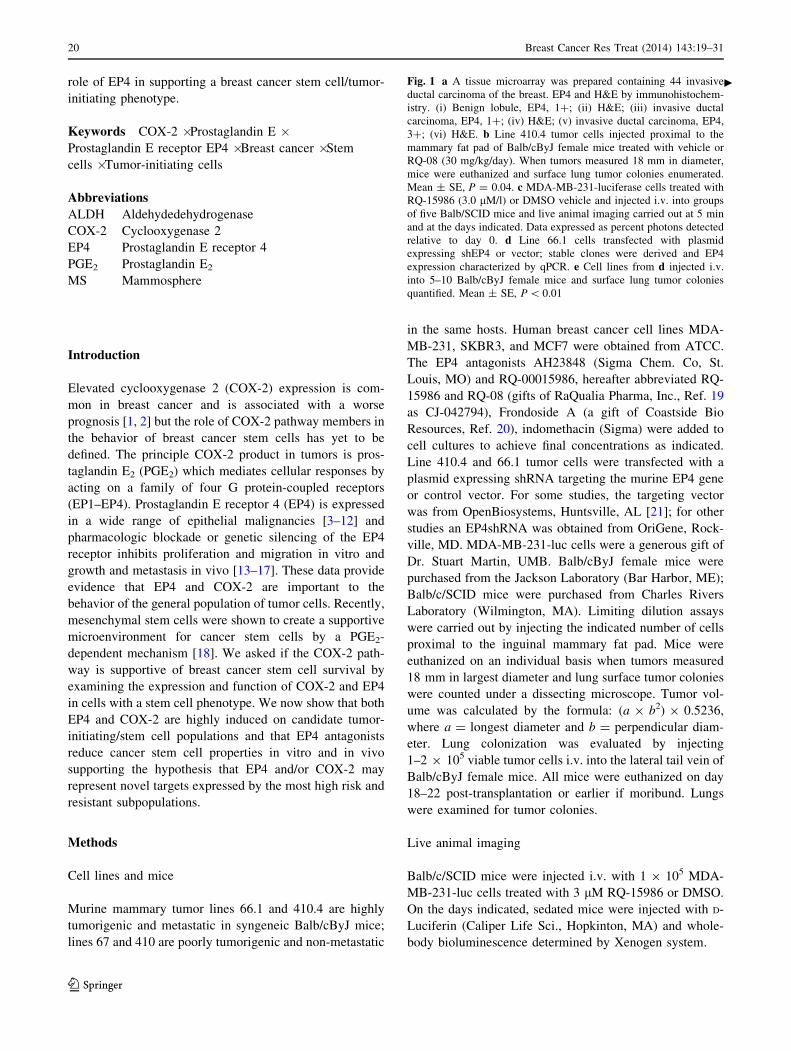
Fig. 1 a A tissue microarray was prepared containing 44 invasive
ductal carcinoma of the breast. EP4 and H&E by immunohistochem-
istry. (i) Benign lobule, EP4, 1?; (ii) H&E; (iii) invasive ductal
carcinoma, EP4, 1?; (iv) H&E; (v) invasive ductal carcinoma, EP4,
3?; (vi) H&E. b Line 410.4 tumor cells injected proximal to the
mammary fat pad of Balb/cByJ female mice treated with vehicle or
RQ-08 (30 mg/kg/day). When tumors measured 18 mm in diameter,
mice were euthanized and surface lung tumor colonies enumerated.
Mean ± SE, P = 0.04. c MDA-MB-231-luciferase cells treated with
RQ-15986 (3.0 lM/l) or DMSO vehicle and injected i.v. into groups
of five Balb/SCID mice and live animal imaging carried out at 5 min
and at the days indicated. Data expressed as percent photons detected
relative to day 0. d Line 66.1 cells transfected with plasmid
expressing shEP4 or vector; stable clones were derived and EP4
expression characterized by qPCR. e Cell lines from d injected i.v.
into 5–10 Balb/cByJ female mice and surface lung tumor colonies
quantified. Mean ± SE, P \ 0.01
c
20 Breast Cancer Res Treat (2014) 143:19–31
123

I ii iii iv v vi
0
15
30
45
60
Ctrl RQ-8
p=0.04
0
20
40
60
80
100
120
ini D1 D2 D3 D8 D15 D22 D29 D36 D45 D57
% o
f p
ho
ton
s at
Day
0
DMSORQ
Cell Line
Vec Clone1 Clone2 Clone3 Clone4
Lu
ng
met
asta
ses
(mea
n±s
.e.)
0
5
10
15
20
25
0.00
0.20
0.40
0.60
0.80
1.00
1.20
Vector clone 1 clone 2 clone 3 clone 4
A
B
C
D E
Breast Cancer Res Treat (2014) 143:19–31 21
123

Immunohistochemistry
A tissue microarray was prepared and described previously
[22]. Immunohistochemistry was carried out after antigen-
retrieval using rabbit anti-human polyclonal EP4 antibody
(Lifespan Biosciences, Seattle, WA) followed by second-
ary antibody (EnVision?dual link System, Dako).
qPCR
Quantitative PCR was conducted using SYBR green (Bio-
Rad, Hercules, CA) and 400–700 ng cDNA per reaction.
Relative expression levels for COX-2 and EP4 were nor-
malized to GAPDH by the DDCt method.
Metastasis PCR array
RNA was extracted from 66.1-vector and 66.1shEP4 cells
and analyzed on a mouse tumor metastasis PCR array
(Qiagen SABiosciences, Valencia, CA) per manufacturer’s
protocol.
Western blotting
Total cellular protein was analyzed for COX-2, EP4, and
beta-actin by immunoblotting; Cox-2 (Cayman Chemical,
Invitrogen, Ann Arbor, MI), EP4 (Cayman), beta-actin
(Sigma AC15), secondary RT for 1 h [KPL anti-mouse
HRP, anti-rabbit HRP (Bio-Rad)] and visualized with ECL
SuperSignal West Pico (Pierce Chem. Co., Rockford, IL).
ELISA
PGE2 levels were determined by PGE2 enzyme immuno-
assay (Cayman Chemical) and expressed as pg PGE2/lg
total protein.
Mammosphere
Cells were grown in MammoCult medium (Stem Cell
Technologies Inc., Vancouver, CA) supplemented with
hydrocortisone, heparin, amphotericin B, and gentamicin
(Sigma) and plated in ultra-low attachment plates (Corning
Inc., Corning, NY). Three-dimensional spheroidal struc-
tures (mammospheres) resulting from the first plating in
low-attachment conditions were designated MS-1; sub-
sequent passage into secondary mammosphere cultures at
days 7–12 were designated as MS-2 cultures.
Flow cytometry
Cells blocked with 1 % FBS and stained with antibodies
recognizing CD44 (FITC-conjugated mouse antihuman
CD44), CD24 (PE-conjugated mouse antihuman CD24) or
appropriate isotype control (all from BD Biosciences, San
Jose, CA). Aldehyde dehydrogenase was detected by Al-
defluor kit (Stem Cell Technologies) used according to
manufacturer’s protocol and analyzed by FACScan flow
cytometry.
Biostatistics
In vivo data was analyzed by non-parametric Kruskal–
Wallis test and pair-wise comparisons were carried out by
exact two-sided Wilcoxon test. Tumor incidence data
analyzed by generalized linear models approach. Data from
in vitro studies analyzed by Student’s t test.
Results
EP4 is widely expressed in primary human breast
cancer and targeting EP4 inhibits metastasis
We examined the expression of EP4 in 44 invasive ductal
carcinomas of the breast by immunohistochemistry. EP4
expression was very low or absent in normal ducts (0, 1?,
Fig. 1a), malignant epithelium was positive for cytoplas-
mic EP4 expression. On a scale of 0–3? staining intensity,
21/44 (48 %) specimens had 1? EP4 expression, 13/44
(29 %) were 2? and 10/44 (23 %) were graded as 3? in
EP4 staining intensity. Nuclear staining was not observed.
EP4 gene silencing or receptor inhibition with small
molecule inhibitors block metastasis in a syngeneic murine
breast cancer model [13, 20, 21, 23]. In this study, we
confirmed, using a second tumor cell line and a different
EP4 antagonist (RQ-08), that metastasis is inhibited by EP4
blockade. Line 410.4 tumor cells were implanted into syn-
geneic Balb/cByJ female mice and oral administration of
RQ-08 (30 mg/kg 9 28 days) was initiated on day ?7.
When tumors achieved an average diameter of 18 mm,
mice were euthanized and metastatic disease was assessed.
The growth of primary tumors was modestly inhibited by
RQ-08 (not shown) but spontaneous metastasis to the lungs
was reduced by 49 % (Fig. 1b, P = 0.04). Metastatic suc-
cess of human MDA-MB-231-luc cells was also reduced by
an EP4 antagonist (Fig. 1c). We studied cell-autonomous
effects of EP4 antagonism on the tumor cell alone, by pre-
treating tumor cells with RQ-15986 (3.0 lM/l) prior to i.v.
injection into Balb/SCID mice. At day 1 after i.v. injection
of tumor cells, less luciferase signal was detected when EP4
was antagonized. As the surviving tumor cell populations
expanded with time, the difference between the two treat-
ment groups became more pronounced. We also created
multiple clones of 66.1 expressing EP4shRNA (Fig. 1d).
Metastatic potential was reduced by 43, 53, 53, and 84 %,
22 Breast Cancer Res Treat (2014) 143:19–31
123

respectively, in comparison to mice injected with vector
control cells (Fig. 1e). Thus, EP4 is widely expressed in
breast cancer and either genetic or pharmacologic com-
promise of EP4 activity reduces metastatic potential.
Metastasis and stem cell-associated genes are
downregulated in shEP4 cells
We employed a qPCR array of known metastasis-associ-
ated genes to compare gene expression patterns of 66.1-
vector versus 66.1shEP4 cells. Table 1 shows genes that
were downregulated by at least 1.5-fold in 66.1shEP4 cells
compared to 66.1-vector cells that included Csf1, c-met,
CXCL12, and CD44. Few genes were upregulated in the
context of EP4 silencing, but included the metastasis-sup-
pressor Nme4 (data not shown). The downregulation of
Csf1, Timp2, and CD44 in 66.1shEP4 cells was confirmed
by qPCR (Fig. 2a). While each of these gene expression
changes may be important to the mechanism of metastasis
inhibition, we focused our further studies on the reduction
in CD44, a phenotypic marker of candidate breast cancer
stem/tumor-initiating cells [24, 25].
Tumor-initiating cells are identified in a syngeneic
model of metastatic breast cancer
A true cancer stem cell should have heightened tumor-
initiating capacity compared to the general population but
a tumor-initiating cell has not been demonstrated in the
murine cell lines under study. We cultured 410.4 cells in
low-attachment conditions to demonstrate that mammo-
spheres will form. These mammospheres can be dissoci-
ated and replated to form secondary and tertiary
mammospheres (dns). Limiting dilution assays compared
the tumorigenic properties of the general population of
410.4 cells versus mammosphere-forming (MS-1) 410.4
cells. Three of 10 mice injected with 100 of 410.4 bulk
cells developed palpable tumors (dashed lines), whereas
8/9 mice injected with MS-1 cells (solid lines) developed
progressively growing tumors (Fig. 3c). In an expanded
limiting dilution assay using additional cell injection
doses (Table 2), five of eight mice (62.5 %) injected with
100 MS-1 developed tumors and an average of 17
metastases per mouse; only 2/8 mice (25 %) injected with
100 cells of the bulk population developed tumors and an
average of 0.5 metastases per animal, indicating height-
ened tumorigenic capacity of mammosphere-forming
cells. At all cell doses, MS-1 cells were more tumorigenic
than the same number of bulk population cells; the stem
cell frequency was calculated as 1/145 MS-1 cells versus
1/526 for bulk cells (P \ 0.00425). The average number
of metastases was also higher in mice injected with MS-1
cells compared to an equal number of bulk population
cells.
EP4 and COX-2 are upregulated in mammospheres
of aggressive breast cancer cells
Several laboratories have described the stem cell properties
of human breast cancer cell lines [26–28] and we con-
firmed that MDA-MB-231 cells are highly enriched for a
stem cell phenotype whereas this population is rare in
MCF7 cells, which have a luminal gene signature. Con-
sistent with the literature, more than 95 % of MDA-MB-
231 cells are CD44hi/CD24low (Fig. 3a). In comparison,
Table 1 Effect of EP4 reduced expression on metastasis-related
genes
Symbol Fold
down-
regulation
Full name
Timp2 -5.22 Tissue inhibitor of metalloproteinase 2
Csf1 -4.29 Colony stimulating factor 1 (macrophage)
Tcf20 -4.06 Transcription factor 20
Ewsr1 -4.01 Ewing sarcoma breakpoint region 1
Plaur -3.89 Plasminogen activator, urokinase receptor
Met -3.53 Met proto-oncogene
Cxcl12 -3.47 Chemokine (C-X-C motif) ligand 12
Chd4 -3.37 Chromodomain helicase DNA binding protein
4
Rorb -2.92 RAR-related orphan receptor beta
Ctbp1 -2.88 C-terminal binding protein 1
CD82 -2.69 CD82 antigen
CD44 -2.56 CD44 antigen
Hras1 -2.54 Harvey rat sarcoma virus oncogene 1
Col4a2 -2.34 Collagen, type IV, alpha 2
Itga7 -2.16 Integrin alpha 7
Rb1 -2.15 Retinoblastoma 1
Mtss1 -2.12 Metastasis-suppressor 1
Kras -2.10 V-Ki-ras2 Kirsten rat sarcoma viral oncogene
homolog
Ctnna1 -2.10 Catenin (cadherin-associated protein), alpha 1
Brms1 -2.09 Breast cancer metastasis-suppressor 1
Smad4 -2.04 MAD homolog 4 (Drosophila)
Etv4 -1.99 Ets variant gene 4(E1A enhancer binding
protein, E1AF)
Fat1 -1.89 FAT tumor suppressor homolog 1 (Drosophila)
Rpsa -1.86 Ribosomal protein SA
Pten -1.82 Phosphatase and tensin homolog
Mta1 -1.74 Metastasis-associated 1
Htatip2 -1.63 HIV-1 tat interactive protein 2, homolog
(human)
Trp53 -1.57 Transformation-related protein 53
RNA isolated from 66.1vector or 66.1shEP4 cells and analyzed on a
mouse Metastasis array. Genes downregulated [1.5-fold in
66.1shEP4 cells relative to 66.1-vector cells are listed
Breast Cancer Res Treat (2014) 143:19–31 23
123

\1.0 % of MCF7 cells are CD44hi (Fig. 3b), however,
these latter cells are highly positive for CD24 expression.
The fraction of aldehydedehydrogenase (ALDH?) positive
cells was highly variable from experiment to experiment;
in one experiment, 5.4 % of MDA-MB-231 cells were
ALDH? (Fig. 3c); 0.23 % of MCF7 cells were ALDH?.
Twenty-six percent of 410.4 cells are ALDH?; 57.4 % of
66.1 cells are positive for this marker (data not shown).
We compared EP4 and COX-2 expression levels in
mammosphere-forming and bulk populations derived from
human or murine breast cancer cell lines. MDA-MB-231
were employed as a model of breast cancer stem cells,
MCF7 as a model of non-stem cells and SKBR3 represent
the HER-2 phenotype. Metastatic murine cell lines 410.4
and 66.1 and non-metastatic cell lines 410 and 67 were
employed for comparison. In both MDA-MB-231 and
SKBR3 cells, EP4 mRNA levels were markedly increased
in MS-1 versus bulk population cells (Fig. 4a) but EP4
mRNA was not elevated in MS-1-forming MCF7 cells.
These patterns have been observed in three independent
experiments. Like MDA-MB-231 and SKBR3 cells,
murine 66.1 and 410.4 MS-1 cells expressed increased EP4
mRNA versus the bulk population (Fig. 4b), but EP4 was
not increased in the comparatively benign 410 and 67 cells
in either MS-1 or MS-2 cultures (Fig. 4c). Like EP4, COX-
2 was increased in MS-1 versus bulk populations of MDA-
MB-231 and SKBR3 cells (Fig. 4d) but slightly elevated in
MCF7 MS-1 cultures. In murine cells, COX-2 was induced
in MS-1 cultures of 66.1 and 410.4 (Fig. 4e), but not in 410
or 67 MS-1 cells (Fig. 4f). Consistent with the mRNA data,
increased COX-2 protein was detected in MS-1 cells of
MDA-MB-231 and SKBR3 grown in either DME or
mammocult media; only a slight increase in COX-2 protein
was detected in MCF-7 cells (Fig. 4g). Increased COX-2 in
MS-1 cells resulted in more PGE2 detected in conditioned
media of MDA-MB-231 and SKBR3, but not MCF7-
derived MS-1 cells (Fig. 4h). Thus, in both human and
mouse, EP4 and COX-2 are elevated in mammospheres
produced by highly tumorigenic, basal-type cell lines.
Luminal-type (MCF7) or less tumorigenic, non-metastatic
(410, 67) cells do not elevate EP4 or COX-2 expression in
MS-1 tumorspheres.
1
0.17
1
0.35
10.86
1
1.42
10.77
1
0.39
1
2.79
1
3.21
0
0.5
1
1.5
2
2.5
3
3.5
Ve
EP
4sh
rna
Ve
EP
4sh
rna
Ve
EP
4sh
rna
Ve
EP
4sh
rna
Ve
EP
4sh
rna
Ve
EP
4sh
rna
Ve
EP
4sh
rna
Ve
EP
4sh
rna
Cd44 Csf1 Ewsrl Cd82 Plaur Timp2 Ctsl Vegfa
0
500
1000
1500
2000
2500
3000
3500
2 3 4 5 6 7 8 9 10 11 12 13 14 15 16
Tu
mo
r vo
lum
e (m
m3 )
Weeks
A
B
Fig. 2 a mRNA isolated from
66.1-vector and 66.1shEP4 cells
and analyzed for expression of
metastasis-related genes by
metastasis pcr array. b 410.4
cells grown in standard culture
or as mammospheres and on day
10 cells were harvested and 100
MS-1 or standard bulk culture
cells injected into the mammary
fat pad of Balb/cByJ female
mice. Tumor growth monitored
by caliper and expressed as
tumor volume. Solid line = 100
MS-1 cells; dashed line = 100
bulk population cells
24 Breast Cancer Res Treat (2014) 143:19–31
123

EP4 antagonists or EP4 gene silencing inhibits
properties of breast cancer stem cells in vitro
We compared the growth properties of mammospheres
derived from 410.4-vector cells to 410.4shEP4 cells
(Fig. 5a). MS-1 mammospheres were disrupted and 5,000
cells from each MS-1 culture was replated and allowed to
form secondary (MS-2) mammospheres. The average
number of MS-2 spheres formed by 410.4-vector cells
(5.6 ± 0.9) was reduced by 57 % in 410.4shEP4 mam-
mospheres (2.4 ± 0.4 spheres). More marked effects of
EP4 gene silencing were detected when the size of mam-
mospheres, as indicated by the number of cells/sphere was
considered. Mammospheres formed by 410.4-vector,
410.4shEP4, 410 or 67 cells were collected from each well
and the total number of sphere-forming cells was deter-
mined (Fig. 5a). Five thousand MS-1 410.4-vector cells
expanded into secondary tumorspheres to produce a total of
280,666 ± 6,992 cells; a 56-fold expansion in cell number
from day 0. In contrast 410.4shEP4 cells expanded by
21-fold. Thus, when mammosphere-induced upregulation
of EP4 was prevented by shEP4, mammosphere-forming
ability (number and size) was compromised. Although line
410 and 67 cells were able to form small secondary
mammospheres, the total average cell number was expan-
ded by 12-fold and 3-fold, respectively. The reduced
sphere-forming ability of 410 and 67 cells is consistent
with the failure to upregulate EP4 or COX-2 in mammo-
spheres of these cells.
We determined the effect of EP4 antagonists or the
COX-1/COX-2 inhibitor indomethacin on sphere-forming
ability of MDA-MB-231 cells. In the presence of indo-
methacin (1.0–50 lM/l), neither the numbers of mammo-
spheres nor the size of spheres was affected by COX
inhibition (Fig. 5b); cell number was 90–115 % of that
observed in vehicle-treated TS-1 cells. In contrast, three
MDA-MB-231, isotype MDA-MB-231, CD44/CD24
MCF7, isotype MCF7, CD44/CD24
MDA-MB-231, ALDH MCF7, ALDH
A
B
C
Fig. 3 a, b MDA-MB-231 and
MCF7 cells grown in standard
culture conditions and analyzed
for CD44 and CD24 expression
by flow cytometry. Isotype
control (left panel), double stain
for CD44 and CD24 (right
panel). c Aldefluor assay of
MDA-MB-231 (left panel) and
MCF7 (right panel) cells
Breast Cancer Res Treat (2014) 143:19–31 25
123

EP4 antagonists, AH23848, Frondoside A and RQ-15986
were all able to inhibit the size of mammospheres (Fig. 5c–e).
In the presence of AH23848, mammospheres were actually
increased in number, but individual spheres were much
smaller as indicated by the dramatic reduction in cells/
sphere. AH23848 at 10, 5, 0.5, or 0.05 lM/l reduced sphere
cellularity by 78, 69, 55, and 2 %, respectively (Fig. 5c). In
contrast, AH23848 did not affect the number of attached
cells at any concentration examined (dns). While the
number of total spheres was not affected by Frondoside A
except at the highest concentration tested (1.0 lM/l), the
sphere size was inhibited by 100, 92, 28, and 1 % in a dose-
dependent manner (Fig. 5d). RQ-15986 (30, 10, 3, and
0.3 lM/l) inhibited sphere-forming cells by 20, 30, 8, or
7 % (Fig. 5e).
Taken together, these data indicate that tumor cells with
heightened stem cell properties are more sensitive than the
general population to EP4 antagonism.
EP4 antagonists inhibit the stem cell phenotype in vitro
To determine if EP4 inhibition would downregulate phe-
notypic markers of breast cancer stem cells, MDA-MB-231
cells were placed in mammosphere culture and on day 8,
RQ-15986 (10 lM/l) was added. On day 10, cultures were
analyzed for ALDH expression. In a typical experiment,
RQ-15986 treatment reduced the proportion of ALDH?
MDA-MB-231 cells from 30.4 % to 16.7 % (Fig. 6a). RQ-
15986, Frondoside A or AH23848 also modestly reduced
the percentage of CD44-positive MS-1 cells. In the pre-
sence of PBS, 64 % of MDA-MB-231 (MS-1) cells were
CD44-positive; Frondoside A (0.25 lM/l) reduced the
fraction of CD44-positive cells to 54.3 %. In the presence
of DMSO, 53.5 % of cells were CD44-positive; RQ-15986
(10 lM/l) or AH23848 (5 lM/l) reduced the CD44-posi-
tive fraction to 45.9 or 46.9 % of cells, respectively. CD44
expression was not changed in attached cells in the pre-
sence of EP4 antagonists (dns). CD24 is poorly expressed
in MS-1 cells and the degree of expression was not chan-
ged by treatment with any EP4 antagonist (dns).
EP4 antagonism inhibits tumor-initiating capacity
in vivo
By limiting dilution assays, we assessed the ability of an
EP4 antagonist to inhibit tumor-initiating cells in vivo.
Balb/cByJ mice were transplanted with 500 or 50 of 410.4
MS-1 cells and, beginning on day ?7, were treated with
vehicle or RQ-08 for 28 days and tumor incidence and size
were assayed. In two independent experiments, EP4
antagonism was able to significantly inhibit the tumor-
forming capacity of small numbers of tumor cells as indi-
cated by tumor incidence (Fig. 6b). When 500 cells were
injected, tumor incidence was reduced from 90 % to 60 %
by RQ-08 (P \ 0.02); RQ-08 inhibited tumor-initiating
capacity from 60 % tumor-positive mice to 20 % tumor-
positive when 50 cells were injected (P \ 0.058). The
calculated stem cell frequency was reduced from 1/126
cells to 1/460 cells by RQ-08 (P \ 0.018). While the
average size of tumors is sometimes variable reflecting the
different latency in individual mice, it is obvious that RQ-
08 has a profound effect on the ability of palpable lesions
to expand (Fig. 6c). In animals that developed palpable
tumors in spite of RQ-08, the percentage of tumor cells that
were CD44hi/CD24low was reduced from 84.2 % ± 1.3
(vehicle) to 74.1 ? 1.3 % (P \ 0.006).
Discussion
EP4 expressed on the malignant cell contributes to meta-
static behavior in a cell-autonomous manner as demon-
strated by reduced metastatic potential of tumor cells in
which EP4 expression has been silenced or antagonized by
small molecule inhibitors. Tumor-derived PGE2 also acts
to suppress the anti-metastatic activities of NK cells by
acting on EP4 expressed on the NK cell [23, 29, 30]. Other
EP4-positive immune effector cells are also inhibited by
PGE2 [31–33]. Thus, the function of multiple EP4 receptor-
positive cells are affected directly by EP4 antagonism.
EP4 promotes multiple cancers [4–17] but little is
known about the role of the COX-2 pathway in cancer stem
cells. Mesenchymal stem cells are stimulated by IL-1 to
produce PGE2 that creates a supportive microenvironment
for cancer stem cells [18]. Stromal fibroblasts support the
induction of MCF7-derived stem cells; in that model, PGE2
Table 2 Tumor-initiating capacity of mammosphere-forming cells
Cell type Tumor
incidence
% Tumor
incidence
Lung
mets
410.4 MS-1, 10 cells 1/8 12.5 7
410.4 Bulk, 10 cells 0/8 0 0
410.4 MS-1, 50 cells 3/8 37.5 0.3
410.4 Bulk, 50 cells 1/8 12.5 0
410.4 MS-1, 100 cells 5/8 62.5 17
410.4 Bulk, 100 cells 2/8 25 0.5
410.4 MS-1, 500 cells 7/8 87.5 3.9
410.4 Bulk, 500 cells 5/9 55.6 3.6
410.4 Bulk, 5 9 105 5/5 100 28.2
MS-1 are mammosphere-derived cells or bulk cells growing under
attached culture conditions and injected at the indicated cell doses
into the mammary fat pad of syngeneic Balb/cByJ female mice.
Tumors palpated twice weekly and degree of metastatic disease
determined at necropsy when tumors measured 18 mm in average
diameter
26 Breast Cancer Res Treat (2014) 143:19–31
123

is not sufficient to directly act on the progenitor stem cells
[34]; rather an indirect mechanism is proposed in which
PGE2 acts indirectly through the cancer-associated fibro-
blast to induce a stem cell phenotype. The relevant EP
receptor was not identified in that study.
Because mammary tumor cells have elevated endoge-
nous COX-2 activity, we investigated the role of PGE2 and
EP4 in an autocrine mechanism that would support breast
cancer stem cells. We now show that a sub-population
enriched in tumor-initiating cells upregulates both EP4 and
Fig. 4 a MDA-MB-231, SKBR3, or MCF7 cells or b 66.1 or 410.4
or c 410 or 67 cells grown as MS-1, MS-2, or bulk cultures. mRNA
harvested and analyzed for human or murine EP4 by qPCR. d–f The
same cells analyzed for COX-2 mRNA expression. g Cell lysates of
MDA-MB-231, SKBR3, or MCF7 cells grown as standard culture or
in mammosphere (MS) assay and maintained in either DME media or
mammocult (MC) media and immunoblotted for COX-2, EP4, or
b-actin. h MDA-MB-231, SKBR3, or MCF7 cells grown as attached
(open bar) or mammosphere (closed bar) in DME; conditioned media
analyzed for PGE2 by ELISA and expressed as Mean ± SE, pg/lg
protein
Breast Cancer Res Treat (2014) 143:19–31 27
123

COX-2. Upregulation of these genes was only observed in
metastatic and/or basal-type human (MDA-MB-231,
SKBR3) and murine (410.4, 66.1) cell lines but not in non-
metastatic or luminal-type cells suggesting that tumor-ini-
tiating cells may contribute to the aggressive phenotype
and may be relatively more sensitive than the non-stem cell
population to direct inhibition by either EP4 antagonists or
COX inhibitors.
The upregulation of EP4 expression in mammospheres
is functionally linked with increased tumor-initiating cell
capacity in vivo of mammosphere-forming cells. MS-1
cells were more tumorigenic than the bulk population with
an increase in stem cell frequency. Importantly, tumori-
genic potential of MS-1 cells was significantly reduced by
an EP4 antagonist in vivo corresponding to a loss of
CD44hi/CD24low cells. In the relatively few RQ-treated
410.4-
vec
410.4s
hEP4
410 67
Sp
her
e-fo
rmin
g C
ells
(m
ean
±s.e
.)
0.0
5.0e+4
1.0e+5
1.5e+5
2.0e+5
2.5e+5
3.0e+5
3.5e+5
2.4
4.3
4.6
5.6
Cel
ls/S
ph
ere
ETOH
Indo
50u
M
Indo
25u
M
Indo
10u
M
Indo
1uM
0
2000
4000
6000
8000
10000
DMSO
AH-10u
M
AH-5uM
AH-0.5u
M
AH-0.05
uM
Cel
ls/S
ph
ere
0
2000
4000
6000
8000
10000
12000
14000
VEH
FA 1uM
FA 0.5u
M
FA 0.1u
M
FA 0.05
uM
Cel
ls/S
ph
ere
0
2000
4000
6000
8000
10000
Cel
ls/S
ph
ere
DMSO
RQ 30uM
RQ 10uM
RQ 3uM
RQ 0.3u
M0
5000
10000
15000
20000
25000
30000
A
C
E
D
B
Fig. 5 a 5 9 103 MS-1 cells of 410.4-vec, 410.4shEP4, 410 or 67
cells (black bars) were re-plated in secondary (MS-2) cultures and on
day 10, sphere number and cellularity were determined for MS-2
cultures. Mean ± SE. b–e MDA-MB-231 cells placed in
mammosphere culture and on day 2, treated with Indomethacin,
AH23848, Frondoside A or RQ-15986 at the indicated concentrations.
On day 10, sphere number and cellularity were determined
28 Breast Cancer Res Treat (2014) 143:19–31
123

mice that developed palpable lesions, the tumors never
expanded. This is in contrast to the modest inhibition of
tumor size when bulk tumor cells are injected and is
consistent with the increased expression of EP4 in MS-1
cells. Consistent with the studies in vivo, in comparison to
410.4-vector cells, 410.4shEP4 cells were less able to
expand to form large mammospheres in culture. Interest-
ingly, growth properties in vitro in conventional attached
conditions are not different for EP4 high versus low cells
supporting the hypothesis that EP4 has important functions
in maintaining the stem/progenitor phenotype. 410.4-vec-
tor cells expanded by 56-fold during 10 days of culture
under low-attachment conditions; the same number of
410.4shEP4 cells were only able to expand by 21-fold.
Poorly tumorigenic 410 and 67 cell lines produced only
small mammospheres. Likewise, the addition of three
different EP4 antagonists to mammosphere-forming cul-
tures were each able to inhibit mammosphere growth.
Interestingly, the COX-1/COX-2 inhibitor indomethacin
did not affect sphere size, indicating that it is not PGE2
synthesis per se, but more likely to be PGE2-mediated cell
signaling that supports sphere formation. The fraction of
CD44hi or ALDH1? cells, both phenotypic markers of
tumor-initiating cells, were reduced by each of three EP4
antagonists.
We did not observe appreciable upregulation of COX-2
in MCF7 tumorspheres consistent with the luminal prop-
erties of this cell line. Singh et al. detected a rare COX-2
highly positive cell in MS-1 cultures of MCF7; in these
cells, the stem cell factor Oct4 was often co-expressed [35].
Those authors propose that the COX-2hi, Oct4? is a breast
cancer stem cell, consistent with the current report.
Veh, ALDH RQ, ALDH
p<0.02
p<0.058
0%
20%
40%
60%
80%
100%
500 Ctl 500 RQ-8 50 Ctl 50 RQ-8
Inci
den
ce
0
100
200
300
400
500
D 7 D 12 D 15 D 19 D 22 D 26 D 29 D 32
Tu
mo
r V
olu
me
(mm
3 )
500 Ctrl
500 RQ-8
50 Ctrl
50 RQ-8
A
B
C
Fig. 6 a MDA-MB-231 cells in
mammosphere culture were
treated with vehicle (left panel)
or RQ-15986 (10 lM/l, right
panel) on day 8 and 2 days
later, Aldefluor positive cells
were determined. b Five
hundred or 50 of 410.4 MS-1
cells were injected into groups
of 10 Balb/cByJ female mice.
On day ?7, RQ-08 or vehicle
administered by gavage (30 mg/
kg/day) and tumor incidence on
day ?46 is plotted. c Tumor
volume for the same mice as in
b that developed palpable
lesions. Mean ± SE
Breast Cancer Res Treat (2014) 143:19–31 29
123

Taken together, these studies support the hypothesis that
EP4-mediated activation can, in an autocrine or paracrine
manner drive stem cell survival. Our studies show, for the
first time, that a clinically relevant EP4 antagonist can
inhibit breast cancer stem cells. Blocking EP4 inhibits
breast cancer metastasis and tumorigenic capacity in vivo
and stem cell properties including mammosphere-forming
ability and phenotypic markers of breast cancer stem cells
in vitro. EP4 antagonism may prove to be safer and more
effective than global COX-2 inhibition. The demonstrated
efficacy of EP4 antagonists in preclinical models of breast
and other malignancies supports the continued evaluation
of EP4 as a new therapeutic target in advanced malignancy.
Acknowledgments We thank the Biostatistics, Genomics, Flow
cytometry, and Tissue Biorepository Shared Services of the Univer-
sity of Maryland Greenebaum Cancer Center. We thank RaQualia
Pharma Inc and Coastside Bio Resources for the gift of EP4 antag-
onists. These studies were supported by the United States Department
of Health and Human Services and the United States Veterans
Administration.
Competing interests The authors declare no competing interests.
Ethical statement All studies comply with current laws of the U.S.A.
Mice were housed, cared for, and used in strict accordance with the U.S.
Department of Agriculture regulations and the NIH Health Guide for
the Care and Use of Laboratory Animals. All animal protocols were
reviewed and approved by the University of Maryland Institutional
Animal Care and Use Committee. The University of Maryland School
of Medicine Animal Facility is fully accredited by the American
Association for the Accreditation of Laboratory Animal Care.
Open Access This article is distributed under the terms of the
Creative Commons Attribution Noncommercial License which per-
mits any noncommercial use, distribution, and reproduction in any
medium, provided the original author(s) and the source are credited.
References
1. Ristimaki A, Sivula A, Lundin J, Lundin M, Salminen T, Hagl-
und C et al (2002) Prognostic significance of elevated cycloox-
ygenase-2 expression in breast cancer. Cancer Res 62:632–635
2. Kerikowske K, Molinaro AM, Gauthier ML, Berman HK,
Waldman F, Bennington J, Sanchez H et al (2010) Biomarker
expression and risk of subsequent tumors after initial ductal
carcinoma in situ diagnosis. J Natl Cancer Inst 102:627–637
3. Reader JC, Holt D, Fulton AM (2011) Prostaglandin E2 EP
receptors as therapeutic targets in breast cancer. Cancer Metas-
tasis Rev 30:449–463
4. Mutoh M, Watanabe K, Kitamura T, Shoji Y, Takahashi M,
Kawamori T et al (2002) Involvement of prostaglandin E receptor
subtype EP4 in colon carcinogenesis. Cancer Res 62:28–32
5. Terada N, Shimizu Y, Kamba T, Inoue T, Maeno A, Kobayashi
T, Nakamura E (2010) Identification of EP4 as a potential target
for the treatment of castration-resistant prostate cancer using a
novel xenograft model. Cancer Res 70:1606–1615
6. Kim J, Lakshmikanthan V, Frilot N, Daaka Y (2010) Prosta-
glandin E2 promotes lung cancer cell migration via EP4-B arr-
estin1-c-Src signalsome. Mol Cancer Res 8:569–577
7. Zhang Y, Ritzenthaler J, Sun X, Roman J, Han S (2009) Pros-
taglandin E2 stimulates human lung carcinoma cell growth
through induction of integrin-linked kinase: the involvement of
EP4 and Sp1. Cancer Res 69:896–904
8. Buchanan FG, Gorden DL, Matta P, Shi Q, Matrisian LM,
DuBois RN (2006) Role of b-arrestin 1 in the metastatic pro-
gression of colorectal cancer. Proc Natl Acad Sci USA 103:
1492–1499
9. Chell SD, Witherden IR, Dobson RR, Moorghen M, Herman AA,
Qualtrough D, Williams AC, Paraskeva C (2006) Increased EP4
receptor expression in colorectal cancer progression promotes
cell growth and anchorage independence. Cancer Res
66:3106–3113
10. Rundhaug JE, Simper MS, Surh I, Fischer SM (2011) The role of
the EP receptors for prostaglandin E2 in skin and skin cancer.
Cancer Metastasis Rev 30:465–480
11. Wu J, Zhang Y, Frilot N, Kim JI, Kim W-J, Daaka Y (2011)
Prostaglandin E2 regulates renal cell carcinoma invasion through
the EP4 receptor-Rap GTPase signal transduction pathway. J Biol
Chem 286:33954–33962
12. Subbaramaiah K, Hudis C, Chang S-H, Hla T, Dannenberg AJ
(2008) EP2 and EP4 receptors regulate aromatase expression in
human adipocytes and breast cancer cells: evidence of a BRCA1
and p300 exchange. J Biol Chem 283:3433–3444
13. Ma X, Kundu N, Rifat S, Walser T, Fulton AM (2006) Prosta-
glandin E receptor EP4 antagonism inhibits breast cancer
metastasis. Cancer Res 66:2923–2927
14. Fulton AM, Ma X, Kundu N (2006) Targeting prostaglandin E EP
receptors to inhibit metastasis. Cancer Res 66:9794–9797
15. Timoshenko A, Guoziong X, Chakrabarti S, Lala P, Chakraborty
C (2003) Role of prostaglandin E2 receptors in migration of
murine and human breast cancer cells. Exp Cell Res 289:265–274
16. Robertson FM, Simeone AM, Mazumdar A, Shah AH, McMurray
JS, Ghosh S et al (2008) Molecular and pharmacological block-
ade of the EP4 receptor selectively inhibits both proliferation and
invasion of human inflammatory breast cancer cells. J Exp Ther
Oncol 7:299–312
17. Yang L, Huang Y, Porta R, Yanagisawa K, Gonzalez A, Segi E
et al (2006) Host and direct antitumor effects and profound
reduction in tumor metastasis with selective EP4 receptor
antagonism. Cancer Res 66:9665–9672
18. Li JH, Reinhardt F, Herschman HR, Weinberg RA (2012) Can-
cer-stimulated mesenchymal stem cells create a carcinoma stem
cell niche via prostaglandin E2 signaling. Cancer Discov 2:
840–855
19. Murase A, Taniguchi Y, Tonai-Kachi H, Nakao K, Takada J
(2007) In vitro pharmacological characterization of CJ-042794, a
novel, potent, and selective prostaglandin EP(4) receptor antag-
onist. Life Sci 82:226–232
20. Ma X, Kundu N, Collin PD, Goloubeva O, Fulton AM (2011)
Frondoside A inhibits breast cancer metastasis and antagonizes
prostaglandin E receptors EP4 and EP2. Breast Cancer Res Treat
132:1001–1008
21. Kundu N, Ma X, Walser T, Goloubeva O, Ostrand-Rosenberg S,
Fulton AM (2009) Antagonism of the prostaglandin E receptor
EP4 inhibits metastasis and enhances NK function. Breast Cancer
Res Treat 117:235–242
22. Ma X, Kundu N, Ioffe O, Goloubeva O, Konger R, Baquet C,
Gimotty P, Fulton AM (2010) Prostaglandin E receptor EP1
suppresses metastasis, is associated with better survival and may
contribute to breast cancer disparities. Mol Cancer Res 8:
1310–1318
30 Breast Cancer Res Treat (2014) 143:19–31
123

23. Ma X, Holt D, Kundu N, Reader J, Goloubeva O, Take Y, Fulton
AM (2013) A prostaglandin E (PGE) receptor EP4 antagonist
protects natural killer cells from PGE2-mediated immunosup-
pression and inhibits breast cancer metastasis. Oncoimmunology
2:e22647
24. Al Hajj M, Wicha MS, Benito-Hernandez A, Morrison SJ, Clarke
MF (2003) Prospective identification of tumorigenic breast can-
cer cells. Proc Natl Acad Sci USA 100:3983–3988
25. Weng D, Penzner JH, Song B, Koido S, Calderwood SK, Gong J
(2012) Metastasis is an early event in mouse mammary carci-
nomas and is associated with cells bearing stem cell markers.
Breast Cancer Res 14:R18
26. Conley SJ, Gheordunescu E, Kakarala P, Newman B, Korkaya H,
Heath AN, Clouthier SG, Wicha MS (2012) Antiangiogenic
agents increase breast cancer stem cells via the generation of
tumor hypoxia. Proc Natl Acad Sci USA 109:2784–2789
27. Kenny PA, Lee GY, Myers CA, Neve RM, Semeiks JR et al
(2007) The morphologies of breast cancer cell lines in three-
dimensional assays correlate with their profiles of gene expres-
sion. Mol Oncol 1:84–96
28. Fillmore CM, Kuperwasser C (2008) Human breast cancer cell
lines contain stem-like cells that self-renew, give rise to pheno-
typically diverse progeny and survival chemotherapy. Breast
Cancer Res 10:R25
29. Holt D, Ma X, Kundu N, Fulton AM (2011) Prostaglandin E2
(PGE2) suppresses natural killer cell function through the PGE2
receptor EP4. Cancer Immunol Immunother 60:1577–1586
30. Holt DH, Ma X, Kundu N, Collin PD, Fulton AM (2012) Mod-
ulation of host natural killer cell functions in breast cancer via
prostaglandin E2 receptors EP2 and EP4. J Immunother
35:179–188
31. Boniface K, Bak-Jensen K, Li Y, Blumenschein W, McGeachy
M, McClanahan T et al (2009) Prostaglandin E2 regulates Th17
cell differentiation and function through cyclic AMP and EP2/
EP4 receptor signaling. J Exp Med 206:535–548
32. Sharma S, Yang S, Zhu L, Reckamp K, Gardner B, Baratelli F
(2005) Tumor cyclooxygenase-2/prostaglandin E2-dependent
promotion of FOXP3 expression and CD4? CD25? T regulatory
cell activities in lung cancer. Cancer Res 65:5211–5225
33. Yao C, Sakata D, Esaki Y, Li Y, Matsuoka T, Kuroiwa K et al
(2009) Prostaglandin E2-EP4 signaling promotes immune
inflammation through Th1 cell differentiation and Th17 cell
expansion. Nat Med 15:633–640
34. Rudnick JA, Arendt LM, Klebba I, Hinds JW, Iyer V, Gupta PB,
Naber SP, Kuperwasser C (2011) Functional heterogeneity of
breast fibroblasts is defined by a prostaglandin secretory pheno-
type that promotes expansion of cancer-stem like cells. PLoS One
6:e24605
35. Singh B, Cook DR, Vincent L, Hall CS, Lucci A (2011) Role of
COX-2 in tumorospheres derived from a breast cancer cell line.
J Surg Res 168:e39–e49
Breast Cancer Res Treat (2014) 143:19–31 31
123