Proprotein interaction with the GPI transamidase
13
Journal of Cellular Biochemistry 88:1025–1037 (2003) Proprotein Interaction With the GPI Transamidase Rui Chen, 1 Vernon Anderson, 2 Yukio Hiroi, 3 and M. Edward Medof 1 * 1 Institute of Pathology, Case Western Reserve University, Cleveland, Ohio 44106 2 Department of Biochemistry, Case Western Reserve University, Cleveland, Ohio 44106 3 Department of Cardiovascular Medicine, University of Tokyo Graduate School of Medicine, Tokyo 133-8655, Japan Abstract For characterizing how the glycosylphosphatidylinositol (GPI) transamidase complex functions, we exploited a two-step miniPLAP (placental alkaline phosphatase) in vitro translation system. With this system, rough microsomal membranes (RM) containing either [ 35 S]-labeled Gaa1p or epitope-tagged Gpi8p, alternative components of the enzymatic complex, were first prepared. In a second translation, unmodified or mutant miniPLAP mRNA was used such that [ 35 S]-labeled native or variant miniPLAP nascent protein was introduced. Following this, the RM were solubilized and anti-PLAP or anti-epitope immunoprecipitates were analyzed. With transamidase competent HeLa cell RM, anti-PLAP or anti-epitope antibody coprecipitated both Gaa1p and Gpi8p consistent with the assembly of the proprotein into a Gaa1p:Gpi8p-containing complex. When RM from K562 mutant K cells which lack Gpi8p were used, anti-PLAP antibody coprecipitated Gaa1p. The proprotein coprecipitation of Gaa1p increased with a nonpermissive GPI anchor addition (o) site. In contrast, if a miniPLAP mutant devoid of its C-terminal signal was used, no coprecipitation occurred. During the transamidation reaction, a transient high Mr band forms. To definitively characterize this product, RM from K cells transfected with FLAG-tagged GPI8 were employed. Western blots of anti-FLAG bead isolates of solubilized RM from the cells showed that the high Mr band corresponded to Gpi8p covalently bound to miniPLAP. Loss of the band following hydrazinolysis demonstrated that the two components were associated in a thioester linkage. The data indicate that recognition of the proprotein involves Gaa1p, that the interaction with the complex does not depend on a permissive o site, and that Gpi8p forms a thioester intermediate with the proprotein. The method could be useful for rapid analysis of nascent protein interactions with transamidase components, and possibly for helping to prepare a functional in vitro transamidase system. J. Cell. Biochem. 88: 1025 – 1037, 2003. ß 2003 Wiley-Liss, Inc. Key words: GPI anchor; transamidase; GPI8; GAA1; miniplap; thioester Posttranslational addition of glycosylphos- phatidylinositol (GPI) moieties is a ubiquitous mechanism for anchoring cell surface proteins to the plasma membrane [reviewed in Tiede et al., 1999]. It is utilized by all eukaryotic cells for tethering molecules with wide-ranging functions. GPI moieties are built upon phos- phatidylinositol in glycosidic linkage to a com- mon core glycan consisting of glucosamine, three mannose residues and a terminal phos- phoethanolamine (p-EthN). They are pre- assembled in the endoplasmic reticulum by stepwise addition of the three core glycan con- stituents to the inositol-phospholipid. The final step in GPI anchor processing is the substitution of preassembled GPI donor moieties for hydrophobic C-terminal signal sequences in nascent proproteins [reviewed in Kodukula et al., 1995; Sevlever et al., 2000]. The substitution occurs at a residue termed the o site which can be one of six small amino acids located 15–30 residues upstream of the C-ter- minus [Moran et al., 1991; Gerber et al., 1992]. Previous studies have shown that the transfer process consists of a transamidation reaction [Maxwell et al., 1995a] in which the small nucleophiles hydrazine (HDZ) or hydroxyla- mine (HDX) can substitute for the free amino group of the terminal p-EthN in GPI donors. Two components of the transamidation ma- chinery that were first characterized are Gaa1p and Gpi8p. In both cases, this initially was ß 2003 Wiley-Liss, Inc. Grant sponsor: NIH; Grant number: R01 DK56309. *Correspondence to: M. Edward Medof, Institute of Pathology, Case Western Reserve University, 2085 Adel- bert Road, Cleveland, Ohio 44106. E-mail: [email protected] Received 22 January 2002; Accepted 29 October 2002 DOI 10.1002/jcb.10439
Transcript of Proprotein interaction with the GPI transamidase

Journal of Cellular Biochemistry 88:1025–1037 (2003)
Proprotein Interaction With the GPI Transamidase
Rui Chen,1 Vernon Anderson,2 Yukio Hiroi,3 and M. Edward Medof1*1Institute of Pathology, Case Western Reserve University, Cleveland, Ohio 441062Department of Biochemistry, Case Western Reserve University, Cleveland, Ohio 441063Department of Cardiovascular Medicine, University of Tokyo Graduate School of Medicine,Tokyo 133-8655, Japan
Abstract For characterizing how the glycosylphosphatidylinositol (GPI) transamidase complex functions, weexploited a two-step miniPLAP (placental alkaline phosphatase) in vitro translation system. With this system, roughmicrosomal membranes (RM) containing either [35S]-labeled Gaa1p or epitope-taggedGpi8p, alternative components ofthe enzymatic complex, were first prepared. In a second translation, unmodified or mutant miniPLAP mRNA was usedsuch that [35S]-labeled native or variant miniPLAP nascent protein was introduced. Following this, the RM weresolubilized and anti-PLAP or anti-epitope immunoprecipitates were analyzed. With transamidase competent HeLa cellRM, anti-PLAP or anti-epitope antibody coprecipitated both Gaa1p and Gpi8p consistent with the assembly of theproprotein into a Gaa1p:Gpi8p-containing complex. When RM from K562 mutant K cells which lack Gpi8p were used,anti-PLAP antibody coprecipitated Gaa1p. The proprotein coprecipitation of Gaa1p increased with a nonpermissive GPIanchor addition (o) site. In contrast, if a miniPLAP mutant devoid of its C-terminal signal was used, no coprecipitationoccurred. During the transamidation reaction, a transient high Mr band forms. To definitively characterize this product,RM from K cells transfected with FLAG-tagged GPI8 were employed. Western blots of anti-FLAG bead isolates ofsolubilizedRM from the cells showed that the highMr band corresponded toGpi8p covalently bound tominiPLAP. Loss ofthe band following hydrazinolysis demonstrated that the two components were associated in a thioester linkage. The dataindicate that recognition of the proprotein involves Gaa1p, that the interaction with the complex does not depend on apermissiveo site, and that Gpi8p forms a thioester intermediate with the proprotein. Themethod could be useful for rapidanalysis of nascent protein interactions with transamidase components, and possibly for helping to prepare a functionalin vitro transamidase system. J. Cell. Biochem. 88: 1025–1037, 2003. � 2003 Wiley-Liss, Inc.
Key words: GPI anchor; transamidase; GPI8; GAA1; miniplap; thioester
Posttranslational addition of glycosylphos-phatidylinositol (GPI) moieties is a ubiquitousmechanism for anchoring cell surface proteinsto the plasma membrane [reviewed in Tiedeet al., 1999]. It is utilized by all eukaryoticcells for tethering molecules with wide-rangingfunctions. GPI moieties are built upon phos-phatidylinositol in glycosidic linkage to a com-mon core glycan consisting of glucosamine,three mannose residues and a terminal phos-phoethanolamine (p-EthN). They are pre-
assembled in the endoplasmic reticulum bystepwise addition of the three core glycan con-stituents to the inositol-phospholipid.
The final step in GPI anchor processing isthe substitution of preassembled GPI donormoieties for hydrophobic C-terminal signalsequences in nascent proproteins [reviewed inKodukula et al., 1995; Sevlever et al., 2000]. Thesubstitution occurs at a residue termed the osite which can be one of six small amino acidslocated 15–30 residues upstream of the C-ter-minus [Moran et al., 1991; Gerber et al., 1992].Previous studies have shown that the transferprocess consists of a transamidation reaction[Maxwell et al., 1995a] in which the smallnucleophiles hydrazine (HDZ) or hydroxyla-mine (HDX) can substitute for the free aminogroup of the terminal p-EthN in GPI donors.
Two components of the transamidation ma-chinery that were first characterized are Gaa1pand Gpi8p. In both cases, this initially was
� 2003 Wiley-Liss, Inc.
Grant sponsor: NIH; Grant number: R01 DK56309.
*Correspondence to: M. Edward Medof, Institute ofPathology, Case Western Reserve University, 2085 Adel-bert Road, Cleveland, Ohio 44106.E-mail: [email protected]
Received 22 January 2002; Accepted 29 October 2002
DOI 10.1002/jcb.10439

accomplished by complementation of yeastmutant lines termed gaa-1 [Hamburger et al.,1995] and gpi-8 [Benghezal et al., 1995] thataccumulated fully assembled GPI donors. Themammalian homologs of the affected genes, i.e.,GAA1 and GPI8, subsequently were isolated by(1) 50 extension of a sequence homologous to thelatter (GPI8) and use of the sequence to rescue ahuman K562 cell line designated mutant K[Chen et al., 1996; Yu et al., 1997] with a similarphenotype, and by (2) cloning a gene withsequence homology to the former (GAA1) andanalyzing the effect of the anti-sense sequencein3T3 cells [Inoue et al., 1999;Hiroi et al., 2000].Human (h) Gaa1p is a 621 amino acid longprotein that spans the ER membrane six timesin its C-terminal region and has a large 328amino acid—long lumenal domain with two N-linked glycans and a short cytoplasmic orientedN-terminus. [Hamburger et al., 1995; Hiroiet al., 2000]. hGpi8p is a 395 amino acid longtype 1 ER protein with a large lumenal domainand a short C-terminal cytoplasmic sequence[Benghezal et al., 1995]. Gpi8p has significantsequence homology with a family of cysteineproteinases including their active site residues,and thus has been proposed to contain theenzymatic site for transamidation.
Although information on the structural orga-nization of the transamidase is beginning toemerge (see Discussion), how it interacts withits proprotein substrates is as yet not under-stood. Moreover, no direct experimental dataare available on how GPI moieties are sub-stituted for C-terminal signal sequences of theproproteins.
The purpose of the present study was toutilize an in vitro translation system to demon-strate directly whether Gaa1p and Gpi8p of thetransamidase machinery are involved in inter-actionwith nascent proproteins, investigate theeffects of altering the o site amino acid or theensuing C-terminal signal sequence, and for-mally establish whether Gpi8p contains theenzymatic activity and, if so, how it functions.
MATERIALS AND METHODS
Reagents and Cells
The coupled transcription and translation(TNT) kit was obtained from Promega, Inc.(Madison, WI). Anti-FLAG mAB M2, biotiny-lated M2 mAB, and anti-FLAG M2-coupledagarose beads were purchased from Sigma
Chemicals (St. Louis, MO). Horseradish perox-idase (HRP) coupled to streptavidin was boughtfrom Gibco Life Technologies (Grand Island,NY). Anti-PLAP antibodies, rabbit reticulocytelysate, [35S] M, T7 polymerase, and HDZ wereobtained as previously described [Chen et al.,1996].
HeLa cells were obtained from the AmericanType Culture Collection (ATCC). The K562 Kmutant cell line [Mohney et al., 1994; Chenet al., 1996; Yu et al., 1997] was derived as pre-viously described. Each cell line was grown asreported [Mohney et al., 1994; Yu et al., 1997].Ribophorin cDNA [Harnik-Ort et al., 1987; FuandKreibich, 2000] was a gift of Dr. G. Kreibich(New York Univ., NY) and ER3 and ER4[Miyawaki et al., 1997; Miyawaki et al., 1999]cameleon cDNAs (calmodulin linked to greenand blue fluorescent proteins) were gifts of Dr.C. Distelhorst (Case Western Reserve Univ.,Cleveland, OH).
Constructs
hGAA1 [cloned into the EcoR1 site of pcDNA3(Invitrogen, Inc., Carlsbad, CA)] was prepar-ed as described [Hiroi et al., 2000]. ChimerichGPI8�FLAG cDNA was prepared by PCR ofhGPI8 sequence in hGPI8/pcDNA3 [Yu et al.,1997] with 50 and 30 primers containingHindIIIand Xho1 sites. Following this, the digestedproduct was ligated into the HindIII and Xho1sites of pFLAG�CTC [E. coli carboxy terminalFLAG expression kit (Sigma Chemicals, St.Louis, MO)]. The hGPI8�FLAG chimeric cDNAinpFLAG�CTCthenwasamplifiedwith50 and30
primers containing Xho1 sites and the res-tricted product cartridged into the Xho1 site ofpcDNA3. miniPLAP, the miniPLAP o mutantW, and the miniPLAP oþ 1 stop construct inpGEM3Zwereprepared as previously described[Kodukula et al., 1991; Gerber et al., 1992].miniPLAP�GAS1 chimeric cDNA was preparedby PCR amplification of nucleotides 1920-2131(encompassing 161 bp of theGAS1 30 end-codingsequence) of GAS1 cDNA in pGOZ [Nuofferet al., 1991]. For this purpose, 50 and 30 primerscontainingNae1 andSal1 sites were used. Afterdigestion, the amplified product was cartridgedinto theminiPLAPNae1 site and pGEM4ZSal1site of miniPLAP/pGEM4Z.
Transfectants
The hGPI8�FLAG/K cell transfectant wasprepared by electroporation using previously
1026 Chen et al.

described methods [Yu et al., 1997]. hGPI8�FLAG/pcDNA3 (50 mg) linearized by Xho1digestion was electroporated into 107 mutantK cells in 0.8 ml of HEPES-buffered saline.Transfected cells were cultured for 24 h in com-plete RPMI 1640 medium and G418 (0.8 mg/ml)was then added. Following selection, survivingneomycin (Neo) resistant cells were labeledwith fluorescein isothiocyanate (FITC)-coupledmurine IA10 anti-decay accelerating factor(DAF) and phycoerythrin (PE)-coupled murineH19 anti-CD59 antibodies (Pharmingen Inc.,La Jolla, CA). The doubly-labeled cells wereselected by three cycles of sorting using aBeckman Coulter Elite cell sorter (Beckman,Coulter, Inc.).
In Vitro Translations
Rough microsomal membranes (RM) wereprepared as previously described [Kodukulaet al., 1992b]. Briefly, pelleted andwashed cells,resuspended in 10 mM triethanolamine, pH 7.5(TEA)/250 mM sucrose, were disrupted for90 min in a N2 cavitation bomb charged to1,200–1,500 psi. The supernatant of the lowspeed pellet of the resulting cell lysate was re-centrifuged for 90 min at 78,000 g in a Ti70rotor. The RM-containing pellet was resus-pended in 250 mM sucrose, 50 mM TEA, pH7.5 and stored at 708C.The two-step microsomal processing reac-
tions were carried out as follows: In the firststep, coupled translation and transcription wasperformed at 308C for 30 min using the TNT kitwith 0.5mg of eitherhGAA1/pcDNA3 orhGPI8-FLAG/pcDNA3 cDNA in conjunction withT7 polymerase and 2.5 ml of RM from a 50OD280 stock. Following this, theRMwere pellet-ed through sucrose cushions, and the pelletsresuspended to three times their originalvolume in 40 mM HEPES, 350 mM potassiumacetate, 2 mM DTT, 1 mM magnesium acetate,pH 7.4 (buffer B) containing 50 mM EDTA.The suspension was then incubated on ice for15min.After spinning through sucrose a secondtime, the RM were rinsed and suspended inbuffer B back to their original volume. For thesecond step,miniPLAPmRNAwas prepared bypriming HindIII-linearized pGEM4Z with SP6RNA polymerase and translating for 90 min at308Cwith rabbit reticulocyte lysate. ThemRNAthen was incubated with the preloaded andEDTA-treated RM in standard fashion as des-cribed [Kodukula et al., 1992b].
Immunoprecipitation/PAGE Gels
For component association studies, mem-branes were lysed with 1% Deoxy big CHAPdetergent (Calbiochem, La Jolla, CA) in bufferB. Immunoprecipitations were performed withrabbit anti-PLAPpolyclonal antibody (AccurateBiochemicals) or with murine anti-FLAGM2 mAB followed by protein A- or proteinC-Sepharose 4B, respectively (Pharmacia,Uppsala, Sweden). Products were eluted with1% SDS buffer, 2.5%mercaptoethanol, separat-ed on 15%SDS–PAGE gels and the gels analyz-ed by autoradiography or phosphorimaging.
Immunoisolation/Western Blotting
For analyses of the high Mr PLAP product,twenty aliquots of 200 ng of miniPLAP�DAF[Chen et al., 2001] mRNA (see Results) wereindividually incubated with 4 ml (50 OD280) ofRM fromGPI8�FLAG/K cell transfectants (pre-pared in the presence of unlabeled M) and theproducts combined. After lysis under standardconditions, the lysate was incubated overnightat 48C with 400 ml of anti-FLAG M2 agarosebeads in buffer B, and following three washeswith the same buffer, the beads were elutedwith nonreducing SDS–PAGE sample buffer.Replicate sets of the eluate and controls wereseparated on nonreducing 15% gels. The pro-teins were transferred to Immobilon-P mem-branes (Millipore, Bedford, MA), and the blotsblocked with 5% bovine serum albumin (BSA)overnight. The blots were cut in themiddle, andthe left half of the blot incubated with biotiny-lated anti-FLAG M2 mAB followed by strepta-vidin-horseradish peroxidase (HRP). The righthalf was incubated with rabbit anti-PLAPpolyclonal antibody followed by HRP-conju-gated goat anti-rabbit Ig (DAKO, Inc., Carpen-teria, CA). The blots were then developed withenhanced chemiluminescence (ECL) reagent.
Hydrazinolysis
Following theminiPLAP�DAF translations inRM fromGPI8�FLAG/K cell transfectants andimmunoisolation of the products with anti-FLAG M2 agarose beads as described above,the beads were eluted with 50 mM Tris-HCl,pH 7.5 containing 4% SDS. The eluate wasincubated for 4 h at 378C alternatively with500 mM HDZ or 500 mM NaCl in the samebuffer. The products were concentrated withUltrafree 0.5 ml centrifugal filters (Millipore,
Proprotein GPI Processing 1027

Bedford,MA). Proteins in the concentrateswerethen separated on 15% SDS–PAGE gels, andthe resolved proteins transferred to Immobilon-P membranes (Millipore). After blocking asabove, the blots were revealed with anti-PLAPpolyclonal antibody followed by HRP-conju-gated goat anti-rabbit Ig and ECL reagent.
RESULTS
The miniPLAP in vitro translation systemhas served as a valuable tool for studying theterminalGPI transfer step in thebiosynthesis ofGPI-anchored proteins. [Kodukula et al., 1991,1992a; Gerber et al., 1992; Udenfriend andKodukula, 1995; Maxwell et al., 1995a; Chenet al., 1996]. It employs a minigene prepared byreplacement of the central 305 amino acids ofnative placental alkaline phosphatase (PLAP)encompassing its glycosylation sites with ashortM-rich stretch of nine residues to enhance[35S]-M incorporation and eliminate molecularweight changes due to glycosylation [Kodukula
et al., 1991]. In this system, N-terminal proces-sing of 28 kDa preprominiPLAP, the primarytranslation product, generates 27 kDa promini-PLAP. Subsequent C-terminal processing ofthis proprotein yields mature 24.7 kDa GPI-anchored miniPLAP. Low level spontaneoushydrolysis or experimental aminolysis withHDZ or HDX yields an alternative 23 kDaproduct. We used this methodology in conjunc-tion with cDNAs encoding the above-describedtwo components of the GPI transfer machineryand a transamidase-deficientmutant cell line toinvestigate how proproteins that undergo GPI-anchoring interact with the GPI transferase.
In preliminary experiments carried out tovalidate the experimental system, we sequen-tially translated GAA1 or GPI8 cDNA followedby miniPLAP mRNA (see Materials and Meth-ods) and examined the microsomal productsdirectly, i.e., without detergent extraction andsubsequent immunoprecipitation. As seen inFigure 1 (panel A), a larger glycosylated Gaa1pproduct was generated indicative of its normal
Fig. 1. Overall Experimental System. RM preloaded with [35S]-labeledGaa1p or epitope-taggedGpi8pwere prepared using therespective cDNAs and the TNT transcription and translationsystem. Following pelleting and 10 mM EDTA-stripping, thepreloaded RMwere resuspended in fresh buffer with [35S]M, andminiPLAPmRNA was translated. The RM were solubilized with1% Deoxy big CHAP and products were immunoprecipitatedwith anti-PLAP or anti-epitope antibody and analyzed on 15%SDS–PAGE gels. Panel A: Gaa1p and Gpi8p products generatedby themselves or in conjunction with miniPLAP in HeLa cell RMprior to extraction and immunoprecipitation. As seen, Gaa1pwas appropriately glycosylated (compare lane 1 and lane 3),Gpi8p N-terminally processed (compare lane 2 and lane 4), and28 kDa preprominiPLAP (ppmPLAP) and 27 kDa prominiPLAP(mPLAP) (lanes 3, 4) were converted to 24.7 kDa GPI-anchored
miniPLAP (mPLAP). The intensities of Gaa1p andGpi8p bands indifferent experiments varied (see text). In studies with micro-somes in which double translations were done, four volumes ofreaction mixtures were used. Panel B: Gaa1p or control PigApmicrosomal products generated by themselves with Deoxy bigCHAP solubilization but without immunoprecipitation (lanes 1,2) and generated in conjunction with miniPLAP and Deoxy bigCHAP solubilization followed by anti-PLAP immunoprecipita-tion (lanes 3, 4). As seen, the anti-PLAP antibody coprecipitatedGaa1p but not PigAp when PIGA cDNA was used as a control.Panel C: Additional controls of ribophorin (Rpn1p) or ERcameleon er3p and er4p [generated with their cDNAs (RPN1,ER3 or ER4)], in conjunction with miniPLAP mRNA and Deoxybig CHAP solubilizationwithout immunoprecipitation (lanes 1–3) or with immunoprecipitation (lanes 4–6).
1028 Chen et al.

N-glycan processing, whereas a Gpi8p productslightly smaller in size than the Gpi8p transla-tion product was produced, consistent withthe cleavage of its N-terminal signal peptide(47 AA) and lack of glycosylation sites. In bothcases, step-wise conversion of 28 kDa prepro-miniPLAP to 27 kDa prominiPLAP and then to24.7 kDa GPI-anchored miniPLAP occurred inaccordance with miniPLAP’s normal N- andC-terminal processing. Different than usuallyobserved, however, the efficiency of formation of24.7 kDa GPI-anchored miniPLAP was lowerthannormally achieved in single step cotransla-tional assays with untreated HeLa RM (see[Kodukula et al., 1992a] and this study, Fig. 2,panel B) and varied in different experiments(Fig. 1, panel A, lane 3 vs. lane 4). This lesserefficiency is consistent with the two-step proto-col in which the miniPLAP translation consti-tutes the second RM reaction using EDTA-washed RM.Studies next were done with the inclusion of
detergent (Deoxy big CHAP) extraction of theRM, followed by immunoprecipitation of solubi-lized products with anti-PLAP antibodies. Asseen in Figure 1 (panel B), the anti-PLAPantibodies coimmunoprecipitated [35S]-labeledGaa1p but not [35S]-labeled phosphatidylinosi-tol glycan A protein (PigAp) which was identi-cally preloaded in the RM as an ER proteincontrol. As additional controls, three other ER
proteinswereused. As observedwithPigAp, theanti-PLAP antibodies also failed to coimmuno-precipitate preloaded ribophorin, a lumenally-oriented multiple ER membrane-spanning pro-tein, and er3p and er4p, two additional lumenalcameleon ER proteins [Miyawaki et al., 1997,1999] (Fig. 1, panelC), confirming the specificityof the prominiPLAP interaction. Comparableresults were obtained in three independentexperiments. In accordance with multiple pre-vious studies in which specific recognition ofPLAP has been achieved with the same anti-PLAP antibody [Kodukula et al., 1991, 1992a,1993; Amthauer et al., 1992; Vidugiriene andMenon, 1995; Maxwell et al., 1995a,b; Rama-lingam et al., 1996; Chen et al., 1998; Vidugir-iene et al., 1999], studies performed in theabsence of miniPLAP translation yielded nobands, and the anti-PLAP antibody did notrecognize Gaa1p on Western blots in otherexperiments (see Fig. 5, below) verifying thatthe recovery of Gaa1p depended on its interac-tion with prominiPLAP.
Comparative immunoprecipitation experi-ments next were carried out with RM preload-ed with [35S]-Gaa1p or [35S]-Gpi8p. For theseanalyses, we took advantage of the fact that theGPI8 construct we used was prepared so as toyield Gpi8p with a FLAG epitope at its C-terminus. As seen in Figure 2 (panel A), bothcomponents were coprecipitated by anti-PLAP
Fig. 2. Panel A: Comparative studies of the interaction ofminiPLAPwith Gaa1p andGpi8p. HeLa cell RMwere preloadedwith [35S]-Gaa1p or [35S]-Gpi8p, the RM stripped, andminiPLAPmRNA then translated as in the legend of Figure 1. When RMfrom Gpi8p-deficient K cells reconstituted with FLAG-taggedGpi8p and anti-FLAG antibody were used, similar coimmuno-precipitation occurred. Both Gaa1p and Gpi8p coprecipitatedwith miniPLAP upon addition of anti-PLAP antibody. Panel B:
RM from K cells deficient in Gpi8p were verified prior todetermination of which component interacts with the nascentminiPLAP proprotein. The products generated following transla-tion ofminiPLAPmRNA in the presence of (1) HeLa cell RM, or(2) K cell RMwithout andwith addedHDZ are shown. K cell RMdeficient in Gpi8p were unable to C-terminally processprominiPLAP in the absence or presence of HDZ, verifying thatthey lacked the respective transamidase component.
Proprotein GPI Processing 1029

antibody. Similar coimmunoprecipitation ofGpi8p occurred if anti-FLAG antibodywas usedin place of anti-PLAP antibody. In the lattercase, lessminiPLAPwas recovered, presumablydue to the fact that prominiPLAP which wasconstantly being generated was in excess. Con-sequently not all of the protein was associatedwith FLAG-Gpi8p.
To distinguish which of the two componentsinteracts with nascent miniPLAP proprotein,studies with our previously described Gpi8p-deficient K562mutant K line [Chen et al., 1996]lacking the entire length of Gpi8p (see Discus-sion) [Yu et al., 1997] were undertaken. As pre-viously reported [Chen et al., 1996] validationthat the cell in fact is transamidase-defective isshown inFigure 2 (panel B).WithHeLa cell RM,processing of prominiPLAP in the absence andpresence of HDZ generated large amounts of24.7 kDa mature GPI-anchored miniPLAP andsmall amounts of the 23.0 kDa hydrazide, andvise versa, respectively. In contrast, with RMfrom Gpi8p-deficient mutant K cells, neitherproduct was generated, and consequently, onlyprominiPLAP was produced.
Using the RM preparation selectively defi-cient in Gpi8p, sequential translations ofGAA1
and miniPLAP followed by immunoprecipita-tion with anti-PLAP antibody next were repeat-ed. As seen in Figure 3 (panel A), when theGpi8p-deficient K cell RM preloaded with [35S]-Gaa1p were used and miniPLAP introduced,anti-PLAP antibodies coimmunoprecipitat-ed Gaa1p. Since only Gpi8p sequence corre-sponding to its cleaved N-terminal signal (seeDiscussion) is encoded in K mutant cells, thecoimmunoprecipitation is indicative of inter-action of the proprotein with the Gaa1p-con-taining transamidase. To establish how theprominiPLAP substrate relates to the two com-ponents when both are present, we exploitedRM from Gpi8p-deficient K cell mutants recon-stituted with FLAG-tagged Gpi8p. The cellswere preloaded with Gaa1p and secondarilyloaded with prominiPLAP by translating mini-PLAP mRNA in standard fashion (as describedin the Materials and Methods). The RM thenwere solubilized with Deoxy big CHAP deter-gent, and the extract added to agarose beadsconjugated with anti-FLAG mAB. Followingwashing and elution with SDS, the eluatewas analyzed on SDS–PAGE gels. As seen inFigure 3 (panel B), both Gaa1p and promini-PLAP were recovered. The results thus in-
Fig. 3. N-terminally processed prominiPLAP binds to Gaa1p.Gaa1pwas preloaded into K cell RMusing its cDNAand the TNTsystem. After washing and resuspension of the RM (seeMaterialsand Methods), miniPLAP mRNA was then translated, the RMsolubilized, and anti-PLAP immunoprecipitates analyzed. PanelA: When K cell RM deficient in Gpi8p were used, coimmuno-precipitation of Gaa1p was observed. Panel B: The nascentpolypeptide, Gaa1p, and Gpi8p exist in a substrate enzymecomplex. Gaa1p was preloaded into RM of K cells reconstituted
with FLAG-Gpi8p. Deoxy big CHAP extracts of the RM wereincubated with agarose beads conjugated to anti-FLAG mAB orunconjugated agarose control. After elution with SDS, proteinswere analyzed on gels. Bands corresponding in size to those ofGaa1p and prominiPLAP were obtained from the anti-FLAG-conjugated beads. Translation controls (TC) for preprominiPLAPandprocessing controls forN-terminally processedprominiPLAP(K cells) routinely included as markers are shown.
1030 Chen et al.

dicated that prominiPLAP associates withboth Gaa1p and Gpi8p in a substrate enzymecomplex.To define the requirements for nascent pro-
protein binding to the complex, we next em-ployed miniPLAP cDNA mutants and HeLaRM. As shown in Figure 4 (panel A), when anonpermissive o site mutant [Gerber et al.,1992] was utilized, coprecipitation of bothGaa1p and Gpi8p was retained denoting a lackof the requirement of a cleavable o site for thebinding. An increase in coprecipitation wasobserved, presumably reflective of ineffectivesubsequent processing of the proprotein byGpi8p (see below and Discussion). When thesignal sequence of yeast GPI-anchored Gas1psurface protein was substituted for that inminiPLAP (Fig. 4, panel B), coprecipitation stilloccurred. In this case, less coprecipitation wasobserved possibly due to diminished avidity ofthe yeast sequence for the hGaa1p/hGpi8pcomplex. Finally, as seen in Figure 4 (panel C),when the C-terminal signal of miniPLAP wasdeleted by placing a stop codon at the oþ 1amino acid residue, coimmunoprecipitationwasabolished indicative of an essential role of theproprotein’s extension peptide for binding to theGaa1p/Gpi8p complex.In previous work with the miniPLAP system
[Chen and Medof, unpublished], a high molec-ular weight band has been observed in anti-PLAP precipitates. The band has been notedmost prominently (1) with miniPLAP in which
itso site amino acid (D) has been replaced by anS residue more efficient in supporting transfer[Gerber et al., 1992] (Fig. 5, panel A, lane 2) and(2)withRMfromcells that aremost active in theGPI transfer reaction (Fig. 5, panel A, lane 4). Ithas also been prominently noted in otherstudies with engineered miniPLAP chimericconstructs [Chen et al., 2001] in which thesequence encoding PLAP’s C-terminal signalhas been replaced with other C-terminal signalsequences, e.g., that of the decay acceleratingfactor (DAF) (Fig. 5, panel A, compare lanes 4and 5) which function more efficiently inconferring GPI-transfer.
Lastly, we performed experiments with RMfrom Gpi8p-defective K cells transfected withFLAG-tagged Gpi8p to ascertain the nature ofthe high molecular weight band and determineif it corresponds to the formation of a mini-PLAP�Gpi8p reaction intermediate. As seen inFigure 5 (panel B), in accordancewith the aboveresults, translation/transcription of GAA1cDNA with ([35S]M) followed by immunopre-cipitation with anti-FLAG antibodies yielded[35S]Gaa1p consistent with the coexistence ofthe two transamidase components in a preexist-ing complex as previously reported [Ohishiet al., 2000]. As a control, no [35S]PigAp wasprecipitated [despite the production of roughlyequivalent amounts of protein (see Fig. 1, panelB)]. Translation of miniPLAP mRNA next wascarried out followed by immunoisolation of theFLAG-tagged Gpi8p-containing complex with
Fig. 4. Effects of varying the o site amino acid and the ensuingC-terminal signal sequence. HeLa cell RM were preloaded withGaa1p, and miniPLAP mRNA translated in the presence of theEDTA-washed RM as in the legend of Figure 1. Panel A: When amutant miniPLAP mRNA encoding a nonpermissive o site (W)was utilized, an increase in coimmunoprecipitation of Gaa1pand Gpi8p was observed. Panel B: When a mutant miniPLAPmRNA in which the yeastGAS1 C-terminal signal sequence was
substituted for the miniPLAP signal sequence, interaction withGaa1pwas not abolished. Panel C: In contrast, whenminiPLAP’sC-terminal signal was deleted, the binding to Gaa1p was totallyabolished. The differences in Gaa1p coprecipitation betweenpanel A in this set of experiments and in Panel A of Figure 2reflects variability inGAA1 translation efficiency, but equivalentresults were obtained in two additional repeat experiments.
Proprotein GPI Processing 1031

anti-FLAG beads. After washing and elution ofthe beads with SDS, the eluate was analyzed onWestern blots employing biotin-labeled anti-FLAG antibodies or anti-PLAP antibodies asalternative revealing reagents. As shown inFigure 5 (panel C), the anti-FLAG antibodiesdetected a major band comigrating with FLAG-tagged Gpi8p and a less intense band �25 kDa
larger in size. In distinction to this result, theanti-PLAP antibodies selectively reacted withthe larger band, establishing that it correspondsto a covalently linked PLAP�Gpi8p product. Asseen in Figure 5 (panel D), preincubation of theeluate with HDZ but not an equimolar NaClcontrol followed by repeat Western blot ana-lyses showed marked diminution of the high
Fig. 5. In all studies, mRNAwas translated with RM for 90 minin standard cotranslational assays as previously described[Gerber et al., 1992; Chen et al., 1996]. Panel A: High Mr bandsometimes seen in in vitro miniPLAP translations. Lanes 1–3:Translations ofminiPLAP inwhich theo site has been changed toserine in the (1) absence of RM, (2) presence of transamidase-competent K562 RM, and (3) presence of Gpi8p-defective K cellRM. Note the highMr band generatedwith K562 cell RM but notK cell RM. Lanes 4 and 5: Translation of chimericminiPLAP�DAF[Chen et al., 2001] andminiPLAPwith its native o site (D) in thepresence of HeLa cell RM which are efficient in transamidation.Note the highMr bandwithminiPLAP�DAF. Panel B: Gaa1p andGpi8p pre-exist in an enzyme complex. Co-immunoprecipita-tion of Gaa1p by anti-FLAG antibody from RM of K cellsreconstituted with FLAG-tagged Gpi8p. Lanes 1–2: Anti-FLAGimmunoprecipitates of RM following translation of (1) GAA1mRNA, or (2) PIGAmRNA. Lanes 3, 4: Nonimmunoprecipitated,(3) GAA1, (4) PIGA control. Panel C: Western blot of protein in
eluates of anti-FLAG beads following processing of miniPLAP inGPI8-FLAG transfectedmutant K cells. Lane1 and left half of lane2, blots revealed with biotin-labeled anti-FLAG M2 mAB andstreptavidin HRP; right half of lane 2 and lane 3, blot revealedwith rabbit anti-PLAP antibody and HRP-conjugated goat anti-rabbit Ig. TwoGpi8p bands are seen (a and b), the higher band (b)containing miniPLAP. The intensity of band b with anti-PLAPantibody is lower presumably because of the transient nature ofthe transamidation reaction. The overall lesser intensity of thebands as compared to those in panel B presumably reflects themore complex immuno-isolation procedure as well as incom-plete recovery at the anti-FLAG bead binding and/or elutionsteps. Panel D: Western blot analyses as in panel C. Lane 1 andleft half of lane 2, blot revealed with anti-FLAG antibody; righthalf of lane 2, lane 3, and lane 4, blot revealed with anti-PLAPantibody. TreatmentwithHDZ (lane 4) removed band bwhereastreatment with NaCl control (lane 3) had no effect. Densitometryshowed that the decrease with HDZ was >80%.
1032 Chen et al.

Mr band, consistent with the high Mr bandbeing a thioester-linked intermediate. Quanti-tative analyses of lanes 3 and 4 indicated thatthe intensity of the labeled high Mr band wasdecreased 5-fold.
DISCUSSION
In previous studies [Kodukula et al., 1991;Benghezal et al., 1995; Hamburger et al., 1995;Chen et al., 1996; Yu et al., 1997; Inoue et al.,1999; Hiroi et al., 2000], the GPI transamidasewas shown to contain the components, Gaa1pand Gpi8p, and evidence was subsequentlypresented implicating the presence of acysteine-proteinase-like active site in Gpi8p[Meyer et al., 2000; Ohishi et al., 2000]. Thisminimal transamidation complex recently hasbeen augmented by the characterization ofPigSp and PigTp, and their yeast orthologsGaa16p and Gaa17p, as two additional compo-nents by both genetic and molecular character-izations [Ohishi et al., 2001]. No directinformation is available, however, as yet onthe function of Gaa1p or whether it is involvedin proprotein interactionwith the transamidasecomplex (see below). In this study, we showedthat (1) interaction of nascent proproteins withthe transamidase involves Gaa1p, (2) the bind-ing to the complex requires the C-terminalsignal peptide but can occur independently ofpermissible o site residues or the precisesequence of the C-terminal extension, (3) thenascent proprotein forms an intermediate pro-protein enzyme complex containing the sub-strate, Gaa1p and Gpi8p, (4) once bound to thecomplex, the nascent protein forms a transientthioester intermediate with Gpi8p, and (5)the substitution of the GPI moiety for the C-terminal peptide occurs by nucleophilic attackof the terminal p-EthN amino group in the GPIon the thioester-linked intermediate. Studieswith HDZ showed that this intermediate isprone to aminolysis consistentwith the covalentintermediate being released as a GPI-anchoredproduct.For conducting our studies, we utilized a
modified miniPLAP in vitro translation system[reviewed in Kodukula et al., 1995] in whichsequential translations of mRNA encoding oneof the transamidase components and then ofminiPLAPwere carried out. In the first transla-tion step, we utilized a coupled TNT system. Toenhance processing efficiency in the second
translation step, we stripped the RM withEDTA. Previous studies have shown that thistreatment removes bound ribosomes andunpro-cessed mRNA [Walter and Blobel, 1983]. To ourknowledge, an experimentally useful two-stepin vitro translation system for similar purposeshas not previously been described.
Our results extend recent studies [Ohishiet al., 2000, 2001] usingCHOcells cotransfectedwith differentially tagged GAA1, GPI8, PIG-S,and PIG-T cDNAs which showed that all fourproteins are included in the complex and re-quired for the transfer of GPI moieties to pro-proteins. How the components interact with theGPI proproteins was not investigated. Cross-linking experiments have begun to characterizethis interaction. A photocrosslinking study ofVidugiriene et al. [2001] employing lysinedeletion variants of miniPLAP with photoreac-tive lysyl-tRNAs yielded bands consistent withthe interaction of the proproteinwithGpi8p andpotential additional contactswithGaa1paswellas other uncharacterized proteins. A study bySpurway et al. [2001] utilizing conventionalminiPLAP with a bis-maleimide cross-linkershowed interaction of the proprotein withGpi8p. One caveat in this latter study, however,is that the inherent specificity of the cross-linker for cysteine favored initial modificationoccurring by virtue of the nucleophilic activesite cysteine of Gpi8p cross-linking to proximalcysteine residues in miniPLAP. These residuesare limited to the hydrophilic sequence onprominiPLAP’s N-terminal side of the o-site asthere are no cysteine residues in the promini-PLAP C-terminal signal sequence. Thus, howthe C-terminal processing signal of transami-dase substrate proteins interacts with thetransamidase complex, and consequentlywherethe specificity of the transamidation reaction islocalized, remain largely uncharacterized. Ourfinding that a thioester is formed between theo-1 residue of prominiPLAP and the active sitecysteine residue of Gpi8p extends the cross-linking study of Spurway et al. [2001] thatlocated the substrate protein near Gpi8p. Inaccordance with the findings of Vidugirieneet al. [2001], the results suggest that an inter-action of the substrate protein with other com-ponents of the complex exists prior to thetransamidation reaction. Additionally, the co-precipitation ofGaa1pwithprominiPLAP in theabsence of Gpi8p is consistent with findings ofOhishi et al. [2001] that Gaa1p, PigSp, and
Proprotein GPI Processing 1033
PigTp can form a stable complexwithoutGpi8p.It further is in accordance with more recentfindings byVainauskas et al. [2002] withGaa1pmutants showing that deletion of the five mostC-terminal lumenal membrane spanning do-mains does not alter the stability or the ERlocalization of the Gaa1p: Gpi8p: PigSp: PigTpcomplex. It, however, abolishes the GPI transa-midation reaction, leading to the suggestion bythe authors that the deleted region of Gaa1ppossibly reacts with the hydrophobic C-term-inal sequence of the proprotein.
To ascertain if Gaa1p participates in theassociation of proproteins with the transami-dase complex, we utilized RM from the Gpi8p-deficient K cell line. This line was derived bymutagenesis of K562 cells [Mohney et al., 1994;Chen et al., 1996]. Since previous RT/PCRanalyses of GPI8 mRNA in K cells showed adeletion of A105T106 resulting in a stop codon at
position 131 [corresponding to amino acid 35(within the 47 AA-longGpi8pN-terminal signalwhich is cleaved)] [Yu et al., 1997], the cell linedoes not make a significant protein productcorresponding to the targeted sequence. Thefindings with HDZ that the K line is unable toconvert prominiPLAP to the miniPLAP hydra-zide [Chen et al., 1996 and the present study]confirmed that it is completely defective intransamidase activity. Translated Gaa1p wascoimmunoprecipitatedbyanti-PLAPantibodiesfrom RM of the Gpi8p-deficient K cell line. Thesimplest explanation would be that Gaa1p isinvolved in the binding of the proprotein to thetransamidase complex. One qualification, how-ever, is that the requirement for the other twocomponents that participate in the complexcannot be excluded.
Our studies with miniPLAP mutants showedthat interaction of the proprotein with Gaa1p in
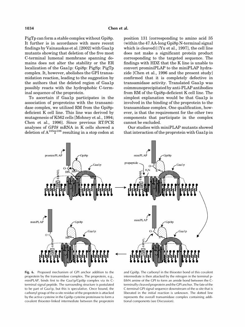
Fig. 6. Proposed mechanism of GPI anchor addition to theproprotein by the transamidase complex. The proprotein, e.g.,miniPLAP, binds first to the Gaa1p/Gpi8p complex via its C-terminal signal peptide. The surrounding structure is postulatedto be part of Gaa1p, but this is speculative. Once bound, thecarbonyl group of the o site residue of the proprotein is attackedby the active cysteine in the Gpi8p cysteine proteinase to form acovalent thioester-linked intermediate between the proprotein
and Gpi8p. The carbonyl in the thioester bond of this covalentintermediate is then attacked by the nitrogen in the terminal p-EthN amine of the GPI to form an amide bond between the C-terminally cleaved proprotein and theGPI anchor. The fate of theC-terminal GPI signal sequence downstream of the o site that isliberated in the initial reaction is unknown. The dotted linerepresents the overall transamidase complex containing addi-tional components (see Discussion).
1034 Chen et al.
the transamidase complex can occur in the abs-ence of permissible proprotein o site residuesand can occur with widely variant sequences,e.g., the yeast Gas1p C-terminal signal sequ-ence, but is absolutely dependent on the pre-sence of the C-terminal peptide. The latter tworesults are in keeping with previous character-izations of the requirements of the properties ofthe C-terminal signal for GPI processing[reviewed in Caras and Weddell, 1989; Caras,1991; Moran et al., 1991; Moran and Caras,1991a,b, 1994; Gerber et al., 1992; Udenfriendand Kodukula, 1995]. The precise manner inwhich amino acid variations in the downstreamsequences affect Gaa1p/Gpi8p binding andwhether the differences correlate with GPIprocessing efficiency [Bon et al., 1997; CrossandBoehme, 2000] arenotknown.Additionally,the site with which the proprotein reacts re-mains to be defined.Several studies [Chen and Medof, unpub-
lished] have documented the presence of a highMr band in anti-PLAP immunoprecipitates.This high Mr band has remained uncharacter-ized. It sometimes is seen transiently. It wasdetected as a putative intermediate in the studyof Spurway et al. [2001]. In this study, weinvestigated the precise nature of the band.Previous analyses of hGpi8p [Ohishi et al.,
2000] and yeast Gpi8p [Meyer et al., 2000] haveshown that mutagenesis of C206 or H164, re-sidues which are conserved in homologousplant cysteine proteinases and in yeast Gpi8p[Benghezal et al., 1995] abolishes its activity,suggesting that they function as catalytic re-sidues in the transamidation reaction. Ourexperiments with K cells transfected withFLAG-tagged GPI8 in the present study show-ing that (1) a high molecular band correspond-ing to miniPLAP covalently bound to Gpi8p canbe isolated and (2) treatment of the band withHDZ abolishes it, directly demonstrate thatGpi8p functions as a modified cysteine protei-nase and formally establish the mechanism ofthe reaction. A proposed schematic diagram ofthe steps which comprise the overall reaction isgiven in Figure 6.The experimental system we developed for
the present investigation opens up the oppor-tunity for further studies with respect to thetransamidase. In addition to mapping impor-tant residues in the C-terminal signal, it couldallow formapping of binding or other functionalsites in the transamidase components. The
double in vitro translation system additionallycould constitute a more general method forstudies of interactions of other ER proteins.
ACKNOWLEDGMENTS
The authors thank Dr. Issei Komuro (Uni-versity of Tokyo Graduate School of Medicine,Tokyo, Japan) for help with GAA1 cDNA, andSara Cechner for manuscript preparation.
REFERENCES
Amthauer R, Kodukula K, Brink L, Udenfriend S. 1992.Phosphatidylinositol-glycan (PI-G)-anchored membraneproteins: Requirement of ATP and GTP for translation-independent COOH-terminal processing. Proc Natl AcadSci USA 89:6124–6128.
Benghezal M, Lipke PN, Conzelmann A. 1995. Identifica-tion of six complementation classes involved in thebiosynthesis of glycosylphosphatidylinositol anchors inSaccharomyces cerevisiae. J Cell Biol 130:1333–1344.
Bon S, Coussen F, Massoulie J. 1997. Sequence require-ments of the GPI cleavage/addition site in rat acetylcho-linesterase: The interaction of GPI anchors withbiological membranes. Splugen, Switzerland: in press.
Caras IW. 1991. Probing the signal for glycophosphatidy-linositol anchor attachment using decay acceleratingfactor as a model system. Cell Biol Int Rep 15:815–826.
Caras IW, Weddell GN. 1989. Signal peptide for proteinsecretion directing glycophospholipid membrane anchorattachment. Science 243:1196–1198.
Chen R, Udenfriend S, Prince GM, Maxwell SE, Ramalin-gam S, Gerber LD, Knez J, Medof ME. 1996. A defect inglycophosphatidylinositol (GPI) transamidase activity inmutant K cells is responsible for their inability to displayGPI surface proteins. Proc Natl Acad Sci USA 93:2280–2284.
Chen R, Walter EI, Parker G, Lapurga JP, Millan JL,Ikehara Y, Udenfriend S, Medof ME. 1998. Mammalianglycophosphatidylinositol anchor transfer to proteinsand posttransfer deacylation. Proc Natl Acad Sci USA95:9512–9517.
Chen R, Knez JJ, Merrick WC, Medof ME. 2001. Compar-ative efficiencies of C-terminal signals of native glyco-phosphatidylinositol (GPI)-anchored proproteins inconferring GPI-anchoring. J Cell Biochem 84:68–83.
Cross GAM, Boehme U. 2000. Mutational analysis of thevariant surface glycoprotein GPI-anchor signal sequencein Trypanosoma brucei. The FASEB summer researchconference: Lipid modifications of proteins. CopperMountain, Colorado: FASEB.
Fu J, Kreibich G. 2000. Retention of subunits of the oligo-saccharyltransferase complex in the endoplasmic reticu-lum. J Biol Chem 275:3984–3990.
Gerber LD, Kodukula K, Udenfriend S. 1992. Phosphati-dylinositol glycan (PI-G) anchored membrane proteins:Amino acid requirements adjacent to the site of cleavage,and PI-G attachment in the COOH-terminal signalpeptide. J Biol Chem 267:12168–12173.
Hamburger D, Egerton M, Riezman H. 1995. Yeast Gaa1pis required for attachment of a completed GPI anchoronto proteins. J Cell Biol 129:629–639.
Proprotein GPI Processing 1035

Harnik-Ort V, Prakash K, Marcantonio E, Colman DR,Rosenfeld MG, Adesnik M, Sabatini DD, Kreibich G.1987. Isolation and characterization of cDNA clones forrat ribophorin I: Complete coding sequence and in vitrosynthesis and insertion of the encoded product into endo-plasmic reticulum membranes. J Cell Biol 104:855–863.
Hiroi Y, Chen R, SawaH, Hosoda T, Kudoh S, Kobayashi Y,Aburatani H, Nagashima K, Nagai R, Yazaki Y, MedofME, Komuro I. 2000. Cloning of murine glycosyl phos-phatidylinositol anchor attachment protein, GPAA1. AmJ Physiol Cell Physiol 279:C205–C212.
Inoue N, Ohishi K, Endo Y, Fujita T, Takeda J, Kinoshita T.1999. Human and mouse GPAA1 (Glycosylphosphatidy-linositol anchor attachment 1) genes: Genomic struc-tures, chromosome loci, and the presence of a minor classintron. Cytogenet Cell Genet 84:199–205.
Kodukula K, Micanovic R, Gerber L, Tamburrini M, BrinkL, Udenfriend S. 1991. Biosynthesis of phosphatidylino-sitol glycan-anchored membrane proteins. Design of asimple protein substrated to characterize the enzymethat cleaves the COOH-terminal signal peptide. J BiolChem 266:4464–4470.
Kodukula K, Amthauer R, Cines D, Yeh E, Brink L,Thomas L, Udenfriend S. 1992a. Biosynthesis of phos-phatidylinositol-glycan (PI-G)-anchored membraneproteins in cell-free systems: PI-G is an obligatory co-substrate for COOH-terminal processing of nascentproteins. Proc Natl Acad Sci USA 89:4982–4985.
Kodukula K, Cines D, Amthauer R, Gerber L, UdenfriendS. 1992b. Biosynthesis of phosphatidylinositol-glycan(PI-G)-anchored membrane proteins in cell-free systems:Cleavage of the nascent protein and addition of the PI-Gmoiety depend on the size of the COOH-terminal signalpeptide. Proc Natl Acad Sci USA 89:1350–1353.
Kodukula K, Gerber LD, Amthauer R, Brink L, UdenfriendS. 1993. Biosynthesis of glycosylphosphatidylinositol(GPI)-anchored membrane proteins in intact cells:Specific amino acid requirements adjacent to the site ofcleavage and GPI attachment. J Cell Biol 120:657–664.
Kodukula K, Maxwell SE, Udenfriend S. 1995. Processingof nascent proteins to glycosylphosphatidylinositol-anchored forms in cell-free systems. Methods Enzymol250:536–547.
Maxwell SE, Ramalingam S, Gerber LD, Brink L, Uden-friend S. 1995a. An active carbonyl formed during glyco-sylphosphatidylinositol addition to a protein is evidenceof catalysis by a transamidase. J Biol Chem 270:19576–19582.
Maxwell SE, Ramalingam S, Gerber LD, Udenfriend S.1995b. Cleavage without anchor addition accompaniesthe processing of a nascent protein to its glycosylpho-sphatidylinositol-anchored form. Proc Natl Acad Sci USA92:1550–1554.
Meyer U, Benghezal M, Imhof I, Conzelmann A. 2000.Active site determination of Gpi8p, a caspase-relatedenzyme required for glycosylphosphatidylinositol anchoraddition to proteins. Biochemistry 39:3461–3471.
Miyawaki A, Llopis J, Heim R, McCaffery JM, Adams JA,Ikura M, Tsien RY. 1997. Fluorescent indicators for Ca2þ
based on green fluorescent proteins and calmodulin.Nature 388:882–887.
Miyawaki A, Griesbeck O, Heim R, Tsien RY. 1999.Dynamic and quantitative Ca2þ measurements using
improved cameleons. Proc Natl Acad Sci USA 96:2135–2140.
Mohney RP, Knez JJ, Ravi L, Sevlever D, Rosenberry TL,Hirose S, Medof ME. 1994. Glycoinositol phospholipidanchor defective K562 mutants with biochemical lesionsdistinct from those in Thy-1-negative murine lymphomamutants. J Biol Chem 269:6536–6542.
Moran P, Caras IW. 1991a. Fusion of sequence elementsfrom non-anchored proteins to generate a fully functionalsignal for glycophosphatidylinositol membrane anchorattachment. J Cell Biol 115:1595–1600.
Moran P, Caras IW. 1991b. A nonfunctional sequenceconverted to a signal for glycophosphatidylinositol mem-brane anchor attachment. J Cell Biol 115:329–336.
Moran P, Caras IW. 1994. Requirements for glycosylpho-sphatidylinositol attachment are similar but not identi-cal in mammalian cells and parasitic protozoa. J Cell Biol125:333–343.
Moran P, Raab H, Kohr WJ, Caras IW. 1991. Glycopho-spholipid membrane anchor attachment. Molecular ana-lysis of the cleavage/attachment site. J Biol Chem 266:1250–1257.
Nuoffer C, Jeno P, Conzelmann A, RiezmanH. 1991. Deter-minants for glycophospholipid anchoring of the Sacchar-omyces cerevisiae GAS1 protein to the plasmamembrane.Mol Cell Biol 11:27–37.
Ohishi K, Inoue N, Maeda Y, Takeda J, Riezman H,Kinoshita T. 2000. Gaa1p and Gpi8p are components of aglycosylphosphatidylinositol (GPI) transamidase thatmediates attachment of GPI to proteins. Mol Biol Cell11:1523–1533.
Ohishi K, Inoue N, Kinoshita T. 2001. PIG-S and PIG-T,essential for GPI anchor attachment to proteins, form acomplex with GAA1 and GPI8. EMBO J 20:4088–4098.
Ramalingam S, Maxwell SE, Medof ME, Chen R, GerberLD, Udenfriend S. 1996. COOH-terminal processing ofnascent polypeptides by the glycosylphosphatidylinositoltransamidase in the presence of hydrazine is governed bythe same parameters as glycosylphosphatidylinositol ad-dition. Proc Natl Acad Sci USA 93:7528–7533.
Sevlever D, Chen R, Medof ME. 2000. Synthesis of the GPIanchor. In: Young NS, Moss J, editors. Paroxysmal noct-urnalhemoglobinuriaand theglycophosphoinositol-linkedproteins. San Diego, CA: Academic Press. pp 199–220.
Spurway TD, Dalley JA, High S, Bulleid NJ. 2001. Earlyevents in glycosylphosphatidylinositol anchor addition.Journal of Biological Chemistry 276:15975–15982.
Tiede A, Bastisch I, Schubert J, Orlean P, Schmidt RE.1999. Biosynthesis of glycosylphosphatidylinositols inmammals and unicellular microbes. Biol Chem 380:503–523.
Udenfriend S, Kodukula K. 1995. Prediction of omega sitein nascent precursor of glycosylphosphatidylinositol pro-tein. Methods Enzymol 250:571–582.
Vidugiriene J, Menon AK. 1995. Soluble constituents of theER lumen are required for GPI anchoring of a modelprotein. EMBO J 14:4686–4694.
Vainauskas S, Maeda Y, Kurniawan H, Kinoshita T,Menon AK. 2002. Structural requirements for the re-cruitment of Gaa1 into a functional glycosylphosphati-dylinositol transamidase complex. J Biol Chem 277(34):30535–30542.
Vidugiriene J, Sharma DK, Smith TK, Baumann NA,Menon AK. 1999. Segregation of the glycosylphosphati-
1036 Chen et al.

dylinositol biosynthetic reactions in a subcompartment ofthe endoplasmic reticulum. J Biol Chem 274:15203–15212.
Vidugiriene J, Vainauskas S, Johnson AE, Menon AK.2001. Endoplasmic reticulum proteins involved in glyco-sylphosphatidylinositol-anchor attachment: photocros-slinking studies in a cell-free system. European Journalof Biochemistry 268:2290–2300.
Walter P, Blobel G. 1983. Preparation of microsomalmembranes for cotranslational protein translocation.Methods Enzymol 96:84–93.
Yu J, Nagarajan S, Knez JJ, Udenfriend S, Chen R,MedofME. 1997. The affected gene underlying the classKsurface protein defects codes for the GPI-transamidase.Proc Natl Acad Sci USA 94:12580–12585.
Proprotein GPI Processing 1037