Proposed Research on Developmental Language …Simon/ Proposed research on developmental language...
28
Proposal, May 2005/ Revised June 2010 Proposed Research on Developmental Language Disorders The auditory system of the brain, from cochlear nerve to temporal lobes – essential for language development My interest is in this small area of the midbrain Question: Could brainstem impairment lead to developmental language disorders? From insults such as: Prenatal exposure to alcohol? Prenatal infection? Asphyxia at birth? Toxic exposure in infancy? Eileen Nicole Simon conradsimon.org 11 Hayes Avenue Lexington, MA 02420 USA 1‐617‐512‐0424 [email protected] Note: To learn about fMRI, I took a course in Cognitive Neuroscience at the Harvard Extension School with Professor Yoohong Jiang In the spring of 2005. I submitted this proposal as my final course project. Professor Jiang is now at the University of Minnesota, https://apps.cla.umn.edu/directory/profiles/jiang166
Transcript of Proposed Research on Developmental Language …Simon/ Proposed research on developmental language...

Proposal, May 2005/ Revised June 2010
Proposed Research on Developmental Language Disorders
The auditory system of the brain, from cochlear nerve to temporal lobes – essential for language development
My interest is in this small area of the midbrain
Question: Could brainstem impairment lead to developmental language disorders? From insults such as: Prenatal exposure to alcohol? Prenatal infection? Asphyxia at birth? Toxic exposure in infancy?
Eileen Nicole Simon conradsimon.org 11 Hayes Avenue Lexington, MA 02420 USA 1‐617‐512‐0424 [email protected]
Note: To learn about fMRI, I took a course in Cognitive Neuroscience at the Harvard Extension School with Professor Yoohong Jiang In the spring of 2005. I submitted this proposal as my final course project. Professor Jiang is now at the University of Minnesota, https://apps.cla.umn.edu/directory/profiles/jiang166

Simon/ Proposed research on developmental language disorders 2
Proposal, May 2005/ Revised June 2010
Motivation: The inferior colliculi appear to be important for speech understanding
Since 1991, with the advent of magnetic resonance imaging (MRI), eleven cases of deafness and auditory agnosia have been reported, associated with selective (and bilateral) damage of the inferior colliculi.
Loss of speech understanding ("word deafness") was the primary problem in nine of these cases [1‐10].
The inferior colliculi are metabolically the most active site in the brain Blood flow and aerobic metabolism are highest in the inferior colliculi [11‐15]
The inferior colliculi are vulnerable to any catastrophic disruption of aerobic metabolism [16‐18, 19‐23].
The inferior colliculi are prominently involved in the brainstem pattern of damage caused by alcohol intoxication, or poisoning by many other toxic substances, which also can often be catastrophic [24‐29, 30‐37].
The inferior colliculi are prominently damaged by a brief period of oxygen deprivation before, during or after birth [38‐39, 40‐49].
Perinatal compromise can cause damage in the inferior colliculi
Impairment of function is likely in infants with prenatal exposure to alcohol and other drugs [50‐55].
Impairment of function is likely in infants who suffer oxygen deprivation during a difficult birth and/or develop neonatal jaundice [56‐65].
Impairment of function should be even more serious for infants than adults who suffer sudden "word deafness" following injury to the inferior colliculi.
Simon/ Proposed research on developmental language disorders 3
Proposal, May 2005/ Revised June 2010
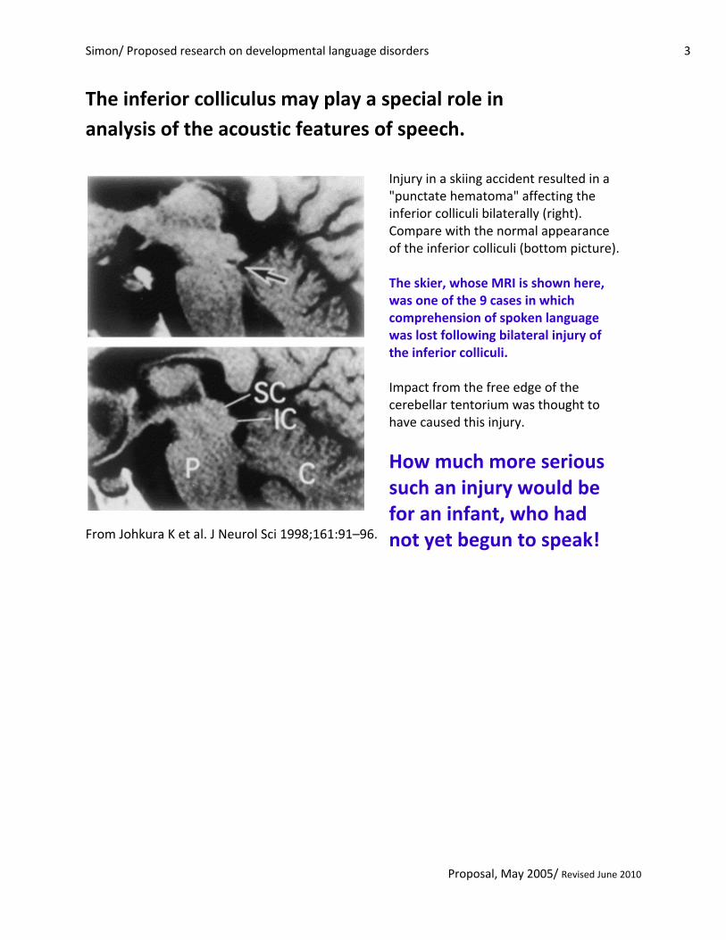
The inferior colliculus may play a special role in analysis of the acoustic features of speech.
From Johkura K et al. J Neurol Sci 1998;161:91–96.
Injury in a skiing accident resulted in a "punctate hematoma" affecting the inferior colliculi bilaterally (right). Compare with the normal appearance of the inferior colliculi (bottom picture). The skier, whose MRI is shown here, was one of the 9 cases in which comprehension of spoken language was lost following bilateral injury of the inferior colliculi. Impact from the free edge of the cerebellar tentorium was thought to have caused this injury.
How much more serious such an injury would be for an infant, who had not yet begun to speak!

Simon/ Proposed research on developmental language disorders 4
Proposal, May 2005/ Revised June 2010
From Leech & Alvord (1977) Archives of Neurology 34:109
In experiments with newborn monkeys (an attempt to produce an animal model of cerebral palsy), damage to the inferior colliculi was found in animals subjected to asphyxia for 8 to 10 minutes (above) [38, 39]. Damage of the inferior colliculi in human infants has also been observed (bottom right) [40‐49].
From Myers RE (1972) Am J Ob Gyn 112:246

Simon/ Proposed research on developmental language disorders 5
Proposal, May 2005/ Revised June 2010
Monkeys subjected to asphyxia did not develop cerebral palsy. The brainstem damage found was dismissed as minimal, thus perhaps the perception that infants can tolerate asphyxia at birth without serious harm.
Brainstem aphasia?
Gilles (1963), citing the experiments with monkeys on asphyxia at birth, and his own observations suggested: “…certain congenital brain stem nuclear ‘aphasias,’ for example, Moebius syndrome, may be related to temporary prenatal or perinatal cardiac failure” [40, p318] Note: This comment appeared in an abstract of a presentation given at the 38th Annual Meeting of the American Association of Neuropathologists, June 16, 1962.
The purpose of the research proposed here is to use functional MRI (fMRI) to investigate: I. The hypothesis put forward by Gilles (1963) that
impairment of function in the inferior colliculi might underlie some developmental language disorders, or
II. Whether some developmental language disorders
occur in the absence of any measurable impairment of auditory function.
Floyd H Gilles, M.D. Neurologist, Children’s Hospital Los Angeles
www.childrenshospitalla.org/

Simon/ Proposed research on developmental language disorders 6
Proposal, May 2005/ Revised June 2010
Functional MRI (fMRI) may provide a way to investigate auditory system function in people with acquired or developmental language disorders.
I wrote a letter in response to the article by CL Pan et al.1 on auditory agnosia caused by a cancerous growth that invaded the inferior colliculi in a 14‐year‐old child. This was one of several attempts I have made to point out the long‐forgotten papers on damage of the inferior colliculi caused by asphyxia at birth.2,3 My letter, with a response from the authors was published in July 2005 [Neurology, 2005 Jul 26;65(2):339], and both are also online at: http://www.neurology.org/cgi/eletters/63/12/2387 Note: In their response, authors Hsieh and Pan point out that frequency components of complex sound spectra are resolved in the inferior colliculi, which may be important for language development. They further point out that inadequate anatomical resolution has up to this time prevented investigation of the inferior colliculi in the language disturbance of children with pervasive developmental disorders, but that now, with magnetic resonance imaging, these hypotheses are ready to be tested!
1Pan CL, Kuo MF, Hsieh ST. Auditory agnosia caused by a tectal germinoma.
Neurology. 2004 Dec 28;63(12):2387‐9. 2Windle WF. Brain damage by asphyxia at birth. Sci Am 1969; 221: 76‐84.
3Myers RE. Two patterns of brain damage and their conditions of occurrence.
Am J Obstet Gynecol 1972; 112:246‐76.

Simon/ Proposed research on developmental language disorders 7
Proposal, May 2005/ Revised June 2010
From: Budd TW et al. Neuroimage 2003; 20:1783
(a) Three orthogonal slices, coronal, horizontal, sagittal – centered on the anatomically defined inferior colliculi (IC), white circles superimposed,
(b) Slices centered on the anatomical center of medial geniculate bodies (MGB)
The inferior colliculi on fMRI scans The special brightness of the inferior colliculi may be because they have the highest blood flow in the brain even in the absence of stimulation.

Simon/ Proposed research on developmental language disorders 8
Proposal, May 2005/ Revised June 2010
The inferior colliculi on fMRI scans
Responses to increasing rates of auditory stimulation From Harms & Melcher J Neurophysiol 2002;88:1433

Simon/ Proposed research on developmental language disorders 9
Proposal, May 2005/ Revised June 2010
The inferior colliculi on fMRI scans
Reponses in people with Lateralized tinnitus (ringing in the ears). From Melcher JR et al. J Neurophysiol 2000; 83:1058

Simon/ Proposed research on developmental language disorders 10
Proposal, May 2005/ Revised June 2010
The inferior colliculi on fMRI scans
Recruitment of greater activity with increasing sound bandwidth. From: Hawley et al. Hear Res. 2005 Jun;204(1‐2):101‐10.

Simon/ Proposed research on developmental language disorders 11
Proposal, May 2005/ Revised June 2010
1. 2. Adults with language problems persisting from childhood, such as autism,
Asperger syndrome, dyslexia, stuttering, or hearing impairment. 3. Children with developmental delay learning to speak associated with autism,
Asperger syndrome, and/or hearing impairment. 4. Children with cochlear implants (note, MRI is being used following implantation
[74‐76]. 5. Children with problems like stuttering or difficulty learning to read. 6. Adults with presbyacusis, which may result from loss of function in the inferior
colliculi [77‐81]. 7. Adults and children (for whom permission can be obtained) with no childhood or
current language disorder.
Subjects for the research proposed here will include children and adults with acquired and developmental language disorders:
1. language difficulties following traumatic head injury
Note: Loss of speech understanding in instances of closed head injury was attributed to trauma caused by impact from the cerebellar tentorium in 3 cases.
Most head injuries result in traumatic injury of wide areas of the brain, but tentorial impact may also occur in many of these cases.
If traumatic injury of the inferior colliculi is seen with MRI (as in the case of the ski accident), decreased activity with fMRI would also be anticipated, and could be compared with activity levels determined in individuals with developmental language disorders.
From: Johkura K. Midbrain deafness with normal brainstem auditory evoked potentials. Neurology. 2002 Oct 22;59(8):1293.

Simon/ Proposed research on developmental language disorders 12
Proposal, May 2005/ Revised June 2010
Testing strategies:
1. For subjects who are mute or with severe verbal disabilities, investigation with: a) clicks and pure tones presented with increasing repetition rate, from
2/second to 35/second, as in experiments on normal subjects by Harms Melcher(2002)
b) pure tones delivered out of phase at each ear, and c) presentation of selected multi‐syllabic words.
2. Recognition of multi‐syllabic words in quiet (WRIQ) and with increasing levels of background noise (WRIN) would be tested in higher‐functioning subjects following some of the methods of Church et al. (1997) [82‐83].
Note: Cardiac gating by the method of Guimaraes et al. (1998) will be used; the high rate of blood flow in the brainstem causes pulsations that mask most auditory stimuli. Quiet MRI as described by Yetkin et al. (2004) will also be employed. The highest rate of blood flow is to the inferior colliculi, as was revealed in the experiments on cerebral circulation by Landau et al. (1955) in radiographic pictures as shown to the right. Harms MP, Melcher JR. Sound repetition rate in the human auditory
pathway: representations in the waveshape and amplitude of fMRI activation. J Neurophysiol. 2002 Sep;88(3):1433-50.
Church MW et al. Hearing, language, speech, vestibular, and dentofacial disorders in fetal alcohol syndrome. Alcoholism, Clinical and Experimental Research 1997; 21:227-237.
Guimaraes AR et al. Imaging subcortical auditory activity in humans. Hum Brain Mapp. 1998;6(1):33-41.
Yetkin FZ et al. Functional magnetic resonance imaging of activation in subcortical auditory pathway. Laryngoscope. 2004 Jan;114(1):96-101.
From: Kety SS. Regional neurochemistry and its application to brain function. In French, JD, ed, Frontiers in Brain Research. New York: Columbia University Press, 1962. pp 97‐120.

Simon/ Proposed research on developmental language disorders 13
Proposal, May 2005/ Revised June 2010
Anticipated results:
1. Observing activity changes with different stimuli in victims of head trauma may suggest auditory stimuli most useful in detecting altered activity in fMRI scans of the inferior colliculi in individuals with known damage of the inferior colliculi.
2. It is anticipated that alteration of activity in the inferior colliculi will be found in
adults and children with fetal alcohol syndrome, especially with “word recognition in noise.”
3. It is anticipated that alteration in activity in the inferior colliculi will be found in
adults with presbyacusis (hearing impairment with aging), especially on tests of word recognition in noise (WRIN).
4. It is anticipated that alteration in activity in the inferior colliculi will be found in
adults with life‐long autism, childhood language delay, or persisting language disorders.
5. Experimentation is planned, to find auditory stimuli that would show a clear
difference from normal subjects, and to look for paradoxical high or low activity.
Children and adults with autism often appear to suffer from hyperacusis, which might be due to loss of inhibitory neurons, but perhaps also increased activity of excitatory neurons [84‐86].
6. It is expected that (as in other investigations) decreased cortical activity with
language tests will be found in the studies proposed here for individuals with autism and other life‐long language disorders.

Simon/ Proposed research on developmental language disorders 14
Proposal, May 2005/ Revised June 2010
Working Hypotheses & Rationale:
From: Just MA et al. Brain. 2004 Aug;127(Pt 8):1811‐21.
Recent fMRI investigations of language disorder in autism implicate “underconnectivity” and differences from normal in brain symmetry [87‐89]
Brain activation of autistic (A) and control (B) groups (Sentence versus Fixation contrast):
Autistic participants show less activation in the left inferior frontal gyrus (LIFG) than the control group, but more activation in the left posterior superior temporal gyrus (LSTG) than the control group.
Differences from normal are evident in the language areas of the cortex! But what impairs development of the cortical language areas?

Simon/ Proposed research on developmental language disorders 15
Proposal, May 2005/ Revised June 2010
Monkeys subjected to asphyxia at birth displayed transient delay in development of motor control. They did eventually appear to “catch up.” But examination of the brain many months or years later revealed that brain growth had not progressed normally [90]. Neurons were sparser in: The oculomotor nuclei, reticular formation, mammillary bodies, hippocampus, amygdala, corpus callosum, cerebellum (Purkinje cells) and cerebral cortex (parietal and frontal) than in normal monkeys. These sites correspond to where changes are found in the brains of people with life‐long autism [91‐92].
Monkey with developmental delay
Normal monkey

Simon/ Proposed research on developmental language disorders 16
Proposal, May 2005/ Revised June 2010
The possible importance of the inferior colliculi for language development is based on four major considerations: 1. Maturation of the cerebral cortex continues after birth guided by
trophic neurotransmitters produced in neurons of brainstem nuclei [96‐98].
2. The brainstem auditory pathway is fully myelinated and functional in the human fetus by 29 gestational weeks [97, 98].
3. Myelination of the later maturing target areas for the brainstem auditory pathway (the temporal and frontal language areas of the cortex) continues over the first four years of post‐natal life.
4. Thus children normally learn to speak “by ear” before the language areas of the cortex are fully developed.
Myelin in the human brainstem auditory pathway at 25 gestational weeks (sagittal view)
Myelin in the human brainstem auditory pathway at 29 gestational weeks (horizontal view).

Simon/ Proposed research on developmental language disorders 17
Proposal, May 2005/ Revised June 2010
The brainstem auditory pathway is one of the earliest systems to become myelinated and functional.
ICol ‐ inferior colliculus (auditory) Scol ‐ superior colliculus (visual) TzB ‐ trapezoid body (auditory) SOl ‐superior olive (auditory)
LLm - lateral lemniscus (auditory) Mlm - medial lemniscus (motor) From Yakovlev & Lecours (1967) with permission from Blackwell Scientific Publishers

Simon/ Proposed research on developmental language disorders 18
Proposal, May 2005/ Revised June 2010
Prenatal first 12 months first decade
Myelin formation in the brain<‐‐ acoustic tectum, fully myelinated prenatally
<‐‐ acoustic radiations (temporal lobes)
Note the acoustic radiations become fully myelinated only between the 3rd and 4th years of life.
Simon/ Proposed research on developmental language disorders 19
Proposal, May 2005/ Revised June 2010
Children learn to speak “by ear”
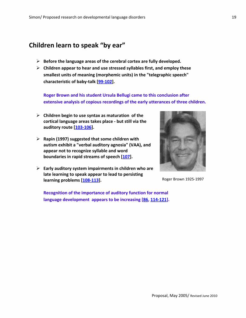
Before the language areas of the cerebral cortex are fully developed. Children appear to hear and use stressed syllables first, and employ these smallest units of meaning (morphemic units) in the "telegraphic speech" characteristic of baby‐talk [99‐102]. Roger Brown and his student Ursula Bellugi came to this conclusion after extensive analysis of copious recordings of the early utterances of three children.
Children begin to use syntax as maturation of the cortical language areas takes place ‐ but still via the auditory route [103‐106].
Rapin (1997) suggested that some children with autism exhibit a "verbal auditory agnosia" (VAA), and appear not to recognize syllable and word boundaries in rapid streams of speech [107].
Early auditory system impairments in children who are late learning to speak appear to lead to persisting learning problems [108‐113]. Recognition of the importance of auditory function for normal language development appears to be increasing [86, 114‐121].
Roger Brown 1925‐1997

Simon/ Proposed research on developmental language disorders 20
Proposal, May 2005/ Revised June 2010
Blood flow and metabolism are greatest in the auditory system Experiments on cerebral circulation (in cats) showed greatest perfusion of a radioactive tracer after 60 seconds, thus greatest blood flow, in nuclei of the brainstem auditory pathway1. Auditory nuclei should therefore be vulnerable to circulatory arrest or asphyxia, or to any factor that disrupts aerobic metabolism. Why is this not always the case? Protective mechanisms go into action that work to preserve metabolic functions during hypoxia or circulatory insufficiency. 1Landau et al. (1955) The local circulation of the living brain; values in the unanesthetized and anesthetized cat. Trans Am Neurol Assoc 80:125
From: Kety SS (1962) Regional neurochemistry and its application to brain function. In French, JD, ed, Frontiers in Brain Research. New York: Columbia University Press, pp 97‐120.

Simon/ Proposed research on developmental language disorders 21
Proposal, May 2005/ Revised June 2010
Protective mechanisms Increase blood flow and delivery of oxygen to the most active areas of the brain during periods of hypoxia or circulatory insufficiency:
Vasodilation1
Release of oxygen by hemoglobin in response to the metabolic end‐product carbon dioxide (the Bohr effect)2
The cerebral cortex is most predictably susceptible to damage from circulatory insufficiency or hypoxia. The brainstem pattern of damage has long been reported and discussed as an unusual finding in cases of complete circulatory arrest (Janzer & Friede 1980). The difference is not a matter of degree, but whether oxygen is totally lacking or only in short supply. The Bohr effect is among the most elegant mechanisms that evolved to maintain homeostasis in multi‐cellular organisms dependent upon aerobic metabolism.
1Kelly PA et al. (1995) Enhanced cerebrovascular responsiveness to hypercapnia following depletion of central serotonergic terminals. Journal of Cerebral Blood Flow and Metabolism 15:706‐713 2Jensen FB (2004) Red blood cell pH, the Bohr effect, and other oxygenation‐linked phenomena in blood O2 and CO2 transport. Acta Physiol Scand.182:215
Christian Bohr (1855‐1911)

Simon/ Proposed research on developmental language disorders 22
Proposal, May 2005/ Revised June 2010
Catastrophic factors prevent protective mechanisms from going into action. Suffocation, circulatory arrest, or any factor that disrupts aerobic metabolism can cause damage or impair function in the auditory system, especially the inferior colliculi. The picture to the right shows damage of the inferior colliculi in a human patient maintained on prolonged parenteral feeding that lacked vitamin B1. Vitamin B1 is an essential coenzyme for aerobic enzymes. Any toxic substance that crosses the blood‐brain barrier can disrupt aerobic enzymes and cause the same kind of damage. Alcohol intoxication has long been known to cause a brainstem pattern of damage that often involves the inferior colliculi.
From Vortmeyer AO et al. (1992) Haemorrhagic thiamine deficient encephalopathy following prolonged parenteral nutrition. Journal of Neurology, Neurosurgery and Psychiatry 55:826‐829.

Simon/ Proposed research on developmental language disorders 23
Proposal, May 2005/ Revised June 2010
Cardiac Arrest Encephalopathy: Damage restricted to the brainstem is only seen after total interference with aerobic metabolism occurs; Janzer and Friede (1980) referred to brainstem pathology as "cardiac arrest encephalopathy;" this was the case in monkeys subjected to several minutes of total asphyxia at birth. Myers (1972) found that prolonged partial hypoxia late in gestation caused damage to wide areas of the cerebral cortex, which was thought far more important that the brainstem pattern. This was cerebral palsy! Miller and Myers (1970, 1972) found also that adult monkeys sustained brainstem damage with complete circulatory arrest, and widespread cortical damage with partial circulatory insufficiency. Janzer RC, Friede RL. Hypotensive brain stem necrosis or cardiac arrest encephalopathy? Acta Neuropathol (Berl). 1980;50(1):53‐6. Myers RE (1972) Two patterns of perinatal brain damage and their conditions of occurrence. American Journal of Obstetrics and Gynecology 112:246‐276 Miller JR, Myers RE (1970) Neurological effects of systemic circulatory arrest in the monkey. Neurology 20:715‐724. Miller JR, Myers RE (1972) Neuropathology of systemic circulatory arrest in adult monkeys. Neurology 22:888‐904.
Simon/ Proposed research on developmental language disorders 24
Proposal, May 2005/ Revised June 2010
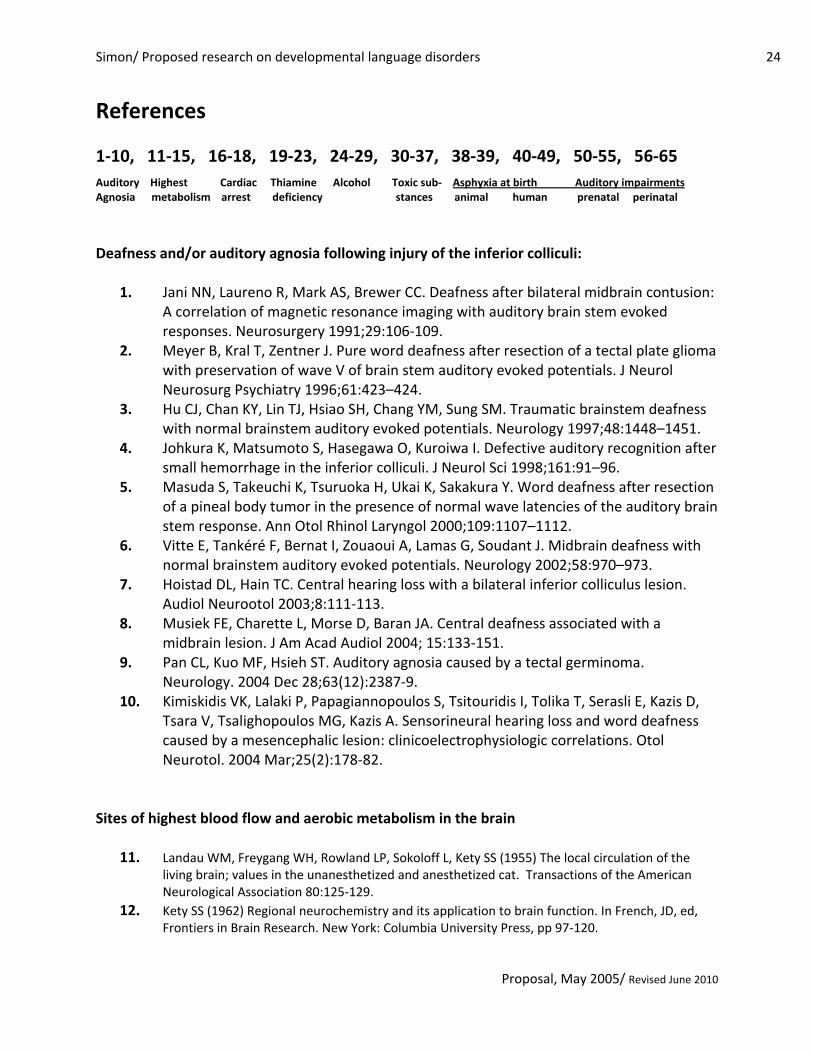
References 1‐10, 11‐15, 16‐18, 19‐23, 24‐29, 30‐37, 38‐39, 40‐49, 50‐55, 56‐65 Auditory Highest Cardiac Thiamine Alcohol Toxic sub‐ Asphyxia at birth Auditory impairments Agnosia metabolism arrest deficiency stances animal human prenatal perinatal
Deafness and/or auditory agnosia following injury of the inferior colliculi:
1. Jani NN, Laureno R, Mark AS, Brewer CC. Deafness after bilateral midbrain contusion: A correlation of magnetic resonance imaging with auditory brain stem evoked responses. Neurosurgery 1991;29:106‐109.
2. Meyer B, Kral T, Zentner J. Pure word deafness after resection of a tectal plate glioma with preservation of wave V of brain stem auditory evoked potentials. J Neurol Neurosurg Psychiatry 1996;61:423–424.
3. Hu CJ, Chan KY, Lin TJ, Hsiao SH, Chang YM, Sung SM. Traumatic brainstem deafness with normal brainstem auditory evoked potentials. Neurology 1997;48:1448–1451.
4. Johkura K, Matsumoto S, Hasegawa O, Kuroiwa I. Defective auditory recognition after small hemorrhage in the inferior colliculi. J Neurol Sci 1998;161:91–96.
5. Masuda S, Takeuchi K, Tsuruoka H, Ukai K, Sakakura Y. Word deafness after resection of a pineal body tumor in the presence of normal wave latencies of the auditory brain stem response. Ann Otol Rhinol Laryngol 2000;109:1107–1112.
6. Vitte E, Tankéré F, Bernat I, Zouaoui A, Lamas G, Soudant J. Midbrain deafness with normal brainstem auditory evoked potentials. Neurology 2002;58:970–973.
7. Hoistad DL, Hain TC. Central hearing loss with a bilateral inferior colliculus lesion. Audiol Neurootol 2003;8:111‐113.
8. Musiek FE, Charette L, Morse D, Baran JA. Central deafness associated with a midbrain lesion. J Am Acad Audiol 2004; 15:133‐151.
9. Pan CL, Kuo MF, Hsieh ST. Auditory agnosia caused by a tectal germinoma. Neurology. 2004 Dec 28;63(12):2387‐9.
10. Kimiskidis VK, Lalaki P, Papagiannopoulos S, Tsitouridis I, Tolika T, Serasli E, Kazis D, Tsara V, Tsalighopoulos MG, Kazis A. Sensorineural hearing loss and word deafness caused by a mesencephalic lesion: clinicoelectrophysiologic correlations. Otol Neurotol. 2004 Mar;25(2):178‐82.
Sites of highest blood flow and aerobic metabolism in the brain
11. Landau WM, Freygang WH, Rowland LP, Sokoloff L, Kety SS (1955) The local circulation of the living brain; values in the unanesthetized and anesthetized cat. Transactions of the American Neurological Association 80:125‐129.
12. Kety SS (1962) Regional neurochemistry and its application to brain function. In French, JD, ed, Frontiers in Brain Research. New York: Columbia University Press, pp 97‐120.

Simon/ Proposed research on developmental language disorders 25
Proposal, May 2005/ Revised June 2010
13. Sokoloff L (1981) Localization of functional activity in the central nervous system by measurement of glucose utilization with radioactive deoxyglucose. Journal of Cerebral Blood Flow and Metabolism 1:7‐36.
14. Calingasan NY, Baker H, Sheu KF, Gibson GE (1994) Distribution of the alpha‐ketoglutarate dehydrogenase complex in rat brain. Journal of Comparative Neurology 346:461‐479.
15. Zeller K, Rahner‐Welsch S, Kuschinsky W (1997) Distribution of Glut1 glucose transporters in different brain structures compared to glucose utilization and capillary density of adult rat brains. Journal of Cerebral Blood Flow and Metabolism 17:204‐209.
Catastrophic disruption of aerobic metabolism I. Cardiac arrest encephalopathy
16. Janzer RC, Friede RL. Hypotensive brain stem necrosis or cardiac arrest encephalopathy? Acta
Neuropathol (Berl). 1980;50(1):53‐6. 17. Miller JR, Myers RE (1970) Neurological effects of systemic circulatory arrest in the monkey.
Neurology 20:715‐724. 18. Miller JR, Myers RE (1972) Neuropathology of systemic circulatory arrest in adult monkeys.
Neurology 22:888‐904. II. Deficiency of thiamine (vitamin B1) ‐ essential for aerobic metabolism
19. Evans CA, Carlson WE, Green EG (1942) The pathology of Chastek paralysis in foxes. A counterpart of Wernicke's hemorrhagic polioencephalitis of man. American Journal of Pathology 18:79‐90.
20. Rinehart JF, Friedman M, Greenberg LD (1949) Effect of experimental thiamine deficiency on the nervous system of the rhesus monkey. Archives of Pathology 48:129‐139.
21. Jubb KV Saunders LZ, Coates HV (1956) Thiamine deficiency encephalopathy in cats. Journal of Comparative Pathology 66:217‐227.
22. Irle E, Markowitsch HJ (1983) Widespread neuroanatomical damage and learning deficits following chronic alcohol consumption or vitamin B1 (thiamine) deficiency in rats. Behavioral Brain Research 9:277‐284.
23. Vortmeyer AO, Hagel C, Laas R (1992) Haemorrhagic thiamine deficient encephalopathy following prolonged parenteral nutrition. Journal of Neurology, Neurosurgery and Psychiatry 55:826‐829.
III. Damage caused by alcohol ‐
24. Wernicke C (1881) Die acute, haemorrhagische Poliencephalitis superior. Lehrbuch der Gehirnkrankheiten für Ärzte und Studirende,Band II. Kassel: Theodor Fischer, pp 229‐242
25. Gamper (1928) Zur Frage der Polioencephalitis haemorrhagica der chronischen Alkoholiker. Anatomische Befunde beim alkoholischen Korsakow und ihre Beziehungen zum klinischen Bild. Deutsche Zeitschrift für Nervenheilkunde 102:122‐129

Simon/ Proposed research on developmental language disorders 26
Proposal, May 2005/ Revised June 2010
26. Kant F (1933) Die Pseudoencephalitis Wernicke der Alkoholiker. (polio‐encephalitis aemorrhagica superior acuta). Archiv für Psychiatrie und Nervenkrankheiten 98:702‐768.
27. Malamud N, Skillicorn SA (1956). Relationship between the Wernicke and the Korsakoff Syndrome. Archives of Neurology and Psychiatry, 76, 585‐596.
28. Torvik A (1987) Topographic distribution and severity of brain lesions in Wernicke's encephalopathy. Clinical Neuropathology 6:25‐29.
29. Victor M, Adams RD, Collins GH (1989) The Wernicke‐Korsakoff syndrome and related neurologic disorders due to alcoholism and malnutrition, 2nd ed, Contemporary Neurology Series v30. Philadelphia, PA : F.A. Davis Co.
IV. Damage caused by other toxic substances
30. Franken L (1959) Étude anatomique d'un cas d'intoxication par le bromure de méthyle. Acta Neurologica et Psychiatrica Belgica 59:375‐383.
31. Goulon M, Nouailhat R, Escourolle R, Zarranz‐Imirizaldu JJ, Grosbuis S, Levy‐Alcover MA (1975). Intoxication par le bromure de methyl: Trois observations, dont une mortelle. Etude neuro‐pathologique d'un cas de stupeur avec myoclonies, suivi pendent cinq ans. Revue Neurologique (Paris) 131:445‐468.
32. Troncoso JC, Johnston MV, Hess KM, Griffin JW, Price DL (1981) Model of Wernicke's encephalopathy. Archives Of Neurology 38:350‐354.
33. Oyanagi K, Ohama E, & Ikuta F. (1989). The auditory system in methyl mercurial intoxication: a neuropathological investigation on 14 autopsy cases in Niigata, Japan. Acta Neuropathologica (Berlin), 77, 561‐568.
34. Squier MV, Thompson J, Rajgopalan B. (1992) Case report: neuropathology of methyl bromide intoxication. Neuropathology and Applied Neurobiology 18: 579‐584.
35. Cavanagh JB (1992) Methyl bromide intoxication and acute energy deprivation syndromes. Neuropathology and Applied Neurobiology 18:575‐578.
36. Cavanagh JB, Nolan CC (1993) The neurotoxicity of alpha‐chlorohydrin in rats and mice: II. Lesion topography and factors in selective vulnerability in acute energy deprivation syndromes. Neuropathology and Applied Neurobiology 19:471‐479.
37. Husain K, Whitworth C, Hazelrigg S, Rybak L (2003) Carboplatin‐induced oxidative injury in rat inferior colliculus. Int J Toxicol. 2003 Sep‐Oct;22(5):335‐42.
Methyl bromide was formerly used as a fire extinguisher, it is still used as an herbicide. Mercury is oto‐toxic, and the neuropathological study of Oyanagi et al. revealed its damaging effects in the brain. Alpha‐chlorohydrin was developed for possible use as a male contraceptive. Carboplatin is used in chemotherapy for cancer patients. Troncoso et al. described the symmetric bilateral lesions of the brainstem caused by pyrithiamine, a poison that displaces thiamine at its enzyme attachment points.

Simon/ Proposed research on developmental language disorders 27
Proposal, May 2005/ Revised June 2010
Damage of the inferior colliculi caused by asphyxia at birth
38. Ranck JB, Windle WF (1959). Brain damage in the monkey, Macaca mulatta, by asphyxia
neonatorum. Experimental Neurology 1:130‐154. 39. Myers RE (1972) Two patterns of perinatal brain damage and their conditions of occurrence.
American Journal of Obstetrics and Gynecology 112:246‐276. Damage of the inferior colliculi observed in human infants
40. Gilles FH (1963) Selective symmetrical neuronal necrosis of certain brain stem tegmental nuclei in temporary cardiac standstill. J Neuropathol Exp Neurol 1963;22:318 [abstract].
41. Gilles FH (1969) Hypotensive brain stem necrosis: selective symmetrical necrosis of tegmental neuronal aggregates following cardiac arrest. Archives of Pathology 88:32‐41.
42. Norman MG (1972) Antenatal neuronal loss and gliosis of the reticular formation, thalamus, and hypothalamus. A report of three cases. Neurology (Minneapolis) 22:910‐916.
43. Griffiths AD, Laurence KM (1974) The effect of hypoxia and hypoglycemia on the brain of the newborn human infant. Developmental Medicine and Child Neurology 16:308‐319.
44. Grunnet ML, Curless RG, Bray PF, Jung AL (1974) Brain changes in newborns from an intensive care unit. Developmental Medicine and Child Neurology 16:320‐328.
45. Smith JF, Rodeck C (1975) Multiple cystic and focal encephalomalacia in infancy and childhood with brain stem damage. Journal of the neurological sciences 25:377‐88
46. Schneider H, Ballowitz L, Schachinger H, Hanefield F, Droeszus J‐U (1975) Anoxic encephalopathy with predominant involvement of basal ganglia, brain stem, and spinal cord in the perinatal period. Acta Neuropathologica (Berlin) 32:287‐298.
47. Leech RW, Alvord EC (1977) Anoxic‐ischemic encephalopathy in the human neonatal period, the significance of brain stem involvement. Archives of Neurology 34:109‐113.
48. Roland EH, Hill A, Norman MG, Flodmark O, MacNab AJ (1988) Selective brainstem injury in an asphyxiated newborn. Annals of Neurology 23:89‐92.
49. Natsume J, Watanabe K, Kuno K, Hayakawa F, Hashizume Y (1995) Clinical, neurophysiologic, and neuropathological features of an infant with brain damage of total asphyxia type (Myers). Pediatric Neurology 13:61‐64.
Prenatal factors associated with auditory system impairments
50. Church MW, Holloway JA (1984) Effects of prenatal ethanol exposure on the postnatal development of the brainstem and auditory evoked potential. Alcoholism, Clinical and Experimental Research. 8:258‐265.
51. Vingan RD, Dow‐Edwards ML, Riley EP (1986) Cerebral metabolic alterations in rats following prenatal alcohol exposure: a deoxyglucose study. Alcoholism, Clinical and Experimental Research 10:22‐26.
52. Dow‐Edwards DL, Freed LA, Fico TA (1990) Structural and functional effects of prenatal cocaine exposure in adult rat brain. Brain Research Developmental Brain Research 57:263‐26

Simon/ Proposed research on developmental language disorders 28
Proposal, May 2005/ Revised June 2010
53. Berman RF, Beare DJ, Church MW, Abel EL (1992) Audiogenic seizure susceptibility and auditory brainstem responses in rats prenatally exposed to alcohol. Alcoholism, Clinical and Experimental Research 16:490‐498.
54. Kelly PA, Ritchie IM, Sharkey J, McBean DE (1994) Alterations in local cerebral blood flow in mature rats following prenatal exposure to cocaine. Neuroscience 60:183‐189.
55. Cone‐Wesson B (2005) Prenatal alcohol and cocaine exposure: influences on cognition, speech, language, and hearing. J Commun Disord. 2005 Jul‐Aug;38(4):279‐302.
Prenatal exposure to alcohol and cocaine are not good, and consideration should be given to the possible deleterious effects of over‐the‐counter drugs, artificial sweeteners, and other nutritional substances generally assumed to be safe.
Perinatal or postnatal factors associated with auditory system impairments
56. Mirsky AF, Orren MM, Stanton L, Fullerton BC, Harris S, Myers RE (1979) Auditory evoked
potentials and auditory behavior following prenatal and perinatal asphyxia in rhesus monkeys. Developmental Psychobiology 12:369‐379.
57. Reiter LW, Heavner GB, Dean KF, Ruppert PH (1981) Developmental and behavioral effects of early postnatal exposure to triethyltin in rats. Neurobehavioral Toxicology and Teratology 3:285‐93.
58. Slawecki CJ, Thomas JD, Riley EP, Ehlers CL (2005) Neurophysiologic consequences of neonatal ethanol exposure in the rat. Alcohol. 2004 Oct‐Nov;34(2‐3):187‐96.
59. Bertoni JM and Sprenkle PM (1989) Lead acutely reduces glucose utilization in the rat brain especially in higher auditory centers. Neurotoxicology 9:235‐242.
60. Worley G, Erwin CW, Goldstein RF, Provenzale JM, Ware RE (1996) Delayed development of sensorineural hearing loss after neonatal hyperbilirubinemia: a case report with brain magnetic resonance imaging. Developmental Medicine and Child Neurology 38:271‐277
61. Shapiro SM, Nakamura H (2001)Bilirubin and the auditory system. J Perinatol. 2001 Dec;21 Suppl 1:S52‐5; discussion S59‐62.
62. Amin SB (2004) Clinical assessment of bilirubin‐induced neurotoxicity in premature infants. Semin Perinatol. 2004 Oct;28(5):340‐7.
63. Amin SB, Charafeddine L, Guillet R (2005)Transient bilirubin encephalopathy and apnea of prematurity in 28 to 32 weeks gestational age infants. J Perinatol. 2005 Jun;25(6):386‐90.
64. Shapiro SM (2005) Definition of the clinical spectrum of kernicterus and bilirubin‐induced neurologic dysfunction (BIND). J Perinatol. 2005 Jan;25(1):54‐9.
65. Strata F, Deipolyi AR, Bonham BH, Chang EF, Liu RC, Nakahara H, Merzenich MM (2005) Perinatal anoxia degrades auditory system function in rats. Proc Natl Acad Sci U S A. 2005 Dec 27;102(52):19156‐61.
This proposal is also posted online at http://www.inferiorcolliculus.org/presentation.html