Propofol inhibits burn injury-induced … inhibits burn injury-induced hyperpermeability through an...
7
Propofol inhibits burn injury-induced hyperpermeability through an apoptotic signal pathway in microvascular endothelial cells K.Y. Tian 1 *, X.J. Liu 2 *, J.D. Xu 1 , L.J. Deng 1 and G. Wang 1 1 Department of Anesthesiology, Guangdong General Hospital, Guangdong Academy of Medical Sciences, Guangzhou, China 2 Department of Anesthesia, Nanfang Hospital, Southern Medical University, Guangzhou, China Abstract Recent studies have revealed that an intrinsic apoptotic signaling cascade is involved in vascular hyperpermeability and endothelial barrier dysfunction. Propofol (2,6-diisopropylphenol) has also been reported to inhibit apoptotic signaling by regulating mitochondrial permeability transition pore (mPTP) opening and caspase-3 activation. Here, we investigated whether propofol could alleviate burn serum-induced endothelial hyperpermeability through the inhibition of the intrinsic apoptotic signaling cascade. Rat lung microvascular endothelial cells (RLMVECs) were pretreated with propofol at various concentrations, followed by stimulation with burn serum, obtained from burn-injury rats. Monolayer permeability was determined by transendothelial electrical resistance. Mitochondrial release of cytochrome C was measured by ELISA. Bax and Bcl-2 expression and mitochondrial release of second mitochondrial-derived activator of caspases (smac) were detected by Western blotting. Caspase-3 activity was assessed by fluorometric assay; mitochondrial membrane potential (Dy m ) was determined with JC-1 (a potential-sensitive fluorescent dye). Intracellular ATP content was assayed using a commercial kit, and reactive oxygen species (ROS) were measured by dichlorodihydrofluorescein diacetate (DCFH-DA). Burn serum significantly increased monolayer permeability (P,0.05), and this effect could be inhibited by propofol (P,0.05). Compared with a sham treatment group, intrinsic apoptotic signaling activation – – indicated by Bax overexpression, Bcl-2 downregulation, Dy m reduction, decreased intracellular ATP level, increased cytosolic cytochrome C and smac, and caspase-3 activation – – was observed in the vehicle group. Propofol not only attenuated these alterations (P,0.05 for all), but also significantly decreased burn-induced ROS production (P,0.05). Propofol attenuated burn-induced RLMVEC monolayer hyperpermeability by regulating the intrinsic apoptotic signaling pathway. Key words: Burn injury; Hyperpermeability; Propofol; Apoptosis; Endothelial cells Introduction Burns are a common form of traumatic injury and are associated with high mortality and morbidity rates (1,2). Resulting from endothelial barrier dysfunction and increased vascular hyperpermeability, blister and edema are consid- ered to be classical signs in burn injury (3,4). Microvascular hyperpermeability leads to shifts and losses of fluid from the circulation, and hypovolemia can occur after large burns if massive volume resuscitation is absent. Although adequate fluid resuscitation can improve the hypovolemia, it can also markedly aggravate the edema process if vascular hyper- permeability persists. Therefore, reversing vascular hyper- permeability plays a key role in protecting cells from burn injury. Some recent studies have suggested that the intrinsic apoptotic signaling cascade is involved in endothelial dysfunction, which is closely associated with vascular hy- perpermeability (5,6). In the intrinsic apoptotic pathway, activated mitochondria release cytochrome C and second mitochondrial-derived activator of caspases (smac), and finally caspase-3 is activated (7). These mitochondrial factors are regulated by proapoptotic and antiapoptotic Bcl-2 family proteins, such as Bax/Bak and Bcl-2/Bcl-xL. In endothe- lial cells, caspase-3 can cleave b-catenin resulting in the dissociation of the vascular endothelial (VE)-cadherin-b- catenin complex (8), which could induce microvascular hyperpermeability (9). Correspondence: Gang Wang: ,[email protected].. *These authors contributed equally to this study. Received May 27, 2014. Accepted November 11, 2014. First published online March 6, 2015. Brazilian Journal of Medical and Biological Research (2015) 48(5): 401-407, http://dx.doi.org/10.1590/1414-431X20144107 ISSN 1414-431X www.bjournal.com.br Braz J Med Biol Res 48(5) 2015
Transcript of Propofol inhibits burn injury-induced … inhibits burn injury-induced hyperpermeability through an...

Propofol inhibits burn injury-inducedhyperpermeability through anapoptotic signal pathway in
microvascular endothelial cells
K.Y. Tian1*, X.J. Liu2*, J.D. Xu1, L.J. Deng1 and G. Wang1
1Department of Anesthesiology, Guangdong General Hospital, Guangdong Academy of Medical Sciences, Guangzhou, China2Department of Anesthesia, Nanfang Hospital, Southern Medical University, Guangzhou, China
Abstract
Recent studies have revealed that an intrinsic apoptotic signaling cascade is involved in vascular hyperpermeability and
endothelial barrier dysfunction. Propofol (2,6-diisopropylphenol) has also been reported to inhibit apoptotic signaling by regulating
mitochondrial permeability transition pore (mPTP) opening and caspase-3 activation. Here, we investigated whether propofol
could alleviate burn serum-induced endothelial hyperpermeability through the inhibition of the intrinsic apoptotic signaling
cascade. Rat lung microvascular endothelial cells (RLMVECs) were pretreated with propofol at various concentrations, followed
by stimulation with burn serum, obtained from burn-injury rats. Monolayer permeability was determined by transendothelial
electrical resistance. Mitochondrial release of cytochrome C was measured by ELISA. Bax and Bcl-2 expression and
mitochondrial release of second mitochondrial-derived activator of caspases (smac) were detected by Western blotting.
Caspase-3 activity was assessed by fluorometric assay; mitochondrial membrane potential (Dym) was determined with JC-1 (a
potential-sensitive fluorescent dye). Intracellular ATP content was assayed using a commercial kit, and reactive oxygen species
(ROS) were measured by dichlorodihydrofluorescein diacetate (DCFH-DA). Burn serum significantly increased monolayer
permeability (P,0.05), and this effect could be inhibited by propofol (P,0.05). Compared with a sham treatment group, intrinsic
apoptotic signaling activation –– indicated by Bax overexpression, Bcl-2 downregulation, Dym reduction, decreased intracellular
ATP level, increased cytosolic cytochrome C and smac, and caspase-3 activation –– was observed in the vehicle group. Propofol
not only attenuated these alterations (P,0.05 for all), but also significantly decreased burn-induced ROS production (P,0.05).
Propofol attenuated burn-induced RLMVECmonolayer hyperpermeability by regulating the intrinsic apoptotic signaling pathway.
Key words: Burn injury; Hyperpermeability; Propofol; Apoptosis; Endothelial cells
Introduction
Burns are a common form of traumatic injury and are
associated with high mortality and morbidity rates (1,2).
Resulting from endothelial barrier dysfunction and increased
vascular hyperpermeability, blister and edema are consid-
ered to be classical signs in burn injury (3,4). Microvascular
hyperpermeability leads to shifts and losses of fluid from the
circulation, and hypovolemia can occur after large burns if
massive volume resuscitation is absent. Although adequate
fluid resuscitation can improve the hypovolemia, it can also
markedly aggravate the edema process if vascular hyper-
permeability persists. Therefore, reversing vascular hyper-
permeability plays a key role in protecting cells from burn
injury.
Some recent studies have suggested that the intrinsic
apoptotic signaling cascade is involved in endothelial
dysfunction, which is closely associated with vascular hy-
perpermeability (5,6). In the intrinsic apoptotic pathway,
activated mitochondria release cytochrome C and second
mitochondrial-derived activator of caspases (smac), and
finally caspase-3 is activated (7). Thesemitochondrial factors
are regulated by proapoptotic and antiapoptotic Bcl-2 family
proteins, such as Bax/Bak and Bcl-2/Bcl-xL. In endothe-
lial cells, caspase-3 can cleave b-catenin resulting in the
dissociation of the vascular endothelial (VE)-cadherin-b-catenin complex (8), which could induce microvascular
hyperpermeability (9).
Correspondence: Gang Wang: ,[email protected]..
*These authors contributed equally to this study.
Received May 27, 2014. Accepted November 11, 2014. First published online March 6, 2015.
Brazilian Journal of Medical and Biological Research (2015) 48(5): 401-407, http://dx.doi.org/10.1590/1414-431X20144107
ISSN 1414-431X
www.bjournal.com.br Braz J Med Biol Res 48(5) 2015

Propofol (2,6-diisopropylphenol) is a widely used short-
acting intravenous anesthetic, and has shown some anti-
oxidative and anti-inflammatory effects (10-12). Furthermore,
propofol also has an inhibitory effect on apoptotic signal-
ing through regulating mitochondrial permeability transition
pore (mPTP) opening, which is a crucial component of in-
trinsic apoptotic signal transduction and caspase-3 activa-
tion (13,14). In the present study, we demonstrated that
propofol could restore burn serum-induced endothelial hy-
perpermeability via regulating the intrinsic apoptotic signal-
ing cascade.
Material and Methods
Animal model and serum collectionThe procedures used in this study and the handling of
study animals followed the National Institutes of Health
Guidelines on the use of experimental animals. The exper-
imental protocol was approved by the Committee on
Research Animal Use of Guangdong Provincial People’s
Hospital. Male Sprague-Dawley rats (n=24) weighing 180-
220 g were purchased from the Experimental Animal
Center of Guangdong Province and allowed to acclimatize
for 1 week before experiments. Animals had ad libitumaccess to chow and water. All the rats were intramuscu-
larly anesthetized using 30 mg/kg sodium pentobarbital
(Shanghai Chemical Plant Co., Ltd., China). Thermal injury
was inflicted with a modifiedWalker andMason burn model
(15). Briefly, a dorsal area equal to 25% total body surface
area (TBSA) was shaved. The rats were placed in a housing
with an adjustable opening that exposed the shaved area
to 1006C water for 30 s. According to the Parkland burn
formula, Ringer’s lactate solution (4 mL?kg-1?%TBSA-1) was
constantly infused through a jugular vein cannula to mimic
clinical conditions of fluid resuscitation. The rats in a sham
treatment group were exposed to water at room temperature
for 30 s. Three hours later, blood samples were collected
and centrifuged at 1200 g for 10 min to harvest the sera,
which were then diluted four-fold with phosphate-buffered
saline (PBS) and used immediately.
Monolayer permeabilityRat lung microvascular endothelial cells (RLMVECs,
ScienCell, USA) were cultured in DMEM/F12 containing
10% fetal bovine serum at 376C in a humidified atmosphere
with 5% CO2 for 48 h to form a confluent monolayer. In a
preliminary, time-dependent experiment, monolayers were
exposed to serum collected from both the sham treatment
and burn groups at 1, 2, or 3 h postsham/burn. Subse-
quently, the 3-h postburn serum was used in the following
experiments. To determine the effect of propofol on mono-
layer hyperpermeability, monolayer cells were pretreated
with 1, 10, or 50 mM propofol for 1 h before exposure to
sham or burn serum. Intralipid (Shanghai Chemical Plant
Co., Ltd.) was selected as the vehicle. Endothelial perme-
ability was measured 2 h after stimulation.
Transendothelial electrical resistanceThe transendothelial electrical resistance (TER) of
RLMVEC monolayers was determined with an STX2 elec-
trode and epithelial volt-ohm meter (EVOM2) accord-
ing to the manufacturer’s instructions (World Precision
Instruments, USA) (16). RLMVECs were seeded at a den-
sity of 16105 cells/cm2 on fibronectin-coated, 6.5 mm
Transwell filters (0.4 mm pore size) and cultured until
reaching full confluency. Resistance values of multiple
Transwell inserts were measured sequentially in each
experimental group and the mean is reported as V cm2
after subtracting the value of a blank cell-free filter.
Measurement of cytosolic cytochrome CCytosolic cytochrome C content was assayed with a
cytochrome C ELISA kit (MBL, USA) (17). RLMVECs were
pretreated with intralipid or 10 mM propofol and ex-
posed to sham or burn serum. After treatment, cells were
lysed in the cold preparation buffer included in the ELISA kit.
Cell homogenates were centrifuged (10,000 g for 60 min at
46C) and the supernatant was collected. Protein concentra-
tion was assayed by the bicinchoninic acid assay (BCA).
The samples were then treated with a conjugate reagent,
and transferred to a cytochrome C antibody-coated micro-
well plate and incubated for 60 min at room temperature.
Next, the plate was washed and treated with a substrate
reagent and incubated for 30 min, followed by the addition of
stop solution. The absorbance was read at 450 nm with an
automatic microplate reader (Spectra Max, M5; Molecular
Devices, USA). Serial dilutions of a cytochrome C calibra-
tor were assayed along with the samples to establish a
standard curve, which was used to calculate the concentra-
tion of cytochrome C.
Measurement of smacCytosol fractions were collected as described above
for cytosolic cytochrome C measurement. Smac expression
was measured by immunoblotting. An equal amount of
protein solution from various treatments was loaded and
analyzed by 12% sodium dodecyl sulfate polyacrylamide gel
electrophoresis (SDS-PAGE). After electrophoresis, pro-
teins were transferred onto polyvinylidene fluoride mem-
branes and blotted with primary antibodies against smac
(Abcam, UK) and glyceraldehyde 3-phosphate dehydrogen-
ase (GAPDH; Tianjin Sungene Biotech Co., Ltd., China).
Membranes were then incubated with horseradish perox-
idase-tagged secondary antibody (Earth, USA), and signals
were developed by an enhanced chemiluminescence
reagent.
Measurement of caspase-3 activityRLMVECs were pretreated with intralipid or 10 mM
propofol and exposed to sham or burn serum. Caspase-3
activity was assayed with a caspase-3 activity assay kit
(Sigma, USA). The cell homogenates were centrifuged at
10,000 g for 60 min, and the supernatant was collected for
402 K.Y. Tian et al.
Braz J Med Biol Res 48(5) 2015 www.bjournal.com.br
caspase-3 assay. Then, the caspase-3 activity was mea-
sured using the caspase-3/CPP32 fluorometric assay kit
(Sigma, USA) following the manufacturer’s instructions.
Detection of Bcl-2 and Bax expressionRLMVECs were pretreated with intralipid or 10 mM
propofol and exposed to sham or burn serum. Cells were
then homogenized and analyzed for Bcl-2 and Bax by
Western blotting as described above.
Measurement of mitochondrial membrane potentialMitochondrial membrane potential (Dym) was assayed
by staining with the potential-sensitive fluorescent dye JC-1
(Sigma) and detected by flow cytometry (18). JC-1 (5 mmol/L)
was loaded on RLMVECs for 15 min at 376C. The stained
cells were washed with PBS, and analyzed by flow cytom-
etry (Becton Dickinson FACScan, USA). A minimum of
10,000 cells per sample was analyzed. JC-1 monomers
emit at 527 nm and ‘‘J-aggregates’’ emit at 590 nm. The
percentage of cells with abnormally low DYm (green fluo-
rescence) was determined.
Measurement of cellular ATPIntracellular ATP was assayed with a luciferase-based
assay kit (CellTiter-Glo, Promega, USA) (18). Cells were
seeded and treated on a standard opaque-walled 96-well
plate. CellTiter-Glo reagent was added to the wells and
allowed to react with cell lysate for 10 min at room
temperature. The luminescence was recorded with an
automatic microplate reader (SpectraMax M5; Molecular
Devices)
Measurement of ROS levelsIntracellular reactive oxygen species (ROS) levels
were assessed with a dichlorodihydrofluorescein diacetate
(DCFH-DA) probe (Sigma) (19). Cells were treated with
DCFH-DA (10 mM), followed by the stimulation with sham/
burn serum for 20 min at 376C. After incubation, the cells
were washed and analyzed using an automatic microplate
reader (Spectra Max, M5; Molecular Devices). The relative
intensity of DCF fluorescence was measured at a wave-
length of 535 nm and compared with the sham treatment
group.
Statistical analysisAll data are reported as means±SD. Differences
between groups were analyzed by one-way analysis of
variance (ANOVA) with the least significant difference (LSD)
multiple-comparison test and Student’s t-test, when appro-
priate. P,0.05 was considered to be significant.
Results
Propofol attenuated burn serum-induced monolayerhyperpermeability
RLMEVC monolayer hyperpermeability was measured
by detecting the TER of cell monolayers. As shown in
Figure 1, a significant increase of monolayer permeability
was observed in RLMECs treated with serum at all the time
points postsham/burn (P,0.05 or P,0.01). Serum from
sham-burn rats did not induce RLMVEC monolayer hyper-
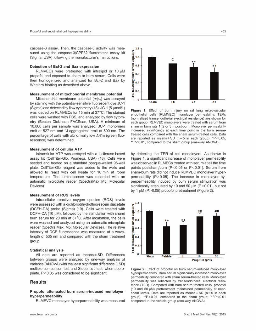
permeability (P.0.05). The increase in monolayer hy-
perpermeability induced by burn serum stimulation was
significantly attenuated by 10 and 50 mM (P,0.01), but not
by 1 mM (P.0.05) propofol pretreatment (Figure 2).
Figure 1. Effect of burn injury on rat lung microvascular
endothelial cells (RLMVEC) monolayer permeability. TERs
(normalized transendothelial electrical resistance) are shown for
each group. RLMVEC monolayers were treated with serum from
sham or burn rats 1, 2 or 3 h post-burn. Monolayer permeability
increased significantly at each time point in the burn serum-
treated cells compared with the sham serum-treated cells. Data
are reported as means±SD (n=5 in each group). *P,0.05,
**P,0.01, compared to the sham group (one-way ANOVA).
Figure 2. Effect of propofol on burn serum-induced monolayer
hyperpermeability. Burn serum significantly increased monolayer
permeability compared with sham serum-treated cells. Monolayer
permeability was reflected by transendothelial electrical resis-
tance (TER). Compared with burn serum-treated cells, propofol
(10 and 50 mM) pretreatment maintained permeability at near-
sham levels. Data are reported as means±SD (n=5 in each
group). **P,0.01, compared to the sham group; ##P,0.01
compared to the vehicle group (one-way ANOVA).
Propofol and endothelial cell hyperpermeability 403
www.bjournal.com.br Braz J Med Biol Res 48(5) 2015

Propofol inhibited burn serum-induced cytochrome Crelease
The cytosolic cytochrome C level in the sham group was
21.4±6.3 ng/mg protein, and reached 75.3±11.6 ng/mg
protein in the vehicle group (P,0.01). The increase of cyto-
solic cytochrome C induced by burn serum was significantly
reduced to 41.5±9.1 ng/mg protein by 10 mM propofol pre-
treatment (P,0.05; Figure 3). These results revealed that
propofol attenuated the burn serum-induced release of
cytochrome C from mitochondria to cytosol.
Propofol inhibited burn serum-induced smac releaseRelease of smac from mitochondria into the cytosol
normally occurs after the opening of mPTPs. As shown in
Figure 4, compared with the sham group, smac content in
the vehicle groupwas significantly increased (395.3±51.6%
of the normal values in the sham group, P,0.01). This
increase was significantly inhibited by 10 mM propofol
pretreatment (228.7±30.5% of normal values in the sham
group; P,0.01). These data indicated that 10 mM propofol
inhibited burn serum-induced release of smac from mito-
chondria into the cytosol.
Propofol inhibited caspase-3 activity induced by burnserum
Activation of caspases plays a central role in the process
of apoptosis. Caspase-3 activity in cells exposed to burn
serum was significantly higher than in cells exposed to
serum from the sham treatment group (198±21.8% of
normal values in the sham group, P,0.01). This increase
was inhibited by 10 mM propofol treatment (128±19.9% of
normal values in the sham group, P,0.05), suggesting that
propofol could inhibit the burn serum-induced caspase-3
activation (Figure 5).
Propofol promoted burn serum-induced Baxupregulation and Bcl-2 downregulation
In the vehicle group, Bax and Bcl-2 expression were
279±33.3 and 51±9.9, respectively (P,0.01). In propo-
fol-treated cells, Bax expression decreased to 174±29.1
(P,0.05), and Bcl-2 expression increased to 82±13.7
(P,0.01). These results revealed that propofol promoted
burn-induced Bax upregulation and Bcl-2 downregulation
(Figure 6).
Propofol inhibited burn serum-induced loss of Dym
RLMEVCDym was determined by flow cytometry, using
the potential-sensitive fluorescent dye JC-1. As shown
in Figure 7, compared with the sham group, burn serum
stimulation effectively increased mitochondrial membrane
potential from 14.6±6.9 to 63.5±10.3% (P,0.01). How-
ever, the effect induced by burn serum was partially
Figure 3. Effect of propofol on burn serum-induced cytochrome C
release. Burn serum induced translocation of cytochrome C from
mitochondria into the cytosol. Propofol pretreatment significantly
inhibited this translocation. Data are reported as means±SD
(n=5 in each group). **P,0.01, compared to the sham group;##P,0.01 compared to the vehicle group (one-way ANOVA).
Figure 4. Effect of propofol on burn serum-induced second
mitochondrial-derived activator of caspases (smac) release.
Compared with the sham group, smac cytosolic content
increased after burn serum exposure. Propofol pretreatment led
to decreased smac content compared with burn serum-exposed
cells. Data are reported as means±SD (n=5 in each group).
**P,0.01, compared to the sham group; ##P,0.01 compared to
the vehicle group (one-way ANOVA).
Figure 5. Effect of propofol on burn serum-induced caspase-3
activation. Compared with sham cells, burn serum significantly
increased caspase-3 activity. Propofol treatment inhibited the
burn serum-induced increase in caspase-3 activity. Data are
reported as means±SD (n=5 in each group). **P,0.01,
compared to the sham group; ##P,0.01 compared to the
vehicle group (one-way ANOVA).
404 K.Y. Tian et al.
Braz J Med Biol Res 48(5) 2015 www.bjournal.com.br

inhibited by 10 mM propofol pretreatment (P,0.01), indi-
cating the protective role of propofol against mitochondrial
depolarization in burn serum-treated RLMVECs.
Propofol alleviated burn serum-inducedmitochondrial dysfunction
Intracellular ATP levels were used to assess mitochon-
drial dysfunction. In the vehicle group, the intracellular ATP
level was 56.0±6.4% of that observed in the sham group
(P,0.01), indicating mitochondrial dysfunction in RLMVECs
after exposure to burn serum. Pretreating cells with 10 mM
propofol increased the ATP level to 81.4±10.3% of that of
the sham group (P,0.01, compared with the burn group),
revealing that propofol improved burn serum-induced
mitochondrial dysfunction (Figure 8).
Propofol reduced burn serum-induced ROSproduction
As shown in Figure 9, 10 mM propofol pretreatment
decreased the ROS levels from 279.5±44.8% of normal
values in the vehicle group to 185.8±32.0%. This result
suggested that propofol had a significant anti-oxidative
effect on burn injury.
Discussion
In the present study, we investigated whether propofol
could attenuate burn serum-induced endothelial hyperper-
meability by inhibiting the activation of intrinsic apoptotic
signaling. Burn serum-induced monolayer hyperpermeabil-
ity was measured in RLMVECs that were exposed to burn
serum collected from rats at 1, 2 or 3 h post-sham/burn for
120 min. Subsequent measurement of TER demonstrated
that any of these collected sera could significantly increase
monolayer permeability. Serum collected from 3-h postburn
rats was used to study the effect of propofol on burn-induced
hyperpermeability. In a concentration-dependent experi-
ment, RLMVECs were pretreated with 1, 10 and 50 mM of
Figure 7. Effect of propofol on burn serum-induced loss of
mitochondrial membrane potential (Dym). Cells with depolarized
mitochondria were significantly increased in the vehicle group
compared with the sham group. Propofol pretreatment reduced the
cells with depolarized mitochondria. RLMVECs: rat lung micro-
vascular endothelial cells. Data are reported as means±SD (n=5
in each group). **P,0.01, compared to the sham group;##P,0.01 compared to the vehicle group (one-way ANOVA).
Figure 8. Effect of propofol on burn serum-induced mitochondrial
dysfunction. Data are reported as % of sham values. The
intracellular ATP level of cells was significantly decreased in the
vehicle group compared with sham cells. Propofol pretreatment
significantly increased the intracellular ATP content. Data are
reported as means±SD (n=5 in each group). **P,0.01, compared
to the sham group; ##P,0.01 compared to the vehicle group (one-
way ANOVA).
Figure 6. Effect of propofol on burn serum-induced
Bax up-regulation and Bcl-2 down-regulation.
Compared with the sham group, burn serum
exposure led to the upregulation of Bax and
downregulation of Bcl-2. Propofol (10 mM) pretreat-
ment significantly improved burn serum-induced
Bax upregulation and Bcl-2 downregulation. Data
are reported as means±SD (n=3 in each group).
*P,0.05, **P,0.01, compared to the sham group;#P,0.05, ##P,0.01 compared to the vehicle
group (one-way ANOVA).
Propofol and endothelial cell hyperpermeability 405
www.bjournal.com.br Braz J Med Biol Res 48(5) 2015

propofol before applying the burn serum stimulus. We found
that burn serum-induced endothelial hyperpermeability was
inhibited by pretreatment with 10 or 50 mM propofol. These
results demonstrated that propofol significantly alleviated
burn serum-induced monolayer hyperpermeability.
Some studies have reported that the intrinsic apoptotic
signaling cascade is involved in endothelial dysfunction,
which may result in vascular hyperpermeability (5,20). Mito-
chondria activation causes the opening of mPTPs. Subse-
quently, cytochrome C, apoptosis-inducing factor, and smac
are released into the cytosol, and finally caspases are
activated (21,22). Caspase-3 is a key executioner enzyme in
apoptosis signal pathways and is activated by apoptosis
signaling. Its activation induces the cleavage of a variety of
cell adherens proteins, including b-catenin (23). b-Cateninacts as a regulator in cadherin-mediated cell-cell adhesion
that connects the intracellular domain of VE-cadherin to
a-catenin, and its cleavage results in disruption of cell-cell
adhesion and hyperpermeability (24). In our study, burn
serum induced an increase of caspase-3 activity and the
release of cytochrome C and smac into the cytsolic fraction.
These alterations were partially alleviated by propofol
pretreatment, indicating that propofol inhibited burn serum-
induced activation mediated by the intrinsic apoptotic path-
way in vitro.The Bcl family, consisting of anti-apoptotic (Bcl-2, Bcl-
xL) and proapoptotic (BAK, BAX) factors, is highly correlated
with apoptosis (25). The pro-apoptotic members of this
family trigger the release of mitochondrial apoptogenic
factors into the cytoplasm by regulating mPTP opening.
The anti-apoptotic members play opposing roles to prevent
apoptosis. Our data suggest that burn serum induces BAX
upregulation and Bcl-2 downregulation, both of which can
be rescued by propofol treatment. mPTP opening is crucial
to apoptotic signaling activation and is responsible for
the release of cytochrome C and smac (26). When mPTP
opening is induced, the inner mitochondrial membrane
potential (Dym) dissipates, leading to the loss of mitochon-
drial functions (e.g., inhibition of energy production and
protein translocation into the organelle) (27). In this study,
low Dym and decreased intracellular ATP were seen in the
cells treated with burn serum, which were restored by pro-
pofol, indicating that propofol inhibited burn serum-induced
mitochondrial dysfunction.
Oxidative stress can induce the translocation of smac
and cytochrome C into the cytoplasm through mPTPs
(28,29); mPTPs are major targets of ROS, which promote
opening (30). In this study, ROS were measured because
they reflect the effects of burn serum-induced oxidative
stress. ROS levels increased in the cells exposed to burn
serum, and decreased after propofol treatment. Our results
showed that propofol inhibited intrinsic apoptotic signaling
through regulating oxidative stress.
In this study, we demonstrated that endothelial hyper-
permeability induced by burn serum can be effectively re-
stored by propofol treatment. Propofol may act through the
intrinsic apoptotic signaling pathway and mediate burn-
induced hyperpermeability through mitochondrial membrane
depolarization, mitochondrial dysfunction, mitochondrial re-
lease of cytochrome C, and activation of caspase 3. Pro-
pofol could have positive therapeutic effects through its
extensive intervention in this pathway. These observations
suggest that propofol holds great promise in preclinical and
clinical settings to interfere with vascular hyperpermeability
caused by burns.
Acknowledgments
Research supported by Science and Technology Plan-
ning Project of Guangdong Province (#2010B080701064)
and Medical Scientific Research Foundation of Guangdong
Province (#A2012354), China.
References
1. Senior K. A positive approach to burn care. Lancet 1999; 353:
1248, doi: 10.1016/S0140-6736(05)66929-2.
2. Kraft R, Herndon DN, Al-mousawi AM, Williams FN, Finnerty
CC, Jeschke MG. Burn size and survival probability in pae-
diatric patients in modern burn care: a prospective observa-
tional cohort study. Lancet 2012; 379: 1013-1021, doi: 10.1016/
S0140-6736(11)61345-7.
3. Lund T, Onarheim H, Reed RK. Pathogenesis of edema
formation in burn injuries. World J Surg 1992; 16: 2-9, doi:
10.1007/BF02067107.
4. Latenser BA. Critical care of the burn patient: the first
48 hours. Crit Care Med 2009; 37: 2819-2826, doi: 10.1097/
CCM.0b013e3181b3a08f.
5. Whaley JG, Tharakan B, Smith B, Hunter FA, Childs EW.
Figure 9. Effect of propofol on burn serum-induced reactive
oxygen species (ROS) increase. Data are reported as % of sham
values. Burn serum exposure significantly increased ROS level
compared with the sham group. Propofol pretreatment inhibited
the burn serum-induced increase of ROS. DCF: dichlorodihydro-
fluorescein diacetate. Data are reported as means±SD (n=5 in
each group). **P,0.01, compared to the sham group; ##P,0.01
compared to the vehicle group (one-way ANOVA).
406 K.Y. Tian et al.
Braz J Med Biol Res 48(5) 2015 www.bjournal.com.br

(-)-Deprenyl inhibits thermal injury-induced apoptotic signal-
ing and hyperpermeability in microvascular endothelial cells.
J Burn Care Res 2009; 30: 1018-1027.
6. Childs EW, Tharakan B, Hunter FA, Tinsley JH, Cao X.
Apoptotic signaling induces hyperpermeability following
hemorrhagic shock. Am J Physiol Heart Circ Physiol 2007;
292: H3179-H3189, doi: 10.1152/ajpheart.01337.2006.
7. Sinha K, Das J, Pal PB, Sil PC. Oxidative stress: the
mitochondria-dependent and mitochondria-independent path-
ways of apoptosis. Arch Toxicol 2013; 87: 1157-1180, doi:
10.1007/s00204-013-1034-4.
8. Childs EW, Tharakan B, Byrge N, Tinsley JH, Hunter FA,
Smythe WR. Angiopoietin-1 inhibits intrinsic apoptotic
signaling and vascular hyperpermeability following hemor-
rhagic shock. Am J Physiol Heart Circ Physiol 2008; 294:
H2285-H2295, doi: 10.1152/ajpheart.01361.2007.
9. Tharakan B, Hellman J, Sawant DA, Tinsley JH, Parrish AR,
Hunter FA, et al. beta-Catenin dynamics in the regulation of
microvascular endothelial cell hyperpermeability. Shock
2012; 37: 306-311, doi: 10.1097/SHK.0b013e318240b564.
10. Shi SS, YangWZ, Chen Y, Chen JP, Tu XK. Propofol reduces
inflammatory reaction and ischemic brain damage in cerebral
ischemia in rats. Neurochem Res 2014; 39: 793-799, doi:
10.1007/s11064-014-1272-8.
11. Cui WY, Liu Y, Zhu YQ, Song T, Wang QS. Propofol induces
endoplasmic reticulum (ER) stress and apoptosis in lung
cancer cell H460. Tumour Biol 2014; 35: 5213-5217, doi:
10.1007/s13277-014-1677-7.
12. Nakajima A, Tsuji M, Inagaki M, Tamura Y, Kato M, Niiya A,
et al. Neuroprotective effects of propofol on ER stress-
mediated apoptosis in neuroblastoma SH-SY5Y cells.
Eur J Pharmacol 2014; 725: 47-54, doi: 10.1016/j.ejphar.2014.
01.003.
13. Zhang Y, Dong Y, Xu Z, Xie Z. Propofol and magnesium
attenuate isoflurane-induced caspase-3 activation via inhi-
biting mitochondrial permeability transition pore. Med Gas
Res 2012; 2: 20, doi: 10.1186/2045-9912-2-20.
14. Zhang B, Tian M, Zhen Y, Yue Y, Sherman J, Zheng H, et al.
The effects of isoflurane and desflurane on cognitive function
in humans. Anesth Analg 2012; 114: 410-415, doi: 10.1213/
ANE.0b013e31823b2602.
15. Huang Q, Xu W, Ustinova E, Wu M, Childs E, Hunter F, et al.
Myosin light chain kinase-dependent microvascular hyper-
permeability in thermal injury. Shock 2003; 20: 363-368, doi:
10.1097/01.shk.0000079425.0000.db.
16. Wang L, Luo H, Chen X, Jiang Y, Huang Q. Functional
characterization of S100A8 and S100A9 in altering mono-
layer permeability of human umbilical endothelial cells. PLoS
One 2014; 9: e90472, doi: 10.1371/journal.pone.0090472.
17. Tharakan B, Corprew R, Hunter FA, Whaley JG, SmytheWR,
Childs EW. 17beta-estradiol mediates protection against
microvascular endothelial cell hyperpermeability. Am J Surg
2009; 197: 147-154, doi: 10.1016/j.amjsurg.2008.10.003.
18. Wang X, Song R, Bian HN, Brunk UT, Zhao M, Zhao KS.
Polydatin, a natural polyphenol, protects arterial smooth
muscle cells against mitochondrial dysfunction and lysoso-
maldestabilization followinghemorrhagic shock.AmJPhysiol
Regul Integr Comp Physiol 2012; 302: R805-R814, doi:
10.1152/ajpregu.00350.2011.
19. Mishra S, Chatterjee S. Lactosylceramide promotes hyper-
trophy through ROS generation and activation of ERK1/2
in cardiomyocytes. Glycobiology 2014; 24: 518-531, doi:
10.1093/glycob/cwu020.
20. Walter DH, Haendeler J, Galle J, Zeiher AM, Dimmeler S.
Cyclosporin A inhibits apoptosis of human endothelial cells by
preventing release of cytochrome C frommitochondria.Circul-
ation 1998; 98: 1153-1157, doi: 10.1161/01.CIR.98.12.1153.
21. Duckett CS. IAP proteins: sticking it to Smac. Biochem J
2005; 385: e1-e2, doi: 10.1042/BJ20041800.
22. Luo X, Budihardjo I, Zou H, Slaughter C, Wang X. Bid, a Bcl2
interacting protein, mediates cytochrome c release from
mitochondria in response to activation of cell surface death
receptors. Cell 1998; 94: 481-490, doi: 10.1016/S0092-8674
(00)81589-5.
23. Steinhusen U, Badock V, Bauer A, Behrens J, Wittman-
Liebold B, Dorken B, et al. Apoptosis-induced cleavage of
beta-catenin by caspase-3 results in proteolytic fragments
with reduced transactivation potential. J Biol Chem 2000;
275: 16345-16353, doi: 10.1074/jbc.M001458200.
24. Marin N, Zamorano P, Carrasco R, Mujica P, Gonzalez FG,
Quezada C, et al. S-Nitrosation of beta-catenin and p120
catenin: a novel regulatory mechanism in endothelial hy-
perpermeability. Circ Res 2012; 111: 553-563, doi: 10.1161/
CIRCRESAHA.112.274548.
25. Yang J, Liu X, Bhalla K, Kim CN, Ibrado AM, Cai J, et al.
Prevention of apoptosis by Bcl-2: release of cytochrome c
from mitochondria blocked. Science 1997; 275: 1129-1132,
doi: 10.1126/science.275.5303.1129.
26. Song R, Bian H, Wang X, Huang X, Zhao KS. Mitochondrial
injury underlies hyporeactivity of arterial smooth muscle in
severe shock.Am J Hypertens 2011; 24: 45-51, doi: 10.1038/
ajh.2010.184.
27. Wang X, Song R, Chen Y, Zhao M, Zhao KS. Polydatin - a
new mitochondria protector for acute severe hemorrhagic
shock treatment. Expert Opin Investig Drugs 2013; 22: 169-
179, doi: 10.1517/13543784.2013.748033.
28. Faizi M, Salimi A, Rasoulzadeh M, Naserzadeh P,
Pourahmad J. Schizophrenia induces oxidative stress and
cytochrome C release in isolated rat brain mitochondria: a
possible pathway for induction of apoptosis and neurode-
generation. Iran J Pharm Res 2014; 13: 93-100.
29. Figarola JL, Singhal J, Rahbar S, Awasthi S, Singhal SS. LR-
90 prevents methylglyoxal-induced oxidative stress and
apoptosis in human endothelial cells. Apoptosis 2014; 19:
776-788, doi: 10.1007/s10495-014-0974-3.
30. Simon HU, Haj-Yehia A, Levi-Schaffer F. Role of reactive
oxygen species (ROS) in apoptosis induction. Apoptosis
2000; 5: 415-418, doi: 10.1023/A:1009616228304.
Propofol and endothelial cell hyperpermeability 407
www.bjournal.com.br Braz J Med Biol Res 48(5) 2015